international journal of anatomy and research, original ... · int j anat res 2016, 4(3):2552-60....
TRANSCRIPT
Int J Anat Res 2016, 4(3):2552-60. ISSN 2321-4287 2552
Original Research Article
A MORPHOLOGICAL STUDY OF MENTAL FORAMEN OF THE DRYADULT HUMAN MANDIBLESKasat PA *1, Muthiyan GG 2, Bhuiyan PS 3.
ABSTRACT
Address for Correspondence: Dr. Payal Arvind Kasat, Speciality Medical Officer, Seth G. S. M. C.& K.E.M. Hospital, Parel, Mumbai, Maharashtra, India. E-Mail: [email protected]
Background: The principal complication of surgery of the mental foramen region is paralysis of the mental nerve.Identification of mental foramen is thus important to dental surgeons in nerve blocks and in doing surgicalprocedures like apicocurettage of mandibular premolars, amalgam filling, periodontal surgery etc. so as toavoid injury to neurovascular bundle. Accessory mental foramina tend to exist in the apical area of the firstmolar and posterior or inferior area of the mental foramen. Verification of the existence of accessory mentalforamina would prevent accessory nerve injury during periapical surgery. In root canal treatment, the possibilityof accessory mental foramina–related nerve paraesthesia seems low unless the mental foramen is injured.These foramina have been found to vary in position in different ethnic groups. Therefore, knowledge of morphologyof mental foramen will enable effective mental block anaesthesia.Materials and Methods: Hundred dry adult human mandibles were examined for number, shape and position ofmental foramen in relation to the roots of mandibular teeth.Results: Mental foramen was present in all hundred observed mandibles and it was bilateral. 1.5% of themandibles showed double mental foramen which was present in the right side. None of the mandibles presentedwith bilateral accessory mental foramen. Taking both sides into consideration, round shaped mental foramenwere observed in 106 mandibles and oval shaped in 94 mandibles. The most common position was on thelongitudinal axis of the second premolar.Conclusions: The morphologic variations of mental foramen are clinically important for dental surgeons andanaesthetists.KEY WORDS: Mental Foramen, Morphology, Mental Anaesthesia, Apicocurettage of Mandibular Premolar, Positionof Mental Foramen.
INTRODUCTION
International Journal of Anatomy and Research,Int J Anat Res 2016, Vol 4(3):2552-60. ISSN 2321-4287
DOI: http://dx.doi.org/10.16965/ijar.2016.271
Access this Article online
Quick Response code Web site:
Received: 19 Jun 2016 Accepted: 15 Jul 2016Peer Review: 20 Jun 2016 Published (O): 31 Jul 2016Revised: None Published (P): 31 Jul 2016
International Journal of Anatomy and ResearchISSN 2321-4287
www.ijmhr.org/ijar.htm
DOI: 10.16965/ijar.2016.271
*1 Speciality Medical Officer, Seth G. S. M. C. & K.E.M. Hospital, Mumbai, Maharashtra, India.2 Assistant Professor of Anatomy, B.Y.L.Nair Charitable Hospital, Mumbai, Maharashtra, India.3 Professor and Head, Department Of Anatomy, Seth G. S. M. C. & K.E.M. Hospital, Parel, Mumbai,Maharashtra, India.
Normally, mental foramen is located below theinterval between the premolars. However,variations in the location of the foramen havebeen reported. It may lie between the apices oflower premolars or below the apex of secondpremolar [1].
The mental foramen is an opening located onthe external surface of the mandible in theregion of the mandibular premolars. Mentalnerve and vessels pass through mental foramen.
Int J Anat Res 2016, 4(3):2552-60. ISSN 2321-4287 2553
Kasat PA et al. A MORPHOLOGICAL STUDY OF MENTAL FORAMEN OF THE DRY ADULT HUMAN MANDIBLES.
In apicocurettage of mandibular premolars, thedentist depends on the relation of the mentalforamen to the lower premolar and to the bodyof ramus; otherwise operation on the lowerpremolar may damage the mental nerve. Themental nerve injury can cause transitory orpermanent sensitive, thermal and tactilechanges in the areas of its innervations [2].Knowledge of the most common position of themental foramen of a local population is veryimportant for oral surgeon both whenadministering regional anesthesia andperforming periapical surgery in the mandiblelike implantation operations. Although it isoften possible to identify the mental foramenby pulpation and radiographically, knowing thenormal range of possible location is essential[2].The presence of small foramina identified asaccessory mental foramina in the surroundingarea of the mental foramen has also beendiscovered [3, 4]. The reported frequency ofoccurrence of accessory mental foramina hasvaried between less than 5% and about 30%[5-7]. Toh et al. [8] described the distributionsof accessory mental nerve emerging from theaccessory mental foramina to the mucous mem-brane and skin of the corner of mouth to thelabial region. The group indicated the possibleoccurrence of pain caused by injury of the nervesemerging from the accessory mental foraminaby an injection via the mucous membrane.Boronat López et al. [9] mentioned theaccessory mental foramina as one of thefactors implicated in regional anesthesiafailure.So, the present study was undertaken to provideinformation on number, shape, position andvariations of mental foramen. The results of thisstudy will furnish a reference for dentists inclinical practices.Aim: To study the morphological characteristicsand variations of mental foramen in the dry adulthuman mandible
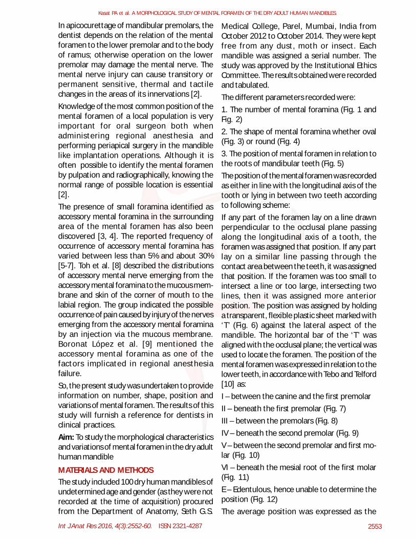
Medical College, Parel, Mumbai, India fromOctober 2012 to October 2014. They were keptfree from any dust, moth or insect. Eachmandible was assigned a serial number. Thestudy was approved by the Institutional EthicsCommittee. The results obtained were recordedand tabulated.The different parameters recorded were:1. The number of mental foramina (Fig. 1 andFig. 2)2. The shape of mental foramina whether oval(Fig. 3) or round (Fig. 4)3. The position of mental foramen in relation tothe roots of mandibular teeth (Fig. 5)The position of the mental foramen was recordedas either in line with the longitudinal axis of thetooth or lying in between two teeth accordingto following scheme:If any part of the foramen lay on a line drawnperpendicular to the occlusal plane passingalong the longitudinal axis of a tooth, theforamen was assigned that position. If any partlay on a similar line passing through thecontact area between the teeth, it was assignedthat position. If the foramen was too small tointersect a line or too large, intersecting twolines, then it was assigned more anteriorposition. The position was assigned by holdinga transparent, flexible plastic sheet marked with‘T’ (Fig. 6) against the lateral aspect of themandible. The horizontal bar of the ‘T’ wasaligned with the occlusal plane; the vertical wasused to locate the foramen. The position of themental foramen was expressed in relation to thelower teeth, in accordance with Tebo and Telford[10] as:I – between the canine and the first premolarII – beneath the first premolar (Fig. 7)III – between the premolars (Fig. 8)IV – beneath the second premolar (Fig. 9)V – between the second premolar and first mo-lar (Fig. 10)VI – beneath the mesial root of the first molar(Fig. 11)E – Edentulous, hence unable to determine theposition (Fig. 12)The average position was expressed as the
MATERIALS AND METHODSThe study included 100 dry human mandibles ofundetermined age and gender (as they were notrecorded at the time of acquisition) procuredfrom the Department of Anatomy, Seth G.S.
Int J Anat Res 2016, 4(3):2552-60. ISSN 2321-4287 2554
Kasat PA et al. A MORPHOLOGICAL STUDY OF MENTAL FORAMEN OF THE DRY ADULT HUMAN MANDIBLES.
mean position rank of the six positions [11]. Theuse of the position rank was necessary becausea linear scale could not be used [12 and 13].Some mandibles were edentulous and positionof mental foramen could not be determined inthem (Fig. 12).4.Any variation if presentInclusion Criteria: All the mandibles availableduring the study periodExclusion Criteria: Damaged bones, bones ofpediatric age group and bones affected bypathology.
Fig. 1: Single mental foramen.
Fig. 2: Double mental foramen.
Fig. 3: Oval shape mental foramen.
Fig. 4: Round shape mental foramen
Fig. 5: Illustration showing position of mental foramenin relation to the mandibular teeth (schematic diagram)
where,The position of the mental foramen expressedin relation to the lower teeth as follows:I– between the canine and the first premolarII – beneath the first premolarIII – between the premolarsIV – beneath the second premolarV –between the second premolar and first mo-larVI – beneath the mesial root of the first molarFig. 6: Transparent plastic sheet used to note the posi-tion of mental foramen.

Int J Anat Res 2016, 4(3):2552-60. ISSN 2321-4287 2555
Kasat PA et al. A MORPHOLOGICAL STUDY OF MENTAL FORAMEN OF THE DRY ADULT HUMAN MANDIBLES.
Fig. 7 - 12 Position of mental foramen in relation to themandibular teeth
Fig. 9: Position IV
Fig. 12: Edentulous
Fig. 7: Position II Fig. 8: Position III
Fig. 10: Position VVVVPosition Position
Fig. 11: Position VI
where,The position of the mental foramen expressedin relation to the lower teeth as follows:II – beneath the first premolarIII – between the premolarsIV – beneath the second premolarV–between the second premolar and first molarVI–beneath the mesial root of the first molarE –Edentulous
foramen while the others were small (satellite)foramina. There was no triple mental foramen.None of the mandibles presented with bilateralaccessory mental foramen.Round shaped mental foramen were observedin 106 sides of the mandibles and oval shapedin 94 sides of the mandibles. On both right andleft sides, mental foramen was oval in 47 % ofmandibles and round in 53 % of mandibles(Fig.13).
RESULTS
Incidences of Mental Foramen: Mentalforamen was present in all one hundredobserved mandibles and it was bilateral.Incidences of Accessory Mental Foramen: 1.5% of the mandibles showed double mentalforamen which was present in the right side(Fig. 2). Out of these, there was a single large
Fig. 13: Shape of mental foramen.
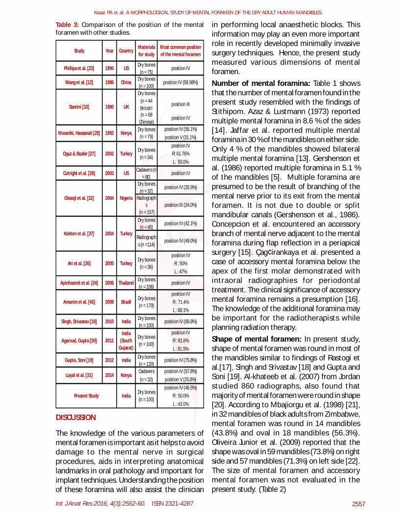
Mental foramen was situated below the apexof second premolar tooth in 46.5 % mandibles(position IV) whereas in 27.5 % it was foundbetween second premolar and first molar(position V). In 11 % of mandibles it wasobserved between first and second premolars(position III). It was seen below the apex of firstpremolar (position II) and below first molar(position VI) equally (2 % each). The mentalforamen was not observed in between canineand first premolar (position I) in any mandible.In 22 sides, position could not be determined asthey were edentulous. Position of accessorymental foramen (in 1 mandible out of 100) wasfound to be situated 13 mm lateral from themental foramen below apex of first molar tooth
Fig. 14: Position of mental foramen.
Int J Anat Res 2016, 4(3):2552-60. ISSN 2321-4287 2556
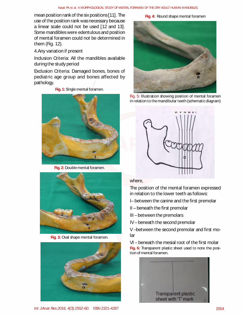
(position VI). It was observed to be located 11mm above and medial to the mental foramenbetween first and second premolar (position III)in 1 mandible out of the same 100 (Fig.14).where,The position of the mental foramen expressedin relation to the lower teeth as follows:I – between the canine and the first premolarII – beneath the first premolarIII – between the premolarsIV – beneath the second premolarV\ –between the second premolar and first mo-larVI – beneath the mesial root of the first molarU – position not determinedLingual fenestrations: A fenestration is adefect in the bony plate overlying the roots ofthe teeth. According to Azaz & Lustmann, in theliving, fenestrations are usually detected dur-ing surgical procedures when a buccal flap israised [14]. Lingual fenestration was found onone side only of a single mandible, close tomental foramen. It was located on the lingualaspect of the junction of right first and secondmolars and the buccal aspect of the right firstmolar (Fig. 15).
Fig. 15: Illustration showing lingual fenestration (probe).
Table 1: Comparison of the numbers of the mentalforamen with other studies.
* Radiologic study ** Cone-beam computed tomographystudy
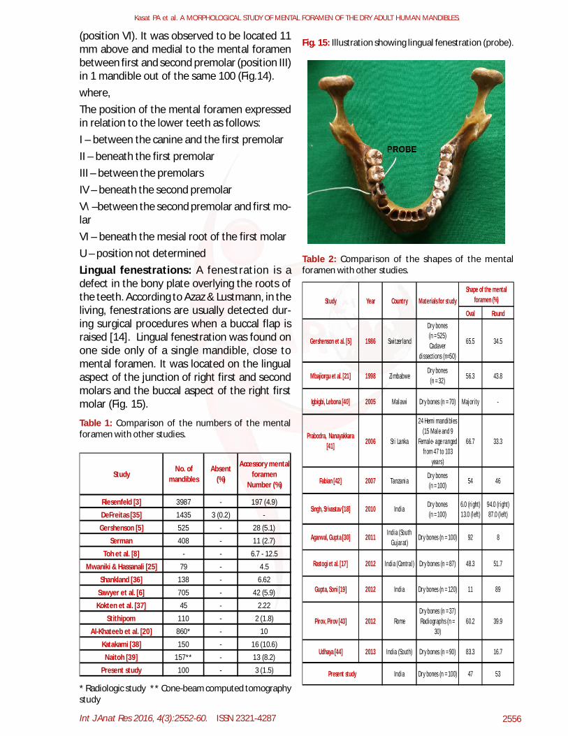
Table 2: Comparison of the shapes of the mentalforamen with other studies.
Kasat PA et al. A MORPHOLOGICAL STUDY OF MENTAL FORAMEN OF THE DRY ADULT HUMAN MANDIBLES.
Riesenfeld [3] 3987 - 197 (4.9)
DeFreitas [35] 1435 3 (0.2) -
Gershenson [5] 525 - 28 (5.1)
Serman 408 - 11 (2.7)
Toh et al. [8] - - 6.7 - 12.5
Mwaniki & Hassanali [25] 79 - 4.5
Shankland [36] 138 - 6.62
Sawyer et al. [6] 705 - 42 (5.9)
Kokten et al. [37] 45 - 2.22
Stithipom 110 - 2 (1.8)
Al-Khateeb et al. [20] 860* - 10
Katakami [38] 150 - 16 (10.6)
Naitoh [39] 157** - 13 (8.2)
Present study 100 - 3 (1.5)
StudyNo. of
mandibles
Accessory mental foramen
Number (%)
Absent (%)
Oval Round
Materials for studyShape of the mental
foramen (%)
Gershenson et al. [5] 1986 Switzerland 65.5 34.5
Study Year
46
Mbajiorgu et al. [21] 1998 Zimbabwe 56.3 43.8
Igbigbi, Lebona [40] 2005 Malawi Majority -
Country
Prabodra, Nanayakkara [41]
Agarwal, Gupta [30] 2011 92 8India (South Gujarat)
Dry bones (n = 100)
Singh, Srivastav [18] 2010 India Dry bones (n = 100)
Dry bones (n = 100)
Dry bones (n = 32)
Dry bones (n = 70)
2006 Sri Lanka
24 Hemi mandibles (15 Male and 9
Female- age ranged from 47 to 103
years)
66.7 33.3
Fabian [42] 2007 Tanzania 54
48.3 51.7
Gupta, Soni [19] 2012 India 11 89
94.0 (right) 87.0 (left)
6.0 (right) 13.0 (left)
Present study Dry bones (n = 100)
Dry bones (n = 37) Radiographs (n =
30)
Dry bones (n = 525) Cadaver
dissections (n=50)
2013 India (South) 83.3 16.7
India 47 53
Udhaya [44] Dry bones (n = 90)
Pirov, Pirov [43] 2012 Rome 60.2 39.9
Dry bones (n = 87)
Dry bones (n = 120)
Rastogi et al. [17] 2012 India (Central )
Int J Anat Res 2016, 4(3):2552-60. ISSN 2321-4287 2557
Table 3: Comparison of the position of the mentalforamen with other studies.
Study Year CountryMaterials for study
Most common position of the mental foramen
Phillips et al. [23] 1990 US Dry bones (n = 75)
position IV
Wang et al. [12] 1986 China Dry bones (n = 100)
position IV (58.98%)
Dry bones(n = 44 British)
position III
(n = 68 Chinese)
position IV
position IV (56.1%)position V (31.1%)
position IVR: 61.76%L: 50.0%
Cutright et al. [28] 2003 US Cadavers (n = 80)
position IV
Dry bones (n = 32)
position IV (35.9%)
Radiographs
(n = 157)Dry bones
(n = 45)position IV (42.1%)
Radiographs (n =114)
position IV (49.0%)
position IVR : 50%L : 47%
Apinhasmit et al. [24] 2006 Thailand Dry bones (n = 106)
position IV
position IVR : 71.4%L : 68.1%
Singh, Srivastav [18] 2010 India Dry bones (n = 100)
position IV (68.8%)
position IVR : 81.6%L : 81.5%
Gupta, Soni [19] 2012 India Dry bones (n = 120)
position IV (75.8%)
Cadavers position IV (57.8%)(n = 32) position V (25.0%)
position IV (46.5%)R : 50.0%L : 43.0%
Santini [33]
position III (34.0%)
Kokten et al. [37] 2004 Turkey
Ari et al. [26] 2005 TurkeyDry bones
(n = 36)
1990 UK
Mwaniki, Hassanali [25] 1992 KenyaDry bones
(n = 79)
Olasoji et al. [32] 2004 Nigeria
Oguz & Bozkir [27] 2002 TurkeyDry bones
(n = 34)
Kenya
Present Study IndiaDry bones (n = 100)
BrazilDry bones (n = 170)
Agarwal, Gupta [30] 2011India
(South Gujarat)
Dry bones (n = 100)
Loyal et al. [31] 2014
Amorim et al. [45] 2008
DISCUSSION
The knowledge of the various parameters ofmental foramen is important as it helps to avoiddamage to the mental nerve in surgicalprocedures, aids in interpreting anatomicallandmarks in oral pathology and important forimplant techniques. Understanding the positionof these foramina will also assist the clinician
in performing local anaesthetic blocks. Thisinformation may play an even more importantrole in recently developed minimally invasivesurgery techniques. Hence, the present studymeasured various dimensions of mentalforamen.Number of mental foramina: Table 1 showsthat the number of mental foramen found in thepresent study resembled with the findings ofStithipom. Azaz & Lustmann (1973) reportedmultiple mental foramina in 8.6 % of the sides[14]. Jaffar et al. reported multiple mentalforamina in 30 % of the mandibles on either side.Only 4 % of the mandibles showed bilateralmultiple mental foramina [13]. Gershenson etal. (1986) reported multiple foramina in 5.1 %of the mandibles [5]. Multiple foramina arepresumed to be the result of branching of themental nerve prior to its exit from the mentalforamen. It is not due to double or splitmandibular canals (Gershenson et al., 1986).Concepcion et al. encountered an accessorybranch of mental nerve adjacent to the mentalforamina during flap reflection in a periapicalsurgery [15]. Çag¢irankaya et al. presented acase of accessory mental foramina below theapex of the first molar demonstrated withintraoral radiographies for periodontaltreatment. The clinical significance of accessorymental foramina remains a presumption [16].The knowledge of the additional foramina maybe important for the radiotherapists whileplanning radiation therapy.Shape of mental foramen: In present study,shape of mental foramen was round in most ofthe mandibles similar to findings of Rastogi etal.[17], Singh and Srivastav [18] and Gupta andSoni [19]. Al-khateeb et al. (2007) from Jordanstudied 860 radiographs, also found thatmajority of mental foramen were round in shape[20]. According to Mbajiorgu et al. (1998) [21],in 32 mandibles of black adults from Zimbabwe,mental foramen was round in 14 mandibles(43.8%) and oval in 18 mandibles (56.3%).Oliveira Junior et al. (2009) reported that theshape was oval in 59 mandibles (73.8%) on rightside and 57 mandibles (71.3%) on left side [22].The size of mental foramen and accessorymental foramen was not evaluated in thepresent study. (Table 2)
Kasat PA et al. A MORPHOLOGICAL STUDY OF MENTAL FORAMEN OF THE DRY ADULT HUMAN MANDIBLES.
Int J Anat Res 2016, 4(3):2552-60. ISSN 2321-4287 2558
than the most common ones are due to a lag inprenatal development [34].Knowledge of position of the mental foramen iscritical to a variety of surgical and diagnosticprocedures. This data is thus clearly of directrelevance in dental teaching and practice.4. Lingual fenestrationsThe occurrence of lingual fenestrations of thelower molar tooth has been reported long back(Sicher, 1965). In present study, the incidenceof lingual fenestration was 1% of the sides ofmandibles (Fig. 15). This was similar to findingof Jaffar et al. [13]. However, in the present itwas found in the right side. In studies by Azaz &Lustmann [1973; 14] and Jaffar et al. [2002; 13],the tendency to the left side was in concordance.Subluxation of root fragments into thesubmandibular space may occur if there is afenestration in the third molar region. As thesefenestrations cannot be recognized by normalradiological procedures, care should be takento avoid subluxation. This can be done byrefraining to apply pressure either along the longaxis of the tooth or in a lingual direction [13].
Position of mental foramen: The present studyshows, the location of mental foramen in linewith the long axes of the second premolar asthe most common position (46.5%). This resultis in consistence with that of Wang et al. [12],Jaffar et al. [13], Azaz & Lustmann [14], Phillipset al. [23], Apinhasmit et al. [24], Mwaniki &Hassanali [25], Ari et al. [26], Oguz & Bozkir[27], Cutright et al. [28] and Green [29]. Thisshows that this is the most common finding inthe majority of the ethnic groups. However, thesecond common position found is the positionbetween the second premolar and first molar(position V) similar to finding of Mwaniki &Hassanali [25], Agarwal & Gupta [30], and Loyalet al. [31]. Olasoji et al. found position III(between the premolars) as the most commonposition for the mental foramen [32]. Thissuggests that traits such as the localization ofmental foramen may not only differ betweenpopulations of different geographic environmentbut also within the inhabitants of the samegeography.The position of mental foramen below the apexof second premolar was 49% by Tebo & Telford[1950; 10], 58.98 % by Wang et al. [1986; 12],52.9 % Santini & Land [1990; 33] and 68.8 % bySingh & Srivastav [2010; 18].The position of tooth is a quick landmark forlocating the mental nerve. Surgeons should beaware that the most common position for themental nerve is at the second premolar.However, this should not be the sole guidance,since the mental nerve may have an aberranttooth position. This also helps in explainingcases of failed anaesthesia, when it is targetedat the second premolar region. In such cases thejunction of the second premolar and first molarshould be considered as a potential alternativesite for the mental nerve (Table 3).A difference in the position of the mental fora-men between the right and the left sides wasobserved in 31% of the mandibles studied. Thiswas not necessarily accompanied by multiplemental foramina on either side.During the early prenatal life mental foramen islocated in the alveolar bone between theprimary canine and first molar (Kjaer, 1989).Therefore, it is speculated that positions other
CONCLUSION
In conclusion, the present analysis revealedvariations in number, shape and position ofmental foramen and accessory mental foramen.The present study suggests that local differ-ences in mental foramen position may occur ina population. These may be related to feedinghabits of different regions which may ultimatelyaffect the development of mandibles. Thisvariability should alert the dental surgeons whileperforming periodontal or endodontic surgery.The mental foramen is difficult to localize asthere are no absolute anatomical landmarks forreference. The mental foramen cannot bevisualized or palpated clinically; hence it islocalized in relation to the lower teeth. Further,when the existence of the accessory mentalforamen is confirmed, it could avoid nerveinjury during periapical surgery. The possibilityof accessory mental foramen related sensorydisturbance is low during root canal treatmentunless the mental foramen and mandibularcanal are injured.If the studies related to variations in the
Kasat PA et al. A MORPHOLOGICAL STUDY OF MENTAL FORAMEN OF THE DRY ADULT HUMAN MANDIBLES.
Int J Anat Res 2016, 4(3):2552-60. ISSN 2321-4287 2559
position, size, incidence and shape of mentalforamen and accessory mental foramen arecarried out in large numbers; it will be ofimmense use to the anthropologists in identify-ing the deceased. Moreover, the data will bereliable for dental surgeons.
Conflicts of Interests: None
REFERENCES
[1]. Collins P. Infratemporal and Pterygopalatine Fos-sae and Temporomandibular Joint. Standring S, edi-tor. Gray’s Anatomy: the Anatomical Basis of Clini-cal Practice. 40th edition. New York: ElsevierChurchill Livingstone; 2008: 527-560.
[2]. Sankar DK, Bhanu SP, Susan PJ. Morphometrical andmorphological study of mental foramen in dry den-tulous mandibles of South Andhra population ofIndia. Indian J Dent Res 2011;22:542-546.
[3]. Riesenfeld A. Multiple infraorbital, ethmoidal, andmental foramina in the races of man. Am J PhysAnthrop. 1956;14:85-100.
[4]. Sutton RN. The practical significance of mandibularaccessory foramina. Aust Dent J. 1974;19:167-173.
[5]. Gershenson A, Nathan H, Luchansky E. Mental Fora-men and Mental Nerve: Changes with Age. Acta Anat(Basel). 1986;126(1):21-28.
[6]. Sawyer DR, Kiely ML, Pyle MA. The Frequency ofAccessory Mental Foramina in Four Ethnic Groups.Arch Oral Biol. 1998 May; 43(5):417-420.
[7]. Agthong S, Huanmanop T, Chentanez V. AnatomicalVariations of the Supraorbital, Infraorbital, andMental Foramina Related to Gender and Side. J OralMaxillofac Surg. 2005 Jun;63(6):800-804.
[8]. Toh H, Kodama J, Yanagisako M, Ohmori T. Anatomi-cal Study of the Accessory Mental Foramen and theDistribution of its Nerves. Okajimas Folia Anat Jpn.1992;69:85-87.
[9]. Boronat López A, Peñarrocha Diago M. Failure oflocoregional anesthesia in dental practice: reviewof the literature. Med Oral Patol Oral Cir Bucal.2006;11:E510-513.
[10]. Tebo HG, Telford IR. An Analysis of the Variations inPosition of the Mental Foramen. Anat Rec.1950;107:61-66.
[11]. Yesilyurt H, Iydinlioglu A, Kavakli A, Ekinci N, ErogluC, Hacialiogullari M, Diyarbakirli S. Local Differ-ences in the Position of the Mental Foramen. FoliaMorphol. 2008;67(1):32-35.
[12]. Wang TM, Shih C, Liu JC, Kuo KJ. A Clinical andAnatomical study of the Location of the Mental Fo-ramen in Adult Chinese Mandibles. ActaAnat.1986;126:29-33.
[13]. Jaffar AA, Al-Zubaidi AF, Al-Salihi AR. AnatomicalFeatures of Clinical Significance in Dry Mandibles.Iraqi Dental Journal. 2002;29:990-118.
[14]. Azaz B, Lustmann J. Anatomical Configurations inDry Mandibles. Br. J .Oral Surg. 1973;2:1-9.
[15]. Concepcion M, Rankow HJ. Accessory branch of themental nerve. J Endod 2000;26: 619-620.
[16]. Çag¢irankaya LB, Kansu H. An accessory mentalforamen: a case report. J Contemp Dent Pract2008;9:98-104.
[17]. Rastogi R, Budhiraja V, Sathpathi DK, Singh S, GourKK, Nair S. Morphology and morphometry of themental foramen in dry adult human mandibles fromcentral India and their clinical correlation. Eur JAnat. 2012;16(1):22-26.
[18]. Singh R, Srivastav AK. Study of Position, Shape, Sizeand Incidence of Mental Foramen and AccessoryMental Foramen in Indian Adult Human Skulls. Int JMorphol. 2010;28(4): 1141-1146.
[19]. Gupta S, Soni JS. Study of Anatomical Variationsand Incidence of Mental Foramen and AccessoryMental Foramen in Dry Human Mandibles. Natl JMed Res. 2012;2(1):28-30.
[20]. Al-Khateeb T, Al-Hadi Hamasha A, Ababneh KT.Position of the Mental Foramen in a NorthernRegional Jordanian Population. Surg Radiol AnatJournal. 2007 Apr; 29(3):231- 237. Epub 2007 Mar21.
[21]. Mbajiorgu EF, Mawera G, Asala SA, Zivanovic S.Position of the Mental Foramen in Adult BlackZimbabwean Mandibles: a Clinical AnatomicalStudy. Cent Afr J Med. 1998 Feb; 44(2):24-30.
[22]. Oliveira Junior EM, Arau´jo ALD, Da Silva CMF et al.Morphological and morphometric study of the men-tal foramen on the M-CP-18 Jiachenjiang point. IntJ Morphol 2009; 27: 231-238.
[23]. Phillips JL, Weller N, Klild JC. The Mental Foramen:Part I. Size, Orientation, and Positional Relation-ship to the Mandibular Second Premolar. J. of Endo-dontics. 1990; 16(5):221-223.
[24]. Apinhasmit W, Chompoopong S, Methathrathip D,Sansuk R, Phetphunphiphat W. Supraorbital Notch/Foramen, Infraorbital Foramen and MentalForamen in Thais: Anthropometric Measurementsand Surgical Relevance. J Med Assoc Thai. 2006May; 89(5):675-682.
[25]. Mwaniki DL, Hassanali J. The Position of Mandibu-lar and Mental Foramina in Kenyan African Man-dibles. East Afr Med J.1992;69:210-213.
[26]. Ari I, Kafa IM, Basar Z, Kurt MA. The Localizationand Anthropometry of Mental Foramen on Late Byz-antine Mandibles. Coll Antropol. 2005Jun;29(1):233-236.
[27]. Oguz O, Bozkir MG. Evaluation of Location of Man-dibular and Mental Foramina in Dry, Young, AdultHuman Male, Dentulous Mandibles. West IndianMed J. 2002;51:14-16.
[28]. Cutright B, Quillopa N, Schubert W. An Anthropo-metric Analysis of the Key Foramina for Maxillofa-cial Surgery. J. Oral Maxillofac. Surg.2003;61:354-357.
[29]. Green RM. The position of the mental foramen: acomparison between the Southern (Hong Kong) Chi-nese and other ethnic and racial groups. Oral SurgOral Med Oral Pathol. 1987;63:287-290.
Kasat PA et al. A MORPHOLOGICAL STUDY OF MENTAL FORAMEN OF THE DRY ADULT HUMAN MANDIBLES.
Int J Anat Res 2016, 4(3):2552-60. ISSN 2321-4287 2560
[30]. Agarwal DR, Gupta SB. Morphometric Analysis ofMental Foramen in Human Mandibles of SouthGujarat. People’s J Sci Res. 2011;4:15-18.
[31]. Loyal PK, Butt F, Ogeng’o JA. The Surgical Relevanceof the Anatomic Position of the Extraosseous Men-tal Nerve in a Kenyan Population. Pan African Medi-cal Journal. 2014;18:51.
[32]. Olasoji HO, Tahir A, Ekanem AU, Abubakar AA. Ra-diographic and Anatomic Locations of Mental Fo-ramen in Northern Nigerian Adults. Niger Postgrad.Med J. 2004 Sep; 11(3):230-233.
[33]. Santini A, Land MA. A Comparison of the Position ofthe Mental Foramen in Chinese and British Man-dibles. Acta Anat.1990;137:208-212.
[34]. Kjaer I: Formation and Early Prenatal Location ofthe Human Mental Foramen. Scandinavian Journalof Dental Research. 1989;97(1):1-7.
[35]. DeFreitas V, Madeira MC, Toledo Filho JL, ChagasCF. Absence of the Mental Foramen in Dry humanMandibles. Acta Anat (Basel). 1979;104(3):353-355.
[36]. Shankland WE 2nd. The Position of the Mental Fo-ramen in Asian Indians. J Oral Implantol.1994;20(2):118-123.
[37]. Kokten G, Buyukanten M, Balcioglu HA. Foramenmentalenin cap ve lokalizasyonunun kuru kemik vepanoramik radyografilerde (Comparison of the Di-ameter and the Location of Mental Foramen in DryMandibles and Panoramic Radiographs). IstanbulUniversitesi Dishekimligi Fakultesi Dergisi. 2004;38(10):65-71.
[38]. Katakami K, Mishima A, Shiozaki K, Shimoda S,Hamada Y, Kobayashi K. Characteristics of Acces-sory Mental Foramina Observed on Limited Cone-Beam Computed Tomography Images. J Endod. 2008Dec; 34(12):1441–1445. Epub 2008 Oct 11.
[39].Naitoh M, Hiraiwa Y, Aimiya H, Gotoh K, Ariji E. Ac-cessory Mental Foramen Assessment Using Cone-Beam Computed Tomography. Oral Surg Oral MedOral Pathol Oral Radiol Endod. 2009Feb;107(2):289-294. Epub 2008 Dec 13.
[40]. Igbigbi PS, Lebona S. The Position and Dimensionsof the Mental Foramen in Adult Malawian Man-dibles. West Afr J Med. 2005 Jul-Sep; 24(3):184-189.
[41]. Prabodra LBL, Nanayakkara BG. The Position, Di-mension and Morphological Variations of MentalForamen in Mandibles. Galle Med J. 2006 Sept;11:13-15.
[42]. Fabian FM. Position, Shape and Direction of Open-ing of the Mental Foramen in Dry Mandibles of Tan-zanian Adult Black Males. Ital J Anat Embryol. 2007Jul-Sep;112(3):169-177.
[43]. Pirov S, Pirov A. Variations of the Mandibular Ca-nal, Mandibular and Mental Orifices. ScientificAnnals of the State University of Medicine and Phar-macy Nicolae Testemiþanu. – Chisinau. 2012;1:84-89.
[44]. Udhaya K, Saraladevi KV, Sridhar J. The Morphomet-ric Analysis of the Mental Foramen in Adult DryHuman Mandibles: A Study on the South Indian Popu-lation. Journal of Clinical and Diagnostic Research.2013 Aug;7(8):1547-1551.
[45]. Amorim MM, Prado FB, Borini CB, Bittar TO, VolpatoMC, Groppo FC, Caria PH. The Mental Foramen inDentate and Edentulous Brazilian’s Mandible. Int JMorpho. 2008;26:981-987.
How to cite this article:Kasat PA, Muthiyan GG, Bhuiyan PS. A MORPHOLOGICAL STUDYOF MENTAL FORAMEN OF THE DRY ADULT HUMAN MANDIBLES.Int J Anat Res 2016;4(3):2552-2560. DOI: 10.16965/ijar.2016.271
Kasat PA et al. A MORPHOLOGICAL STUDY OF MENTAL FORAMEN OF THE DRY ADULT HUMAN MANDIBLES.