international journal of chemtech research -...
TRANSCRIPT

International Journal of ChemTech Research CODEN (USA): IJCRGG ISSN: 0974-4290
Vol.8, No.7, pp 351-362, 2015
Antitumor activity of biosynthesized silver nano particlesfrom leaves of Momordica charantia against MCF-7 cell line
Gandhiraj.V1*, Sathish Kumar.K2, Madhusudhanan.J1, Sandhya.J1
1Shri Andal Alagar College of Engineering, Tamilnadu, India.2SSN College of Engineering, Tamilnadu, India.
Abstract: Nano materials are utilized in the pharmaceutical field which can be developed intonovel agents with less side effects and cost effective process. Biosynthesized silvernanoparticle from Momordica charantialeaves aqueous extract and Invitro study ofanticancer activity against Breast cancer cell line MCF-7 showed significant activity. TheMTT assay methods were followed for evaluating the cell viability and cytotoxicity for bothplant extract and plant-silver nanoparticle. The subjected cells were drawn for the DNAfragmentation assay and the DNA bands were visualized in GEL-Documentation and theDNA ladder of 1KB used as Marker for reference. The Cell toxicity of 63.26% on plantextract and 67.16% on plant –silver naoparticles synthesized activity were recorded againstMCF-7 cell line and DNA bands were sheered and recorded at 1.5 % for plant extract and2.5% of plant –silver naoparticles. The FT-IR analysis of silver nano particle from plantextract showed presence of alkynes, alkene and amine functional group and SEM-EDXanalysis showed the particle size of 96.3nm and presence of Cl, Ag, O, Na, Mg, Si and Al.The functional development of novel compounds from silver nano particle frombiosynthesized process has good potential than other characteristic drugs used.Key words: Silver nano particle, Biosynthesis, MTT, Fragmentation, Cancer.
Introduction:
Nano materials are of reduced physical, chemical and biological materials which can be used to developnovel agents can be used over resistant pathogens and act as anticancer agents1.The sustainability in theinitiatives of using green chemistry to develop and focus issues in multi disciplinary fields of research bysignificantly cost effective methods to produce nanomaterials which are used in multiple applications includingbiological and biomedical fields which is reduced in toxicity and depends based on the size, shape, compositionand uniformal chemistry to prolong its lifespan of Nanoparticles as it is important to develop stabilizing agentsand mechanism which can be produced to biological environment2.
The physicochemical properties for nanoparticles synthesis include photochemical reduction, laserablation and electrochemistry which are expensive. There are different methods have been developed tosynthesis of nanoparticles by physical, chemical and biological3.The biological or green synthesis has receivedcomplete attention in area of research and Nanoparticle synthesis by using living organism, cellular extracts,and plant based products, bacteria and fungi. And it could be as simple, viable and non-toxic forms compared toother methods of synthesis 4.Silver nanoparticles are studied and applied in variant fields of catalysis, photonics,biosensing, diagnostic and antimicrobial agents5 and promoted to heal wounds and possess anticancerousactivity 6.

Gandhiraj.V et al /Int.J. ChemTech Res. 2015,8(7),pp 351-362. 352
The presence of secondary metabolities in plants makes to redox reaction and can be exploited forbiosynthesis of nanoparticles. Theses metabolities poses active biocompounds which are stable and caninvestigate themechanism followed by possible medical value and treatment7. The research groups around theworld have successfully demonstrated the efficacy of silver nanoparticles having potential cytotoxicity againstcancer cells8 and antiangiogenic property in micro vascular endothelial cells9.
Naturally produced plant materials could be beneficial to develop as an agent with target basedmedication which is possible by nanoformulations and properties regarding plans and its constituents wereanalyzed and information should be gathered from medicinal practioners10.
Cancer is a disease which is accumulated in a range of 10-12 million new cases, 5-7 million deaths byits multiple forms11. The leading forms of cancer treatment were ineffective which has lent to develop newerforms of methods to achieve medical formulations12. Though the various cytotoxic agents used in treatment ofbreast cancer like doxorubicin, cisplatin, bleomycin and many other has temporary relief and its effects areuncertain, for that it is necessary to develop novel treating molecule and its mechanism13.
The reactive oxygen species (ROS) in the cell is to produce apoptosis and generation of ROS in cells byoxidative stress and treating those free radicals which is produced during cellular metabolism by inhibiting theJNK pathway leads to mitochondrial- dependent apoptosis and includes cytotoxicity by oxidative celldamage14.The ROS has role in DNA damage and Caspase enzyme is a important molecule in apoptosis bycleaving of inhibitory and translocation of caspase activated DNAse to nucleus results in DNA fragmentation15.Because the major compounds of phenolic and flavonoid possess antioxidant molecule which can reduceoxidative stress and lipid peroxidation of the cells leads to either cell damage to form malignant cells16.
Momordica charantia(Bitter guard) is a flowering plant in the family of Cucurbitaccae has simpleleaves with 5-7 palmately lobed tendrils branched or unbranched. Fruits are ovoid, spindle shaped and ridgedwith seeds are white in appearance before ripening and red after it, the outer skin is edible and both vegetableand fruit is used as raw vegetable. It is used as agents to anthelmintic, antibacterial, antidiabetic, antimicrobial,antioxidant, antitumor, hypoglycemic and cytotoxic17. Among various nanoparticles, silver nanoparticle has awide area of applications in multiple fields and has been taken for the present study by synthesizingnanoparticles using leaves of Momordica charantiaand treating against cancer cell line.
Experimental:
Collection of plant:
The plant leaves were collected from the locals of chengalpet region of Kanchipuram District, TamilNadu, India with the help of local villagers and washed with running tap water to remove dust materials.
Preparation of leaf extract
The leaves of Momordica charantia were rinsed with distilled water and shade dried in roomtemperature for 3 weeks and then 150g of dried leaves were powdered using kitchen blender. 5g of thepowdered leaf was dissolved in 100ml of distilled water in 250ml of conical flask .The mixture wasallowed to boil for 20mins at 55-600C.After that the leaf extract was filtered through Whatman filter paperNo. 1 and stored at 4°C.Then the solution was used as stock solution.
Preparation of 1mM silver nitrate solution:
Analytical grade silver nitrate (AgNO3) was purchased from Krishna scientific chemical suppliers. A1mM stock solution of AgNO3 in distilled water was prepared according to the following calculations
The molecular weight of AgNO3:[Ag-107.87, N-14, O-16] = 107.87 + 14 + (16 × 3) = 169.87Therefore, Molar mass of AgNO3 = 169.87 g

Gandhiraj.V et al /Int.J. ChemTech Res. 2015,8(7),pp 351-362. 353
Molecular weight x Required Molarity x Required volumeMolarity = -------------------------------------------------------------------- 1000
0.169g of silver nitrate was added in 1000ml of distilled water to prepare 1Mm silver nitrate solution.
Synthesis of silver nanoparticle
The 10ml of Momordica charantia leaf extract was added to 90 ml of 1mM aqueous of AgNO3for reduction of silver nitrate in to silver ions. Aqueous mixture was incubating in direct sunlight condition.After the sunlight exposure color changes from yellowish to dark brown color due to the reduction of silvernitrate into silver ions. The color changes indicate the formation of silver nanoparticles.
Characterization methods for synthesis of silver nanoparticle:
UV-Vis spectroscopy
UV-Vis spectroscopy can be comprehended as absorption spectroscopy in the spectral region of ultra-violet and visible spectra. It uses light in visible and near-UV range to promote outer electrons to higher energylevels. The UV-Vis spectra have broad features that are of limited use for sample identification but are veryuseful for quantitative measurements. The concentration of analytes in solution can be determined by measuringthe absorbance at specific wavelength and applying the Beer-Lambert Law, the UV-Vis range spans the rangeof human visual acuity and to observe the optical property of biosynthesized silver nanoparticles, samples wereperiodically analyzed for UV–Vis spectroscopic studies at room temperature at 200-700nm range and it isuseful to characterize the absorption, transmission, and reflectivity of a variety of technologically importantmaterials, such as pigments, coatings etc.
Scanning electron microscopy and Energy dispersive x-ray spectroscopy
Scanning electron microscopy (SEM) is a technique that uses electrons instead of light to form anoutput image and allowed researchers to examine a much larger variety of specimens. It has a large depth offield, which allows more of a specimen to be in focus at one time. The SEM also has much higher resolutionclosely spaced specimens can be magnified at much higher levels.Energy Dispersive X-Ray Spectroscopy(EDX) is a technique that provides the elemental curve as output. This analytical technique is generally used inconjunction with the Scanning Electron Microscopy (SEM). EDX technique primarily detects the X-raysemitted from the sample during the process of bombardment by an electron beam for characterizing theelemental composition of the sample of interest. Quantitative results can be obtained from the relative x-raycounts at the characteristic energy levels for the sample constituents. Some typical applications include alloyidentification, foreign material analysis, coating composition analysis etc. EDX helped to verify the presence ofsilver in the sample and its percentage as well.
Transmission electron microscopy
Transmission electron microscopy (TEM) was performedfor characterizing size and shape ofbiosynthesizedsilver nanoparticles. The sample was firstsonicated (Vibronics VS 80) for 15 min. A drop ofthissolution was loaded on carbon-coated coppergrids, and solvent was allowed to evaporate underInfrared light for30 min. TEM measurements wereperformed on Philips model CM 200 instrumentoperated at an acceleratingvoltage at 200 kV.
X-ray diffraction (XRD)
X-ray diffraction (XRD) measurements of film of thebiologically synthesized silver nanoparticlessolutioncast onto glass slides were done on a εMMAdiffractometer operating at a voltage of 40 kV and currentof 20 mA with Cu K (α) radiation of1.54187 nm wavelength. The scanning as done inthe region of 2h from20°C to 80°C at 0.02°/min and thetime constant was 2s.

Gandhiraj.V et al /Int.J. ChemTech Res. 2015,8(7),pp 351-362. 354
Fourier Transform infrared spectroscopy
For Fourier transform infrared spectroscopy (FTIR)measurements, the reaction mixture wascentrifugedat 15,000 rpm for 15 min after complete reduction ofAgNO3 by the Momordica charantialeafextract to separate Agnanoparticles from biomass or other bioorganiccompounds which may interfere inanalyzing molecule–AgNPs interaction. The Ag nanoparticles pelletobtained after centrifugation were re-dispersed inwater and washed (centrifugation and re-dispersion)with distilled water for three times. Finally,thesamples were dried and grinded with KBr pellets andanalyzed on a Nicolet IR 200 (Thermo electroncorp)model.
Antitumor activity:
The anticancer activity was performed on the breast cancer cell line (MCF-7) which is procured formNCCS, Pune, India and maintained in specific medium for its growth and carried MTT assay for plant extractand Silver nanoparticle of the plant extract.
The MTT assay18 is based on the ability of live but not dead cells to reduce a yellow tetrazolium dye toa purple formazan product. Cells were maintained in DMEM medium, supplemented with 10% Fetal BovineSerum, at 37oC in humidified atmosphere with 5% CO2.The cells were plated in 96 well flat bottom tissueculture plates at a density approximately1.2 X 104 cells/well and allowed to attach overnight at 37oC. Themedium was then discarded and cells were incubated with different concentrations of the extracts for 24 hours.After the incubation, medium was discarded and 100µl fresh medium was added with 10µl of MTT (5mg/ml).After 4 hours, the medium was discarded and 100µl of DMSO was added to dissolve the formazan crystals.Then, the absorbance was read at 570nm in a microtitre plate reader. Cyclophosphamide was used as a positivecontrol.
Cell survival was calculated by the following formula:Viability % = (Test OD/ Control OD) X 100Cytotoxicity % = 100 – Viability%
DNA fragmentation assay:
The experiment was characterized by the activation of endogenous endonucleases with subsequentcleavage of chromatin DNA into internucleosomal fragments of roughly 50 base pairs (bp) and multiples of(100, 150 etc.). This effect can be used to detect apoptosis, for example via the DNA laddering assay.
In 24 flat-wells plate, incubate 2x10⁵ cancer cells (triplicate wells of 105 per well) with differentsamples (GS, CS and quercetin IC 50 range concentrations) (105 target cells per well). Add fresh DMEMmedium and allow for 24 hour incubation, collect the cell sample in 1.5 ml eppendorf tube, spin down,resuspend with 0.5 ml PBS in 1.5 ml eppendorf tubes, and add 55µl of lysis buffer (40 ml of 0.5 M EDTA 5 mlof 1 M TrisCl buffer pH 8.0 5 ml of 100% Triton X-100 50 ml of H2O) for 20 min on ice (4°C). Centrifuge theeppendorf tubes in cold at 12,000 g for 30 minutes. Transfer the samples to new 1.5 ml eppendorf tubes andthen extract the supernatant with 1:1 mixture of phenol: chloroform (gentle agitation for 5 min followed bycentrifugation) and precipitate in two equivalence of cold ethanol and one-tenth equivalence of sodium acetate.Spin down, decant, and resuspend the precipitates in 30ul of deionized water-RNase solution (0.4ml water + 5ulof RNase) and 5ul of loading buffer for 30 minutes at 37°C. Also insert 2ul of DNA ladder (marker) on theouter lanes. Run the 1.2% gel at 5V for 5min before increasing to 100V. After the dye front reach ¾ of the gel,observe the image of DNA shearing in 312nm UV illuminator19.
Results:
Synthesis of Nanoparticles:
The aqueous plant extract was prepared and preceded to synthesis of silver nano particles undercontrolled conditions. The plant extract color change from initial color to brown color shows the conversion andsynthesis of nanoparticles by redox reaction and subjected characterization studies (Figure 1).

Gandhiraj.V et al /Int.J. ChemTech Res. 2015,8(7),pp 351-362. 355
Figure 1: a)Before synthesis of AgNPsb)After synthesis of AgNPs
UV-Vis spectroscopy analysis:
Figure 2: UV spectrophotometer analysis of synthesized nano particle.
The UV-VIS spectrometer analysis consists of plot which is recorded the scanning limits employed fornano particle synthesis and dielectric constant of solution helps to create uniform size of the particle with noevidence for aggregation or variance until the 410-490 nm during reaction period (Figure 2) and consists ofsilver nano particle which is confirmed by SEM and TEM analysis.
Scanning electron microscopy and Energy dispersive x-ray spectroscopy
Scanning electron microscopy provided further insight into the morphology and size details of the silvernanoparticles. The SEM image showing the high density silver nanoparticles synthesized by the M.charantiaextract further confirmed the development of silver nanostructures and EDAX analysis at size of approximately91.63nm(Figure 3).
Figure 3a Figure 3bFig 3a)SEM image with 5µm Fig 3b) SEM image with 2µm

Gandhiraj.V et al /Int.J. ChemTech Res. 2015,8(7),pp 351-362. 356
Figure3c Figure 3dFig 3c) SEM image with 1µm Fig 3d) SEM image with 500nm
Figure 3:SEM images with different scales
Figure 4: EDAX analysis of synthesized nano particle.
The peak observed in different ranges showing the principal structures of Cl, C, Ag, O, Na, Mg, Si andAl which including synthesized nanoparticles of silver and plant constituents which is reduced along withNaoparticles (Figure 4)
Transmission electron microscopy
The transmission electron spectrometer resulted in outer surface of the synthesized nanoparticle imagesand exhibiting its resolution in multiple nm(Figure 5)
Figure 5a Figure 5bFig 5a) TEM image with 20nm. Fig 5b) TEM image with 50nm

Gandhiraj.V et al /Int.J. ChemTech Res. 2015,8(7),pp 351-362. 357
Figure 5cFig 5c) TEM image with 100nmFigure 5: TEM images with different scales
X-ray diffraction analysis
The XRD pattern of synthesized Silvernanoparticle using Momordica charantia extract were recordedand typical XRD pattern is shown in The diffraction peaks are indexed as(111), (200), (220), (311) planes ofapure face centered crystalline (fcc) structureof silver. Crystallite size of AgNPs asestimated from the FWHM ofdifferent peaksusing the Scherer’s formula and diffractionlines observed at 2θ angle. Apart from these peaks,therecorded XRD pattern shows additional unassigned peaks. This may be due to theformation of the crystallinebio-organic compounds/metalloproteins that are present in Momordica charantia (Figure 6).
Figure 6: XRD analysis.
Fourier transform infrared spectroscopy analysis:
Figure 7: FT-IR analysis of Momordica charantia.

Gandhiraj.V et al /Int.J. ChemTech Res. 2015,8(7),pp 351-362. 358
The functional group determination through FT-IR analysis has ensured the presence of multiplepatterns at 3276.15, 2921.30, 2852.13, 1569.66, 1385.96, 1040.42, 616.30, 474.68 and 420.83 cm-1 of silvernanoparticles of Momordica charantiashowing the presence of primary amines, alkanes, alkynes and aromaticfunctional groups (Figure 7).
MTT assay:
The cell viability and toxicity assay were carried out for the plant extract and silver nano particlesofMomordica charantiawere tabulated in table 1.
Table 1: Viability of the MCF-7 Cancer cell line against Plant and Ag nanoparticle extract.
Concentration in µg/ml Plant leaf extractviability
Silver nanoparticle withplant leaf extract
viability12 90.51 88.0125 84.36 78.0650 79.18 69.08
100 36.74 32.84
From the inferred data the plant extract cytotoxicity is 63.26% at the concentration 100 µg/ml of stockconcentration (Figure 9) and silver nanoparticle extract showed cytotoxicity of 67.16% at concentration of 100µg/ml (Figure 10), a fraction higher than the plant extract which can be developed into active substance bystudying and characterizing its active principle.
The graphical figure shows the viability of the Plant and silver nanoparticle extract of Momordicacharantia(Figure 8).
Figure 8: The viability of plant and nanoparticle of the extract.
The figure below represented the activity of Plant and Nanoparticle extract against MCF-7 cell line.

Gandhiraj.V et al /Int.J. ChemTech Res. 2015,8(7),pp 351-362. 359
Figure 9a Figure 9bToxicity-12µg/ml Toxicity-25µg/ml
Figure 9c Figure 9dToxicity-50µg/ml Toxicity-100µg/ml
Figure 9 a, b, c,d).Cytotoxicity of plant leaf extract against MCF-7 cell line byMTT cell viability assay.
Figure 10a Figure 10bToxicity-12µg/ml Toxicity-25µg/ml
Figure 10c Figure 10dToxicity-50µg/ml Toxicity-100µg/ml
Figure 10 a, b, c,d).Cytotoxicity of synthesized silver nanoparticle with plant leafextract againstMCF-7 cell line by MTT cell viability assay.
Gandhiraj.V et al /Int.J. ChemTech Res. 2015,8(7),pp 351-362. 360
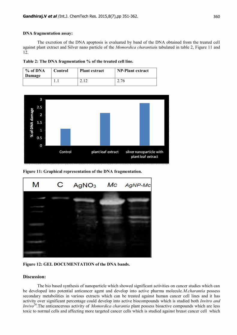
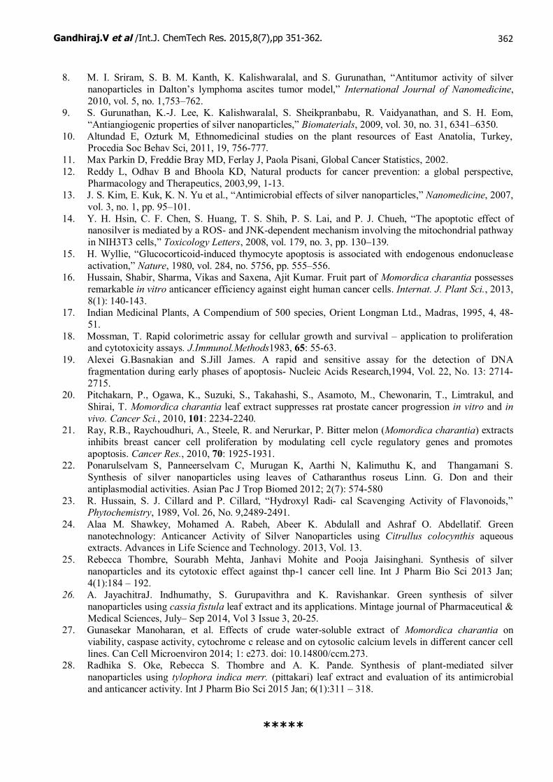
DNA fragmentation assay:
The excretion of the DNA apoptosis is evaluated by band of the DNA obtained from the treated cellagainst plant extract and Silver nano particle of the Momordica charantiais tabulated in table 2, Figure 11 and12.
Table 2: The DNA fragmentation % of the treated cell line.
% of DNADamage
Control Plant extract NP-Plant extract
1.1 2.12 2.76
Figure 11: Graphical representation of the DNA fragmentation.
Figure 12: GEL DOCUMENTATION of the DNA bands.
Discussion:
The bio based synthesis of nanoparticle which showed significant activities on cancer studies which canbe developed into potential anticancer agent and develop into active pharma molecule.M.charantia possesssecondary metabolities in various extracts which can be treated against human cancer cell lines and it hasactivity over significant percentage could develop into active biocompounds which is studied both Invitro andInvivo20.The anticancerous activity of Momordica charantia plant possess bioactive compounds which are lesstoxic to normal cells and affecting more targeted cancer cells which is studied against breast cancer cell which

Gandhiraj.V et al /Int.J. ChemTech Res. 2015,8(7),pp 351-362. 361
proliferated by producing cell cycle regulatory genes which promotes apoptosis21. Dose dependent cytotoxicityagainst DLA cells through caspase enzyme were evaluated Invitro against silver nano particles which showedactivity and it is confirmed by histopathological studies of DLA cells8.
Silver nanoparticles synthesized from Catharanthus roseus leaves aqueous extract possesantiplasmodial activity with the nanoparticle showing 35-55 nm in size and it is recorded againstP.flaciparum22. The methanol extract of M.charantia fruit were studied against different cancer cell lines uponthat Breast cancer cell line (MCF-7) shows significant activity around 99% of cytotoxicity at 100 µg/mlconcentration and aqueous extract showing 42% cytotoxic activity at 100 µg/ml concentration which can bedeveloped into anticancerous agent23.
Invitro analysis of anticancer potentials of green synthesized nanoparticle from Citrullus colocynthiswere studied against different cancer cell lines of Colon (HCT-116), Breast (MCF-7), Liver (Hep-G2) andIntestine (CACO-2) and shows significant activity in HCT-116 and Hep-G2 with lesser activity against MCF-7cell lines24. Combined activity of silver nanoparticle and chemotherapeutic agents like Cyclophosphamide,mercaptopurine and busulfan which is used to treat cancer showed significant activity over 75% cytotoxicitywhen compared to tested separately against THP-1 cancer cell line25. Jayachitra et al., 2014 has tested MCF-7cancer cell line against nanoparticle synthesized from Cassia fistula leaf extract and showed significantanticancer activity by dose dependent manner by increasing concentration of Silver nanoparticles26.
Water extracts of Momordica charantiaon cell viability and its cellular mechanism of action wasstudied against six different cancer cell lines on dose dependent manner by MTT assay, it shows significantactivity and mechanism of action by caspases 3 and 9 showed increases in levels of free calcium through releaseof cytochrome – c which resulted in the mitochondrial apoptosis27.Plant mediated silver nanoparticle synthesisbased on phytoconstituents which is tested on Breast cancer cell line (MCF-7) using MTT assay and IC50values evaluated by dose dependent manner28.
Conclusion:
The plant M.charantia poses important phytoconstituents and it could add only to beneficiary effect ofgreen synthesizing the nanoparticle from the plant. It could develop into active agent of chemotherapy based onthe inferred data and activity over significance of 65%. The potential activity may because of the plantphytoconstituents and synthesizing silver nano particle leads to develop a new variant in the field of anticanceragents and present study adds to the case of registering the potential source of it.
Conflict of Interest:
Authors declare no conflict of interest in this research.
Reference:
1. Juliet H.A. Bell, John W.Haycock. Next Generation Nerve Guides: Materials, Fabrication, GrowthFactors, and Cell Delivery. Tissue Engineering Par B: Re-Views. April 2012, 18 (2): 116-128.
2. W. J. Parak, D. Gerion, T. Pellegrino, D. Zanchet, C. Micheel, S. C. Williams, R. Boudreau, M. A. LeGros and C. A. Larabell and A. P. Alivisatos, “Biological Applications of Colloidal Nanocrystals,”Nanotechnology,2003, Vol. 14, No. 7, 15-27.
3. Lu, X., et al., Chemical synthesis of novel plasmonic nanoparticles. Annual review of physicalchemistry, 2009. 60: 167-192.
4. Iravani, S., Green synthesis of metal nanoparticles using plants. Green Chemistry, 2011. 13(10): 2638-2650.
5. Songping W, Shuyuan M. Preparation of ultra fine silver powder using ascorbic acid. Mater. Chem.Phy. 2005, 89: 423-427.
6. Rani APV, Hande M P, Valiyaveettil S Antiproliferative activity of silver nanoparticles. BMC CeelBiol., 2009, 10:65.
7. N. Ahmad, S. Sharma, M. K. Alam, V. N. Singh, S. F. Shamsi, B. R. Mehta and A. Fatma, “RapidSynthesis of Silver Nanoparticles Using Dried Medicinal Plant of Basil,” Colloids and Surfaces B:Biointerfaces, 2010, Vol. 81, No. 1, 81-86.
Gandhiraj.V et al /Int.J. ChemTech Res. 2015,8(7),pp 351-362. 362
8. M. I. Sriram, S. B. M. Kanth, K. Kalishwaralal, and S. Gurunathan, “Antitumor activity of silvernanoparticles in Dalton’s lymphoma ascites tumor model,” International Journal of Nanomedicine,2010, vol. 5, no. 1,753–762.
9. S. Gurunathan, K.-J. Lee, K. Kalishwaralal, S. Sheikpranbabu, R. Vaidyanathan, and S. H. Eom,“Antiangiogenic properties of silver nanoparticles,” Biomaterials, 2009, vol. 30, no. 31, 6341–6350.
10. Altundad E, Ozturk M, Ethnomedicinal studies on the plant resources of East Anatolia, Turkey,Procedia Soc Behav Sci, 2011, 19, 756-777.
11. Max Parkin D, Freddie Bray MD, Ferlay J, Paola Pisani, Global Cancer Statistics, 2002.12. Reddy L, Odhav B and Bhoola KD, Natural products for cancer prevention: a global perspective,
Pharmacology and Therapeutics, 2003,99, 1-13.13. J. S. Kim, E. Kuk, K. N. Yu et al., “Antimicrobial effects of silver nanoparticles,” Nanomedicine, 2007,
vol. 3, no. 1, pp. 95–101.14. Y. H. Hsin, C. F. Chen, S. Huang, T. S. Shih, P. S. Lai, and P. J. Chueh, “The apoptotic effect of
nanosilver is mediated by a ROS- and JNK-dependent mechanism involving the mitochondrial pathwayin NIH3T3 cells,” Toxicology Letters, 2008, vol. 179, no. 3, pp. 130–139.
15. H. Wyllie, “Glucocorticoid-induced thymocyte apoptosis is associated with endogenous endonucleaseactivation,” Nature, 1980, vol. 284, no. 5756, pp. 555–556.
16. Hussain, Shabir, Sharma, Vikas and Saxena, Ajit Kumar. Fruit part of Momordica charantia possessesremarkable in vitro anticancer efficiency against eight human cancer cells. Internat. J. Plant Sci., 2013,8(1): 140-143.
17. Indian Medicinal Plants, A Compendium of 500 species, Orient Longman Ltd., Madras, 1995, 4, 48-51.
18. Mossman, T. Rapid colorimetric assay for cellular growth and survival – application to proliferationand cytotoxicity assays. J.Immunol.Methods1983, 65: 55-63.
19. Alexei G.Basnakian and S.Jill James. A rapid and sensitive assay for the detection of DNAfragmentation during early phases of apoptosis- Nucleic Acids Research,1994, Vol. 22, No. 13: 2714-2715.
20. Pitchakarn, P., Ogawa, K., Suzuki, S., Takahashi, S., Asamoto, M., Chewonarin, T., Limtrakul, andShirai, T. Momordica charantia leaf extract suppresses rat prostate cancer progression in vitro and invivo. Cancer Sci., 2010, 101: 2234-2240.
21. Ray, R.B., Raychoudhuri, A., Steele, R. and Nerurkar, P. Bitter melon (Momordica charantia) extractsinhibits breast cancer cell proliferation by modulating cell cycle regulatory genes and promotesapoptosis. Cancer Res., 2010, 70: 1925-1931.
22. Ponarulselvam S, Panneerselvam C, Murugan K, Aarthi N, Kalimuthu K, and Thangamani S.Synthesis of silver nanoparticles using leaves of Catharanthus roseus Linn. G. Don and theirantiplasmodial activities. Asian Pac J Trop Biomed 2012; 2(7): 574-580
23. R. Hussain, S. J. Cillard and P. Cillard, “Hydroxyl Radi- cal Scavenging Activity of Flavonoids,”Phytochemistry, 1989, Vol. 26, No. 9,2489-2491.
24. Alaa M. Shawkey, Mohamed A. Rabeh, Abeer K. Abdulall and Ashraf O. Abdellatif. Greennanotechnology: Anticancer Activity of Silver Nanoparticles using Citrullus colocynthis aqueousextracts. Advances in Life Science and Technology. 2013, Vol. 13.
25. Rebecca Thombre, Sourabh Mehta, Janhavi Mohite and Pooja Jaisinghani. Synthesis of silvernanoparticles and its cytotoxic effect against thp-1 cancer cell line. Int J Pharm Bio Sci 2013 Jan;4(1):184 – 192.
26. A. JayachitraJ. Indhumathy, S. Gurupavithra and K. Ravishankar. Green synthesis of silvernanoparticles using cassia fistula leaf extract and its applications. Mintage journal of Pharmaceutical &Medical Sciences, July– Sep 2014, Vol 3 Issue 3, 20-25.
27. Gunasekar Manoharan, et al. Effects of crude water-soluble extract of Momordica charantia onviability, caspase activity, cytochrome c release and on cytosolic calcium levels in different cancer celllines. Can Cell Microenviron 2014; 1: e273. doi: 10.14800/ccm.273.
28. Radhika S. Oke, Rebecca S. Thombre and A. K. Pande. Synthesis of plant-mediated silvernanoparticles using tylophora indica merr. (pittakari) leaf extract and evaluation of its antimicrobialand anticancer activity. Int J Pharm Bio Sci 2015 Jan; 6(1):311 – 318.
*****