international workshop on stem cell differentiation0601influids/international workshop on stem...
TRANSCRIPT
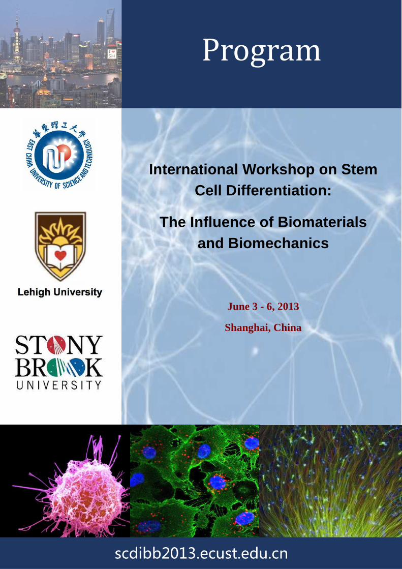
International Workshop on Stem Cell Differentiation:
The Influence of Biomaterials and Biomechanics
June 3 - 6, 2013
Shanghai, China
Program
scdibb2013.ecust.edu.cn

International Workshop on Stem Cell Differentiation
The Influence of Biomaterials and Biomechanics
East China University of Science and Technology,
International Workshop on Stem Cell Differentiation
he Influence of Biomaterials and Biomechanics
June 3 - 6, 2013
East China University of Science and Technology, Shanghai, China
Co-Organized by
Lehigh University
International Workshop on Stem Cell Differentiation:
he Influence of Biomaterials and Biomechanics
Shanghai, China

CONFERENCE CHAIRS
Professor Changsheng Liu, East China University of Science and Technology, China Professor H. Daniel Ou-Yang, Lehigh University, USA
ORGANISING COMMITTEE
Secretary Prof. Yongsheng Li, East China University of Science and Technology, China
Organizing Committee Members Prof. Sabrina S. Jedlicka, Lehigh University, USA
Prof. Miriam Rafailovich, Stony Brook University, USA
Prof. Dimitrios Vavylonis, Lehigh University, USA
Prof. Marcia Simon, Stony Brook University, USA
Prof. Jiaxue Huang, Union Stem Cell & Gene Engineering Co. Ltd, China
Dr. Xue Qu, East China University of Science and Technology, China
Dr. Shengmiao Zhang, Huazhong University of Science and Technology, China
Prof. Qixin Zhuang, East China University of Science and Technology, China
SECRETARIAT CONTACT
Yongsheng Li
East China University of Science and Technology
130 Meilong Road
Xuhui District, Shanghai, 200237, China
Tel: +86-21-64250740
E-mail: [email protected]
CONFERENCE WEBSITE
http://scdibb2013.ecust.edu.cn

Contents
General Information Conference Themes Sponsors Program Schedule Oral Presentations

GENERAL INFORMATION HOTEL INFORMATION Shanghai Grand Sky Light Hotel(上海园林格兰云天大酒店)
Address: 100 Baise Road, Xuhui District, Shanghai 200231(上海徐汇区百色路 100号);
http://www.grandskylighthotel.com/
REGISTRATION DESK The registration/information desk will be located in Shanghai Grand Skylight Gardens Hotel during the
daytime on Monday 3th June 2013.
MEETING ROOMS Main Meeting Room: Lecture Room in Yifu Building ECUST(华东理工大学逸夫楼演讲厅)
Secondary Meeting Room: 2nd Meeting Room in Peace Building(华东理工大学和平楼第二会议室)
Refreshment: Youyi Restaurant from where boxed lunch will be available(华东理工大学友谊餐厅)
BADGES For identification purposes, security reasons and catering please make sure you wear your conference badge.
Replacements for lost badges are offered on the registration desk.
CONFERENCE BAGS & DELEGATE MANUALS Please make sure that you insert a business card or name card in your bag. Please also write your name in
your program book and do not leave either your book or bag unattended at the conference at any time.
EVENING RECEPTIONS & BANQUET Locations of receptions and banquets can be found in the Program Schedule.
SPONSORS
National Natural Science Foundation of China
National Science Foundation, USA
Ministry of Education, China
Ministry of Science and Technology, China
Science and Technology Commission of Shanghai Municipality
Tayee Medical Technology Co., Ltd.
CONFERENCE THEMES INTRODUCTION
"Stem Cell Differentiation: The Influence of Biomaterials and Biomechanics" (SCD) is an international
workshop on Stem Cell Differentiation, co-sponsored by the East China University of Science and
Technology (ECUST), Lehigh University and Stony Brook University. The goal of this workshop is to bring
together scientists from China, Europe, and the United States and highlight the latest research emphasizing
the influence of the cell-materials interface (chemistry, mechanics, and morphology) on stem cell
differentiation. Through this conference, we hope to promote interdisciplinary as well as international
collaborations which will further the field of stem cell research, suggest clinical applications, and discuss
ethical concerns and government funding policies. The participation of young faculty, post-doctoral
associates, and graduate students is strongly encouraged.
Topics
• The theoretical simulation and experimental methods toward the control of stem cell division and
differentiation
• The role of biochemistry and biomechanics in control of stem cell differentiation
• The features and applications of biological surface materials and nanomaterials in the control of stem
cell division differentiation
• Simulation and control of induced stem cells early differentiation and post-mature cell environment
• Progress and prospect of regulation of stem cell differentiation in clinical medicine
• Comparison and discussion of attention and support for stem cell by scientific research departments of
different countries

PROGRAM SCHEDULE Monday, June 3rd 2013
8:00-18:30 Registration at Shanghai Grand Sky Light Hotel
18:30-20:00 Reception at Shanghai Grand Sky Light Hotel
Tuesday, June 4th 2013
8:10-8:30 Opening Address
Lecture Room in Yifu Building 8:30-10:10 4 lectures: [A-1] ~ [A-4]
10:10-10:45 Coffee/Tea Break
10:45-12:25 4 lectures: [B-1] ~ [B-4]
12:25-13:30 Lunch at Youyi Restaurant
13:30-15:10 4 lectures: [C-1] ~ [C-4]
2nd Meeting Room in Peace Building 15:10-15:55 Coffee/Tea Break
15:55-18:00 5 lectures: [D-1] ~ [D-5]
18:30 Banquet at Shangyuan Hotel
Wednesday, June 5th 2013
8:30-9:30 2 lectures: [E-1] ~ [E-2]
2nd Meeting Room in Peace Building 9:30-10:00 Coffee/Tea Break
10:00-12:05 5 lectures: [F-1] ~ [F-5]
12:05-13:30 Lunch and Panel-style Discussion
13:30-15:10 4 lectures: [G-1] ~ [G-4]
Lecture Room in Yifu Building 15:10-15:55 Coffee/Tea Break/Poster
15:55-18:00 5 lectures: [H-1] ~ [H-5]
18:30 Dinner at Youyi Restaurant and Tour on Huangpu River
Friday, June 7th 2013
9:00- City Tour
Thursday, June 6th 2013
8:30-9:30 2 lectures: [I-1] ~ [I-2]
Lecture Room in Yifu Building 9:30-10:00 Coffee/Tea Break/Poster
10:00-12:05 5 lectures: [J-1] ~ [J-5]
12:05-13:30 Lunch at Youyi Restaurant
13:30-15:10 4 lectures: [K-1] ~ [K-4] Lecture Room in Yifu Building
15:10-15:30 Closing remarks
Monday, June 3rd 2013
8:00-18:30 Registration at Shanghai Grand Skylight Gardens Hotel
18:30-20:00 Reception at Shanghai Grand Skylight Gardens Hotel
Tuesday, June 4th 2013
Room Lecture Room in Yifu Building
8:10-8:30 Opening Address
Chairman Changsheng Liu, East China University of Science and Technology, China
8:30-8:55 [A-1] The role of chemical and mechanical heterogeneity in stem cell differentiation Miriam Rafailovich, State University of New York at Stony Brook, USA
8:55-9:20 [A-2] Guide stem cell differentiation by regulation of cell signaling pathways via biomaterials design Yin Xiao, Queensland University of Technology, Australia
9:20-9:45 [A-3] Autologous chondrocyte implantation for cartilage repair Miomir Knežević, Educell Ltd., Slovenia
9:45-10:10 [A-4] Hydrogel based on RGD mimetic cross-linker with tunable stiffness to regulate Malcolm Xing, University of Manitoba, Canada
10:10-10:45 Coffee/Tea Break
Chairman Megan T. Valentine, University of California, Santa Barbara, USA
10:45-11:10 [B-1] Mechanical cues affects Ca2+ signaling in stem cells Yingxiao Wang, University of California, San Diego, USA
11:10-11:35 [B-2] Mathematical modeling of contractile actomyosin bundle assembly Dimitrios Vavylonis, Lehigh University, USA
11:35-12:00
[B-3] Mechanical regulation of stem cell differentiation: from mechanical cues to mechanical responses Zhi Pan, L'Oréal, USA
12:00-12:25 [B-4] Biophysical regulation of epigenetic state and cell reprogramming
Timothy Downing, University of California, Berkeley, USA
12:25-13:30 Lunch at Youyi Restaurant
Room 2nd Meeting Room in Peace Building
Chairman Malcolm Xing, University of Manitoba, Canada
13:30-13:55 [C-1] Instructive nanofibrous scaffolds for cardiovascular repair Patrick Ching-Ho Hsieh, National Cheng Kung University, Taiwan
13:55-14:20
[C-2] Role of Fhod3-mediated actin assembly in myofibrillogenesis during cardiac differentiation Ryu Takeya, Kyushu University, Japan

14:20-14:45 [C-3] Force switches on the P-selectin-mediated Ca2+-responding of neutrophils Jianhua Wu, South China University of Technology, China
14:45-15:10
[C-4] The influence of whole-organ heart extracellular matrix on induced pluripotent stem cells differentiation Changyong Wang, Academy of Military Medical Sciences, China
15:10-15:55 Coffee/Tea Break
Chairman Hongjun Wang, Stevens Institute of Technology, USA
15:55-16:20
[D-1] The differentiation of dental pulp stem cells is determined by dynamic cooperation between cells and substrate Marcia Simon, State University of New York at Stony Brook, USA
16:20-16:45 [D-2] Cellular responses of bioactive materials for bone regeneration Changsheng Liu, East China University of Science and Technology, China
16:45-17:10
[D-3] Osteogenic differentiation of mesenchymal stem cells through the use of novel scaffolds Min Wang, The University of Hong Kong, Hong Kong
17:10-17:35
[D-4] Enhanced osteogenic differentiation potential in dedifferentiation- reprogrammed mesenchymal stem cells Gang Li, The Chinese University of Hong Kong, Hong Kong
17:35-18:00
[D-5] Tissue engineering and regenerative medicine for bone deficiency and dental implantation, a novel option for oral function restoration Xinquan Jiang, Shanghai Ninth People Hospital, China
18:30 Banquet at Shangyuan Hotel
Wednesday, June 5th 2013
Room 2nd Meeting Room in Peace Building
Chairman Miomir Kneževi ć, Educell Ltd., Slovenia
8:30-9:00 [E-1] Biothics and funding policy: participate by video conference Brooke Ellison, State University of New York at Stony Brook, USA
9:00-9:30 [E-2] Topics on human embryonic stem cell research Dena S. Davis, Lehigh University, USA
9:30-10:00 Coffee/Tea Break
Chairman Sanjay Kumar, University of California, Berkeley, USA
10:00-10:25 [F-1] Integrating mechanical and chemical signaling to guide cell differentiation Paul Janmey, University of Pennsylvania, USA
10:25-10:50
[F-2] Combined impacts of substrate stiffness, topography, and size on stem cell differentiation Mian Long, Institute of Mechanics, Chinese Academy of Sciences, China
10:50-11:15 [F-3] Modulation of stem cell differentiation with nanoscale materials Xingjie Liang, National Center for Nanoscience and Technology, China
11:15-11:40
[F-4] Synchronized hyperthermochemotherapy targeting CD44+ cancer subpopulation augments therapeutic efficacy in vivo Dar-Bin Shieh, National Cheng Kung University, Taiwan
11:40-12:05 [F-5] Towards 4P medicine: a view from cancer research Ping Ao, Shanghai Jiao Tong University, China
12:05-13:30 Lunch and Panel-style Discussion
Room Lecture Room in Yifu Building
Chairman Yin Xiao, Queensland University of Technology, Australia
13:30-13:55 [G-1] Biomaterials surfaces for stem cell culture Keith Mclean, Commonwealth Scientific and Industrial Research Organisation, Australia
13:55-14:20
[G-2] Biomaterial systems with controlled temporal and spatial signal presentation for regulating stem cell behavior Eben Alsberg, Case Western Reserve University, USA
14:20-14:45
[G-3] Cell-mediated biomineralization is dictated by substrate morphology and chemistry Aneel K. Bherwani, Ziauddin University, Pakistan
14:45-15:10
[G-4] Design of cell-recognizable nano-biomaterials for ES/iPS cell proliferation and
differentiation ----cadherin-based "cell-cooking plate"
Toshihiro Akaike, Tokyo Institute of Technology, Japan
15:10-15:55 Coffee/Tea Break/Poster
Chairman Renke Li, University of Toronto, Canada
15:55-16:20 [H-1] 3D-woven PCL with hMSCs: Differentiation and integration with native bone Benjamin Lee Larson, Harvard-MIT Health Sciences and Technology, USA
16:20-16:45
[H-2] Biomaterials with bioactive composition and nano-structure for osteogenic differentiation of stem cells and bone regeneration Jiang Chang, Shanghai Institute of Ceramics, Chinese Academy of Sciences, China
16:45-17:10 [H-3] Tumor suppressor WWOX inhibits stem cell development Nan-Shan Chang, National Cheng Kung University, Taiwan
17:10-17:35
[H-4] Recreation of developmental signals in biomimetic biomaterial scaffold to guide hMSC chondrogenesis Liming Bian, The Chinese University of Hong Kong, Hong Kong
17:35- 18:00
[H-5] Direct viewing of single-molecule behavior elucidates fine tuning of cell mechanics Naoki Watanabe, Tohoku University, Japan
18:30 Dinner at Youyi Restaurant and Tour on Huangpu River
Friday, June 7th 2013
9:00 City Tour
Thursday, June 6th 2013
Room Lecture Room in Yifu Building
Chairman Keith Mclean, Commonwealth Scientific and Industrial Research Organisation, Australia
8:30-9:00 [I-1] Government funding panel
Rosemarie Hunziker, National Institutes of Health, USA
9:00-9:30 [I-2] Government funding panel
Kaiming Ye, National Science Foundation, USA
9:30-10:00 Coffee/Tea Break/Poster
Chairman Eben Alsberg, Case Western Reserve University, USA
10:00-10:25 [J-1] Biomimetic nanofibers and cell phenotypic expression
Hongjun Wang, Stevens Institute of Technology, USA
10:25-10:50 [J-2] Development of novel magnetic tweezers technologies: Applications to biomaterials characterization and cell matrix actuation
Megan T. Valentine, University of California, Santa Barbara, USA
10:50-11:15
[J-3] MRTFs are essential transcription factors in cellular differentiation and development Cuntong Zhang, Tianjin University of Science and Technology, China
11:15-11:40 [J-4] Adverse effects of gold nanoparticles on human skin cells
Tatsiana Mironava, State University of New York at Stony Brook, USA
11:40-12:05
[J-5] Multifunctional gelatin-organosilica nanoparticles for efficient tumor-targeted gene transfection Lei Ren, Xiamen University, China
12:05-13:30 Lunch at Youyi Restaurant
Chairman Yingxiao Wang, University of California, San Diego, USA
13:30-13:55 [K-1] Mechanobiological regulation of normal and tumor stem cells in the brain Sanjay Kumar MD, University of California, Berkeley, USA
13:55-14:20 [K-2] Engineered myocardial milieu for aged cell rejuvenation
Renke Li, University of Toronto, Canada
14:20-14:45 [K-3] Effects of dynamic perfusion on the construction of a large-scale tissue
Tingting Tang, Shanghai Ninth People Hospital, China
14:45-15:10 [K-4] Directed differentiation of hematopoietic stem cells
Jiaxue Huang, Union Stem Cell & Gene Engineering Co. Ltd, China
15:10-15:30 Closing remarks H. Daniel Ou-Yang, Lehigh, USA& Changsheng Liu, ECUST, China
ORAL PRESENTATIONS
[A-1] Miriam Rafailovich , State University of New York at Stony Brook, USA
The role of chemical and mechanical heterogeneity in stem cell differentiation
[A-2] Yin Xiao , Queensland University of Technology, Australia
Guide stem cell differentiation by regulation of cell signaling pathways via biomaterials design
Cell differentiation and tissue formation are specifically regulated by either the programmed cell signaling pathways or manipulated process to the environments. For example, low oxygen pressure (hypoxia) in vivo plays a pivotal role in coupling angiogenesis with osteogenesis via progenitor cell recruitment, differentiation and angiogenesis. Hypoxia activates a series of angiogneic processes mediated by the hypoxia inducing factor-1 (HIF-1) transcription factor. HIF-1α initiates the expression of a number of genes associated with tissue regeneration and skeletal tissue development and has been shown to enhanced fracture repair. Hypoxia can be mimicked artificially by stabilizing HIF-1α expression , such as by the application of Co2+ ions, and has been suggested as a potential strategy to promote neovascularization. We have developed hypoxia-mimicking scaffolds by incorporating ionic Co2+. The results showed that low amounts of Co (< 5%) incorporated into scaffolds had no significant cytotoxicity and that their incorporation significantly enhanced HIF-1α expression, VEGF protein secretion, and bone-related gene expression in BMSCs. Our results indicate that incorporating cobalt ions into scaffolds is a viable option for preparing hypoxia-mimicking tissue engineering scaffolds and significantly enhanced hypoxia function.
Lithium (Li) has been widely used as a long-term mood stabilizer in the treatment of bipolar and depressive disorders. Li+ ions are thought to enhance the remyelination of peripheral nerves and also stimulate the proliferation of neural progenitor cells and retinoblastoma cells via activation of the Wnt/β-catenin signalling pathway. We incorporated the Li+ ions into the scaffolds and showed that this approach yielded scaffolds with a favourable composition, microstructure and mesopore properties for cell attachment, proliferation, and cementogenic differentiation of periodontal ligament cells (PDLCs). It was found that Li+ ions by themselves significantly enhanced the proliferation, differentiation, cementogenic gene expression of PDLCs, most likely via activation of Wnt/β-catenin signalling pathway. Our results indicate that incorporation of Li+ ions into bioactive scaffolds is a viable means of enhancing the Wnt canonical signalling pathway to stimulate osteogenic/cementogenic differentiation of PDLCs.
[A-3] Miomir Kneževi ć, Educell Ltd., Slovenia
Autologous chondrocyte implantation for cartilage repair
Miomir Knežević1, ArianaBarlič1, Damjan Radosavljevič2, Matej Drobnič2, NevenkaKregar Velikonja1
1 Educelld.o.o., Ljubljana, Slovenia, 2 Department of Orthopaedic Surgery, University Medical Centre, Ljubljana, Slovenia
The idea of tissue repair is to establish tissue structure similar to those of native tissue in order to enable the repair tissue proper physiological function.
Cartilage tissue regeneration using autologous cells has first been investigated for knee cartilage repair. Products for knee cartilage repair are being tested through clinical trials and are becoming part of routine clinical practice in last decade and this approach is being applied also for other joints (ankle, hip).
The aspects that need to be considered in planning of cell therapy approach are: 1. Biological: allow the cells to express cell phenotype, similar to native cartilage; 2. Tissue engineering to use scaffold that allows cell survival, is biodegradable, non-immunogenic and is easy to handle; 3. Surgical: the concept should allow minimally invasive procedure, good clinical outcome.
Biopsy of articular cartilage, taken from non-load bearing surface is usually used as cell source for these tissue engineering applications. When chondrocytes are isolated from cartilage tissue and cultured as monolayers, they lose their cartilage phenotype and transform into flattened fibroblast-like cells upon repeated passages. Gene expression studies have shown a change in expression of typical chondrocyte markers of hyaline cartilage (collagen type II, aggrecan) to collagen type I and versican, which are predominant in dedifferentiated chondrocytes and fibrocartilage. In principle, chondrocyte dedifferentiation can be reversed by transferring cells into an environment supporting a spherical morphology. Specific gene expression was tested to measure the influence of the scaffold on cell phenotype.
The cell therapy approach is mainly used in large, recidivant lesions over 2 cm2. Different forms of products for cartilage repair (ChondroArt 1D, 2D, 3D) differ in a view of combination with scaffold materials to allow an optimal treatment of different lesion types.
The first ACI implantations in Ljubljana took place in 1996. So we are able to provide one of the longest follow-up evaluations in Europe of the classical ACI (ChondroArt 1D). Clinical results demonstrated stable performance of operated knees over ten years. High incidence of knee osteoarthritis in patients with concomitant ACL reconstruction and low incidence of OA in osteochondritisdissecans patients indicate that ACI works best for localized low-impact cartilage lesions in young patients.
Due to logistics and cost-effectiveness issues together with legislative considerations of cell cultivation the research and clinical interest of last years has been focused on "non-cellular" scaffolds. They are populated with recipient’s cells from bone marrow aspirate, subchondral bone MSC, or peripheral cartilage when implanted. In spite of their promising initial results, they are currently indicated for small-sized lesions only.
This leaves the novel scaffold based ACI (together with osteo-chondral allografts in certain situations) a current clinical standard for medium to large cartilage defects.
Tissue engineering of cartilage is also the basis for UroArtTM cell product for treatment of vesicoureteral reflux and for cell based testing system for in vitro assessment of anti-TNF substances or other antireumatic drugs.
[A-4] Malcolm Xing , University of Manitoba, Canada
Hydrogel based on RGD mimetic cross-linker with tunable stiffness to regulate stem cell differentiation
We report a functionalized cationic poly (amido amine) crosslinker synthesized by Michael addition of peptide mimetic agmatine to N,N’-bis(acryloyl)cystamine. A polyethylene glycol diacrylate (PEGDA) hybrid hydrogel was developed using functionalized crosslinker with cell adhesion capability and tunable stiffness. This engineered hydrogel presents a 3-D in-growth of cells and shows remarkable morphology of the gel. The tunable stiffness of this hydrogel can regulate the osteogenic differentiation of mesenchymal stem cells from RT-PCR. Both in vitro and in vivo tests demonstrate adjustable degradation rate and biocompatibility. The crosslinker and its hydrogels hold promising potential for tissue engineering applications.
[B-1] Yingxiao Wang, University of California, San Diego, USA
Mechanical cues affects Ca2+ signaling in stem cells
Tae-Jin Kim1, Yingxiao Wang1, 2
1Neuroscience Program, University of Illinois, Urbana-Champaign, Urbana, IL, 61801, USA, 2Department of Bioengineering, University of California, San Diego, 92093, USA
Mechanical forces play crucial roles in regulating cellular functions, such as cell spreading, traction forces, and stem cell differentiation. However, it is not clear how they influence early cell signaling events such as calcium in living stem cells. Using highly-sensitive Ca2+ biosensors-based on fluorescence resonance energy transfer (FRET), we investigated the molecular mechanism by which mechanical forces affect calcium signaling in human mesenchymal stem cells (hMSCs). Spontaneous Ca2+ oscillations were observed inside the cytoplasm and the endoplasmic reticulum (ER) using the FRET biosensors targeted at subcellular locations in cells plated on rigid dishes. Lowering the substrate stiffness to 1 kPa significantly inhibited both the magnitudes and frequencies of the cytoplasmic Ca2+ oscillation in comparison to stiffer or rigid substrate. This Ca2+ oscillation was shown to be dependent on ROCK, a downstream effector molecule of RhoA, but independent of actin filaments, microtubules, myosin light chain kinase, or myosin activity. Lysophosphatidic acid, which activates RhoA, also inhibited the frequency of the Ca2+ oscillation. Consistently, either a constitutive active mutant of RhoA (RhoA-V14) or a dominant negative mutant of RhoA (RhoA-N19) inhibited the Ca2+ oscillation. Further experiments revealed that HMSCs cultured on gels with low elastic moduli displayed low RhoA activities.
Therefore, our results demonstrate that RhoA and its downstream molecule ROCK may mediate the substrate rigidity-regulated Ca2+ oscillation, which determines the physiological functions of HMSCs. We further investigated the molecular and biophysical mechanisms by which mechanical force regulates Ca2+ signaling at subcellular level in HMSCs, integrating optical laser tweezers and Ca2+ FRET biosensor. Laser-tweezer-traction on a fibronectin-coated bead at the plasma membrane induces intracellular Ca2+ oscillations caused by Ca2+ release from endoplasmic reticulum (ER) in the absence of extracellular Ca2+. Ca2+ oscillations produced by ER Ca2+ release upon mechanical force are mediated not only by the mechanical support of cytoskeleton and actomyosin contractility, but also by mechanosensitive Ca2+ channels on the plasma membrane, specifically TRPM7. As the ER Ca2+ release is inhibited, the mechanical force can induce intracellular Ca2+ increase via mechanosensitive Ca2+ channels in the presence of extracellular Ca2+, which is mediated by the cytoskeletal structure but not actomyosin contractility. Taken together, our results indicate that active actomyosin contractility is essential for the force transmission into the deep intracellular organelles, ER but dispensable for the mechanical regulation of plasma membrane channels. Therefore, our results clearly suggest a crucial role of mechanical force in regulating calcium signaling in live stem cells with a well-coordinated molecular hierarchy.
[B-2] Dimitrios Vavylonis , Lehigh University, USA
Mathematical modeling of contractile actomyosin bundle assembly
Actin filaments, myosin motors, and actin filament cross-linkers self-organize into contractile structures within cells. The subcellular distribution of these structures is important in cellular mechanosensing, the development and mechanical integrity of tissue, and stem cell differentiation. I will present work focusing on analysis and mathematical modeling of the kinetics of this organization in single cells. Model organism fission yeast assembles a contractile actomyosin ring by the condensation of a broad band of membrane-bound nodes containing myosin motors. Formins nucleate actin filaments from these membrane nodes and establish transient actomyosin connections among them. The movements of the nodes depend on actin filament cross-linkers that bundle the growing actin filaments. We developed numerical simulations modeling actin filaments as semiflexible polymers polymerized by formins, pulled by myosin, and represented cross-linking as an attractive interaction. The simulations show that actin cross linkers regulate actin-filament orientations inside actin bundles and organize the actin network. These simulations reproduce experimental observations by D. Laporte and J.-Q. Wu who observed that mutations and changes of the concentrations of cross-linker alpha-actinin in live cells causes the nodes to condense into different morphologies, forming clumps at low cross-linking and extended meshworks at high concentrations. To study the process of stress fiber formation in animal cells we observed the kinetics of stress fiber formation in HeLa cells expressing myosin light chain marker MRLC-GFP, after removal of myosin inhibition by blebbistatin. We observe transient myosin foci formation on the cell cortex that corresponds to myosin minifilaments. This is followed by contractile activity and alignment that leads to stabilization of the foci. A 2D Monte Carlo simulation incorporating the above features can explain the kinetics of stress-fiber network formation observed in experiments. This work supports a hierarchical process of self-organization involving components drawn together from distant parts of the cell followed by progressive stabilization and alignment by cross-linker and other proteins.
[B-3] Zhi Pan, L'Oréal, USA
Mechanical regulation of stem cell differentiation: from mechanical cues to mechanical responses
Similar to biochemical signals, mechanical cues, such as substrate mechanics, surface topography or external applied forces, can regulate tissue cell behaviors. Recently more investigations have shown that they can also trigger stem cell differentiation in complex microenvironments where inducers for multiple cell types are present. To provide better guidance for stem-cell based functional tissue engineering, it is crucial to understand how these external mechanical cues are related to different cellular responses and how the relations are dependent on different cell types and/or cell densities. Here we reported studies on human dermal fibroblasts and corneal limbal epithelial cells, as representatives of individual cells and collective cell sheet respectively, on physiologically relevant substrates with different stiffness or specific surface topographies. The influence of related mechanical cues on cell behaviors including morphology, adhesion and migration was illustrated through actin cytoskeleton reorganization and cell-to-substrate/cell-to-cell contact formation. In addition, by combining the digital image speckle correlation (DISC) technique with finite element method (FEM), we have analyzed the redistribution of cellular traction stresses during cell movement in real time with high spatial resolution, which in turn can mechanically explain and predict cells’ responses. All together these findings and techniques show us the potential of how we can regulate stem cell differentiation through mechanical cues.
[B-4] Timothy Downing, University of California, Berkeley, USA
Biophysical regulation of epigenetic state and cell reprogramming
Timothy L. Downing1,2, Jennifer Soto1,2,Constant Morez2,3, Timothee Houssin2,4, Falei Yuan2, Julia Chu2, Ashley Fritz5, Shyam Patel2, David V. Schaffer1,2,5, and Song Li1,2
1UC Berkeley & UCSF Joint Graduate Program in Bioengineering, 2Department of Bioengineering, UC Berkeley, CA, USA; 3Department of Biomedical Engineering, Ecole Polytechnique, Palaiseau, France; 4UniversityLille Nord de France, F-59000 Lille, France;
5Department of Chemical Engineering, UC Berkeley, CA, USA
Cell reprogramming represents a major advancement in cell biology and has wide applications in regenerative medicine, disease modeling, and drug screening. Induced pluripotent stem cells can be produced from adult fibroblastswith the use of transcription factors and chemical compounds;however, the role of biophysical factors in cell reprogrammingis not known. Here we show, for the first time, that biophysical cues, in the form of parallel microgrooves on the surface of cell-adhesive substrates, can significantly improve reprogramming efficiency and replace the effects of small molecule epigenetic modifiers. The mechanism behind this biophysical enhancement of cell reprogramming relies on mechanomodulation of epigenetic state –specifically, increases in histone H3 acetylation (AcH3) and histone H3 trimethylation (H3k4me3) on microgroove surfaces. Further investigation reveals that microtopography

dramatically downregulates histone deacytylase (HDAC) activityand increases the expressionofWD repeat domain 5 (Wdr5) protein–a subunit of H3 methyltransferase. Furthermore, microtopography promotes the upregulation of key epithelial-related genes, suggesting the initiation of a mesenchymal-to-epithelial transition, a required step in cell reprogramming. Disruption of actin-myosin contractility abolishes microtopographical regulation of epigeneticsand cell reprogramming, confirming that the actin cytoskeleton is required for this epigenetic mechanomodulation. In addition, adult fibroblasts show a similar response onaligned nanofibersurfacesin cell reprogramming and histone modifications, providing additional evidence that various topographical cues can regulate cell reprogramming. Furthermore, micropatterning cells on islands of matrix proteins for tight regulation of cell shape demonstrates the critical role that cell deformation plays in actively changing epigenetic state. In summary, this study unravels how the microfeatures of cell-adhesive substrates can induce epigenetic modifications, which in turn lead to enhanced cell reprogramming. We believe this novel biophysical regulation of epigenetics and cell reprogramming has important implications in a number of complex biological processes and in the broad scope of cell biology.
[C-1] Patrick Ching-Ho Hsieh MD, National Cheng Kung University, Taiwan
Instructive nanofibrous scaffolds for cardiovascular repair
Angiogenic therapy is a promising approach for tissue repair and regeneration. However, recent clinical trials using protein delivery or gene therapy to promote angiogenesis have failed to provide therapeutic effects. A key factor for achieving effective revascularization is the durability of the microvasculature and the formation of new arterial vessels. Accordingly, we carried out experiments to test if intramyocardial injection of self-assembling peptide nanofibers (NF) combined with vascular endothelial growth factor (VEGF) may create an intramyocardial microenvironment with prolonged VEGF releasing to improve post-infarction neovascularization in rats. Our data showed that when injected with NF, VEGF delivery was sustained within the myocardium for up to 14 days, and the side effects of systemic edema and proteinuria were significantly reduced to the same level as control. We also found NF/VEGF injection significantly improved angiogenesis (2.8 folds increase in capillary density), arteriogenesis (4.9 and 4.4 folds increase in arteriole and artery densities, respectively) and cardiac performance (51% increase in left ventricular fraction shortening) 28 days after myocardial infarction. As a result, NF/VEGF injection not only allowed controlled local delivery, but also transformed the injected site to a favorable microenvironment that recruited endogenous myofibroblasts and helped achieve effective revascularization. Strikingly, the engineered vascular niche further attracted a new population of cardiomyocyte-like cells to home to the injected sites, suggesting cardiomyocyte regeneration. Furthermore, large animal study also revealed consistent benefits. In summary, this study demonstrates a new strategy for cardiovascular repair with great potential for future clinical translation.
[C-2] Ryu Takeya, Kyushu University, Japan
Role of Fhod3-mediated actin assembly in myofibrillogenesis during cardiac differentiation
In striated muscle, thin actin filaments and thick filaments of myosin are highly organized to form myofibrils. Formation of myofibrils (myofibrillogenesis) is essential for the final step in the differentiation of cardiomyocytes as well as for successful regeneration of cardiac tissue from stem cells. However, the molecular mechanisms by which myofibrils are organized during cardiogenesis remain poorly understood. Here we show that Fhod3, a member of formin family proteins that regulate nucleation and polymerization of actin filaments, plays an essential role in myofibrillogenesis at a developmental stage of the heart. Fhod3-null mice grow normally up to embryonic day (E) 8.5, when the developing heart composed of immature myofibrils initiates spontaneous contraction. However, these undifferentiated myofibrils fail to mature further and myocardial development does not continue, leading to embryonic lethality by E11.5. Transgenic expression of wild-type Fhod3 in the heart restores myofibril maturation and cardiomyogenesis, which allow Fhod3–/– embryos to further develop. Furthermore, cardiomyopathic changes with immature myofibrils are caused in mice overexpressing a mutant Fhod3, defective in binding to actin. These findings indicate that actin dynamics regulated by Fhod3 participates in myofibril organization during cardiogenesis and thus plays a crucial role in cardiac differentiation.
[C-3] Jianhua Wu, South China University of Technology, China
Force switches on the P-selectin-mediated Ca2+-responding of neutrophils
In vascular inflammation process, binding of P-selectin glycoprotein ligand-1 (PSGL-1) on flowing leukocytes to selectins on vascular endothelial cells mediates the initial cell adhesion and triggers the downstream signaling. The “inside-out” signaling is [Ca2+]in-dependent usually so that the concentration of [Ca2+] in is a criterion of cell activation. The [Ca2+]in responding of flowing cells is related to either chemokine or fluid shear stress, but it still remains unknown that whether and how the force regulates the [Ca2+]in responding of leukocyte. With real-time fluorescence imaging system combined with flow chamber, we herein examined P-selectin-mediated calcium responding of human neutrophils in flows, for better understanding the regulation of fluid shear stress coupling and chemokine on intracellular signaling in pathophysiological process. We found that shear stress triggers the P-selectin-mediated Ca2+-response of neutrophils in flows. Increasing shear stress lengthens first and then shortens the mechanical relaxation time, the shear stress threshold occurs at about 0.5dyn/cm2. The peak of Ca2+-responding in neutrophils on P-selectin was apparent at shear stress from 0.2 to 6 dyn/cm2, and either decreased with shear stress below the stress threshold or increased with shear stress above the stress threshold until a saturation state reached at shear stress of 1 dyn/cm2. Our results suggest that fluid force on neutrophils adhered to E-selectin participates to intracellular outside-in signaling cascade through bonding of selectin to its ligands expressed on neutrophils, leading to Ca2+-response and activation of neutrophils, suggesting a mechano-chemical regulation mechanism for the activation and the immuno-functional realization of neutrophils.
[C-4] Changyong Wang, Academy of Military Medical Sciences, China
The influence of whole-organ heart extracellular matrix on induced pluripotent stem cells differentiation
Jin Zhou, Weizhen Chen, Cuimi Duan, Qiuxia Lin, Yan Wang, Changyong Wang
Cardiovascular disease (CVD) is one of the leading causes of morbidity and mortality worldwide. For the treatment of end stage heart disease, orthotopic cardiac transplantation is a highly successful procedure. But the limited number of available donor hearts and immune rejection after transplantation issues still remained unsettled. The emerging whole-heart reconstruction research that based on heart whole-organ decellularization and recellularization could potentially solve this, which brings new opportunities to the treatment of heart disease.
In our study, we design one rat whole-heart perfusion decellularization system at first. By using it we make a comparison of the results from different decellularization ways with specific detergent solutions and developed a high-throughput, safe and convenient decellularized heart matrix scaffolds preparation system as well as corresponding decellularization method. After that, we detected the basic properties and the biocompatibility of the decellularized construct in vitro. The decellularized whole-organ heart matrix or the decellularized whole-organ heart matrix-made sheet scaffold was then recellularized with induced pluripotent stem cells(iPSCs) as seed cells, so as to investigate the regularity of survival, growth and cardiovascular lineage differentitation potential of iPSCs in decellularized heart matrix microenvironment. Finally, we implanted the iPSCs-recelularized tissue-engineered cardiac patch or the matrix-made sheet scaffold onto the infarct bed in a rat model of myocardial infarction to enhance the differentiation ability of the iPSCs and also test the practicality of myocardial infarction therapy.
Our research proves that the prepared decellularized whole heart construct retained the three-dimensional microarchitecture, the essential extracellular matrix components and vasculature structure. It could also been used as good scaffolds for constructing heart tissue in vitro and direct differentiation of the stem cells, like iPSCs, into the cardiovascular lineage. After in vivo tranplantation of iPSCs-recelularized constructed cardiac patch or only the matrix-made sheet scaffold in myocardial infarction rat model, the implanted slices show the potential contribute to the new microenvironment for arteriogenesis and cardiac repair. Besides, the implanted iPSCs were immunostained with various cardiac specific markers after four weeks growth in vivo, which suggested that the differentiation ability of implanted iPSCs were enhanced and the iPSC also played a role in the process of myocardial tissue regeneration after myocardial infarction. This study also demonstrates alternative use of the heart whole-organ decellualrized matrix and a new strategy for cardiovascular repair, which could have potential for future clinical application.
[D-1] Marcia Simon, State University of New York at Stony Brook, USA
The differentiation of dental pulp stem cells is determined by dynamic cooperation between cells and substrate
[D-2] Changsheng Liu, East China University of Science and Technology, China
Cellular responses of bioactive materials for bone regeneration
Most bio-tissues, ranging from the hard to the soft, have mesoscopically well-ordered structures, which play a crucial role in executing the functions of mineralization and possess a micrometer-scaled, highly porous structure with remarkable mechanical strength. These delicate structures in nature inspire researchers to create versatile materials with special microstructures and characteristics.
The refined mesoscopic nanostructure can provide new avenues to regulate the cell responsive activities. Observations on osteoblast response demonstrated that the huge specific surface area of mesoporous topological structure could promote osteoblastic proliferation via up-regulated the integrin α5 at an early stage during cell attachment due to its higher absorptive capability of protein and faster formation of apatite layer. In addition, scaffolds possessed macro-meso porous structure appeared to provide a better microenvironment for hMSC attachment, spreading, proliferation, and osteogenic differentiation. Moreover, the µCT and histological evaluation confirmed that the hierarchical scaffolds improved the efficiency of bone regeneration in vivo.
[D-3] Min Wang , The University of Hong Kong, Hong Kong
Osteogenic differentiation of mesenchymal stem cells through the use of novel scaffolds
Mesenchymal stem cells (MSCs) have the potential to differentiate along multiple lineage cell types, including bone, cartilage, muscle, etc., under different environments/stimuli. In the bone regeneration process in human bodies, MSCs are recruited from the bone marrow and periosteum and may responsively differentiate into chondrocytes, osteoblasts or fibroblasts, depending on the biological and other conditions. MSCs from other sources such as adipose, umbilical cord and muscle may also be regulated for osteogenic differentiation in vivo by cytokines, hormones, growth factors, etc. secreted by surrounding cells. For in vitro osteogenic differentiation of MSCs, some inducing factors including both chemical factors and physical factors are necessary. Several members of the transforming growth factor (TGF)-β superfamily, such as bone morphogenetic protein-2 (BMP-2) and BMP-7, have shown promising results in stimulating osteogenic differentiation of MSCs. In our research in bone tissue engineering, nanocomposite scaffolds containing recombinant human bone morphogenetic protein-2 (rhBMP-2) could be produced using selective laser sintering, a
rapid prototyping technology. Furthermore, hybrid nanofibrous scaffolds with incorporated rhBMP-2 could be fabricated via electrospinning. Using these scaffolds, it has been demonstrated through in vitro and in vivo studies that osteogenic differentiation of MSCs, such as C3H10T1/2 cells, human bone marrow-derived MSCs and human umbilical cord-derived MSCs, could be induced. The studies have also shown that the osteogenic differentiation is influenced by a number of scaffold parameters.
[D-4] Gang Li, The Chinese University of Hong Kong, Hong Kong
Enhanced osteogenic differentiation potential in dedifferentiation-reprogrammed mesenchymal stem cells
Yun-feng Rui and Gang Li
Mesenchymal stem cells (MSCs) have capacities of self-renewal and multi-differentiation potentials, are widely used for bone regenerative therapy. However, low cell survival rate and osteogenic differentiation in vivo after MSCs transplantation has significantly hindered the effectiveness of stem cell therapy. Dedifferentiation has been considered as one of the means rerouting cell fate and reversing the differentiated cells to an earlier, more primitive phenotype characterized by alterations in gene expression patterns. One recent study demonstrated that dedifferentiated neu-MSCs exhibited enhanced cell survival and higher efficacy in neuronal differentiation compared to untreated MSCs both in vitro and in vivo. We hypothesized that de-differentiation process could reprogram osteogenic MSCs to a distinct phenotype with enhanced cell survival and osteogenic potential. In this study, we aimed to (1) establish and characterize the dedifferentiated osteogenic MSCs (De-OS-rPBMSCs) using rat peripheral blood-derived MSCs (rPBMSCs) in vitro; (2) compare the cell survival capacity, cell proliferative capacity, colony forming ability, cell migration capacity and osteogenic/ adipogenic/ chondrogenic differentiation potential of the De-OS-rPBMSCs and untreated rPBMSCs in vitro; (3) To compare the osteogenic differentiation potential of of the De-OS-rPBMSCs and untreated rPBMSCs in vivo.
[D-5] Xinquan Jiang, Shanghai Ninth People Hospital, China
Tissue engineering and regenerative medicine for bone deficiency and dental implantation, a novel option for oral function restoration
Clinical problems requiring bone regeneration in oral and maxillofacial region are diverse and challenging in modern dentistry. Reconstruction of alveolar bone is an important part of therapeutic procedures. Several approaches have been developed for treating bone loss/defect, including autologous bone grafts, distraction osteogenesis, and guided bone regeneration (GBR). These methods may achieve certain effect on bone regeneration, but also present limitations for clinical application. Recently, the development of bone tissue engineering and regeneration medicine has made it an attractive alternative for alveolar bone regeneration with great potential. Stem cells, novel bioactive matrix as well as different growth factors have been adopted to achieve the purpose. In this presentation, animal studies as well as preliminary clinical applications of bone regeneration together with oral functional restoration by dental
implantation would be discussed. Vertical alveolar ridge augmentation, sinus augmentation and tissue engineering techniques and systematically evaluated in large animal models. Besides, the introduction of the method to clinic would also be reviewed. Finally, future challenges and prospvascularization of tissue engineering bone, as well as how to improve osteoof dental implants with fabricated tissue engineered bone would be discussed. In summary, tissue enginimplantation belongs to a promising novel option for oral function restoration.
[E-1] Brooke Ellison
For the SCD conference, I will be preresearch purposes. I am on the Ethics Committee of New York's Empire State Stem Cell Board, and in 2009, New York became the first public stem cell research funding authority to provide compensation to wodecision was the outcome of over two years of ethical debate regarding concerns over inducements, informed consent, paternalism, and unintended consequences. The decision made by the Empire State Stem Cell Board wagreat deal of public discussion, but has also provided the groundwork for tremendous scientific strides in New York.compensation for this procedure. I will discuss hyperstimulation syndrome, prevalence of the condition, and the connection it may or may not have to oocyte donation. I will also discuss the debates that took place in the Empire State Stem Cell Board, and how these have given riseforth by the International Society for Stem Cell Research.
[E-2] Dena S. Davis
This talk will discuss some of the issues in human embryonic stem cell research today, especially with respect to NIH guidelincontroversy over whether embryos created for the purpose of reproduction, but with donor gametes, may be used for stem cell derivation without the consent of the gamete donors. Another topic will be the meancontext. Do limits on how long embryos can remain frozen, and other constraints, limit donor options to the extent that it is no longer correct to say that they have given "informed and voluntary consent"?
[F-1] Paul Janmey,
Integrating mechanical and chemical signaling to guide cell differentiation
Cells are sensitive to mechanical signals produced either by application of exogenous force, or by the resistance to cellof the materials to which they adhere. The range of stiffness over which different cell types respond can vary over a wide range and generally reflects the elastic modulus of the tissue from which the cells
implantation would be discussed. Vertical alveolar ridge augmentation, sinus augmentation and more challenging mandible defect repairing were carried out by tissue engineering techniques and systematically evaluated in large animal models. Besides, the introduction of the method to clinic would also be reviewed. Finally, future challenges and prospect including the regeneration of large mandible bone defect, the vascularization of tissue engineering bone, as well as how to improve osteoof dental implants with fabricated tissue engineered bone would be discussed. In summary, tissue engineering and regeneration medicine for bone deficiency and dental implantation belongs to a promising novel option for oral function restoration.
Brooke Ellison, State University of New York at Stony Brook, USA
For the SCD conference, I will be presenting the ethics of oocyte donation for stem cell research purposes. I am on the Ethics Committee of New York's Empire State Stem Cell Board, and in 2009, New York became the first public stem cell research funding authority to provide compensation to women who donate oocytes for research. This decision was the outcome of over two years of ethical debate regarding concerns over inducements, informed consent, paternalism, and unintended consequences. The decision made by the Empire State Stem Cell Board was groundbreaking, subject to a great deal of public discussion, but has also provided the groundwork for tremendous scientific strides in New York. I will discuss the ethics of oocyte donation and providing compensation for this procedure. I will discuss the concerns of ovarian hyperstimulation syndrome, prevalence of the condition, and the connection it may or may not have to oocyte donation. I will also discuss the debates that took place in the Empire State Stem Cell Board, and how these have given riseforth by the International Society for Stem Cell Research.
Dena S. Davis, Lehigh University, USA
This talk will discuss some of the issues in human embryonic stem cell research today, especially with respect to NIH guidelines. Included will be a discussion of the ongoing controversy over whether embryos created for the purpose of reproduction, but with donor gametes, may be used for stem cell derivation without the consent of the gamete donors. Another topic will be the meaning of "voluntary" donation in an international context. Do limits on how long embryos can remain frozen, and other constraints, limit donor options to the extent that it is no longer correct to say that they have given "informed and voluntary consent"?
Paul Janmey, University of Pennsylvania, USA
Integrating mechanical and chemical signaling to guide cell differentiation
Cells are sensitive to mechanical signals produced either by application of exogenous force, or by the resistance to cell-generated forces caused by the viscoelastic properties of the materials to which they adhere. The range of stiffness over which different cell types respond can vary over a wide range and generally reflects the elastic modulus of the tissue from which the cells were isolated. Many cell types can alter their own
implantation would be discussed. Vertical alveolar ridge augmentation, sinus more challenging mandible defect repairing were carried out by
tissue engineering techniques and systematically evaluated in large animal models. Besides, the introduction of the method to clinic would also be reviewed. Finally, future
ect including the regeneration of large mandible bone defect, the vascularization of tissue engineering bone, as well as how to improve osteo-integration of dental implants with fabricated tissue engineered bone would be discussed. In
eering and regeneration medicine for bone deficiency and dental implantation belongs to a promising novel option for oral function restoration.
State University of New York at Stony Brook, USA
senting the ethics of oocyte donation for stem cell research purposes. I am on the Ethics Committee of New York's Empire State Stem Cell Board, and in 2009, New York became the first public stem cell research funding
men who donate oocytes for research. This decision was the outcome of over two years of ethical debate regarding concerns over inducements, informed consent, paternalism, and unintended consequences. The
s groundbreaking, subject to a great deal of public discussion, but has also provided the groundwork for tremendous
I will discuss the ethics of oocyte donation and providing the concerns of ovarian
hyperstimulation syndrome, prevalence of the condition, and the connection it may or may not have to oocyte donation. I will also discuss the debates that took place in the Empire State Stem Cell Board, and how these have given rise to recommendations put
This talk will discuss some of the issues in human embryonic stem cell research today, es. Included will be a discussion of the ongoing
controversy over whether embryos created for the purpose of reproduction, but with donor gametes, may be used for stem cell derivation without the consent of the gamete
ing of "voluntary" donation in an international context. Do limits on how long embryos can remain frozen, and other constraints, limit donor options to the extent that it is no longer correct to say that they have given
Integrating mechanical and chemical signaling to guide cell differentiation
Cells are sensitive to mechanical signals produced either by application of exogenous ated forces caused by the viscoelastic properties
of the materials to which they adhere. The range of stiffness over which different cell types respond can vary over a wide range and generally reflects the elastic modulus of
were isolated. Many cell types can alter their own
stiffness to match that of the substrate to which they adhere. The maximal elastic modulus that cells can attain is similar to that of crosslinked actin networks at the concentrations in the cell cortex. Mechanosensing appears to require an elastic connection between cell and substrate, mediated by transmembrane proteins. Transmission and transduction of these physical stimuli depend on the type of adhesion receptors by which the cells bind their surroundings, and on crosstalk between signals derived from cell-cell and cell-matrix interactions. Integration of chemical and physical stimuli can lead to changes in cell structure and function that cannot be achieved by either signal alone.
[F-2] Mian Long , Institute of Mechanics, Chinese Academy of Sciences, China
Combined impacts of substrate stiffness, topography, and size on stem cell differentiation
[F-3] Xingjie Liang , National Center for Nanoscience and Technology, China
Modulation of stem cell differentiation with nanoscale materials
Recent advances in nanotechnology have provided various new tools for biomedical research and clinical applications. Nano-biology is one of the disciplines that will benefit from this technology the most. Bionanotechnology is to utilize the developed technologies and/or materials smaller than 100 nanometers in one dimension and takes advantages of properties that are only presented at this nano level. Nanoscale materials, with their unique size-dependent physical and chemical properties, have showed promising advantages in biological research fields such as intercellular targeted molecular labeling, vehicles traffic monitoring, precisely silence of targeted genes, controllable release of payload including chemical compounds and/or siRNA, and so on. However, to develop sensitive and efficient nanomaterials with these promising progresses, the effective penetration of plasma membrane and low toxicology of engineered nanomaterials as carriers or probes exist as a potential barrier and has caught much attention. Therefore, Designing nanomaterials for expected bioeffects without significant toxicity has become an important issue for clinical and basically biological application of novel nanobiomaterials. It is critical to identify novel nanomaterials with an exclusive biological function in vitro and in vivo, for example, to modulate the stem cell differentiation. Recently, innovative materials assembled at nanosize for biological activities measurement have been developed with negligible cytotoxicity in tissue culture and without detectable side effects in vivo. The unique properties of nanostructures still needs further study to precisely monitor or modulate the differentiation of stem cells. The developed nanotechnology may inspire researchers to innovate a new generation of nanoscale biomaterials with inventive non-traditional approach for biological studies. The development of targeted nanoscale biostructures will allow for more efficiently monitor and measurement of molecular behaviors at different levels.
[F-4] Dar-Bin Shieh
Synchronized h
[F-5] Ping Ao, University, China
There has been a tremendous progress in cancer research. However, it appears the current dominant cancer research framework of regarding cancer as leads impasse. Naturally questions have been asked that whether it is possible to develop alternative frameworks such that they can connect both to mutations and other genetic/genomic effects and to environmental factors. Furthermore, sucan be made quantitative and with predictions experimentally testablegood candidate exampleperspective suggests that that gives grounds for hope. verifications will be presented
[G-1] Keith McleanAustralia
Laurence Meagher,
The ability to design and engineer surfaces to elicit specific cellular responses (eprevention of noncell fate, including that of stem cells, by presentation of specific biological ligands; patterning of cells) is of interest and relevance to the development of mattissue engineering and regenerative medicine. A number of coating strategies have been used to achieve this goal, however, many are limited with respect to their function and transferability between different substrate materials. In our group wesuite of methods to design and construct surfaces able to elicit specific cellular responses including the use of plasma polymerisation; layersurface initiated polymerisation (SIP). In this presentation I will descriwhich result in a robust coating procedure that leads to highly defined architectures. This approach, which can be applied to many substrate materials, is based on the grafting of polymers from surface immobilised polymerisation initiators. Coatings produced by this method can be designed to display highly defined coating architectures with a variety of controlled properties, including the reduction of non
Bin Shieh, National Cheng Kung University, Taiwa
hyperthermochemotherapy targeting CD44+ cancer augments therapeutic efficacy in vivo
, Shanghai Center for Systems Biomedicine, Shanghai Jiao Tong University, China
Towards 4P medicine: a view from cancer
There has been a tremendous progress in cancer research. However, it appears the current dominant cancer research framework of regarding cancer as leads impasse. Naturally questions have been asked that whether it is possible to develop alternative frameworks such that they can connect both to mutations and other genetic/genomic effects and to environmental factors. Furthermore, sucan be made quantitative and with predictions experimentally testable
example to illustrate the potential of 4P medicine paradigm shift. Such suggests that beneath cancer’s daunting complexity may
that gives grounds for hope. Its rationales, predictions, and its initial experimental verifications will be presented.
Keith Mclean, Commonwealth Scientific and Industrial Research Organisation,
Biomaterials surfaces for stem cell culture
Laurence Meagher, Helmut Thissen, Paul J. Pasic, Katie Styan, Veronica Glattauer, Kelly Tsang, David N. Haylock, Keith M. McLean
The ability to design and engineer surfaces to elicit specific cellular responses (eprevention of non-specific protein adsorption and subsequent cell attachment; control of cell fate, including that of stem cells, by presentation of specific biological ligands; patterning of cells) is of interest and relevance to the development of mattissue engineering and regenerative medicine. A number of coating strategies have been used to achieve this goal, however, many are limited with respect to their function and transferability between different substrate materials. In our group wesuite of methods to design and construct surfaces able to elicit specific cellular responses including the use of plasma polymerisation; layersurface initiated polymerisation (SIP). In this presentation I will descriwhich result in a robust coating procedure that leads to highly defined architectures. This approach, which can be applied to many substrate materials, is based on the grafting of polymers from surface immobilised "living" or controlled freepolymerisation initiators. Coatings produced by this method can be designed to display highly defined coating architectures with a variety of controlled properties, including the reduction of non-specific interactions with biomolecules and the dis
National Cheng Kung University, Taiwan
therapy targeting CD44+ cancer subpopulation in vivo
Systems Biomedicine, Shanghai Jiao Tong
ancer research
There has been a tremendous progress in cancer research. However, it appears the current dominant cancer research framework of regarding cancer as diseases of genome leads impasse. Naturally questions have been asked that whether it is possible to develop alternative frameworks such that they can connect both to mutations and other genetic/genomic effects and to environmental factors. Furthermore, such framework can be made quantitative and with predictions experimentally testable, thus provides a
to illustrate the potential of 4P medicine paradigm shift. Such eneath cancer’s daunting complexity may have a simplicity
predictions, and its initial experimental
Commonwealth Scientific and Industrial Research Organisation,
ulture
Helmut Thissen, Paul J. Pasic, Katie Styan, Veronica Glattauer, Kelly Tsang,
The ability to design and engineer surfaces to elicit specific cellular responses (e.g. specific protein adsorption and subsequent cell attachment; control of
cell fate, including that of stem cells, by presentation of specific biological ligands; patterning of cells) is of interest and relevance to the development of materials for tissue engineering and regenerative medicine. A number of coating strategies have been used to achieve this goal, however, many are limited with respect to their function and transferability between different substrate materials. In our group we have developed a suite of methods to design and construct surfaces able to elicit specific cellular responses including the use of plasma polymerisation; layer-by-layer methods and surface initiated polymerisation (SIP). In this presentation I will describe SIP methods which result in a robust coating procedure that leads to highly defined architectures. This approach, which can be applied to many substrate materials, is based on the
or controlled free radical polymerisation initiators. Coatings produced by this method can be designed to display highly defined coating architectures with a variety of controlled properties, including
specific interactions with biomolecules and the display of
biologically active signals (peptides, proteins and small molecules). The influence of surface chemistry and architecture on stem cell response to a variety of biomaterials surfaces will be described.
[G-2] Eben Alsberg, Case Western Reserve University, USA
Biomaterial systems with controlled temporal and spatial signal presentation for regulating stem cell behavior
Tissue engineering holds the promise of producing functional biologic replacements to repair damaged and diseased tissues in the body. We use the complex signals that are implicated in tissue morphogenesis, repair and homeostasis as a template for the development of innovative biomaterial systems for tissue regeneration. Stem cells are a particularly promising cell source for these applications, and through the precise temporal and spatial presentation of soluble bioactive factors, mechanical forces, and biomaterial physical and biochemical properties, we aspire to create biomaterials and microenvironments that regulate stem cell gene expression and new tissue formation. During today’s talk, I will first discuss some of our recent work with high-density stem cell systems that has exciting clinical potential for treating diseases such as osteoarthritis. By providing defined bioactive signals from within stem cell aggregates and sheets, it is possible to enhance chondrogenesis without extensive in vitro culture. Next, our efforts to control the delivery of genetic material will be described. New three-dimensional macroscopic biopolymer scaffolds have been engineered to retain and protect the genetic material locally and release it in a sustained and tunable manner to prolong its effect on incorporated cells and on cells directly at the site of interest for tissue engineering. Lastly, I will discuss a new biodegradable biomaterial system we’ve engineered that allows for independent modulation of the soluble (e.g., growth factors via affinity binding) and insoluble (e.g., cell adhesion signals) biochemical signaling environment and biomaterial physical properties (e.g., the elastic moduli). The capacity to independently control and pattern these environmental parameters can be a powerful tool for elucidating their individual and combined effects on cell function for regenerative medicine applications.
[G-3] Aneel K. Bherwani, Ziauddin University, Pakistan
Cell-mediated biomineralization is dictated by substrate morphology and chemistry
Aneel K Bherwani BDS MS1, Giulia Suarato MS2, Sisi Qin2, Chungchueh Chang PhD2, Vladimir Jurukovsky PhD1,2,3, Miriam Rafailovich PhD2, Marcia Simon PhD1.
1 Oral Biology and Pathology, Stony Brook University, Stony Brook, USA; 2 Materials Science Engineering, Stony Brook University, USA; 3 Suffolk Community College, USA
Dental implantology has changed the face of dentistry and opened up new horizons. This includes replacing lost teeth to enhancing orthodontic treatment. Current research in this area has renewed focus on cell-material interaction to achieve enhanced osseointegration. In this regard we tested the effect of substrate morphology and chemistry by culturing dental pulp stem cells on flat films and electrospunfibres of
polymethylmethacrylate (PMMA) and Poly4VinylPyridine (P4VP). For experiments on differentiation, cultures were grown in alpha-MEM supplemented with 10% fetal bovine serum, 0.2 mM L-ascorbic acid 2-phosphate, 2 mM glutamine, 10 mM beta-glycerol phosphate either with or without 10 nM dexamethasone. After 21-days samples were examined using confocal microscopy of cells stained with Alexafluor 488-linked phalloidin and propidium iodide, and by scanning electron microscopy (SEM) and Energy Dispersive X-ray Analysis (EDAX). Using SEM, biomineralization was observed on all surfaces induced by dexamethasone. Extensive biomineralization was also observed in the case of P4VP in the absence of dexamethasone, where the deposits were templated along the fibers. Minimal biomineralization was observed on PMMA fibers. EDAX spectra indicated that the deposits on both P4VP and PMMA fibers were comprised primarily of Ca and phosphorous, in a ratio consistent with hydroxyapatite. Carbonaceous deposits were appreciated on PMMA in the absence of dexamethasone. mRNA expression of osteogenic markers which include Osteocalcin (OCN) and Alkaline Phosphatase (ALP) mRNA expression was up regulated on P4VP fibers regardless of induction whereas OCN and ALP induction on PMMA required dexamethasone. The data suggest that substrate morphology and chemistry both influence protein conformation on fibers, which effect cell differentiation and biomineralization.
[G-4] Toshihiro Akaike , Tokyo Institute of Technology, Japan
Design of cell-recognizable nano-biomaterials for ES/iPS cell proliferation and differentiation ----cadherin-based "cell-cooking plate"
Biomaterials can play central role in biomedical engineering and regenerative medicine by facilitating cellular behavior and function, such as those where extracellular matrix (ECM) can facilitate embryonic stem (ES) cell growth, proliferation and differentiation. Both biologically derived and synthetic materials have been explored as an ECM in regenerative medicine and tissue engineering. However, complexities associated with natural materials, including complex structural composition, purification, immunogenicity and pathogen transmission have driven the development of synthetic biomaterials for use as 2D or 3D extracellular microenvironments. Moreover, the natural ECMs generate a complex environment and can place considerable stress on the differentiating cells during ES cell culture. The designing of artificial ECM should enable more efficient and scalable culture of ES cells, as well as greater control over material properties and tissue responses. It is expected that development of a new recombinant ECM using chimera proteins for tissue engineering and regenerative medicine will play a significant role in providing an alternative to organ or tissue transplantation.
Here, we summarizes some of the most recent developments on the construction of novel ECM using chimera proteins of adhesion molecules (e.g., E-cadherin and N-cadherin) or growth factors such as leukocyte inhibitory factor, LIF, epidermal growth factor, EGF and hepatocyte growth factor, HGF for ES cell proliferation and differentiation. To construct these chimera proteins, we fused functional domains of adhesion molecules and growth factors to IgG-Fc region (abbreviated as E-cad-Fc, N-cad-Fc, LIF-Fc, and EGF-Fc).
Until recently few successes have been reported on proliferation of ES cells in single cell cultures with homogeneous environment. Here, we showed that mouse ES cells
cultured on E-cad-Fc coated surface had unique single cell morphology, higher proliferative ability and transfection efficiency than those grown under conventional conditions (Fig. 1). Furthermore, they require less LIF, probably due to the homogeneous exposure of cell to this cytokine.
The biological signals of growth factors and cytokines are mediated by two different forms, the secreted form and the cell membrane- or matrix-anchored form, which release different signal transduction cascades. As growth factors are required in only very tiny quantities to elicit biological response, designing artificial matrices for controlled growth factor presentation is necessary. We showed that immobilized LIF and E-cadherin can maintain ES cells efficiently with lower dependency of ES cells on LIF. In addition to ES cell proliferation, E-cad-Fc and N-cad-Fc immobilized ECM can be used to induce controlled and efficient hepatic and neural differentiation at a single cell level.
Here, we report that recombinant E-cadherin substratum can guide the stepwise differentiation of ES cells to cells with characteristics of definitive endoderm, hepatic progenitor cells, and finally cells with phenotypic and functional characteristics of hepatocytes under homogeneous culture condition. Moreover, we established a second artificial substratum, N-cad-Fc, for controlling the neural differentiation. We stated that N-caherin can significantly enhance neurite outgrowth in ES/iPS cell-derived neural cells. Finally, the synergistic application of E-cadherin and N-cadherin for extracellular substratum is sufficient to induce homogeneous and complete neural conversion of ES/iPS cells in presence of specific soluble factors.
[H-1] Benjamin Lee Larson, Harvard-MIT Health Sciences and Technology, USA
3D-Woven poly (ε-Caprolactone) (PCL) with human mesenchymal stem cells (hMSCs): Differentiation and integration with native bone in vivo and in vitro
Introduction: Tissue engineered repair of articular cartilage based on scaffolds and human mesenchymal stem cells (hMSCs) is complicated by incomplete graft integration as well as the potential for hypertrophic differentiation of the hMSCs. However, the mechanisms leading to hMSC hypertrophy are not fully understood. In the present work, composites based on three-dimensionally woven poly(ε-caprolactone) (PCL) scaffolds with cartilage-mimetic mechanical properties, hMSCs, and bone explants were studied with respect to construct structural and functional development and hMSC differentiation in vivo and in vitro.
Materials & Methods: The hMSCs were obtained from adult human bone marrow, expanded, and seeded onto scaffolds at passage 2. The PCL scaffolds were made by three dimensional weaving of 156 µm diameter multi-filament PCL yarn into a five-layered fabric on a custom built loom, and punching the fabric into 8 mm diameter x 0.7 mm thick discs. The vital bone was obtained from fresh bovine calf femurs and cut into 8 mm diameter x 2 mm thick discs. An in vivo ectopic bone integration study was done with specimens implanted subcutaneously in nude rats. The specimen groups were: PCL alone, PCL sutured onto vital bone, hMSCs freshly seeded onto PCL then sutured onto vital bone, and

Chondro pre-cultured constructs. Explants were harvested after eight weeks. In vitro studies were performed to explore mechanisms underlying our in vivo observations. In one in vitro study, hMSCs were pre-cultured for three weeks in Chondro medium, then cultured for an additional three weeks in Chondro, Osteo, or CCM, either alone or with vital bone present. This bone was either separated from the construct by a trans-well membrane or was sutured to the construct. In a second in vitro study, hMSCs were freshly seeded onto PCL scaffolds and cultured for three weeks in Chondro, Osteo, or CCM, either alone or with vital bone present. Biochemical analyses of constructs included measuring sulfated glycosaminoglycans (GAG) and calcium (Ca2+); these were performed after removal (if present) of vital bone and capsule. Histological analyses of constructs included safranin-O/fast green and von Kossa/eosin staining. Mechanical analyses included measuring confined compressive modulus (HA).
Results & Discussion: Three dimensionally woven PCL scaffolds seeded with hMSCs and pre-cultured in chondrogenic differentiation medium integrated well with bone explants and underwent a process resembling endochondral ossification over eight weeks of ectopic implantation. Additional in vitro results suggested that soluble factors derived from the bone were responsible for the ossification observed in vivo. We speculate that in an ectopic in vivo model of hMSCs implanted adjacent to bone, soluble signals strongly favor ossification of the engineered cartilage, whereas in an orthotopic model of hMSCs implanted into an articular joint, a more balanced set of chondrogenic and osteogenic signals may preserve the chondrogenic phenotype of the hMSCs.
[H-2] Jiang Chang, Shanghai Institute of Ceramics, Chinese Academy of Sciences, China
Biomaterials with bioactive composition and nano-structure for osteogenic differentiation of stem cells and bone regeneration
It is known that both materials chemistry and microstructure of the biomaterials play key roles in cell-materials interaction and may significantly affect cell behavior and tissue regeneration.
One example is the silicate based bioactive glasses and ceramics. Studies have shown that the dissolution products of these bioactive materials stimulated bone cell proliferation, differentiation, and gene expression related to bone tissue regeneration, and this stimulatory effect may be considered as a new criterion for evaluation of material bioactivity. In our recent studies, we designed and fabricated a series of silicate ceramics and investigated their mechanical property, bioactivity, in vitro degradability and cytocompatibility and in vivo biocompatibility. Our results showed that some silicate ceramics exhibited good bioactivity, degradability and improved mechanical properties as compared to calcium phosphate bioceramics, could stimulate proliferation and differentiation of bone marrow and adipose derived stem cells and enhance bone regeneration in bone defect animal model. These silicate bioceramics may be used for fabrication of bone implants and bone tissue engineering scaffolds.
Besides the bioactive composition of biomaterials, the structure of the materials in micro and nano-scale may greatly affect the function of the materials as cell or drug
carrier. Therefore, fabrication of biomaterials such as bioceramics with controllable micro- and nano-structure is of great importance for tissue regeneration, tissue engineering and drug delivery applications. In this aspect, we have developed novel a hydrothermal technology to control the nano-structure of bioceramics, and the nano-structured bioceramics showed positive effect on osteogenic differentiation of stem cells.
[H-3] Nan-Shan Chang, National Cheng Kung University, Taiwan
Tumor suppressor WWOX inhibits stem cell development
Tumor suppressor WWOX (FOR or WOX1) is frequently down regulated in metastatic cancer cells due to gene alterations or epigenetic modifications. These cells acquired increased invasiveness in vivo due, in part, to augmented expression of hyaluronidase Hyal-1 and Hyal-2. Most strikingly, WWOX-negative cells failed to recognize WWOX-positive parental cells. They underwent retrograde migration and subsequent division when encountering WWOX-positive cells. Upon restoration with WWOX, they migrated collectively and fended off any cells without WWOX. WWOX is anchored, in part, in cell membrane/cytoskeleton. A surface-exposed SDRrepl epitope of WWOX was determined to fend off WWOX-negative cells, and its repellence activity was lost upon phosphorylation at Tyr287. Interestingly, formation of stem cell colonies occurred when WWOX-negative mouse embryonic fibroblasts (MEF) were in close contact with WWOX-negative cells, but not with WWOX-positive cells. Also, wild type or heterozygous WWOX MEF cells did not form stem cell colonies upon interacting with WWOX-negative cells. Thus, these observations suggest that WWOX inhibits stem cell development, and that WWOX down regulation in cancer cells tends to render stem cell status.
[H-4] Liming Bian , The Chinese University of Hong Kong, Hong Kong
Recreation of developmental signals in biomimetic biomaterial scaffold to guide hMSC chondrogenesis
Methacrylated hyaluronic acid (MeHA) hydrogels provide a favorable environment that supports the chondrogenesis of MSCs. Many studies have attempted to improve the biological functionality of biomaterial scaffolds by tethering ECM molecules or other bioactive groups. However, little attention has focused on mimicking early cell-cell interactions in tissue engineering scaffolds for stem cell-based cartilage regeneration. N-cadherin is widely considered to be the key factor in directing these cell-cell interactions during mesenchymal condensation. The evolutionarily conserved His-Ala-Val (HAV) motif in the first extracellular domain (ECD1) of N-cadherin is critical to the homotypic protein interaction that mediates the cell to cell adhesion. The aim of this study is to emulate cell to cell surface interactions that are critical to early condensation events by conjugating N-cadherin mimetic peptides onto HA hydrogels.
[H-5] Naoki Watanabe
Direct viewing of single
[I-1] Rosemarie Hunziker
[I-2] Kaiming Ye
[J-1] Hongjun Wang
Biomimetic nanofibers and cell phenotypic expression
To appropriately maintain the phenotype of cells, particularly for the functional tissue formation, it is desirable for the scaffold to maximally recapitulate the major features of native extracellular matrix (ECM) on a multiscale, from the composition, morphology, to spatial organization and spatial distribution of various biomolecules. Cells of in vivo tissues are either embedded in ECM fibers, or reside on the top of basement membraconsisting of tightly crossconsidered advantageous in terms of providing a biomimetic environment to tissue cells as a result of their morphological similarity to the native ECM. Indeed, the advof nanofiber scaffolds in promoting cell growth and maintaining proper phenotype have been demonstrated in a number of studies, which is a result of the synergistic effects of both nanoscale surface features and chemical signaling. Its potential apfunctional tissue formation has been proposed. In this talk, a detailed discussion will be made to address how nanofibers especially their composition and spatial arrangement regulates the phenotype of tissue cells.
] Naoki Watanabe, Tohoku University, Japan
Direct viewing of single-molecule behavior elucidates fine tuning of cell mechanics
Rosemarie Hunziker, National Institutes of Health, USA
Government funding panel
Kaiming Ye, National Science Foundation, USA
Government funding panel
Hongjun Wang, Stevens Institute of Technology, USA
Biomimetic nanofibers and cell phenotypic expression
To appropriately maintain the phenotype of cells, particularly for the functional tissue formation, it is desirable for the scaffold to maximally recapitulate the major features of
tive extracellular matrix (ECM) on a multiscale, from the composition, morphology, to spatial organization and spatial distribution of various biomolecules. Cells of in vivo tissues are either embedded in ECM fibers, or reside on the top of basement membraconsisting of tightly cross-linked ECM fibers with pores. To this end, nanofibers are considered advantageous in terms of providing a biomimetic environment to tissue cells as a result of their morphological similarity to the native ECM. Indeed, the advof nanofiber scaffolds in promoting cell growth and maintaining proper phenotype have been demonstrated in a number of studies, which is a result of the synergistic effects of both nanoscale surface features and chemical signaling. Its potential apfunctional tissue formation has been proposed. In this talk, a detailed discussion will be made to address how nanofibers especially their composition and spatial arrangement regulates the phenotype of tissue cells.
elucidates fine tuning of cell mechanics
, USA
Biomimetic nanofibers and cell phenotypic expression
To appropriately maintain the phenotype of cells, particularly for the functional tissue formation, it is desirable for the scaffold to maximally recapitulate the major features of
tive extracellular matrix (ECM) on a multiscale, from the composition, morphology, to spatial organization and spatial distribution of various biomolecules. Cells of in vivo tissues are either embedded in ECM fibers, or reside on the top of basement membrane
linked ECM fibers with pores. To this end, nanofibers are considered advantageous in terms of providing a biomimetic environment to tissue cells as a result of their morphological similarity to the native ECM. Indeed, the advantages of nanofiber scaffolds in promoting cell growth and maintaining proper phenotype have been demonstrated in a number of studies, which is a result of the synergistic effects of both nanoscale surface features and chemical signaling. Its potential application for functional tissue formation has been proposed. In this talk, a detailed discussion will be made to address how nanofibers especially their composition and spatial arrangement

[J-2] Megan T. Valentine, University of California, Santa Barbara, USA
Development of novel magnetic tweezers technologies: Applications to biomaterials characterization and cell matrix actuation
Characterization of the mechanical properties of cells, as well as the tissues and extracellular matrices (ECM) in which they reside, requires microscale manipulation platforms that allow precise measurement of their local rheology. To achieve this, my laboratory has developed a suite of NdFeB-based magnetic tweezers devices optimized for biomaterials characterization. In this talk, I will present the design and construction of three new microscope-mounted magnetic tweezers devices that allow controlled forces to be applied locally to networks, cells, and tissues while their deformation is determined with nanometer accuracy: (1) high-force devices that enable the application of nN forces; (2) ring magnet devices that enable oscillatory microrheology without prestress; and (3) portable magnetic tweezers that enable visualization of the microscale deformation of soft materials under applied force through simultaneous fluorescence imaging. The utility of these devices will be demonstrated by measuring the mechanics of dense networks of microtubules, which are rigid cytoskeletal polymers. We find that crosslinker dynamics profoundly affect network elasticity and dynamics, and that it is possible to predict macroscale stiffness, strength, and stress propagation from the force-sensitive unbinding kinetics and compliance of single crosslinkers. This suggests new avenues for the design and actuation of ECM materials to optimize and control matrix properties in a manner that enables the guidance of cell growth, motility, and differentiation.
[J-3] Cuntong Zhang, Tianjin University of Science and Technology, China
MRTFs are essential transcription factors in cellular differentiation and development
[J-4] Tatsiana Mironava, State University of New York at Stony Brook, USA
Adverse effects of gold nanoparticles on human skin cells
Gold nanoparticles (AuNPs) are being used for diagnostic and therapeutic purposes. Nanobased materials also facilitate stem cell therapy for bone tissue engineering and wound healing. However, their potential health risk is still under investigation. We have investigate the effect of 13 nm and 45 nm AuNPs on human adipose derived stromal cells (ADSCs) and found that AuNPs uptake is a function of time, AuNP size and concentration. Exposure to AuNPs affects the ADSC differentiation and reduces secretion of adiponectin and accumulation of lipids. These changes are consistent with ability of AuNPs to transiently increase the expression of DLK1 a known suppressor of adipogenesis. AuNPs also altered ADSC matrix deposition and ADSC ability to migrate and contract collagen. Interestingly, the small NPs more effectively inhibited migration while the larger NPs more effectively suppressed adipogenesis. These effects were reversed by growth in AuNP-free medium as demonstrated by enhanced lipogenesis.

[J-5] Lei Ren, Xiamen University, China
Multifunctional gelatin-organosilica nanoparticles for efficient tumor-targeted gene transfection
Gene therapy is a promising new approach for treating a variety of genetic and acquired diseases. While viral vectors are highly efficient for gene therapy, their use is associated with high toxicity and immunogenicity. Synthetic or non-viral vectors are attractive alternatives to viral vectors because of their low immunogenicity and low acute toxicity. Organosilica nanoparticle with low cytotoxicity is emerging as an alternative non-viral vector for gene therapy. However, the efficacy presented by silica-based vectors is insufficient, therefore better-performing strategies are needed. Herein we fabricated gelatin–silica nanohybrids (GSNPs) by a two-step sol–gel procedure. To improve the transfection efficacy, different fusion peptides (Tat, HA2, R8, Tat/HA2, and Tat/R8) modified GSNPs were prepared for particle size, zeta potential, cellular uptake, hemolysis activity at physiological pH (7.0) or acidic pH (5.0), and condensation of plasmid DNA (pGL3). The results suggest that the sizes and zeta potentials of GS-peptide conjugates were 147 - 161 nm and 19 - 33 mV, respectively; GS-peptide conjugates exhibited low cytotoxicity against Hela cells; the plasmid DNA is readily entrapped at a GS-peptide/pDNA weight ratio of 50 - 200. The in vitro and in vivo studies demonstrated that the synergistic effects of cell penetrating peptide Tat and fusogenic peptide HA2 could promote the efficient cellular internalization, endosome escape, and nuclear targeting of the therapeutics nucleic acid, hence delivering genes efficiently.
[K-1] Sanjay Kumar , University of California, Berkeley, USA
Mechanobiological regulation of normal and tumor stem cells in the brain
It is now widely appreciated that the structure, dimensionality, and stiffness of the microenvironment can play important roles in regulating the self-renewal and differentiation of stem cells. However, the molecular mechanisms that underlie these effects and their significance in vivo remain significantly less clear. Here I will discuss our recent efforts to understand and dissect the contributions of physical microenvironmental signals to normal and tumor stem cell biology in the central nervous system. First, we have shown that extracellular matrix (ECM) stiffness strongly instructs lineage commitment in hippocampal adult neural stem cells, and that these signals are processed through the small GTPases RhoA and Cdc42 during a critical time window following ECM adhesion. Lentiviral injection studies reveal that activation of RhoA also substantially suppresses neurogenesis in vivo. Second, we have investigated connections between mechanosensing, invasion, and tumorigenesis in primary patient-derived human glioblastoma tumor initiating cells. We find that the adhesion and motility of these cells are surprisingly insensitive to ECM stiffness but can be rendered sensitive to this cue through pharmacologic or genetic induction of contractility. This intervention has the additional effect of severely restricting three-dimensional invasion in vitro and significantly extending survival and limiting diffuse infiltration in a mouse orthotopic xenograft model. These studies highlight the importance of the physical microenvironment in controlling normal and tumor stem cell behavior and support the notion that the underlying signaling systems may offer valuable molecular targets.
[K-2] Renke Li , University of Toronto, Canada
Engineered myocardial milieu for aged cell rejuvenation
[K-3] Tingting Tang , Shanghai Ninth People Hospital, China
Effects of dynamic perfusion on the construction of a large-scale tissue engineered bone
One major challenge to construct the tissue engigeered bone at the large scale is the survivor of seeding cells in the scaffold interior. We have developed a customized perfusion bioreactor to promote the proliferation and osteogenic differentiation of stem cells in the scaffold. In our study, the porous β-TCP cylindrical scaffolds loaded with the bone marrow derived mesenchymal stem cells (MSCs) were continuously perfused by a perfusion bioreactor (dynamic culture) at different flow rate or were immersed in the medium without perfusion (static culture). The cell proliferation, distribution and differentiation were evaluated by the daily glucose consumption, the cell viability, real-time PCR of osteogenic marker, SEM and undecalcified histological study. We demonstrated that the stem cells could survive in the deep zone of scaffold after 4 weeks dynamic culture, while the cells only survive in the superficial zone after 4 weeks static culture. The dynamic culture at the flow rae of 6ml/min could promote the stem cell’s proliferation and the culture at the flow rate of 9ml/min could promote the stem cell’s differentiation. A animal model with segmental bone defect was also used to evaluate the repairing efficiency of tissue engineered bones constructed in static or dynamic culture. The engineered bone was implanted in the 3-cm long tibial defect of the goat. The defect healing was evaluate by the X-ray and histology observation. We found that the new bone substitute the degradated scaffold materials with time in both group. the dynamic cultured tissue engineering bone could repair the bone defect more effectively compared with the stataic cultured one. Our results suggested that the dynamic culture in perfusion bioreactor is an efficieny way to construct the tissue engineered bone to repair the segmental bone defect.
[K-4] Jiaxue Huang, Union Stem Cell & Gene Engineering Co. Ltd, China
Directed differentiation of hematopoietic stem cells
