interocular transfer in normal humans, and those who lack ... · pdf fileboth eyes (hubel and...
TRANSCRIPT

Perception, 1972, volume 1, pages 483-490
Interocular transfer in normal humans, and those who lack stereopsis
J A Movshon, B E I Chambers, C Blakemore The Psychological and Physiological Laboratories, University of Cambridge, Cambridge, England Received 29 May 1973
Abstract.Interocular transfer of the tilt aftereffect was investigated in normal humans with good stereopsis and in subjects without stereoscopic vision. These latter subjects were divided into two groups: those with and those without a history of strabismus. Strabismic subjects showed grossly reduced interocular transfer of the effect (12% mean transfer). Nonstrabismic subjects had moderate transfer (49%) and normal subjects showed approximately 70% mean transfer. All normal subjects showed greater transfer from the dominant eye to the nondominant than vice versa.
The results are discussed with respect to developmental effects in the visual system of cats and humans, and the nature of the tilt aftereffect.
1 Introduction Visual experience during early development can have a profound influence on certain aspects of visual perception, and recent neurophysiological experiments on cats have cast light on the possible mechanisms underlying developmental disorders in man. The properties of neurones in the visual cortex of an adult cat are determined to a large extent by the animal's early experience.
1.1 Modification of binocularity Cells in the visual cortex of a normal adult cat can almost all be influenced through both eyes (Hubel and Wiesel, 1962) and it is thought that these binocular neurones provide a mechanism for binocular fusion and stereoscopic depth perception (Barlow et al, 1967). However, binocularity is almost totally lost if one eye is briefly covered or made to deviate (artificial strabismus or squint) early in the kitten's life (Wiesel and Hubel, 1965; Hubel and Wiesel, 1965). In humans, a variety of developmental anomalies, in particular strabismus, aniseikonia (unequal image size in the two eyes) and anisometropia (unequal refractive power), early in life, can lead to a loss of stereoscopic vision. These conditions are sometimes accompanied by a dramatic reduction in visual acuity (amblyopia) in one eye, which cannot be remedied by optical correction and is therefore presumed to be a central deficit.
It is obviously tempting to speculate that human visual neurones, like those of the cat, lose their input from one eye or the other if they do not receive simultaneous congruent binocular stimulation early in life. This loss in binocularity could explain the failure of stereoscopic vision in cases of ocular misalignment and optical inequality in the two eyes.
1.2 Modification of orientation selectivity The adequate stimulus for a neurone in the visual cortex of a normal adult cat is a contour, of a particular orientation, moving across the appropriate part of the visual field: different neurones respond to different orientations of edge (Hubel and Wiesel, 1962). These orientation-selective properties of cortical neurones are also crucially dependent on early visual experience. If a kitten is reared in an environment that consists entirely of edges of one orientation, all its cortical neurones will adopt that
484 J A Movshon, B E I Chambers, C Blakemore
angle of edge as their preferred orientation (Blakemore and Cooper, 1970; Hirsch and Spinelli, 1971; Blakemore and Mitchell, 1973). Such an animal has higher acuity for patterns of the orientation it experienced when young than for the orthogonal orientation (Blakemore and Cooper, 1970; Muir and Mitchell, 1973).
In humans there is a developmental disorder, known as meridional amblyopia, that closely parallels these modifications in the kitten cortex. Children who suffer extreme astigmatic error, which defocuses retinal images at one orientation, are left with an uncorrectable acuity reduction for patterns of that orientation (Mitchell et al, 1973).
1.3 Interocular transfer as a measure of binocularity After prolonged exposure to a pattern of stripes, people experience a variety of aftereffects that depend on the orientation of the adapting stripes. It has been argued that these aftereffects depend on stimulation of orientation-selective neurones in the human visual system (see, for example, Coltheart, 1971). One piece of evidence that these aftereffects occur at some central neural site is the fact that they generally transfer quite efficiently from one eye to the other and interocular transfer even survives pressure blinding of the adapted eye. For instance, adapting to a vertical grating through one eye alone causes a rise in contrast threshold for test gratings of the same orientation and bar width presented to either eye (Gilinsky and Doherty, 1969; Blakemore and Campbell, 1969). The magnitude of this interocular transfer is typically about 70%.
Perhaps the most striking orientational aftereffect is the tilt aftereffect, first described by Gibson (1933). If one adapts to a grating tilted slightly from vertical, a vertical test pattern seems rotated in the opposite direction for a short time. Campbell and Maffei (1971) reported that this aftereffect shows total interocular transfer.
It seems reasonable to hypothesize that the transfer of orientation-specific aftereffects relies on orientation-selective neurones with binocular input. In that case the degree of interocular transfer should reflect the proportion of binocular, as opposed to monocular, neurones. So here is a possible test for the idea that humans who lack stereopsis have a reduced population of binocular cortical cells. Thus, we have measured the degree of interocular transfer of the tilt aftereffect for observers with normal binocular vision and for those with no stereopsis.
2 Methods 2.1 Definitions Spatial frequency: the number of cycles (dark plus light bar) of a grating per degree
of visual angle. Contrast: (/max_4iin)/(4iax+Anin) where 7max and Imin are the maximum and
minimum luminances in the grating. Mean Luminance: K^max+^min)-
The angle subtended at the eye is given in 'deg\ to differentiate it from the angle of tilt which is shown in ' ° \
2.2 Apparatus The tilt aftereffect was induced by adapting to a tilted high-contrast grating. The strength of the effect was tested by adjusting the orientation of a thin bright line falling on an unadapted part of the retina until it matched the apparent angle of an objectively vertical test grating falling on the adapted region.
The tilted adapting grating was a transilluminated negative masked down to a circular patch, subtending 3 deg in diameter at a viewing distance of 114 cm. Its contrast was approximately 0-7 and its luminance profile, orthogonal to the stripes, was sinusoidal.

Stereopsis and interocular transfer 485
The vertical test grating was generated by a television technique on the face of an oscilloscope, which was also masked down to a 3 deg circular patch, the centre of which was 9 deg below the middle of the adapting display. Both adapting and test gratings had spatial frequencies of 7-1 cycles deg-1 and there was a thin fixation circle, 0-5 deg in diameter, in the centre of each one. They were matched in colour (yellow-green) and in mean luminance, which was 4-8 cd m~2. The dim background was ~0-5 cd m~2.
The thin, bright measuring line was generated on a second oscilloscope. It was 3 deg long, 0-17 deg wide, and its centre was 5 deg below that of the test grating. While fixating the test grating the subject adjusted the angle of the measuring line by turning a potentiometer. The voltage across a ganged potentiometer was displayed on a digital voltmeter and could be printed out to record the orientation of the line when it matched the apparent angle of the grating. The standard error of the mean of ten such settings made by naive untrained subjects rarely exceeded 0*3°.
2.2 Optimizing the test and adapt conditions The magnitude of the tilt aftereffect is inversely related to the contrast of the test pattern (Parker, 1972). Therefore the contrast of the test grating was set 0-3-0-5 log units above the subject's contrast threshold to ensure that it was always clearly visible but that the aftereffect was maximized.
We performed a control experiment to optimize the orientation of the adapting grating. Three observers adapted to the rotatable grating set in turn to a number of angles, from vertical to 90° clockwise to vertical, using the procedure described below. The aftereffect in each case was measured by matching the measuring line to the apparent orientation of the vertical test grating. The results, shown in figure 1, indicate that the maximum aftereffect occurs after adapting to a grating tilted 10° to the vertical, so this adapting orientation was used throughout the following experiments.
2.3 Procedure The subject looked at the stimulus display through a diving mask, in front of which was a sliding occluder under the experimenter's control, to allow either eye alone to view the display. Before viewing the adapting grating each subject made a series of
i i i i i i i i i i 0° 30° 60° 90°
Orientation of adapting pattern (clockwise from vertical)
Figure 1. The magnitude of the tilt aftereffect for a vertical test grating plotted as a function of the orientation of the adapting grating. First the subjects made ten unadapted settings were used measuring line to match the orientation of the vertical test grating and these setting were used as a baseline (zero on the ordinate and abscissa) against which to measure the change in apparent orientation after adaptation. Positive angles on the ordinate are anticlockwise errors in the setting of the measuring line. Each point is the mean of ten settings. J A M . and BJEJ.C. are two of the authors and both are well practised observers, while M.E.B. is a naive inexperienced observer. For this control experiment the display was viewed binocularly throughout.

486 J A Movshon, B E I Chambers, C Blakemore
ten baseline settings, matching the measuring line to the orientation of the vertical test grating, while fixating the circle in the middle of the latter. This was done separately in each eye and the means of these settings were used as a baseline from which to estimate any change after adaptation.
The subject then adapted through one eye alone for 2-5 min by moving his gaze around the fixation circle of the 10° off-vertical grating. At a signal from the experimenter he fixated the test grating below and set the orientation of the measuring line as quickly as possible, usually within 3 s. He then printed out his setting and immediately returned his gaze to the adapting pattern and readapted for 25 s before looking down to make another reading. Now, each time the subject shifted his gaze, the experimenter could slide the occluder to select which eye alone could see the test display. In any one session only one eye was adapted, but the adapted and unadapted eyes were alternately exposed during each test setting. Ten readings were obtained for each eye in this manner. After a break of at least 15 min the whole procedure was repeated, this time adapting the second eye throughout. Thus we obtained a measure of the aftereffect in each eye after adapting that eye or the other one.
2.4 Subjects The observers (19-29 years of age) were all members of the University of Cambridge and were naive to the aims of the experiment. For the purpose of analysis they were divided into three groups on the basis of clinical history and performance on two tests of stereopsis—random dot stereograms of varying degrees of decorrelation between the two fields, and the Gulick-Lawson conventional stereogram with geometric patterns of different disparity (Julesz, 1971): Group I. Normal subjects, with no history of visual disorder apart from refractive error, which was always corrected. All these observers appreciated depth in the Gulick-Lawson stereogram and they could see depth in the Julesz stereogram above about 70% correlation. Group II. Subjects with no stereopsis and no history of strabismus. They were totally unable to appreciate stereoscopic depth in the tests but they had no clinical history of uncorrected or corrected strabismus, nor did they appear strabismic on simple observation. Group III. Strabismic subjects, who lacked stereopsis completely and had a clinical history of strabismus. In three cases the squint had been surgically corrected but in no case was the correction made before the age of 2 years.
All subjects wore their normal correcting lenses during the experiment.
2.5 Estimating ocular dominance A simple sighting method was used as a measure of eye dominance for each subject. The subject was asked to approach a card with a small aperture in it, and view a distant object through the aperture with both eyes open. The eye used to sight the object through the opening was determined by asking the observer to close each eye in turn to see if the object disappeared. The eye used to sight the object was judged to be dominant.
3 Results The outcome of these experiments is summarized in figures 2 and 3. For figure 2 the percentage transfer from the nondominant eye to the dominant is plotted against that in the reverse direction. Percentage transfer is defined as the magnitude of the aftereffect (measured through one eye) produced by adapting in the opposite eye expressed as a percentage of that produced by adapting in the same eye. The straight line with unity slope describes identical transfer in both directions.

Stereopsis and interocular transfer 487
The eight normal subjects (Group I), plotted as open circles, showed between 59% and 105% transfer from the dominant eye to the nondominant, and in every case the degree of transfer was less in the other direction. Five of the eight normal observers experienced significantly smaller tilt aftereffects through the dominant eye than through the nondominant after adapting in the eye being tested. One of the normals showed a significantly larger aftereffect in the dominant than in the nondominant eye, while the remaining two showed no significant difference between the two eyes, when adapting in the eye being tested. The actual mean aftereffect after adapting and testing in the same eye varied from 115° to 3-74° from subject to subject.
The four strabismic subjects (Group III), plotted as solid squares in figure 2, had greatly reduced interocular transfer in the two directions. The actual magnitude of the aftereffect, on adapting and testing in the same eye, was not obviously different from that in normal subjects (range 2-24° to 5*62°).
c I B o
120h
100h
80 h
60
• •o 4 0
20
o1-
o Group I normal subjects A Group II nonstrabismic subjects without stereopsis • Group III strabismic subjects
120 20 40 60 80 100 Interocular transfer: dominant to
nondominant (%) Figure 2. Percentage interocular transfer from the nondominant to the dominant eye plotted against transfer from the dominant to the nondominant eye for all subjects. The diagonal line is the line of equal interocular transfer.
Normal
3
£ oL s • 3
2 2
l h
oL
Nonstrabismic
Strabismic Q
0 10 20 30 40 50 60 70 80 90 100 Mean interocular transfer (%)
Figure 3. Histograms of mean interocular transfer for the three groups of subjects.

488 J A Movshon, B E I Chambers, C Blakemore
The three nonstrabismic subjects (Group II), plotted as solid triangles, showed transfer levels intermediate between those of the normal and strabismic observers. One of these three produced the greatest difference in transfer for the two directions of any of our observers: 12% from nondominant to dominant and 100% in the other direction. Again, the magnitude of the aftereffect in a single eye was not significantly different from that of normals (range 2-82° to 4-19°).
The histograms in figure 3 describe the average interocular transfer in the two directions for the three groups of observers. The results for the three groups were as follows:
Group I (normal) II (nonstrabismic) III (strabismic)
Range (%) 52-82 43-56 0-28
Mean (%) 70 49 12
N 8 3 4
4 Discussion Our results have shown gross reductions in the degree of interocular transfer of the tilt aftereffect in human observers who lack stereoscopic vision. Stimson and Beckingham (1971) reported similar results in a preliminary study of the transfer of the threshold-elevation phenomenon in a strabismic observer. These findings lend weight to the notion that stereopsis depends on the presence of binocular neurones and that developmental anomalies of binocular vision can lead to a reduction in the population of binocular cells in the human visual system.
4.1 Comparison with experiments on kittens The effects of artificial strabismus (Hubel and Wiesel, 1965) and monocular deprivation in kittens (Wiesel and Hubel, 1965) are summarized in figure 4, using Hubel and Wiesel's (1962) method of categorizing cortical neurones into seven ocular dominance groups. Strabismus leads to a loss of binocular neurones, leaving two
60 r
40
| z 2 0
Normal
1 2 3 4 5 6 7
180
150
120
90
60
30
0 L
Strabismic
EM 1 2 3 4 5 6 7
Ocular dominance
180
150
120
90
60
30 h
i~ Monocularly deprived
1 2 3 4 5 6 7
Figure 4. Ocular dominance histograms for 224 cells from normal cats (Hubel and Wiesel, 1962); 384 cells from four kittens reared with artificial strabismus (Hubel and Wiesel, 1965), and 184 cells from five kittens reared with monocular occlusion of the eye contralateral to the hemisphere in which neurones were recorded (Wiesel and Hubel, 1965). Ocular dominance groups: 1 cells driven only by the contralateral eye, 2 contralateral input much stronger than ipsilateral, 3 contralateral slightly stronger than ipsilateral, 4 equal input, 5 ipsilateral slightly stronger than contralateral, 6 ipsilateral much stronger than contralateral, 7 cells only driven by the ipsilateral eye.
Stereopsis and interocular transfer 489
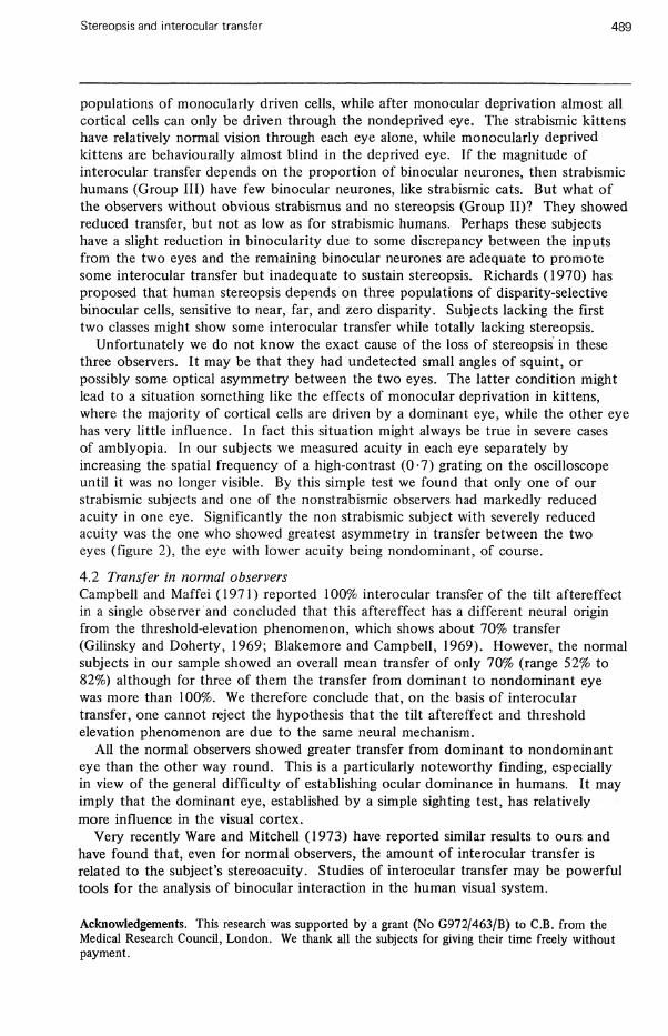
populations of monocularly driven cells, while after monocular deprivation almost all cortical cells can only be driven through the nondeprived eye. The strabismic kittens have relatively normal vision through each eye alone, while monocularly deprived kittens are behaviourally almost blind in the deprived eye. If the magnitude of interocular transfer depends on the proportion of binocular neurones, then strabismic humans (Group III) have few binocular neurones, like strabismic cats. But what of the observers without obvious strabismus and no stereopsis (Group II)? They showed reduced transfer, but not as low as for strabismic humans. Perhaps these subjects have a slight reduction in binocularity due to some discrepancy between the inputs from the two eyes and the remaining binocular neurones are adequate to promote some interocular transfer but inadequate to sustain stereopsis. Richards (1970) has proposed that human stereopsis depends on three populations of disparity-selective binocular cells, sensitive to near, far, and zero disparity. Subjects lacking the first two classes might show some interocular transfer while totally lacking stereopsis.
Unfortunately we do not know the exact cause of the loss of stereopsis in these three observers. It may be that they had undetected small angles of squint, or possibly some optical asymmetry between the two eyes. The latter condition might lead to a situation something like the effects of monocular deprivation in kittens, where the majority of cortical cells are driven by a dominant eye, while the other eye has very little influence. In fact this situation might always be true in severe cases of amblyopia. In our subjects we measured acuity in each eye separately by increasing the spatial frequency of a high-contrast (0-7) grating on the oscilloscope until it was no longer visible. By this simple test we found that only one of our strabismic subjects and one of the nonstrabismic observers had markedly reduced acuity in one eye. Significantly the non strabismic subject with severely reduced acuity was the one who showed greatest asymmetry in transfer between the two eyes (figure 2), the eye with lower acuity being nondominant, of course.
4.2 Transfer in normal observers Campbell and Maffei (1971) reported 100% interocular transfer of the tilt aftereffect in a single observer and concluded that this aftereffect has a different neural origin from the threshold-elevation phenomenon, which shows about 70% transfer (Gilinsky and Doherty, 1969; Blakemore and Campbell, 1969). However, the normal subjects in our sample showed an overall mean transfer of only 70% (range 52% to 82%) although for three of them the transfer from dominant to nondominant eye was more than 100%. We therefore conclude that, on the basis of interocular transfer, one cannot reject the hypothesis that the tilt aftereffect and threshold elevation phenomenon are due to the same neural mechanism.
All the normal observers showed greater transfer from dominant to nondominant eye than the other way round. This is a particularly noteworthy finding, especially in view of the general difficulty of establishing ocular dominance in humans. It may imply that the dominant eye, established by a simple sighting test, has relatively more influence in the visual cortex.
Very recently Ware and Mitchell (1973) have reported similar results to ours and have found that, even for normal observers, the amount of interocular transfer is related to the subject's stereoacuity. Studies of interocular transfer may be powerful tools for the analysis of binocular interaction in the human visual system.
Acknowledgements. This research was supported by a grant (No G972/463/B) to C.B. from the Medical Research Council, London. We thank all the subjects for giving their time freely without payment.

490 J A Movshon, B E I Chambers, C Blakemore
References Barlow, H. B., Blakemore, C , Pettigrew, J. D., 1967, "The neural mechanism of binocular depth
discrimination",/. Physiol., 193, 327-342. Blakemore, C, Campbell, F. W., 1969, "On the existence of neurones in the human visual system
selectively sensitive to the orientation and size of retinal images", J. Physiol., 203, 237-266. Blakemore, C, Cooper, G. F., 1970, "Development of the brain depends on the visual
environment",Nature, 228, 477-478. Blakemore, C, Mitchell, D.E., 1973, "Environmental modification of the visual cortex and the
neural basis of learning and memory", Nature, 241, 467-468. Campbell, F. W., Maffei, L., 1971, "The tilt after-effect: a fresh look", Vision Res., 11, 833-840. Coltheart, M., 1971, "Visual feature-analyzers and aftereffects of tilt and curvature", Psychol.
Rev., 78, 114-121. Gibson, J. J., 1933, "Adaptation, after-effect and contrast in the perception of curved lines",
/. Exp. Psychol., 16, 1-31. Gilinsky, A. S., Doherty, R.S., 1969, "Interocular transfer of orientational effects", Science, 164,
454-455. Hirsch, H. V. B., Spinelli, D. N., 1971, "Modification of the distribution of receptive field
orientations by selective visual exposure during development", iijtp. Brain Res., 13, 509-527. Hubel, D.H., Wiesel, T.N., 1962, "Receptive fields, binocular interaction and functional
architecture in the cat's visual cortex",/. Physiol., 160, 106-154. Hubel, D. H., Wiesel, T. N., 1965, "Binocular interaction in striate cortex of kittens reared with
artificial squint",/. NeurophysioL, 28, 1041-1059. Julesz, B., 197'1, Foundations of Cyclopean Perception (University of Chicago Press, Chicago). Mitchell, D. E., Freeman, R. D., Millodot, M., Haegerstrom, G., 1973, "Meridional amblyopia:
evidence for modification of the human visual system by early visual experience", Vision Res., 13,535-558.
Muir, D., Mitchell, D. E., 1973, "Visual resolution and experience: acuity deficits in cats following early selective visual deprivation", Science, 180, 420-422.
Parker, D.H., 1972, "Contrast and size variables and the tilt after-effect", Quart. J. Exp. Psychol., 24, 1-7.
Richards, W., 1970, "Stereopsis and stereoblindness",£xp. Brain Res., 10, 380-388. Stimson, A. T., Beckingham, D., 1971, "A preliminary investigation of amblyopia in a human
subject", B.A. Dissertation, Department of Physiology, University of Cambridge. Ware, C , Mitchell, D. E., 1973, "Interocular transfer of after-effects", Paper presented at the
meeting of the Association lor Research in Vision and Ophthalmology, Sarasota, Florida, May 3-7.
Wiesel, T. N., Hubel, D. H., 1965, "Comparison of the effects of unilateral and bilateral eye closure on cortical unit responses in kittens",/. Neurophysiol, 28, 1029-1040.
p © 1973 a Pion publication printed in Great Britain