interplay of dna damage and cell cycle signaling at the ... filesupplementary data for: interplay of...
TRANSCRIPT

Supplementary Data for: Interplay of DNA damage and cell cycle signaling at the level of human Replication Protein A Gloria E.O. Borgstahl1*, Kerry Brader1, Adam Mosel2, Shengqin Liu2, Elisabeth Kremmer5, Kaitlin A. Goettsch3, Carol Kolar1, Heinz-Peter Nasheuer4 and Greg G. Oakley2 1Eppley Institute for Research in Cancer and Allied Diseases, University of Nebraska Medical Center, Omaha, NE, 68198, USA 2Department of Oral Biology, University of Nebraska Medical Center, Omaha, NE, 68583, USA 3School of Interdisciplinary Informatics, College of Information Science and Technology, University of Nebraska at Omaha, Omaha, NE, 68182, USA 4Centre for Chromosome Biology, School of Natural Sciences, National University of Ireland, Galway, Ireland 5Institute for Molecular Immunology, Helmholtz Zentrum München, German Research Center for Environmental Health (GmbH), 81377 München, Germany * To whom correspondence should be addressed. Tel: +1-402-559-8578; Fax: +1-402-559-3739; Email: [email protected]
1

2
A B
Supplementary Figure S1. Visualizing Zeocin induced double-strand breaks using a modified neutral comet assay. (A) Composite images of 75 cells per sample after normalization for total fluorescent intensity where UM-SCC-38 expressing WT RPA2 cells were treated with Zeocin. (B) Comet profiles of composite images (thin vertical bar = %DNA in head; thick vertical bar = %DNA in Tail; distance between thin and thick vertical bars represents tail length; black curve = average comet profile; light grey curve = DNA in head; dark grey curve = DNA in tail). Individual comet images were normalized for total fluorescent intensity, thereby making each comet image directly comparable in terms of %DNA content and subsequent Olive tail moment (%DNA in tail x tail length). Normalization does not affect the tail length values. The 75 normalized images (comets) for each sample were then averaged into their respective composite image. The composite image and its comet profile (all of which have the same area under the curve) directly reflects the average Olive tail moment for that sample.

A
B
C
D
E
pS12
pS4/8
RPA2
HA
RPA1-CT
Supplementary Figure S2 . Identification of capillary IEF immunoassay peaks specific for RPA G2 isoforms. Peggy data on a pH 3-10 gradient from several RPA antibodies: (A) pS4/8 antibody (Bethyl) diluted 1:50; (B) pS12 antibody diluted 1:50; (C) RPA2 antibody (Bethyl) diluted 1:500; (D) HA antibody (Neomarkers) diluted 1:25; (E) RPA1-CT antibody not diluted. In parts A and B, DNA-damaged lysates are shown (t1, pink line). In parts C, D and E control lysates that were not treated are shown (blue line). A star indicates the peaks that are in common for all antibodies and are specific for RPA isoforms under these experimental conditions.

Supplementary Figure S3. To make sure the RPA2(pS29) antibody was active we tested against mitotic cell lysates as a positive control. UM-SCC-38 WT RPA cells synchronized to M phase with nocodazole were harvested and lysed. Whole cell lysate was separated on a 10% Bis-Tris SDS PAGE gel. Proteins were transferred to a nitrocellulose membrane and probed with anti-RPA2(pS29). Protein loaded left to right: 10 µg, 20 µg, 40 µg. Thus, the antibody sample is good and the lack of signal in the westerns is due to very low levels of RPA2(pS29).
4

Supplementary Figure S4. Capillary isoelectric focusing data for RPA2 pSer12 antibody. This antibody does not appear to work well with native protein. , DNA-damaged (t1, pink line) and control lysates that were not treated (blue line) are shown.
5
pS12

Supplementary Figure S5. Western blots of S phase HA-RPA2 IP of chromatin-bound fraction with respective RPA1 controls. Correlating to Figure 4B in the paper.
6

7
Supplementary Figure S6. Western blots of G2 phase HA-RPA2 IP of chromatin-bound fraction with respective RPA1 controls. Correlating to Figure 4C in the paper.
8
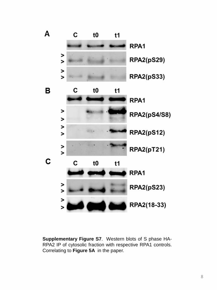
Supplementary Figure S7. Western blots of S phase HA-RPA2 IP of cytosolic fraction with respective RPA1 controls. Correlating to Figure 5A in the paper.

9
Supplementary Figure S9. Western blots of G2 phase HA-RPA2 IP of cytosolic fraction with respective RPA1 controls. Correlating to Figure 5B in the paper.

10
Table S1. PIKK and CDK phosphorylation sites of human RPA.
Protein Residue sequence
Kinase Cell cycle Treatment Ab
available?
Reference
RPA1 Ser 38 TTGNSPPRY CK1, CDK ayn none No [1, 2]
Thr 180 TSGGTQSKV PIKK, CK1 asyn IR, UV, none No [3-5]
Ser 207 VTNKSQIRT PIKK asyn PMA No [6]
Thr 483 QACPTQDCN PIKK asyn none No [7]
RPA2 Ser 4,8 MWNSGFESYGSS PIKK S, asyn, M UV, HU, IR, ETOP Yes [8-11]
Ser 11,12,13 ESYGSSSYGGA PIKK asyn, M UV, none, ETOP Yes [9, 11, 12]
Thr 21 AGGYTQSPG PIKK, CHK2 S, asyn UV, HU, IR, CPT, ETOP Yes [8, 9, 11, 13, 14]
Ser 23 GYTQSPGGF CDK S/G2, M, asyn IR, UV Yes [9, 10, 12, 15]
Ser 29 GGFGSPAPS CDK S/G2, M, asyn IR, UV Yes [9, 10, 13, 15, 16]
Ser 33 SPAPSQAEK PIKK asyn, S UV, HU, ETOP Yes [8, 9, 11, 13, 14]
Ser 52 PCTISQLLS PIKK/GSK3 in vitro None No [8]
Ser 72 NVEISQVTI PIKK in vitro None No [8]
Ser 174 SKANSQPSA PIKK in vitro None No [8]
Treatments: CPT = camptothecin; ETOP = etoposide; HU=hydroxyurea; IR=ionizing radiation; PMA = phorbol 12-myristate 13-acetate
Kinase: PIKK = ATM/ATR or DNA-PKcs; CDK = cyclin dependent kinase; CHK = checkpoint homolog kinase, CK = casein kinase, GSK = glycogen
synthase kinase

11
Table S2. Other phosphorylation sites of human RPA.
Sub-unit
Residue Sequence Kinasea Reference
RPA1 Ser 6 VGQLSEGAI [1, 5, 17]
Thr 34,35 IRPITTGNSP CAMK2 [17]
Ser 135 APAASPAAS GSK3 [17, 18]
Ser 174 GPSLSHTSG [1, 2]
Ser 177 LSHTSGGTQ CK1 [1]
Ser 182 GGTQSKVVP [1]
Thr 191 IASLTPYQS [2, 5, 18-20]
Ser 315 KSKDSLVDI CK1, Aurora [1, 19]
Ser 371 KFDGSRQPV [1]
Ser 384 GARVSDFGG PKA [1, 5, 6, 12, 17]
Ser 432 SDLKSGGVG CK1 [17]
Ser 438 GVGGSNTNW PKA [5, 6, 12]
Thr 440 GGSNTNWKT [17]
Tyr 461 DKPDYFSSV [1]
Ser 463,464 PDYFSSVATV [17]
Thr 467 SSVATVVYL CK1 [17]
Tyr 470 ATVVYLRKE [17]
Tyr 478 ENCMYQACP [1, 7]
Ser 569 ANFRSFIFR [18]
Thr 580 VKVETYNDE [18]
Tyr 581 KVETYNDES EGFR [1]
Ser 585 YNDESRIKA [17, 18]
Thr 590 RIKATVMDV Aurora [1, 5, 18]
Tyr 599 KPVDYREYG [1, 5]
RPA2 Tyr 9 GFESYGSSS [1, 21]
Ser 39 AEKKSRARA [1, 12, 22]
Thr 75 ISQVTIVGI CK1 [17]
Thr 88 EKAPTNIVY [1, 6]
Thr 98 IDDMTAAPM [18]
Ser 238 LKHMSVSSI [17]
Ser 241 MSVSSIKQA CK1 [1]
12
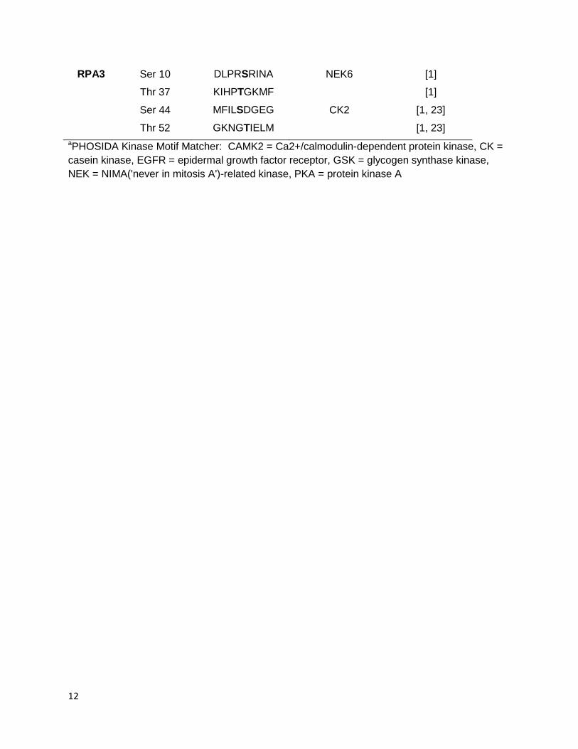
RPA3 Ser 10 DLPRSRINA NEK6 [1]
Thr 37 KIHPTGKMF [1]
Ser 44 MFILSDGEG CK2 [1, 23]
Thr 52 GKNGTIELM [1, 23] aPHOSIDA Kinase Motif Matcher: CAMK2 = Ca2+/calmodulin-dependent protein kinase, CK = casein kinase, EGFR = epidermal growth factor receptor, GSK = glycogen synthase kinase, NEK = NIMA('never in mitosis A')-related kinase, PKA = protein kinase A

13
Table S3. Primary antibodies, source and dilutions used
Antibody Source Host Traditional
western
Capillary
IEF
ATM Abcam ab17995 Rabbit 1:1000
pATM(pSer1981) Abcam ab81292 Rabbit 1:1000
CHK1 (G-4) Santa Cruz sc-8408 Mouse 1:3000
pCHK1(pSer345) Cell Signaling #2341 Rabbit 1:500
CHK2 Abcam ab47433 Rabbit 1:1000
pCHK2(pThr68) Abcam ab38461 Rabbit 1:1000
H2AX Cell Signaling #2595 Rabbit 1:1000
ɣH2AX(pS139) Abcam ab2893 Rabbit 1:1000
Actin, N-terminal Sigma A2103 Rabbit 1:1000
α-tubulin (10D8) Santa Cruz sc-53646 Mouse 1:2000
RPA1 (RPA70-9) Millipore NA13 Mouse 1:1000
RPA1(aa 525-616)a Nasheuer lab RAC-4D9 [24] Rat No dilution
RPA2 (aa 225-270) Bethyl A300-244A Mouse 1:500
pRPA2(pSer4/Ser8) Abcam87277
Bethyl A300-245A
Rabbit
Rabbit
1:1000
1:50
pRPA2(pSer12) Oakley lab Rabbit 1:3000 1:50
pRPA2(pThr21) Abcam ab61065 Rabbit 1:1000 1:25
pRPA2(pSer23) Nasheuer lab RPA1-8H3 [10] Rat 1:5 No dilution
PRPA2(pSer29) Nasheuer lab RPA2-8C7 Rat 1:2 No dilution
pRPA2(pSer33) Abcam ab87278 [10]
Bethyl A300-246A
Rabbit
Rabbit
1:1000
1:25
RPA2(aa 18-33 not phospho) Nasheuer lab RPA-3A2 Rat 1:5 No dilution
Hemaglutinin (HA Ab1) Thermo Scientific RB-1438 Rabbit 1:25 a aa = amino acids
14
Supplementary References
1. P.V. Hornbeck, J.M. Kornhauser, S. Tkachev, B. Zhang, E. Skrzypek, B. Murray, V. Latham, and M. Sullivan, PhosphoSitePlus: a comprehensive resource for investigating the structure and function of experimentally determined post-translational modifications in man and mouse, Nucleic Acids Res. 40 (2012) D261-70.
2. H. Zhou, S. Di Palma, C. Preisinger, M. Peng, A.N. Polat, A.J. Heck, and S. Mohammed, Toward a comprehensive characterization of a human cancer cell phosphoproteome, J Proteome Res 12 (2013) 260-71.
3. S. Matsuoka, B.A. Ballif, A. Smogorzewska, E.R. McDonald, 3rd, K.E. Hurov, J. Luo, C.E. Bakalarski, Z. Zhao, N. Solimini, Y. Lerenthal, Y. Shiloh, S.P. Gygi, and S.J. Elledge, ATM and ATR substrate analysis reveals extensive protein networks responsive to DNA damage, Science 316 (2007) 1160-6.
4. M.P. Stokes, J. Rush, J. Macneill, J.M. Ren, K. Sprott, J. Nardone, V. Yang, S.A. Beausoleil, S.P. Gygi, M. Livingstone, H. Zhang, R.D. Polakiewicz, and M.J. Comb, Profiling of UV-induced ATM/ATR signaling pathways, Proc. Natl. Acad. Sci. USA 104 (2007) 19855-60.
5. J.V. Olsen, M. Vermeulen, A. Santamaria, C. Kumar, M.L. Miller, L.J. Jensen, F. Gnad, J. Cox, T.S. Jensen, E.A. Nigg, S. Brunak, and M. Mann, Quantitative phosphoproteomics reveals widespread full phosphorylation site occupancy during mitosis, Sci. Signal. 3 (2010) ra3.
6. K.T. Rigbolt, T.A. Prokhorova, V. Akimov, J. Henningsen, P.T. Johansen, I. Kratchmarova, M. Kassem, M. Mann, J.V. Olsen, and B. Blagoev, System-wide temporal characterization of the proteome and phosphoproteome of human embryonic stem cell differentiation, Sci. Signal. 4 (2011) rs3.
7. Y. Bai, J. Li, B. Fang, A. Edwards, G. Zhang, M. Bui, S. Eschrich, S. Altiok, J. Koomen, and E.B. Haura, Phosphoproteomics identifies driver tyrosine kinases in sarcoma cell lines and tumors, Cancer Res 72 (2012) 2501-11.
8. E. Olson, C.J. Nievera, V. Klimovich, E. Fanning, and X. Wu, RPA2 is a direct downstream target for ATR to regulate the S-phase checkpoint, The Journal of biological chemistry 281 (2006) 39517-33.
9. M. Zernik-Kobak, K. Vasunia, M. Connelly, C.W. Anderson, and K. Dixon, Sites of UV-induced Phosphorylation of the p34 Subunit of Replication Protein A from HeLa Cells, J. Biol. Chem. 272 (1997) 23896-23904.
10. H. Stephan, C. Concannon, E. Kremmer, M.P. Carty, and H.P. Nasheuer, Ionizing radiation-dependent and independent phosphorylation of the 32-kDa subunit of replication protein A during mitosis, Nucleic Acids Res. 37 (2009) 6028-41.
11. S. Liu, S.O. Opiyo, K. Manthey, J.G. Glanzer, A.K. Ashley, C. Amerin, K. Troksa, M. Shrivastav, J.A. Nickoloff, and G.G. Oakley, Distinct roles for DNA-PK, ATM and ATR in RPA phosphorylation and checkpoint activation in response to replication stress, Nucleic Acids Res. 40 (2012) 10780-94.
12. N. Dephoure, C. Zhou, J. Villen, S.A. Beausoleil, C.E. Bakalarski, S.J. Elledge, and S.P. Gygi, A quantitative atlas of mitotic phosphorylation, Proc. Natl. Acad. Sci. USA 105 (2008) 10762-7.
13. H. Niu, H. Erdjument-Bromage, Z.Q. Pan, S.H. Lee, P. Tempst, and J. Hurwitz, Mapping of amino acid residues in the p34 subunit of human single-stranded DNA-binding protein phosphorylated by DNA-dependent protein kinase and Cdc2 kinase in vitro, J. Biol. Chem. 272 (1997) 12634-41.
14. W.D. Block, Y. Yu, and S.P. Lees-Miller, Phosphatidyl inositol 3-kinase-like serine/threonine protein kinases (PIKKs) are required for DNA damage-induced
15
phosphorylation of the 32 kDa subunit of replication protein A at threonine 21, Nucleic Acids Res. 32 (2004) 997-1005.
15. V.F. Liu and D.T. Weaver, The ionizing radiation-induced Replication Protein A phosphorylation response differs between ataxia telangiectasia and normal human cells, Mol. Cell. Biol. 13 (1993) 7222-7231.
16. F. Fang and J.W. Newport, Distinct roles of cdk2 and cdc2 in RP-A phosphorylation during the cell cycle, J. Cell Sci. 106 ( Pt 3) (1993) 983-94.
17. F. Gnad, J. Gunawardena, and M. Mann, PHOSIDA 2011: the posttranslational modification database, Nucleic Acids Res. 39 (2011) D253-60.
18. J.E. Nuss, S.M. Patrick, G.G. Oakley, G.M. Alter, J.G. Robison, K. Dixon, and J.J. Turchi, DNA damage induced hyperphosphorylation of replication protein A. 1. Identification of novel sites of phosphorylation in response to DNA damage, Biochemistry 44 (2005) 8428-37.
19. T. Shiromizu, J. Adachi, S. Watanabe, T. Murakami, T. Kuga, S. Muraoka, and T. Tomonaga, Identification of missing proteins in the neXtProt database and unregistered phosphopeptides in the PhosphoSitePlus database as part of the Chromosome-centric Human Proteome Project, J Proteome Res 12 (2013) 2414-21.
20. S. Gauci, A.O. Helbig, M. Slijper, J. Krijgsveld, A.J. Heck, and S. Mohammed, Lys-N and trypsin cover complementary parts of the phosphoproteome in a refined SCX-based approach, Anal. Chem. 81 (2009) 4493-501.
21. A.B. Iliuk, V.A. Martin, B.M. Alicie, R.L. Geahlen, and W.A. Tao, In-depth analyses of kinase-dependent tyrosine phosphoproteomes based on metal ion-functionalized soluble nanopolymers, Mol Cell Proteomics 9 (2010) 2162-72.
22. E.L. Huttlin, M.P. Jedrychowski, J.E. Elias, T. Goswami, R. Rad, S.A. Beausoleil, J. Villen, W. Haas, M.E. Sowa, and S.P. Gygi, A tissue-specific atlas of mouse protein phosphorylation and expression, Cell 143 (2010) 1174-89.
23. V. Mayya, D.H. Lundgren, S.I. Hwang, K. Rezaul, L. Wu, J.K. Eng, V. Rodionov, and D.K. Han, Quantitative phosphoproteomic analysis of T cell receptor signaling reveals system-wide modulation of protein-protein interactions, Sci. Signal. 2 (2009) ra46.
24. P.E. Pestryakov, K. Weisshart, B. Schlott, S.N. Khodyreva, E. Kremmer, F. Grosse, O.I. Lavrik, and H.P. Nasheuer, Human replication protein A. The C-terminal RPA70 and the central RPA32 domains are involved in the interactions with the 3'-end of a primer-template DNA, J. Biol. Chem. 278 (2003) 17515-24.