interventional cardiology - startseite · jose m. wiley, fernando pastor, and cristina sanina 27...
TRANSCRIPT


Interventional CardiologyPrinciples and PracticeSecond Edition
Edited by
George D. Dangas, MD, PhD, FACC, FSCAI, FESC, FAHAProfessor of MedicineDirector, Cardiovascular InnovationMount Sinai Medical CenterNew York, NYUSA
Carlo Di Mario, MD, PhD, FRCP, FACC, FSCAI, FESCConsultant CardiologistRoyal Brompton HospitalProfessor of Clinical CardiologyNational Heart & Lung Institute Imperial College LondonLondonUK
Nicholas N. Kipshidze, MD, PhD, FACC, FESC, FSCAIProfessor of Medicine and SurgeryConsultant CardiologistN. Kipshidze Central University HospitalTbilisi, GeorgiaDirector,New York Cardiovascular Research,New York, NYUSA
Associate Editors
Peter Barlis and Tayo Addo
Foreword by
Patrick W. Serruys, MD, PhD

This edition first published 2017 © 2011, 2017 by John Wiley & Sons, Ltd
Registered OfficeJohn Wiley & Sons, Ltd, The Atrium, Southern Gate, Chichester, West Sussex, PO19 8SQ, UK
Editorial Offices9600 Garsington Road, Oxford, OX4 2DQ, UK1606 Golden Aspen Drive, Suites 103 and 104, Ames, Iowa 50010, USA
For details of our global editorial offices, for customer services and for information about how to apply for permission to reuse the copyright material in this book please see our website at www.wiley.com/wiley‐blackwell.
The right of the author to be identified as the author of this work has been asserted in accordance with the UK Copyright, Designs and Patents Act 1988.
All rights reserved. No part of this publication may be reproduced, stored in a retrieval system, or transmitted, in any form or by any means, electronic, mechanical, photocopying, recording or otherwise, except as permitted by the UK Copyright, Designs and Patents Act 1988, without the prior permission of the publisher.
Designations used by companies to distinguish their products are often claimed as trademarks. All brand names and product names used in this book are trade names, service marks, trademarks or registered trademarks of their respective owners. The publisher is not associated with any product or vendor mentioned in this book. It is sold on the understanding that the publisher is not engaged in rendering professional services. If professional advice or other expert assistance is required, the services of a competent professional should be sought.
The contents of this work are intended to further general scientific research, understanding, and discussion only and are not intended and should not be relied upon as recommending or promoting a specific method, diagnosis, or treatment by health science practitioners for any particular patient. The publisher and the author make no representations or warranties with respect to the accuracy or completeness of the contents of this work and specifically disclaim all warranties, including without limitation any implied warranties of fitness for a particular purpose. In view of ongoing research, equipment modifications, changes in governmental regulations, and the constant flow of information relating to the use of medicines, equipment, and devices, the reader is urged to review and evaluate the information provided in the package insert or instructions for each medicine, equipment, or device for, among other things, any changes in the instructions or indication of usage and for added warnings and precautions. Readers should consult with a specialist where appropriate. The fact that an organization or Website is referred to in this work as a citation and/or a potential source of further information does not mean that the author or the publisher endorses the information the organization or Website may provide or recommendations it may make. Further, readers should be aware that Internet Websites listed in this work may have changed or disappeared between when this work was written and when it is read. No warranty may be created or extended by any promotional statements for this work. Neither the publisher nor the author shall be liable for any damages arising herefrom.
Library of Congress Cataloging‐in‐Publication Data
Names: Dangas, George D., editor. | Di Mario, Carlo, editor. | Kipshidze, Nicholas N., editor.Title: Interventional cardiology : principles and practice / edited by George D. Dangas, Carlo Di Mario, Nicholas N. Kipshidze ; foreword by Patrick W. Serruys.Other titles: Interventional cardiology (Di Mario)Description: Second edition. | Chichester, West Sussex ; Ames, Iowa : John Wiley & Sons, Inc., 2017. | Includes bibliographical references and index.Identifiers: LCCN 2016018228 (print) | LCCN 2016018438 (ebook) | ISBN 9781118976036 (cloth) | ISBN 9781118975930 (pdf) | ISBN 9781118976067 (epub)Subjects: | MESH: Cardiovascular Diseases–therapy | Cardiac Surgical Procedures | Cardiovascular Diseases–diagnosisClassification: LCC RD598 (print) | LCC RD598 (ebook) | NLM WG 166 | DDC 617.4/12–dc23LC record available at https://lccn.loc.gov/2016018228
A catalogue record for this book is available from the British Library.
Wiley also publishes its books in a variety of electronic formats. Some content that appears in print may not be available in electronic books.
Cover image: Courtesy of the Editors and Associate Editors.
Set in 9/11pt Minion by SPi Global, Pondicherry, India
1 2017
iii
About the Companion Website viContributors viiForeword xviPreface xviiAcknowledgments xviii
Part I Principles and Techniques 1
Section I Basic Knowledge 3
1 Atherogenesis and Inflammation 3Umair Hayat, Vikas Thondapu, Tim Tsay, and Peter Barlis
2 The Essentials of Femoral Vascular Access and Closure 17Ted Feldman and Mohammad Sarraf
3 Radial Artery, Alternative Arm Access, and Related Techniques 27Thomas J. Ford, Martin K.C. Ng, Vikas Thondapu, and Peter Barlis
4 Optimal Angiographic Views for Coronary Angioplasty 34Gioel Gabrio Secco and Carlo Di Mario
5 Material Selection 44Sahil A. Parikh, Michele Pighi, and Carlo Di Mario
Section II Imaging and Physiology 59
6 Physiologic Assessment in the Cardiac Catheterization Laboratory: CFR, FFR, iFR, and Beyond 59Sukhjinder Nijjer and Justin Davies
7 Intravascular Ultrasound and Virtual Histology: Principles, Image Interpretation, and Clinical Applications 71Adriano Caixeta, Akiko Maehara, and Gary S. Mintz
8 Optical Coherence Tomography, Near‐Infrared Spectroscopy, and Near‐Infrared Fluorescence Molecular Imaging 91Ismail Dogu Kilic, Roberta Serdoz, Enrico Fabris, Farouc Amin Jaffer, and Carlo Di Mario
9 Complementary Imaging Techniques: Multislice Computed Tomography of Coronary Arteries 107Omosalewa O. Lalude, Francesca Pugliese, Pim J. de Feyter, and Stamatios Lerakis
10 Cardiovascular Magnetic Resonance Imaging 126Omosalewa O. Lalude and Stamatios Lerakis
Section III PCI in Different Clinical Settings 138
11 Stable Coronary Artery Disease 138Abhiram Prasad and Bernard J. Gersh
12 PCI Strategies in Acute Coronary Syndromes without ST Segment Elevation (NSTEACS) 148Georgios E. Christakopoulos, Subhash Banerjee, and Emmanouil S. Brilakis
13 Primary and Rescue PCI in Acute Myocardial Infarction and Elements of Myocardial Conditioning 155Tayo Addo, Neil Swanson, and Anthony Gershlick
14 The Management of Cardiogenic Shock and Hemodynamic Support Devices and Techniques 163Bimmer E.P.M. Claessen, Dagmar Ouweneel, and José P.S. Henriques
Section IV PCI in Different Lesion Types 168
15 Percutaneous Coronary Intervention in Unprotected Left Main 168Gill Louise Buchanan, Alaide Chieffo, and Antonio Colombo
16 Bifurcation Lesion Stenting 175Yves Louvard, Thierry Lefevre, Bernard Chevalier, and Philippe Garot
17 Risk Stratification Approach to Multivessel Coronary Artery Disease 185Davide Capodanno and Corrado Tamburino
18 Chronic Total Coronary Occlusion 190Gerald S. Werner and Emmanouil S. Brilakis
19 Percutaneous Coronary Intervention of Arterial and Vein Grafts 201Bimmer E.P.M. Claessen, José P.S. Henriques, and George D. Dangas
20 Interventional Approach in Small Vessel, Diffuse, and Tortuous Coronary Artery Disease 205Robert Pyo
21 In‐Stent Restenosis in New Generation DES Era 213Marco G. Mennuni and Patrizia Presbitero
Section V Special Techniques and Complications 224
22 Laser, Rotational, and Orbital Coronary Atherectomy 224Kaleab N. Asrress, Peter O’Kane, Robert Pyo, and Simon R. Redwood
23 Thrombus‐Containing Lesions 233Giovanni Luigi De Maria and Adrian P. Banning
24 Specialized Balloons in Percutaneous Coronary Intervention: Cutting, Scoring, Gliding, and Drug‐Eluting Balloons 244Bimmer E.P.M. Claessen, José P.S. Henriques, and George D. Dangas
Contents

iv Contents
25 Coronary Artery Dissections, Perforations, and the No‐Reflow Phenomenon 248Adriano Caixeta, Luiz Fernando Ybarra, Azeem Latib, Flavio Airoldi, Roxana Mehran, and George D. Dangas
26 Access Site Complications 267Jose M. Wiley, Fernando Pastor, and Cristina Sanina
27 Renal Insufficiency and the Impact of Contrast Agents 274Michael Donahue and Carlo Briguori
28 Radiation Management in Interventional Cardiology 282Stephen Balter and Charles E. Chambers
29 Concepts of Cell Therapy and Myocardial Regeneration 290Kevin O’Gallagher, Zoë Astroulakis, Alex Sirker, and Jonathan M. Hill
Section VI Clinical Trials in Coronary Heart Disease 296
30 Statistical Essentials in the Design and Analysis of Clinical Trials 296Usman Baber and Stuart J. Pocock
31 Historical Perspective of Sirolimus and Paclitaxel‐Eluting Stent Clinical Studies 301Adriano Caixeta, Leonardo Guimarães, Philippe Généreux, and George D. Dangas
32 Cobalt‐Chromium Everolimus‐Eluting Stents 313Vikas Thondapu, Yoshinobu Onuma, Bimmer E.P.M. Claessen, Patrick W. Serruys, and Peter Barlis
33 Platinum‐Chromium Everolimus‐Eluting Stents 326Vikas Thondapu, Bimmer E.P.M. Claessen, George D. Dangas, Patrick W. Serruys, and Peter Barlis
34 Bioresorbable Stents 335Gianluca Caiazzo, Alessio Mattesini, Ciro Indolfi, and Carlo Di Mario
35 The Biolimus Stent Family 344Anna Franzone, Raffaele Piccolo, and Stephan Windecker
36 The Biotronik Stent Family 360Anna Franzone, Raffaele Piccolo, and Stephan Windecker
37 Novel Drug‐Eluting Stent Systems 368J. Ribamar Costa, Jr., Adriano Caixeta, and Alexandre A.C. Abizaid
Part II Interventional Pharmacology 377
Section I Fundamentals of Interventional Pharmacology 379
38 Basics of Antiplatelet and Anticoagulant Therapy for Cardiovascular Disease 379Piera Capranzano and Dominick J. Angiolillo
39 Balance of Ischemia and Bleeding in Selecting Antithrombotic Regimens 389Bimmer E.P.M. Claessen and José P.S. Henriques
Section II Pharmacological Agents 397
40 Oral Antiplatelet Agents in PCI 397Jonathan A. Batty, Joseph R. Dunford, George D. Dangas, and Vijay Kunadian
41 Parenteral Anticoagulant Agents in PCI 408Piera Capranzano, Corrado Tamburino, and George D. Dangas
42 Parenteral Antiplatelet Agents in PCI 415Piera Capranzano, Giuseppe Gargiulo, and Corrado Tamburino
43 Role of Parenteral Agents in PCI for Stable Patients 421Joanna Ghobrial, David A. Burke, and Duane S. Pinto
44 Vasoactive and Antiarrhythmic Drugs During PCI 432Bimmer E.P.M. Claessen and José P.S. Henriques
45 The Optimal Duration of Dual Antiplatelet Therapy After PCI 436Mikkel Malby Schoos, Roxana Mehran, and George D. Dangas
46 Triple Antiplatelet Therapy and Combinations with Oral Anticoagulants After PCI 443Jonathan A. Batty, Joseph R. Dunford, Roxana Mehran, and Vijay Kunadian
Section III Pharmacological Testing 453
47 Peri‐procedural Platelet Function Testing in Risk Stratification and Clinical Decision Making 453Paul A. Gurbel, Fang Liu, Gailing Chen, and Udaya S. Tantry
48 Genetics and Pharmacogenetics in Interventional Cardiology 459Hillary Johnston‐Cox, Johan L.M. Björkegren, and Jason C. Kovacic
49 Monitoring and Reversal of Anticoagulation and Antiplatelet Agents 469Gregory W. Yost and Steven R. Steinhubl
Part III Hypertension and Structural Heart Disease 485
Section I Systemic and Pulmonary Hypertension 487
50 Right Heart Catheterization and Pulmonary Hemodynamics 487P. Christian Schulze
51 Treatment of Pulmonary Embolism: Medical, Surgical, and Percutaneous 491Ian del Conde and Barry T. Katzen
52 Renal Denervation for Resistant Hypertension 499Hitesh C. Patel, Carl Hayward, Sebastian Ewen, and Felix Mahfoud
Section II Structural Heart Interventions 507
53 Antithrombotic Strategies in Valvular and Structural Heart Disease Interventions 507Mikkel Malby Schoos, Davide Capodanno, and George D. Dangas
54 Alcohol Septal Ablation for Hypertrophic Obstructive Cardiomyopathy 517Amir‐Ali Fassa, George D. Dangas, and Ulrich Sigwart
55 Left Atrial Appendage Exclusion 525Jorge G. Panizo and Jacob S. Koruth
56 Cryptogenic Stroke, Patent Foramen Ovale, and ASD Closure 530Barry Love

Contents v
57 Paravalvular Leak Closure and Ventricular Septal Defect Closure 540Saurabh Sanon, Mackram F. Eleid, Allison K. Cabalka, and Charanjit S. Rihal
Section III Valvular Heart Disease Interventions 546
58 Aortic Valvuloplasty and Large‐Bore Percutaneous Arterial Access 546Matthew I. Tomey, Annapoorna S. Kini, Samin K. Sharma, and Jason C. Kovacic
59 Transfemoral Aortic Valve Implantation: Preparation, Implantation, and Complications 558Brandon M. Jones, Samir R. Kapadia, Amar Krishnaswamy, Stephanie Mick, and E. Murat Tuzcu
60 Transthoracic Aortic Valve Implantation 569Giuseppe Bruschi, Kaleab N. Asrress, Paola Colombo, and Vinayak N. Bapat
61 New Aortic Valve Technologies 575Dimytri Alexandre Siqueira and Alexandre A.C. Abizaid
62 Transseptal Puncture 582Alec Vahanian, Dominique Himbert, Fabrice Extramiana, Gregory Ducrocq, and Eric Brochet
63 Principles of Carpentier’s Reconstructive Surgery in Degenerative Mitral Valve Disease 592 Farzan Filsoufi and Alain Carpentier
64 Mitral Valve Repair: MitraClip and Emerging Techniques 599Ted Feldman, Mohammad Sarraf, Mayra Guerrero, and Francesco Maisano
65 Balloon Mitral Valvuloplasty 606C.N. Manjunath, Nagaraja Moorthy, and Upendra Kaul
66 Pulmonary Artery and Valve Catheter‐Based Interventions 619Kasey Chaszczewski, Damien Kenny, and Ziyad M. Hijazi
67 Imaging for Planning and Guidance for Structural Heart Interventions 629Ankit Parikh and Stamatios Lerakis
Part IV Vascular Disease for the Interventionalist 641
Section I Cerebrovascular Disease 643
68 Acute Stroke Intervention 643Stefan C. Bertog, Iris Q. Grunwald, Anna Luisa Kühn, Laura Vaskelyte, Ilona Hofmann, Sameer Gafoor, Markus Reinartz, Predrag Matic, and Horst Sievert
69 Carotid Artery Angioplasty and Stenting 653Alberto Cremonesi, Shane Gieowarsingh, and Fausto Castriota
70 Cerebral Aneurysms: Diagnosis, Indications, and Strategies for Endovascular Treatment 671Gyula Gál
Section II Aorta and Branch Diseases 677
71 Management of Acute Aortic Syndromes 677Christoph A. Nienaber and Rachel E. Clough
72 Thoracic Endovascular Aortic Aneurysm Repair 687Paul S. Lajos and Michael L. Marin
73 Endovascular Aortic Aneurysm Repair 692William Beckerman, Paul S. Lajos, and Peter L. Faries
74 Acute and Chronic Mesenteric Ischemia 698Robert J. Rosen, Amit Jain, and Jennifer Drury
75 Renal Artery Interventions 705Mark Shipeng Yu, Kun Xiang, Steven T. Haller, and Christopher J. Cooper
76 Revascularization for Arteries in the Pelvis 713Femi Philip and Jason H. Rogers
Section III Peripheral Arterial Disease 721
77 Iliac Interventions 721Manish Taneja and Apoorva Gogna
78 Superficial Femoral Artery Interventions 726Cristina Sanina, Pedro R. Cox‐Alomar, Prakash Krishnan, and Jose M. Wiley
79 Popliteal Artery Interventions 733Karthik Gujja, Gopi Punukollu, Vishal Kapur, and Prakash Krishnan
80 Below the Knee Interventions in Critical Limb Ischemia 738Karthik Gujja, Katarzyna Nasiadko, Arthur Tarricone, and Prakash Krishnan
81 Subclavian, Vertebral, and Upper Extremity Vascular Disease 748Ian Del Conde, Cristina Sanina, and Jose M. Wiley
Section IV Venous Disease/Interventions 754
82 Antithrombotic Strategies in Endovascular Interventions: Current Status and Future Directions 754Mehdi H. Shishehbor
83 Chronic Venous Insufficiency 759Karthik Gujja, Cristina Sanina, and Jose M. Wiley
84 Cardiac Vein Anatomy and Transcoronary Sinus Catheter Interventions in Myocardial Ischemia 768Werner Mohl, Levente Molnár, and Béla Merkely
Index 776

vi
Don’t forget to visit the companion website for this book:
www.wiley.com/go/dangas/cardiology
There you will find valuable material designed to enhance yourlearning, including:• 19 procedural videos illustrating key procedures• More than 400 interactive multiple choice questions
Scan this QR code to visit the companion website:
About the Companion Website
vii
Alexandre A.C. Abizaid, MD, PhDDirector, Invasive Cardiology DepartmentInstituto Dante Pazzanese de Cardiologia (IDPC);Hospital do Coração—Associação do Sanatório Sírio (HCor);Hospital Israelita Albert EinsteinSão Paulo, Brazil
Tayo Addo, MDAssociate Professor of MedicineUniversity of Texas Southwestern Medical CenterDallas, TX, USA
Flavio Airoldi, MDDirectorInterventional Cardiology UnitIRCCS MultimedicaSesto San Giovanni, Italy
Dominick J. Angiolillo, MD, PhDDepartment of Medicine, Division of CardiologyUniversity of Florida College of Medicine—JacksonvilleJacksonville, FL, USA
Kaleab N. Asrress, MA, PhD, MRCPDepartment of CardiologySt. Thomas’ Hospital;King’s College London British Heart Foundation Centre of ExcellenceThe Rayne Institute, St. Thomas’ HospitalLondon, UK
Zoë Astroulakis, MBBS, MRCP, PhDConsultant in Interventional CardiologyDepartment of Cardiology, St George’s HospitalLondon, UK
Usman Baber, MD, MSDirector of Clinical BiometricsCardiovascular Institute Assistant Professor of MedicineIcahn School of Medicine at Mount SinaiNew York, NY, USA
Stephen Balter, PhDProfessor of Radiology and MedicineColumbia University Medical CenterNew York, NY, USA
Subhash Banerjee, MDChief of CardiologyVA North Texas Health Care System;Professor of MedicineUniversity of Texas Southwestern Medical CenterDallas, TX, USA
Adrian P. Banning, MBBS, MD, FRCP, FESCConsultant CardiologistOxford Heart Centre, Oxford University HospitalsJohn Radcliffe HospitalOxford, UK
Vinayak N. Bapat, MBBS, MS, FRCS.CThDepartment of Cardiology and Cardiothoracic SurgerySt Thomas’ Hospital;King’s College London British Heart Foundation Centre of ExcellenceThe Rayne Institute, St. Thomas’ HospitalLondon, UK
Peter Barlis, MBBS, MPH, PHD, FACC, FESC, FRACPProfessor of MedicineFaculty of Medicine, Dentistry & Health SciencesThe University of MelbourneVictoria, Australia
Jonathan A. Batty, BSc, MBChBInstitute of Cellular MedicineNewcastle University;The Royal Victoria InfirmaryNewcastle upon Tyne NHS Foundation TrustNewcastle upon Tyne, UK
William Beckerman, MDResident in Vascular SurgeryDivision of Vascular SurgeryIcahn School of Medicine at Mount SinaiNew York, NY, USA
Stefan C. Bertog, MDCardioVascular Center FrankfurtFrankfurt, Germany
Johan L.M. Björkegren, MD, PhDThe Zena and Michael A. Wiener Cardiovascular Institute and the Department of Genetics & Genomic SciencesInstitute of Genomics and Multiscale BiologyIcahn School of Medicine at Mount SinaiNew York, NY, USA
Carlo Briguori, MD, PhD, FACC, FSCAIChief of Interventional CardiologyLaboratory of Interventional Cardiology and Department of Cardiology Clinica MediterraneaNaples, Italy
Emmanouil S. Brilakis, MD, PhDMinneapolis Heart InstituteMinneapolis, MN, USA;Professor of MedicineUniversity of Texas Southwestern Medical CenterDallas VA Medical CenterDallas, TX, USA
ContributorsContributors
viii Contributors
Eric Brochet, MDCardiologistEchocardiography LaboratoryDepartment of CardiologyHôpital Bichat‐Claude BernardParis, France
Giuseppe Bruschi, MD, FESCDepartment of Cardiology and Cardiothoracic SurgeryNiguarda Ca’ Granda HospitalMilan, Italy
Gill Louise Buchanan, MBChBDepartment of CardiologyNorth Cumbria University NHS TrustCarlisle, UK
David A. Burke, MDDivision of Cardiovascular Medicine, Department of MedicineBeth Israel Deaconess Medical CenterHarvard Medical SchoolBoston, MA, USA
Allison K. Cabalka, MDDivision of Pediatric CardiologyMayo Clinic College of MedicineRochester, MN, USA
Gianluca Caiazzo, MD, PhDDivision of Cardiology, Department of Medical and Surgical SciencesMagna Graecia UniversityCatanzaro, Italy;National Institute of Health Research (NIHR)Royal Brompton & Harefield NHS Foundation TrustLondon, UK
Adriano Caixeta, MD, PhDInterventional CardiologistHospital Israelita Albert Einstein;Professor of MedicineUniversidade Federal de São Paulo,São Paulo, Brazil
Davide Capodanno, MD, PhDAssociate Professor of CardiologyUniversity of Catania;Interventional CardiologistFerrarotto HospitalCatania, Italy
Piera Capranzano, MDCardiovascular DepartmentFerrarotto Hospital, University of CataniaCatania, Italy
Alain Carpentier, MD, PhDHôpital Européen Georges‐PompidouParis, France
Fausto Castriota, MDMaria Cecilia HospitalGVM Care & ResearchCotignola, Italy
Charles E. Chambers, MDProfessor of Medicine and RadiologyPenn State Hershey Medical CenterHershey, PA, USA
Kasey Chaszczewski, MDRush University Medical CenterChicago, IL, USA
Gailing Chen, MDSinai Center for Thrombosis ResearchCardiac Catheterization LaboratoryBaltimore, MD, USA
Bernard Chevalier, FESCInstitut Cardiovasculaire Paris Sud Hôpital Privé Jacques Cartier, Massy; Hôpital Privé Claude Galien Quincy, France
Alaide Chieffo, MDConsultant Interventional CardiologistInterventional Cardiology UnitSan Raffaele Scientific HospitalMilan, Italy
Georgios E. Christakopoulos, MDResearch FellowVA North Texas Health Care System;University of Texas Southwestern Medical CenterDallas, TX, USA
Bimmer E.P.M. Claessen, MD, PhDDepartment of CardiologyAcademic Medical Center—University of AmsterdamAmsterdam, The Netherlands
Rachel E. Clough, MDUniversity Heart Center Rostock Department of Internal MedicineCardiology, Pulmonology, Intensive Care MedicineRostock School of MedicineRostock, Germany
Antonio Colombo, MDInterventional Cardiology UnitSan Raffaele Scientific HospitalMilan, Italy
Paola Colombo, MD, PhDDepartment of Cardiology and Cardiothoracic SurgeryNiguarda Ca’ Granda HospitalMilan, Italy
Christopher J. Cooper, MDDepartment of Medicine, Cardiovascular MedicineUniversity of ToledoToledo, OH, USA
J. Ribamar Costa, Jr., PhDChief of the Medical Section of Coronary Intervention of the Instituto Dante Pazzanese de Cardiologia (IDPC);Hospital do Coração—Associação do Sanatório Sírio (HCor)São Paulo, Brazil
Pedro R. Cox‐Alomar, MD, MPH, FACCInterventional Cardiology FellowDivision of CardiologyUniversity of Florida College of MedicineUF Health Medical CenterJacksonville, FL, USA

Contributors ix
Alberto Cremonesi, MDMaria Cecilia HospitalGVM Care & ResearchCotignola, Italy
George D. Dangas, MD, PhD, FACC, FSCAI, FESC, FAHAProfessor of MedicineDirector, Cardiovascular InnovationDepartment of CardiologyMount Sinai Medical CenterNew York, NY USA
Justin Davies, BSc, MBBS, MRCP, PhDImperial College LondonLondon, UK
Giovanni Luigi De Maria, MDOxford Heart Centre, Oxford University HospitalsJohn Radcliffe HospitalOxford, UK
Ian del Conde, MD, FACCMiami Cardiac and Vascular InstituteMiami, FL, USA
Carlo Di Mario, MD, PhD, FRCP, FACC, FSCAI, FESCConsultant CardiologistNational Institute of Health Research (NIHR)Royal Brompton & Harefield NHS Foundation Trust, London;Professor of Clinical CardiologyNational Heart & Lung Institute Imperial College LondonLondon, UK
Michael Donahue, MDInterventional CardiologistLaboratory of Interventional Cardiology and Department of CardiologyClinica MediterraneaNaples, Italy
Jennifer Drury Physician AssistantLenox Hill Heart and Vascular InstituteNew York, NY USA
Gregory Ducrocq, MDHôpital Bichat‐Claude BernardParis, France
Joseph R. Dunford, MResInstitute of Cellular MedicineNewcastle UniversityNewcastle upon Tyne, UK
Mackram F. Eleid, MDDivision of Cardiovascular DiseasesMayo Clinic College of MedicineRochester, MN, USA
Sebastian Ewen, MDKlinik für Innere Medizin IIIUniversitätsklinikum des SaarlandesHomburg‐Saar, Germany
Fabrice Extramiana, MDHôpital Bichat‐Claude BernardParis, France
Enrico Fabris, MDInterventional CardiologistNational Institute of Health Research (NIHR)Royal Brompton & Harefield NHS Foundation Trust, London;NHLI Imperial College LondonLondon, UK;Cardiovascular Department Ospedali Riuniti and University of Trieste Trieste, Italy
Peter L. Faries, MD, FACSThe Franz W. Sichel Professor of SurgeryChief, Division of Vascular SurgeryProfessor of Surgery & RadiologyIcahn School of Medicine at Mount SinaiNew York, NY, USA
Amir‐Ali Fassa, MDLa Tour HospitalGeneva, Switzerland
Ted Feldman, MD, FESC, FACC, MSCAIDirector, Cardiac Catheterization LaboratoriesEvanston HospitalNorthShore University HealthSystemEvanston, IL, USA
Pim J. de Feyter, MD, PhD, FESC, FACCProfessor of Cardiac ImagingDepartments of Cardiology and RadiologyErasmus MC University Medical CenterRotterdam, The Netherlands
Farzan Filsoufi, MDProfessorDepartment of Cardiovascular SurgeryIcahn School of Medicine at Mount SinaiNew York, NY USA
Thomas J. Ford, MBChBDepartment of CardiologySt. George Hospital;Faculty of MedicineUniversity of New South WalesSydney, New South Wales Australia
Anna Franzone, MDDepartment of CardiologyBern University HospitalBern, Switzerland
Sameer Gafoor, MDCardioVascular Center FrankfurtFrankfurt, Germany;Swedish Medical Center Seattle, WA, USA
Gyula Gál, MDDepartment of Radiology, Section of NeuroradiologyOdense University HospitalOdense, Denmark

x Contributors
Giuseppe Gargiulo, MDCardiovascular DepartmentFerrarotto HospitalUniversity of CataniaCatania, Italy
Philippe Garot, FESCInstitut Cardiovasculaire Paris SudHôpital Privé Jacques Cartier, Massy;Hôpital Privé Claude GalienQuincy, France
Philippe Généreux, MDClinical InstructorDivision of CardiologyCenter for Interventional Vascular TherapyColumbia University Medical CenterNew York, NY, USA
Bernard J. Gersh, MBChB, DPhil, FACCProfessor of MedicineMayo Clinic and Mayo Clinic College of MedicineRochester, MN, USA
Anthony Gershlick, MBBS, BSc, FRCPUniversity of LeicesterLeicester, UK
Joanna Ghobrial, MD, MSDivision of Cardiovascular MedicineDepartment of MedicineBeth Israel Deaconess Medical CenterHarvard Medical SchoolBoston, MA, USA
Shane Gieowarsingh, MBBS, METMaria Cecilia HospitalGVM Care & ResearchCotignola, Italy
Apoorva Gogna, MBBS, FRCRConsultant, Interventional RadiologySingapore General HospitalSingapore
Iris Q. Grunwald, MD, PhDPost Graduate Medical InstituteAnglia Ruskin UniversityChelmsford, UK;Southend University HospitalSouthend‐on‐Sea, UK
Mayra Guerrero, MD, FACC, FSCAINorthShore University HealthSystemEvanston, IL, USA
Leonardo Guimarães, MDInterventional CardiologistHospital Israelita Albert Einstein; Universidade Federal de São PauloSão Paulo, Brazil
Karthik Gujja, MD, MPHAssistant Professor of MedicineIcahn School of Medicine at Mount Sinai;Assistant Director of Endovascular FellowshipThe Zeta and Michael A. Weiner Cardiovascular InstituteIcahn School of Medicine at Mount SinaiNew York, NY, USA
Paul A. Gurbel, MDDirector, Inova Center for Thrombosis Research and Drug DevelopmentInova Heart and Vascular InstituteFalls Church, VA, USA
Steven T. Haller, PhDDepartment of MedicineCardiovascular MedicineUniversity of ToledoToledo, OH, USA
Umair Hayat Melbourne Medical School Faculty of Medicine, Dentistry and Health SciencesThe University of MelbourneVictoria, Australia
Carl Hayward, MB, BChir, MRCP, MACardiology Research FellowNational Institute of Health Research (NIHR)Royal Brompton & Harefield NHS Foundation TrustLondon, UK
José P.S. Henriques, MD, PhDDepartment of CardiologyAcademic Medical Center—University of AmsterdamAmsterdam, The Netherlands
Ziyad M. Hijazi, MD, MPH, MSCAIProfessor of PediatricsWeill Cornell MedicineChair, Department of PediatricsSidra Medical and Research CenterDoha, Qatar
Jonathan M. Hill, MDDepartment of CardiologyKing’s College Hospital NHS Foundation TrustLondon, UK
Dominique Himbert, MDCardiologistDepartment of CardiologyHôpital Bichat‐Claude BernardParis, France
Ilona Hofmann, MDCardioVascular Center FrankfurtFrankfurt, Germany
Ciro Indolfi, MDDivision of Cardiology, Department of Medical and Surgical SciencesMagna Graecia UniversityCatanzaro, Italy
Farouc Amin Jaffer, MD, PhDAssociate ProfessorCardiology DivisionMassachusetts General Hospital, Harvard Medical SchoolBoston, MA, USA
Amit Jain, MDLenox Hill Heart and Vascular InstituteNew York, NY, USA

Contributors xi
Hillary Johnston‐Cox, MD, PhDThe Zena and Michael A. Wiener Cardiovascular Institute and the Department of Genetics & Genomic SciencesInstitute of Genomics and Multiscale BiologyIcahn School of Medicine at Mount SinaiNew York, NY, USA
Brandon M. Jones, MDFellow in Cardiovascular Medicine and Interventional CardiologyRobert and Suzanne Tomsich Department of Cardiovascular MedicineCleveland ClinicCleveland, OH, USA
Samir R. Kapadia, MDDirector, Sones Cardiac Catheterization LaboratorySection Head, Interventional CardiologyProfessor of MedicineRobert and Suzanne Tomsich Department of Cardiovascular MedicineCleveland ClinicCleveland, OH, USA
Vishal Kapur, MD, FACCAssistant Professor of CardiologyThe Zena and Michael A. Weiner Cardiovascular InstituteIcahn School of Medicine at Mount SinaiNew York, NY, USA
Barry T. Katzen, MDMiami Cardiac and Vascular InstituteMiami, FL, USA
Upendra Kaul, MD, DM, FCSI, FSCAI, FACC, FAMSExecutive Director and Dean CardiologyFortis Escorts Heart InstituteNew Delhi, India
Damien Kenny, MB, MD, FACC, FSCAIConsultant CardiologistOur Lady’s Children’s HospitalDublin, Ireland
Ismail Dogu Kilic, MDDepartment of CardiologyPamukkale University HospitalsDenizli, Turkey
Annapoorna S. Kini, MDThe Zena and Michael A. Wiener Cardiovascular InstituteThe Marie‐Josée and Henry R. Kravis Cardiovascular Health CenterIcahn School of Medicine at Mount SinaiNew York, NY, USA
Jacob S. Koruth, MDDirector, Experimental LabHelmsley Electrophysiology Center;Assistant Professor of Medicine and CardiologyMount Sinai HospitalNew York, NY, USA
Jason C. Kovacic, MD, PhDThe Zena and Michael A. Wiener Cardiovascular InstituteThe Marie‐Josee and Henry R. Kravis Cardiovascular Health CenterIcahn School of Medicine at Mount SinaiNew York, NY, USA
Prakash Krishnan, MD, FACC, FSCAIAssistant Professor of Medicine‐Cardiology and RadiologyIcahn School of Medicine at Mount Sinai;Director of Endovascular ServicesThe Zena and Michael A. Weiner Cardiovascular InstituteIcahn School of Medicine at Mount SinaiNew York, NY, USA
Amar Krishnaswamy, MDAssociate Program Director, Interventional CardiologyRobert and Susan Tomsich Department of Cardiovascular MedicineCleveland ClinicCleveland, OH, USA
Anna Luisa Kühn, MD, PhDDepartment of RadiologyUniversity of Massachusetts Medical SchoolWorcester, MA, USA
Vijay Kunadian, MBBS, MD, FRCP, FESC, FACCInstitute of Cellular MedicineFaculty of Medical Sciences, Newcastle UniversityNewcastle upon Tyne;Freeman Hospital Newcastle upon Tyne Hospital NHS Foundation TrustNewcastle upon Tyne, UK
Paul S. Lajos MD, RPVIAssociate Chief of Vascular SurgeryMount Sinai Queens;Division of Vascular SurgeryAssistant Professor of Surgery & RadiologyDepartment of Surgery The Mount Sinai HospitalIcahn School of Medicine at Mount SinaiNew York, NY, USA
Omosalewa O. Lalude, MBBS, FACCMedical Director, Adult Cardiac ImagingMemorial Healthcare SystemHollywood, FL, USA
Azeem Latib, MBBChInterventional CardiologistInterventional Cardiology UnitSan Raffaele Scientific InstituteMilan, Italy
Thierry Lefevre, FESC, FSCAIInstitut Cardiovasculaire Paris SudHôpital Privé Jacques CartierMassy;Hôpital Privé Claude GalienQuincy, France
Stamatios Lerakis, MD, PhDProfessor of Medicine (Cardiology), Radiology and Imaging SciencesAdjunct Professor of Biomedical EngineeringEmory University School of Medicine and Georgia Institute of TechnologyDirector of Imaging for the Emory Structural and Valve Heart CenterDirector of Cardiac MRI at Emory University Hospital and Emory ClinicAtlanta, GA, USA
Fang Liu, MDSinai Center for Thrombosis ResearchCardiac Catheterization LaboratoryBaltimore, MD, USA

xii Contributors
Yves Louvard, FSCAIInstitut Cardiovasculaire Paris SudHôpital Privé Jacques CartierMassy;Hôpital Privé Claude GalienQuincy, France
Barry Love, MDAssistant Professor of Pediatrics and MedicineIcahn School of MedicineMount Sinai Medical CenterNew York, NY, USA
Akiko Maehara, MDColumbia University Medical Center;Cardiovascular Research FoundationNew York, NY, USA
Felix Mahfoud, MDKlinik für Innere Medizin IIIUniversitätsklinikum des SaarlandesHomburg‐Saar, Germany;Harvard‐MIT Biomedical EngineeringInstitute of Medical Engineering and ScienceCambridge, MA, USA
Francesco Maisano, MD, FESCDivision of Cardiac and Vascular SurgeryUniversity Hospital ZurichZurich, Switzerland
C.N. Manjunath, MD, DMProfessor and Head of Department of CardiologySri Jayadeva Institute of Cardiovascular Sciences and ResearchBangalore, India
Michael L. Marin, MD, FACSThe Jacobson Professor of Surgery Chairman, Department of Surgery Icahn School of Medicine at Mount SinaiSurgeon-In-ChiefMount Sinai Health SystemNew York, NY, USA
Predrag Matic, MDCardioVascular Center FrankfurtFrankfurt, Germany
Alessio Mattesini, MDDepartment of Heart and VesselsAOUC CareggiFlorence, Italy
Roxana Mehran, MDDepartment of CardiologyMount Sinai Medical CenterNew York, NY, USA
Marco G. Mennuni, MDInterventional CardiologistDepartment of CardiologyHumanitas Research HospitalRozzano, Milan, Italy
Béla Merkely, MD, PhD, DScChairman and DirectorHeart and Vascular Center, Semmelweis UniversityBudapest, Hungary
Stephanie Mick, MDDepartment of Cardiovascular SurgeryCleveland ClinicCleveland, OH, USA
Gary S. Mintz, MDChief Medical OfficerColumbia University Medical Center;Cardiovascular Research FoundationNew York, NY, USA
Werner Mohl, MD, PhDProfessor of SurgeryDepartment of Cardiac SurgeryMedical University of ViennaVienna, Austria
Levente Molnár, MDAssistant LecturerSemmelweis UniversityBudapest, Hungary
Nagaraja Moorthy, MD, DMAssistant ProfessorDepartment of CardiologySri Jayadeva Institute of Cardiovascular Sciences and ResearchBangalore, India
Katarzyna Nasiadko, MD, MHAResearch AssistantIcahn School of Medicine at Mount SinaiNew York, NY, USA
Martin K.C. Ng, PhD, MBBSUniversity of New South Wales Medical School,The University of Sydney;Department of Cardiology, Royal Prince Alfred HospitalSydney, New South WalesAustralia
Christoph A. Nienaber, MD, PhDUniversity Heart Center Rostock, Department of Internal Medicine ICardiology, Pulmology, Intensive Care MedicineRostock School of MedicineRostock, Germany
Sukhjinder Nijjer, BSc, MBChB, MRCP, PhDHammersmith HospitalImperial College Healthcare NHS TrustLondon, UK
Kevin O’Gallagher, BA, MBBS, MRCPRegistrar in Interventional CardiologyDepartment of Cardiology, King’s College Hospital NHS Foundation TrustLondon, UK
Peter O’Kane, MDDorset Heart CentreRoyal Bournemouth HospitalBournemouth, UK
Yoshinobu Onuma, MDResearch FellowThoraxcenterErasmus Medical CenterRotterdam, The Netherlands

Contributors xiii
Dagmar Ouweneel, MScDepartment of CardiologyAcademic Medical Center—University of AmsterdamAmsterdam, The Netherlands
Jorge G. Panizo, MDHelmsley Electrophysiology CenterMount Sinai HospitalNew York, NY, USA
Ankit Parikh, MDEmory University School of MedicineAtlanta, GA, USA
Sahil A. Parikh, MD, FACC, FSCAIAssistant Professor of MedicineCase Western Reserve University School of MedicineDirector, Center for Research and InnovationDirector, Interventional Cardiology Fellowship ProgramDirector, Experimental Interventional Cardiology LaboratoryUniversity Hospitals Case Medical Center, Harrington Heart & Vascular InstituteCleveland, OH, USA
Fernando Pastor, MDMedical Director & Director Cardiac Catheterizations LaboratoryInstituto Cardiovascular CuyoSanatorio La MercedVilla Mercedes, Argentina
Hitesh C. Patel, BSc, MB, BS, MRCPCardiology Research FellowNational Institute of Health Research (NIHR)Royal Brompton & Harefield NHS Foundation TrustLondon, UK
Femi Philip, MDDivision of Cardiovascular MedicineUniversity of California, Davis Medical CenterSacramento, CA, USA
Raffaele Piccolo, MDDepartment of CardiologyBern University HospitalBern, Switzerland
Michele Pighi, MDNational Institute of Health Research (NIHR) Royal Brompton & Harefield NHS Foundation Trust London, UK
Duane S. Pinto, MD, MPHDivision of Cardiovascular MedicineDepartment of MedicineBeth Israel Deaconess Medical CenterHarvard Medical SchoolBoston, MA, USA
Stuart J. Pocock, PhDProfessor and ChairLondon School of Hygiene and Tropical MedicineUniversity of LondonLondon, UK
Abhiram Prasad, MD, FRCP, FESC, FACCProfessor of Interventional CardiologySt George’s, University of LondonLondon, UK
Patrizia Presbitero, MDSenior Consultant in Interventional CardiologyDepartment of CardiologyHumanitas Research HospitalRozzano, Milan, Italy
Francesca Pugliese, MDErasmus MC University Medical CenterRotterdam, The Netherlands
Gopi Punukollu, MDInterventional CardiologyLenox Hill Hospital (North Shore LIJ)New York, NY, USA
Robert Pyo, MDMontefiore Medical CenterAlbert Einstein College of MedicineNew York, NY, USA
Simon R. Redwood, MB, BS, MD, FRCP, FACCProfessor of Interventional CardiologyConsultant Interventional CardiologistDepartment of CardiologySt. Thomas’ Hospital;King’s College London British Heart Foundation Centre of ExcellenceThe Rayne Institute, St Thomas’ HospitalLondon, UK
Markus Reinartz, MDCardioVascular Center FrankfurtFrankfurt, Germany;Herz-Jesu-KrankenhausDernbach, Germany
Charanjit S. Rihal, MD, MBADivision of Cardiovascular DiseasesMayo Clinic College of MedicineRochester, MN, USA
Jason H. Rogers, MDDirector, Interventional CardiologyDivision of Cardiovascular MedicineUniversity of California, Davis Medical CenterSacramento, CA, USA
Robert J. Rosen, MDLenox Hill Heart and Vascular InstituteNew York, NY, USA
Cristina Sanina, MDPostdoctoral Fellow, ISCIUniversity of Miami Miller School of MedicineMiami, FL, USA
Saurabh Sanon, MD, FACCDivision of Cardiovascular DiseasesMayo Clinic College of MedicineRochester, MN, USA
Mohammad Sarraf, MDNorthShore University HealthSystemEvanston, IL, USA
Mikkel Malby Schoos, MD, PhDDepartment of CardiologyZealand University HospitalDenmark

xiv Contributors
P. Christian Schulze, MD, PhDDepartment of Internal Medicine IDivision of Cardiology, AngiologyPneumology and Intensive Medical CareFriedrich-Schiller-University JenaJena, Germany
Gioel Gabrio Secco, MD, PhDInterventional CardiologistDepartment of CardiologySanti Antonio e Biagio e Cesare Arrigo HospitalAlessandria, Italy
Roberta Serdoz, MDNational Institute of Health Research (NIHR)Royal Brompton & Harefield NHS Foundation Trust, London;NHLI Imperial College LondonLondon, UK
Patrick W. Serruys, MD, PhDFaculty of MedicineNational Heart & Lung InstituteImperial College LondonLondon, UK
Samin K. Sharma, MDThe Zena and Michael A. Wiener Cardiovascular InstituteThe Marie‐Josée and Henry R. Kravis Cardiovascular Health CenterIcahn School of Medicine at Mount SinaiNew York, NY, USA
Mehdi H. Shishehbor, DO, MPH, PhDDirector, Endovascular ServicesAssociate Program DirectorInterventional CardiologyHeart & Vascular InstituteCleveland ClinicCleveland, OH, USA
Horst Sievert, MD, PhDCardioVascular Center FrankfurtFrankfurt, Germany;Anglia Ruskin UniversityChelmsfordEssex, UK
Ulrich Sigwart Emeritus ProfessorGeneva University HospitalsGeneva, Switzerland
Dimytri Alexandre Siqueira, MD, PhDDante Pazzanese Institute of CardiologySão Paulo, Brazil
Alex Sirker, MA (Cantab), MB, BChir, MRCP, PhDConsultant in Interventional CardiologyDepartment of Cardiology, UCLH and St Bartholomew’s HospitalLondon, UK
Steven R. Steinhubl, MDScripps Translational Science InstituteSan Diego, CA, USA
Neil Swanson, MBChBUniversity of LeicesterLeicester, UK
Corrado Tamburino, MD, PhDProfessor of CardiologyUniversity of Catania;Director, Cardio‐Thoracic‐Vascular DepartmentFerrarotto HospitalCatania, Italy
Manish Taneja, MBBS, FRCRSpecialist in Interventional Radiology and Interventional NeuroradiologyRaffles Hospital, Singapore
Udaya S. Tantry, PhDDirector, Thrombosis Research LabInova Center for Thrombosis Research and Drug DevelopmentInova Heart and Vascular InstituteFalls Church, VA, USA
Arthur Tarricone, BSSenior Associate ResearcherIcahn School of Medicine at Mount SinaiNew York, NY, USA
Vikas Thondapu Melbourne Medical SchoolFaculty of Medicine, Dentistry and Health SciencesThe University of MelbourneVictoria, Australia
Matthew I. Tomey, MDAssistant Professor of Medicine (Cardiology)The Zena and Michael A. Wiener Cardiovascular Institute, and The Marie‐Josée and Henry R. Kravis Cardiovascular Health Center Icahn School of Medicine at Mount SinaiNew York, NY, USA
Tim Tsay Melbourne Medical School Faculty of Medicine, Dentistry and Health SciencesThe University of MelbourneVictoria, Australia
E. Murat Tuzcu, MDProfessor of Medicine, Interventional CardiologyChief Academic OfficerChief, Department of Cardiovascular MedicineCleveland ClinicAbu Dhabi, United Arab Emirates
Alec Vahanian, MD, FESC, FACCHead of CardiologyHôpital Bichat‐Claude BernardParis, France
Laura Vaskelyte, MDCardioVascular Center FrankfurtFrankfurt, Germany
Gerald S. Werner, MD, PhD, FACC, FSCAI, FESCProfessor of Cardiology and DirectorMedizinische Klinik I (Cardiology & Intensive Care)Klinikum Darmstadt GmbHDarmstadt, Germany
Jose M. Wiley, MD, MPH, FACC, FACP, FSCAIAssociate Professor of Clinical MedicineAlbert Einstein College of MedicineDirector of Endovascular InterventionsDivision of CardiologyMontefiore Einstein Center for Heart & Vascular CareBronx, NY, USA

Contributors xv
Stephan Windecker, MDDepartment of CardiologyBern University HospitalBern, Switzerland
Kun Xiang, MD, PhDDepartment of Medicine, Cardiovascular MedicineUniversity of ToledoToledo, OH, USA
Luiz Fernando Ybarra, MDInterventional CardiologistHospital Israelita Albert Einstein;Universidade Federal de São PauloSão Paulo, Brazil
Gregory W. Yost, DODepartment of CardiologyGeisinger Medical CenterDanville, PA, USA
Mark Shipeng Yu, MDDepartment of Medicine, Cardiovascular MedicineUniversity of ToledoToledo, OH, USA
xvi
The development of interventional cardiology has followed the evolutionary trend of internal medicine. After World War II and during the latter part of the twentieth century, the number of subspecialties in the field of Internal Medicine exploded. The “Great Internal Medicine” became Cardiology, Pneumonology, Gastroenterology, Endocrinology, and so on.
Interventional cardiology was originally created by Andreas Grüntzig when, for the first time in a conscious patient, he applied the technique of percutaneous transluminal angioplasty: the whole intervention consisted of “inflating” a balloon inside the narrowed section of a coronary artery. It took almost two decades to diversify the approach of the percutaneous treatment of coronary artery disease with devices such as directional atherectomy, rotational atherectomy, and stenting.
The ground‐breaking work in congenital treatment, the first pulmonary balloon angioplasty, the first closure of an atrial septum defect, almost went unnoticed by the “mature” interventional cardiologist. It was not until Alain Cribier’s pioneering work that the field of adult valvular intervention ushered in the specialty of interventional cardiology outside the coronary arteries.
In the 1990s the term TCT was coined (transcatheter treatment) and this further evolved into the concept of percutaneous coronary intervention (PCI), which today englobes and comprises intracra-nial treatment, carotid treatment, aortic arch reconstruction, descending aorta, femoral, popliteal, pedal artery, most of the congenital abnormalities including ASD, VSD, patent ductus arteriosus, and also left atrial appendage and most recently the extraordinary explosion of devices to treat aortic stenosis, aortic regurgitation, and mitral valve stenosis … as well as others such as the alcoholization of the interventricular septum in hypertrophic cardiomyopathy.
As a consequence of this diversification, we see highly specialized doctors in interventional cardiology, who dedicate their time to total chronic occlusion, bifurcation, aortic stenosis with TAVR, mitral clips, mitral valve replacement, and so on.
What is striking is that the development of a highly specialized subspecialty requires an in‐depth knowledge of very specific details to efficiently and safely perform these interventions. For instance, the transseptal punctures for left appendage closure or clip implantation are quite different and necessitate 3D imaging online with precise measurement in 3D dimension of the site of the puncture, which has to be a few millimeters below, above, at the back, in the front of the septum, and so on. Thus, the new generation gets involved in a very granular analysis of the syndromes, techniques, type of lesion, and type of imaging, and may, at some point, lose the “helicopter view of the field.” Therefore we must commend the editors of the second edition of the Interventional Cardiology for having a very broad and wide description of the field, which is an increasingly challenging endeavor.
It would be easy to be laudative about the content. It is apparent that all the authors have done their utmost to cover the field. It is always very challenging to start and maintain a so‐called “ textbook.” In my personal experience as an editor/co‐editor of 42 (text) books; I can frankly say that a textbook in the field is a most exacting activity, in the sense that only a few great names have been able to repeat the experience multiple times during their lifetime and their textbook became such a historical entity that the baton was taken up by other people.
So once more I can only congratulate the editors for having con-ceived, constructed, and finalized the expanded second edition of this textbook. A textbook is a matter of endurance and repetition, and while it might be easy to criticize the content, the real question is this: will these three magnificent editors be able to continue the work of updating their textbook throughout their careers, because that’s what really gives a sense of purpose to a textbook. I sincerely hope that they will.
Patrick W. Serruys, MD, PhDJanuary 2016
Foreword
xvii
There is no doubt that our specialty has been expanding in every direction at a fast pace in recent years. Just in the 5 years since publica-tion of the first edition of Interventional Cardiology, almost its entire content has been revised extensively. The updated edition includes not only current data and critical new information on the most important subjects, but also introduces new subspecialties that have been developing in the intervening years. Additionally, specialists from other areas of medicine and surgery are taking part in the treatment of patients with interventional, percutaneous, minimally invasive methods and techniques. The technological advances are vast and highlight the overall growth of cardiovascular interventions.
In this edition, we have been fortunate to have a group of interna-tionally recognized authorities in many fields contributing chapters describing the use of these techniques in a wide range of cardiovas-cular diseases.
Accordingly, we have expanded considerably the second edition of this textbook to cover four major sections: coronary interven-tions, interventional pharmacology, structural heart interventions, and endovascular therapy. We believe that the reader will find this approach useful and practical. Each section includes key subjects presented in an organized way: starting with the pathophysiological background and relevant pathology, followed by mechanisms of
treatment, device description, procedural techniques, follow‐up care, risks, contraindications, and complications, where applicable. The inclusion of multiple choice questions with each online chapter allows both self‐assessment as well as completion of accredited learning hours.
The modular presentation of this textbook, both as a printed book, and as an e‐book CD-ROM or web‐based program reflects the efforts of the publisher and the editors to reach out to many generations of physicians in training. The evolution of specialty certification and recertification has indeed made life learning a reality in our era. Therefore, the present textbook must also fulfill the quest to approach the new and tech‐savvy learner, those ahead of an initial certification examination, those in advanced clinical practice who need practical instruction for a certain specialized subject, as well as those who have been practicing for a long time and need to refresh their knowledge with or without a recertifica-tion examination ahead of them. We have tried our best, and we certainly hope the reader will concur.
George D. DangasCarlo Di Mario
Nicholas N. Kipshidze
Preface

xviii
Acknowledgments
In a time when interventional cardiology has become too com-plex to be mastered by one or even three individuals, we decided to involve the best scholars in the field to cover the various topics of this book: without their help we could not have achieved this final result.
Our masters have taught us more than to push catheters. They made us love our profession and love teaching: we are delighted that many of them also contributed to this textbook.
Our Fellows have told us with their questions and doubts that not everything can be found in the many existing textbooks and the Internet. They inspired us to embark on this endeavor and
acted as a continuous source of inspiration to draw enough atten-tion to practical details.
Finally, we have neglected our spouses and children to spend long hours in front of a computer screen. We are confident that our wives already understand us and we hope one day our children will see this textbook on the shelves of the family library, read some pages, and forgive us.
George D. DangasCarlo Di Mario
Nicholas N. Kipshidze

Part I
Principles and techniques


Interventional Cardiology: Principles and Practice, Second Edition. Edited by George D. Dangas, Carlo Di Mario, and Nicholas N. Kipshidze. © 2017 John Wiley & Sons, Ltd. Published 2017 by John Wiley & Sons, Ltd.
3
Atherosclerosis and its clinical consequences are the leading cause of death in Western nations [1]. Several factors have been implicated in the evolution, progression, and destabilization of atherosclerotic plaque highlighting its multifaceted nature. Atherosclerosis, now considered a chronic inflammatory disease, begins at a young age and progresses slowly for decades [2–4]. The clinical symptoms of atheroma occur in adults and usually involve plaque rupture and thrombosis [5–7].
While several advances have helped curb some of the complications resulting from atherosclerosis, this disease still represents an ongoing challenge with several new insights raising optimism that help to improve clinical outcomes is at hand. This chapter reviews the pathogenesis of atherosclerosis and the inflammatory cascades leading to plaque progression and destabilization. New coronary imaging modalities and developments in computer modeling are critiqued as tools to help improve the understanding of cardiovascular diseases.
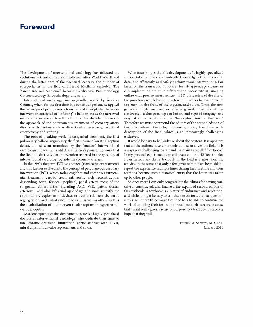
Pathogenesis of atherosclerosisAtherosclerosis is an inflammatory fibro‐proliferative process in which plaque forms in the intima, bringing about stenosis or thrombosis and hence ischemia [8–10]. Though the exact initiator of plaque formation remains unknown, there is a general consensus that the triggering episode is endothelial damage, which could be caused by factors such as cigarette smoke toxins, hypertension, or immune injury [4,11–15]. Damaged cells become more permeable, ultimately causing subendothelial macrophages to consume circulating low density lipoproteins (LDL) which are altered in the intima to induce further endothelial damage [8,9,16]. More macrophages are then recruited, after which they remain in the intima as lipid‐rich foam cells [9,10,17–19]. Meanwhile, in an attempt to restore endothelial function, smooth muscle cells migrate from the media to the intima to proliferate and generate a connective tissue matrix to cap the lipid core, further thickening the lesion [8,19,20]. Plaques enlarge as the process becomes chronic, classified as stable or unstable (Figures 1.1 and 1.2), either of which can lead to clinical sequelae [8,17,21].
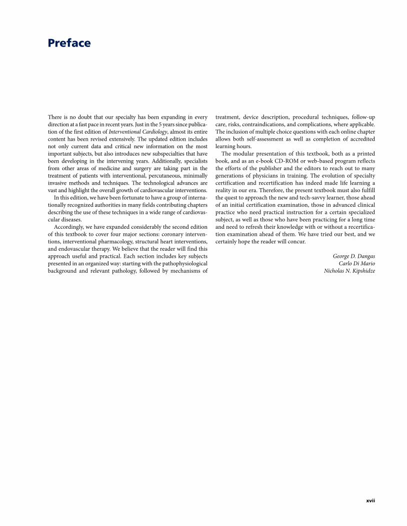
Clinical featuresThe first indication of coronary artery disease (CAD) may be sudden death, or patients can present with silent ischemia, stable angina, or an acute coronary syndrome (ACS) [22]. ACSs comprise a range of syndromes resulting from atherosclerotic plaque disruption or rupture and are divided into unstable angina (UA), non‐ ST‐elevation myocardial infarction (NSTEMI), and ST‐ elevation myocardial infarction (STEMI) [21,23,24]. An unstable plaque, characterized by a large lipid core covered by a thin and unstable fibrous cap, is prone to rupture [21,25–27]. The sudden rupture can cause thrombus formation, in turn leading to ACS (Figure 1.2) [26,28,29]. Conversely, a stable plaque has a thick fibrous cap which is not easily ruptured (Figure 1.1), causing the chronic condition of stable angina through episodes of ischemia experienced upon physical exertion [25,27,30].
Consequences of atherosclerosisThe risk of major thrombotic and thromboembolic complications of atherosclerosis appears to be related more to the stability of atheromatous plaques than to the extent of disease [31,32]. Stable angina is associated with smooth fibrous coronary artery plaques (stable plaque), whereas unstable angina, acute myocardial infarction (AMI), and sudden cardiac death are almost invariably associated with destabilization of plaques [29]. Similarly, in patients with carotid artery atherosclerotic disease, plaque irregularity and rupture are closely associated with cerebral ischemic events, and patients with irregular or ulcerated plaque demonstrate a higher risk of ischemic stroke irrespective of the degree of luminal stenosis [33].
Much attention has been placed on trying to identify plaques at high risk of disruption leading to thrombosis. Such “vulnerable plaques” have also been areas of intense research using novel intracoronary imaging modalities: optical coherence tomography (OCT) [6,29]. OCT offers the advantages over intravascular ultrasound or angiography of ultra‐high resolution and superiority in imaging the vessel wall and lumen interface [34–36].
Atherogenesis and Inflammation
Umair Hayat1, Vikas Thondapu1, Tim Tsay1, and Peter Barlis2
1 Melbourne Medical School, The University of Melbourne, Australia2 The University of Melbourne, Australia
ChAPter 1
SecTion i Basic Knowledge
4 PART I Principles and Techniques SECTION I Basic Knowledge
Low densitylipoprotein (LDL)
Macrophageuptakes
oxidized LDL
Macrophagetransforms
into the foam cell
Foam cell diesreleases
lipid debris
LymphocyteSmooth muscle cell
transformsinto the foam cell
Migrationof smoothmuscle cells
Tunica media
Tunica intima
Plaque rupture
Platelets
EndotheliumFibrin
Thrombus
Erythrocyte
Internal elastic lamina
Foam cell diesreleaseslipid debrisSmooth musclecell uptakesoxidized LDL
Fibrous cap
Foam cell
Macrophage
Low densitylipoprotein (LDL)
oxidizes
Monocyte migratesinto the tunica intima
Figure 1.2 Unstable atherosclerotic plaque characterized by the presence of a thin fibrous cap rich in inflammatory macrophagic foam cells and T lymphocytes. Rupture of the fibrous cap at the shoulder region has resulted in thrombus formation.
Low densitylipoprotein (LDL) Fibrous cap Core Platelets
Endothelium
Tunica intima
Foam cell diesreleaseslipid debris
Smooth musclecell uptakesoxidized LDL
Internal elastic lamina
Tunica media
Migrationof smoothmuscle cells
Smooth muscle celltransforms
into the foam cell
LymphocyteFoam cell diesreleases
lipid debris
Macrophagetransforms intothe foam cell
Macrophageuptakes
oxidized LDL
Macrophage
Low densitylipoprotein (LDL)
oxidizes
Foam cell
Monocyte migrates into the tunica
intima
Artery lumen
Figure 1.1 Stable atherosclerotic plaque characterized by the presence of a low inflammatory infiltrate. This type of lesion is constituted by a lipid core (extracellular lipid, cholesterol crystals, and necrotic debris) covered by a thick fibrous cap consisting principally of smooth muscle cells (SMC) in a collagenous–proteoglycan matrix, with varying degrees of infiltration by macrophages and T lymphocytes. HDL, high density lipoprotein.

CHAPTER 1 Atherogenesis and Inflammation 5
Considerable data exist to sustain the hypothesis that several morphologic and molecular markers identifying unstable plaques could be expressed during plaque vulnerability. As shown by a number of anatomical and clinical studies, these vulnerable plaques are more often associated with rupture and thrombosis than stable plaques covered by a thin fibrous cap and show an extensive inflammatory infiltrate [28,37].
Unlike the stable plaque that shows a chronic inflammatory infiltrate, the vulnerable and ruptured plaque is characterized by features of acute inflammation [37,38]. There are a large number of studies showing that “active” inflammation mainly involves T lymphocytes and macrophages which are activated toward a pathway of inflammatory response, secrete cytokines and lytic enzymes which in turn cause thinning of the fibrous cap, predisposing to plaque rupture. Recent research has furnished new insight into the molecular mechanisms that cause transition from a stable to an unstable phase of atherosclerosis and points to inflammation as the playmaker in the events leading to plaque destabilization and suggest that alterations in shear stress may also play a pivotal part [39,40].
A current challenge is to identify morphological and molecular markers able to discriminate stable plaques from vulnerable ones allowing the stratification of “high risk” patients for acute cardiac and cerebrovascular events before clinical syndromes develop. Bearing this aim in mind, this chapter focuses on cellular and molecular mechanisms affecting plaque progression and serum markers correlated to plaque inflammation.
Insights from coronary imagingTraditionally, coronary angiography has been the gold standard to detect extent and severity of CAD. These findings form the foundation of the interventionist’s clinical decision‐making process and whether to proceed to percutaneous therapy. It is widely acknowledged, however, that angiography has several limitations. First, it maintains a relatively low image resolution. Second, it represents a luminogram of the artery and stenosis. Therefore little detail is provided as to the composition of the underlying plaque causing the stenosis and, finally, it is a 2D imaging method used to assess what are complex 3D structures.
intravascular ultrasoundIntravascular ultrasound (IVUS) utilizes ultrasound waves that reflect off vascular tissues to yield real‐time images [41,42]. While angiography only portrays a luminal silhouette [41], IVUS, with a resolution of 100–150 μm, captures details not retrievable with angiography—cross‐sections of the lumen and vessel wall, even a differentiation of its layers [43–46]. Thus, IVUS enables study of the atherosclerotic process through the visualization of plaque in the vessel wall [41,47–49]. Indeed, the technology has demonstrated a greater prevalence of atherosclerosis than initially claimed with angiography [44].
optical coherence tomographyOCT, the optical analog of IVUS, employs the reflection of near‐infrared (NIR) light instead of sound. Initially applied in ophthalmology, advancement in the technology has now enabled OCT to capture non‐transparent tissues such as coronary vessels [50,51]. OCT offers real‐time, in vivo and in situ cross‐sectional imaging of vascular structures with a resolution 10‐fold that of IVUS (15 μm versus 150 μm) and a penetration depth similar to that of histology [34,43,50–53].
By virtue of its superior resolution, OCT can provide near‐histological analysis of atherosclerotic plaques in real time (Figure 1.3). OCT definition of thin cap fibroatheroma (TCFA) follows the findings of autopsy studies of sudden death patients that had revealed the presence of fibrous caps <65 μm in the majority of plaques that had ruptured. These thin ruptured caps were also found to have an infiltrate of macrophages [54]. Whereas OCT is well placed in precisely defining the thinness of fibrous cap, macrophage infiltration seen as punctate, signal‐rich spots at the junction of fibrous cap and lipid pool has been described less consistently. Previous autopsy studies had also shown that plaque rupture, erosion, and calcified nodules were the three leading underlying mechanisms for luminal thrombosis with a frequency of 65%, 30%, and 5%, respectively [25]. In recent years OCT has enabled this type of information to be obtained in vivo and has confirmed similar prevalence of plaque morphologies in patients presenting with STEMI and NSTEMI [55].
Plaque rupture on OCT is identified by a clear‐cut disruption in the signal‐rich thin fibrous cap overlying a signal‐poor necrotic core resulting in extrusion of highly thrombogenic material into the lumen. Plaque erosion on the other hand is identified by the presence of luminal thrombus adjacent to a plaque that has an irregular but intact, thicker fibrous cap. Such plaques are mostly devoid of necrotic core. Calcified nodules are the least common etiology in ACS and are less well defined. They are recognized by sharp nodules protruding into the lumen causing discontinuation of the fibrous cap (Figure 1.4).
In patients with stable CAD, coronary imaging can provide lesion level information and help to show the changes in plaque microstructure in response to pharmacotherapies. Kataoka et al. [56] evaluated 293 and 122 lipid and fibrous plaques in 280 stable statin‐treated patients with CAD and reported that patients with LDL‐C levels <50 mg/dL were less likely to have lipid plaques, and had more features of plaque stability such as thicker fibrous caps and smaller lipid arcs.
the vulnerable plaqueAtherosclerotic lesions, according to the classification of the American Heart Association modified by Virmani et al. [29], are divided in two groups: (i) non‐atherosclerotic intimal lesions and (ii) progressive atherosclerotic lesions which include stable, vulnerable, and thrombotic plaques.
The different pathologic characterization of atherosclerotic lesions largely depends on the thickness of the fibrous cap and its grade of inflammatory infiltrate, which is in turn largely constituted by macrophages and activated T lymphocytes. Typically, the accumulating plaque burden is initially accommodated by an adaptive positive remodeling with expansion of the vessel external elastic lamina and minimal changes in lumen size [57,58]. The plaque contains monocyte‐derived macrophages, smooth muscle cells, and T lymphocytes. Interaction between these cells types and the connective tissue appears to determine the development and progression of the plaque itself, including important complications such as thrombosis and rupture.
The lesions classified as vulnerable or TCFA identify a plaque prone to rupture and thrombosis characterized by a large necrotic core containing numerous cholesterol clefts. The overlying cap is thin and rich in inflammatory cells, macrophages, and T lymphocytes with few smooth muscle cells [28,29,59]. Burke et al. [54] identified a cut‐off value for cap thickness of 65 μm to define a vulnerable coronary plaque. Despite the predominant hypothesis

6 PART I Principles and Techniques SECTION I Basic Knowledge
focusing on the responsibility of a specific vulnerable atherosclerotic plaque rupture [5,7] for acute coronary syndromes, some pathophysiologic, clinical, and angiographic observations seem to suggest the possibility that the principal cause of coronary instability is not to be found in the vulnerability of a single atherosclerotic plaque, but in the presence of multiple vulnerable plaques in the
entire coronary tree, correlated with the presence of a diffuse inflammatory process [37,38,60,61].
Within this context, recent angiographic studies have demonstrated the presence of multiple vulnerable atheromatous plaques in patients with unstable angina [20,62] and in those affected by transmural myocardial infarction [61]. Recently, by means of flow
(a) (b)
0.5 mm
Thrombus
Super�cialcalcium
Fibrous cap withirregular surface
Rupturedcap40 μm
Figure 1.4 Unstable coronary plaques seen on OCT. (a) Plaque erosion: intact fibrous cap with irregular luminal surface and superficial calcium. (b) Plaque rupture with luminal thrombus. At the 11 o’clock position a thin cap fibro‐atheroma (TCFA) is seen (fibrous cap thickness measured 40 μm, marked with small white bar).
(a) (b)
(c)
Figure 1.3 Stable coronary plaques seen on optical coherence tomography (OCT). (a) Calcified plaque is seen at 7‐ to 10 o’clock position. It is characterized by sharply delineated borders and heterogeneous core. (b) Calcified plaque is outlined with white dotted line. (c) Lipid‐rich plaque marked by white lines. It is characterized by dark, signal‐poor core with ill‐defined margins and a bright thick fibrous cap (>65 μm). As the light rapidly attenuates through the necrotic core, OCT cannot be used to measure the depth of such plaques.

CHAPTER 1 Atherogenesis and Inflammation 7
cytometry Spagnoli et al. [38] have demonstrated the presence of an activated and multicentric inflammatory infiltrate in the coronary vessels of individuals who died of AMI. Similar results have been obtained by Buffon et al. [60], who, through the determination of the neutrophil myeloperoxidase activity, have proved the presence of a diffuse inflammation in the coronary vessels in individuals affected by unstable angina. These results have been confirmed by a morphological study which demonstrated the presence of a high inflammatory infiltrate constituted by macrophages and T lymphocytes activated in the whole coronary tree, also present in the stable plaques of individuals who died of AMI. These plaques showed a two‐ to fourfold higher inflammatory infiltrate than aged‐matched individuals dying from non‐cardiac causes with chronic stable angina (SA) or without clinical cardiac history (CTRL), respectively [37]. Moreover, it has also been demonstrated that activated T lymphocytes infiltrate the myocardium both in the peri‐infarcted area and in remote unaffected myocardial regions in patients who died of a first myocardial infarction [63].
The simultaneous occurrence of diffuse coronary and myocardial inflammation in these patients further supports the concept that both coronary and myocardial vulnerabilities concur in the pathogenesis of fatal AMI.
AMI—at least associated with unfavorable prognosis—is therefore likely to be the consequence of a diffuse “active” chronic inflammatory process which determines the destabilization of both the entire coronary tree and the whole myocardium, not only the part of it affected by infarction. The causes of the diffuse inflammation associated with myocardial infarction are scarcely known. The presence of activated T lymphocytes suggests the “in situ” presence of an antigenic stimulus which triggers adaptive immunity.
role of inflammation in the natural history of atherosclerosisinception of the plaqueEndothelium injury has been proposed to be an early and clinically relevant pathophysiologic event in the atherosclerotic process [4,32]. Patients with endothelial dysfunction have an increased risk for future cardiovascular events including stroke [64]. Endothelial dysfunction was described as the ignition step in atherogenesis. From this point on, an inflammatory response leads to the development of the plaque.
Endothelial damage can be caused by physical and chemical forces, by infective agents or by oxidized LDL (ox‐LDL). Dysfunctional endothelium expresses P‐selectin (stimulation by agonists such as thrombin) and E‐selectin (induced by IL‐1 or TNF‐α). Expression of intercellular adhesion molecule‐1 (ICAM‐1) both by macrophages and endothelium and vascular adhesion molecule‐1 (VCAM‐1) by endothelial cells is induced by inflammatory cytokines such as interleukin‐1 (IL‐1), tumor necrosis factor‐1 (TNF‐α), and γ‐interferon (IFNγ).
Monocytes recalled in the subintimal space ingest lipoproteins and morph into macrophages. These generate reactive oxygen species (ROS), which convert ox‐LDL into highly oxidized LDL. Macrophages upload ox‐LDL via scavenger receptors until foam cells form. Foam cells with leukocytes migrate at the site of damage and generate the fatty streak. The loss of biologic activity of endothelium determines nitric oxide (NO) reduction together with increased expression of prothrombotic factors, proinflammatory adhesion molecules cytokines, and chemotactic factors. Cytokines may decrease NO bioavailability increasing the production of ROS. ROS
reduces NO activity both directly, reacting with endothelial cells, and indirectly via oxidative modification of eNOS or guanylyl cyclase [65]. Low NO bioavailability can upregulate VCAM‐1 in the endothelial cell layer that binds monocytes and lymphocytes in the first step of invasion of the vascular wall, via induction of nuclear factor κB (NFκB) expression [66]. In addition, NO inhibits leukocyte adhesion [67] and NO reduction results in induction of monocyte chemotactic protein‐1 (MCP‐1) expression which recruits monocytes [68]. NO is in a sensitive balance with endothelin‐1 (ET‐1) regulating vascular tone [69]. Plasma ET‐1 concentrations are increased in patients with advanced atherosclerosis and correlate with the severity of the disease [70,71]. In addition to its vasoconstrictor activity, ET‐1 also promotes leukocyte adhesion [72] and thrombus formation [73]. Dysfunctional endothelium expresses P‐selectin (stimulation by agonists such as trombin) and E‐selectin (induced by IL‐1 or TNF‐α) [74]. The expression of both ICAM‐1 by macrophages and endothelium, and VCAM‐1 by endothelial cells is induced by inflammatory cytokines such as IL‐1, TNF‐α, and IFNγ. Endothelial cells also produce monocyte chemotactic protein‐1 (MCP‐1), monocyte colony‐stimulating factor, and IL‐6 which further amplify the inflammatory cascade [75]. IL‐6 production by smooth muscle cells represents the main stimulus for C‐reactive protein (CRP) production [3]. Recent evidence suggests that CRP may contribute to the proinflammatory state of the plaque both mediating recruitment of monocytes and stimulating monocytes to release IL‐1, IL‐6, and TNF‐α [76]. The damaged endothelium allows the passage of lipids into the subendothelial space. Fatty streaks represent the first step in the atherosclerotic process.
evolving fibro‐atheromatous plaqueThe atheroma evolution is modulated by innate and adaptive immune responses [3,77,78]. The most important receptors for innate immunity in atherothrombosis are the scavenger receptors and the toll‐like receptors (TLRs) [79]. Adaptive immunity is much more specific than innate immunity but may take several days or even weeks to become fully mobilized. It involves an organized immune response leading to generation of T‐ and B‐cell receptors and immunoglobulins, which can recognize foreign antigens [80].
Stable plaqueMacrophages take up lipid deposited in the intima via a number of receptors, including scavenger receptor‐A, and CD36. Deregulated uptake of modified LDL through scavenger receptors leads to cholesterol accumulation and “foam cell” formation. The lipid laden macrophages (foam cells) forming the fatty streak secrete proinflammatory cytokines that amplify the local inflammatory response in the lesion, matrix metalloproteinases (MMPs), tissue factor into the local matrix, as well as growth factors, which stimulate the smooth muscle replication responsible for lesion growth. Macrophages colony‐stimulating factor (M‐CSF) acts as the main stimulator in this process, next to granulocyte–macrophage stimulating factor (MGGM‐CSF) and IL‐2 for lymphocytes [81]. Lymphocytes enter the intima by binding adhesion molecules: VCAM‐1, P‐selectin, ICAM‐1, MCP‐1 (CCL2), IL‐8 (CxCL8) [75]. Such infiltrate constituted mainly by CD4+ T lymphocytes recognize antigens bound to MHC class II molecules involved in antigen presentation to T lymphocytes thus provoking an immune response [2]. The major histocompatibility complex molecules (MHC II) are expressed by endothelial cells, macrophages, and vascular smooth muscle cells in proximity to activated T lymphocytes in the atherosclerotic plaque. Proinflammatory cytokines manage a central transcriptional control

8 PART I Principles and Techniques SECTION I Basic Knowledge
point mainly mediated by NFκB. Macrophage/foam cells produce cytokines that activate neighboring smooth‐muscle cells (SMCs), resulting in extracellular matrix production [2].
Repeated inflammatory stimuli induce foam cells to secrete growth factors that induce proliferation and migration of SMCs into the intima. The continuous influx of cells in the subintimal space convert the fatty streak in a more complex and advanced lesion in which inflammatory cells (monocytes/macrophages, lymphocytes), SMCs, necrotic debris mainly resulting from cell death, ox‐LDL elicit a chronic inflammatory response by adoptive immune system. SMCs form a thick fibrous cap that cover the necrotic core and avoid the exposition of thrombogenic material to the bloodstream. The volume of lesion grows and protrudes into the arterial lumen causing variable degrees of lumen stenosis. These lesions are advanced complicated “stable” atherosclerotic lesions, asymptomatic and often unrecognized [82,83].
Vulnerable plaque: a shift toward Th1 patternEarly phases of the plaque development are characterized by an acute innate immune response against exogenous (infectious) and endogenous non‐infectious stimuli. Specific antigens activate adaptive immune system leading to proliferation of T and B cells. A first burst of activation might occur in regional lymph nodes by dendritic cells (DCs) trafficking from the plaque to the lymph node. Subsequent cycles of activation can be sustained by interaction of activated/memory T cells re‐entering in the plaque by selective binding to endothelial cell surface adhesion molecules with plaque macrophages expressing MHC class II molecules. In this phase of the atherogenic process the selective recruitment of a specific subtype of CD4+ cells play a major part in determining the future development of the lesion. Two subtypes of CD4+ cells have a juxtaposed role: Th1 and Th2 cells [84].
Th1 cells secreting proinflammatory cytokines, such as IFNγ, promote macrophage activation, inflammation, and atherosclerosis, whereas Th2 cells (cytokine pattern IL‐4, IL‐5, and IL‐10) mediate antibody production and generally have anti‐inflammatory and antiatherogenic effects [64]. Therefore the switch to a selective recruitment of Th1 lymphocyte represents a key point toward plaque vulnerability and disruption. T cells in the plaque may encounter antigens such as ox‐LDL. Moreover, T‐cell response can be triggered by heat shock proteins of endogenous or microbial origin [85]. It is still unknown why the initial inflammatory response becomes a chronic inflammatory condition. However, when the plaque microenvironment triggers the selective recruitment and activation of Th1 cells they in turn determine a potent inflammatory cascade.
The combination of IFNγ and TNF‐α upregulates the expression of fractalkine (CX3CL1) [86]. IL‐1 and TNFα‐activated endothelium express also fractalkine (membrane bound form) which directly mediates the capture and adhesion of CX3CR1 expressing leukocytes providing a further pathway for leukocyte activation [87]. This cytokine network promotes the development of the Th1 pathway which is strongly proinflammatory and induces macrophage activation, superoxide production, and protease activity.
role of inflammation as vulnerability factorHomeostasis of plaque “microenvironment” (i.e., the balance between cell migration and cell proliferation, extracellular matrix production and degradation, macrophages and lymphocytes interplay) appears strictly related to the transition of a stable plaque into a vulnerable one.
A limited number of T cells, following the Th1 pathway, initiates the production of large amounts of molecules downstream in the cytokine cascade orchestrating the transition from the stable to unstable plaque [77,88].
Within the plaque, inflammatory cells such as foam cells and monocyte‐derived macrophages are induced to produce matrix‐degrading enzymes, cytokines, and growth factors strictly implicated in extracellular matrix homeostasis. In particular, cytokines such as INFγ suppress collagen synthesis, a major component of the fibrous cap [75]. Moreover, infiltration of mononuclear cells results in release of proteases which causes plaque disruption [89]. The production of ROS within the atherosclerotic plaque has important implications for its structural integrity [65]. Deregulated oxidant production has the potential to promote the elaboration and activation of matrix degrading enzymes in the fibrous cap of the plaque. Moreover, impaired NO function coupled with oxidative excess can activate MMPs [90], namely MMP‐2 and MMP‐9, which weaken the fibrous cap. Another mechanism that can determine the thinning of the fibrous cap is the apoptosis of smooth muscle cells. In fact, there is evidence for extensive apoptosis of SMCs within the cap of advanced atherosclerosis, as well as those cultured from plaques [32,91].
A very important role, not yet well studied, is that of dendritic cells, namely cells specialized in antigen presentation with a key role in the induction of primary immune response and in the regulation of T‐lymphocyte differentiation, as well as in mechanisms of central and peripheral tolerance aiming at the elimination of T lymphocytes that are potentially self‐reactive toward self‐antigens [92,93]. A characteristic of dendritic cells is also the ability to polarize T‐cell responses toward a T‐helper phenotype (Th1) in response to bacterial antigens. Molecules expressed by activated T lymphocytes, like CD40L, OX40, stimulate the release from dendritic cells of chemokines (fractalkines) able to attract other lymphocytes toward the inflammation site, amplifying the immune response [94].
Patients with ACS are characterized by the expansion of an unusual subset of T cells, CD4+CD28null T cells, with functional activities that predispose for vascular injury [95,96]. CD4+CD28null T cells are a population of lymphocytes rarely found in healthy individuals. Disease‐associated expansions of these cells have been reported in inflammatory disorders such as rheumatoid arthritis. CD4+CD28null T cells are characterized by their ability to produce high amounts of IFNγ [96]. Equally importantly, CD4+CD28null T cells have been distinguished from classic Th cells by virtue of their ability to function as cytotoxic effector cells. Possible targets in the plaque are SMCs and endothelial cells, as recently shown [97]. In vivo, CD4+CD28null cells have a tendency to proliferate with the frequent emergence of oligoclonality, raising the possibility of continuous antigenic stimulation, as it is the case in certain autoimmune disorders and in chronic infections. The demonstration of oligoclonality within the CD4+CD28null T‐cell subsets and sharing of T‐cell receptor sequences in expanded T‐cell clones of patients with ACS strongly support the notion that these cells have expanded and are activated in response to a common antigenic challenge [98]. CD4+CD28null T cells are long‐lived cells. Clonality and longevity of these cells are associated with defects in apoptotic pathways [99]. Moreover, CD28 is relevant for the expansion of naïve T cells, thus the absence of this molecule contributes to the senescence of lymphocytes. The excessive expansion of a pool of senescent T lymphocytes might compromise the efficacy of the immune responses direct against exogenous antigens as well as determinate autoimmune responses.

CHAPTER 1 Atherogenesis and Inflammation 9
Recently, a subpopulation of T CD4+ cells, expressing IL‐2 receptor, CD25 membrane marker, has been pointed out. Such lymphocytes represent 7–10% of T CD4+ cells and their homeostasis is due to some co‐stimulatory molecules, such as CD28 receptor expressed by T cells and B7 molecules expressed by dendritic cells [100]. The current knowledge of the role of this specific subset of T cells in human atherogenesis is still incomplete, even though a very recent study carried out on mice has demonstrated an antiatherogenic effect of T CD4+CD25+ cells [101].
Th1 cells and T regulatory 1 cells have been demonstrated to play opposite roles in rupture of atherosclerotic lesion. The role of novel subset of T regulatory cells, known as CD4+CD25+Foxp3+ T cells, has been recently studied in CAD. Han et al. [102] found that the reduction of CD4+CD25+Foxp3+ T lymphocytes was consistent with the expansion of Th1 cells in patients with unstable CAD. The reversed development between CD4+CD25+ Tregs and Th1 cells might contribute to plaque destabilization.
Serum markers correlated to plaque inflammationIn recent years, a number of studies have correlated different serologic biomarkers with cardiovascular disease [4,103] leading to a rapid increase in the number of biomarkers available (Table 1.1). These biomarkers are useful in that they can identify a population at risk of an acute ischemic event and detect the presence of so‐called vulnerable plaques and/or vulnerable patients [104,105]. Ideally, a biomarker must have certain characteristics to be a potential predictor of incident or prevalent vascular disease. Measurements have to be reproducible in multiple independent samples, the method for determination should be standardized, variability controlled, and the sensitivity and specificity should be good. In addition, the biomarker should be independent from other established risk markers, substantively improve the prediction of risk with established risk factors, be associated with cardiovascular events in multiple population cohorts and clinical trials, and the cost of the assays has to be acceptable. Finally, to be clinically useful a biomarker should correctly reflect the underlying biological process associated with plaque burden and progression.
Traditional biomarkers for cardiovascular risk include LDL cholesterol and glucose. However, 50% of heart attacks and strokes occur in individuals who have normal LDL cholesterol, and 20% of major adverse events occur in patients with no accepted risk factors [106]. Therefore, in light of changing atherosclerotic models, vulnerable blood may be better described as blood that has an increased level of activity of plasma determinants of plaque progression and rupture.
In this context, proposed biomarkers fall into nine general categories: inflammatory markers, markers for oxidative stress, markers of plaque erosion and thrombosis, lipid‐associated markers, markers of endothelial dysfunction, metabolic markers, markers of neovascularization, and genetic markers. The last six biomarker categories are not treated in this chapter but only listed in Table 1.1. Some of these markers may indeed reflect the natural history of atherosclerotic plaque growth and may not be directly related to an increased risk of cardiovascular events. On the contrary, other markers are more related to complex plaque morphological features and may reflect an active process within the plaque which is in turn related to the onset of local complications and onset of acute clinical events.
However, it is important to emphasize that, in any individual patient, it is not yet clear how these biomarkers relate to quantitative
risk of major adverse cardiovascular events. The best outcomes may be achieved by a panel of markers that will capture all of the different processes involved in plaque progression and plaque rupture, and that will enable clinicians to quantify an individual patient’s true cardiovascular risk. In all likelihood, a combination of genetic (representing heredity) and serum markers (representing the net interaction between heredity and environment) will ultimately be the ones that should be utilized in primary prevention. Finally, different non‐invasive and invasive imaging techniques may be coupled with biomarkers detection to increase the specificity, sensitivity, and overall predictive value of each potential diagnostic technique.
Markers of inflammationMarkers of inflammation include CRP, inflammatory cytokines soluble CD40L (sCD40L), soluble vascular adhesion molecules (sVCAM), and TNF.
CRP is a circulating pentraxin that has a major role in the human innate immune response [107] and provides a stable plasma biomarker for low grade systemic inflammation. CRP is produced predominantly in the liver as part of the acute phase response. However, CRP is also expressed in SMCs within diseased atherosclerotic arteries [108] and has been implicated in multiple aspects of atherogenesis and plaque vulnerability, including expression of adhesion molecules, induction of NO, altered complement function, and inhibition of intrinsic fibrinolysis [109]. CRP is considered to be an independent predictor of unfavorable cardiovascular events in patients with atherosclerotic disease. Beyond the ability of CRP to predict risk among both primary and secondary prevention patients, interest in it has increased with the recognition that statin‐induced reduction of CRP is associated with less progression in adverse cardiovascular events that is independent of the lipid‐associated changes [110] and that the efficacy of statin therapy may be related to the underlying level of vascular inflammation as detected by high‐sensitivity CRP (hs‐CRP). Among patients with stable angina and established CAD, plasma levels of hs‐CRP have consistently been shown associated with recurrent risk of cardiovascular events [111,112]. Similarly, during acute coronary ischemia, levels of hs‐CRP are predictive of high vascular risk even if troponin levels are non‐detectable, suggesting that inflammation is associated with plaque vulnerability even in the absence of detectable myocardial necrosis [113,114]. Despite these data, the most relevant use of hs‐CRP remains in the setting of primary prevention. To date, over two dozen large‐scale prospective studies have shown baseline levels of hs‐CRP to independently predict future myocardial infarction, stroke, cardiovascular death, and incidence of peripheral arterial disease [115,116]. Moreover, eight major prospective studies have had adequate power to evaluate hs‐CRP after adjustment for all Framingham covariates, and all have confirmed the independence of hs‐CRP [117]. Despite this evidence, it is important to recognize that there remain no firm data to date that lowering CRP levels per se will lower vascular risk. Further, as with other biomarkers of inflammation, it remains controversial whether CRP has a direct causal role in atherogenesis [118], and ongoing work with targeted CRP‐lowering agents are required to fully test this hypothesis. However, the clinical utility of hs‐CRP has been well established, and on the basis of data available through 2002, the Centers for Disease Control and Prevention and the American Heart Association endorsed the use of hs‐CRP as an adjunct to global risk prediction, particularly among those at “intermediate risk” [119]. Data available since 2002 strongly reinforce these recommendations and suggest

10 PART I Principles and Techniques SECTION I Basic Knowledge
expansion to lower risk groups, as well as those taking statin therapy. Perhaps most importantly, data for hs‐CRP provides evidence that biomarkers beyond those traditionally used for vascular risk detection and monitoring can have important clinical roles in prevention and treatment.
Cellular adhesion molecules can be considered potential markers of vulnerability because such molecules are activated by inflammatory cytokines and then released by the endothelium [120]. These molecules represent the one available marker to assess endothelial activation and vascular inflammation. The Physicians’ Health Study evaluated more than 14,000 healthy subjects and demonstrated ICAM‐1 expression positive correlation with cardiovascular risk and showed that subjects in the higher quartile of ICAM‐1 expression showed 1.8 times higher risk than subjects in the lower quartile [121]. Furthermore, soluble ICAM‐1 and VCAM‐1 levels showed a positive correlation with atherosclerosis disease burden [122]. IL‐6 is expressed during the early phases of inflammation and it is the principal stimulus for CRP liver production. In addition, CD40 ligand, a molecule expressed on cellular membrane, is a TNFα homologue which stimulates activated
macrophages proteolytic substances production [123]. CD40 and CD40L have been found on platelets and several other cell types in functional‐bound and soluble (sCD40L) forms. Although many platelet‐derived factors have been identified, recent evidence suggests that CD40L is actively involved in the pathogenesis of ACS. CD40L drives the inflammatory response through the interaction between CD40L on activated platelets and the CD40 receptor on endothelial cells. Such interactions facilitate increased expression of adhesion molecules on the surface of endothelial cells and release of various stimulatory chemokines. These events, in turn, facilitate activation of circulating monocytes as a trigger of atherosclerosis. Beyond known proinflammatory and thrombotic properties of CD40L, experimental evidence suggests that CD40L‐induced platelet activation leads to the production of reactive oxygen and nitrogen species, which are able to prevent endothelial cell migration and angiogenesis [124]. As a consequence of inhibiting endothelial cell recovery, the risk of subsequent coronary events may be greater. Clinical studies have supported the involvement of CD40L in ACS and the prognostic value in ACS populations. Levels of sCD40L have been shown to be an independent predictor of
Table 1.1 Serologic markers of vulnerable plaque/patient.
Reflecting metabolic and immune disorders Reflecting hypercoagulability Reflecting complex atherosclerotic plaque
Abnormal lipoprotein profile (i.e., high LDL, low HDL, lipoprotein [a], etc.)
Non‐specific markers of inflammation (hs‐CRP, CD40L, ICAM‐1, VCAM, leukocytosis and other immuno‐related serologic markers which may not be specific for atherosclerosis and plaque inflammation
Serum markers of metabolic syndrome (diabetes or hypertriglyceridemia)
Specific markers of immune activation (i.e., anti‐LDL antibody, anti‐HSP antibody
Markers of lipid peroxidation (i.e., ox‐LDL and ox‐HDL)
Homocysteine
PAPP‐A
Circulating apoptosis markers (i.e., Fas/Fas ligand)
ADMA/DDAH/
Circulating NEFA
Markers of blood hypercoagulability (i.e., fibrinogen, d‐dimer, factor V Leiden)
Increased platelet activation and aggregation (i.e., gene polymorphism of platelet glycoproteins IIb/IIIa, Ia/IIa, and Ib/IX)
Increased coagulation factors (i.e., clotting of factors V, VII, VIII, XIII, von Willebrand factor)
Decreased anticoagulation factors (i.e., proteins S and C, thrombomodulin, antithrombin III)
Decreased endogenous fibrinolysis activity (i.e., reduced tissue plasminogen activator, increased type I PAI, PAI polymorphisms)
Prothrombin mutation (i.e., G20210A)
Thrombogenic factors (i.e., anticardiolipin antibodies, thrombocytosis, sickle cell disease, diabetes, hypercholesterolemia)
Transient hypercoagulability (i.e., smoking, dehydration, infection)
Morphology/structure• Cap thickness• Lipid core size• Percentage stenosis• Remodeling (positive vs. negative)• Color (yellow, red)• Collagen content vs. lipid content• Calcification burden and pattern• Shear stress
Activity/function• Plaque inflammation (macrophage density, rate of
monocyte and activated T‐cell infiltration)• Endothelial denudation or dysfunction (local nitric
oxide production, anti/procoaugulation properties of the endothelium)
• Plaque oxidative stress• Superficial platelet aggregation and fibrin
deposition• Rate of apoptosis (apoptosis protein markers,
microsatellite)• Angiogenesis, leaking vasa vasorum, intraplaque
hemorrhage• Matrix metalloproteinases (MMP‐2, ‐3, ‐9)• Microbial antigens (Chlamydia pneumoniae)
Temperature
Pan arterial• Transcoronary gradient of vulnerability biomarkers• Total calcium burden• Total coronary vasoreactivity• Total arterial plaque burden (intima media
thickness)
ADMA, asymmetric dimethylarginine; CRP, C‐reactive protein; DDAH, dimethylarginine dimethylaminohydrolase; HDL, high density lipoprotein; HSP, heat shock protein; ICAM, intercellular adhesion molecule; LDL, low density lipoprotein; NEFA, non‐esterified fatty acids; PAI, plasminogen activator; PAPP‐A, pregnancy‐associated plasma protein A; VCAM, vascular adhesion molecule.