interventional management of pleural infections management of pleural infections john e. heffner,...
TRANSCRIPT
DOI 10.1378/chest.08-2956 2009;136;1148-1159Chest
John E. Heffner, Jeffrey S. Klein and Christopher Hampson InfectionsInterventional Management of Pleural
http://chestjournal.chestpubs.org/content/136/4/1148.full.htmland services can be found online on the World Wide Web at: The online version of this article, along with updated information
ISSN:0012-3692)http://chestjournal.chestpubs.org/site/misc/reprints.xhtml(
of the copyright holder.may be reproduced or distributed without the prior written permission Northbrook, IL 60062. All rights reserved. No part of this article or PDFby the American College of Chest Physicians, 3300 Dundee Road,
2009Physicians. It has been published monthly since 1935. Copyright CHEST is the official journal of the American College of Chest
© 2009 American College of Chest Physicians by Jonathan Ball on November 2, 2009chestjournal.chestpubs.orgDownloaded from

Interventional Management of PleuralInfections
John E. Heffner, MD, FCCP; Jeffrey S. Klein, MD, FCCP;and Christopher Hampson, MD
Pleural infections represent an important group of disorders that is characterized by the invasionof pathogens into the pleural space and the potential for rapid progression to frank empyema.Previous epidemiologic studies have indicated that empyema is increasing in prevalence, whichunderscores the importance of urgent diagnosis and effective drainage to improve clinicaloutcomes. Unfortunately, limited evidence exists to guide clinicians in selecting the idealdrainage intervention for a specific patient because of the broad variation that exists in theintrapleural extent of infection, presence of locules, comorbid features, respiratory status, andvirulence of the underlying pathogen. Moreover, many patients experience delays in both therecognition of infected pleural fluid and the initiation of appropriate measures to drain thepleural space. The present review provides an update on the pathogenesis and interventionaltherapy of pleural infections with an emphasis on the unique role of image-guided drainage withsmall-bore catheters. (CHEST 2009; 136:1148–1159)
Abbreviations: ACCP � American College of Chest Physicians; BTS � British Thoracic Society; MIST1 �Multicenter Intrapleural Sepsis Trial; rtPA � recombinant tissue plasminogen activator; US � ultrasonography;VATS � video-assisted thoracoscopic surgery
I nfections of the pleural space present in a highlyvariable manner and affect a heterogeneous pop-
ulation of patients with diverse underlying etiologicconditions (Table 1). All pleural infections, however,share in common a considerable potential for deathand lifelong morbidity.1,2 Most case series2–6 havereported mortality rates between 7% and 33%, withmortality rates at � 50% among elderly patients withcomorbidities.2,6–8 More recent epidemiologic stud-ies9,10 have indicated that empyema has increased inincidence during the last 2 decades. Hospital dis-
charge data in Washington state demonstrate anincrease in the empyema incidence rate by 2.8% peryear from 1987 to 2004.9 Similar data show a 12.4%age-adjusted increase in the empyema incidence ratein Canada from 1995 to 2003, which affected mostlychildren and the elderly.10 An aging population,longer survival times for immunocompromised pa-tients and those with comorbid diseases, and chang-ing virulence of pleural pathogens11,12 suggest thatthese incidence trends will continue.
Because of the considerable mortality and morbid-ity associated with pleural infections, experts recom-mend13 adherence to modern principles of empyemamanagement that promote early diagnosis and promptpleural drainage. Delays in initiating effective drainageprolong hospital stays, increase the likelihood that moreinvasive drainage procedures will be required, andincrease mortality and morbidity.3,14,15 Unfortunately,studies3,14,15 have demonstrated that physicians com-monly delay diagnosis and drainage for patients withpleural infections. These delays may occur because noclinical features or laboratory studies clearly identifywhich patients with pneumonia have pleural infections.Consequently, every patient who is at risk for pleural
Manuscript received December 15, 2008; revision acceptedMarch 4, 2009.Affiliations: From the Department of Medicine (Dr. Heffner),Providence Portland Medical Center, Oregon Health and Sci-ence Center, Portland, OR; and Fletcher Allen Health Care (Drs.Klein and Hampson), University of Vermont College of Medi-cine, Burlington, VT.Correspondence to: John E. Heffner, MD, Providence Port-land Medical Center, 5050 NE Hoyt St., Suite 540, Portland, OR97213; e-mail: [email protected]© 2009 American College of Chest Physicians. Reproductionof this article is prohibited without written permission from theAmerican College of Chest Physicians (www.chestjournal.org/site/misc/reprints.xhtml).DOI: 10.1378/chest.08-2956
CHEST Postgraduate Education CornerCHEST IMAGING AND PATHOLOGY REVIEW
1148 Postgraduate Education Corner
© 2009 American College of Chest Physicians by Jonathan Ball on November 2, 2009chestjournal.chestpubs.orgDownloaded from

infections should undergo an initial evaluation to detectpleural fluid, determine the likelihood that the fluid isinfected, and ensure prompt drainage when indicated.
The present review summarizes the pathophysio-logic principles of empyema formation, classificationand staging systems for empyema, and the relativevalue of different approaches to draining the pleuralspace. Image-guided small-bore catheter drainagereceives special emphasis because of the unique valueit provides for patients with both nonloculated andcomplex multiloculated infected pleural effusions.
Pathophysiologic Classification andClinical Staging of Pleural Infections
Pleural effusions develop when the balance ofpleural fluid formation and removal is altered. Pleu-ral effusions secondary to pneumonia are termedparapneumonic effusions. Most of these effusionsremain sterile and resolve with antibiotic therapy(termed uncomplicated parapneumonic effusions),but infections of the pleural space develop in a smallsubset of patients and require drainage for fullrecovery (termed complicated parapneumonic effu-sions). Without effective drainage, complicated para-
pneumonic effusions progress to frank intrapleuralpus, which defines the presence of an empyema.This progression may occur rapidly over a few daysand necessitate surgical drainage (Fig 1).
Progression to empyema occurs in three phases.16
The exudative phase develops when inflammatoryfluid enters the pleural space across vascular andvisceral pleural membranes that have increased per-meability due to pneumonia. Pleural fluid is nonvis-cous, free-flowing, and readily drained by thoracen-tesis or chest tube. Unremitting inflammation depositsfibrin that coats the visceral pleura and promotes theformation of locules that impede lung reexpansionduring attempts at fluid drainage. Pleural fluid be-comes purulent and increasingly viscous. This fibrin-opurulent phase may respond to therapy with anti-biotics and chest tube drainage but often requiresintervention to break down adhesions. If a fibropu-rulent effusion remains undrained, fibroblasts even-tually deposit fibrotic tissue that encases the lung ininelastic peels. At this organizing phase, resolution ofthe empyema requires surgical procedures to drainpus, obliterate the empyema space, and reexpand thelung.
The three phases of empyema represent a contin-uum of events with no clear demarcations. Biomar-kers, such as pleural fluid pH, glucose, and lactatedehydrogenase, have been proposed to classify pa-tients into a phase to guide therapy.17 The AmericanCollege of Chest Physicians (ACCP)13 and the Brit-ish Thoracic Society (BTS)5 recommend these andother biomarkers for staging pleural infections andlinking each stage with prognosis and treatment(Tables 2, 3). Because experts agree that intrapleuralpus must be drained, staging systems provide valuefor guiding the management of nonpurulent effu-sions. Limited retrospective data exist18 to establishthat both the BTS and ACCP systems have high
Table 1—Underlying Etiologic Conditions for PleuralSpace Infections
Community-acquired and health-care-associated pneumoniaBronchial obstruction from tumor or foreign bodyRuptured lung abscessBronchiectasisThoracic and abdominal surgeryChest and abdominal traumaThoracic interventional procedures, such as thoracentesis and
esophagoscopyPrimary pleural infection from hematogenous spreadExtension of infection from neck, abdomen, or mediastinum
Figure 1. Serial chest radiographs and CT scan images demonstrating rapid progression of infected pleural fluid to an empyema thatrequired surgical drainage. A: a chest radiograph obtained at hospital admission demonstrates a right pleural effusion and parenchymaldensity at the right lung base. Therapy with antibiotics was begun, but thoracentesis was not performed. The effusion became massive3 days later (B) when a noncontrasted CT scan (C) demonstrated multiple locules that contained viscous pus during surgical drainage.Without contrast, the CT scan could not clearly differentiate in some areas between loculated fluid and lung consolidation.
www.chestjournal.org CHEST / 136 / 4 / OCTOBER, 2009 1149
© 2009 American College of Chest Physicians by Jonathan Ball on November 2, 2009chestjournal.chestpubs.orgDownloaded from

sensitivity but only moderate specificity for identify-ing patients with nonpurulent effusions who requiredrainage.
Of note, these staging systems only apply to parap-neumonic effusions. Parapneumonic effusions repre-sent the most common cause of exudative effusionsand occur in 20 to 57% of hospitalized patients withpneumonia.17 Empyemas occur in 1 to 5% of hospi-talized patients with parapneumonic effusions.19,20
Causes of pleural infections other than pneumoniahave a more complex pathophysiology. For instance,pleural infections after chest trauma develop withinaltered anatomic planes, and tissue hemorrhage ac-celerates intrapleural locules and complicates pleuralfluid drainage. Staging systems provide only generalroadmaps for managing infected effusions affectingpatients without pneumonia (Table 1).
Therapeutic Approaches to PleuralInfections
Recommendations for managing pleural infectionsare limited because of the shortcomings of the
evidence base. Clinical guidelines make general rec-ommendations for managing pleural infections,5,13
but none provides explicit suggestions for specifictherapies based on the unique clinical features of thepatients, underlying etiologies, and phase of empy-ema. Most treatment studies have observational de-signs. The few available randomized controlled trialsenrolled small numbers of patients, aggregated di-verse etiologies of empyema, did not explicitly stagepleural infections, and seldom used best practices inall study arms. Consequently, the patterns for treat-ing individual patients typically derive from localinstitutional expertise and preferences and demon-strate considerable variation.
It is important, therefore, for physician groups toassess the clinical outcomes of the approaches theyadopt and ensure that their outcomes match thosereported in the literature. Basic principles of man-agement (ie, rapid detection of infected pleural fluidand prompt, complete drainage when necessary) areoften more important than the specific proceduresused. The complexity of the conditions of thesepatients and the availability of advanced imaging and
Table 3—BTS Stages of Parapneumonic Effusions
Fluid Characteristics Simple Parapneumonic EffusionsComplicated
Parapneumonic Effusions Empyema
Macroscopic appearance Clear Clear or cloudy/turbid PusFluid analysis
pH � 7.20 � 7.20Lactate dehydrogenase
concentrations� 1,000 IU/L � 1,000 IU/L
Glucose concentration � 40 mg/dL � 40 mg/dL May be positive on Gram stain/culture
Gram stain No organisms found on Gram stain or culture May be positive on Gramstain and/or culture
Comments Usually resolves with antibiotics alone;perform chest tube drainage for symptomrelief if required
Requires chest tubedrainage
Requires chest tube drainage; nobiochemical tests necessary;pH measurements notnecessary
The table was modified from the work of Davies et al.5
Table 2—ACCP System for Staging Pleural Infections and Recommending Drainage
Effusion Stage Pleural Space Features BacteriologyPleural Fluid
Chemistry Thoracentesis/Drainage
I (uncomplicatedparapneumonic)
Minimal, free-flowing effusion(� 10 mm on lateral decubitus)
Culture and Gram stainresults unknown
pH unknown No/No
II (uncomplicatedparapneumonic)
Small-to-moderate free-flowingeffusion (� 10 mm and less thanone-half hemithorax)
Negative culture andGram stain
pH � 7.20 or glucose� 60 mg/dL
Yes/No
III (complicatedparapneumonic)
Large, free-flowing effusion (one-halfhemithorax or greater); loculatedeffusion; effusion with thickenedparietal pleura
Positive culture orGram stain
pH � 7.20 or glucose� 60 mg/dL
Yes/Yes
IV (empyema) Pus Tests not indicated Yes/Yes
Note: Uncomplicated parapneumonic effusions left undrained should have thoracentesis repeated if the effusion enlarges or the clinical conditiondeteriorates. Modified from the work of Colice et al.13
1150 Postgraduate Education Corner
© 2009 American College of Chest Physicians by Jonathan Ball on November 2, 2009chestjournal.chestpubs.orgDownloaded from

therapeutic interventions warrant a multidisciplinaryapproach that coordinates pulmonary, thoracic sur-gery, and interventional radiology expertise. If pleu-ral drainage is initiated, modifications of the treat-ment plan should occur based on early and frequentmonitoring of the adequacy of drainage. Serial tho-racenteses, blind or image-guided insertion of large-caliber or small-caliber chest tubes, intrapleural fi-brinolytic therapy, thoracoscopy, and thoracotomyare the available drainage techniques. Advancedimaging studies play an important role in applyingeach of these modalities.
Serial Thoracentesis
When the clinical presentation and pleural fluidanalysis do not establish a clear indication for pleuralfluid drainage, the ACCP guidelines recommendrepeating diagnostic thoracentesis to measure pleu-ral fluid biomarkers again and reassess the need fordrainage.13 No outcome data, however, validate thisapproach. Some centers recommend daily therapeu-tic thoracentesis with or without pleural lavage wheninfected effusions reaccumulate after initial thora-centesis to allow patients with free-flowing fluid orsingle pleural locules to avoid chest tube or surgicaldrainage until antibiotics resolve the infection.21–23
Serial thoracentesis also allows outpatient manage-ment.23 This approach may require an average ofeight thoracenteses in � 2 to 4 weeks.21,22 Mostexperts avoid repeating multiple thoracenteses be-cause more effective and minimally invasive drainageprocedures using small-bore catheters allow fasterrecovery and shorter hospital stays.
Chest Tube Drainage
Insertion of a chest tube into the pleural spacerepresents the traditional approach to draining in-
fected pleural fluid. Chest tubes vary in size but canbe classified as large-bore (� 24F to 34F) or small-bore (8F to 24F). They can be inserted withoutimaging guidance by a “blind” technique that directsthe tube toward dependent regions of pleural fluid.24
Alternatively, tubes can be guided by fluoroscopy,ultrasonography (US), or CT imaging.25–27 Tech-niques for the insertion of chest tubes includeintercostal incisions (for large-bore tubes) or use of atrocar or Seldinger technique (8F to 28F tubes).Complete reexpansion of the lung, as demonstratedby repeat imaging, resolution of clinical and labora-tory signs of infection, and avoidance of surgicaldrainage define successful drainage.
For patients with viscous pleural pus, the surgicaltradition recommends the use of large-bore chesttubes (28F to 32F) to ensure adequate drainage.28,29
In vitro studies30 support this recommendation bydemonstrating lower flow rates of viscous secretionsthrough smaller bore tubes. Multiple uncontrolledclinical studies, however, indicate that small-bore pig-tail catheters (� 12F) can successfully drain infectedpleural fluid, including loculated empyemas, in 70% to100% of instances (Fig 2).24,25,31–42 Keeling et al31
observed similar dwell times for patients treated with8F to 12F catheters for empyema compared withnoninfectious causes of pleural effusions. Six to 20% ofpatients treated initially with small-bore catheters even-tually require surgical drainage.25,31,43
Several factors promote the efficacy of small-borecatheters. Most case series of successful outcomesemphasize the importance of monitoring chest tubefunction and flushing tubes with a saline solutionseveral times a day.44 Malfunctioning tubes are imme-diately repositioned or replaced. Many studies37,45 aug-mented pleural drainage by the use of intrapleuralfibrinolytic drugs (eg, urokinase, streptokinase, recom-binant tissue plasminogen activator [rtPA]) to lyse
Figure 2. Portable chest radiograph (A) shows dense airspace consolidation in the left lower lobe andlingula with fluid tracking laterally (arrows). The patient underwent image-guided drainage of thick puswith a small-bore catheter. A sagittal sonographic image (B, cephalad to the left of the image) showsno residual fluid. The sonogram (B) demonstrates the echogenic visceral pleural line peripherally(arrows).
www.chestjournal.org CHEST / 136 / 4 / OCTOBER, 2009 1151
© 2009 American College of Chest Physicians by Jonathan Ball on November 2, 2009chestjournal.chestpubs.orgDownloaded from
fibrin adhesions (see next section). Most reports31
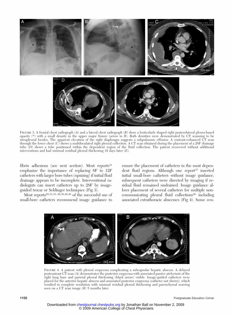
emphasize the importance of replacing 8F to 12Fcatheters with larger bore tubes (upsizing) if initial fluiddrainage appears to be incomplete. Interventional ra-diologists can insert catheters up to 28F by image-guided trocar or Seldinger techniques (Fig 3).
Most reports25,31,33–36,38,40,45 of the successful use ofsmall-bore catheters recommend image guidance to
ensure the placement of catheters in the most depen-dent fluid regions. Although one report37 insertedinitial small-bore catheters without image guidance,subsequent catheters were directed by imaging if re-sidual fluid remained undrained. Image guidance al-lows placement of several catheters for multiple non-communicating pleural fluid collections36 includingassociated extrathoracic abscesses (Fig 4). Some cen-
Figure 3. A frontal chest radiograph (A) and a lateral chest radiograph (B) show a lenticularly shaped right posterolateral pleura-basedopacity (*) with a small density in the upper major fissure (arrow in B). Both densities were demonstrated by CT scanning to beintrapleural locules. The apparent elevation of the right diaphragm suggests a subpulmonic effusion. A contrast-enhanced CT scanthrough the lower chest (C) shows a multiloculated right pleural collection. A CT scan obtained during the placement of a 28F drainagetube (D) shows a tube positioned within the dependent region of the fluid collection. The patient recovered without additionalinterventions and had minimal residual pleural thickening 19 days later (E).
Figure 4. A patient with pleural empyema complicating a subcapsular hepatic abscess. A delayedpostcontrast CT scan (A) demonstrates the posterior empyema with associated passive atelectasis of theright lung base and parietal pleural thickening (black arrow) visible. Image-guided catheters wereplaced for the anterior hepatic abscess and associated posterior empyema (catheter not shown), whichresulted in complete resolution with minimal residual pleural thickening and parenchymal scarringseen on a CT scan image (B) 5 months later.
1152 Postgraduate Education Corner
© 2009 American College of Chest Physicians by Jonathan Ball on November 2, 2009chestjournal.chestpubs.orgDownloaded from
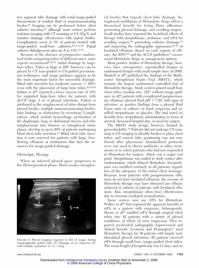
ters augment tube drainage with serial image-guidedthoracentesis of residual fluid or noncommunicatinglocules.45 Imaging can be performed before chestcatheter insertion,45 although most centers performreal-time imaging with CT scanning or US (Fig 5) andmonitor drainage effectiveness with repeat studies.Complications occur in 3% of patients treated withimage-guided small-bore catheters.25,31,43,46 Pigtailcatheter dislodgement rates are 8 to 13%.31,43
Because of the absence of prospective random-ized trials comparing tubes of different sizes, someexperts recommend5,29,47 initial drainage by large-bore tubes. Tubes as large as 28F, however, can beplaced by CT scan-guided or US-guided percutane-ous techniques, and image guidance appears to bethe most important factor for successful drainage.Blind tube insertion has moderate success (� 50%)even with the placement of large-bore tubes.22,39,48
Solaini et al49 reported a lower success rate of 12%for unguided large-bore tubes for patients withACCP stage 3 or 4 pleural infections. Failure isattributed to the misplacement of tubes distant frompleural locules, multiple noncommunicating locules,tube kinking, or obstruction by secretions. Compli-cations, which include hemorrhage; perforation ofthe diaphragm, lung, or abdominal viscera; and tubemisplacement into fissures or extrapleural tissueplanes, develop in up to 20% of patients undergoingblind chest tube insertion.43 Blind chest tube inser-tion is now reserved for patients with large, free-flowing effusions at institutions that lack the re-sources for image-guided drainage.
Fibrinolytic Therapy
When an infected pleural space progresses tothe fibrinopurulent phase, fibrin creates intrapleu-
ral locules that impede chest tube drainage. In-trapleural instillation of fibrinolytic drugs offers atheoretical benefit for lysing fibrin adhesions,promoting pleural drainage, and avoiding surgery.Small studies have reported the beneficial effects oftherapy with streptokinase, urokinase, and rtPA foravoiding surgery,50 promoting catheter drainage,51
and improving the radiographic appearance45,52 ofloculated effusions. Based on early reports of effi-cacy, the BTS5,53 and the ACCP guidelines13 recom-mend fibrinolytic drugs as management options.
Most positive studies of fibrinolytic therapy, how-ever, have retrospective, uncontrolled designs orrandomized designs with small sample sizes. In 2005,Maskell et al20 published the findings of the Multi-center Intrapleural Sepsis Trial (MIST1), whichremains the largest randomized controlled trial offibrinolytic therapy. Study centers placed small-borechest tubes (median size, 12F) without image guid-ance in 427 patients with complicated parapneumo-nic effusions (pleural fluid pH � 7.20, with signs ofinfection, or positive findings from a pleural fluidGram stain or culture) or frank empyema and in-stilled streptokinase or placebo. The trial noted nobenefits from streptokinase administration in terms ofsurvival, decreased hospital stay, or need for surgery.
The MIST1 study design, however, limited itsgeneralizability.54 Patients did not undergo CT scan-ning or US imaging to identify locules or place chesttubes, and correct tube positioning was not con-firmed after placement. Standardized protocolswere not used to direct antibiotic or other treat-ments or to select patients who had not respondedto fibrinolysis for surgery, which was a major endpoint. Streptokinase was mailed to study centers afterrandomization, which delayed fibrinolysis. Streptoki-nase was instilled routinely in all patients regard-less of the adequacy of the initial chest drainage.Because most patients with parapneumonic effu-sions do not have loculated effusions, the overuse offibrinolytic therapy may have obscured any efficacyachieved in subsets of patients with loculated effu-sions. Also, streptokinase often loses effectivenessdue to immune-mediated neutralization.
Some centers now use rtPA for fibrinolysis.Walker et al55 first reported the apparent benefits ofrtPA in a patient with empyema. Subsequently,Skeete et al56 instilled rtPA through surgical chesttubes into 42 patients with a variety of pleuralconditions, of which 12 were empyemas. They re-ported accelerated radiographic improvement andclinical benefit. Levinson and Pennington37 usedfibrinolytic therapy for 30 patients with largely mul-tiloculated pleural infections; 20 patients receivedrtPA through small-bore, image-guided chest tubes.The mean length of hospital stay was 11 days, and no
Figure 5. Pleural imaging (superior to left of image) duringsonographically guided tube (T) drainage of an empyema (E)with multiple septations (S). L � lung.
www.chestjournal.org CHEST / 136 / 4 / OCTOBER, 2009 1153
© 2009 American College of Chest Physicians by Jonathan Ball on November 2, 2009chestjournal.chestpubs.orgDownloaded from

patient required surgical drainage. Gervais et al45
reported their experience with rtPA instilled throughimage-guided 8.5F to 16F catheters in 66 patients, ofwhom 53 had empyemas or complicated parapneu-monic effusions. In the study by Gervais et al,45
patients were selected for fibrinolysis if the initialpleural fluid drainage was incomplete. The overallsuccess rate was 86%, although the outcomes werenot specifically reported for the 53 patients withpleural infections. Based on CT imaging studiesobtained before chest tube insertion that demon-strated multiple locules, the authors opined thatrtPA successfully drained effusions that would oth-erwise have required surgery.
Since the publication of the MIST1 findings, twometaanalyses57,58 appraised the evidence for fibrino-lytic therapy and drew similar conclusions. The currentevidence does not support routine fibrinolytic therapyfor unselected patients with parapneumonic effusions.Because of the significant heterogeneity of the treat-ment effects among the trials, however, subgroups ofpatients with loculated or septated infected pleuraleffusions may benefit. As a prudent approach, pendingfuture clinical trials would reserve therapy with fibrino-lytic drugs for patients whose pleural effusions fail todrain completely after initial catheter insertion. Chesttubes should be sized appropriately for the fluid viscos-ity, with timely catheter upsizing performed as needed(Fig 6). Definitive surgical drainage should not bedelayed for appropriate operative candidates if fibrino-lysis fails to drain the effusion rapidly and completely.59
The viscosity of pus is largely attributable to itsdeoxyribose nucleoprotein content. Fibrinolyticdrugs have negligible effects on decreasing the vis-cosity of empyema pus in contrast to agents thatdepolymerize DNA, such as human recombinantdeoxyribonuclease.60 Recombinant deoxyribonucle-ase has been reported61 to improve drainage in asingle patient who did not respond to fibrinolytictherapy.
The complications of fibrinolysis include chestpain, fever, hemothorax, hematuria, and allergicreactions to streptokinase (Fig 7).20,56 With the useof rtPA, systemic hemorrhage has not been reportedexcept in patients receiving concomitant full-doseanticoagulation.45 A Cochrane review58 reported thatintrapleural fibrinolytic therapy has not been shownto increase the number of adverse events resultingfrom chest tube drainage, but the confidence inter-val around this observation is too wide to firmlyexclude this possibility.
Thoracoscopy
Thoracoscopy provides minimally invasive accessto the pleural space for patients with free-flowing ormultiloculated effusions to suction viscous pleuralfluid, lyse adhesions to promote drainage of locules,and place chest tubes in dependent regions ofpleural fluid under direct visualization. Visual in-spection of the pleural space after debridementdetermines whether patients should be converted totherapy with decortication by thoracotomy; the in-ability of the reinflated lung to expand to the chestwall and diaphragm indicates an unsuccessful thora-coscopy and a need for thoracotomy.62 The advan-tages of thoracoscopic pleural drainage comparedwith thoracotomy include less postoperative pain,lower costs, shorter hospital stays, and better cos-metic results. Available thoracoscopic proceduresinclude medical thoracoscopy and video-assistedthoracoscopic surgery (VATS).
Medical thoracoscopy can drain some establishedempyemas.63,64 Pulmonary physicians or surgeonscan perform the procedure in endoscopy suites usinglocal anesthetics and moderate sedation. US identi-fies an entry site for the thoracoscope where theeffusion is the largest and most distant from thediaphragm.28 The advantages of medical thoracos-copy compared with VATS include lower cost and
Figure 6. Chest CT scan image of a multiloculated empyema (A) that required percutaneousplacement of a large-bore catheter. After subsequent instillation of rtPA, a contrast-enhanced scan (B)at the level of the aortic arch shows the tube in the pleural space posteriorly with minimal residualpleural fluid or thickening (white curved arrows) and regions of edema (black curved arrows) of theextrapleural fat (black straight arrows), a finding often seen on CT scans of patients with empyema.
1154 Postgraduate Education Corner
© 2009 American College of Chest Physicians by Jonathan Ball on November 2, 2009chestjournal.chestpubs.orgDownloaded from

better tolerance by frail patients who may not toler-ate lung deflation, which is required for VATS.65,66
For carefully selected patients with fibrinopurulentpleural infections and locules within reach of thethoracoscope, medical thoracoscopy has a reportedsuccess rate of 93% with a small proportion ofpatients needing conversion to VATS or open surgi-cal drainage.28
A thoracic surgeon performs VATS with patientsunder general anesthesia using a three-entry portand a double-lumen endotracheal tube, althoughlocal or regional anesthesia and two-port approacheshave been reported.67 Decortication and pleurec-tomy can be performed. VATS provides wide accessto the pleural space in many patients but may beinadequate to reach all fluid collections for advancedempyemas and dense adhesions or widely distributedlocules.62 The overall success rate, as defined bycomplete recovery without requiring thoracotomy, is60 to 100% for fibrinopurulent effusions.68–71 Manycenters reserve VATS for the treatment of patientswith fibrinopurulent effusions,72 although some sur-geons initially treat empyemas in the organizingphase with VATS, with conversion to thoracotomy ifnecessary.62,73–75 Roberts62 has supported this ap-proach by emphasizing that preoperative evaluationscannot establish with certainty the phase of a pleuralspace infection, which requires assessment underdirect VATS visualization. The series of patients withfibrinopurulent or organizing empyemas treatedinitially by VATS had a success rate of 38% and ahospital mortality rate of 6.6%. All of the deathsoccurred in those patients who did not respond totreatment with VATS and who then requiredthoracotomy.
No large randomized studies directly compare theutility of chest tube drainage with or without fibrino-lytic therapy vs thoracoscopy.68 A small, randomizedtrial76 compared chest tube drainage plus fibrinolytictherapy with VATS and found shorter hospitaliza-tions with VATS. This study was limited by its smallsize and methodological problems.77 Another ran-domized trial78 of 70 patients compared treatmentwith VATS with chest tube drainage without fibrino-lytic therapy and observed shorter hospitalizationsand less need for open decortication after primarytreatment with VATS. Chest tubes were placedwithout image guidance, however, and the clinicianswho made the treatment decisions were not blinded.Epidemiologic data from the state of Washington9
noted a lower overall mortality for patients withempyema from 1987 to 2004, during which timeprogressively more patients underwent surgicaldrainage (either thoracotomy or thoracoscopy), asopposed to chest tube drainage. Patients treated withchest tube drainage, however, most likely underwentstandard incisional chest tube insertion without im-age guidance. Modern protocols calling for the earlyuse of small-bore, image-guided catheters may com-pare more favorably with VATS in future trials.
In the absence of data from adequate trials, thedecision to proceed directly to thoracoscopy vs aninitial trial of chest tube drainage remains ill de-fined.49 Some experts79,80 have proposed initial tho-racoscopy for all patients with fibrinopurulent ororganized empyemas, while others81 have recom-mended a trial of image-guided catheter drainagewith or without fibrinolytic therapy. Regardless ofthe approach, definitive surgical drainage should notbe delayed inappropriately if initial drainage by chest
Figure 7. Hemothorax complicating intrapleural instillation of rtPA for a loculated empyema. An unenhanced CT scan (A) shows rightanterior, posterolateral, and paraspinal and small left pleural fluid collections with a pigtail catheter entering the right chest wall (arrow)with its tip terminating in the posterolateral fluid collection (not shown in A). Intrapleural rtPA was instilled into the anterior fluidcollection through a second pigtail catheter. Three days later (B), the anterior collection drained but posterolateral collection persisted.After the instillation of additional rtPA, pleural drainage became bloody, and a repeat unenhanced CT scan (C) demonstrated a large,anterior fluid collection with high-attenuation material dependently (black arrow) reflecting a loculated hemothorax that displaced theanterior catheter (white arrows). The posterior fluid collection in C increased slightly compared with B, suggesting posterior accumulationof blood from the anterior hemorrhage. This series of images demonstrates the difficulty in establishing by CT scan whether differentpleural fluid collections intercommunicate.
www.chestjournal.org CHEST / 136 / 4 / OCTOBER, 2009 1155
© 2009 American College of Chest Physicians by Jonathan Ball on November 2, 2009chestjournal.chestpubs.orgDownloaded from
tubes proves unsuccessful.81 Experts variably definethe acceptable durations of catheter trials beforethoracoscopy as 1 to 7 days.5,29,36,53,59,79 The disad-vantages of delaying thoracoscopy for a catheter trialare ill defined. One study observed that primarytherapy with VATS for the drainage of complicatedparapneumonic effusions had a higher success ratecompared with secondary VATS after a failed trial ofcatheter drainage with fibrinolysis.82 The viewpointsof the patients regarding the risks and benefits ofattempting chest tube drainage in an effort to avoidsurgery should enter into decision making. In ourexperience, the failure to aspirate pleural fluid through
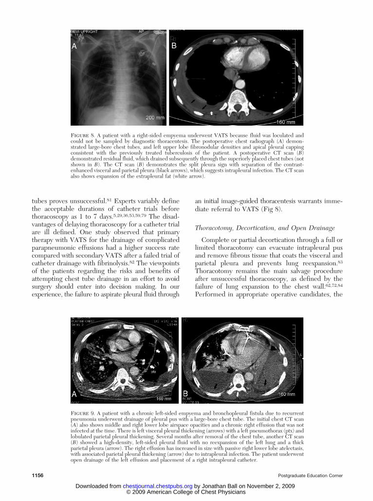
an initial image-guided thoracentesis warrants imme-diate referral to VATS (Fig 8).
Thoracotomy, Decortication, and Open Drainage
Complete or partial decortication through a full orlimited thoracotomy can evacuate intrapleural pusand remove fibrous tissue that coats the visceral andparietal pleura and prevents lung reexpansion.83
Thoracotomy remains the main salvage procedureafter unsuccessful thoracoscopy, as defined by thefailure of lung expansion to the chest wall.62,72,84
Performed in appropriate operative candidates, the
Figure 8. A patient with a right-sided empyema underwent VATS because fluid was loculated andcould not be sampled by diagnostic thoracentesis. The postoperative chest radiograph (A) demon-strated large-bore chest tubes, and left upper lobe fibronodular densities and apical pleural cappingconsistent with the previously treated tuberculosis of the patient. A postoperative CT scan (B)demonstrated residual fluid, which drained subsequently through the superiorly placed chest tubes (notshown in B). The CT scan (B) demonstrates the split pleura sign with separation of the contrast-enhanced visceral and parietal pleura (black arrows), which suggests intrapleural infection. The CT scanalso shows expansion of the extrapleural fat (white arrow).
Figure 9. A patient with a chronic left-sided empyema and bronchopleural fistula due to recurrentpneumonia underwent drainage of pleural pus with a large-bore chest tube. The initial chest CT scan(A) also shows middle and right lower lobe airspace opacities and a chronic right effusion that was notinfected at the time. There is left visceral pleural thickening (arrows) with a left pneumothorax (ptx) andlobulated parietal pleural thickening. Several months after removal of the chest tube, another CT scan(B) showed a high-density, left-sided pleural fluid with no reexpansion of the left lung and a thickparietal pleura (arrow). The right effusion has increased in size with passive right lower lobe atelectasis,with associated parietal pleural thickening (arrow) due to intrapleural infection. The patient underwentopen drainage of the left effusion and placement of a right intrapleural catheter.
1156 Postgraduate Education Corner
© 2009 American College of Chest Physicians by Jonathan Ball on November 2, 2009chestjournal.chestpubs.orgDownloaded from

mortality rate is 3% to 10%85,86 with a median postsur-gery hospital stay of 7 days.85 Patients with organizedempyemas who cannot tolerate thoracotomy or havetrapped lungs can undergo rib resection with opendrainage. Pus drains through a chest wound placed for� 6 months.5,87–89 Chronic empyemas with broncho-pleural fistulas also may require long-term open drain-age to prevent persistent pleural suppuration when pa-tients are treated with chest tube drainage alone (Fig 9).Recently, the use of an image-guided, small-bore cath-eter has been described90 for the long-term drainage ofchronic empyemas that are not amenable to surgery.
Decortication by thoracotomy is also indicated forseriously ill and toxic patients with associated medi-astinitis or bronchopleural fistulas who require me-diastinal drainage or fistula closure.72 Also, someexperts72 recommend proceeding directly to thora-cotomy (or VATS in selected instances) without priorchest tube drainage for toxic patients with virulentmulti-drug-resistant pathogens and multiorgan dys-function, who have a high mortality rate and maybenefit from immediate drainage.
Conclusion
Modern principles of managing pleural space infec-tions emphasize the importance of the early detectionof effusions in patients with pneumonia, and theprompt drainage of complicated parapneumonic effu-sions and empyemas. Delays in effective drainageincrease morbidity and mortality. As outlined in thisreview, multiple interventions exist to detect and drainpleural fluid with limited data from rigorous clinicaltrials to establish the superiority of any single approach.Clinicians should establish standardized protocols intheir institutions for early identification and managementbased on available expertise and resources. Regardless ofthe approach adopted, measured outcomes should matchthe best practices reported in the literature.
Acknowledgments
Financial/nonfinancial disclosures: The authors have re-ported to the ACCP that no significant conflicts of interest existwith any companies/organizations whose products or servicesmay be discussed in this article. This review did not requireinstitutional review board approval.
References1 Ferguson AD, Prescott RJ, Selkon JB, et al. The clinical
course and management of thoracic empyema. QJM 1996;89:285–289
2 Davies CWH, Kearney SE, Gleeson FV, et al. Predictors ofoutcome and long-term survival in patients with pleural
infection. Am J Respir Crit Care Med 1999; 160:1682–1687
3 Ashbaugh DG. Empyema thoracis: factors influencing mor-bidity and mortality. Chest 1991; 99:1162–1165
4 Bouros D, Schiza S, Patsourakis G, et al. Intrapleural strep-tokinase versus urokinase in the treatment of complicatedparapneumonic effusions: a prospective, double-blind study.Am J Respir Crit Care Med 1997; 155:291–295
5 Davies CW, Gleeson FV, Davies RJ. BTS guidelines for themanagement of pleural infection. Thorax 2003; 58(suppl):ii18–ii28
6 Mandal AK, Thadepalli H, Mandal AK, et al. Outcome ofprimary empyema thoracis: therapeutic and microbiologicaspects. Ann Thorac Surg 1998; 66:1782–1786
7 Smith JA, Mullerworth MH, Westlake GW, et al. Empyemathoracis: 14-year experience in a teaching center. Ann ThoracSurg 1991; 51:39–42
8 Tsai TH, Jerng JS, Chen KY, et al. Community-acquiredthoracic empyema in older people. J Am Geriatr Soc 2005;53:1203–1209
9 Farjah F, Symons RG, Krishnadasan B, et al. Management ofpleural space infections: a population-based analysis. J ThoracCardiovasc Surg 2007; 133:346–351
10 Finley C, Clifton J, Fitzgerald JM, et al. Empyema: anincreasing concern in Canada. Can Respir J 2008; 15:85–89
11 Lin YC, Tu CY, Chen W, et al. An urgent problem of aerobicgram-negative pathogen infection in complicated parapneu-monic effusions or empyemas. Intern Med 2007; 46:1173–1178
12 Foster S, Maskell N. Bacteriology of complicated parapneu-monic effusions. Curr Opin Pulm Med 2007; 13:319–323
13 Colice GL, Curtis A, Deslauriers J, et al. Medical and surgicaltreatment of parapneumonic effusions: an evidence-basedguideline. Chest 2000; 118:1158–1171
14 Heffner JE, McDonald J, Barbieri C, et al. Management ofparapneumonic effusions: an analysis of physician practicepatterns. Arch Surg 1995; 130:433–438
15 Chu MW, Dewar LR, Burgess JJ, et al. Empyema thoracis:lack of awareness results in a prolonged clinical course. CanJ Surg 2001; 44:284–288
16 Andrews NC, Parker EF, Shaw RR, et al. Management ofnontruberculous empyema. Am Rev Respir Dis 1962; 85:935–936
17 Light RW. Parapneumonic effusions and empyema. Proc AmThorac Soc 2006; 3:75–80
18 Manuel Porcel J, Vives M, Esquerda A, et al. Usefulness ofthe British Thoracic Society and the American College ofChest Physicians guidelines in predicting pleural drainageof non-purulent parapneumonic effusions. Respir Med 2006;100:933–937
19 Ahmed RA, Marrie TJ, Huang JQ. Thoracic empyema inpatients with community-acquired pneumonia. Am J Med2006; 119:877–883
20 Maskell NA, Davies CW, Nunn AJ, et al. UK Controlled trialof intrapleural streptokinase for pleural infection. N EnglJ Med 2005; 352:865–874
21 Simmers TA, Jie C, Sie B. Minimally invasive treatment ofthoracic empyema. Thorac Cardiovasc Surg 1999; 47:77–81
22 Storm HK, Krasnik M, Bang K, et al. Treatment of pleuralempyema secondary to pneumonia: thoracocentesis regimenversus tube drainage. Thorax 1992; 47:821–824
23 Sasse S, Nguyen T, Teixeira LR, et al. The utility of dailytherapeutic thoracentesis for the treatment of early empy-ema. Chest 1999; 116:1703–1708
24 Stavas J, vanSonnenberg E, Casola G, et al. Percutaneousdrainage of infected and noninfected thoracic fluid collec-tions. J Thorac Imaging 1987; 2:80–87
www.chestjournal.org CHEST / 136 / 4 / OCTOBER, 2009 1157
© 2009 American College of Chest Physicians by Jonathan Ball on November 2, 2009chestjournal.chestpubs.orgDownloaded from

25 Moulton JS, Benkert RE, Weisiger KH, et al. Treatment ofcomplicated pleural fluid collections with image-guideddrainage and intracavitary urokinase. Chest 1995; 108:1252–1259
26 Klein JS, Schultz S, Heffner JE. Interventional radiology ofthe chest: Image-guided percutaneous drainage of pleuraleffusions, lung abscess, and pneumothorax. Am J Roentgenol1995; 164:581–588
27 Rosenberg ER. Ultrasound in the assessment of pleuraldensities. Chest 1983; 84:283–285
28 Tassi GF, Davies RJ, Noppen M. Advanced techniques inmedical thoracoscopy. Eur Respir J 2006; 28:1051–1059
29 Yim AP. Paradigm shift in empyema management. Chest1999; 115:611–612
30 Park JK, Kraus FC, Haaga JR. Fluid flow during percutane-ous drainage procedures: an in vitro study of the effects offluid viscosity, catheter size, and adjunctive urokinase. Am JRoentgenol 1993; 160:165–169
31 Keeling AN, Leong S, Logan PM, et al. Empyema andeffusion: outcome of image-guided small-bore catheter drain-age. Cardiovasc Intervent Radiol 2008; 31:135–141
32 Liang SJ, Chen W, Lin YC, et al. Community-acquiredthoracic empyema in young adults. South Med J 2007;100:1075–1080
33 Ulmer JL, Choplin RH, Reed JC. Image-guided catheterdrainage of the infected pleural space. J Thorac Imaging1991; 6:65–73
34 Westcott JL. Percutaneous catheter drainage of pleural effu-sion and empyema. Am J Roentgenol 1985; 144:1189–1193
35 Merriam MA, Cronan JJ, Dorfman GS, et al. Radiographicallyguided percutaneous catheter drainage of pleural fluid col-lections. Am J Roentgenol 1988; 151:1113–1116
36 Akhan O, Ozkan O, Akinci D, et al. Image-guided catheterdrainage of infected pleural effusions. Diagn Interv Radiol2007; 13:204–209
37 Levinson GM, Pennington DW. Intrapleural fibrinolyticscombined with image-guided chest tube drainage for pleuralinfection. Mayo Clin Proc 2007; 82:407–413
38 Shankar S, Gulati M, Kang M, et al. Image-guided percuta-neous drainage of thoracic empyema: can sonography predictthe outcome? Eur Radiol 2000; 10:495–499
39 Moulton JS. Image-guided management of complicated pleu-ral fluid collections. Radiol Clin North Am 2000; 38:345–374
40 Moulton JS, Moore PT, Mencini RA. Treatment of loculatedpleural effusions with transcatheter intracavitary urokinase.Am J Roentgenol 1989; 153:941–945
41 vanSonnenberg E, Nakamoto SK, Mueller PR, et al. CT- andultrasound-guided catheter drainage of empyemas afterchest-tube failure. Radiology 1984; 151:349–353
42 Silverman SG, Mueller PR, Saini S, et al. Thoracic empyema:management with image-guided catheter drainage. Radiology1988; 169:5–9
43 Horsley A, Jones L, White J, et al. Efficacy and complicationsof small-bore, wire-guided chest drains. Chest 2006; 130:1857–1863
44 Cheng G, Vintch JR. A retrospective analysis of the manage-ment of parapneumonic empyemas in a county teachingfacility from 1992 to 2004. Chest 2005; 128:3284–3290
45 Gervais DA, Levis DA, Hahn PF, et al. Adjunctive intrapleu-ral tissue plasminogen activator administered via chest tubesplaced with imaging guidance: effectiveness and risk forhemorrhage. Radiology 2008; 246:956–963
46 Parulekar W, Di Primio G, Matzinger F, et al. Use ofsmall-bore vs large-bore chest tubes for treatment of malig-nant pleural effusions. Chest 2001; 120:19–25
47 Hyde J, Sykes T, Graham T. Reducing morbidity from chestdrains. BMJ 1997; 314:914–915
48 Ali I, Unruh H. Management of empyema thoracis. AnnThorac Surg 1990; 50:355–359
49 Solaini L, Prusciano F, Bagioni P. Video-assisted thoracicsurgery in the treatment of pleural empyema. Surg Endosc2007; 21:280–284
50 Diacon AH, Theron J, Schuurmans MM, et al. Intrapleuralstreptokinase for empyema and complicated parapneumoniceffusions. Am J Respir Crit Care Med 2004; 170:49–53
51 Bouros D, Schiza S, Tzanakis N, et al. Intrapleural urokinaseversus normal saline in the treatment of complicated parap-neumonic effusions and empyema: a randomized, double-blind study. Am J Respir Crit Care Med 1999; 159:37–42
52 Davies RJO, Traill ZC, Gleeson FV. Randomised controlledtrial of intrapleural streptokinase in community acquiredpleural infection. Thorax 1997; 52:416–421
53 Balfour-Lynn IM, Abrahamson E, Cohen G, et al. BTSguidelines for the management of pleural infection in chil-dren. Thorax 2005; 60(suppl):i1–i21
54 Heffner JE. Multicenter trials of treatment for empyema:after all these years. N Engl J Med 2005; 352:926–928
55 Walker CA, Shirk MB, Tschampel MM, et al. Intrapleuralalteplase in a patient with complicated pleural effusion. AnnPharmacother 2003; 37:376–379
56 Skeete DA, Rutherford EJ, Schlidt SA, et al. Intrapleuraltissue plasminogen activator for complicated pleural effu-sions. J Trauma 2004; 57:1178–1183
57 Tokuda Y, Matsushima D, Stein GH, et al. Intrapleuralfibrinolytic agents for empyema and complicated parapneu-monic effusions: a meta-analysis. Chest 2006; 129:783–790
58 Cameron R, Davies HR. Intra-pleural fibrinolytic therapyversus conservative management in the treatment of adultparapneumonic effusions and empyema. Cochrane DatabaseSyst Rev (database online). Issue 1, 2008
59 Bouros D, Tzouvelekis A, Antoniou KM, et al. Intrapleuralfibrinolytic therapy for pleural infection. Pulm PharmacolTher 2007; 20:616–626
60 Simpson G, Roomes D, Heron M. Effects of streptokinaseand deoxyribonuclease on viscosity of human surgical andempyema pus. Chest 2000; 117:1728–1733
61 Simpson G, Roomes D, Reeves B. Successful treatment ofempyema thoracis with human recombinant deoxyribonucle-ase. Thorax 2003; 58:365–366
62 Roberts JR. Minimally invasive surgery in the treatment ofempyema: intraoperative decision making. Ann Thorac Surg2003; 76:225–230
63 Loddenkemper R, Boutin C. Thoracoscopy: present diagnos-tic and therapeutic indications. Eur Respir J 1993; 6:1544–1555
64 Mathur PN, Loddenkemper R. Medical thoracoscopy: role inpleural and lung diseases. Clin Chest Med 1995; 16:487–496
65 Colt HG. Thoracoscopy: a prospective study of safety andoutcome. Chest 1995; 108:324–329
66 Soler M, Wyser C, Bolliger CT, et al. Treatment of earlyparapneumonic empyema by “medical” thoracoscopy. SchweizMed Wochenschr 1997; 127:1748–1753
67 Drain AJ, Ferguson JI, Sayeed R, et al. Definitive manage-ment of advanced empyema by two-window video-assistedsurgery. Asian Cardiovasc Thorac Ann 2007; 15:238–239
68 Waller DA. Thoracoscopy in management of postpneumonicpleural infections. Curr Opin Pulm Med 2002; 8:323–326
69 Cassina PC, Hauser M, Hillejan L, et al. Video-assistedthoracoscopy in the treatment of pleural empyema: stage-based management and outcome. J Thorac Cardiovasc Surg1999; 117:234–238
70 Sasse SA. Parapneumonic effusions and empyema. Curr OpinPulm Med 1996; 2:320–326
71 Silen ML, Naunheim KS. Thoracoscopic approach to the
1158 Postgraduate Education Corner
© 2009 American College of Chest Physicians by Jonathan Ball on November 2, 2009chestjournal.chestpubs.orgDownloaded from

management of empyema thoracis: indications and results.Chest Surg Clin N Am 1996; 6:491–499
72 Potaris K, Mihos P, Gakidis I, et al. Video-thoracoscopic andopen surgical management of thoracic empyema. Surg Infect2007; 8:511–517
73 Lackner RP, Hughes R, Anderson LA, et al. Video-assistedevacuation of empyema is the preferred procedure for manage-ment of pleural space infections. Am J Surg 2000; 179:27–30
74 Suzuki T, Kitami A, Suzuki S, et al. Video-assisted thoraco-scopic sterilization for exacerbation of chronic empyemathoracis. Chest 2001; 119:277–280
75 Waller DA, Rengarajan A. Thoracoscopic decortication: a rolefor video-assisted surgery in chronic postpneumonic pleuralempyema. Ann Thorac Surg 2001; 71:1813–1816
76 Wait MA, Sharma S, Hohn J, et al. A randomized trial ofempyema therapy. Chest 1997; 111:1548–1551
77 Coote N, Kay E. Surgical versus non-surgical management ofpleural empyema. Cochrane Database Syst Rev (databseonline). Issue 3, 2005
78 Bilgin M, Akcali Y, Oguzkaya F. Benefits of early aggressivemanagement of empyema thoracis. ANZ J Surg 2006; 76:120–122
79 Brutsche MH, Tassi GF, Gyorik S, et al. Treatment ofsonographically stratified multiloculated thoracic empyemaby medical thoracoscopy. Chest 2005; 128:3303–3309
80 Sendt W, Forster E, Hau T. Early thoracoscopic debridementand drainage as definite treatment for pleural empyema. EurJ Surg 1995; 161:73–76
81 Lim TK, Chin NK. Empirical treatment with fibrinolysis and
early surgery reduces the duration of hospitalization in pleuralsepsis. Eur Respir J 1999; 13:514–518
82 Petrakis IE, Kogerakis NE, Drositis IE, et al. Video-assistedthoracoscopic surgery for thoracic empyema: primarily, or afterfibrinolytic therapy failure? Am J Surg 2004; 187:471–474
83 Thurer RJ. Decortication in thoracic empyema: indications andsurgical technique. Chest Surg Clin N Am 1996; 6:461–490
84 Angelillo Mackinlay TA, Lyons GA, Chimondeguy DJ, et al.VATS debridement versus thoracotomy in the treatment ofloculated postpneumonia empyema. Ann Thorac Surg 1996;61:1626–1630
85 Pothula V, Krellenstein DJ. Early aggressive surgical manage-ment of parapneumonic empyemas. Chest 1994; 105:832–836
86 Grotenhuis BA, Janssen PJ, Eerenberg JP. The surgicaltreatment of stage III empyema: the effect on lung function.Minerva Chir 2008; 63:23–27
87 Deslauriers J, Jacques LF, Gregoire J. Role of Eloesser flapand thoracoplasty in the third millennium. Chest Surg Clin NAm 2002; 12:605–623
88 Athanassiadi K, Gerazounis M, Kalantzi N. Treatment ofpost-pneumonic empyema thoracis. Thorac Cardiovasc Surg2003; 51:338–341
89 Thourani VH, Lancaster RT, Mansour KA, et al. Twenty-sixyears of experience with the modified eloesser flap. AnnThorac Surg 2003; 76:401–406
90 Davies HE, Rahman NM, Parker RJ, et al. Use of indwellingpleural catheters for chronic pleural infection. Chest 2008;133:546–549
www.chestjournal.org CHEST / 136 / 4 / OCTOBER, 2009 1159
© 2009 American College of Chest Physicians by Jonathan Ball on November 2, 2009chestjournal.chestpubs.orgDownloaded from

DOI 10.1378/chest.08-2956 2009;136; 1148-1159Chest
John E. Heffner, Jeffrey S. Klein and Christopher HampsonInterventional Management of Pleural Infections
November 2, 2009This information is current as of
& ServicesUpdated Information
l.htmlhttp://chestjournal.chestpubs.org/content/136/4/1148.fulhigh-resolution figures, can be found at:Updated Information and services, including
References
8.full.html#ref-list-1http://chestjournal.chestpubs.org/content/136/4/114accessed free at:This article cites 88 articles, 55 of which can be
Open AccessoptionFreely available online through CHEST open access
Permissions & Licensing
http://www.chestjournal.org/site/misc/reprints.xhtml(figures, tables) or in its entirety can be found online at: Information about reproducing this article in parts
Reprints http://www.chestjournal.org/site/misc/reprints.xhtml
Information about ordering reprints can be found online:
Email alerting service
online article.article. Sign up in the box at the top right corner of the Receive free email alerts when new articles cite this
formatImages in PowerPoint
format. See any online article figure for directions downloaded for teaching purposes in PowerPoint slide Figures that appear in CHEST articles can be
© 2009 American College of Chest Physicians by Jonathan Ball on November 2, 2009chestjournal.chestpubs.orgDownloaded from