intra-genomic internal transcribed spacer region sequence
TRANSCRIPT

Title Intra-genomic internal transcribed spacer region sequenceheterogeneity and molecular diagnosis in clinical microbiology
Author(s) Zhao, Y; Tsang, CC; Xiao, M; Cheng, J; Xu, Y; Lau, SKP; Woo,PCY
Citation International Journal of Molecular Sciences, 2015, v. 16 n. 10, p.25067-25079
Issued Date 2015
URL http://hdl.handle.net/10722/223223
Rights This work is licensed under a Creative Commons Attribution-NonCommercial-NoDerivatives 4.0 International License.
Int. J. Mol. Sci. 2015, 16, 25067-25079; doi:10.3390/ijms161025067
International Journal of
Molecular Sciences ISSN 1422-0067
www.mdpi.com/journal/ijms
Article
Intra-Genomic Internal Transcribed Spacer Region Sequence Heterogeneity and Molecular Diagnosis in Clinical Microbiology
Ying Zhao 1,2, Chi-Ching Tsang 2, Meng Xiao 1, Jingwei Cheng 1,3, Yingchun Xu 1,
Susanna K. P. Lau 2,4,5,6,* and Patrick C. Y. Woo 2,4,5,6,*
1 Department of Clinical Laboratory, Peking Union Medical College Hospital,
Chinese Academy of Medical Sciences, Beijing 100730, China;
E-Mails: [email protected] (Y.Z.); [email protected] (M.X.);
[email protected] (J.C.); [email protected] (Y.X.) 2 Department of Microbiology, The University of Hong Kong, Hong Kong;
E-Mail: [email protected] 3 Graduate School, Peking Union Medical College, Chinese Academy of Medical Sciences,
Beijing, China 4 State Key Laboratory of Emerging Infectious Diseases, The University of Hong Kong, Hong Kong 5 Research Centre of Infection and Immunology, The University of Hong Kong, Hong Kong 6 Carol Yu Centre for Infection, The University of Hong Kong, Hong Kong
* Authors to whom correspondence should be addressed;
E-Mails: [email protected] (P.C.Y.W.); [email protected] (S.K.P.L.);
Tel.: +852-2255-4892 (P.C.Y.W. & S.K.P.L.);
Fax: +852-2855-1241 (P.C.Y.W. & S.K.P.L.).
Academic Editor: Michael Ibba
Received: 11 August 2015 / Accepted: 14 October 2015 / Published: 22 October 2015
Abstract: Internal transcribed spacer region (ITS) sequencing is the most extensively used
technology for accurate molecular identification of fungal pathogens in clinical microbiology
laboratories. Intra-genomic ITS sequence heterogeneity, which makes fungal identification
based on direct sequencing of PCR products difficult, has rarely been reported in pathogenic
fungi. During the process of performing ITS sequencing on 71 yeast strains isolated
from various clinical specimens, direct sequencing of the PCR products showed
ambiguous sequences in six of them. After cloning the PCR products into plasmids for
sequencing, interpretable sequencing electropherograms could be obtained. For each of
the six isolates, 10–49 clones were selected for sequencing and two to seven intra-genomic
OPEN ACCESS

Int. J. Mol. Sci. 2015, 16 25068
ITS copies were detected. The identities of these six isolates were confirmed to be
Candida glabrata (n = 2), Pichia (Candida) norvegensis (n = 2), Candida tropicalis (n = 1)
and Saccharomyces cerevisiae (n = 1). Multiple sequence alignment revealed that one to
four intra-genomic ITS polymorphic sites were present in the six isolates, and all these
polymorphic sites were located in the ITS1 and/or ITS2 regions. We report and describe
the first evidence of intra-genomic ITS sequence heterogeneity in four different pathogenic
yeasts, which occurred exclusively in the ITS1 and ITS2 spacer regions for the six isolates
in this study.
Keywords: internal transcribed spacer region; sequencing; heterogeneity; yeast;
molecular identification
1. Introduction
Molecular identification of fungal pathogens in clinical microbiology laboratories most commonly
involves polymerase chain reaction (PCR) amplification of genes and/or intergenic regions in the fungal
genome and direct nucleotide sequencing of the purified PCR products. The most extensively used
targets for such purpose are the internal transcribed spacer (ITS) region comprising the ITS1-5.8S-ITS2
nuclear ribosomal DNA (nrDNA) gene cluster and 25S nrDNA [1,2]. Since most fungi possess more
than one nrDNA operon in their genomes [3,4], whether molecular characterization can be used to achieve
fungal identification depends very much on the perfect intra-genomic sequence homogeneity in the
multiple copies of the nrDNA operons of fungi.
Intra-genomic ITS sequence heterogeneity has rarely been described in pathogenic fungi. When present,
such ITS sequence heterogeneity within a single genome makes fungal identification based on direct
sequencing of the PCR product difficult, because multiple PCR products will result and double or multiple
nucleotide peaks will be observed in the sequencing electropherograms. Recently, during the process
of performing ITS sequencing on 71 isolates of yeasts recovered from various clinical specimens, direct
sequencing of the PCR products showed ambiguous sequences in six of them (unpublished data).
For these six isolates, which belong to four different fungal species, double or multiple peaks were
frequently observed in the sequencing electropherograms. We hypothesize that intra-genomic ITS
sequence heterogeneity is present in these six clinical isolates. In order to test for this hypothesis,
the PCR products of the ITS of these six isolates were cloned and 10 to 49 clones from each isolate
were selected for sequencing. In this study, we describe this phenomenon of ITS sequence heterogeneity
within the genomes of four different pathogenic fungal species. The importance of intra-genomic ITS
sequence heterogeneity on molecular identification and typing of fungal species in clinical microbiology
laboratories is also discussed.

Int. J. Mol. Sci. 2015, 16 25069
2. Results
2.1. Clinical Characteristics
The clinical characteristics of the five patients from whom the six yeast isolates were recovered are
summarized in Table 1. Two patients were male and three were female, with a median age of 60
(range, 30 to 86). Two patients had type 2 diabetes mellitus, while one had cervical carcinoma and another
one had chronic renal failure. All patients suffered from invasive yeast infections, including peritonitis,
mediastinitis, as well as lung abscess and empyema, with the yeasts recovered from the corresponding
clinical specimens. For the 66-year-old male patient with esophageal perforation and mediastinitis, yeasts
were recovered from both the patient’s blood and pleural fluid, which were collected on separate days.
2.2. Direct PCR Product Sequencing
PCR of the ITS of the six patient isolates yielded DNA bands of about 900 bp (PUMY010
and PUMY011), 500 bp (PUMY020 and PUMY021), 550 bp (PUMY040) and 800 bp (PUMY065).
Direct sequencing of the purified PCR products revealed that double or multiple peaks were present
frequently in the sequencing electropherograms (Figure S1), and so the actual sequences of the ITS of
the isolates could not be determined successfully. DNA extraction and direct PCR product sequencing
were independently performed more than three times but the sequence ambiguity remained.
2.3. Sequencing of Cloned PCR Products and Phylogenetic Analyses
After cloning the PCR products into plasmids for sequencing, interpretable sequencing
electropherograms could be obtained (Figure S1). Out of the 17, 12, 45, 49, 10 and 15 clones selected
for sequencing for the isolates PUMY010, PUMY011, PUMY020, PUMY021, PUMY040 and PUMY065,
respectively, two to seven intra-genomic ITS copies were observed (Table 1 and Figure S1). Multiple
sequence alignment revealed that one to four intra-genomic ITS polymorphic sites were present in
the six isolates, and all these polymorphic sites were located in the ITS1 and/or ITS2 regions (Table 1,
Figure 1, Figures S1 and S2). For PUMY010, the four polymorphic sites present in the seven intra-genomic
ITS copies involved transition (A ↔ G and T ↔ C) and transversion (A ↔ C) substitutions as well as
an insertion/deletion, and these intra-genomic ITS copies possessed 99.05%–99.41% sequence identities
to the ITS of Candida glabrata CBS 138T; for PUMY011, the three polymorphic sites present in the
three intra-genomic ITS copies involved transversion (A ↔ T and A ↔ C) substitutions as well as an
insertion/deletion, and these intra-genomic ITS copies possessed 98.82%–99.29% sequence identities
to the ITS of C. glabrata CBS 138T; for PUMY020, the three polymorphic sites present in the four
intra-genomic ITS copies involved a transition (A ↔ G) substitution and insertions/deletions,
and these intra-genomic ITS copies possessed 98.68%–99.12% sequence identities to the ITS of
Pichia (Candida) norvegensis ST 3481-03; for PUMY021, the three polymorphic sites present in the
five intra-genomic ITS copies involved transition (A ↔ G) and transversion (A ↔ T) substitutions as
well as insertions/deletions, and these intra-genomic ITS copies possessed 98.68%–99.12% sequence
identities to the ITS of P. norvegensis ST 3481-03; for PUMY040, the two polymorphic sites present
in the three intra-genomic ITS copies involved only insertions/deletions and these intra-genomic ITS

Int. J. Mol. Sci. 2015, 16 25070
copies possessed 99.59%–100% sequence identities to the ITS of C. tropicalis CBS 94T; while for
PUMY065, the single polymorphic site present in the two intra-genomic ITS copies involved only an
insertion/deletion and these intra-genomic ITS copies possessed 99.73%–99.87% sequence identities
to the ITS of Saccharomyces cerevisiae NRRL Y-12632T. Phylogenetic analyses based on all the
intra-genomic ITS copies of each yeast isolate showed unambiguously that isolates PUMY010 and
PUMY011 were clustered with C. glabrata CBS 138T; isolates PUMY020 and PUMY021 were
clustered with P. norvegensis ST 3481-03; isolate PUMY040 was clustered with C. tropicalis
CBS 94T; and isolate PUMY065 was clustered with S. cerevisiae NRRL Y-12632T (Figure 2).
Figure 1. Schematic diagrams showing the multiple alignments of ITS sequences of the six
yeast isolates. The internal transcribed spacer regions ITS1 and ITS2 are highlighted in
gray. Intra-genomic polymorphisms are represented by the corresponding nucleotides or the
symbol “▲” (meaning deletion) at the respective sites. The diagrams are not drawn to scale.
Figure 2. Cont.
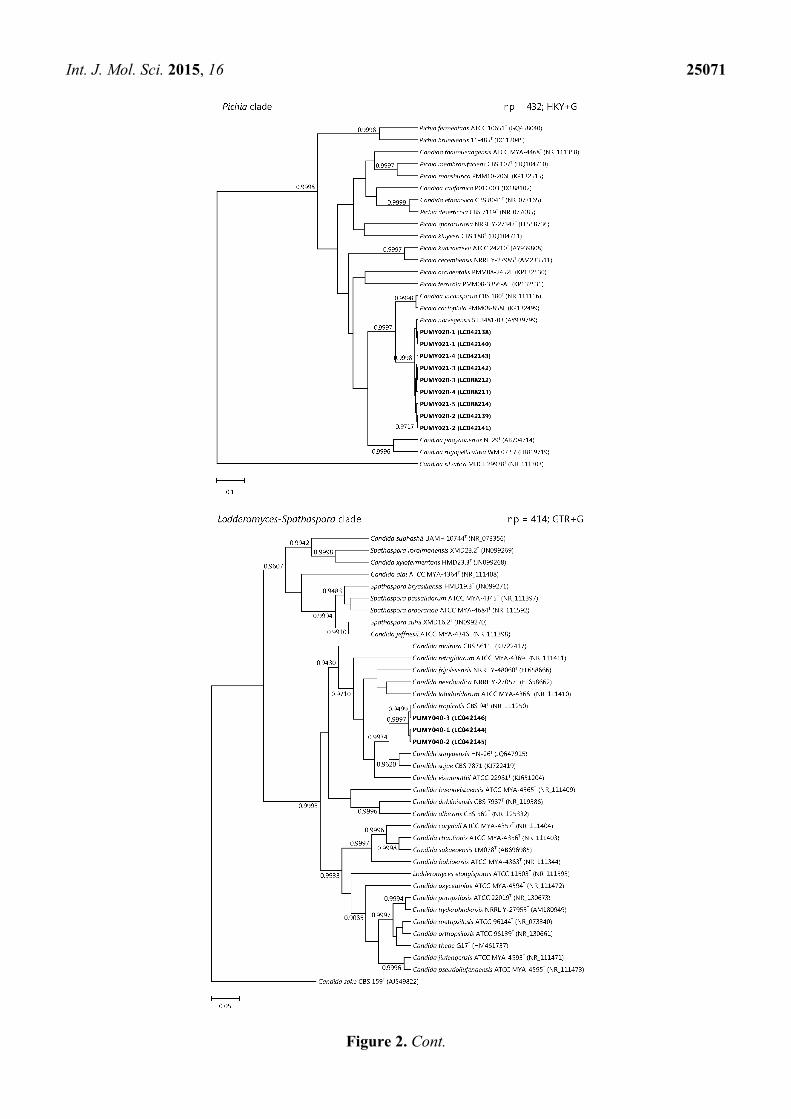
Int. J. Mol. Sci. 2015, 16 25071
Figure 2. Cont.

Int. J. Mol. Sci. 2015, 16 25072
Figure 2. Phylogenetic trees showing the relationship of the six yeast isolates to other
members of the Nakaseomyces, Pichia, Lodderomyces-Spathaspora, or Saccharomyces clades.
The trees were inferred from ITS sequence data by the Bayesian inference method and the
numbers of nucleotide positions (np) of the trimmed, aligned sequences included and the
substitution models employed (HKY = Hasegawa-Kishino-Yano model; GTR = general
time reversible model; G = γ-distributed rate variation; and I = estimated proportion of
invariable sites) for phylogenetic analyses are shown at the top right corner of each tree,
and the trees were rooted using Candida castellii NRRL-17070T, C. silvatica MUCL 29938T,
C. sake CBS 159T, and Kazachstania africana NRRL Y-8276T, respectively. The scale bars
indicate the estimated numbers of substitutions per base. Numbers at nodes indicate levels
of posterior probability support based on the Bayesian analyses of the data sets, and posterior
probability values lower than 0.9 are not shown. All accession numbers (in parentheses) are
given as cited in the DDBJ/ENA/GenBank databases. The clinical isolates reported in this
study are highlighted in bold type.
3. Discussion
We report and describe the first evidence of intra-genomic ITS sequence heterogeneity in a variety
of yeast species recovered from clinical specimens. So far, most cases of intra-genomic ITS sequence
heterogeneity were reported in environmental fungi of several fungal phyla, including Ascomycota [5–9],
Basidiomycota [6,10–13], Glomeromycota [14–20], Microsporidia [21,22] and the subphylum
Mucoromycotina [23]. Most of these fungi are molds, whereas only three yeasts species [6] and
one dimorphic fungus [8] have been found to possess intra-genomic ITS sequence heterogeneity. On
the other hand, this phenomenon has rarely been reported in fungal pathogens identified in clinical
microbiology laboratories. In our previous study, we documented the presence of intra-genomic ITS
sequence heterogeneity in four of the 28 Rhizopus microsporus isolates recovered during an outbreak
investigation of gastrointestinal mucormycosis [23]. In the present study, this phenomenon could also
be observed in six of the 71 yeast isolates subject to ITS sequencing. These six isolates, of four different
fungal species, were isolated from five patients who suffered from severe fungal infections due to the
corresponding yeasts (Table 1). As one of the major reasons for intra-genomic ITS sequence heterogeneity
is the result of DNA insertion/deletion, minute differences in the sizes of the PCR products would
be generated during amplification of the ITS region. This may give rise to thicker bands observed in

Int. J. Mol. Sci. 2015, 16 25073
agarose gel electrophoresis if the nucleotide fragments inserted/deleted is long enough, as we observed
in our previous study [11]. In the present study, this “thicker band” phenomenon was not obvious for all
the six isolates as the different intra-genomic ITS copies observed were only due to substitutions and/or
very short insertions/deletions involving only one to four nucleotides. More importantly, multiple peaks
were always observed in the sequencing electropherograms if the PCR products were sequenced directly
since two or more types of PCR products were simultaneously sequenced (Figure S1). In this study,
cloning and subsequent sequencing of the ITS PCR products of the six yeast isolates showed that for
each of the six isolates, more than one type of ITS sequences were observed (Figure 1, Figures S1 and S2).
This confirmed intra-genomic ITS sequence heterogeneity in all six yeast isolates. In addition, all
the four ITS copies of the isolate PUMY020 could also be found in the isolate PUMY021 (copies 1–4).
Although PUMY021 possessed an additional fifth ITS copy which could not be observed in
PUMY020, the electropherogram of PUMY020 obtained by direct PCR product sequencing showed that
the presence of such an ITS copy (with ten “T” residues followed by two “A” residues at the ITS2
polymorphic site) in the genome of PUMY020 is possible (Figure S1). Given the recovery rate of this
ITS copy in PUMY021 was just one in 49 clones, it is possible that the clones selected for sequencing
were not sufficient in number to reveal this ITS copy in PUMY020. As a result, it is reasonable to
believe that the isolates PUMY020 and PUMY021, which were isolated from the same patient from
different clinical specimens collected on different dates, are actually the same.
Concerted evolutionary mechanisms are employed to maintain sequence uniformity among the
many different copies of the nrDNA operon within a fungal genome. However, when error occurs,
intra-genomic nrDNA sequence variation would result and this gives rise to nrDNA sequence
heterogeneity within a single genome. As for the positions of intra-genomic ITS sequence variation, they
occurred only in ITS1 and/or ITS2, but not in the 5.8S nrDNA region, for all the six yeast isolates in
this study (Figures 1 and S2). In fungi which possess intra-genomic ITS sequence heterogeneity,
the sites of nucleotide difference are also much more often present in ITS1 and/or ITS2, and
intra-genomic 5.8S nrDNA variation is uncommonly observed [5,7–12,17–23]. This phenomenon may
likely be explained by the fact that the 5.8S nrRNA, after maturation and together with the mature 25S
and 5S rRNAs, forms the large ribosomal subunit, which is essential to the organism for protein
translation. Given the importance of the function of the 5.8S nrRNA and its short length of about 160
nucleotides, it is not surprising to observe minimal sequence variation in this gene. On the other hand,
although ITS1 and ITS2 are also transcribed together with the 18S, 5.8S and 25S nrDNA to give the
45S rRNA precursor, upon maturation these two spacers are spliced and removed and they are not
involved in the functioning of the ribosome. Therefore, the two spacers ITS1 and ITS2 are under fewer
evolutionary constraints than the 5.8S nrDNA and thus more sequence variations could be observed.

Int. J. Mol. Sci. 2015, 16 25074
Table 1. Characteristics of the six yeast strains in this study.
Patient Sex/Age a Underlying Diseases b Clinical Diagnosis Clinical
Specimens Strains c
Number of
Intra-Genomic
ITS Copies d
Number of
Intra-Genomic ITS
Polymorphic Sites d
Location of
Intra-Genomic ITS
Polymorphic Sites d
Final Identification
1 F/30
Carcinoma of cervix, post-total
abdominal hysterectomy and bilateral
salpingo-oophorectomy and radiotherapy
Suppurative peritonitis Peritoneal
fluid PUMY010 7 4 ITS1 and ITS2 Candida glabrata
2 F/86 N/A Intestinal obstruction Intraperitoneal
drainage fluid PUMY011 3 3 ITS1 Candida glabrata
3 M/66 Type 2 diabetes mellitus Esophageal perforation
and mediastinitis Pleural fluid PUMY020 4 3 ITS1 and ITS2
Pichia (Candida)
norvegensis
Blood PUMY021 5 3 ITS1 and ITS2 Pichia (Candida)
norvegensis
4 M/60 Chronic renal failure Peritonitis Peritoneal
fluid PUMY040 3 2 ITS2 Candida tropicalis
5 F/58 Type 2 diabetes mellitus Lung abscess and
empyema Empyema pus PUMY065 2 1 ITS1
Saccharomyces
cerevisiae
a F, female; M, male; b N/A, not available; c Strain PUMY021 was isolated two days after the isolation of strain PUMY20 from the same patient; d ITS, internal
transcribed spacer.

Int. J. Mol. Sci. 2015, 16 25075
Intra-genomic ITS sequence heterogeneity poses difficulties in both the identification and typing of
the corresponding fungal strains. Rapid and accurate identification of fungal pathogens is particularly
important to clinical microbiology laboratories because it is essential to the prescription of the appropriate
antifungal treatment to the patients. In the presence of intra-genomic ITS sequence heterogeneity, the
direct sequencing results of the PCR products may be uninterpretable and it takes much more time to
clone the PCR products for subsequent sequencing to obtain interpretable sequences corresponding to
the different intra-genomic ITS copies. The additional experimental steps may require skilled personnel
and the lengthy procedures may hinder the prescription of the correct treatment regimens in a timely
manner. As for strain- or geno-typing of the fungal pathogen, which are important to the study of infection
epidemiology, the different intra-genomic ITS copies may complicate the typing analysis. For example,
in the Nakaseomyces clade phylogenetic tree in this study, the seven ITS sequence types of the isolate
PUMY010 formed three distinct clusters and these three separated clusters may represent three
individual strain types (Figure 2). Such phenomenon could also be observed in other yeast isolates
characterized in this study (Figure 2). This makes it difficult to conclude to which strain types these
isolates actually belong and hence hinders epidemiological study. Therefore, when double or multiple
peaks of strong signal intensities are observed in the sequencing electropherograms of ITS PCR products,
reference laboratories proficient in molecular technology should be consulted for further identification
and typing of such fungal strains.
4. Materials and Methods
4.1. Patients and Strains
The six strains used in this study were isolated from clinical specimens of five patients hospitalized
in Peking Union Medical College Hospital (PUMCH), Beijing, China, during a period of 10 months
(May 2009 to February 2010) (Table 1). The medical records of the patients were retrospectively reviewed
and their demographic information and clinical statuses (including sex, age, underlying diseases, clinical
diagnosis, and type of specimens from which the yeast strains were isolated) were collected. This study
was approved by the Human Research Ethics Committee of PUMCH (Number S-263, dated 28
October 2009), where the study protocols were performed in accordance with the approved guidelines
and written informed consents were obtained.
4.2. DNA Extraction, PCR and Direct PCR Product Sequencing
Fungal genomic DNA was extracted from a single clone of each yeast isolate. Briefly, for each
yeast isolate, cells from a single colony were subcultured on Sabouraud dextrose agar (SDA) (Oxoid,
Hampshire, UK) supplemented with chloramphenicol (50 µg/mL) (Sigma-Aldrich, St. Louis, MO,
USA). After 24–48 h of incubation at 25 °C the fresh cells were harvested with a sterile cotton swab
and suspended in 1 mL of autoclaved distilled water with glass beads (212–300 µm in diameter)
(Sigma-Aldrich). The cells were then disrupted using TissueLyser II (QIAGEN, Hilden, Germany) at
a frequency of 30 Hz for 1 min. Genomic DNA from the homogenized cell suspension was then
purified using the QIAquick PCR Purification Kit (QIAGEN) according to the manufacturer’s
protocol. Subsequent PCR amplification and DNA sequencing of the ITS region for the six yeast isolates
Int. J. Mol. Sci. 2015, 16 25076
were performed according to our previous publication [24] using the primer pair ITS1/ITS4 [25].
The sequencing electropherograms obtained were viewed using Chromas Lite 2.1.1 (Technelysium,
South Brisbane, Australia). For each yeast isolate, PCR amplification was performed at least twice to
ensure that the sequence ambiguity was not due to DNA elongation error of the polymerase.
4.3. Cloning and Sequencing
Freshly-prepared gel-purified PCR products were cloned into the pCRII-TOPO vector using the
TOPO TA Cloning Kit (Invitrogen, Carlsbad, CA, USA) according to the manufacturer’s protocol.
The TA-ligated plasmids were then transformed into Escherichia coli DH5α (TaKaRa Bio, Kusatsu,
Japan) by electroporation. The electroporated cells were then grown on LB agar, Lennox (Difco, BD
Diagnostic Systems, Sparks, MD, USA) with kanamycin (50 µg/mL) (Sigma-Aldrich) for the selection
of positive transformants, as well as isopropyl β-D-1-thiogalactopyranoside (IPTG) (40 µg/mL)
(Sigma-Aldrich) and 5-bromo-4-chloro-3-indolyl-beta-D-galactopyranoside (X-gal) (100 µg/mL)
(Promega, Madison, WI, USA) for blue-white screening. White colonies were then selected and grown
in LB broth, Lennox (Difco) with kanamycin (50 µg/mL) overnight and the plasmids in the bacterial
cells were extracted using the QIAprep Spin Miniprep Kit (QIAGEN) according to the manufacturer’s
protocol. The purified plasmids were then sequenced using the PCR primers directly. The sequencing
electropherograms obtained were viewed using Chromas Lite 2.1.1.
4.4. Comparative Sequence Identity Analyses and Phylogenetic Analyses
The sequences obtained from the cloned PCR products were comparatively analyzed by pairwise
alignment, with the optimal GLOBAL alignment parameters, using BioEdit 7.2.0 [26]. The sequences
of the PCR products were also compared with sequences of closely related species from GenBank by
multiple sequence alignment using MUSCLE 3.8 [27] and were then end-trimmed. Poorly aligned or
divergent regions of the aligned, end-trimmed DNA sequences were removed using Gblocks 0.91b [28,29]
with relaxed parameters. Tests for substitution models were performed using MEGA 6.06 [30] and
phylogenetic tree construction, by the Bayesian inference method, was performed using BEAST 1.8.0 [31].
For each analysis, ten million generations were run with trees sampled every 1,000th generation to yield
10,000 trees. The trees were then summarized as a single consensus tree for each clade using TreeAnnotator
1.8.0 and viewed using FigTree 1.4.0.
4.5. Nucleotide Sequence Accession Number
The ITS sequences of the six yeast isolates have been deposited in the DDBJ/ENA/GenBank databases
with the accession numbers LC042127-LC042133, LC042135-LC042148, and LC088212-LC088214.
Supplementary Materials
Supplementary materials can be found at http://www.mdpi.com/1422-0067/16/10/25067/s1.
Int. J. Mol. Sci. 2015, 16 25077
Acknowledgments
This work is partly supported by the Cheng Yu Tung Fellowship, Strategic Research Theme Fund,
Mary Sun Medical Scholarship, Wong Ching Yee Medical Postgraduate Scholarship, and University
Postgraduate Scholarship, The University of Hong Kong, Hong Kong; and the Croucher Senior Medical
Research Fellowship, Croucher Foundation, Hong Kong.
Author Contributions
Ying Zhao and Chi-Ching Tsang performed all the experimental work. Ying Zhao, Chi-Ching Tsang,
Susanna K. P. Lau and Patrick C. Y. Woo analyzed the data obtained and wrote the manuscript.
Meng Xiao, Jingwei Cheng and Yingchun Xu provided the clinical data. All authors reviewed
the manuscript.
Conflicts of Interests
The authors declare no conflict of interests.
References
1. Kwiatkowski, N.P.; Babiker, W.M.; Merz, W.G.; Carroll, K.C.; Zhang, S.X. Evaluation of nucleic
acid sequencing of the D1/D2 region of the large subunit of the 28S rDNA and the internal
transcribed spacer region using SmartGene IDNS software for identification of filamentous fungi
in a clinical laboratory. J. Mol. Diagn. 2012, 14, 393–401.
2. Irinyi, L.; Serena, C.; Garcia-Hermoso, D.; Arabatzis, M.; Desnos-Ollivier, M.; Vu, D.; Cardinali, G.;
Arthur, I.; Normand, A.C.; Giraldo, A.; et al. International Society of Human and Animal Mycology
(ISHAM)-ITS reference DNA barcoding database—The quality controlled standard tool for
routine identification of human and animal pathogenic fungi. Med. Mycol. 2015, 53, 313–337.
3. Black, J.; Dean, T.; Byfield, G.; Foarde, K.; Menetrez, M. Determining fungi rRNA copy number
by PCR. J. Biomol. Tech. 2013, 24, 32–38.
4. Johnson, S.; Carlson, E.; Pappagianis, D. Determination of ribosomal DNA copy number and
comparison among strains of Coccidioides. Mycopathologia 2015, 179, 45–51.
5. O’Donnell, K.; Cigelnik, E. Two divergent intragenomic rDNA ITS2 types within a monophyletic
lineage of the fungus Fusarium are nonorthologous. Mol. Phylogenet. Evol. 1997, 7, 103–116.
6. Ganley, A.R.D.; Kobayashi, T. Highly efficient concerted evolution in the ribosomal DNA repeats:
Total rDNA repeat variation revealed by whole-genome shotgun sequence data. Genom. Res.
2007, 17, 184–191.
7. Simon, U.K.; Weiß, M. Intragenomic variation of fungal ribosomal genes is higher than previously
thought. Mol. Biol. Evol. 2008, 25, 2251–2254.
8. Alper, I.; Frenette, M.; Labrie, S. Ribosomal DNA polymorphisms in the yeast Geotrichum candidum.
Fungal Biol. 2011, 115, 1259–1269.
9. Naidoo, K.; Steenkamp, E.T.; Coetzee, M.P.A.; Wingfield, M.J.; Wingfield, B.D. Concerted
evolution in the ribosomal RNA cistron. PLoS ONE 2013, 8, e59355.

Int. J. Mol. Sci. 2015, 16 25078
10. Wang, D.M.; Yao, Y.J. Intrastrain internal transcribed spacer heterogeneity in Ganoderma species.
Can. J. Microbiol. 2005, 51, 113–121.
11. Pannecoucque, J.; Höfte, M. Detection of rDNA ITS polymorphism in Rhizoctonia solani AG 2-1
isolates. Mycologia 2009, 101, 26–33.
12. Lindner, D.L.; Banik, M.T. Intragenomic variation in the ITS rDNA region obscures phylogenetic
relationships and inflates estimates of operational taxonomic units in genus Laetiporus. Mycologia
2011, 103, 731–740.
13. Vydryakova, G.A.; Van, D.T.; Shoukouhi, P.; Psurtseva, N.V.; Bissett, J. Intergenomic and
intragenomic ITS sequence heterogeneity in Neonothopanus nambi (Agaricales) from Vietnam.
Mycology 2011, 3, 89–99.
14. Sanders, I.R.; Alt, M.; Groppe, K.; Boller, T.; Wiemken, A. Identification of ribosomal DNA
polymorphisms among and within spores of the Glomales: Application to studies on the genetic
diversity of arbuscular mycorrhizal fungal communities. New Phytol. 1995, 130, 419–427.
15. Lloyd-Macgilp, S.A.; Chambers, S.M.; Dodd, J.C.; Fitter, A.H.; Walker, C.; Young, J.P.W. Diversity
of the ribosomal internal transcribed spacers within and among isolates of Glomus mosseae and
related mycorrhizal fungi. New Phytol. 1996, 133, 103–111.
16. Redecker, D.; Thierfelder, H.; Walker, C.; Werner, D. Restriction analysis of PCR-amplified
internal transcribed spacers of ribosomal DNA as a tool for species identification in different
genera of the order Glomales. Appl. Environ. Microbiol. 1997, 63, 1756–1761.
17. Lanfranco, L.; Delpero, M.; Bonfante, P. Intrasporal variability of ribosomal sequences in the
endomycorrhizal fungus Gigaspora margarita. Mol. Ecol. 1999, 8, 37–45.
18. Redecker, D.; Hijri, M.; Dulieu, H.; Sanders, I.R. Phylogenetic analysis of a dataset of fungal 5.8S
rDNA sequences shows that highly divergent copies of internal transcribed spacers reported from
Scutellospora castanea are of ascomycete origin. Fungal Genet. Biol. 1999, 28, 238–244.
19. Hijri, M.; Hosny, M.; van Tuinen, D.; Dulieu, H. Intraspecific ITS polymorphism in
Scutellospora castanea (Glomales, Zygomycota) is structured within multinucleate spores.
Fungal Genet. Biol. 1999, 26, 141–151.
20. Jansa, J.; Mozafar, A.; Banke, S.; McDonald, B.A.; Frossard, E. Intra- and intersporal diversity of
ITS rDNA sequences in Glomus intraradices assessed by cloning and sequencing, and by SSCP
analysis. Mycol. Res. 2002, 106, 670–681.
21. O’Mahony, E.M.; Tay, W.T.; Paxton, R.J. Multiple rRNA variants in a single spore of the
Microsporidian Nosema bombi. J. Eukaryot. Microbiol. 2007, 54, 103–109.
22. Tay, W.T.; O’Mahony, E.M.; Paxton, R.J. Complete rRNA gene sequences reveal that the
microsporidium Nosema bombi infects diverse bumblebee (Bombus spp.) hosts and contains
multiple polymorphic sites. J. Eukaryot. Microbiol. 2005, 52, 505–513.
23. Woo, P.C.Y.; Leung, S.Y.; To, K.K.W.; Chan, J.F.W.; Ngan, A.H.Y.; Cheng, V.C.C.; Lau, S.K.P.;
Yuen, K.Y. Internal transcribed spacer region sequence heterogeneity in Rhizopus microsporus:
implications for molecular diagnosis in clinical microbiology laboratories. J. Clin. Microbiol.
2010, 48, 208–214.
24. Woo, P.C.Y.; Ngan, A.H.Y.; Tsang, C.C.C.; Ling, I.W.H.; Chan, J.F.W.; Leung, S.Y.; Yuen, K.Y.;
Lau, S.K.P. Clinical spectrum of Exophiala infections and a novel Exophiala species, Exophiala
hongkongensis. J. Clin. Microbiol. 2013, 51, 260–267.
Int. J. Mol. Sci. 2015, 16 25079
25. White, T.; Bruns, T.; Lee, S.; Taylor, J. Amplification and direct sequencing of fungal ribosomal
RNA genes for phylogenetics. In PCR Protocols: A Guide to Methods and Applications; Innis, M.,
Gelfand, D., Shinsky, J., White, T., Eds.; Academic Press: San Diego, CA, USA, 1990; pp. 315–322.
26. Hall, T.A. BioEdit: A user-friendly biological sequence alignment editor and analysis program for
Windows 95/98/NT. Nucleic Acids Symp. Ser. 1999, 41, 95–98.
27. Edgar, R.C. MUSCLE: Multiple sequence alignment with high accuracy and high throughput.
Nucleic Acids Res. 2004, 32, 1792–1797.
28. Castresana, J. Selection of conserved blocks from multiple alignments for their use in phylogenetic
analysis. Mol. Biol. Evol. 2000, 17, 540–552.
29. Talavera, G.; Castresana, J. Improvement of phylogenies after removing divergent and ambiguously
aligned blocks from protein sequence alignments. Syst. Biol. 2007, 56, 564–577.
30. Tamura, K.; Stecher, G.; Peterson, D.; Filipski, A.; Kumar, S. MEGA6: Molecular evolutionary
genetics analysis version 6.0. Mol. Biol. Evol. 2013, 30, 2725–2729.
31. Drummond, A.J.; Suchard, M.A.; Xie, D.; Rambaut, A. Bayesian phylogenetics with BEAUti and
the BEAST 1.7. Mol. Biol. Evol. 2012, 29, 1969–1973.
© 2015 by the authors; licensee MDPI, Basel, Switzerland. This article is an open access article
distributed under the terms and conditions of the Creative Commons Attribution license
(http://creativecommons.org/licenses/by/4.0/).