intracellular targeting of plga nanoparticles ... - bgufohs.bgu.ac.il/homes/stepensky/mypapers/25....
TRANSCRIPT
rXXXX American Chemical Society A dx.doi.org/10.1021/mp200198c |Mol. Pharmaceutics XXXX, XXX, 000–000
ARTICLE
pubs.acs.org/molecularpharmaceutics
Intracellular Targeting of PLGA Nanoparticles EncapsulatingAntigenic Peptide to the Endoplasmic Reticulum of Dendritic Cellsand Its Effect on Antigen Cross-Presentation in VitroHadas Sneh-Edri,† Diana Likhtenshtein,† and David Stepensky*
Department of Pharmacology, Ben-Gurion University of the Negev, Beer-Sheva, Israel
’ INTRODUCTION
Many drugs act on intracellular targets and require efficientendocytosis and permeation to the site of action in a specificorganelle in order to exert their pharmacological effects. Com-plexity of the cellular endocytosis and trafficking pathways1,2 andhigh compartmentalization of the cells into different organelleslead to suboptimal magnitude and duration of pharmacologicaleffects in the organelle of interest as well as to nonspecific effectsdue to exposure of other organelles to the drug. Therefore,encapsulation of the intracellularly acting drugs into specializeddrug delivery systems (DDSs) that are targeted to specificorganelle and deliver the drug in a controlled fashion is requiredin order to obtain efficient and selective pharmacological effects.3,4
Intracellularly targeted DDSs can be based on drug-encapsulat-ing particles or vesicles (liposomes) decorated with organelle-specific targeting moieties. Efficient targeting of the drug to theorganelle of interest requires recognition of the targeting moi-eties by the endogenous intracellular trafficking mechanisms.3,4
Feasibility of targeted delivery of drugs andmodel compoundsinto individual organelles has been assessed in several studies(see review5) that claim preferential drug delivery to targetorganelles. Targeting moieties that were used for this purposeincluded (1) peptide sequences that are recognized by cytosolictransport systems of the host cell, such as endoplasmic reticulum(ER) signal peptide or ER-retrieval sequence, nuclear localiza-tion signal (NLS), mitochondrial localization signal, etc.; (2)peptide or nonpeptide molecules that preferentially interact withthe membrane of the target organelle, e.g. mitochondriotropic
arginine-rich peptides or positively charged compounds. Theabove-mentioned studies, however provide limited quantitativeandmechanistic insights into effect of organelle-specific targetingresidues on the intracellular trafficking of nanoparticle formula-tions and its effect on the resulting pharmacological activities ofthe drug.
In this research project we sought to perform quantitativeassessment of intracellularly targeted nanoparticulate DDS andto determine the effect of the targeting ligands on the nanopar-ticles’ intracellular localization and the drugs’ pharmacologicaleffect. To this end, a delivery system was developed for intra-cellularly targeted drug delivery (see Figure 1) and its in vitroefficiency assessed in experimental cells. An experimental setupwas applied based on the delivery of an antigenic peptide toantigen-presenting cells comprising one of the steps of cancerimmunotherapy using anticancer vaccines.6,7 For efficient vacci-nation, antigenic peptides should reach the intracellular orga-nelles within the antigen-presenting cells where the antigencross-presentation process takes place (predominantly the ERand endosomal compartments).8 The setup and the specificantigenic peptide (SIINFEKL) were chosen based on the avail-ability of specific and highly sensitive reagents and experimentalsystems (specific antibodies and assays) needed for quantitative
Received: February 2, 2011Accepted: June 10, 2011Revised: May 31, 2011
ABSTRACT: Intracellularly targeted delivery system based onPLGA nanoparticles decorated with endoplasmic reticulum(ER)-targeting or control peptides and encapsulating antigenicpeptide and fluorescent marker, was developed and character-ized. The cellular uptake by dendritic cells (murine DC2.4cells), intracellular trafficking, and cross-presentation efficiencyof this delivery system were studied in vitro. The preparednanoparticles (an average diameter of ∼350 nm) efficientlyencapsulated antigenic peptide and fluorescent marker and gradually released them over several days. Yet, the nanoparticles’ size wassmall enough to allow their efficient endocytosis by the antigen-presenting cells in vitro. Surface conjugation of the targeting orcontrol peptides enhanced the endocytosis of the nanoparticles, affected their intracellular trafficking, and induced prolonged low-magnitude cross-presentation of the antigenic peptide. We demonstrated in vitro that the intracellular fate of nanoparticulate drugdelivery systems can be altered by their surface decoration with peptidic targeting residues.More detailed investigation is required todetermine the mechanisms and therapeutic potential of intracellular targeting of nanodelivery systems in vivo for the goal of ananticancer vaccine.
KEYWORDS: targeted drug delivery, intracellular targeting, nanoparticle formulations, antigenic peptide, anticancer vaccination
B dx.doi.org/10.1021/mp200198c |Mol. Pharmaceutics XXXX, XXX, 000–000
Molecular Pharmaceutics ARTICLE
assessment of the intracellular fate and pharmacological effect(i.e., cross-presentation efficiency) upon application of the targeteddelivery nanoparticles or control formulations. We choose to assesstargeting to the ER since this organelle is a major site of peptideloading on the MHC class I molecules, and efficient delivery ofexogenous peptide to this organelle can dramatically enhanceits cross-presentation efficiency. As a targeting signal we used apeptide containing specific ER-targeting moieties (KKXX signal)that was previously shown to target intracellular proteins to theER.9,10
’MATERIALS AND METHODS
Materials, Antibodies and Cells. Poly(DL-lactide-co-glycolide)polymer (PLGA, 50:50 monomer ratio, with free carboxylic endgroups, MW 31�58 kDa) was from LACTEL (DURECT Corp.,USA). SIINFEKL, branching (ADGADGADG), and propiolicacid N-conjugated targeting (AAKKAA) and control (scrambled,KAAAAK) peptides were synthesized by GL Biochem, China. Thepeptides were synthesized using a regular Fmoc solid phase synth-esis method, and propiolic acid was attached to the amino group ofN-terminal residue of the targeting and control peptides at the lastelongation step, followed by the regular deprotection, cleavageand ashing steps. Bovine serum albumin labeled with fluoresceinisothiocyanate (BSA-FITC), poly(ethylene-alt-maleic anhydride)(PEMA), N-(3-dimethylaminopropyl)-N0-ethylcarbodiimidehydrochloride (EDC), N-hydroxysuccinimide (NHS), 2-(N-morpholino)ethanesulfonic acid (MES), and chlorophenolred-β-D-galactopyranoside (CPRG) were from Sigma-Aldrich(Rehovot, Israel). DMSO, ethanol, methanol, isopropanol, dichlor-omethane, and other analytical grade solvents were from BioLab,Israel. All other reagents were of analytical grade.The following antibodies were used: rat monoclonal antibody
against GRP94 (ER marker; clone 9G10, SPA-850, Stressgen),rabbit anti-EEA1 polyclonal antibody (early endosomes marker;50313, Abcam), mouse monoclonal antibody that recognizes ofH-2Kb-SIINFEKL complexes (25-D1.16 clone, generously pro-vided by Prof. Lea Eisenbach, The Weizmann Institute of Science,Israel), Alexa Fluor 594 donkey anti-rat IgG (Invitrogen),Alexa Fluor 546 F(ab’)2 fragment of goat anti-rabbit IgG(Invitrogen), and Cy5-conjugated donkey anti-mouse IgG(Millipore).DC2.4 mouse dendritic cells (H-2Kb-positive, immature cells)
and B3Z cells (lacZ-inducible CD8-OVA 1.3 T-T hybridomacells that recognize SIINFEKL-H-2Kb complexes11) were kindly
provided by Prof. Peter Cresswell, Yale University, CT, USA.DC2.4 cells were cultured in RPMI 1640 medium (BiologicalIndustries, Beit-Haemek, Israel) supplemented with 10% fetalbovine serum, 2 mM L-glutamine, 50 mg/mL gentamycin sulfate,1% nonessential amino acids, and 50 μM β-mercaptoethanol.B3Z cells were cultured in the same medium lacking β-mercap-toethanol. Both types of cells were grown in a humidifiedatmosphere with 5% CO2 and 37 �C, and undergo incubationwith the studied formulations at the same conditions (seebelow).Nanoparticle Preparation.Nanoparticles were prepared by a
double emulsion technique (w/o/w emulsion). A solution ofBSA-FITC and SIINFEKL in PBS (2 mg/mL of each compo-nent, 200 μL) or PBS (to generate empty nanoparticles, anegative control) was added to a solution of PLGA in dichlor-omethane (100 mg/mL, 2 mL), and sonication was performedusing Vibracell probe sonicator (Sonics, CT, USA) with probeNo. 3 for 2 min on ice. To the resulting w/o emulsion was addedan aqueous solution of PEMA (20 mg/mL, saturated with DCM,5 mL), and sonication was carried out for 5 min at the sameconditions to form a w/o/w emulsion. This emulsion wastransferred to an aqueous solution of PEMA (3 mg/mL,50 mL) and vigorously stirred for 5 min. After that, an aqueoussolution of PEMA with isopropanol (9:1, v:v, 50 mL) was addedand the formulation stirred for 3 h in the chemical safety cabinetfor complete evaporation of the organic solvents. The nanopar-ticles were sedimented by centrifugation, washed, resuspended indouble distilled water and lyophilized using FreeZone 2.5 PlusLyophilizer (Labconco, MO, USA).Decoration of the Nanoparticles’ Surface with Targeting
Residues. At the first stage, a branching peptide was conjugatedto the nanoparticles’ surface using a carbodiimide reaction.The nanoparticles (10 mg) were resuspended in MES buffer(100 mM, pH 5.8) and underwent a reaction with EDC andNHS (10 and 5M, respectively) for 30 min at room temperature.The nanoparticles with activated carboxylic groups were thenwashed, resuspended in borate buffer (200 mM, pH 8.5), toundergo a reaction with the branching peptide (600 mM) for 2 hat room temperature, and were then washed with PBS (100 mM,pH 7.4).In the next stage, linker (3-azidopropylamine, 5 M) was
conjugated to the nanoparticles decorated with branching pep-tide using a carbodiimide reaction under the same synthesisconditions. The linker was synthesized from sodium azide and3-chloropropylamine according to the procedure described byJiang et al.12
In the last stage, the targeting or control peptide was con-jugated to the linker using a Click reaction. The nanoparticlesdecorated with the branching peptide and linker were resuspendedin aqueous solution of copper sulfate and sodium ascorbate(100 mM and 500 mM, respectively), propiolic acid N-conjugatedtargeting or control peptide (1.5M) was added, and the suspensionwas incubated for 3 h at room temperature with constant stirring.The nanoparticles were sedimented by centrifugation, washed,resuspended in double distilled water and lyophilized.The success of the individual conjugation steps was qualita-
tively assessed by Fourier transform infrared analysis (FTIR).Nanoparticle samples were placed on ZnSe crystals and analyzedusing IR Scope II microscope equipped with EQUINOX 55/SFTIR spectrometer (Bruke, MA, USA). Three spectra (in thewavelength range of 600�4000 cm�1) were collected fromrepresentative parts of the samples and averaged.
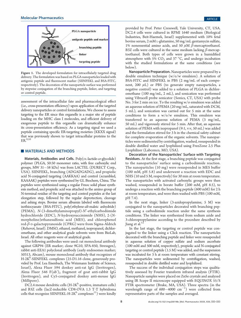
Figure 1. The developed formulation for intracellularly targeted drugdelivery. The formulation was based on PLGA nanoparticles loaded withantigenic peptide and fluorescent marker (SIINFEKL and BSA-FITC,respectively). The decoration of the nanoparticle surface was performedby stepwise conjugation of the branching peptide, linker, and targetingor control peptide.
C dx.doi.org/10.1021/mp200198c |Mol. Pharmaceutics XXXX, XXX, 000–000
Molecular Pharmaceutics ARTICLE
Characterization of the Nanoparticles. Nanoparticle mor-phology was studied with scanning electron microscopy (SEM).Samples of the lyophilized formulations were placed on carbonadhesive tape, coated with gold, and imaged using a Quanta 200scanning electron microscope (Hillsbro, OR, USA) at theInstitute of Applied Research (Ben-Gurion University, Beer-Sheva, Israel). Quantitative analysis of the SEM images deter-mined nanoparticle size by using ImageJ software (version1.40C, NIH, USA13). Nanoparticle ζ-potential was measuredby Laser Doppler Velocimetry using a ZetaPlus instrument(Brookhaven Instruments Corporation Ltd., NY, USA) anddouble distilled water as dispersion medium in the range of10�1000 nm at the National Institute of Biotechnology (Ben-Gurion University, Beer-Sheva, Israel).The encapsulation efficiency was determined by analysis of the
supernatants of nanoparticle suspensions (taken in the last stageof their preparation, after evaporation of the organic solvents)using a QuantiPro BCA assay kit (Sigma). In vitro release rate ofthe encapsulated materials from the nanoparticles was deter-mined by incubating 5 mg samples of the formulations in 500 μLof PBS solution (1mM, pH 7.4) at 37 �Cwith constant mixing bymagnetic stirrer. Samples were centrifuged at 3 h, 1, 2, 3, 4, and 7days, supernatant was collected and replaced with fresh PBSsolution, and the samples were thoroughly resuspended byvortexing. BSA-FITC release in the supernatant was quantifiedusing an Infinite M200 microplate reader (Tecan, Switzerland,excitation at 492 nm and emission at 520 nm), and the releasedpeptides and protein were quantified usingQuantiPro BCA assaykit (Sigma). Solutions with known concentrations of BSA-FITCand SIINFEKL served as controls.Uptake of the Nanoparticles by DC2.4 Cells in Vitro and
Analysis of Intracellular Localization. In all in vitro experi-ments, DC2.4 cells were grown on 24-well or 96-well plates(100,000 or 40,000 cells/well, respectively) and incubated withthe individual formulations or with an equivalent amount ofSIINFEKL solution (2 μg/mL of free or encapsulated SIINFEKL).For assessment of NP uptake, DC2.4 cells were incubated with
the individual formulations for 2 h, extensively washed with PBS,and harvested using trypsin�EDTA solution (Biological Indus-tries, Beit-Haemek, Israel), and the cells’ fluorescence in thegreen channel was analyzed using the Guava EasyCyte mini flowcytometry system (Millipore, USA) and FlowJo software (v. 7.6.1,Tree Star Inc., USA).For analysis of intracellular nanoparticle localization: DC2.4
cells grown on glass coverslips in 24-well plates were (1)incubated with the individual formulations for 2 h, (2) exten-sively washed with PBS, and then fixed with 2.5% formaldehydein PBS, (3) permeabilized with 0.1% Triton X-100 solution, (4)stained with antibody against GRP94 (ER marker) followed byAF594 donkey anti-rat secondary antibody, or with an antibodyagainst EEA1 (early endosomesmarker) followed by AF546 goatanti-rabbit secondary antibody, and (5) mounted on slides usingMowiol 4-88 with DABCO antifading agent. Cells stained withsecondary antibodies and/or incubated with empty nanoparticleswere used as controls.Representative images of the prepared slides at the individual
fluorescence channels were sequentially collected with an Olym-pus FV100-IX81 confocal microscope (Tokyo, Japan) equippedwith 60� oil objective. All experimental samples were imaged onthe same day using a constant set of imaging parameters (thatwere initially adjusted to keep all the samples’ fluorescence withina linear range). The collected images were analyzed using a
custom-written “IntraCell” plugin in ImageJ software14,15 toidentify the borders of the individual cells and the organelles(endoplasmic reticulum and early endosomes), and to quantifythe relative amount of the nanoparticles inside and outside theseorganelles within a specific cell. For each formulation, at least50 cells were analyzed using this approach.Analysis of Cross-Presentation. For FACS analysis of cross-
presentation, DC2.4 cells were grown on 24-well plates and wereincubated for 2 hwith the individual formulations or with equivalentamount of SIINFEKL and BSA-FITC solution (2 μg/mL of freeor encapsulated SIINFEKL). They were then extensively washedwith PBS, harvested using trypsin�EDTA solution, washed,blocked, resuspended in 1% BSA and 0.1% sodium azide solu-tion, and stained with 25-D1.16 antibody followed by Cy5-conjugated donkey anti-mouse secondary antibody. Fluorescenceof the cells was analyzed using FACScanto II flow cytometer (BD,USA) and FlowJo software (v. 7.6.1, Tree Star Inc., USA).For B3Z T cell activation assay of cross-presentation, DC2.4
cells were grown on 96-well plates and were incubated for 2, 4,24, or 48 h with the individual formulations or with solution ofSIINFEKL peptide and BSA-FITC. The DC2.4 cells were thenextensively washed with PBS and incubated with B3Z cells for20 h, the medium was discarded, the cells were lysed withsolution containing 0.3 mM CPRG reagent and incubated for1 h, and absorbance at 630 nm, which is indicative of amountβ-galactosidase secreted by B3Z cells, was determined usingInfinite M200 microplate reader (Tecan, Switzerland).Statistical Analysis. The data are presented as mean (
standard deviation. Differences in the studied parameters be-tween the experimental groups were analyzed using two-tailedt test or ANOVA with Tukey�Kramer or Dunnett post-testusing InStat 3.0 software (GraphPad Software Inc.). The p valueless than 0.05 was termed significant.
’RESULTS
Nanoparticle Preparation and Characterization. Sphericalnanoparticles with narrow size distribution were prepared(Figures 2A and 2B). The diameter of unconjugated nanoparti-cles was 344 ( 75 nm, and the ζ-potential was �32 ( 1 mV,which is consistent with presence of free carboxylic groups on thenanoparticles’ surface. Following individual conjugation steps,characteristic changes of the FTIR spectra of the nanoparticleswere observed (e.g., appearance of azide peak at ∼2100 cm�1
following conjugation of the linker, appearance of triazole ring at1500�1600 cm�1 following conjugation of the targeting orcontrol peptide that indicates successful Click reaction betweenazide group of the linker and alkyne group of the propiolic-acidN-conjugated peptide), and were consistent with successfulconjugation of branching peptide, linker, and the targeting orcontrol peptides. Multistep conjugation procedure did not affectthemorphology or the size distribution of the nanoparticles (datanot shown), but decreased the ζ-potential to �27 ( 1 mV,consistent with attachment of positively charged targeting orcontrol peptides to the nanoparticles’ surface.The generated nanoparticles efficiently encapsulated the anti-
genic peptide andmarker molecules: the encapsulation efficiencyof unconjugated nanoparticles was 33 ( 8%, corresponding toapproximately 0.67 mg of SIINFEKL and 0.67 mg of BSA-FITCper each mg nanoparticle weight (i.e., approximately 105 and 103
SIINFEKL and BSA-FITC molecules per nanoparticle, re-spectively). Analysis of fluorescence of the samples obtained in

D dx.doi.org/10.1021/mp200198c |Mol. Pharmaceutics XXXX, XXX, 000–000
Molecular Pharmaceutics ARTICLE
in vitro release experiments (see Figure 2C) indicates that BSA-FITC was gradually released from the studied formulations andthat the conjugation procedure slightly enhanced its release rateduring the first days of the experiment. In addition to BSA-FITC,these samples contained substantial amounts of peptides as canbe seen from comparison between content of total protein andpeptides vs BSA-FITC alone (see Figure 2D and Figure 2C,respectively). Comparison of these curves for unconjugatednanoparticles indicates that release of SIINFEKL peptide (theonly peptide component of this formulation) exhibits a moderate“burst effect” (a phenomenon of rapid release of the surface-adsorbed or superficially located material in the formulation)during the first hours of incubation, followed by gradual releaseover 3�5 days. On the other hand, targeting and control peptide-conjugated nanoparticles released also substantial amounts ofmaterials that were attached to them during the multistageconjugation procedure (i.e., branching peptide, linker, andtargeting or control peptide). We noticed that empty nanopar-ticles and unconjugated nanoparticles, but not the targeting orcontrol peptide-conjugated nanoparticles, had substantial ten-dency to aggregate during in vitro release experiments. Thisphenomenon was not observed in other experiments, and noneof the studied formulations exhibited tendency for aggregation in
the cell medium during cellular uptake and intracellular traffickingexperiments, as determined by confocalmicroscopy of nanoparticlesalone, or for cells incubated with the nanoparticles (see below).Nanoparticle Uptake and Intracellular Trafficking. The
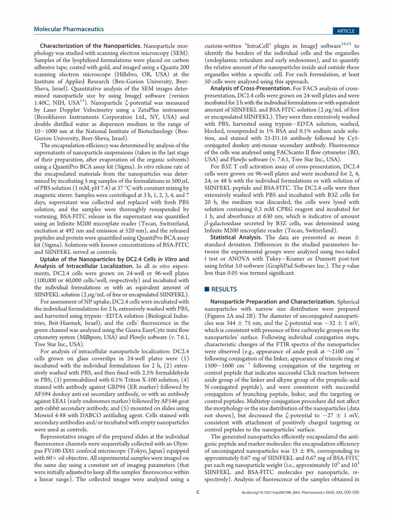
endocytosis and intracellular fate of the nanoparticles in theDC2.4 cells was influenced by the presence and sequence of theconjugated peptides. FACS analysis of cells incubated withdifferent formulations indicated significantly higher uptake oftargeting or control peptide-conjugated nanoparticles, as com-pared to the unconjugated nanoparticles (see Figure 3).Immunofluorescence analysis of both ER and endosomes in
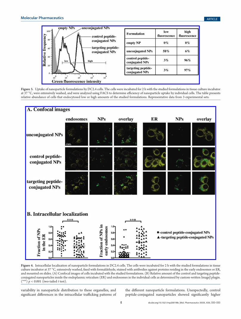
the same samples of DC2.4 cells was unsuccessful due to thetechnical limitations related to the background staining andcross-emission of the fluorophores (“bleed-through” phenom-enon). Therefore, staining of the ER and of the endosomes wasperformed separately (see Figure 4A). Confocal imaging colla-borates the conclusions of FACS analysis and indicates thatDC2.4 cells were able to endocytose the multiple targeting orcontrol peptide-conjugated nanoparticles, but a significantly smallernumber of unconjugated nanoparticles were endocytosed.Quantitative analysis of the confocal images using custom-
written “IntraCell” ImageJ plugin15 revealed a high colocalizationof the nanoparticles with the ER and endosomes, high intercell
Figure 2. Characterization of the generated nanoparticle formulations. (A) Cryo-TEM analysis of morphology of unconjugated nanoparticles. (B)Nanoparticle size distribution based quantitative analysis of Cryo-TEM images of unconjugated nanoparticles using ImageJ program. Other studiedformulations were characterized by similar morphology and size distribution. (C) In vitro release of BSA-FITC from the studied formulations into thePBS solution at 37 �C as determined by fluorescence analysis of the samples. (D) In vitro release of protein and peptides from the studied formulationsinto the PBS solution at 37 �C as determined by analysis of the samples using QuantiPro BCA assay. The inset shows a magnified release curve of theunconjugated nanoparticles. The in vitro release data curves were obtained following subtraction of background values obtained from samples of emptynanoparticles. Panels A�Dpresent representative data from at least 3 experimental sets for each analysis. The error bars represent SD of triplicates. (**) p< 0.01 in comparison to the control and targeting peptide-conjugated nanoparticles (ANOVA with Tukey�Kramer post-test).
E dx.doi.org/10.1021/mp200198c |Mol. Pharmaceutics XXXX, XXX, 000–000
Molecular Pharmaceutics ARTICLE
variability in nanoparticle distribution to these organelles, andsignificant differences in the intracellular trafficking patterns of
the different nanoparticle formulations. Unexpectedly, controlpeptide-conjugated nanoparticles showed significantly higher
Figure 3. Uptake of nanoparticle formulations by DC2.4 cells. The cells were incubated for 2 h with the studied formulations in tissue culture incubatorat 37 �C, were extensively washed, and were analyzed using FACS to determine efficiency of nanoparticle uptake by individual cells. The table presentsrelative abundance of cells that endocytosed low or high amounts of the studied formulations. Representative data from 3 experimental sets.
Figure 4. Intracellular localization of nanoparticle formulations in DC2.4 cells. The cells were incubated for 2 h with the studied formulations in tissueculture incubator at 37 �C, extensively washed, fixed with formaldehyde, stained with antibodies against proteins residing in the early endosomes or ER,and mounted on slides. (A) Confocal images of cells incubated with the studied formulations. (B) Relative amount of the control and targeting peptide-conjugated nanoparticles inside the endoplasmic reticulum (ER) and endosomes in the individual cells as determined by custom-written ImageJ plugin.(***) p < 0.001 (two-tailed t test).
F dx.doi.org/10.1021/mp200198c |Mol. Pharmaceutics XXXX, XXX, 000–000
Molecular Pharmaceutics ARTICLE
accumulation in the ER (0.56 ( 0.23) and a significantly loweraccumulation in the endosomes (0.16 ( 0.18), as compared tothe targeting peptide-conjugated nanoparticles (0.38( 0.22 and0.50 ( 0.28, respectively, see Figure 4B).Cross-Presentation Efficiency of the Studied Formula-
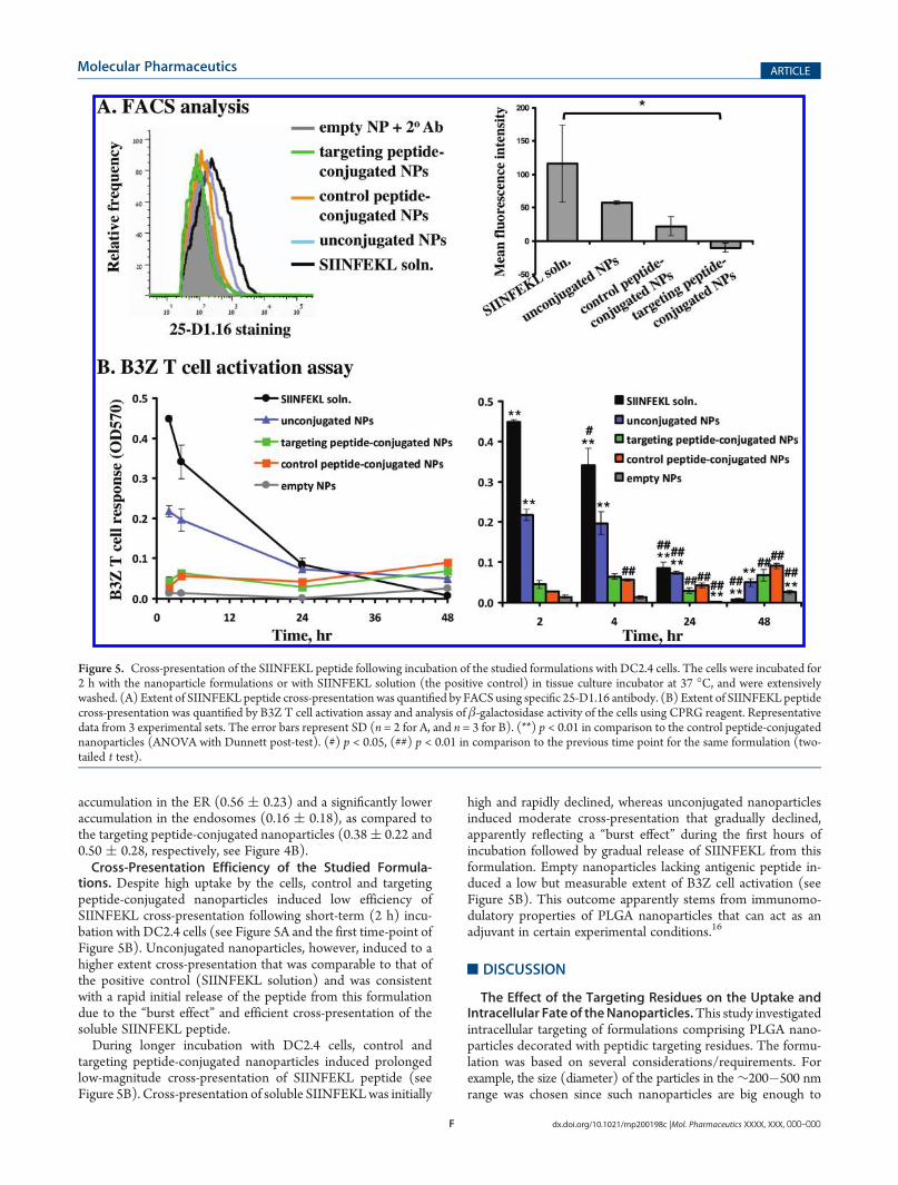
tions. Despite high uptake by the cells, control and targetingpeptide-conjugated nanoparticles induced low efficiency ofSIINFEKL cross-presentation following short-term (2 h) incu-bation with DC2.4 cells (see Figure 5A and the first time-point ofFigure 5B). Unconjugated nanoparticles, however, induced to ahigher extent cross-presentation that was comparable to that ofthe positive control (SIINFEKL solution) and was consistentwith a rapid initial release of the peptide from this formulationdue to the “burst effect” and efficient cross-presentation of thesoluble SIINFEKL peptide.During longer incubation with DC2.4 cells, control and
targeting peptide-conjugated nanoparticles induced prolongedlow-magnitude cross-presentation of SIINFEKL peptide (seeFigure 5B). Cross-presentation of soluble SIINFEKLwas initially
high and rapidly declined, whereas unconjugated nanoparticlesinduced moderate cross-presentation that gradually declined,apparently reflecting a “burst effect” during the first hours ofincubation followed by gradual release of SIINFEKL from thisformulation. Empty nanoparticles lacking antigenic peptide in-duced a low but measurable extent of B3Z cell activation (seeFigure 5B). This outcome apparently stems from immunomo-dulatory properties of PLGA nanoparticles that can act as anadjuvant in certain experimental conditions.16
’DISCUSSION
The Effect of the Targeting Residues on the Uptake andIntracellular Fate of theNanoparticles.This study investigatedintracellular targeting of formulations comprising PLGA nano-particles decorated with peptidic targeting residues. The formu-lation was based on several considerations/requirements. Forexample, the size (diameter) of the particles in the∼200�500 nmrange was chosen since such nanoparticles are big enough to
Figure 5. Cross-presentation of the SIINFEKL peptide following incubation of the studied formulations with DC2.4 cells. The cells were incubated for2 h with the nanoparticle formulations or with SIINFEKL solution (the positive control) in tissue culture incubator at 37 �C, and were extensivelywashed. (A) Extent of SIINFEKL peptide cross-presentation was quantified by FACS using specific 25-D1.16 antibody. (B) Extent of SIINFEKL peptidecross-presentation was quantified by B3Z T cell activation assay and analysis of β-galactosidase activity of the cells using CPRG reagent. Representativedata from 3 experimental sets. The error bars represent SD (n = 2 for A, and n = 3 for B). (**) p < 0.01 in comparison to the control peptide-conjugatednanoparticles (ANOVA with Dunnett post-test). (#) p < 0.05, (##) p < 0.01 in comparison to the previous time point for the same formulation (two-tailed t test).
G dx.doi.org/10.1021/mp200198c |Mol. Pharmaceutics XXXX, XXX, 000–000
Molecular Pharmaceutics ARTICLE
contain a sufficient amount of encapsulated drug (water-solubleantigenic peptide) and gradually release it over several days, yetthey are small enough to be endocytosed by the antigen-presenting cells,17,18 and can be potentially moved intracellularlyby the endogenous trafficking mechanisms.5,19,20 The choice ofthe polymer (PLGA) was based on presence of carboxylic groupsin its chemical structure that are suitable for conjugation of thetargeting residues (using a well-studied carbodiimide chemistryin aqueous buffer) that were claimed to enhance nanoparticles’cytosolic delivery following endocytosis (“endosomal escape”21).The work was successful in generating nanoparticle formulationswith desired properties that were efficiently loaded with SIIN-FEKL peptide and BSA-FITC marker. In vitro release kinetics ofthe encapsulated material from the unconjugated nanoparticlesusing two analytical methods (see Figures 2C and 2D) revealedthat the encapsulated compounds are gradually released from theformulation. It can be assumed that the difference betweencurves in panels C and D of Figure 2 originates from release ofpeptide(s), demonstrating that release of SIINFEKL peptideexhibits a moderate “burst effect” (∼30% of the content) duringthe first hours of incubation, and is slightly more rapid than thatof BSA-FITC (3�4 days vs 7 or more days, respectively).A 3-step conjugation approach was used for stepwise decora-
tion of the unconjugated nanoparticles with the branchingpeptide, linker, and the targeting or control peptide. To enhancethe conjugation efficiency of the targeting residues, we usedPEMA as a surfactant/stabilizer (that contains free carboxylicgroups22,23) and conjugation of branching peptide to increase thenumber of carboxylic groups on the nanoparticles’ surface. Theapplied conjugations affected the key properties of the formula-tion, including ζ-potential, FTIR spectrum, uptake and intracel-lular trafficking in the cells. Outcomes of in vitro releaseexperiments indicate that the conjugation procedure had limitedeffect on the content and release kinetics of BSA-FITC from thenanoparticles. However, effect of the conjugation procedure onthe content and release kinetics of SIINFEKL peptide cannot bereadily determined as it was masked by release of other peptidesfrom the targeting and control peptide-conjugated nanoparticles.Based on indirect data (kinetics of SIINFEKL release fromunconjugated nanoparticles and the overall duration of theconjugation procedure), targeting and control peptide-conju-gated nanoparticles are estimated to contain approximately 30%less SIINFEKL in comparison to unconjugated nanoparticles dueto its partial release into the aqueous solution (the “burst effect”)during the multistep conjugation procedure. On the other hand,targeting and control peptide-conjugated nanoparticles weredecorated with high amounts of branching peptide, linker, andtargeting or control peptide that were gradually released fromthese formulations during in vitro release experiments (see Figure 2D).Unfortunately, the efficiency of the individual conjugation stepscould not be determined due to limited sensitivity and specificity ofthe available analyticalmethods. It is expected that the unconjugatedPLGA nanoparticles used in this study contained at least severalhundred surface carboxylic groups. This estimation is based onreport of conjugation efficiency of 433 peptide residues pernanoparticle in a similar experimental system (PLGA nanoparticlesof similar size but prepared using polyvinyl alcohol andnot PEMAasa surfactant/stabilizer) by Misra et al.20 PEMA substantiallyincreases the amount of surface carboxylic groups22,24 and canincrease the conjugation efficiency up to thousands of peptideresidues per nanoparticle.25 Based on these reports and the presentresults of the in vitro release experiments, we conservatively
estimate that the applied conjugation approach resulted in decora-tion of nanoparticle surface by at least dozens or hundreds oftargeting or control residues.Intracellular distribution of nanoparticles was characterized by
high intercell variability (see Figure 4B) that ranged from 0% to80�100%. This outcome apparently derives from existence ofseveral competing pathways of nanoparticle endocytosis andintracellular trafficking. It is expected that the mechanism ofuptake and trafficking of the individual nanoparticle is dependenton its size, ζ-potential, sequence and density of surface peptides,and other formulation-derived factors. This variability obscuresthe effect of the surface peptidic residues on the nanoparticletargeting to specific organelles. Nevertheless, we were able todetermine that decoration of the nanoparticles with peptidicresidues profoundly affected their uptake and intracellular traf-ficking in the cells, indicating substantial differences in the surfaceproperties between the different formulations. These differencescannot be attributed to the change of the surface charge uponconjugation of the peptidic residues (change of ζ-potential from�32 ( 1 to �27 ( 1 mV). Based on available scientific data oninteraction between nanoparticles and immune cells, these smallchanges in the nanoparticles’ ζ-potential are not expected to affectthe efficiency of their endocytosis by the dendritic cells,17 mono-cytes or macrophages.26 It is possible that differences in efficiency ofendocytosis of the studied formulations derive from specific ornonspecific interactions of surface peptidic residues with the cellmembrane that affected the mechanisms of endocytosis. Themechanisms responsible for the lower uptake of unconjugatednanoparticles, as compared to the targeting or control peptide-conjugated nanoparticles, require additional detailed investigation.Despite the similar extent of endocytosis, control peptide-
conjugated nanoparticles preferentially accumulated in the ER,and not the endosomal compartment, while targeting peptide-conjugated nanoparticles have accumulated to a similar extent inthese compartments (Figure 4B). The applied pixel-based anal-ysis of the images apparently overestimated the nanoparticleaccumulation in the individual organelles15 due to a limitedresolution between the ER (mesh-like network, 50�100 nmdiameter of the tubules27) and endosomes (tubules with 60�100 nm diameter and up to 4 μm length28) and compactmorphology/small size of DC2.4 cells. Nevertheless, this analysiscan be applied for qualitative or semiquantitative analysis of NPlocalization in the studied organelles,15 and the obtained resultsindicate substantial differences in the intracellular traffickingpatterns of the studied nanoparticle formulations.The preferential colocalization of control peptide-conjugated
nanoparticles in the ER was not expected and requires furtherinvestigation. It is possible that surface peptidic residues affectthe efficiency of individual endocytosis pathways of the nano-particles and affect their ability to reach the cytosol and the targetorganelle. The extent of targeting, therefore, can be affected bythe relative efficiency of the individual endocytosis mechanismsin the specific cell type.29 These factors are apparently respon-sible for the different extent of endocytosis and ER delivery ofpeptide-conjugated formulations in dendritic cells (this study) vsendothelial cells (HeLa cells15). Further studies are required toreveal the mechanisms of uptake and of intracellular trafficking ofthese formulations by the cells and to determine the potential ofdecoration of nanoparticles with peptidic residues for intracellu-larly targeted drug delivery. For this purpose, potent targetingresidues should be identified and their targeting properties shouldbe quantitatively assessed (e.g., using imaging and biochemical
H dx.doi.org/10.1021/mp200198c |Mol. Pharmaceutics XXXX, XXX, 000–000
Molecular Pharmaceutics ARTICLE
approaches30) to determine the minimal required amount oftargeting moieties, maximal nanoparticle size, and other factorsthat can limit effectiveness of intracellular drug targeting.5
The Effect of Intracellular Targeting of Antigenic Peptideson Their Cross-Presentation. The experimental results(Figure 5) apparently reflect existence of two routes of antigenicpeptide cross-presentation under the applied conditions (in vitroincubation of DC2.4 cells with the nanoparticles in 24-wellplates). The first route involves loading the H-2Kb moleculeswith soluble SIINFEKL peptide present in the medium.31,32 Thepeptide source in this route is the exogenously added SIINFEKLsolution, or peptide released into the medium from the nano-particles that have not been endocytosed (e.g., due to the “bursteffect”). Cross-presentation of soluble peptide can take place atthe cell surface,31,33 or at the endosomal (phagosomal or ER-endosomal) compartment,34 and the magnitude of these pro-cesses declines rapidly (see the SIINFEKL solution curve inFigure 5B) reflecting rapid degradation of the soluble peptide inthe medium. As compared to in vitro experiments, this route isnot expected to contribute significantly to prolonged peptidecross-presentation under in vivo conditions due to a high dilutionand rapid degradation of the unprotected/soluble peptide in thetissue fluids following administration.The second route involves endocytosis of nanoparticles,
prolonged release of the encapsulated antigenic peptide at theorganelles that were reached by the nanoparticles, and peptidetrafficking and interaction with intracellular H-2Kb molecules. Asdiscussed previously, control peptide-conjugated nanoparticlesaccumulated to a higher extent in the ER, as compared to thetargeting peptide-conjugated nanoparticles (see Figure 4B). Weexpected that this accumulation in the ERwould be accompaniedby increased ER delivery and enhanced cross-presentation ofantigenic peptide. However, cross-presentation efficiency ofcontrol and targeting peptide-conjugated nanoparticles wassimilar, and was lower as compared to other formulations (seeFigure 5). It is possible that this outcome stems from low amountof the antigenic peptide delivered to the intracellular organellesby these formulations (i.e., following substantial release of theantigenic peptide during the conjugation procedure). Therefore,the nanoparticles’ preparation and conjugation procedure shouldbe further optimized to increase the loading capacity of theconjugated formulations.Future studies should also assess the in vivo anticancer
vaccination efficiency of the developed formulations in animalswith established tumors and in tumor protection assays. Thedeveloped formulations are expected to be endocytosed by theantigen-presenting cells following vaccination, and that gradualrelease of the antigenic peptide in the ER and endosomalcompartments (the major sites of antigen cross-presentation)will result in prolonged cross-presentation of the antigen by theantigen-presenting cells leading to enhanced activation of cyto-toxic T lymphocytes (CTLs) directed against the tumorcells.35,36 During vaccination experiments, part of the adminis-tered formulations will be endocytosed also by the cells ofthe mononuclear phagocyte system (MPS, formerly knownas reticuloendothelial system), primarily by monocytes andmacrophages.37,38 Some macrophages are capable to cross-present endocytosed antigens, and both macrophages andmonocytes play a role in tumor immunology and can affectthe outcomes of anticancer vaccination experiments. There-fore, planning of vaccination experiments should take intoaccount endocytosis and intracellular trafficking of the studied
formulations by the cells of the mononuclear phagocyte systemand subsequent pharmacological and immunological conse-quences of these processes.Efficient targeting of antigenic peptides to the ER for the
purpose of anticancer vaccination was reported previously byNakagawa et al.39,40 In these studies a different experimentalapproach was used: the targeting residues were attached to the drugitself (the antigenic peptide) and not to the carrier (the nanopar-ticles or liposomes). Poly(γ-glutamic acid) nanoparticles39 orfusogenic liposomes40 encapsulating the ER-targeted antigenicpeptides induced potent in vitro CD8+ T cell activation andenhanced in vivo CTL activation and antitumor effects in animalmodels. The authors suggested that this outcome stems fromprolonged cross-presentation of the antigenic peptide due to itsefficient targeting to the ER and long-term retention in thisorganelle (which is characterized by low proteolytic activity ascompared to the cytosol).We chose a different experimental approach and conjugated
the targeting residues to the carrier (the nanoparticles) and notto the drug itself (the antigenic peptide). Success of this approachis dependent on capability of the intracellular trafficking mechan-isms to handle cargo (nanoparticles) of the size comparable tosome intracellular vesicles (hundreds of nanometers in dia-meter). The drawbacks of this approach include use of a relativelysophisticated 3-step procedure for conjugation of peptide resi-dues that leads to partial release of the encapsulated materialsduring the conjugation and washing steps. Moreover, quantita-tive characterization of the peptide-conjugated nanoparticles interms of conjugation efficiency and stability of surface peptidicresidues in vitro and in vivo is difficult due to analytical limitations.On the other hand, the applied approach is based on thecommonly used PLGA nanoparticles and well-studied chemicalreactions that are suitable for medical applications and can beeasily adapted for different cargo (encapsulation of other drug ormultiple drugs) and targeting to different organelle (conjugationof different targeting moieties using the same chemistry).
’CONCLUSIONS
Intracellularly targeted drug delivery is a promising newapproach for enhancing and controlling the drug pharmacologi-cal activities. It appears that conjugation of specific targetingresidues can affect the intracellular fate of the nanoparticle drugdelivery systems and result in its preferential drug accumulationwithin specific organelles. Unexpectedly, following endocytosisby DC2.4 cells, nanoparticles decorated with ER-targeting pep-tide accumulated to a lower extent in the ER as compared to thecontrol peptide-conjugated nanoparticles. We attribute thisfinding to effect of control and targeting peptides on efficiencyof the individual endocytosis pathways of the studied formula-tions and on their subsequent intracellular distribution. Studiesthat quantitatively assess the mechanisms, barriers, and efficiencyof intracellular drug delivery are required to identify potenttargeting residues, to attain efficient intracellular targeting ofnanodelivery systems and to determine its therapeutic potentialfor anticancer vaccination and other applications.
’AUTHOR INFORMATION
Corresponding Author*Department of Pharmacology, Ben-Gurion University of theNegev, POB 653, Beer-Sheva 84105, Israel. Tel: +972-8-6477381. Fax: +972-8-6479303. E-mail: [email protected].
I dx.doi.org/10.1021/mp200198c |Mol. Pharmaceutics XXXX, XXX, 000–000
Molecular Pharmaceutics ARTICLE
Author Contributions†These authors equally contributed to this project.
’ACKNOWLEDGMENT
This study was supported by the New Faculty Member Grant(Ben-Gurion University of the Negev) and Prof. Yannai TabbCancer Research Foundation Grant to D.S.We thankMrs. MazalRubin for technical assistance and Prof. Peter Cresswell (Dept. ofImmunobiology, Yale University), Dr. Ayelet David and Prof.Sofia Schreiber-Avissar (Dept. of Pharmacology, Ben-GurionUniversity), Prof. Lea Eisenbach (Dept. of Immunology, Weiz-mann Institute of Science), Prof. Smadar Cohen (Dept. ofBiotechnology Engineering, Ben-Gurion University), and Prof.Angel Porgador (Dept. of Microbiology and Immunology, Ben-Gurion University) for reagents and research tools.
’REFERENCES
(1) Burgdorf, S.; Kautz, A.; Bohnert, V.; Knolle, P. A.; Kurts, C.Distinct pathways of antigen uptake and intracellular routing in CD4 andCD8 T cell activation. Science 2007, 316, 612–6.(2) Miaczynska, M.; Stenmark, H. Mechanisms and functions of
endocytosis. J. Cell Biol. 2008, 180, 7–11.(3) Torchilin, V. P. Recent approaches to intracellular delivery of
drugs and DNA and organelle targeting. Annu. Rev. Biomed. Eng. 2006,8, 343–75.(4) Breunig, M.; Bauer, S.; Goepferich, A. Polymers and nanoparti-
cles: intelligent tools for intracellular targeting?. Eur. J. Pharm. Biopharm.2008, 68, 112–28.(5) Stepensky, D. Quantitative aspects of intracellularly-targeted
drug delivery. Pharm. Res. 2010, 27, 2776–80.(6) Reichert, J. M.;Wenger, J. B. Development trends for new cancer
therapeutics and vaccines. Drug Discovery Today 2008, 13, 30–7.(7) Purcell, A. W.; McCluskey, J.; Rossjohn, J. More than one reason
to rethink the use of peptides in vaccine design.Nat. Rev. Drug Discovery2007, 6, 404–14.(8) Amigorena, S.; Savina, A. Intracellular mechanisms of antigen
cross presentation in dendritic cells. Curr. Opin. Immunol. 2010, 22,109–17.(9) Teasdale, R. D.; Jackson, M. R. Signal-mediated sorting of
membrane proteins between the endoplasmic reticulum and the Golgiapparatus. Annu. Rev. Cell Dev. Biol. 1996, 12, 27–54.(10) Andersson, H.; Kappeler, F.; Hauri, H. P. Protein targeting to
endoplasmic reticulum by dilysine signals involves direct retention inaddition to retrieval. J. Biol. Chem. 1999, 274, 15080–4.(11) Sanderson, S.; Shastri, N. LacZ inducible, antigen/MHC-
specific T cell hybrids. Int. Immunol. 1994, 6, 369–76.(12) Jiang, X.; Zhang, J.; Zhou, Y.; Xu, J.; Liu, S. Facile preparation of
core-crosslinked micelles from azide-containing thermoresponsive dou-ble hydrophilic diblock copolymer via Click chemistry. J. Polym. Sci., PartA: Polym. Chem. 2008, 46, 860–71.(13) Rasband, W. S. ImageJ; U. S. National Institutes of Health:
Bethesda, Maryland, USA, http://rsb.info.nih.gov/ij/, 1997�2010.(14) Stepensky, D. “IntraCell”—intracellular localization of nano-
particles in the individual organelles, plugin for ImageJ. Online. 2011.Available from http://rsb.info.nih.gov/ij/plugins/intracell/.(15) Sneh-Edri, H.; Stepensky, D. “IntraCell” plugin for assessment
of intracellular localization of nano-delivery systems and their targetingto the individual organelles. Biochem. Biophys. Res. Commun. 2011, 405,228–33.(16) Waeckerle-Men, Y.; Groettrup, M. PLGA microspheres for
improved antigen delivery to dendritic cells as cellular vaccines. Adv.Drug Delivery Rev. 2005, 57, 475–82.(17) Foged, C.; Brodin, B.; Frokjaer, S.; Sundblad, A. Particle size
and surface charge affect particle uptake by human dendritic cells in an invitro model. Int. J. Pharm. 2005, 298, 315–22.
(18) Bachmann, M. F.; Jennings, G. T. Vaccine delivery: a matter ofsize, geometry, kinetics andmolecular patterns.Nat. Rev. Immunol. 2010,10, 787–96.
(19) Hoshino, A.; Fujioka, K.; Oku, T.; Nakamura, S.; Suga, M.;Yamaguchi, Y.; Suzuki, K.; Yasuhara, M.; Yamamoto, K. Quantum dotstargeted to the assigned organelle in living cells. Microbiol. Immunol.2004, 48, 985–94.
(20) Misra, R.; Sahoo, S. K. Intracellular trafficking of nuclearlocalization signal conjugated nanoparticles for cancer therapy. Eur. J.Pharm. Sci. 2010, 39, 152–63.
(21) Panyam, J.; Zhou, W. Z.; Prabha, S.; Sahoo, S. K.; Labhasetwar,V. Rapid endo-lysosomal escape of poly(DL-lactide-co-glycolide)nanoparticles: implications for drug and gene delivery. FASEB J. 2002,16, 1217–26.
(22) Keegan, M. E.; Royce, S. M.; Fahmy, T.; Saltzman, W. M. Invitro evaluation of biodegradable microspheres with surface-boundligands. J. Controlled Release 2006, 110, 574–80.
(23) Wang, Q.; Wang, J.; Lu, Q.; Detamore, M. S.; Berkland, C.Injectable PLGA based colloidal gels for zero-order dexamethasonerelease in cranial defects. Biomaterials 2010, 31, 4980–6.
(24) Lo, C. T.; Van Tassel, P. R.; Saltzman, W. M. Simultaneousrelease of multiple molecules from poly(lactide-co-glycolide) nanopar-ticles assembled onto medical devices. Biomaterials 2009, 30, 4889–97.
(25) Zhang, N.; Chittasupho, C.; Duangrat, C.; Siahaan, T. J.;Berkland, C. PLGA nanoparticle�peptide conjugate effectively targetsintercellular cell-adhesionmolecule-1.BioconjugateChem.2008, 19, 145–52.
(26) Verma, A.; Stellacci, F. Effect of surface properties on nano-particle-cell interactions. Small 2011, 6, 12–21.
(27) Voeltz, G. K.; Prinz, W. A. Sheets, ribbons and tubules - howorganelles get their shape. Nat. Rev. Mol. Cell Biol. 2007, 8, 258–64.
(28) Marsh, M.; Griffiths, G.; Dean, G. E.; Mellman, I.; Helenius, A.Three-dimensional structure of endosomes in BHK-21 cells. Proc. Natl.Acad. Sci. U.S.A. 1986, 83, 2899–903.
(29) Cartiera, M. S.; Johnson, K. M.; Rajendran, V.; Caplan, M. J.;Saltzman, W. M. The uptake and intracellular fate of PLGA nanopar-ticles in epithelial cells. Biomaterials 2009, 30, 2790–8.
(30) Cardarelli, F.; Serresi, M.; Albanese, A.; Bizzarri, R.; Beltram, F.Quantitative analysis of Tat peptide binding to import carriers revealsunconventional nuclear transport properties. J. Biol. Chem. 2011,286, 12292–9.
(31) Met, O.; Buus, S.; Claesson, M. H. Peptide-loaded dendriticcells prime and activate MHC-class I-restricted T cells more efficientlythan protein-loaded cross-presenting DC. Cell. Immunol. 2003, 222,126–33.
(32) Porgador, A.; Gilboa, E. Bone marrow-generated dendritic cellspulsed with a class I-restricted peptide are potent inducers of cytotoxic Tlymphocytes. J. Exp. Med. 1995, 182, 255–60.
(33) Smyth, L. A.; Harker, N.; Turnbull, W.; El-Doueik, H.;Klavinskis, L.; Kioussis, D.; Lombardi, G.; Lechler, R. The relativeefficiency of acquisition of MHC:peptide complexes and cross-presenta-tion depends on dendritic cell type. J. Immunol. 2008, 181, 3212–20.
(34) Ackerman, A. L.; Cresswell, P. Cellular mechanisms governingcross-presentation of exogenous antigens. Nat. Immunol. 2004, 5, 678–84.
(35) Audran, R.; Peter, K.; Dannull, J.; Men, Y.; Scandella, E.;Groettrup, M.; Gander, B.; Corradin, G. Encapsulation of peptides inbiodegradable microspheres prolongs their MHC class-I presentation bydendritic cells and macrophages in vitro. Vaccine 2003, 21, 1250–5.
(36) Waeckerle-Men, Y.; Allmen, E. U.; Gander, B.; Scandella, E.;Schlosser, E.; Schmidtke, G.;Merkle, H. P.; Groettrup,M. Encapsulationof proteins and peptides into biodegradable poly(D,L-lactide-co-glycolide)microspheres prolongs and enhances antigen presentation by humandendritic cells. Vaccine 2006, 24, 1847–57.
(37) Panagi, Z.; Beletsi, A.; Evangelatos, G.; Livaniou, E.; Ithakissios,D. S.; Avgoustakis, K. Effect of dose on the biodistribution andpharmacokinetics of PLGA and PLGA-mPEG nanoparticles. Int. J.Pharm. 2001, 221, 143–52.
(38) Semete, B.; Booysen, L.; Lemmer, Y.; Kalombo, L.; Katata, L.;Verschoor, J.; Swai, H. S. In vivo evaluation of the biodistribution and
J dx.doi.org/10.1021/mp200198c |Mol. Pharmaceutics XXXX, XXX, 000–000
Molecular Pharmaceutics ARTICLE
safety of PLGA nanoparticles as drug delivery systems. Nanomedicine2010, 6, 662–71.(39) Matsuo, K.; Yoshikawa, T.; Oda, A.; Akagi, T.; Akashi, M.;
Mukai, Y.; Yoshioka, Y.; Okada, N.; Nakagawa, S. Efficient generation ofantigen-specific cellular immunity by vaccination with poly(gamma-glutamic acid) nanoparticles entrapping endoplasmic reticulum-targetedpeptides. Biochem. Biophys. Res. Commun. 2007, 362, 1069–72.(40) Hayashi, A.; Wakita, H.; Yoshikawa, T.; Nakanishi, T.; Tsutsumi,
Y.; Mayumi, T.; Mukai, Y.; Yoshioka, Y.; Okada, N.; Nakagawa, S. Astrategy for efficient cross-presentation of CTL-epitope peptides leadingto enhanced induction of in vivo tumor immunity. J. Controlled Release2007, 117, 11–9.