intracranial hypotension (nxpowerlite)
TRANSCRIPT

Sasitorn Petcharunpaisan, M.D.
Department of RadiologyKing Chulalongkorn Memorial Hospital
Bangkok, Thailand
Epidemiology
Not rare, an important cause of new daily persistent headaches among young & middle age individuals
Prevalence: ~1 per 50,000, previously probably underdiagnosed
F:M ~ 2:1, onset in 4th or 5th decadeAssociated with connective tissue
disorders (Marfan, Ehler Danlos)JAMA 2006.;295(19):2286-96
Etiology & Pathogenesis
Generally caused by spinal CSF leakPrecise cause remains largely unknown,
underlying structural weakness of spinal meninges is suspected
Hx of trivial traumatic events elicited in 1/3Wide variety of dural defects; simple dural
hole, fragile meningeal diverticula, absence of dura cover spinal nerve root
JAMA 2006.;295(19):2286-96
Etiology & Pathogenesis
Decreased CSF volume may be final common pathway in pathophysiology
Altered distribution of craniospinal elasticity due to spinal CSF leak may be final common pathway
So, “spontaneous spinal CSF leak” are preferred terms
JAMA 2006.;295(19):2286-96

Diffuse pachymeningeal (dural) enhancement Bilateral subdural effusion/hematomas Downward displacement of brain Enlargement of pituitary gland Engorgement of dural venous sinuses Prominence of spinal epidural venous plexus Venous sinus thrombosis & isolated cortical vein
thrombosisAJNR 2008.; 29:1164-70
Monroe-Kellie Rule
Sum of volumes of intracranial blood, CSF & cerebral tissue must remain constant in an intact cranium
Loss of CSF can be compensated by increased vascular component or by increased intracranial CSF component
JAMA 2006.;295(19):2286-96
Monroe-Kellie Rule
Accounting for pachymeningeal enhancement, engorged venous structures, pituitary hyperemia and subdural effusions
Subdural hematoma may caused by tearing of bridging veins or rupture of thin wall vessels in subdural zone
Sagging of brain is caused by loss of CSF buoyancy
JAMA 2006.;295(19):2286-96

JAMA 2006.;295(19):2286-96
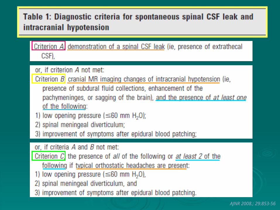
AJNR 2008.; 29:853-56
Diffuse, uniform thicknessLocated at convexity, along falx cerebri, tentorium & posterior fossa duraDisappears after successful treatment
Diffuse Pachymeningeal, (Dural) Enhancement
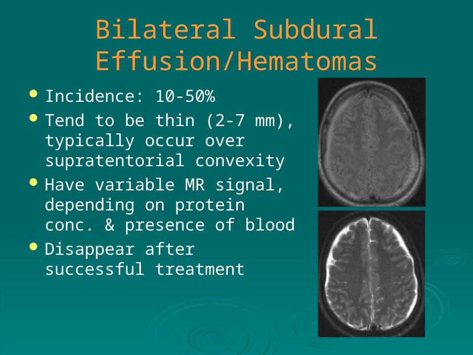
Bilateral Subdural Effusion/Hematomas
Incidence: 10-50% Tend to be thin (2-7 mm),
typically occur over supratentorial convexity
Have variable MR signal, depending on protein conc. & presence of blood
Disappear after successful treatment

Downward Displacement of The Brain
Low lying cerebellar tonsils Effacement of prepontine
cistern, flattening of pons against clivus
Effacement of perichiasmatic cistern with bowing of optic chiasm over pituitary fossa
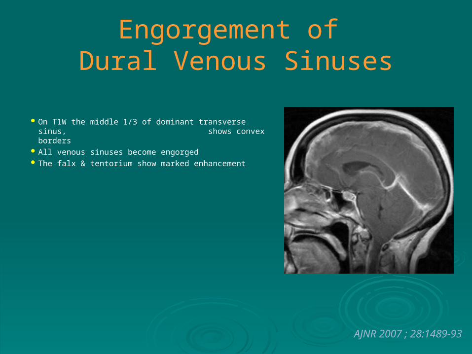
Engorgement of Dural Venous Sinuses
On T1W the middle 1/3 of dominant transverse sinus, shows convex borders
All venous sinuses become engorged The falx & tentorium show marked enhancement
AJNR 2007 ; 28:1489-93
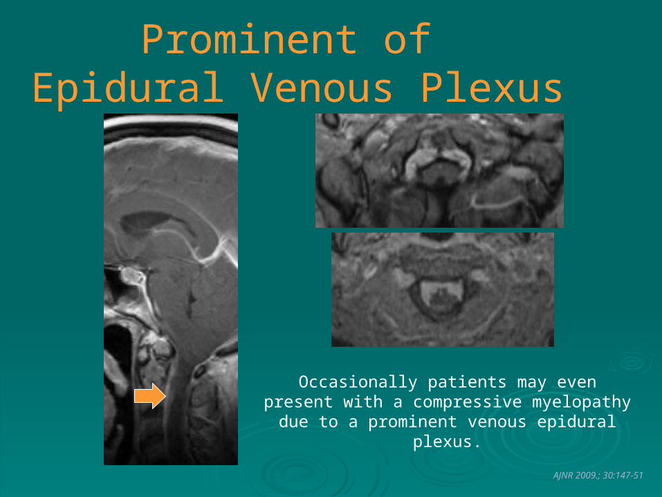
Prominent of Epidural Venous Plexus
AJNR 2009.; 30:147-51
Occasionally patients may even present with a compressive myelopathy due to a prominent
venous epidural plexus.

Spinal Extradural Fluid Collections
From: AJNR 2009.; 30:147-51
Treatment
Many cases resolved spontaneouslyThere is no randomized control trial
evaluation of the treatment optionConservative approach: bed rest, oral
hydration, caffeine intake, use of abdominal binder
JAMA 2006.;295(19):2286-96
Treatment
Mainstay of treatment is epidural blood patch (EBP) - epidural injection of autologous blood into epidural space
Effective in relieving symptoms in ~1/3, presumable by dural temponade and sealing the leak
If unsuccessful, it can be repeated
JAMA 2006.;295(19):2286-96
Treatment
If EBP fail, direct EBP or percutaneous placement of fibrin sealant is recommended Requires knowledge of exact site of CSF leak
Surgical Rx is reserved for Pt who failed nonsurgical Rx
Often successful when focal CSF leak is identified
Ligation or placement of muscle pledget
JAMA 2006.;295(19):2286-96
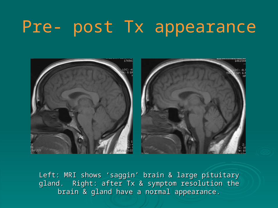
Pre- post Tx appearance
Left: MRI shows ‘saggin’ brain & large pituitary gland. Right: after Tx & Left: MRI shows ‘saggin’ brain & large pituitary gland. Right: after Tx & symptom resolution the brain & gland have a normal appearance.symptom resolution the brain & gland have a normal appearance.

Pituitary gland changes in Intracranial Hypotension
Pre- & post treatment changes. The pituitary gland was initially enlarged & after Pre- & post treatment changes. The pituitary gland was initially enlarged & after Tx it becomes normal in size.Tx it becomes normal in size.
AJNR 2008.; 29:853-56
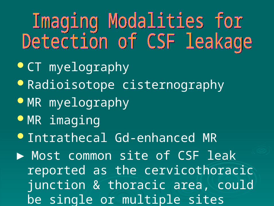
CT myelographyRadioisotope cisternographyMR myelography MR imagingIntrathecal Gd-enhanced MR► Most common site of CSF leak reported as
the cervicothoracic junction & thoracic area, could be single or multiple sites
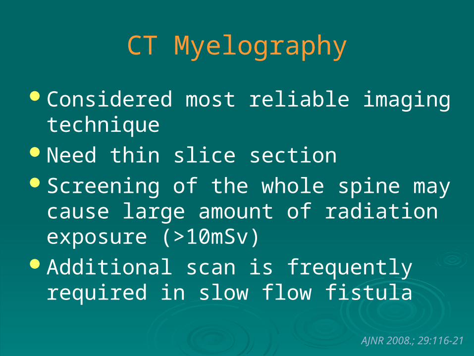
CT Myelography
Considered most reliable imaging technique Need thin slice sectionScreening of the whole spine may cause
large amount of radiation exposure (>10mSv)
Additional scan is frequently required in slow flow fistula
AJNR 2008.; 29:116-21
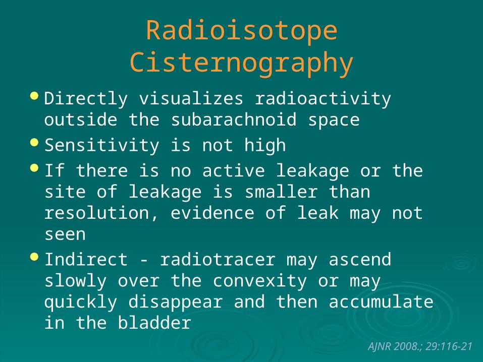
Radioisotope Cisternography
Directly visualizes radioactivity outside the subarachnoid space
Sensitivity is not highIf there is no active leakage or the site of
leakage is smaller than resolution, evidence of leak may not seen
Indirect - radiotracer may ascend slowly over the convexity or may quickly disappear and then accumulate in the bladder
AJNR 2008.; 29:116-21
MR Myelography
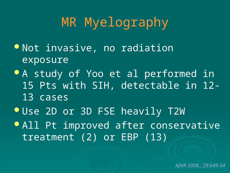
Not invasive, no radiation exposure A study of Yoo et al performed in 15 Pts
with SIH, detectable in 12-13 casesUse 2D or 3D FSE heavily T2WAll Pt improved after conservative treatment
(2) or EBP (13)
AJNR 2008.; 29:649-54
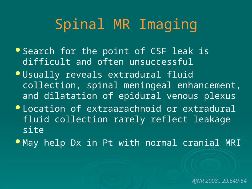
Spinal MR Imaging
Search for the point of CSF leak is difficult and often unsuccessful
Usually reveals extradural fluid collection, spinal meningeal enhancement, and dilatation of epidural venous plexus
Location of extraarachnoid or extradural fluid collection rarely reflect leakage site
May help Dx in Pt with normal cranial MRI
AJNR 2008.; 29:649-54
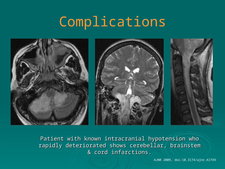
Complications
Patient with known intracranial hypotension who rapidly Patient with known intracranial hypotension who rapidly deteriorated shows cerebellar, brainstem & cord infarctions.deteriorated shows cerebellar, brainstem & cord infarctions.
AJNR 2009, doi:10.3174/ajnr.A1749
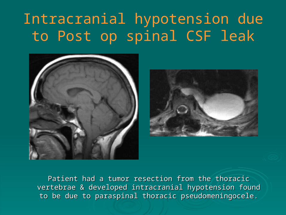
Intracranial hypotension due to Post op spinal CSF leak
Patient had a tumor resection from the thoracic vertebrae & developed Patient had a tumor resection from the thoracic vertebrae & developed intracranial hypotension found to be due to paraspinal thoracic intracranial hypotension found to be due to paraspinal thoracic
pseudomeningocele.pseudomeningocele.
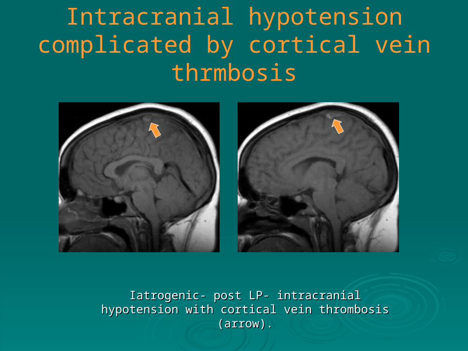
Intracranial hypotension complicated by cortical vein thrmbosis
Iatrogenic- post LP- intracranial hypotension with cortical Iatrogenic- post LP- intracranial hypotension with cortical vein thrombosis (arrow).vein thrombosis (arrow).
References1. Spontaneous spinal cerebrospinal fluid leaks and intracranial hypotension. JAMA 2006;
295(19):2286-962. Diffuse pachymeningeal hyperintensity and subdural effusion/hematoma by FLAIR MRI in
patients with spontaneous intracranial hypotension. AJNR 2008; 29:1164-703. The venous distention sign: a diagnostic sign of intracranial hypotension. AJNR 2008;
28:1489-934. Intradural spinal vein enlargement in intracranial hypotension. AJNR 2005; 26:34-385. Diagnostic criteria for spontaneous spinal CSF leaks and intracranial hypotension. AJNR
2008; 29:853-566. Detection of CSF leak in spinal CSF leak syndrome using MR myelography: correlation with
radioisotope cisternography. AJNR 2008; 29:649-547. Gadolinium-enhanced MR cisternography to evaluate dural leaks in intracranial hypotension
syndrome. AJNR 2008; 29:116-218. Diagnostic value of spinal MRI in spontaneous intracranial hypotension syndrome. AJNR
2009; 30:147-519. False localizing sign of C1-2 CSF leak in spontaneous intracranial hypotension. J Neurosurg
2004; 100:639-44