intraductal proliferative lesions - bc · pdf fileintraductal proliferative lesions –...
TRANSCRIPT

Intraductal Proliferative Lesions –DIN diagnosis and management
Dr. Kathy CeballosSurgical Oncology Network Breast Cancer Update
October 24, 2009

Conflict of Interest Conflict of Interest ---- NoneNone

Asked to address….Asked to address….Classification/ new terminology (DIN) Classification/ new terminology (DIN) ––Why?Why?When is excisional biopsy recommended When is excisional biopsy recommended following a core?following a core?How likely is DIN1A on a core to have How likely is DIN1A on a core to have cancer on excision? cancer on excision? What is the risk of future breast cancer What is the risk of future breast cancer with DIN1A?with DIN1A?

Intraductal proliferative breast lesionsIntraductal proliferative breast lesions‘‘Risk lesions’: neoplastic and non Risk lesions’: neoplastic and non neoplasticneoplasticExamples:Examples:
Usual ductal hyperplasia (UDH, epitheliosis) Usual ductal hyperplasia (UDH, epitheliosis) Flat epithelial atypia (DIN1A)Flat epithelial atypia (DIN1A)Columnar cell changeColumnar cell changeAtypical ductal hyperplasia (ADH)Atypical ductal hyperplasia (ADH)Low grade DCISLow grade DCISGrade 2 and 3 DCISGrade 2 and 3 DCIS
lobule
Terminal duct

Columnar cell lesions and Columnar cell lesions and Flat epithelial atypia (DIN1A)Flat epithelial atypia (DIN1A)

Columnar cell lesions (CCL)Columnar cell lesions (CCL)
Columnar cell change Columnar cell change (CCC), columnar cell (CCC), columnar cell hyperplasia (CCH)hyperplasia (CCH)+/+/-- atypiaatypia
Normal CCC
Without atypia With atypia

Flat epithelial atypia (DIN1A)Flat epithelial atypia (DIN1A)
Neoplastic intraductal alteration 1 Neoplastic intraductal alteration 1 –– 5 5 layers of mildly atypical cells resulting in a layers of mildly atypical cells resulting in a distension of TDLUsdistension of TDLUs
−− Synonyms: clinging carcinoma, columnar Synonyms: clinging carcinoma, columnar cell change with atypia, columnar cell cell change with atypia, columnar cell hyperplasia with atypiahyperplasia with atypia
Associated with invasive tubular carcinoma Associated with invasive tubular carcinoma and LCIS. May be the immediate precursorand LCIS. May be the immediate precursorFrequently multicentric, multifocal and bilatFrequently multicentric, multifocal and bilat

Flat epithelial atypia (DIN1A)Flat epithelial atypia (DIN1A)
Commonly associated Commonly associated with calcificationswith calcificationsMay be mammo target May be mammo target lesionlesion

DIN1A DIN1A –– issues of reproducibilityissues of reproducibility

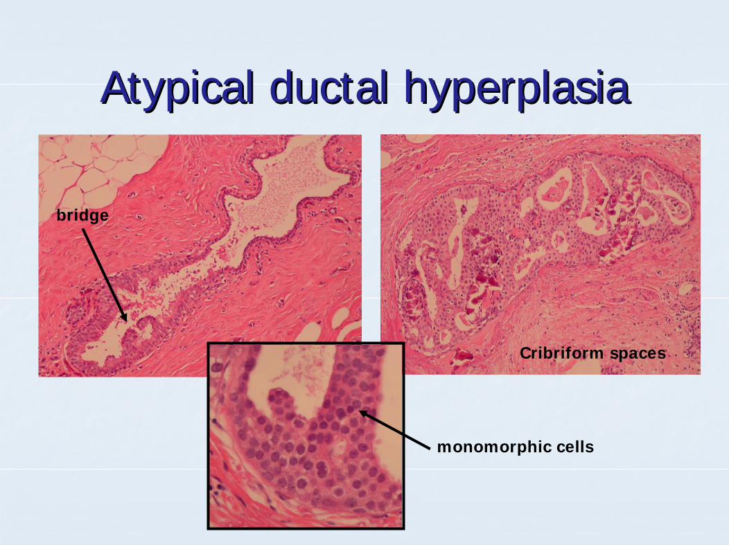
Atypical ductal hyperplasia Atypical ductal hyperplasia (DIN1B)(DIN1B)
Neoplastic intraductal lesion consisting of Neoplastic intraductal lesion consisting of monomorphic cells forming arcades, monomorphic cells forming arcades, bridges, papillae or solid masses.bridges, papillae or solid masses.Morphologically IDENTICAL to low grade Morphologically IDENTICAL to low grade DCIS but insufficient in quantity (< 2 DCIS but insufficient in quantity (< 2 complete duct spaces or 2 mm)complete duct spaces or 2 mm)Concept invented to prevent over treatment Concept invented to prevent over treatment of minimal diseaseof minimal disease
Atypical ductal hyperplasiaAtypical ductal hyperplasia
monomorphic cells
Cribriform spaces
bridge
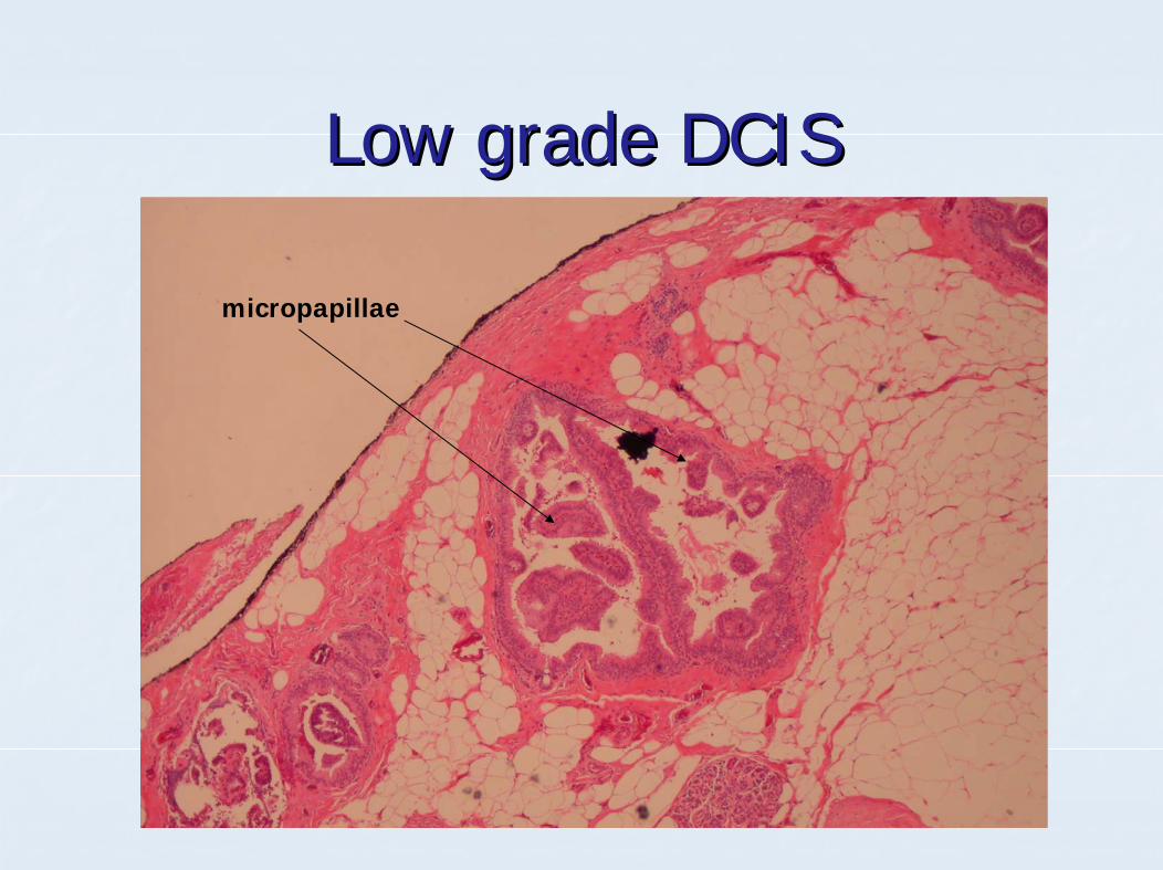
Low grade DCISLow grade DCIS
micropapillae


ClassificationClassification
TraditionalTraditional−− Usual ductal hyperplasia Usual ductal hyperplasia −− Flat epithelial atypiaFlat epithelial atypia−− Atypical Ductal Hyperplasia (ADH)Atypical Ductal Hyperplasia (ADH)−− Grade 1 DCISGrade 1 DCIS−− Grade 2 DCISGrade 2 DCIS−− Grade 3 DCISGrade 3 DCIS

Ductal intraepithelial neoplasia Ductal intraepithelial neoplasia (DIN) terminology(DIN) terminology
WHY!?!!!!

Ductal intraepithelial neoplasia Ductal intraepithelial neoplasia (DIN) terminology (DIN) terminology ---- WHY?!WHY?!
Traditional terminology did not reflect the biologyTraditional terminology did not reflect the biologyObjections to the term ductal Objections to the term ductal carcinomacarcinoma in situin situ
Carcinoma usually assoc. metastatic potentialCarcinoma usually assoc. metastatic potentialNon obligate precursor to invasive diseaseNon obligate precursor to invasive disease
Objections to term atypical ductal Objections to term atypical ductal hyperplasiahyperplasiaNeoplastic lesionsNeoplastic lesionsADH and low grade DCIS are morphologically identical yet ADH and low grade DCIS are morphologically identical yet one is carcinoma and one is hyperplasia one is carcinoma and one is hyperplasia
Trend in other organ systems (CIN, PIN, etc)Trend in other organ systems (CIN, PIN, etc)

Ductal Intraepithelial neoplasia Ductal Intraepithelial neoplasia (DIN) terminology**(DIN) terminology**
Usual ductal hyperplasia = UDH Usual ductal hyperplasia = UDH DIN 1A = flat epithelial atypiaDIN 1A = flat epithelial atypiaDIN 1B = atypical ductal hyperplasiaDIN 1B = atypical ductal hyperplasiaDIN 1C = low grade DCIS (grade 1)DIN 1C = low grade DCIS (grade 1)DIN 2 = grade 2 DCISDIN 2 = grade 2 DCISDIN 3 = grade 3 DCISDIN 3 = grade 3 DCIS
**will likely evolve with molecular genetics**will likely evolve with molecular genetics

DIN1A on core biopsyDIN1A on core biopsy
Studies: Up to 1/3 of DIN1A on core bx Studies: Up to 1/3 of DIN1A on core bx upgraded on excisionupgraded on excisionLimited by:Limited by:
NumbersNumbersDiagnostic criteriaDiagnostic criteriaNot all ‘pure’ DIN1A (coexisting ADH)Not all ‘pure’ DIN1A (coexisting ADH)Mammographic findings unknownMammographic findings unknownMany include ADH/DIN1B as ‘upgrade’Many include ADH/DIN1B as ‘upgrade’

DIN1A on core biopsyDIN1A on core biopsy
Senetta et al Senetta et al (2009):(2009): 392 cores for 392 cores for calcificationscalcifications
CCLs target calcs in 37% casesCCLs target calcs in 37% cases41 pts flat epithelial atypia (DIN1A) only41 pts flat epithelial atypia (DIN1A) only
36 surgery 36 surgery –– no DCIS, or invasive disease, no DCIS, or invasive disease, 13% ADH, 23% ALH/LCIS, 34% DIN1A13% ADH, 23% ALH/LCIS, 34% DIN1A
Recommendation: No need to exciseRecommendation: No need to excise

DIN1A on core biopsyDIN1A on core biopsy
Kunju Kunju (2007):(2007): 14 cases pure DIN1A from 14 cases pure DIN1A from 20002000--20052005
1 had low grade DCIS, 2 Inv Ca on 1 had low grade DCIS, 2 Inv Ca on excision (21% upgraded)excision (21% upgraded)5 ADH/DIN1B, 2 LCIS/ALH, 2 no resection5 ADH/DIN1B, 2 LCIS/ALH, 2 no resectionMammo Indication: 12 calcs, 2 densitiesMammo Indication: 12 calcs, 2 densitiesRecommendation: ExcisionRecommendation: Excision
**usually 14 gauge needle, avg of 5 passes**usually 14 gauge needle, avg of 5 passes
DIN1A on core biopsyDIN1A on core biopsy
Martel et al Martel et al (2007):(2007): 1751 core bx reviewed 1751 core bx reviewed retrospectively, 63 pure DIN1Aretrospectively, 63 pure DIN1A
24 excisions up to 10 yrs later 24 excisions up to 10 yrs later –– 9 Inv Ca9 Inv Ca5 of 24 excisions happened within 3 5 of 24 excisions happened within 3 months, NO Inv Camonths, NO Inv CaRecommendation: Risk factor Recommendation: Risk factor –– screeningscreening
Do not exciseDo not excise

DIN1A on core biopsyDIN1A on core biopsy
Piubello et al Piubello et al (2009):(2009): 875 core bx over 5 yr 875 core bx over 5 yr period reviewed, pure DIN1A in 33 casesperiod reviewed, pure DIN1A in 33 cases
0 of 20 excisions had DCIS or inv. Ca0 of 20 excisions had DCIS or inv. CaRecommendation: Do not exciseRecommendation: Do not excise

DIN1A on core biopsy DIN1A on core biopsy –– BCCABCCA(unpublished)(unpublished)
145 core biopsies from BCWHHC and BCCA 145 core biopsies from BCWHHC and BCCA with pure DIN1A underwent excisionwith pure DIN1A underwent excision
8 excisions (5.5%) showed upgrades of 8 excisions (5.5%) showed upgrades of significancesignificance
6 DCIS6 DCIS2 Invasive cancers2 Invasive cancers
?Nature of mammographic abnormality?Nature of mammographic abnormalityRecommendation: Do not exciseRecommendation: Do not excise

ExcisionExcision
ADH (DIN1B) ADH (DIN1B) –– 1010--39% upgraded on 39% upgraded on excision (usually DCIS) excision (usually DCIS) All papillary lesions (25% upgraded to ADH All papillary lesions (25% upgraded to ADH or DCIS) or DCIS) Radial scar Radial scar ---- ~20% ADH, ~20% DCIS, up ~20% ADH, ~20% DCIS, up to 12% IDCto 12% IDCMucocele like lesions Mucocele like lesions ---- ~30% DCIS or IDC~30% DCIS or IDC

No excisionNo excision
Lobular neoplasia (ALH/LN1, LCIS/LN2)Lobular neoplasia (ALH/LN1, LCIS/LN2)Flat epithelial atypia (DIN1A) Flat epithelial atypia (DIN1A)
This assumes rigorous radiological This assumes rigorous radiological pathological correlationpathological correlation

DIN1A DIN1A –– Risk of subsequent CancerRisk of subsequent Cancer

DIN1A RiskDIN1A Risk
Bijker et al Bijker et al (2001):(2001): EORTC trial 10853 EORTC trial 10853 (recurrences or mets post breast conserving sx for DCIS)(recurrences or mets post breast conserving sx for DCIS)
0/59 cases DIN1A (called low gr clinging 0/59 cases DIN1A (called low gr clinging DCIS) recurred, followDCIS) recurred, follow--up 5.4 yrsup 5.4 yrs
Eusebi et al Eusebi et al (1989):(1989):
2/32 (6%) cases recurred as grade 1 2/32 (6%) cases recurred as grade 1 DCIS, followDCIS, follow--up 17.4 yrsup 17.4 yrs
Conclusion: ?risk is lowConclusion: ?risk is low

DIN1A Risk DIN1A Risk –– BCCABCCA(unpublished)(unpublished)
All benign breast bxs 1989All benign breast bxs 1989--1996 reviewed1996 reviewed133 cases DIN1A (109 excisions, 24 cores)133 cases DIN1A (109 excisions, 24 cores)34 coexisting ADH (DIN1B), 24 ALH/LCIS, 1 34 coexisting ADH (DIN1B), 24 ALH/LCIS, 1 invasive tubular Cainvasive tubular CaAll untreated after initial biopsyAll untreated after initial biopsyBCCA records searched for follow up to 2007BCCA records searched for follow up to 2007
DIN1A Risk DIN1A Risk –– BCCABCCAOutcomesOutcomes
Worse lesion developed in 28/133 women Worse lesion developed in 28/133 women (21%)(21%)
13 cases DCIS13 cases DCIS15 cases invasive carcinoma (2 lobular)15 cases invasive carcinoma (2 lobular)
18 lesions were in the ipsilateral breast, 10 in the 18 lesions were in the ipsilateral breast, 10 in the contralateral breastcontralateral breast

In summary…In summary…New WHO classification (DIN)New WHO classification (DIN)Excision:Excision:
ADH/DIN1B, radial scars, mucocele like lesions, papillary ADH/DIN1B, radial scars, mucocele like lesions, papillary lesions, DCIS, invasive Calesions, DCIS, invasive Ca
No excision:No excision:FEA/DIN1A, ALH, LCISFEA/DIN1A, ALH, LCIS
Likelihood DIN1A on a core to have cancer on Likelihood DIN1A on a core to have cancer on excision:excision:
very low in our lab (5%)very low in our lab (5%)
Future risk of breast cancer with DIN1AFuture risk of breast cancer with DIN1A?similar to ADH/DIN1B (10%)?similar to ADH/DIN1B (10%)


ADH/DIN1B on core biopsyADH/DIN1B on core biopsy
Eby et al Eby et al (2009):(2009): 991 consecutive core bx 991 consecutive core bx 20012001--2006, 141 cases ADH/DIN1B (14.2%)2006, 141 cases ADH/DIN1B (14.2%)
26/123 upgraded to DCIS or inv Ca on 26/123 upgraded to DCIS or inv Ca on excision (21.1%)excision (21.1%)Recommendation: ExciseRecommendation: Excise