intramyocardial injection of recombinant adeno...
TRANSCRIPT

Research ArticleIntramyocardial Injection of RecombinantAdeno-Associated Viral Vector CoexpressingPR39Adrenomedullin Enhances Angiogenesis and ReducesApoptosis in a Rat Myocardial Infarction Model
Rui An1 Cong Xi2 Jian Xu1 Ying Liu1 Shumiao Zhang3
YueminWang3 Yuewen Hao1 and Lijun Sun1
1Department of Radiology Xijing Hospital Fourth Military Medical University Xirsquoan China2Department of Neurology Baoji City Peoplersquos Hospital Baoji China3Department of Physiology Fourth Military Medical University Xirsquoan China
Correspondence should be addressed to Yuewen Hao 1982 edifier163com and Lijun Sun sunljfmmu163com
Received 2 December 2016 Revised 29 January 2017 Accepted 9 February 2017 Published 2 March 2017
Academic Editor Massimo Collino
Copyright copy 2017 Rui An et al This is an open access article distributed under the Creative Commons Attribution License whichpermits unrestricted use distribution and reproduction in any medium provided the original work is properly cited
Cotransfer of angiogenic and antiapoptotic genes could be the basis of new gene therapy strategies for myocardial infarctionIn this study rAAV-PR39-ADM coexpressing antimicrobial peptide (PR39) and adrenomedullin (ADM) was designed with themediation of recombinant adeno-associated virus In vitro CRL-1730 cells were divided into four groups namely the sham groupthe AAV-null group the NS (normal saline) group and the PR39-ADM group Immunocytochemistry analysis CCK-8 assaysMatrigel assays and apoptotic analysis were performed in vivo myocardial infarction model was established through ligation ofthe left coronary artery on rats and treatment groups corresponded to those used in vitro Myocardial injury cardiac performanceand the extent of myocardial apoptosis were assessed Results suggested that rAAV-PR39-ADM administration after myocardialinfarction improved cell viability and cardiac function attenuated apoptosis and myocardial injury and promoted angiogenesisSubsequently levels of 6timesHis HIF-1120572 VEGF p-Akt Akt ADM Bcl-2 and Bax were measured by western blot rAAV-PR39-ADMincreased p-Akt HIF-1120572 and VEGF levels and induced higher Bcl-2 expression and lower Bax expression In conclusion ourresults demonstrate that rAAV-PR39-ADM mitigates myocardial injury by promoting angiogenesis and reducing apoptosis Thisstudy suggests a potential novel gene therapy-based method that could be used clinically for myocardial infarction
1 Introduction
Coronary heart disease with myocardial infarction (MI) asthe acute manifestation remains the leading threat to humanhealth worldwide [1ndash4] Numerous treatment methods basedon the pathogenesis and related to timely reperfusion [56] and reinfarction or ventricular remodelling reduction inthe later stage of MI using pharmacological agents haveflourished recently [7]
The therapeutic delivery of nucleic acid polymers intocells as a drug to treat disease is known as gene therapythis approach has many components including adrenergicmanipulation Ca cycling protein handling angiogenesis
cardiac regeneration and targeting cardiac arrhythmias [8]In general antiapoptosis proangiogenesis and the dilation ofcorresponding vessels are the main aspects of MI treatmentInhibiting apoptosis in both cardiomyocytes and endothe-liocytes might reduce the likelihood of reinfarction at theintracellular level Collateral vessel growth induced by growthfactors such as vascular endothelial growth factor (VEGF)and the dilation of corresponding vessels induced by vasodi-lation substances such as prostaglandin and adrenomedullinsupply the at-risk myocardium with necessary oxygen andnutrition and deplete the tissue of metabolites Howeverprevious gene therapy approaches have usually focused onthe role of a single protective gene on cytoprotection vessel
HindawiOxidative Medicine and Cellular LongevityVolume 2017 Article ID 1271670 13 pageshttpsdoiorg10115520171271670
2 Oxidative Medicine and Cellular Longevity
dilation or angiogenesis of themyocardial infarction and thetherapeutic effects were actually modest
PR39 a proline- (P-)arginine- (R-) rich antimicrobialpeptide of 39 amino acids was originally isolated from pigintestine and identified in neutrophil azurophilic granulesandmacrophages [9 10] Previous studies have demonstratedthat PR39 plays an important role in reducing cardiac injurythrough the induction of angiogenesis by inhibiting HIF-1120572degradation [11] and in antiapoptosis through the expressionof IAP-2 [12]
Adrenomedullin (ADM) a potent and long-lasting vaso-active peptide was originally isolated fromhuman pheochro-mocytoma in 1993 [13] Its functions including dilating thecoronary artery inhibiting vascular remodelling and reduc-ing fibroblast proliferation and extracellular matrix synthesisresult in therapeutic effects such as reducing infarct sizeischemia-reperfusion injury and ischemia-induced arrhyth-mias This consequently lowers mortality in rodent modelsaccording to previous reports [14ndash19]
Accordingly based on previous work [20 21] an adeno-associated viral vector coexpressing PR39 andADMwas con-structed The aim of this study was to assess this strategy forthe protection of the jeopardized myocardium through bothcytoprotection and angiogenesis (vectors were constructedwith the support of Xirsquoan HuaGuang Bioengineering Co(Xirsquoan China) and China patents were applied (CN1919344ACN1919343A CN100589846C and CN100589847C))
2 Materials and Methods
21 Cell Culture Immortalized CRL-1730 cells and HEK-293 cells (provided by Xirsquoan HuaGuang Bioengineering Cor-poration Shaanxi Province of China) were maintained inRPMI 1640 complete medium (Sigma Corporation USA)with supplemental growth factors and antibiotics accordingto company specifications Hypoxia conditionwas performedwith 1 O
2and 5 CO
2cell incubator (provided by the
Department of Physiology of the Fourth Military MedicalUniversity Xirsquoan China)
22 Strains and Plasmids Bacillus coli TOP10 TTAT-Hisand pBV220NT4 AAV vector pSSHGCMV that contains31015840LTR and 51015840LTR and two helper plasmids (pAAVAdPFG140) were provided by Xirsquoan HuaGuang BioengineeringCorporation Shaanxi Province of China pGEM-T-Easy wassupplied by Promega Corporation USA T4 DNA ligasewas supplied by Fermentas Corporation USA Nae I BamHI EcoR I Eco721 and Taq DNA polymerase were boughtfrom Xirsquoan Hua Mei Bioengineering Corporation ShaanxiProvince of China
23 Construction of NT4-Intron-6timesHis-PR39-Splicing-ADMBox Recombinant propeptide was used to realize bioactivepeptide mediated gene therapy (China Patent NumbersCN1919344A CN1919343A and CN100589846C) Recombi-nant vector (HRE-NT4-Intron-6timesHis-PR39-splicing-ADMbox) was constructed by the connection of cDNA of PR39ADM Intron and signal peptide (NT4) hypoxia-responsive
element (HRE) and splicing Then vectors were transducedinto target cells with the help of plasmid (China Patent Num-ber CN100589847C) following transcription of pre-mRNAtwo kinds of biological active peptides (PR39 andADM)wereexpressed We adopted the technology of multiple proteinsand peptides secretory expression with the guidance of singlesignal peptide and realized the expression of PR39 and ADMin the same carrier Vectors were testified by Sangon Biotech(Shanghai) Co Ltd
24 Immunocytochemistry Analysis First CRL-1730 cellswere cultured for 12 hours in RPMI 1640 medium with10 fetal bovine serum and were respectively transfectedwith 100 120583L 34 times 109 pfu AAV-NT4-TAT-6His-PR39-ADM(abbreviated as PR39-ADM below) or equal volume of AAV-null for 48 hours [NS (normal saline) was performed as con-trol] Cells were washed twice with PBS fixed with acetonefor 15min and soaked in 075 H
2O2for 30min and 05
Triton X 100 for 30minThen cells were incubated for 4 hourswith antibodies against 6timesHis (1 500) and enzyme-labelingantibodies against goat IgG (1 500) for DAB colorationTheexpression of the fusion proteins was indirectly verified by6timesHis expression in cells
Second CRL-1730 cells were pretransfected with PR39-ADM or AAV-null for 48 hours (NS was performed ascontrol) and then incubated in hypoxia (37∘C 1 O
2 N2
and 5 CO2) for 10 hours the expression of HIF-1120572 was
detected with the incubation of its antibody (1 500 methodmentioned above)
Digital photomicrographs of the 6timesHis-positivemdashandHIF-1120572-positivemdashcellswere taken fromfive randomly chosenfields per section to estimate the intensity of protein 6timesHisand HIF-1120572 expression which were described as opticaldensity (OD) value by the Image Pro Plus (IPP) imageprocessing software
25 CCK-8 Assay Cell proliferation was detected by CellCounting Kit-8 (CCK-8) (Sigma-Aldrich St Louis MOUSA) assays The assays were performed at 12 hours 24hours and 36 hours with different titers of PR39-ADM (datanot shown) the effect of hypoxia on CRL1730 cells was alsotested AAV-null and NS were established as control groupsAll CRL-1730 cells were incubated with 10 120583L of CCK-8tetrazolium salt for 2 hours and the absorbance was detectedby microplate spectrofluorometer at a 450 nm wavelengthThe proliferation experiments were repeated three times withtriplicate wells for each condition
26 Matrigel Assay Prior to Matrigel (BD Bioscience Bed-ford MA USA) assays the CRL-1730 cells were pretreatedwith PR39-ADM AAV-null andNS for 48 hours 96-well cellculture plate was coated with 50 120583L of ice cold Matrigel as atube formation base After allowing the gel to settle for 30minin a 37∘C 5 CO
2incubator the endothelial cells (5 times 104
per well) from different groups were seeded onto theMatrigeland incubated at 37∘C in a 5 CO
2incubator 12 hours after
incubation the extent of tube formation was then recordedusing microscope [22]
Oxidative Medicine and Cellular Longevity 3
27 Apoptotic Analysis CRL-1730 cells were pretransfectedwith 100 120583L 34 times 109 pfu PR39-ADM or equal volume ofAAV-null (NS was performed as control) in 37∘C 5 CO
2
incubator for 48 hours Then three groups of cells wereplaced in the hypoxic incubator (37∘C 1 O
2 N2 and 5
CO2) for 10 hours digested by trypsin and centrifugated and
fixed by absolute alcohol All groupsrsquo apoptosis rates weredetermined by flow cytometry (FCM)
28 Animals All experiments were performed on healthyadult male Sprague-Dawley rats (body weight 220 gndash250 g)that were obtained from the Fourth Military Medical Uni-versity Animal Center The rats were kept under pathogen-free conditions at about 22∘C on a 12-hour light-dark cyclewith free access to food and water The present study wasperformed in accordance with the Guide for the Care andUse of Laboratory Animals published by the US NationalInstitutes of Health (National Institutes of Health PublicationNumber 85-23 revised 1996) and the experimental protocolwas approved by the Ethics Committee of the FourthMilitaryMedical University
29 Production of Acute Myocardial Infarction and Intramy-ocardial Gene Transfer Rats were anaesthetized with 35chloral hydrate by intraperitoneal injection Next rats wereventilated by respiratory mask (independently developedby the Department of Physiology of the Fourth MilitaryMedical University Patent Number ZL2008101509278) witha pressure-controlled ventilator After thoracotomy MI wasinduced by permanent ligation of the left anterior descendingcoronary artery (LAD) with a 7-0 polypropylene sutureSubsequently chest and skin were closed in layers sham-operated animals were subjected to the same proceduresas the experimental animals just without the LAD ligationPR39-ADM AAV-null or NS was respectively injecteddirectly into the ischemic border zone of the myocardiumby using an insulin syringe with a 30-gauge needle at fiveseparate sites (60120583L to each site) The chest was closedand the animals were allowed to recover immediately afterinjection
210 Experimental Protocol Fifty-six rats were randomlyassigned to four groups the sham group (119899 = 14) received thesham operation and no LAD ligation MI groups includingthe MI + PR39-ADM group (119899 = 14) received the LADligation and PR39-ADM (300 120583L 3 times 109 pfu) the MI +AAV-null group (119899 = 14) received the LAD ligation and thesame quantity of AAV-null the MI + NS group (119899 = 14)received the LAD ligation and the same quantity of normalsaline
211 Evaluation of Cardiac Function by Echocardiography andInvasive Hemodynamic Assessment Transthoracic echocar-diographic examinations were established under isofluraneanesthesia (2) of rats in each group 4 weeks after MIAn ACUSON echocardiography instrument equipped witha 13MHz phased-array transducer (Siemens USA) was usedto obtain echocardiographic images The M-mode images
of left ventricular (LV) dimensions were obtained The leftventricular ejection fraction (LVEF) and left ventricularfractional shortening (LVFS) were recorded All the abovemeasurements represent the mean of five consecutive cardiaccycles After the echocardiography a high-fidelity pressuretransducing catheter was inserted via the right carotid arteryinto the left ventricle to measure the left ventricular pressure(LVP) When the rats returned to stable conditions leftventricular systolic pressure (LVSP) left ventricular end-diastolic pressure (LVEDP) and their first derivative withrespect to time (plusmn 119889119901119889119905 max) were continuously measuredas before [23]
212 Evaluation of Infarct Size by Pathological Staining Ratswere immediately sacrificed after both echocardiography andhemodynamicmeasurements were obtainedThe hearts werearrested and tissue sections of the myocardium were stainedwith hematoxylin-eosin (HampE) and Massonrsquos Trichromelight microscopy was used to evaluate the morphologicalchanges at a magnification of 400x Interstitial collagendepositionwas visualized usingMasson staining based on thepercentage of blue staining and analyzed by the software ofImage Pro Plus 60
213 Evaluation of Apoptosis Rate by TUNEL Staining Theparaffin-embedded tissue was cut into sections 4-5 120583mthick and the terminal deoxynucleotidyl transferased UTPnick-end labeling (TUNEL) assays were performed to ana-lyze myocardial apoptosis according to the manufacturerrsquosinstructions (Roche (Mannheim Germany)) The apoptoticcells were analyzed by fluoresce microscopy Green flu-orescence represents the TUNEL-positive cells and bluefluorescence represents the nuclei The apoptotic index wascalculated as the ratio of the number of TUNEL-positivecardiomyocytes to the total number of nuclei
214 Assessment of Relative Proteins by Western BlottingProteins of 6timesHis HIF-1120572 VEGF p-Akt Akt ADM Bcl-2 and Bax were detected at 4 weeks after myocardialinfarction moreover proteins of HIF-1120572 and VEGFwere alsodetected at 1week after infarction Left ventricularmyocardialtissues were collected and lysed with lysis buffer Aftersonication the lysates were centrifuged and the proteinswere separated using SDS-PAGE and then transferred toImmobilon-NC membranes (Millipore Boston MA USA)After being blocked with 5 skim milk in Tris-bufferedsaline at room temperature for 2 h the membrane wasincubated with primary antibodies against 6timesHis HIF-1120572VEGF p-Akt Akt ADM Bcl-2 Bax and GAPDH (1 1000)overnight at 4∘C (Antibodies against 6timesHis HIF-1120572 VEGFAkt phospho-Akt (Ser473) Bcl-2 Bax and GAPDH werepurchased from Cell Signaling Technology (Beverly MAUSA)) antibody against ADM was purchased from Abcam(Cambridge Massachusetts UK) The membranes wereincubated with secondary antibodies that conjugated withhorseradish peroxidase for 1 h at 37∘C (The rabbit anti-goatgoat anti-rabbit and goat anti-mouse secondary antibodieswere purchased fromBeyotime (Shanghai China))The blots
4 Oxidative Medicine and Cellular Longevity
Control A Control B PR39-ADM(a)
Control A Control B PR39-ADM(b)
Control A Control B PR39-ADM(c)
Figure 1 Expression of protein 6timesHis (a) and HIF-1120572 (b) in CRL-1730 cells Matrigel assays (c) Cells were seeded on growth factor depletedMatrigel in the absence of serum and in the presence of PR39-ADM NS or AAV-null There was more cord formation in PR39-ADM treatedcells than in other control cells (original magnification times40) (control A Ns group control B AAV-null group)
Cel
l via
bilit
y (
)
Hypoxia for 10h
Normoxia NS AAV-null PR39-ADM
lowast
lowast
0
20
40
60
80
100
120
(a)
Col
lage
n vo
lum
e fra
ctio
n (
)
Sham NS AAV-null PR39-ADM
lowastlowast
0
10
20
30
40
(b)
Figure 2 The change of cell viability in hypoxia condition (a) Collagen volume fraction assessment (b) In the collagen volume fractionassessment data were expressed as mean plusmn SEM (119899 = 8 for each group) lowast119875 lt 005 versus Normoxia group 119875 lt 005 versus NS group
were imaged using a Bio-Rad imaging system (Bio-RadHercules CA USA) and quantified using the Quantity Onesoftware package (West Berkeley CAUSA)The value for thesham group was defined as 100
215 Statistical Analysis SPSS 180 was used to analyze thedata which are presented as the mean plusmn standard error ofthe mean (SEM) in this study Comparisons among multiplegroups were assessed by one-way analysis of variance The
LSD 119905-test was used tomake intergroup comparisons A valueof 119875 lt 005 was considered statistically significant
3 Results
31 Transfection Efficiency of PR39-ADM in CRL-1730 CellsImmunostaining showed that the CRL-1730 cells transfectedwith PR39-ADM significantly expressed 6timesHis (indirectlyrepresenting the expression of PR39 and ADM Figure 1(a))
Oxidative Medicine and Cellular Longevity 5
Sham NS
AAV-null PR39-ADM
PI
103
102
101
100
PI
103
102
101
100
PI
103
102
101
100
PI
103
102
101
100
Annexin V-FITC10
010
110
210
3
Annexin V-FITC10
010
110
210
3
Annexin V-FITC10
010
110
210
3
Annexin V-FITC10
010
110
210
3
(a)
Apop
tosis
inde
x (
)
Sham NS AAV-null PR39-ADM
lowastlowast
0
5
10
15
20
25
(b)
Figure 3 Apoptosis detection through flow cytometry (a) Cells pretreated with corresponding treatments were collected suspended andthen stained by PI and Annexin-V and finally analyzed by FCM The experiments were carried out in triplicate (b) The apoptosis index indifferent groups lowast119875 lt 005 versus sham group 119875 lt 005 versus NS group
and HIF-1120572 (Figure 1(b)) which was not observed in othergroups
32 Effects of PR39-ADM on CRL-1730 Cells After transfec-tion and hypoxia induction cell viability and proliferationwere determined in each group Viability and proliferationrates in the PR39-ADM group were higher than those in theother groups (119875 lt 005) However there was no significantdifference between the AAV-null group and the NS group(119875 gt 005) (Figure 2)
The rate of apoptosis detected by flow cytometry in thePR39-ADM group was significantly lower compared to thatin other groups (119875 lt 005) and no significant difference wasdetected between the AAV-null group and NS group (119875 gt005) (Figure 3)
To determine whether PR39-ADM treatment could trig-ger tubulogenesis we performed an in vitro Matrigel assaywith CRL-1730 cells in the presence of PR39-ADM AAV-null and normal saline to analyze the extent of tube for-mation We observed a significant increase in the formation
6 Oxidative Medicine and Cellular Longevity
Sham NS
AAV-null PR39-ADM
(a)
Sham NS AAV-null PR39-ADM
lowast lowast
LVEF
()
100
80
60
40
20
0
(b)Sham NS AAV-null PR39-ADM
lowast lowast
0
20
40
60
LVFS
()
(c)
Figure 4 Echocardiography assessment (a) The evaluation of cardiac function by echocardiography and representative M-mode imageswere shown (b) Left ventricle ejection fraction (EF) (c) Left ventricular fractional shortening (FS) The results were expressed as the meanplusmn SEM (119899 = 8 for each group) lowast119875 lt 005 versus sham group 119875 lt 005 versus NS group
of capillary-like structures in the endothelial cells exposedto PR39-ADM as compared to that observed with otherconditions Quantification of the tubes using a microscopeshowed more branch points in PR39-ADM-treated cells thanin cells subjected to other treatments (Figure 1(c))
33 PR39-ADM Increased Cardiac Function after InfarctionFour weeks after myocardial infarction cardiac function wasassessed by echocardiography As shown in Figure 4 PR39-ADM significantly restored myocardial impairment inducedby LAD ligation in the PR39-ADM group when compared tothat in the other groups (119875 lt 005)
In addition invasive hemodynamic assessment was per-formed immediately after echocardiography to assess cardiacfunction after infarction As shown in Figure 5 PR39-ADM significantly increased LVSP and LV plusmn119889119875119889119905 max anddramatically decreased LVEDP (compared to that in the otherMI groups 119875 lt 005)
34 PR39-ADM Attenuated Myocardial Injury Indicated byPathological Staining Myocardial damage was evaluated byHampE and Masson staining Cardiomyocytes were intact andthere was no necrosis or inflammatory cell infiltration in thesham group and the cardiac muscle cross striations wereclearly visible However in theMI groups eosinophilic stain-ing neutrophil infiltration and granulation tissue formationwere commonly seen in myocardial infarcted areas In thePR39-ADM group the degree of neutrophil infiltration andmyocardial lesion area were lower compared to those in theAAV-null (119875 lt 005) and NS groups (119875 lt 005) Myocardialinfarct size was also attenuated with the use of PR39-ADMcompared to that in other MI groups (119875 lt 005) (Figures 6(a)and 6(b))
As shown in Figure 2(b) PR39-ADM treatment resultedin the relatively regular arrangement of collagen fibers anddecreased collagen volume fraction compared to those in theAAV-null group (119875 lt 005) and NS group (119875 lt 005)
Oxidative Medicine and Cellular Longevity 7
0
50
100
150
200LV
SP (m
mH
g)
Sham NS AAV-null PR39-ADM
lowast lowast
(a)
minus5
0
5
10
15
LVED
P (m
mH
g)
Sham NS AAV-null PR39-ADM
lowast lowast
(b)
0
1000
2000
3000
4000
5000
LV +
dpdt
max
(mm
Hg
s)
Sham NS AAV-null PR39-ADM
lowast lowast
(c)
minus5000
minus4000
minus3000
minus2000
minus1000
0
LV minus
dpdt
max
(mm
Hg
s)
Sham NS AAV-null PR39-ADM
lowast lowast
(d)
Figure 5 Hemodynamic assessment The results were expressed as the mean plusmn SEM (119899 = 8 for each group) LVSP left ventricular systolicpressure LVEDP left ventricular end-diastolic pressure LV plusmn119889119875119889119905 max the instantaneous first derivation of left ventricle pressure lowast119875 lt005 versus sham group 119875 lt 005 versus NS group
35 PR39-ADM Alleviated Myocardial Apoptosis in MI RatsMyocardial apoptosis was evaluated by TUNEL As shownin Figure 7 the apoptotic index was significantly increasedin MI groups and PR39-ADM significantly decreased thisparameter compared to that in the AAV-null group (119875 lt005) and NS group (119875 lt 005)
36 Relative Expression of Proteins Assessed by Western BlotTo further investigate the molecular mechanism underlyingcardioprotectionmediated by PR39-ADM againstMI 6timesHisHIF-1120572 VEGF p-Akt Akt ADM Bcl-2 and Bax proteinlevels were detected by western blot As shown in Figure 9p-Akt was significantly increased after MI compared to thatin the sham group and PR39-ADM significantly increasedp-Akt levels compared to that in the AAV-null (119875 lt 005)and NS groups (119875 lt 005) however there was no significantdifference between AAV-null and NS groups The 6timesHis tagused to label PR39-ADM was assessed to indirectly validatethe successful transfection of PR39-ADM As expected theexpression of 6timesHis was significantly higher than that in theother groups and so does the expression of ADM whichsignificantly increased in the PR39-ADM groupWe detectedthe expression of HIF-1120572 and VEGF at both 1 and 4 weeksafter infarction shown in Figure 8 HIF-1120572 was significantly
increased after MI and PR39-ADM dramatically increasedthe expression of HIF-1120572 compared to that in other groups at1 week after infarction no significance was detected amongMI groups at 4 weeks VEGF is a downstream effector ofHIF-1120572 consistent with our expectation MI significantlyincreased the expression of VEGF in the PR39-ADM groupand the expression was even higher compared to that in theother groups at 1 week after infarction No significance wasdetected at 4 weeks among the groups MI surgery induceda dramatic increase in Bax and a dramatic decrease in Bcl-2 expression relative to those in the sham group PR39-ADMadministration significantly decreasedBax and increasedBcl-2 expression
4 Discussion
MI or acute myocardial infarction (AMI) is usually causedby coronary artery occlusion and leads to persistent ischemiaand consequently results in local necrosis of the heart andcorresponding cardiac dysfunction being responsible fornearly 73 million deaths each year worldwide Current ther-apies include surgical procedures such as coronary bypassballoon angioplasty stents and heart transplant as the lastoption [24] Pharmacological treatments generally facilitate
8 Oxidative Medicine and Cellular Longevity
Sham NS
AAV-null PR39-ADM(a)
Sham NS AAV-null PR39-ADM
lowastlowast
Infa
rctio
n ra
tio (
)
06
04
02
00
(b)
Figure 6 Hematoxylin-eosin staining suggests that PR39-ADM attenuates myocardial injury (a) Representative images of HampE staining areshown (magnification times200 119899 = 8 for each group) (b) Myocardial infarction size comparison Data are expressed as mean plusmn SEM (119899 = 8 foreach group) lowast119875 lt 005 versus sham group 119875 lt 005 versus NS group
surgical interventions to improve outcomes in patientsLimiting myocardial damage and adverse remodelling withnovel therapies in the acute phase of MI continues to be animportant objective
Recently newmolecular and cellular targets togetherwithgenomic proteomic and other biotechnological advanceshave led to the discovery of novel pharmaceutical agentsfor the treatment of MI [25] Chang et al reported thatthe functional benefits of cell therapy were accompanied bydifferential regulation of protein expression in the recipi-ent myocardium which might contribute to the improvedcardiac function [26] The administration of growth factorswith the aim of promoting angiogenesis chemotaxis stemcell differentiation cardiomyocyte survival and proliferationreduction of apoptosis and remodelling holds great promiseas a therapy for MI [25] However limitations such as thelow half-life and systemic side effects should be seriouslyconsidered [27] Therefore specified gene delivery with theutilization of vectors such as adenovirus lentivirus andadeno-associated virus has been extensively used for thetreatment of cardiovascular diseases and this approach was
estimated to represent 84 of all gene therapy trials reportedin 2012 [8]
AAV a small and nonpathogenic human virus thatbelongs to the parvovirus family was originally discovered inthe mid-1960s as a contaminant of cell culture also infectedwith adenovirus [28 29] The high efficiency of in vivotransduction of postmitotic tissues such as heart brain andretina combined with its low immunogenicity led to thewidespread use of AAV as a transfer method for gene therapyin these organs [30] In this study PR39 and ADM wereintegrated into the recombinant AAV vector to express thesetwo proteins in the ischemic region after MI
AAV vectors that coexpress PR39 and ADM can be stablytransfected into the ischemic area by means of local myocar-dial injectionWith its characteristic of long-term persistencein muscle cells the expression of PR39 and ADM wasconsistently increased with their corresponding protectivefunctions PR39 increased the expression of not only HIF-1120572-dependent genes including VEGF and its receptor VEGF-R1but alsoHIF-1120572-independent genes such as VEGF-R2 FGFR-1 and syndecan-4 [11] PR39 has been reported to function in
Oxidative Medicine and Cellular Longevity 9
Sham NS AAV-null PR39-ADM
TUNEL
DAPI
Merge
(a)
Sham NS AAV-null PR39-ADM
lowast lowast
Apop
tosis
rate
05
04
03
02
01
00
(b)
Figure 7 Evaluation of apoptosis rate by TUNEL staining (a) Representative images of apoptosis are shownThe apoptotic cells were detectedby TUNEL (green) and the nuclei were detected by DAPI (blue)The scale bar was 20 120583m (b)The results were expressed as the mean plusmn SEM(119899 = 8 for each group) lowast119875 lt 005 versus sham group 119875 lt 005 versus NS group
angiogenesis mainly by enhancing HIF-1120572-dependent geneexpression by selectively inhibiting proteasomal degradationof this transcription factor whichwas shown in a pigmodel ofchronic myocardial ischemia [31] Consistent with a previousstudy the quantity of HIF-1120572 as well as corresponding VEGFin the ischemic region was significantly increased with theuse of PR39-ADM and Matrigel assays indicated that PR39-ADM significantly accelerated the formation of visible ringsand cords of CRL-1730 cells Wu et al reported that PR39decreases caspase-3 activity and inhibits hypoxia-inducedapoptosis in endothelial cells through an increase of IAP-2expression they also demonstrated PR39 to be an antiapop-totic factor in endothelial cells during hypoxia [12] In ourstudy the rate of apoptosis was detected in CRL-1730 cellsand TUNEL assays were performed in addition to probing forapoptosis-associated proteins such as Bcl-2 and Bax resultssuggested that PR39-ADM treatment significantly decreasedthe apoptosis induced by MI
ADM executes numerous actions including vasodilationnatriuresis and evasion of apoptosis and stimulation of
nitric oxide (NO) production mainly through the calcitoninreceptor-like receptor (CLR) and a specific receptor-activitymodifying protein and activates the second messenger sig-nal resulting in an increase in cAMP and NO synthesisADM also acts on various intracellular signal transductionpathways such as protein kinase B phosphorylation and pro-tein tyrosine kinase activation [32 33] Among them PI3Kand its downstream serinethreonine kinase Akt (also knownas protein kinase B or PKB) regulate cellular activationinflammatory responses chemotaxis and apoptosis [23 34]It has been demonstrated that PI3KAkt pathway activationis protective against myocardial ischemia-reperfusion injury[35ndash38] The results of our study showed that PR39-ADMtreatment significantly increasedAkt phosphorylationwhichindicated that the PI3KAkt signalling pathway is involvedin the protective effect of PR39-ADM and should be furtherinvestigated using specific inhibitors of this signalling path-way
Caution should be taken when interpreting the resultsof this study We separately detected the two key proteins
10 Oxidative Medicine and Cellular Longevity
Sham NS AAV-null PR39-ADM Sham NS AAV-null PR39-ADM
1week 4weeks
HIF-1120572
VEGF
GAPDH
HIF-1120572
VEGF
GAPDH
Sham NS AAV-null PR39-ADMSham NS AAV-null PR39-ADM
lowast lowast
lowast
lowastlowast
00
05
10
15
Expr
essio
n of
HIF
-1120572
relat
ive t
o G
APD
H
00
01
02
03
04
Expr
essio
n of
HIF
-1120572
relat
ive t
o G
APD
H(a)
Sham NS AAV-null PR39-ADM Sham NS AAV-null PR39-ADM
1week 4weeks
HIF-1120572
VEGF
GAPDH
HIF-1120572
VEGF
GAPDH
Sham NS AAV-null PR39-ADMSham NS AAV-null PR39-ADM
lowastlowast
000
005
010
015
020
025
Expr
essio
n of
VEG
F re
lativ
e to
GA
PDH
00
05
10
15
Expr
essio
n of
VEG
F re
lativ
e to
GA
PDH
(b)
Figure 8 Effect of PR39-ADM on the expression of HIF-1120572 and VEGF at 1 week and 4 weeks after myocardial infarction The values wereexpressed as the mean plusmn SEM (119899 = 6 for each group at 1 week 119899 = 8 for each group at 4 weeks) lowast119875 lt 005 versus sham group 119875 lt 005versus NS group
(HIF-1120572 and VEGF) regulated during cardiac protection1 week and 4 weeks after the injection of PR39-ADMInterestingly at 1 week after injection HIF-1120572 significantlyincreased in the MI groups and in the PR39-ADM groupits expression increased to a higher level compared to thatin other groups HIF-1120572 was induced by hypoxia and PR39-ADM significantly increased its expression by selectivelyinhibiting proteasome degradation of this transcription fac-tor Consequently VEGF the key downstream growth factorwas significantly increased which resulted in angiogenesis
However at 4 weeks after treatment the quantity of HIF-1120572 and VEGF of MI groups was slightly higher than that inthe sham group but no significant difference was detectedamong MI groups Potential reasons are as follows at thelater stage of ischemia PR39-ADM increased angiogenesisdilated periphery microvessels was antiapoptotic restoredthe myocardium and recovered blood flow to the ischemicarea Consequently hypoxia was to some extent reversedHIF-1120572 degradation was increased and the quantity of HIF-1120572 and downstream VEGF returned to relatively low levels
Oxidative Medicine and Cellular Longevity 11
Sham N
S
AAV
-nul
l
PR39
-AD
M
6timesHis
ADM
p-Akt
Akt
Bcl-2
Bax
GAPDH
Sham N
S
AAV
-nul
l
PR39
-AD
M
Sham N
S
AAV
-nul
l
PR39
-AD
M
Sham N
S
AAV
-nul
l
PR39
-AD
M
Sham N
S
AAV
-nul
l
PR39
-AD
M
Relat
ive t
o sh
am (A
DM
)
4
3
2
1
0
p-A
ktA
kt
10
08
06
04
02
00
lowast lowast
Relat
ive t
o sh
am (B
cl-2
)
15
10
05
00
lowastlowast
Relat
ive t
o sh
am (B
ax)
8
6
4
2
0
lowastlowast
Figure 9 Effect of PR39-ADM on the expression of 6timesHis ADM phosphorylation-Akt Bcl-2 and Bax following myocardial infarctionThevalues were expressed as the mean plusmn SEM (119899 = 8 for each group) lowast119875 lt 005 versus sham group 119875 lt 005 versus NS group
[39] Moreover in the results comparison part no significantdifference was found between the NS group and AAV-nullgroup indicating that the introduction of AAV as a gene-carrier has no significant side effects in cardiomyocytes Itshould also be noted that AAV-associated gene therapy hasthe potential advantage of maintaining the expression oftarget genes at a sufficient concentration over a long periodfrom a single administration However long-term expressionof transfected genes whichmay lead to unwanted side effectswas determined to be the potential shortfall of this kind ofgene therapy For example VEGF has been one of the mostpopular downstream and direct angiogenesis stimulatorsinvestigated for gene delivery however persistent expressionof VEGF can promote the formation of endothelial cell-derived intramural vascular tumours near the implantationsite as well as dysmetabolism [40 41] Therefore precursorsof proangiogenic factors such as PR39 have been exten-sively researched recently however limitations describedabove still remained Consequently the shutdown theorywas introduced and the HRE promoter was designed in ourAAV vectors to tackle this problem With the recovery ofperipheral vessels as well as the influx of oxygen during thelater stages of treatment hypoxia triggered HRE activationwas shut down and consequently terminated the persistentexpression of PR39 and ADM along with HIF-1120572 and VEGFthus possible side effects were reduced
Apoptosis and peripheral vessel blockage are two primarycauses of MI occurrence and progression Therefore gene
transfer using combinations of antiapoptotic and angiogenicgenes could be the basis for new strategies tomitigateMI pro-gression In this study an AAV vector coexpressing PR39 andADMattenuatedmyocardial dysfunction induced byMI bothin vivo and in vitro In conclusion hypoxia-induced HRE-mediated PR39-ADM expression could quickly dilate bloodvessels increase blood supply and promote myocardiumrepair in the early stages of MI and consistently pro-tected myocardium through its antiapoptotic effect and bypromoting new vascular formation It then restored car-diac function as well as reduced myocardial remodellingat the later stage Subsequently HRE was repressed withthe recovery of oxygen and consequently prohibited theoverexpression of both upstream and downstream factorsin addition to mitigating corresponding side effects Thisstrategy provides an alternative gene therapy method for MIand might have important practical clinical implications inthe future
Competing Interests
The authors declare that there is no conflict of financialinterests
Authorsrsquo Contributions
Rui An and Cong Xi contributed equally to this work
12 Oxidative Medicine and Cellular Longevity
Acknowledgments
This study was supported by grants from the NationalNatural Science Foundation of China (81170185 81501434and 81570319)
References
[1] C B Fordyce B J Gersh GW Stone andC B Granger ldquoNoveltherapeutics in myocardial infarction targeting microvasculardysfunction and reperfusion injuryrdquo Trends in PharmacologicalSciences vol 36 no 9 pp 605ndash616 2015
[2] T A Gaziano ldquoCardiovascular disease in the developing worldand its cost-effective managementrdquo Circulation vol 112 no 23pp 3547ndash3553 2005
[3] E G Nabel and E Braunwald ldquoA tale of coronary arterydisease and myocardial infarctionrdquoThe New England Journal ofMedicine vol 366 no 1 pp 54ndash63 2012
[4] D Mozaffarian E J Benjamin A S Go et al ldquoHeart diseaseand stroke statisticsmdash2015 update a report from the AmericanHeart AssociationrdquoCirculation vol 131 no 4 pp e29ndashe39 2015
[5] K A A Fox P G Steg K A Eagle et al ldquoDecline in ratesof death and heart failure in acute coronary syndromes 1999ndash2006rdquo Journal of the American Medical Association vol 297 no17 pp 1892ndash1900 2007
[6] A Bagai G D Dangas GW Stone and C B Granger ldquoReper-fusion strategies in acute coronary syndromesrdquo CirculationResearch vol 114 no 12 pp 1918ndash1928 2014
[7] S D Fihn J M Gardin J Abrams et al ldquo2012 ACCFAHAACPAATSPCNASCAISTS guideline for the diagnosis andmanagement of patients with stable ischemic heart diseaserdquoJournal of the American College of Cardiology vol 60 no 24pp e44ndashe164 2012
[8] M C Scimia A M Gumpert and W J Koch ldquoCardiovasculargene therapy for myocardial infarctionrdquo Expert Opinion onBiological Therapy vol 14 no 2 pp 183ndash195 2014
[9] J Shi C R Ross T L Leto and F Blecha ldquoPR-39 a proline-richantibacterial peptide that inhibits phagocyte NADPH oxidaseactivity by binding to Src homology 3 domains of p47 phoxrdquoProceedings of the National Academy of Sciences of the UnitedStates of America vol 93 no 12 pp 6014ndash6018 1996
[10] J Li L F Brown R J Laham R Volk and M SimonsldquoMacrophage-dependent regulation of syndecan gene expres-sionrdquo Circulation Research vol 81 no 5 pp 785ndash796 1997
[11] J Li M Post R Volk et al ldquoPR39 a peptide regulator ofangiogenesisrdquo Nature Medicine vol 6 pp 49ndash55 2000
[12] J Wu C Parungo G Wu et al ldquoPR39 inhibits apoptosis inhypoxic endothelial cells role of inhibitor apoptosis protein-2rdquoCirculation vol 109 no 13 pp 1660ndash1667 2004
[13] K Kitamura K Kangawa M Kawamoto et al ldquoAdrenom-edullin a novel hypotensive peptide isolated from humanpheochromocytomardquo Biochemical and Biophysical ResearchCommunications vol 192 no 2 pp 553ndash560 1993
[14] T Tsuruda J Kato K Hatakeyama et al ldquoAntifibrotic effectof adrenomedullin on coronary adventitia in angiotensin II-induced hypertensive ratsrdquoCardiovascular Research vol 65 no4 pp 921ndash929 2005
[15] T Eto ldquoA review of the biological properties and clinicalimplications of adrenomedullin and proadrenomedullin N-terminal 20 peptide (PAMP) hypotensive and vasodilatingpeptidesrdquo Peptides vol 22 no 11 pp 1693ndash1711 2001
[16] R Nakamura J Kato K Kitamura et al ldquoAdrenomedullinadministration immediately aftermyocardial infarction amelio-rates progression of heart failure in ratsrdquoCirculation vol 110 no4 pp 426ndash431 2004
[17] H Nishida T Sato M Miyazaki and H Nakaya ldquoInfarct sizelimitation by adrenomedullin protein kinase A but not PI3-kinase is linked to mitochondrial KCa channelsrdquo Cardiovascu-lar Research vol 77 no 2 pp 398ndash405 2008
[18] Y H Looi K A Kane A R McPhaden and C L WainwrightldquoAdrenomedullin acts via nitric oxide and peroxynitrite toprotect against myocardial ischaemia-induced arrhythmias inanaesthetized ratsrdquoBritish Journal of Pharmacology vol 148 no5 pp 599ndash609 2006
[19] H Okumura N Nagaya T Itoh et al ldquoAdrenomedullininfusion attenuates myocardial ischemiareperfusion injurythrough the phosphatidylinositol 3-kinaseAkt-dependentpathwayrdquo Circulation vol 109 no 2 pp 242ndash248 2004
[20] L Sun Y Hao X Nie X Zhang G Yang and Q WangldquoConstruction of PR39 recombinant AAV under control ofthe HRE promoter and the effect of recombinant AAV ongene therapy of ischemic heart diseaserdquo Experimental andTherapeutic Medicine vol 4 no 5 pp 811ndash814 2012
[21] L Sun YHao XNie et al ldquoRecombinantAAV-PR39-mediatedhypoxia-inducible factor 1120572 gene expression attenuates myocar-dial infarctionrdquo International Journal ofMolecularMedicine vol33 no 1 pp 171ndash177 2014
[22] I Arnaoutova and H K Kleinman ldquoIn vitro angiogenesisendothelial cell tube formation on gelled basement membraneextractrdquo Nature Protocols vol 5 no 4 pp 628ndash635 2010
[23] R An L Zhao C Xi et al ldquoMelatonin attenuates sepsis-induced cardiac dysfunction via a PI3KAkt-dependentmecha-nismrdquo Basic Research in Cardiology vol 111 no 1 article 8 2016
[24] Y Toyoda T Sloane Guy and A Kashem ldquoPresent statusand future perspectives of heart transplantationrdquo CirculationJournal vol 77 no 5 pp 1097ndash1110 2013
[25] S Pascual-Gil E Garbayo P Dıaz-Herraez F Prosper andM JBlanco-Prieto ldquoHeart regeneration after myocardial infarctionusing synthetic biomaterialsrdquo Journal of Controlled Release vol203 pp 23ndash38 2015
[26] Y-H Chang L Ye W Cai et al ldquoQuantitative proteomicsreveals differential regulation of protein expression in recipientmyocardium after trilineage cardiovascular cell transplanta-tionrdquo Proteomics vol 15 no 15 pp 2560ndash2567 2015
[27] S M Jay and R T Lee ldquoProtein engineering for cardiovasculartherapeutics untapped potential for cardiac repairrdquo CirculationResearch vol 113 no 7 pp 933ndash943 2013
[28] M D Hoggan N R Blacklow and W P Rowe ldquoStudies ofsmall DNA viruses found in various adenovirus preparationsphysical biological and immunological characteristicsrdquo Pro-ceedings of the National Academy of Sciences of the United Statesof America vol 55 no 6 pp 1467ndash1474 1966
[29] R W Atchison B C Casto andW M Hammon ldquoAdenovirus-associated defective virus particlesrdquo Science vol 149 no 3685pp 754ndash756 1965
[30] S Zacchigna L Zentilin and M Giacca ldquoAdeno-associatedvirus vectors as therapeutic and investigational tools in thecardiovascular systemrdquo Circulation Research vol 114 no 11 pp1827ndash1846 2014
[31] M J Post K Sato M Murakami et al ldquoAdenoviral PR39improves blood flow and myocardial function in a pig modelof chronic myocardial ischemia by enhancing collateral forma-tionrdquo American Journal of PhysiologymdashRegulatory Integrative
Oxidative Medicine and Cellular Longevity 13
and Comparative Physiology vol 290 no 3 pp R494ndashR5002006
[32] H KWong T T Cheung and BM Cheung ldquoAdrenomedullinand cardiovascular diseasesrdquo JRSM Cardiovascular Disease vol1 no 5 Article ID cvd2012012003 2012
[33] B M Y Cheung and F Tang ldquoAdrenomedullin exciting newhorizonsrdquo Recent Patents on Endocrine Metabolic and ImmuneDrug Discovery vol 6 no 1 pp 4ndash17 2012
[34] L CCantley ldquoThephosphoinositide 3-kinase pathwayrdquo Sciencevol 296 no 5573 pp 1655ndash1657 2002
[35] Y Fujio T Nguyen D Wencker R N Kitsis and K WalshldquoAkt promotes survival of cardiomyocytes in vitro and protectsagainst ischemia-reperfusion injury in mouse heartrdquo Circula-tion vol 101 no 6 pp 660ndash667 2000
[36] S Hernandez-Resendiz C Palma-Flores S De Los Santos etal ldquoReduction of no-reflow and reperfusion injury with thesynthetic 17120573-aminoestrogen compound Prolame is associatedwith PI3KAkteNOS signaling cascaderdquo Basic research incardiology vol 110 no 2 article 1 2015
[37] A K Jonassen M N Sack O D Mjoslashs and D M YellonldquoMyocardial protection by insulin at reperfusion requires earlyadministration and is mediated via Akt and p70s6 kinase cell-survival signalingrdquoCirculation Research vol 89 no 12 pp 1191ndash1198 2001
[38] C Penna M Brancaccio F Tullio et al ldquoOverexpression ofthe muscle-specific protein melusin protects from cardiacischemiareperfusion injuryrdquo Basic Research in Cardiology vol109 article 418 2014
[39] G N Masoud andW Li ldquoHIF-1120572 pathway role regulation andintervention for cancer therapyrdquo Acta Pharmaceutica Sinica Bvol 5 no 5 pp 378ndash389 2015
[40] R J Lee M L Springer W E Blanco-Bose R Shaw P CUrsell and H M Blau ldquoVEGF gene delivery to myocardiumdeleterious effects of unregulated expressionrdquo Circulation vol102 no 8 pp 898ndash901 2000
[41] P Korpisalo J P Hytnen J T T Laitinen et al ldquoCapillaryenlargement not sprouting angiogenesis determines beneficialtherapeutic effects and side effects of angiogenic gene therapyrdquoEuropean Heart Journal vol 32 no 13 pp 1664ndash1672 2011
Submit your manuscripts athttpswwwhindawicom
Stem CellsInternational
Hindawi Publishing Corporationhttpwwwhindawicom Volume 2014
Hindawi Publishing Corporationhttpwwwhindawicom Volume 2014
MEDIATORSINFLAMMATION
of
Hindawi Publishing Corporationhttpwwwhindawicom Volume 2014
Behavioural Neurology
EndocrinologyInternational Journal of
Hindawi Publishing Corporationhttpwwwhindawicom Volume 2014
Hindawi Publishing Corporationhttpwwwhindawicom Volume 2014
Disease Markers
Hindawi Publishing Corporationhttpwwwhindawicom Volume 2014
BioMed Research International
OncologyJournal of
Hindawi Publishing Corporationhttpwwwhindawicom Volume 2014
Hindawi Publishing Corporationhttpwwwhindawicom Volume 2014
Oxidative Medicine and Cellular Longevity
Hindawi Publishing Corporationhttpwwwhindawicom Volume 2014
PPAR Research
The Scientific World JournalHindawi Publishing Corporation httpwwwhindawicom Volume 2014
Immunology ResearchHindawi Publishing Corporationhttpwwwhindawicom Volume 2014
Journal of
ObesityJournal of
Hindawi Publishing Corporationhttpwwwhindawicom Volume 2014
Hindawi Publishing Corporationhttpwwwhindawicom Volume 2014
Computational and Mathematical Methods in Medicine
OphthalmologyJournal of
Hindawi Publishing Corporationhttpwwwhindawicom Volume 2014
Diabetes ResearchJournal of
Hindawi Publishing Corporationhttpwwwhindawicom Volume 2014
Hindawi Publishing Corporationhttpwwwhindawicom Volume 2014
Research and TreatmentAIDS
Hindawi Publishing Corporationhttpwwwhindawicom Volume 2014
Gastroenterology Research and Practice
Hindawi Publishing Corporationhttpwwwhindawicom Volume 2014
Parkinsonrsquos Disease
Evidence-Based Complementary and Alternative Medicine
Volume 2014Hindawi Publishing Corporationhttpwwwhindawicom
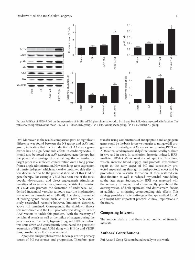