intraocular pressure - university of utah
TRANSCRIPT

© U N I V E R S I T Y O F U T A H H E A L T H
INTRAOCULAR PRESSURE
Natalie K Modersitzki, Dustin Randall MS4 , Alex Vitale BS, Lydia Sauer MD, Alex Tolman MS2

© U N I V E R S I T Y O F U T A H H E A L T H
EYE ANATOMY
Wikimedia –commons

© U N I V E R S I T Y O F U T A H H E A L T H
CORNEA
• The cornea is the transparent tissue that covers the iris, pupil and anterior chamber of the eye
• The cornea accounts for about 60 Diopters of refractive power. Its innate ability to bend light comes from its convex nature
• The cornea is used as a point of reference when measuring intraocular pressure (IOP)

© U N I V E R S I T Y O F U T A H H E A L T H
CORNEA
Moran axis

© U N I V E R S I T Y O F U T A H H E A L T H
• Corneal rigidity is a direct result of the way that the collagen fibers line up in the corneal matrix
• Corneal edema also affects pressure readings because of the fluid imbalance in the tissue
• IOP is a measure of resistance when we depress the cornea. Thus, corneal thickness also affects IOP measurement
BIOMECHANICS

© U N I V E R S I T Y O F U T A H H E A L T H
CORNEAL EDEMA
Moran axis

© U N I V E R S I T Y O F U T A H H E A L T H
• IOP is the fluid pressure inside the eye• It is an integral part of a patient’s work-up
as it is used as a screening tool for glaucoma
• Pressure is a measure of force per area • IOP is determined by the amount
of weight required to flatten 3.06 mm of the cornea
• Most tonometers measure pressure in millimeters of mercury (mmHg)
INTRAOCULAR PRESSURE
IOP= (F/C) +P
F= aqueous flow C= aqueous outflow P= episcleral venous pressure
Imbert-Fick Principle
© U N I V E R S I T Y O F U T A H H E A L T H
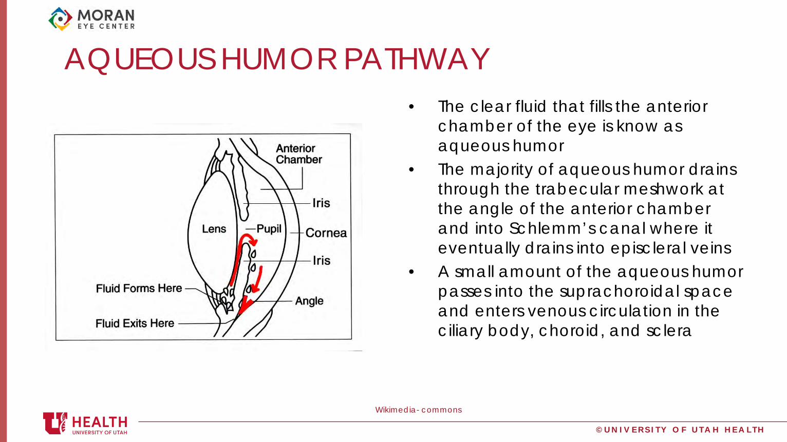
AQUEOUS HUMOR PATHWAY• The clear fluid that fills the anterior
chamber of the eye is know as aqueous humor
• The majority of aqueous humor drains through the trabecular meshwork at the angle of the anterior chamber and into Schlemm’s canal where it eventually drains into episcleral veins
• A small amount of the aqueous humor passes into the suprachoroidal space and enters venous circulation in the ciliary body, choroid, and sclera
Wikimedia- commons

© U N I V E R S I T Y O F U T A H H E A L T H
PHYSIOLOGY
• The primary determinant of IOP is the production, drainage, and pathway of aqueous humor in the anterior chamber
• The vitreous humor in the posterior segment has a fixed volume, and therefore it does not affect IOP measurements
• IOP also can increase with elevated systemic blood pressure

© U N I V E R S I T Y O F U T A H H E A L T H
ANGLE
Moranaxis

© U N I V E R S I T Y O F U T A H H E A L T H
IMPORTANCE OF IOP
• IOP is carefully regulated• Pressure in the eye helps maintain the integrity
of eye structure and shape• Irregular IOP can suggest pathologies such as
glaucoma, uveitis, or retinal detachment

© U N I V E R S I T Y O F U T A H H E A L T H
NORMAL PRESSURE
• Although the pressure in the eyes can fluctuate throughout the day, 10-22 mmHg is considered normal IOP
Timroot.com/glaucoma/ moran axis

© U N I V E R S I T Y O F U T A H H E A L T H
MEASURING IOP
• There are many different ways to eye pressure including: pneumotonometry, rebound tonometry, air-puff tonometry, and the gold standard – applanation.

© U N I V E R S I T Y O F U T A H H E A L T H
MEASURING IOP: TONOPEN
reichert
1. Instill a drop of topical anesthetic into the eye
2. Position the patient, seated supine, and have the patient fixate on a target
3. Position the pen in front of the cornea and make contact with the central cornea

© U N I V E R S I T Y O F U T A H H E A L T H
MEASURING IOP: TONOMETER
ICare
1. Position the patient, seated supine, and have the patient fixate on a target
2. Position the tonometer in front of the cornea and click the button to measure the pressure with the probe
3. Its light-weight probe makes momentary contact with the cornea
4. This device uses rebound technology to measure intraocular pressure

© U N I V E R S I T Y O F U T A H H E A L T H
MEASURING IOP: GOLDMAN APPLANATION
Wikkicommons/ Moran axis
© U N I V E R S I T Y O F U T A H H E A L T H
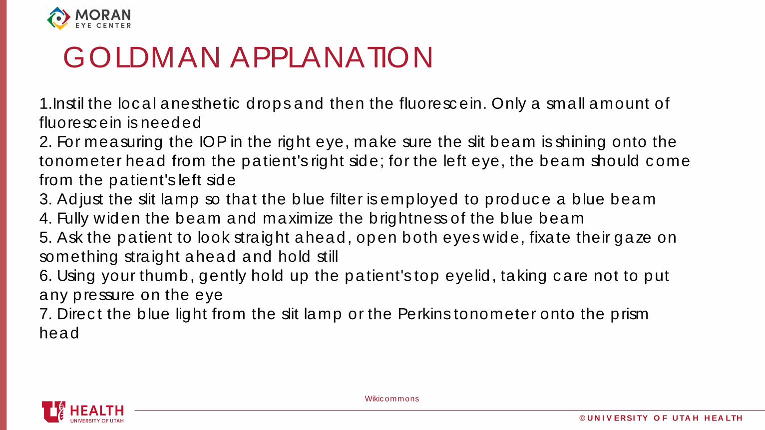
GOLDMAN APPLANATION
Wikicommons
1.Instil the local anesthetic drops and then the fluorescein. Only a small amount of fluorescein is needed2. For measuring the IOP in the right eye, make sure the slit beam is shining onto the tonometer head from the patient's right side; for the left eye, the beam should come from the patient's left side3. Adjust the slit lamp so that the blue filter is employed to produce a blue beam4. Fully widen the beam and maximize the brightness of the blue beam5. Ask the patient to look straight ahead, open both eyes wide, fixate their gaze on something straight ahead and hold still6. Using your thumb, gently hold up the patient's top eyelid, taking care not to put any pressure on the eye7. Direct the blue light from the slit lamp or the Perkins tonometer onto the prism head

© U N I V E R S I T Y O F U T A H H E A L T H
GOLDMAN APPLANATION
• 8. Align the tonometer head level with the patient’s eye• 9. Move the tonometer forward slowly until the prism rests on the center of the
cornea• 10. With the other hand, turn the calibrated dial on the tonometer clockwise until
the two fluorescein semi-circles in the prism head are seen to meet and form a horizontal ‘S’ shape. The correct end point is when the inner edges of the two fluorescein semi-circle images touch
• 11. The reading on the dial is the IOP measurement • 12. Withdraw the prism from the corneal surface and wipe its tip

© U N I V E R S I T Y O F U T A H H E A L T H
Wikimediacommons
© U N I V E R S I T Y O F U T A H H E A L T H
MEASURING IOP: PALPATION
• It is possible to detect very high IOP using your fingertips• Relative assessment of IOP • Variable evidence in showing correlation between tactile
assessment of IOP and tonometry – accuracy is limited and is generally more effective with very high and very low IOPs– may be useful in estimating IOP in patients who cannot undergo traditional
applanation tonometry (i.e. severe ocular surface disease, post-operative penetrating keratoplasty)

© U N I V E R S I T Y O F U T A H H E A L T H
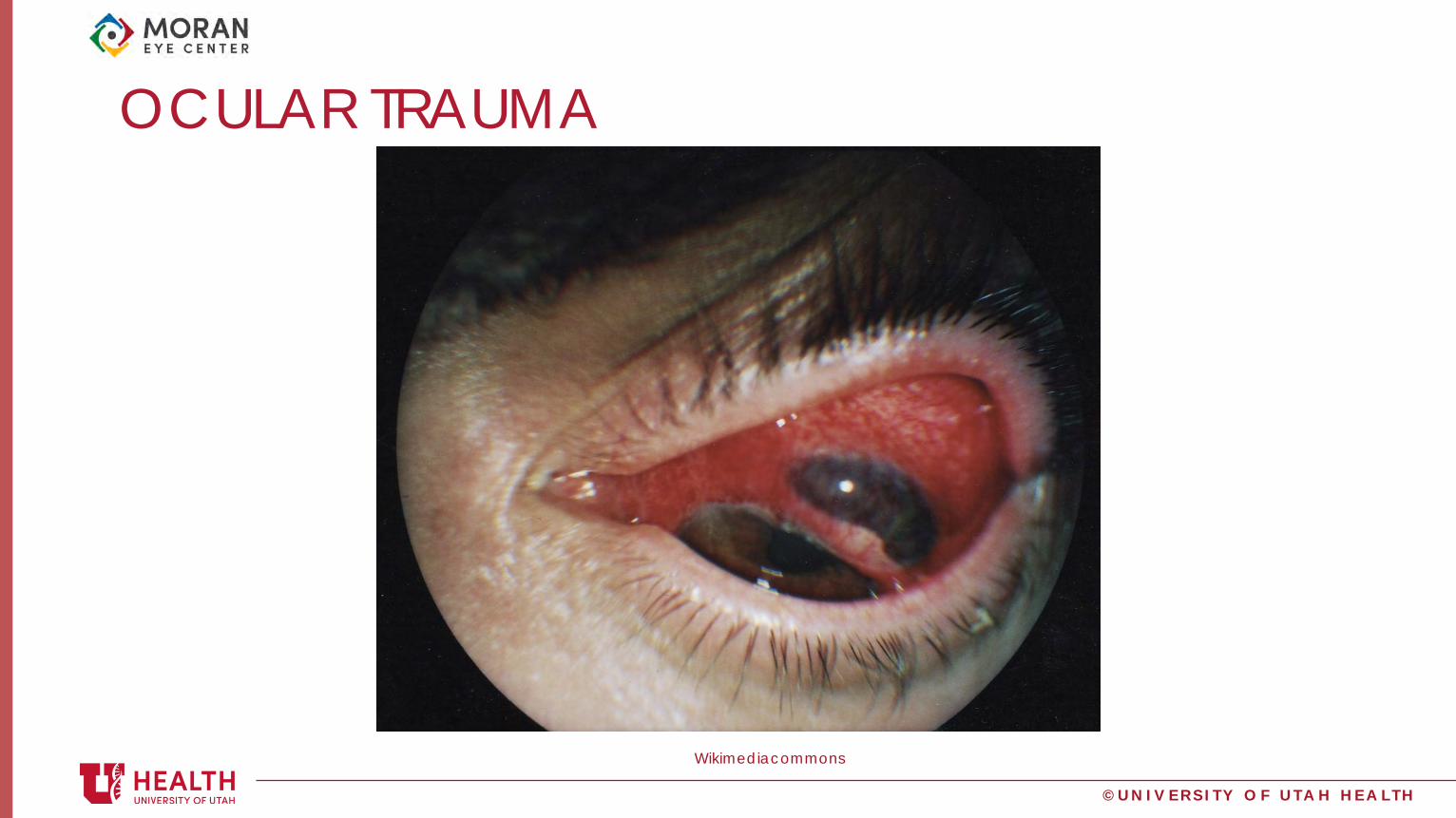
CONTRAINDICATIONS• Contraindications to measuring IOP include:
– Trauma – Corneal ulcer– Open globe
• Additional pressure in these conditions can cause further damage to the globe and lead to extrusion of aqueous and/or vitreous humor

© U N I V E R S I T Y O F U T A H H E A L T H
CORNEAL ULCER
morancore
© U N I V E R S I T Y O F U T A H H E A L T H
OCULAR TRAUMA
Wikimediacommons

© U N I V E R S I T Y O F U T A H H E A L T H
Conditions associated with high Intraocular Pressure
© U N I V E R S I T Y O F U T A H H E A L T H
GLAUCOMA: OPEN-ANGLE GLAUCOMA
• Glaucoma is a group of optic neuropathies usually (but not always) characterized by increased IOP
• Open-angle glaucoma is an optic neuropathy resulting in a progressive loss of retinal ganglion cell axons
– Usually is asymptomatic, but rarely can manifest initially as visual field loss and can lead to irreversible blindness if left untreated
• A leading cause of irreversible blindness world-wide
– A decrease in aqueous outflow or an increase in aqueous production are possible mechanisms for increased IOP

© U N I V E R S I T Y O F U T A H H E A L T H
GLAUCOMA- OPEN ANGLE

© U N I V E R S I T Y O F U T A H H E A L T H
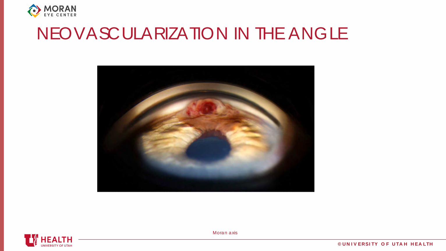
NEOVASCULAR GLAUCOMA
• A secondary glaucoma characterized by a development of new vessels over the iris and iridocorneal angle
• Primarily occurs in diabetic patients or those with a previous retinal vein occlusion
– Posterior segment ischemia induces VEGF production, which stimulates iris and angle neovascularization – Formation of new vessels and fibrous membrane blocks the iridocorneal angle, leading to an obstruction
of aqueous humor outflow– Outflow obstruction leads to an increased IOP
• Associated with poor visual prognosis
Moran axis

© U N I V E R S I T Y O F U T A H H E A L T H
NEOVASCULAR GLAUCOMA
Moran axis
© U N I V E R S I T Y O F U T A H H E A L T H
NEOVASCULARIZATION IN THE ANGLE
Moran axis

© U N I V E R S I T Y O F U T A H H E A L T H
PSEUDOEXFOLIATION SYNDROME (PXF)
• Systemic condition in which a basement membrane particulate material is deposited at the edge of the pupil, on the lens, in the drainage structures, and throughout other structures in the front of the eye
• The material adheres to the anterior lens capsule and can rub pigment off the iris and subsequently clog the trabecular meshwork, causing an increase in IOP
• Can cause zonular instability– Leads to surgical complications

© U N I V E R S I T Y O F U T A H H E A L T H
PXF DEPOSITS ON THE LENS
Moran Axis
© U N I V E R S I T Y O F U T A H H E A L T H
PIGMENT DISPERSION SYNDROME (PDS)
• Occurs when the posterior surface of the iris rubs against the zonules• Pigment clumps float in the aqueous humor and eventually clog the
trabecular meshwork, leading to an increase in IOP • Causes a transillumination defect in the iris • Most prevalent in young, white, myopic males
© U N I V E R S I T Y O F U T A H H E A L T H
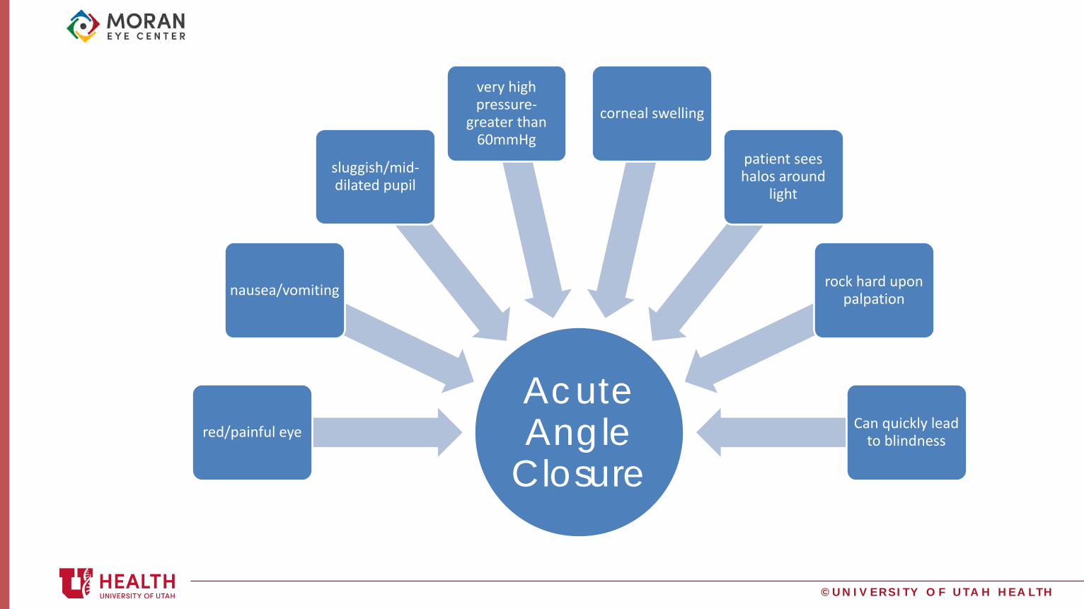
ACUTE ANGLE CLOSURE
• A medical emergency in which the lens is located too far forward anatomically and plasters against the iris
– The resistance creates a pressure gradient across the iris that forces the lens and iris to an even more anterior position
– As a result, the irido-corneal angle can become sealed off, completely blocking the trabecular meshwork, leading to a rapid, painful rise in IOP
• Without immediate treatment, the increase in pressure can damage the optic nerve and many other integral structures in the eye
© U N I V E R S I T Y O F U T A H H E A L T H
Acute Angle
Closure red/painful eye
nausea/vomiting
sluggish/mid-dilated pupil
very high pressure-
greater than 60mmHg
corneal swelling
patient sees halos around
light
rock hard upon palpation
Can quickly lead to blindness

© U N I V E R S I T Y O F U T A H H E A L T H
NARROW ANGLE

© U N I V E R S I T Y O F U T A H H E A L T H
UVEITIS
• Inflammation of the uvea, which is the middle portion of the eye• Elevated IOP affects 5- 19% of uveitis patients • Many different uveitic syndromes can lead to increased IOP
including: varicella zoster virus, herpes simplex virus, cytomegalovirus, sarcoidosis, toxoplasmosis, syphilis, acute uvetic angle closure and anterior uveitis (synechiae)
• Chronic and acute corticosteroid usage can also contribute to an increase in IOP in these patients
– Corticosteroids decrease outflow of aqueous humor
Aao.org

© U N I V E R S I T Y O F U T A H H E A L T H
UVEITIS

© U N I V E R S I T Y O F U T A H H E A L T H
TREATMENT FOR HIGH IOP• Decrease aqueous humor production:
– Beta blocker –timolol• Systemic side effects can occur from nasal absorption of topical Beta blockers,
making it especially important to ask your patients if they have asthma, COPD, or any cardiac problems
– Alpha agonists -brimonidine– Carbonic anhydrase inhibitor (CAI) - acetazolamide
• Increase uveoscleral outflow: – Prostaglandin analogues latanoprost, bimatoprost
• They can make eyelashes grow longer , and in a few patients it may darken their iris color, turning green and blue eyes brown

© U N I V E R S I T Y O F U T A H H E A L T H
TREATMENT FOR HIGH IOP• Increase uveoscleral outflow:
– Prostaglandin analogues latanoprost, bimatoprost• They can make eyelashes grow longer , and in a few patients it may darken their iris
color, turning green and blue eyes brown
• Increase trabecular outflow: – M3 agonist- pilocarpine, carbachol
• Surgical options– Trabeculectomy – creates an alternate drainage pathway – Bleb- reservoir for aqueous to flow into under the conjunctiva – Argon laser trabeculoplasty(ALT) – used to burn parts of the
trabecular meshwork causing increase in outflow

© U N I V E R S I T Y O F U T A H H E A L T H
Conditions with Low Intraocular Pressure

© U N I V E R S I T Y O F U T A H H E A L T H
HYPOTONY
• Defined as low IOP (below 10mmHg) • Often associated with decreased vision• Causes include:
– Post surgical wound leak– Chronic inflammation – Retinal detachment

© U N I V E R S I T Y O F U T A H H E A L T H
MACULAR FOLDS FROM HYPOTONY

© U N I V E R S I T Y O F U T A H H E A L T H
LEAKING WOUND • Loss of intraocular fluid leads to low eye pressure• Complaints of epiphora, defined as unintentional (not
caused by crying) overflow of tears onto the face• Physical manifestations include:
– shallow anterior chamber– large corneal folds– choroidal effusion – optic nerve edema
• Seidel positive upon examination

© U N I V E R S I T Y O F U T A H H E A L T H
SEIDEL POSITIVE
Moranaxis

© U N I V E R S I T Y O F U T A H H E A L T H
LEAKING BLEB

© U N I V E R S I T Y O F U T A H H E A L T H
SUMMARY
• IOP is a measure of resistance when we depress the cornea.
• Different diseases can cause fluctuations in pressure readings
• IOP is an important part of a comprehensive eye exam

© U N I V E R S I T Y O F U T A H H E A L T H
SPECIAL THANKS
• James Gilman, CRA, FOPS• Sophia Fang, MD

© U N I V E R S I T Y O F U T A H H E A L T H
REFERENCES
• www.RootAtlas.com• www.Wikimediacommons.com• www.AAO.org/intraocularpressure

Thank you