intraperitoneal injection of ginseng extract … injection of ginseng...ginseng enhances...
TRANSCRIPT

The American Journal of Chinese Medicine, Vol. 32, No. 1, 75–88© 2004 World Scientific Publishing Company
& Institute for Advanced Research in Asian Science and Medicine
Correspondence to: Dr. Jerming Tseng, Department of Medical Education and Research, Changhua ChristianHospital, 135 Nanhsiao Street, Changhua 500, Taiwan. E-mail: [email protected]
Intraperitoneal Injection of Ginseng ExtractEnhances Both Immunoglobulin and
Cytokine Production in Mice
Chian-Jiun LiouKang-Ning Junior College of Medical Care and Management
Nei-Hu, Taipei, Taiwan
Ming-Liang LiDepartment of Biology, National Taiwan Normal University
Taipei 116, Taiwan
Jerming TsengDepartment of Medical Education and Research, Changhua Christian Hospital
Changhua 500, Taiwan
Abstract: Ginseng is one of the most widely used Chinese herbal medicines. In this report, therelatively short-term effect of ginseng extract on the immunoglobulin production and cytokineproduction was studied. The ginseng extract was prepared by boiling the ground ginseng rootin 50% ethanol. The specific pathogen-free mice were intraperitoneally (i.p.) injected withvarious doses of ginseng extract for 3 consecutive days. The results indicated that the serumlevels of immunoglobulin (Ig)M, IgG and IgA were significantly elevated after the mice werei.p. injected with 4 g/kg/day of ginseng extract. Under in vitro condition, the lipopolysaccharide(LPS)-stimulated spleen cells showed a dose-dependent increase in secretion of IgM, IgG andIgA. However, at a higher dosage (4 g/kg/day), the amount of IgA secretion began to decline.The serum level of interleukin (IL)-2, interferon (IFN)-γ [T-helper (Th)1-type cytokines] andIL-4 and IL-10 (Th2-type cytokines) were significantly elevated after the mice were i.p. injectedwith 2 g/kg/day or higher doses of ginseng extract. The amount of cytokine secretion byconcanavalin A (Con A)-stimulated spleen cells was also significantly enhanced after themice were i.p. injected with 0.4 g/kg/day or higher dose of ginseng extracted. To furtherconfirm the results from enzyme-linked immunosorbent assay (ELISA), the spleen cells werecultured for 36 hours in the presence of 1 µg/ml of Con A. Total mRNA was isolated andassayed for mRNA expression using reverse transcriptase-polymerase chain reaction (RT-PCR). The results revealed that expression of IL-2 and IFN-γ mRNA were dose-dependentlyenhanced by the ethanol extract of ginseng. The levels of IL-4 and IL-10 mRNA expression
75
00177.p65 26/02/04, 1:52 PM75

C.-J. LIOU et al.76
were also elevated in the spleen cells of ginseng-treated mice in comparison with that of thecontrol group. In addition, we observed that the concentrations of IgG1, IgG2a and IgG2b inculture supernatants of spleen cells were dose-dependently increased by in vivo treatment ofginseng extract, suggesting that both Th1- and Th2-type cytokines were involved in IgGproduction. Our observation in this study demonstrated that the Chinese herbal drug ginsengwas able to regulate antibody production by augmenting Th1- (IL-2, IFN-γ) and Th2-type(IL-4, IL-10) cytokine production.
Keywords: Ginseng; Extract Preparation; Immunoglobulin; Cytokines; Mice.
Introduction
Ginseng (Panax ginseng, C.A. Meyer) has been a popular herbal remedy used in easternAsian cultures for thousands of years. Based on its major pharmacological effects, Panaxginseng is used in traditional Chinese medicine to enhance stamina and capacity to copewith fatigue and physical stress. The mechanisms of ginseng actions remain unclear, althoughthere is an extensive literature that deals with the effects of ginseng on a variety of diseasesas well as various infections (Awang, 1999; Ernst, 2002). Panax ginseng has also beenshown to accelerate hepatic lipogenesis and increase glycogen storage, which could contributeto an antidiabetic effect (Yokozawa et al., 1975). Persons who consumed fresh ginseng didshow a significantly reduced risk ratio for cancer (Yun and Choi, 1998). Furthermore, ginsenghas been proposed to be a potent immunomodulator. Ginseng enhanced production ofmacrophages, B- and T-cells, natural killer (NK) cells and colonic activity of bone marrow(Klein et al., 2000). The ethanol-insoluble fraction of an aqueous extract of ginseng wasfound to induce proliferation of splenocytes and generate activated killer cells in vitro(Yun et al., 1993). A systematic evaluation of multiple factors in the immune system revealedthat Panax ginseng stimulated basal NK cell activity and promoted a recovery of NK functionfrom cyclophosphamide-induced immunosuppression in mice (Kim et al., 1990). In addition,the extract of Ginseng radix was demonstrated to enhance the anti-Candida activity ofmacrophages in vitro and to prolong the survival time of Candida albicans-infected C3H/HeJ mice (Akagawa et al., 1996).
An acidic polysaccharide from ginseng named ginsan induced the generation of CD8+
LAK cells from both NK and T cells. The same report also demonstrated that ginsan inducedthe expression of mRNA for interleukin (IL)-2, interferon (IFN)-γ, IL-1α and granulocytemacrophage colony stimulating factor (GM-CSF) in C57BL/6 mouse spleen cells when thecells were cultured in the medium containing concanavalin A (Con A) plus ginsan(Kim et al., 1998). Recently, the anti-inflammatory effects of several ginsenosides derivedfrom Panax ginseng were studied. These ginsenosides suppressed expression ofcyclooxygenase-2 and activation of NF-kappaB in mice (Surh et al., 2002).
The previous documents suggested that ginseng showed profound effects on the humoralimmune response. Oral administration of the aqueous extract of ginseng for 5 to 6 dayssignificantly increased the primary immunoglobulin (Ig)M response by 50%, and augmentedthe secondary IgG and IgM response by 50% and 100%, respectively (Jie et al., 1984). The
00177.p65 26/01/04, 2:21 PM76

GINSENG ENHANCES IMMUNOGLOBULIN AND CYTOKINE PRODUCTION 77
volunteers receiving oral administration of a standardized extract of ginseng root, GinsanaG115, induced a higher immune response in vaccination against influenza in comparisonwith the individuals receiving placebo (Scaglione et al., 1996). However, in an athymic ratmodel, subcutaneous administration with ginseng extracts after rats were challenged withPseudomonas aeruginosa reduced serum anti-P. aeruginosa IgM and IgA antibody levels(Song et al., 1997a). In a rat model of chronic P. aeruginosa pneumonia, subcutaneousinjection of an aqueous extract of ginseng resulted in an increase in IgG2a level but a decreasein IgG1 level in comparison with that in the control group. The authors suggested that thechange from IgG1 to IgG2a subclasses indicated a possible shift from T-helper (Th)2- toTh1-type immune response (Song et al., 1998). Long-term oral administration of ginsengextract decreased the serum level of γ-globulin by 56%. Among the immunoglobulin isotypes,serum IgG1 was dose-dependently decreased to 68% of control value (Kim et al., 1997). Arecent study in lactating cows indicated that the ginsenoside R (b1) significantly augmentedantibody production and lymphocyte proliferation in response to pokeweed mitogen (PWM),Con A and Staphylococcus aureus antigens (Hu et al., 2003).
In this study, the specific pathogen-free mice were intraperitoneally injected with 50%ethanol extract of ginseng for 3 consecutive days. Both the immunoglobulin (IgG, IgA,IgM) and cytokine (IL-2, IFN-γ, IL-4, IL-10) production by the spleen cells were measuredafter the treatment. The relatively short-term effect of ginseng extract was estimated, and thepossible relation between cytokine production and immunostimulating effects of ginsengwas discussed.
Materials and Methods
Preparation of Herbal Drug Extract
A batch of herbal medications was purchased from the Lao-Chen-Ge Chinese herbal drugstore,De-Hua Street, Taipei. The herbs were ground into dried powder. The ginseng (5 g) powderwas mixed and suspended in 100 ml of 50% ethanol. The drug suspension was boiled untilhalf the volume of liquid remained. The suspension was then spun at 10,000 g for 30 minutesand the supernatant was collected and dried with a Speed Vac. The dried extracts werereconstituted using sterile phosphate buffered saline (PBS) to make a stock of 100 mgdrug/ml and sterilized with a 0.2 µm Millipore filter before used.
Animal Treatment and Cell Culture
For the drug treatment, the animals were divided into four groups and injected intraperitoneally(i.p.) with 0.5 ml of ginseng extract, ranging from 0.4, 2 to 4 g/kg body weight daily (g/kg/day)for three days. The control group was composed of mice injected with an equal volume ofPBS. Mice were sacrificed at day 4. The serum from individual mice was collected and thespleen cells isolated. The spleen cells (5 × 10⁵ cells/ml) were cultured in a medium containingRPMI 1640 supplemented with 10% fetal bovine serum (FBS), 2 mM L-glutamine, antibioticsand lipopolysaccharide (LPS; 1 µg/105 cells) for 5 days. The immunoglobulin concentrations
00177.p65 26/01/04, 2:21 PM77
C.-J. LIOU et al.78
in the culture supernatants were estimated using the enzyme-linked immunosorbent assay(ELISA) technique. To investigate the effects of ginseng on cytokine production, the micewere also treated as described, but the spleen cells were cultured in the presence of 1 µg/mlCon A for 3 days. Culture supernatants of spleen cell were collected, and the cytokineconcentrations were measured using the ELISA technique.
ELISA
IgA, IgG, IgM, IgG1, IgG2a and IgG2b concentrations were measured using a sandwichELISA technique. The capture antibody for the assay was a rabbit anti-mouse IgG + IgA +IgM antibody (Zymed Laboratory, CA). The secondary antibody for the assay was thehorseradish peroxidase (HRP)-conjugated goat anti-mouse IgA (1:500 diluted; ZymedLaboratory, CA), goat anti-mouse IgG (1:10,000 diluted; whole IgG molecule; JacksonImmunoresearch, PA), and goat anti-mouse IgM (1:2000 diluted; heavy chain-specific;Jackson Immunoresearch, PA). The HRP-conjugated goat anti-mouse IgG1, IgG2a andIgG2b (Zymed Laboratory, CA) were diluted to 1:1000. Briefly, a 96-well microtiter plate(Nunc-Immuno Plate, MaxiSorp, Nunc, Denmark) was precoated with 100 ng/well of captureantibodies at 4°C overnight. The plate was washed with PBS–0.05% Tween 20 solution andblocked with PBS-1% gelatin. After the blocking, the properly diluted samples and standardwere added (100 µl/well). Standards for IgG, and IgA had a range from 0.2 to 0.0031 µg/ml,standards for IgM had a range from 1 to 0.0312 µg/ml, and standards for IgG1, IgG2a andIgG2b had a range from 0.1 to 0.0016 µg/ml. The plate was then incubated at 37°Cfor 2 hours. At the end of incubation, a HRP-conjugated secondary antibody was added(100 µl/well). After 1 hour of incubation at 37°C, the color was developed using asubstrate solution containing 0.1 M citrate buffer, pH 4.5, 0.03% H2O2 and 0.1% ofo-phenylenediamine. The absorbance at 490 nm in each well was read using an ELISAreader (EL311, BioTek, VT), and the data was analyzed using log-logit model.
For the quantitative analysis of IL-2, IFN-γ, IL-4 and IL-10, cytokine ELISA set purchasedfrom R&D Systems (MN, USA) was used. The capture antibody was a rat monoclonalantibody to mouse cytokine and the detection antibody was a biotinylated goat anti-cytokinepolyclonal antibody. The color was developed by incubating the plate with an HRP-conjugatedstreptoavidin (Zymed, CA, USA), followed by a substrate solution containing hydrogenperoxide and tetramethylbenezidine (TMB). The reaction continued for 30 minutes at roomtemperature, and was stopped by adding 100 µl of 2N of sulfuric acid. The absorbance at450 nm in each well was read using an ELISA reader (EL311, BioTek, Winooski, VT), andthe data was analyzed using a log-logit model.
Synthesis of cDNA
The level of Th1 type (IL-2, IFN-γ) and Th2 type (IL-4, IL-10) mRNA in the spleen cellswas estimated using reverse transcriptase-polymerase chain reaction (RT-PCR) analysis
00177.p65 26/01/04, 2:21 PM78

GINSENG ENHANCES IMMUNOGLOBULIN AND CYTOKINE PRODUCTION 79
36 hours after culture in the presence of 1 µg/ml of Con A. Spleen cells (1 × 10⁷ cells)isolated from ginseng-treated animals were washed twice with 1 × RNase-free PBS. The cellpellet was mixed with 1 ml of TRIzol reagent (GIBCO-BRL, MD), and the mixture wasforced to pass through a 25G needle five times to release RNA from the cells. This homogenatewas then vigorously mixed with 0.2 ml chloroform. After sitting at room temperature for10 minutes, the mixture was spun at 4°C for 15 minutes to separate the organic and aqueouslayers. The aqueous layers were removed into a new tube and RNA was precipitated with0.5 ml of isopropanol. The precipitate was then resuspended in 30 µl of RNase-free water,and a 5 µl aliquot was removed for RNA quantification using GeneQuant II (PharmaciaBiotech, Piscataway, NJ). RNA in RNase-free water (2 µg in 10 µl) was mixed with 2.5 µl ofoligo (dT)15 solution (40 pM; Promega, WI). The solution was heated at 70°C for 10 minutes,followed by cooling at room temperature for 10 minutes and then transferred onto ice. Areaction mixture containing 4 dNTPs (Boehringer Mannheim, Germany), DTT (GIBCO/BRL, MD), reverse transcriptase (M-MLV; Promega, WI) and RNasin (Promega, WI) wassubsequently mixed with RNA. The reaction was carried out at 37°C for 60 minutes tosynthesize the cDNA.
PCR
cDNA (5 µl) was mixed with 0.5 µl 4 dNTP, 10 µl primer mix (2.5 µM each), 0.5 µlTaq polymerase, 2 µl MgCl2 (2 mM), and 5 µl PCR buffer. The DEPC-treated waterwas then added to make a volume up to 50 µl in total. The primers (Promega, WI)used for PCR amplification were as follows: IL-10 sense primer: 5′-ATGCAGGACTTTAAGGGTTACTTG-3′; IL-10 antisense primer: 5′-TAGACACCTTGGTCTTGGAGCTTA-3′; IL-4 sense primer 5′-TGCCTCCAAGAACACAACTG-3′;IL-4 antisense primer: 5′-AACGTACTCTGGTTGGCTTC-3′; IL-2 sense primer:5′-GACACTTGTGCTCCTTGTCA-3′; IL-2 antisense primer: 5′-TCAATTCTGTGGCCTGCT TG-3′; IFN-γ sense primer: 5′-GCAGAGCCAAATTGTCTCCT-3′; IFN-γantisense primer: 5′-ATGCTCTTCGACCTCGAAAC-3′; β2 microglobulin (internal control)sense primer: 5′-TGACCGGCTTGTATGCTATC-3′; β2 microglobulin antisense primer:5′-CAGTGTGAGCCAGGATATAG-3′. The PCR conditions were denaturation at 94°C for50 seconds, annealing at 60°C for 45 seconds and primer extension at 72°C for 45 seconds.After 35 cycles (40 cycles for IL-10) of amplification, the PCR products were subjected togel electrophoresis through 1.5% agarose (Sigma, MO) containing ethidium bromide at80 V. The amplicons were visualized under UV light.
Statistical Analysis
Data from the control or drug-treatment groups were tested by ANOVA. The differencebetween the two means was assessed using the Student’s t-test. Probability values of < 0.05were considered to be significant.
00177.p65 26/01/04, 2:21 PM79

C.-J. LIOU et al.80
Results
Ginseng Induced an Increase in Immunoglobulin Production
Both the sera and spleen cells isolated from the mice injected with various doses of ginsengor the same volume of PBS (control group) were collected. The spleen cells were culturedin vitro for 5 days with the presence of 1 µg/ml LPS. The amount of immunoglobulins (IgG,IgA and IgM) in sera and culture supernatants were measured using ELISA. The resultsindicated that the serum levels of IgM, IgG and IgA were significantly elevated after themice were i.p. injected with 4 g/kg/day of 50% ethanol-extract of ginseng for 3 consecutivedays (Fig. 1). The IgM level was evenly elevated at the relatively low dose (2 g/kg/day).Under in vitro condition, the LPS-stimulated spleen cells showed a dose-dependent increasein secretion of IgM and IgG (Fig. 2). For the spleen cells isolated from the mice treated with0.4 and 2 g/kg/day of ginseng extract, the amount of IgA in culture supernatant was alsoincreased. However, at a higher dosage (4 g/kg/day), the amount of IgA secretion began todecline but was still significantly higher than that of the control group.
The predominant immunoglobulin in serum was IgG, which had a range between10 to 20 mg/ml (Fig. 1). The serum level of IgM was relatively low, which was only 0.1 to0.2 mg/ml. However, the predominant immunoglobulin secreted by the LPS-stimulated spleencells was IgM, which was between 0.5 to 1.5 µg/ml in culture supernatant (Fig. 2). Both theIgG and IgA secretion were less than 80 ng/ml. This simply demonstrated that LPS was aT-independent antigen. The LPS-activated B-lymphocytes predominantly produced IgMand showed no class switching. Therefore, the ginseng-induced increase in immunoglobulinsecretion might be due to either the class-switching by ethanol-extract of ginseng beinginduced or the secretion of the pre-existing immunoglobulin producers being augmented.
Ginseng Induced an Increase in Cytokine Production
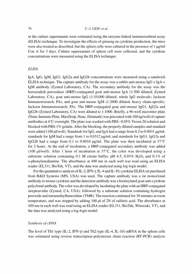
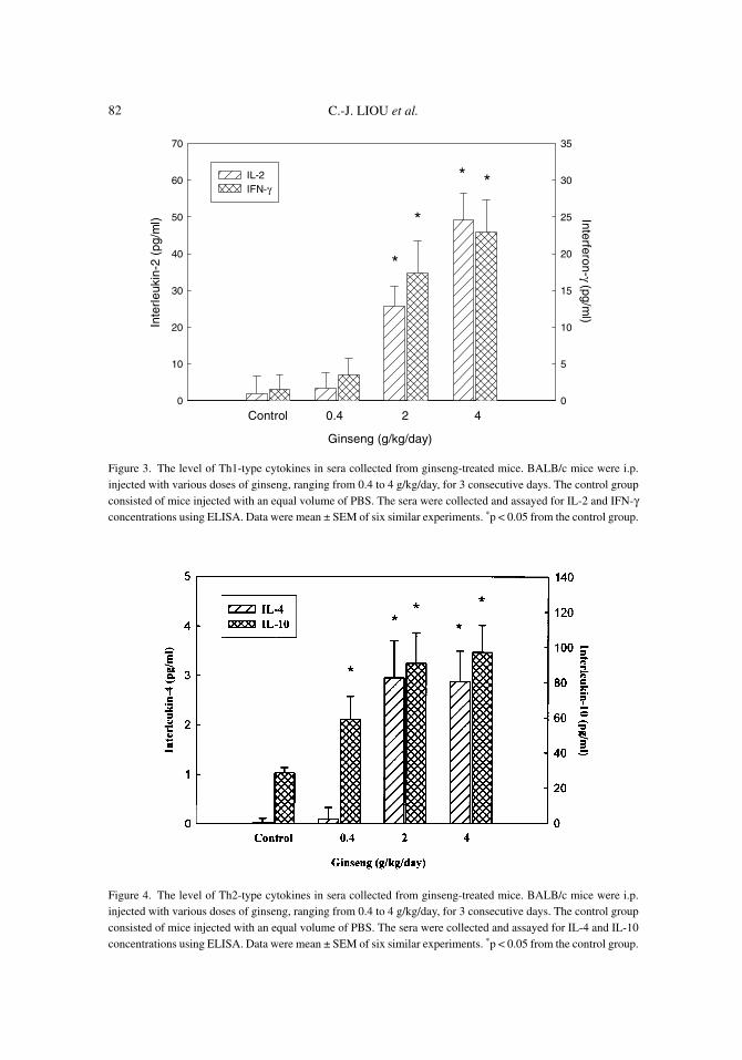
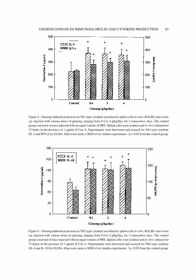
In order to study the possible role of cytokines in ginseng-induced increase of immunoglobulinproduction, both the sera and spleen cells isolated from the ginseng-treated mice were assayedfor cytokine level and secretion, respectively. The results indicated that the serum level ofIL-2, IFN-γ (Th1-type cytokines) was significantly elevated after the mice were i.p. injectedwith 2 g/kg/day or higher dose of 50% ethanol-extracted of ginseng for 3 consecutive days.The serum concentration of IL-2 increased five to ten folds, and that of IFN-γ also increasedabout ten to 15 folds (Fig. 3). The serum level of IL-4 and IL-10 (Th2-type cytokines) werealso elevated by the ginseng treatment. Ginseng treatment dose-dependently increased theserum level of IL-10. The mice injected with 2 g/kg/day or more of ginseng extract alsoshowed a significant increase in the serum level of IL-4. However, the serum level of IL-4was in a range between 0.1 to 3 pg/ml, which was approximately 100-fold lower than that ofIL-10 (Fig. 4). For the spleen cells isolated from ginseng-treated mice, the concentrations ofTh1- and Th2-type cytokines in culture supernatants were also increased significantly incomparison to that of the control group (Figs. 5 and 6). However, the amount ofTh1-type cytokines produced by spleen cells was approximately two-fold higher than that ofTh2-type cytokines.
00177.p65 26/01/04, 2:21 PM80

GINSENG ENHANCES IMMUNOGLOBULIN AND CYTOKINE PRODUCTION 81
Figure 1. The level of immunoglobulin in sera collected from ginseng-treated mice. BALB/c mice were i.p.injected with various doses of ginseng, ranging from 0.4 to 4 g/kg/day, for 3 consecutive days. The control groupconsisted of mice injected with an equal volume of PBS. Sera were collected and assayed for IgG, IgA and IgMconcentrations using ELISA. Data were mean ± SEM of six similar experiments. *p < 0.05 from the control group.
Figure 2. Ginseng induced an increase in immunoglobulin secretion by spleen cells in vitro. BALB/c mice werei.p. injected with various doses of ginseng, ranging from 0.4 to 4 g/kg/day, for 3 consecutive days. The controlgroup consisted of mice injected with an equal volume of PBS. Spleen cells were isolated after the treatment andthen incubated for 5 days with 1 µg/ml of LPS. Supernatants were harvested and assayed for Ig concentrationsusing ELISA. Data were mean ± SEM of six similar experiments. *p < 0.05 from the control group.
00177.p65 26/01/04, 2:21 PM81
C.-J. LIOU et al.82
Figure 4. The level of Th2-type cytokines in sera collected from ginseng-treated mice. BALB/c mice were i.p.injected with various doses of ginseng, ranging from 0.4 to 4 g/kg/day, for 3 consecutive days. The control groupconsisted of mice injected with an equal volume of PBS. The sera were collected and assayed for IL-4 and IL-10concentrations using ELISA. Data were mean ± SEM of six similar experiments. *p < 0.05 from the control group.
Figure 3. The level of Th1-type cytokines in sera collected from ginseng-treated mice. BALB/c mice were i.p.injected with various doses of ginseng, ranging from 0.4 to 4 g/kg/day, for 3 consecutive days. The control groupconsisted of mice injected with an equal volume of PBS. The sera were collected and assayed for IL-2 and IFN-γconcentrations using ELISA. Data were mean ± SEM of six similar experiments. *p < 0.05 from the control group.
Inte
rleu
kin-
2 (p
g/m
l)
0
10
20
30
40
50
60
70Interferon-γ (pg/m
l)
0
5
10
15
20
25
30
35
IL-2IFN-γ
Ginseng (g/kg/day)
Control 0.4 2 4
*
*
* *
00177.p65 26/01/04, 2:21 PM82
GINSENG ENHANCES IMMUNOGLOBULIN AND CYTOKINE PRODUCTION 83
Figure 5. Ginseng induced an increase in Th1-type cytokine secretion by spleen cells in vitro. BALB/c mice werei.p. injected with various doses of ginseng, ranging from 0.4 to 4 g/kg/day, for 3 consecutive days. The controlgroup consisted of mice injected with an equal volume of PBS. Spleen cells were isolated and in vitro cultured for72 hours in the presence of 1 µg/ml of Con A. Supernatants were harvested and assayed for Th1-type cytokine(IL-2 and IFN-γ) by ELISA. Data were mean ± SEM of six similar experiments. *p < 0.05 from the control group.
Figure 6. Ginseng induced an increase in Th2-type cytokine secretion by spleen cells in vitro. BALB/c mice werei.p. injected with various doses of ginseng, ranging from 0.4 to 4 g/kg/day, for 3 consecutive days. The controlgroup consisted of mice injected with an equal volume of PBS. Spleen cells were isolated and in vitro cultured for72 hours in the presence of 1 µg/ml of Con A. Supernatants were harvested and assayed for Th2-type cytokine(IL-4 and IL-10) by ELISA. Data were mean ± SEM of six similar experiments. *p < 0.05 from the control group.
00177.p65 26/01/04, 2:21 PM83

C.-J. LIOU et al.84
Ginseng Augmented Expression of Th1- and Th2-Type Cytokine mRNA
To further confirm the results from ELISA, the spleen cells were isolated after the treatmentand cultured for 36 hours in the presence of 1 µg/ml of Con A. Total mRNA was isolatedand assayed for mRNA expression using RT-PCR. Results of RT-PCR correlated well withthat of ELISA in which expression of IL-2 and IFN-γ mRNA were dose-dependently enhancedby ethanol extract ginseng (Fig. 7). The levels of IL-4 and IL-10 mRNA expression werealso elevated in the spleen cells from ginseng-treated mice in comparison to that of thecontrol group (Fig. 7).
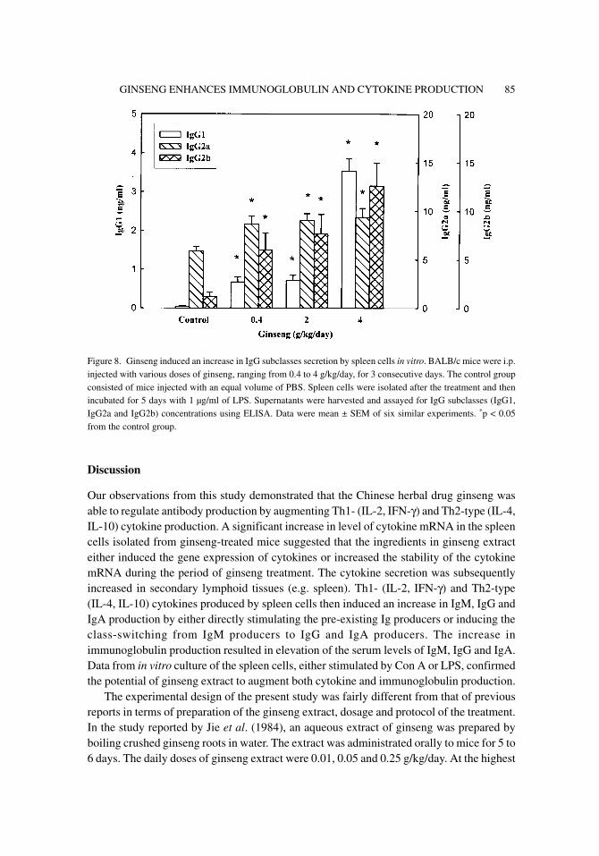
Ginseng Extract Increased Secretion of IgG Subtypes by Spleen Cells
To demonstrate that Th1- and Th2-type cytokines were involved in IgG production, theamount of IgG1, IgG2a and IgG2b secretion by spleen cells in vitro were assayed. Resultsindicated that the concentrations of IgG1 and IgG2b in culture supernatants of spleen cellswere dose-dependently increased by in vivo treatment of ginseng extract (Fig. 8). An increasein IL-4 production resulted in class-switching from IgM producer to IgG1 and IgG2b producer.Therefore, it further confirmed that IL-4 production was enhanced by ginseng extract. Theamount of IgG2a secretion was significantly increased after the mice were treated with ginsengextract at as low as 0.4 g/kg/day. Since IFN-γ was a potential inducer for class-switchingfrom IgM to IgG2a, this result also confirmed that ginseng-treatment did enhance IFN-γproduction.
Figure 7. Detection of cytokine mRNA expression in spleen cells isolated from ginseng-treated mice. BALB/cmice were i.p. injected with various doses of ginseng, ranging from 0.4 to 4 g/kg/day, for 3 consecutive days. Thecontrol group consisted of mice injected with an equal volume of PBS. Spleen cells were isolated and in vitro
cultured for 36 hours in the presence of 1 µg/ml of Con A. The total mRNA was isolated from the spleen cells(1 × 10⁷ cells). cDNA was synthesized and then amplified using PCR. Lane 1: control, Lane 2: 0.4 g/kg/day,Lane 3: 2 g/kg/day, and Lane 4: 4 g/kg/day.
00177.p65 26/01/04, 2:21 PM84
GINSENG ENHANCES IMMUNOGLOBULIN AND CYTOKINE PRODUCTION 85
Discussion
Our observations from this study demonstrated that the Chinese herbal drug ginseng wasable to regulate antibody production by augmenting Th1- (IL-2, IFN-γ) and Th2-type (IL-4,IL-10) cytokine production. A significant increase in level of cytokine mRNA in the spleencells isolated from ginseng-treated mice suggested that the ingredients in ginseng extracteither induced the gene expression of cytokines or increased the stability of the cytokinemRNA during the period of ginseng treatment. The cytokine secretion was subsequentlyincreased in secondary lymphoid tissues (e.g. spleen). Th1- (IL-2, IFN-γ) and Th2-type(IL-4, IL-10) cytokines produced by spleen cells then induced an increase in IgM, IgG andIgA production by either directly stimulating the pre-existing Ig producers or inducing theclass-switching from IgM producers to IgG and IgA producers. The increase inimmunoglobulin production resulted in elevation of the serum levels of IgM, IgG and IgA.Data from in vitro culture of the spleen cells, either stimulated by Con A or LPS, confirmedthe potential of ginseng extract to augment both cytokine and immunoglobulin production.
The experimental design of the present study was fairly different from that of previousreports in terms of preparation of the ginseng extract, dosage and protocol of the treatment.In the study reported by Jie et al. (1984), an aqueous extract of ginseng was prepared byboiling crushed ginseng roots in water. The extract was administrated orally to mice for 5 to6 days. The daily doses of ginseng extract were 0.01, 0.05 and 0.25 g/kg/day. At the highest
Figure 8. Ginseng induced an increase in IgG subclasses secretion by spleen cells in vitro. BALB/c mice were i.p.injected with various doses of ginseng, ranging from 0.4 to 4 g/kg/day, for 3 consecutive days. The control groupconsisted of mice injected with an equal volume of PBS. Spleen cells were isolated after the treatment and thenincubated for 5 days with 1 µg/ml of LPS. Supernatants were harvested and assayed for IgG subclasses (IgG1,IgG2a and IgG2b) concentrations using ELISA. Data were mean ± SEM of six similar experiments. *p < 0.05from the control group.
00177.p65 26/01/04, 2:21 PM85

C.-J. LIOU et al.86
dose, i.e. 0.25 g/kg/day, both the primary IgM response and the secondary IgG and IgMresponses were increased by 50%. The author also observed an enhancement of interferonproduction in non-stimulated spleen cells. We had similar findings, but the extract of ginsengwas prepared by boiling the ground ginseng roots in 50% ethanol, instead. The extract wasadministrated i.p. for 3 days. The daily doses of ginseng extract were 0.4, 2 and 4 g/kg/day,which was significantly higher than that of Jie’s study. However, we observed an increase insecretion of immunoglobulin at the lowest dose (0.4 g/kg/day), which was only slightlyhigher than the dose used by Jie’s group.
However, Song’s group showed different results. In an athymic rat model, theysubcutaneously administrated the rats with an aqueous extract of ginseng after the animalswere challenged with Pseudomonas aeruginosa. The daily dosage of the ginseng extractwas 0.025 g/kg/day and the rats were treated with the extract for 10 days. After the treatment,they observed a reduction in serum levels of anti-P. aeruginosa IgM and IgA antibodies(Song et al., 1997b). The extract used by Song’s group was water-soluble instead of the 50%ethanol-soluble fraction of ginseng. The dose used in Song’s study was relatively low andthe period of treatment in their study was longer than that in our study. In addition, theymonitored the effect of ginseng on the antigen-specific antibody response instead of onpolyclonal antibody production.
In a rat model of chronic P. aeruginosa pneumonia, subcutaneous injection of an aqueousextract of ginseng at a dose of 0.025 g/kg/day for 2 weeks resulted in an increase in IgG2alevel but a decrease in IgG1 level in comparison to that of the control group (Song et al.,1998). The authors suggested that the change from IgG1 to IgG2a subclasses indicated apossible shift from Th2- to Th1-type immune response. Kim’s group studied the long-termoral administration of ginseng extract to healthy female mice. They found that oraladministration of ginseng at doses of 0.03 and 0.15 g/kg/day for 52 days decreased theserum level of γ-globulin by 56%. However, the Ig isotypes, including IgG2a, IgG2b, IgG3,IgM and IgA, in serum was unchanged, but serum IgG1 was dose-dependently decreased to68% of the control value (Kim et al., 1997). Namely, the healthy condition and the length ofadministration might result in an opposite effect on the production of Ig isotypes. Kim’sgroup previously studied ginsan, an acidic polysaccharide from ginseng. When the spleencells were cultured in vitro with the presence of ginsan, they found that ginsan inducedthe generation of CD8+ LAK cells from both NK and T cells. The same report alsodemonstrated that ginsan induced the expression of mRNA for IL-2, IFN-γ, IL-1α andGM-CSF in C57BL/6 mouse spleen cells when the cells were cultured with Con A plusginsan (Kim et al., 1997). In our study, we were unable to find any preference between Th1-and Th2-type immune responses after the mice were i.p. injected with 50% ethanol extractof ginseng.
Although ginseng has been used in Eastern Asian countries for more than a thousandyears, the biological activity, pharmacological effect and its active ingredients remain to bedetermined. Evidence from controlled clinical trials did not support the use of ginseng totreat its established indications (Ernst, 2002). Furthermore, the way to prepare ginseng extract,the dosage, the route of administration, and the protocol of treatment can indeed affect theefficacy of ginseng. Therefore, more studies are needed to establish the critical parameters.
00177.p65 26/01/04, 2:21 PM86

GINSENG ENHANCES IMMUNOGLOBULIN AND CYTOKINE PRODUCTION 87
Acknowledgments
This work was presented by Chian-Jiun Liou to the Department of Biology, National TaiwanNormal University in partial fulfillment of the requirement for a PhD degree.
References
Akagawa, G., S. Abe, S. Tansho, K. Uchida and H. Yamaguchi. Protection of C3H/HE J mice fromdevelopment of Candida albicans infection by oral administration of Juzen-taiho-to and itscomponent, Ginseng radix: possible roles of macrophages in the host defense mechanisms.Immunopharmacol. Immunotoxicol. 18: 73–89, 1996.
Awang, D.V.C. Immune stimulants and antiviral botanicals: echincea and ginseng. In: J. Janick (ed.)Perspectives on New Crops and New Uses. ASHS Press, Alexandria, VA, 1999, pp. 450–456.
Ernst, E. The risk-benefit profile of commonly used herbal therapies: ginkgo, St. John’s wort, ginseng,echinacea, saw palmetto, and kava. Ann. Intern. Med. 136: 42–53, 2002.
Hu, S., C. Concha, F. Lin and W.K. Persson. Adjuvant effect of ginseng extracts on the immuneresponses to immunization against Staphylococcus aureus in dairy cattle. Vet. Immunol.Immunpathol. 91: 29–37, 2003.
Jie, Y.H., S. Cammisuli and M. Baggiolini. Immunodulatory effects of panax ginseng C. A. Meyer inthe mouse. Agent. Action 15: 386–391, 1984.
Kim, J.Y., D.R. Germolec and M.I. Luster. Panax ginseng as a potential immunomodulator: studies inmice. Immunopharmacol. Immunotoxicol. 12: 257–276, 1990.
Kim, K.H., Y.S. Lee, I.S. Jung, S.Y. Park, H.Y. Chung, I.R. Lee and Y.S. Yun. Acidic polysaccharidefrom Panax ginseng, ginsan, induces Th1 cell and macrophage cytokines and generates LAKcells in synergy with rIL-2. Planta Med. 64: 110–115, 1998.
Kim, Y.W., D.K. Song, W.H. Kim, K.M. Lee, M.B. Wie, Y.H. Kim, S.H. Kee and M.K. Cho. Long-term oral administration of ginseng extract decreases serum gamma-globulin and IgG1 isotypein mice. J. Ethnopharmacol. 58: 55–58, 1997.
Klein, C., T. Sato, M.M. Meguid and G. Miyata. From food to nutritional support to specificnutraceuticals: a journey across time in the treatment of disease. J. Gastroenterol. 35: 1–6, 2000.
Scaglione, F., G. Cattaneo, M. Alessandria and R. Cogo. Efficacy and safety of the standardizedginseng extract G115 for potentiating vaccination against the influenza syndrome and protectionagainst the common cold. Drug Exp. Clin. Res. 22: 65–72, 1996.
Song, Z., H.K. Johansen, V. Fabber, C. Moser, A. Kharazmi, J. Rygaard and N. Hoiby. Ginsengtreatment reduces bacterial load and lung pathology in chronic Pseudomonas aeruginosapneumonia in rats. Antimicrobiol. Agent. Chemother. 41: 961–964, 1997a.
Song, Z.J., H.K. Johansen, V. Faber and N. Høiby. Ginseng treatment enhances bacterial clearanceand decreases lung pathology in athymic rats with chronic P. aeruginosa pneumonia. APMIS105: 438–444, 1997b.
Song, Z., A. Kharazmi, H. Wu, V. Faber, C. Moser, H.K. Johansen, J. Rygaard and N. Høiby.Effects of ginseng treatment on neutrophil chemiluminescence and immunoglobulin Gsubclasses in a rat model of chronic Pseudomonas aeruginosa pneumonia. Clin. Diagn. Lab.Immunol. 5: 882–887, 1998.
Surh, Y.J., J.Y. Lee, K.J. Choi and S.R. Ko. Effects of selected ginsenosides on phorbol ester-inducedexpression of cyclooxygenase-2 and activation of NF-kappaB and ERK 1/2 in mouse skin.Ann. N. Y. Acad. Sci. 973: 396–401, 2002.
00177.p65 26/01/04, 2:21 PM87

C.-J. LIOU et al.88
Yokozawa, T., H. Seno and H. Oura. Effect of ginseng extract on lipid and sugar metabolism. I.Metabolic correlation between liver and adipose tissue. Chem. Pharm. Bull. (Tokyo). 23: 3095–3100, 1975.
Yun, T.K. and S.Y. Choi. Non-organ specific cancer prevention of ginseng: a prospective study inKorea. Int. J. Epidemiol. 27: 359–364, 1998.
Yun, Y.S., Y.S. Lee, S.K. Jo and I.S. Jung. Inhibition of autochthonous tumor by ethanol insolublefraction from Panax ginseng as an immunomodulator. Planta Med. 59: 521–524, 1993.
00177.p65 26/01/04, 2:21 PM88
