introduction and definitions - welcome to d...
TRANSCRIPT

TYPE 1 OR TYPE 2 DIABETES? WHAT’S IN A NAME. THE CONTROVERSY AND CONUNDRUM OF DIABETES TYPE IN YOUTH AND THE PUBLIC HEALTH
IMPACT
by
Melissa A. Buryk
B.S., United States Naval Academy, 2003
M.D., Uniformed Services University of the Health Sciences, 2007
Submitted to the Graduate Faculty of
Graduate School of Public Health in partial fulfillment
of the requirements for the degree of
Master of Public Health
University of Pittsburgh
2014
UNIVERSITY OF PITTSBURGH
GRADUATE SCHOOL OF PUBLIC HEALTH
This essay is submitted
by
Melissa Buryk
on
July 25, 2014
and approved by
Essay Advisor:David Finegold, MD ______________________________________Director, Multidisciplinary MPH ProgramProfessor, Department of Human GeneticsGraduate School of Public HealthUniversity of Pittsburgh
Essay Reader:Dorothy Becker, MBBCh ______________________________________ProfessorDepartment of Pediatric EndocrinologyChildren’s Hospital of Pittsburgh of UPMC

Copyright © by Melissa A. Buryk
2014

ABSTRACT
Type 1a diabetes (T1D) is an autoimmune condition characterized by islet cell
destruction and progressive insulin deficiency, characteristically present with weight loss and
thin habitus. With the rising incidence of obesity over the past several decades, the prevalence of
obesity at onset of diabetes has increased. Furthermore, the incidences of both Type 2 diabetes
(T2D) and T1D have significantly increased in recent years. Therefore, it has become
increasingly difficult to distinguish between T1D and T2D at onset of disease in children with
several hypotheses available to attempt to link these diagnoses with the theory that obesity itself
may be playing a role in the increasing onset of diabetes in children. The distinction between
T1D and T2D may not be of initial therapeutic importance, and may not even be a valid
classification system. However, the differentiation is of importance to public health because it
helps to characterize the co-morbidities of the rising obesity epidemic and plan for future health
care delivery and the costs of these conditions and their comorbidities rise. With obesity leading
to diabetes, improved public health efforts to stem the obesity epidemic may ultimately be able
to decrease the long-term costs to the healthcare system resulting from increasing obesity
epidemic.
Diabetes associated autoantibodies are the traditional method used to differentiate T1D
from T2D. However, patients with phenotypic T1D may be negative for autoantibodies at
disease diagnosis with commonly used testing methods. Therefore, absence of antibodies, even
David Finegold, MD
TYPE 1 OR TYPE 2 DIABETES? WHAT’S IN A NAME. THE CONTROVERSY AND CONUNDRUM OF DIABETES TYPE IN YOUTH AND THE
PUBLIC HEALTH IMPACT
Melissa Buryk, MPH
University of Pittsburgh, 2014
in the presence of obesity, does not necessarily indicate the presence of T2D. We have used other
markers of autoimmunity (diabetes associated T cell responses) and HLA typing to identify
additional autoimmune findings in antibody negative, insulin-requiring diabetes. This enhances
the argument that many autoimmune children may be classified with T2D due to the presence of
obesity and evidence of insulin resistance and that autoantibody negative diabetes (and pediatric
diabetes in general) may is heterogeneous, with our current classification system not adequate to
fully characterize this condition.

TABLE OF CONTENTS
1.0 INTRODUCTION AND DEFINITIONS...................................................................1
1.1 PREVALENCE AND INCIDENCE OF T1D AND T2D..................................2
1.2 OBESITY AND THE RISING INCIDENCE OF T1D.....................................3
1.2.1 Evidence for and against the accelerator hypothesis....................................5
1.2.2 Potential over and underestimation of T2D and T1D prevalence...............8
1.3 WHY IS THE DISTINCTION BETWEEN T1D AND T2D IMPORTANT? 9
2.0 EVALUATION FOR AUTOIMMUNE FACTORS IN AUTOANTIBODY
NEGATIVE INSULIN REQUIRING DIABETES...................................................................11
2.1 SPECIFIC AIMS AND HYPOTHESIS...........................................................12
2.2 RESEARCH DESIGN AND METHODS........................................................12
2.2.1 Study population.............................................................................................12
2.2.2 Autoantibody assays..........................................................................................13
2.2.3 T cell proliferation assay...................................................................................14
2.2.4 Molecular typing of HLA alleles.......................................................................15
2.2.5 C-Peptide............................................................................................................15
2.2.6 Data analyses and statistics...............................................................................16
2.3 RESULTS............................................................................................................16
2.3.1 Course over 2 years............................................................................................17
2.4 CONCLUSIONS FROM EVALUATION OF ANTIBODY NEGATIVE
SUBJECTS...........................................................................................................................19
3.0 CONCLUSIONS.........................................................................................................20
BIBLIOGRAPHY........................................................................................................................22

LIST OF TABLES
Table 1. Comparison of Immune Features in Autoantibody Negative Versus Autoantibody
positive insulin requiring diabetes children...................................................................................17

LIST OF FIGURES
Figure 1: Autoimmune cascade for development of diabetes.........................................................4
Figure 2: Comparison of the acceleration hypotheses.....................................................................6


1.0 INTRODUCTION AND DEFINITIONS
John Snow’s removal of the Broad Street pump handle and the subsequent remission of the
cholera outbreak of 1854 was a landmark in public health1. However, as described by Omran, the
epidemiologic transition hypothesis predicts that as societies evolve, the incidence of infectious
diseases decreases with subsequent rise in chronic disease.2 Thus, the face of public health has
shifted from controlling infectious disease to containment of the consequences of chronic
disease, with heart disease and other obesity related conditions now top causes of mortality.3
The prevalence of overweight and obesity in America’s youth has risen sharply since the
1960s.4,5 Consequently the current generation of children in America may be the first to have a
shorter life span than their parents.6
Co-incident with the rise in obesity, rates of both Type 1 diabetes (T1D), characterized by
insulin deficiency, presence of autoantibodies and reliance on subcutaneous insulin for survival,
and Type 2 diabetes (T2D), typically seen in obese, insulin resistant individuals and
characterized by the absence of antibodies, lack of ketosis and ability to treat without insulin,
have dramatically increased in children over recent decades. 7 Most would not argue that the rise
in T2D is related to the rise in obesity. However, there is much speculation as to the significance
of the concomitant rise of obesity at onset of T1D and the rising incidence of T1D8,9
The “obesity epidemic”, both in the general population and in diabetic children, has led to
difficulty in characterizing the “type” of diabetes in the new-onset, obese, insulin-requiring,

autoantibody negative diabetic child. This distinction is relevant for estimation of disease
burdens and potential cost estimates, potential treatments, and the epidemiologic study of the
origins of disease, as well as for implementing public health primary and secondary prevention
programs. 10,11 Here, we discuss the possible role obesity may play in the rising incidence of T1D
through review of the current literature, the mechanisms for distinction between T1D and T2D
that are currently available and the overall implication of these arguments. We also present data
supporting the argument that autoimmunity is present even in the autoantibody negative insulin
requiring diabetic children and discuss what future efforts may help characterize childhood
diabetes.
1.1 PREVALENCE AND INCIDENCE OF T1D AND T2D
The most recent prevalence estimates for T1D and T2D, up to 2009, were reported by Dabelea et
al in the SEARCH study. 7 SEARCH is an observational, multicenter study, evaluating physician
diagnosed diabetes in youth <20 years of age, for the purpose of estimating the prevalence and
incidence of diabetes by type, age, gender and ethnicity.12 Although the SEARCH estimates are
the most inclusive national data we have on diabetes incidence and prevalence, in the absence of
a national registry, data is collected from 5 geographic locations including: California, Colorado,
Ohio, South Carolina, and Washington State as well as selected Indian reservations in Arizona
and New Mexico. The case definition of type 1 diabetes for this study was: clinical diagnosis of
T1D with confirmatory diabetes autoantibodies (GAD and IA2 measured); the presence of 1
autoantibody confirming the diagnosis. The definition of T2D was clinician diagnosis plus the
absence of autoantibodies and presence of insulin resistance based on a clamp validated index for
T2D.13
The prevalence estimates from this study are as follows. In 2001, 4958 of 3.3 million
youths were diagnosed with type 1 diabetes for a prevalence of 1.48 per 1000. In 2009, 6666 of
3.4 million youths were diagnosed with T1D for a prevalence of 1.93 per 1000. These figures
equate to a 21.1% (95% CI 15.6%-27%) increase in T1D over the period of observation.
Likewise, in 2001 588 of 1.7 million youth were diagnosed with T2D for a prevalence of 0.34
per 1000. In 2009, 819 of 1.8 million were diagnosed with T2D for a prevalence of 0.46 per
1000; an increase of 30.5% (95% CI, 17.3% -45.1%) of T2D over 8 years.
Likewise, according to the same SEARCH methodology have shown an increased
incidence of T1D among non-hispanic white youth from 24.4/100,000 (95% CI 23.9-24.8) in
2002 to 27.4/100,000 (95% CI 26.9-27.9) in 2009, an increase of approximately 2.7% per year
over that time.14 These incidence estimates are higher than those from individual registries in
Philadelphia 21, Chicago15, Colorado17 and in Pittsburgh16, however the trends are consistent with
other reports of increasing T1D incidence, with some of the potential skewing possibly due to
study of only the white population.
1.2 OBESITY AND THE RISING INCIDENCE OF T1D
The incidence of T1D is rising at a rate of 3-5% per year in the U.S. and worldwide. 7,17,18,19,20,21.
Additionally, according to some reports, there has been a steep increase in T1D incidence in the
very young (<5 years old).18,22 Concomitant with the worldwide increase in childhood obesity,
there has been a rise in obesity at onset of T1D in children.5,21,23,2425 The term “double diabetes”

was first applied to pediatrics in Pittsburgh in 2003 by Libman et al, describing a child with
clinical features of T1D including ketosis and autoimmunity, who later developed signs of T2D
including acanthosis and obesity.26 The phenotype of children with T1D and features of
metabolic syndrome (hyperlipidemia, hypertension, and insulin resistance) has, unfortunately,
become common in recent years. Our group in Pittsburgh developed the hypothesis that obesity
may accelerate the onset of T1D in a child with genetic susceptibility and a putative
environmental trigger.26
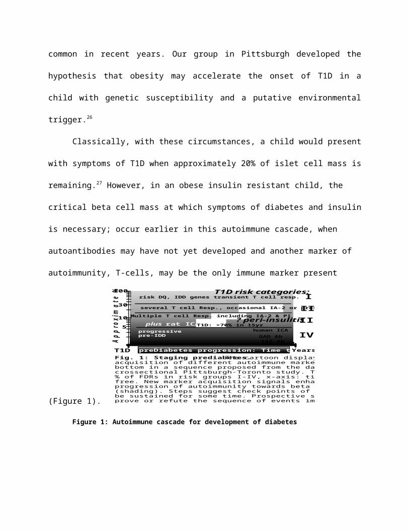
Classically, with these circumstances, a child would present with symptoms of T1D when
approximately 20% of islet cell mass is remaining.27 However, in an obese insulin resistant child,
the critical beta cell mass at which symptoms of diabetes and insulin is necessary; occur earlier
in this autoimmune cascade, when autoantibodies may have not yet developed and another
marker of autoimmunity, T-cells, may be the only immune marker present (Figure 1).
App
roxi
mat
e %
of F
DR
s
I
III
IV
T1D risk categories:
II
T1D
risk DQ, IDD genes transient T cell resp.
plus rat ICA:human ICA
GAD AbIA2 Ab
10
5
30
100
Multiple T cell Resp. including IA-2 & PI? peri-insulitis
progressive pre-IDD
several T cell Resp., occasional IA-2 or proinsulin
T1D: >70% in 15yr
preDiabetes progression: Time to overt T1D Years
Fig. 1: Staging prediabetes. The cartoon displays the acquisition of different autoimmune markers from top to bottom in a sequence proposed from the data obtained in the crossectional Pittsburgh-Toronto study. The y-axis shows the % of FDRs in risk groups I-IV, x-axis: time remaining disease-free. New marker acquisition signals enhanced risk for progression of autoimmunity towards beta cell destruction (shading). Steps suggest check points of progression that can be sustained for some time. Prospective studies are required to prove or refute the sequence of events implied.
Figure 1: Autoimmune cascade for development of diabetes
Independently and contemporaneously with our theory for the acceleration of onset of
diabetes, the accelerator hypothesis was developed by Wilkins. This hypothesis has been
discussed and tested in many ways and is discussed in further detail below. There are other
hypotheses, unrelated to obesity, for the increasing incidence of T1D however these will not be
discussed in this paper.
1.2.1 Evidence for and against the accelerator hypothesis
Wilkin’s theorizes that according to Occam’s razor, the simplest explanation that explains all
factors of a theory is most likely to be correct.28 With this logic in mind, an alternative
explanation for the concomitant rise in T1D incidence and obesity is the “accelerator hypothesis”
by which Wilkins proposes that type 1 and type 2 diabetes are the same disorder of insulin
resistance set against different backgrounds; with insulin resistance itself as the trigger leading to
beta cell stress, subsequent autoimmunity and later clinical diabetes.28 The cornerstone of this
argument is that, in fact, T1D and T2D are part of the same spectrum but that the difference
between these diagnoses of convention is that the typical picture of T2D is slow tempo and T1D
is fast tempo.
According to this concept, the age at presentation of diabetes is dependent upon the
percent of beta cell function relative to insulin resistance, with those with low insulin resistance
and low genetic susceptibility having low percent probability of developing diabetes and those
with high risk and high insulin resistance having the highest risk to develop diabetes and all other
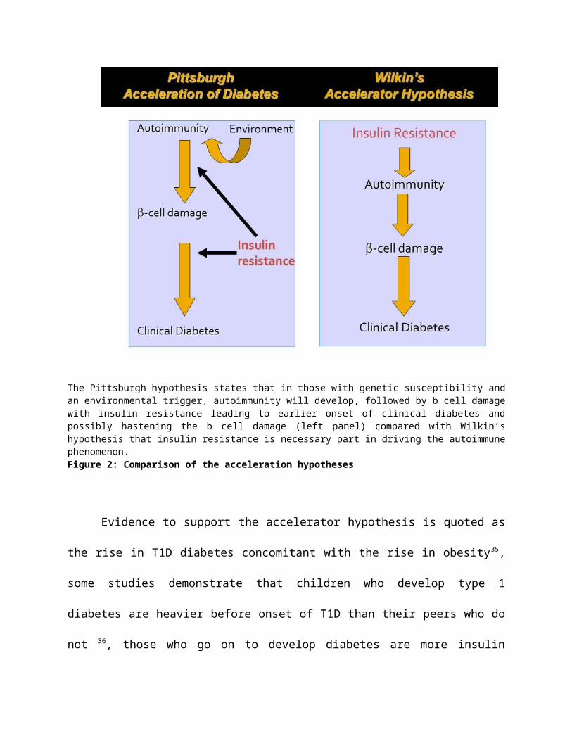
permutations having middle levels of risk and tempos of diabetes onset. 29 Figure 2 contrasts the
acceleration of diabetes onset hypothesis with the “accelerator hypotheis”.
The accelerator hypothesis goes on to explain that autoimmunity, rather than being an
absolute cause of diabetes is the immune system response is the mechanism of clearance of
apoptotic beta cells, and as such should be antigen specific. In the most severe forms, this
response may recruit T-cells which enhance the destructive process.30 This theory could explain
why some adults diagnosed with “type 2” are also antibody positive. Finally, the accelerator
hypothesis attempts to link the many and varied forms of diabetes: Type 1 diabetes, Type 2
diabetes, Type 1 ½ diabetes31, “LADA” (latent autoimmune diabetes in adulthood)32, “LADY”
(latent autoimmune diabetes in youth)33, “double diabetes”26, “Ketosis-prone diabetes”34 as
different variations of one diagnosis, rather than many different diagnoses.
The Pittsburgh hypothesis states that in those with genetic susceptibility and an environmental trigger, autoimmunity will develop, followed by b cell damage with insulin resistance leading to earlier onset of clinical diabetes and possibly hastening the b cell damage (left panel) compared with Wilkin’s hypothesis that insulin resistance is necessary part in driving the autoimmune phenomenon. Figure 2: Comparison of the acceleration hypotheses
Evidence to support the accelerator hypothesis is quoted as the rise in T1D diabetes
concomitant with the rise in obesity35, some studies demonstrate that children who develop type
1 diabetes are heavier before onset of T1D than their peers who do not 36, those who go on to
develop diabetes are more insulin resistant than those who do not 37,38, and the convergence of
the phenotypes of type 1 and type 2 diabetes. 39,40
Although the accelerator hypothesis is an attractively simple one, there is evidence to its
contrary, or at least not supportive. Most of the evidence used to support the “accelerator
hypothesis” is based on epidemiologic observation rather than mechanistic data; leading to the

possibility of ecological fallacy. Using SEARCH data, the accelerator hypothesis was tested by
comparing fasting C-peptide, age and BMI at onset of T1D. The relationship between reduced
age at onset and higher BMI was only significant in those children with the most impaired c-
peptide.41 Both the Diabetes Autoimmunity Study in the Young (DAISY)42, and the
BABYDIAB43 studies followed prospectively several thousand children at high risk for T1D
from the time of birth. Neither study provided support for a role of insulin resistance as the cause
of islet autoimmunity or progression to T1D. Additionally, as opposed to the previously
mentioned DPT-1 38, which did demonstrate a link between insulin resistance and progression to
T1D among islet cell positive FDRs, the ENDIT trial44 only demonstrated this effect in those
with fasting plasma insulin response <10th percentile. While these studies support the concept
that insulin resistance may accelerate the progression to overt diabetes among persons with pre-
existing islet autoimmunity and significant beta cell defect, they do not lend full support that
insulin resistance alone is the primary driver of autoimmunity.
Although the Occam’s Razor explanation is an attractive one, current evidence simply
does not provide support to distill diabetes down to one disease with differing tempos. Instead,
Albert Einstein’s philosophy “everything should be made as simple as possible, but no simpler”
seems to be more correct in this case, as argued by Marian Rewers.45
This leaves us with the facts that both T1D and T2D are increasing in incidence, along
with childhood obesity, with obesity possibly accelerating the onset of T1D in those who have an
environmental trigger, appropriate genetic (HLA) predisposition, and existing autoimmunity.

1.2.2 Potential over and underestimation of T2D and T1D prevalence
While the recent SEARCH estimates are the most rigorous estimates we have to date7 and using
the capture, re-capture method likely record the vast majority of diabetes cases, the previously
defined case definitions may lead to OVER estimation of T2D with subsequent UNDER
estimation of T1D. The SEARCH definition may OVERestimate T2D prevelance by only testing
2 antibodies (GAD and IA2). This testing strategy may only classify up to 83% with autoimmune
diabetes. 46 Therefore, a more exhaustive antibody testing strategy, including ICA and IAA
and/or ZnT8 could be employed to have the highest sensitivity to identify autoimmunity; a
strategy that by some estimates left only 2-4% of patients autoantibody negative.47,48 The T2D
definition including a C-peptide measurement of >3.7ng/mL at onset of diabetes likely rules out
the severe insulin deficiency and secretory impairment that are typical of the onset of T1D.
Thereby, by potentially misclassifying a patient with T2D, the prevalence of T1D could be
underestimated.
1.3 WHY IS THE DISTINCTION BETWEEN T1D AND T2D IMPORTANT?
While this distinction may not be important for initial therapeutic purposes and may not even
exist, as our current system stands, assessing the burden of diabetes in youth by diabetes type is
crucial for implementing public health primary and secondary prevention programs and planning
health care delivery services11.

While an explanation that both T1D and T2D are part of the same spectrum is appealing,
in either case, when a patient is diagnosed with diabetes, they should be treated based on their
symptoms. In an insulin deficient patient, the answer is to give insulin and in an insulin resistant
patient the answer may be weight loss and insulin sensitizers.47 However, since we have yet to
come up with a new system of classification (or non-classification as the case may be) for
diabetes in childhood, under the current regime it may still be important to classify our patients.10
One reason this distinction is important is the projection of long-term healthcare needs.
Recent reports in the New York Times demonstrate nearly $26, 000 spent annually on the care of
patients with T1D using insulin infusion pumps. While this estimate varies depending upon the
level of sophistication of treatment, cost estimates have shown a quadrupling in the care of T1D
since 1987. The majority of this rise in costs consists of prescription drug costs, with the cost of
inpatient admission and outpatient care remaining relatively stable.49
Although the short term costs for the treatment of T2D are much less than the costs of
T1D, there is a projection for a nearly four-fold increase , compared with 3x increase in T2D, in
the incidence of type 2 diabetes by the year 2050, even with current incidence rates remaining
stable, due to the increasing U.S. population. 11 With improvements in T1D care, patients with
T1D are also living longer than in previous generations, leading to an increase in prevalence of
T1D and even further increased cost to healthcare system. While co-morbidities of T1D may be
decreasing, despite increased costs related to treatment, the co-morbidities related to T2D are in
fact increasing, with many obese children diagnosed with T2D already having evidence of
vascular co-morbidities at the time of presentation with T2D.50
Proving a role for autoimmunity in patients with supposed T2D may also help with
mechanistic explanations for autoimmune diabetes and ultimately help move toward a better
understanding of the disease. Finally, whether T1D or T2D, it seems that public health
interventions aiming to decrease the rates of obesity amongst children across the globe could
ultimately decrease the rates of diabetes as well--- regardless of the underlying theory. Below,
we present data arguing that many children with insulin-requiring diabetes in fact, have markers
of autoimmunity.
2.0 EVALUATION FOR AUTOIMMUNE FACTORS IN AUTOANTIBODY
NEGATIVE INSULIN REQUIRING DIABETES
In order to improve the distinction between autoimmune and non-autoimmune or T1D and T2D,
as the current systems classify diabetes, we sought to evaluate patients for additional markers of
autoimmunity beyond the traditional islet autoantibody. One method of evaluating diabetes risk
is measurement of HLA. HLA (human leukocyte antigen) is a protein that is present on the
surface of cells. The immune system uses the HLA present antigen to the thymus in order to
distinguish “self” from “non-self”. There are many sub-types of HLA, some of which are known
to lead to increased risk for autoimmune conditions. The presence of HLA subtypes DQ2 and
DQ8 increase the risk of type 1 diabetes along with other autoimmune conditions, such as celiac
disease.51 Although the highest risk alleles are present in up to 90% of children with T1D, HLA
accounts for only 50% of T1D susceptibility and many people with high risk HLA do not in fact
develop diabetes. 52 Additionally, certain HLA genotypes, DQB0602 and Asp/Asp are known to
be protective from autoimmune conditions, although this protection is not complete.53
T-cell autoimmunity has been used in adults to identify autoimmunity in those with
clinician diagnosed type 2 diabetes and in children with diabetes.54,5556 and has been
demonstrated to appear early in the pathogenesis of T1D.57 We feel that the combination of these
methods could help us identify additional non-autoimmune children with insulin-requiring
diabetes, many of whom are obese, with markers of autoimmunity.
2.1 SPECIFIC AIMS AND HYPOTHESIS
The aim of this study was to investigate if autoantibody negative, mostly obese insulin treated,
diabetic children possess other T1D related autoimmune markers. We hypothesized that many
autoantibody negative diabetics have T1D high-risk HLA alleles and T-cell proliferative
responses typical of T1D, ultimately developing autoantibodies over the 2-year period following
diagnosis as autoimmune antigen spreading continues. To answer this question we analyzed our
autoantibody negative insulin-requiring diabetes subgroup. The goal of our study was
demonstrate that despite the fact that many children are obese at onset of diabetes, these children
many time do in fact have autoimmune disease, further supporting a link between obesity and
increasing incidence of T1D. Additionally, we aimed to follow these children over 2 years
following their diabetes diagnosis to see how their clinical and autoimmune course would
progress with the hypothesis that many of these children would develop islet autoantibodies over
2 years.
2.2 RESEARCH DESIGN AND METHODS
2.2.1 Study population
Children, <19 years of age with insulin-requiring diabetes, diagnosed consecutively between
January 2004 and June 2008 at Children’s Hospital of Pittsburgh, were recruited for consent and
enrollment in the Juvenile Onset Diabetes (JOD)/Antigen Spreading Study (AGS). All clinical
data were obtained within 1 week of diagnosis and at initial follow up, 2-3 months later. Blood
for measures of autoimmunity was drawn at onset and/or 3 months. Of 351 patients recruited,
287 provided T cell/autoantibody samples, but 23 of these were excluded because of insufficient
sample volumes and 3 additional were excluded because of inadequate T-cell measurement.
Data of 261 diabetes patients were analyzed: age 9.7 ± 4 years (range 1.2-18.9), 60%
male, and 92% Caucasian, 6% African American, 2% “other”. There were no differences in
mean hemoglobin A1c (A1c), BMIz, gender, race or serum c-peptide levels between the included
(n=261) and excluded (n=90) subjects. The excluded group was slightly younger with mean age
7.7 years (p=0.03), because of the difficulty in obtaining sufficient research blood volume in the
younger patients. As most children with new onset T1D regain pre-diagnosis weight loss by the
initial follow up visit (2-3 month), BMI and waist circumference measures were analyzed at that
time to approximate pre-diagnosis habitus. Of these 261 subjects analyzed, 27 subjects were
negative for islet autoantibodies at onset.
We compared all parameters in the diabetes group to a control group chosen to be first
degree relatives (FDRs) of the probands, with protective biallelic Asp57/Asp57 HLA genotype58,
matched by age, race, gender, BMIz and waist circumference.
2.2.2 Autoantibody assays
We measured autoantibodies to IA-2, GAD-65, insulin (IAA) and ICA. Anti-GAD and -IA2
autoantibodies were assayed in triplicate using in vitro translated/transcribed [35S]-Methionine-
labelled human GAD65 and IA2/BDC, the latter containing the intracellular domains59. Insulin
autoantibodies were measured using a modification of the 125I-labelled insulin assay with
precipitation of IgG by protein A,60 in samples obtained within one week of insulin treatment.
ICA was measured using both human group O and cafeteria-fed rat pancreas substrates by
immunohistochemistry61. Based on the 97th percentile of controls, positive ICA was defined as
≥5 JDF units for human and ≥10 JDF units for rat substrates. These assays have been
standardized in International and National workshops (Diabetes Autoantibody Standardization
Program or DASP). Sensitivities and specificities have been consistently 80-100% and for ICA,
GAD, and IA2 autoantibody and 60% and 93% for IAA.
2.2.3 T cell proliferation assay
Blood samples for T cell assays were collected in preservative-free heparin, blinded and shipped
to Toronto by overnight courier in uncooled Styrofoam boxes. Viable peripheral blood
mononuclear cells were enriched on Ficoll-Hypaque gradients and 1x10⁵ cells/flat-bottom
microculture well were incubated in 200 serum- and protein-free Hybrimax 2897 medium
(Sigma, St. Louis, MO) with or without 0.005 -10µg/mL of the test antigens (see below). The
various test antigens (‘analytes’, 20 µL in medium) were preloaded to replicate dry wells prior to
addition of other non-cellular culture ingredients and stored frozen until used. Recombinant
human IL2 (10 units) was added to test analytes to detect anergic T cells. 62 After 6 days, cultures
were pulsed overnight with 1µCi 3H-Thymidine, harvested and submitted to scintillation
counting. Data were calculated as average counts per minute (cpm) and mean stimulation indices
(SI, cpm test/cpm unstimulated culture). A diabetic sample was sent regularly as a positive
control for the assays. A positive response was identified as SI ≥ 3 SD above the mean of
ovalbumin-stimulated responses, which corresponded to an SI >1.5 in 98±1% of viable
samples.62 Tetanus toxoid stimulation was used as a non-autoimmunity related positive control.
The presence of 4 or more positive reactivities to analytes was considered abnormal and has been
shown to distinguish diabetic from unrelated control subjects62 and has been confirmed in a

blinded, national T cell assay workshops.62 63 This assay provides comparable results to those of
the ‘Seattle assay’, despite rather different assay technologies.64 Table 1 lists the 10 previously
identified diabetes-associated test antigens, 2 positive and 2 negative controls.
2.2.4 Molecular typing of HLA alleles
Molecular HLA typing was carried out using sequence-specific priming and exonuclease-
released fluorescence (SSPERF), as previously described.64, 65,66Briefly, this methodology
involves designing and purifying double-labelled fluorescent probes for the detection of class I
and class II alleles in PCR amplified DNA samples without the need of agarose gels for reading
results.
2.2.5 C-Peptide
Serum c-peptide levels were obtained at onset prior to insulin administration (measured by the
clinical laboratory assay (Advia Centaur (Siemens)), with lower limit of detection of 0.5 ng/mL,
and again post-prandially at the first follow-up visit. All correlations were performed with this 2-
3 month remission sample assayed in the research laboratory using a human c-peptide
radioimmunoassay kit (Linco Research, St. Charles, Missouri) with a lower detection limit of 0.1
ng/ml and linearity to 5.0 ng/mL. Levels of >5.0 ng/mL were re-measured using dilutions. Inter-
and intra-assay coefficients of variation were 0.047 and 0.046 respectively. For purposes of
analysis, assay results below the level of detection were replaced with the midpoint between 0
and the lower limit of detection. (e.g., 0.25 for values at diagnosis and 0.05 for values obtained at
follow-up visits).

2.2.6 Data analyses and statistics
Data were summarized using means and standard deviations (SDs) or median and interquartile
range for continuous variables and frequencies for discrete variables. The distributions of
variables were assessed for normality. T-tests were used to compare means of 2 groups of
continuous variables. Chi-square test was used to compare frequencies of categorical variables
and Fisher’s exact test with <5 expected counts. Wilcoxon rank sum test and median tests were
used to compare medians of non-parametric data. There was no statistical difference in the
immune markers collected at the zero and three month time frames, therefore these samples were
combined to increase the power of multivariate analyses. All statistical analyses were performed
using SAS version 9.3 (SAS Institute, Cary, NC, USA), significance was set to 5%.
2.3 RESULTS
Twenty seven of these autoantibody negative subjects had positive, diabetes specific, T cell
responses with a median of 10 [10-10] responses, similar to the antibody positive group. One
child, an obese Hispanic 2 year old with ketosis at onset of diabetes, was negative for both
conventional autoantibodies and T cells. The mean age of the antibody negative was slightly
higher, as was mean BMIz (table 1). The autoantibody negative group was more likely to be non-
white than the autoantibody positive group. Both groups had similar frequency of HLA
DQ2/DQ8. Five of the 28 autoantibody negative subjects were positive for a protective HLA
(Asp/Asp=3 and 0602=2). Both groups had similarly low c-peptide concentrations at 3 months
following diagnosis, during the recovery or “honeymoon” period.
Table 1. Comparison of Immune Features in Autoantibody Negative Versus Autoantibody positive insulin
requiring diabetes children
AA- (n=28) AA+ (n=234) p
Age (yrs) (Mean+ SD) 11.6+ 3.1 9.9+ 3.8 0.04*
Gender (% Male) 60 64 NS
Non-white (%) 25 10 0.02
BMIz (Mean+SD) 1.3 + 0.7 0.9 +0.5 0.005
Waist (cm) Med [IQR] 68.3 [57-74] 65 [57-85] NS
C-peptide (ng/mL) at 3 months following diagnosis
1.7 [0.5-3.1] 1.5 [0.5-1.8] NS
DQ2/DQ8 or both (%) 70 81 NS
Number of positive T-cells (median/IQR)
10 [10-10] 10 [10-10] NS
2.3.1 Course over 2 years
Twenty seven out of 28 subjects had follow-up data available over 2 years. Over this time period,
3 subjects became antibody positive for the conventional autoantibodies. 2 of these subjects
became positive for Gad and ICA and the other subject positive for Gad and IA2. A total of 17
patients were positive for ICA measured on rat pancreas when followed over 2 years, including
the three individuals who were positive for conventional autoantibodies. A total of 20/27 (74%)
of the antibody negative were positive for the high risk DQ2/DQ8, 4 of whom were not positive
for antibodies. Therefore, when combining the antibody and HLA results, 21/28 of the antibody
negative subjects had some measure of immune risk at the end of 2 years of follow-up.
Additionally, the one subject who was negative for both antibodies and T cells became positive
for ICA on rat pancreas and carried the protective asp/asp genotype. He also developed
autoimmune hypothyroidism and his clinical diabetes course progressed with typical insulin
requirements. Two of three patients with Asp/Asp genotype became positive for at least one
autoantibody. The third remained negative for antibodies over 2 years. Of those with the
protective 0602 HLA type, one developed positive rat ICA and one remained antibody negative.
Five subjects, all obese with BMIz range 2.0-3.1 (3 of whom became positive for rat ICA
and the other 2 negative for all autoantibodies and negative for DQ2/DQ8 alleles) came off of
insulin therapy with a mean time of 12 months following diagnosis, all after significant weight
loss. Four of these individuals restarted insulin, while one remained off insulin at the last time
point of follow up (4 years). One of the individuals who was negative for DQ2/DQ8 did in fact
have all negative antibodies, obesity and carried the protective Asp/Asp genotype. None of the
individuals in the autoantibody negative group were diagnosed with monogenic diabetes
(MODY) although 3 individuals where tested for this condition based on family history and low
insulin requirements.
Of the 6 subjects who remained negative for autoantibodies and lacked the high risk
HLA, 2 of them were in the group with protective alleles. The other 4 remaining did not have
anything unusual about their diabetes course; with the disease course behaving like that of a
typical diabetic.
2.4 CONCLUSIONS FROM EVALUATION OF ANTIBODY NEGATIVE
SUBJECTS
It is clear that the autoantibody negative group of our diabetes patients is quite heterogeneous but
with generally older age, higher BMIz score, and a higher percentage of non-white individuals.
These factors, along with antibody negativity may lead physicians to label these individuals with
T2D. However, all of these patients had markers of autoimmunity (T cells) and many had high
risk HLA alleles at 70% with the prevalence in the general population of only 30%. In over half
of our patients who were able to come off of insulin altogether, autoantibodies were present
when followed up over 2 years.
This heterogeneous group of children perhaps gives us the most pause when we consider
what, in fact, is the difference between T1D and T2D. Several obese patients were able to
transiently discontinue insulin therapy, but did ultimately develop autoantibodies. Several
patients remained negative for all markers of immunity (other than T cells) but had a typical T1D
course. One patient was negative for both autoantibodies and T cells and even had a protective
Asp/Asp, but had evidence of autoimmunity with thyroid disease. It is possible that some of the
patients who are negative for autoantibodies would be positive for other immune markers that we
do not yet measure, or possibly even different epitopes of the currently measured antibodies.

3.0 CONCLUSIONS
There is no question that the incidence of pediatric diabetes is increasing. Likewise, there is no
question that over the past several decades the prevalence of obesity in children has risen
sharply, becoming a major public health concern. Many studies reinforce the finding that the
prevalence of obesity at the onset of diabetes has been increasing as well. Both the Wilkins
accelerator hypothesis and the Pittsburgh theory for acceleration of the onset of diabetes have
been developed as way to link these observations. While the available data do not fully support
the Wilkins accelerator hypothesis in its entirety, the Pittsburgh theory for the acceleration of the
onset of diabetes theory remains logical and attractive. The data we present here shows several
cases of obese individuals who were antibody negative at onset and developed antibodies over 2
years of follow-up, fitting into this paradigm. However, not all patients fit exactly into this
hypothesis and it is likely that our failure to fully characterize all patients with a specific “type”
of diabetes is indicative that we do not have a full understanding of the disease itself. Although
our current classification regime is useful to some extent to help forecast future disease burden, it
is likely merely a way to fit a square peg into a round hole rather than a full understanding of the
pathophysiologic mechanisms underlying the disease in each individual patient. Public health
measures to decrease obesity and thus the impact of its presence on the appearance of diabetes
(regardless of type) and planning to help manage the possible burden on the healthcare system is
also important. Future studies to examine the ontogeny of autoimmunity including both B and T
cells immunity, prior to development of diabetes in children will be important to understand
these mechanisms. Finally, genetic testing may a way in the future to help us better understand
the underlying defects in all types of diabetes.
BIBLIOGRAPHY

1 Paneth N, Assessing the contributions of John Snow to Epidemiology; 150 years after the removal of the Broad Street pump handle. Epidemiology 2004: 15; 514-516.
2 Omran A, The epidemiologic transition: A theory of the epidemiology of population change. The Milbank Memorial Fund Quarterly. 1971: 49; 509-538.
3 Hoyert DL, Xu JQ. Deaths: preliminary data for 2011. National vital statistics reports; 61 (6). Hyattsville, MD: National Center for Health Statistics. 2012.
4 Ogden CL, Carroll MD, Curtin LR, McDowell MA, Tabak CJ, Flegal KM. Prevalance of overweight and obesity in the United States, 1999-2004. JAMA 2006: 295; 1549-1555.
5 Ogden CL, Carroll MD, Kit BK, Flegal KM. Prevelance of childhood and adult obesity in the United States, 2011-2012. JAMA 2015: 311; 806-814.
6 Olshansky SJ, Passaro DJ, Hershow RC, Layden J, Carnes BA, Brody J, Hayflick L, Butler RN, Allison DB, Ludwig DJ. A potential decline in life expectancy in the United States in the 21st Century. N Engl J Med. 2005: 11; 1138-45.
7 Dabelea D, Mayer-Davis EJ, Saydah S, Imperatore G, Linder B, Divers J, Bell R, Badaru A, Talton J, Crume T, Lises A, Merchant A, Lawrence J, Reynolds K, Dolan L, Liu L, Hamman R; for the SEARCH for Diabetes in Youth Study. Prevalence of Type 1 and Type 2 diabetes among children and adolescents from 2001 to 2009. JAMA 2014: 17; 1778-1786.
8 Libman IM, Pietropaolo M, Arslanian SA, LaPorte RE, Becker DJ. Changing prevalence of overweight children and adolescents at onset of insulin-treated diabetes. Diabetes Care 2003: 26; 2871-2875.
9 Liu LL, Lawrence JM, Davis C, Liese AD, Pettitt DJ, Pihoker C, Dabelea D, Hamman R, Waitzfelder B, Kahn HS. Prevalence of overweight and obesity in youth with diabetes in USA: the SEARCH for Diabetes in Youth Study. Pediatric Diabetes 2010: 11; 4-11.
10 Pinhas-Hamiel O, Zeitler P. The importance of a name. New Engl J Med 1999: 18; 1418-1421.11 Imperatore G, Boyle JP, Thompson TJ, Case D, Dabalea D, Hamman RF, Lawrence JM, Liese
AD, Liu LL, Mayer-Davis EJ, Rodriguez BL, Standiford D. Projections of Type 1 and Type 2 diabetes burden in the U.S. population ages <20 years through 2050: dynamic modeling of incidence, mortality, and population growth. Diabetes Care 2012: 35; 2515-2520.
12 The SEARCH Study Group. SEARCH for Diabetes in Youth: a multicenter study of the prevalence, incidence and classification of diabetes mellitus in youth. Controlled Clin Trials 2004: 25; 458-471.
13 Dabelea D, Pihoker C, Talton JW, et al; SEARCH for Diabetes in Youth Study. Etiological approach to characterization of diabetes type. Diabetes Care 2011: 34; 1628-1633.
14 Lawrence J, Imperatore G, Dabelea D, Mayer-Davis E, Linder B, Saydah S, Klingensmith G, Dolan L, Standiford D, Pihoker C, Pettitt D, Talton J, Thomas J, Bell R, D’Agostino R for the SEARCH for diabetes in youth study group. Trends in incidence of Type 1 Diabetes month non-hispanic white youth in the United States, 2002-2009. Diabetes 2014;1-27.
15 Smith TL, Drum ML, Lipton RB: Incidence of childhood type I and non-type I diabetes mellitus in a diverse population: the Chicago Childhood Diabetes Registry, 1994 to 2003. J Pediatr Endocrinol Metab 2007: 20; 1093-1107.
16 Libman I, LaPorte R, Becker D, Dorman J, Drash A, Kuller L: Was there an epidemic of diabetes in nonwhite adolescents in Allegheny County, Pennsylvania? Diabetes Care 1998: 21; 1278–1281.
17 Vehik K, Hamman RF, Lezotte D, et al. Increasing incidence of type 1 diabetes in 0- to 17- year- old Colorado youth. Diabetes Care. 2007: 3; 503-509.
18 Harjutsalo V, Sund R, Knip M, Groop PH. Incidence of type 1 diabetes in Finland. JAMA 2013: 4; 427-428.
19 Patterson CC, Gyurus E, Rosenbauer J, et al. Trends in childhood type 1 diabetes incidence in Europe during 1989-2008: evidence of nonuniformity over time in rates of increase. Diabetalogia 2012: 55; 2142-214.
20 DIAMOND Project Group. Incidence and trends of childhood Type 1 diabetes worldwide 1990-1999. Diabet Med 2006: 23; 857-866.
21 Lipman TH, Levitt Katz LE, Ratcliffe SJ et al. Increaseing incidence of type 1 diabetes in youth: twenty years of the Phildelphia Pediatric Diabetes Registry. Diabetes Care 2013: 36: 1597-1603.
22 Gale EM. The rise of childhood type 1 diabetes in the 20th century. Diabetes 2002: 51; 3353-3361.
23 Libman I, Pietropaolo M, Arslanian S, LaPorte R, Becker D. Changing Prevalence of Overweight Children and Adolescents at Onset of Insulin-Treated Diabetes. Diabetes Care 2003: 26; 2871–2875.
24 Kaminski B, Klingensmith G, Beck R, Tamborlane W, Lee J, Hassan K, Schatz D, Kollman C, Redondo M for the Pediatric Diabetes Consortium. Body mass index at the time of diagnosis of autoimmune Type 1 Diabetes in children. J Pediatr 2013: 162; 736-40.
25 Pinhas-Hamiel O, Levek-Motola N, Kaidar K, Boyko V, Tisch E, Mazor-Aronovitch K, Graf-Barel C, Landau Z, Lerner-Geva L, Ben-David R. Prevelance of Overweight, Obesity and Metabolic syndrome compenents in children, adolescents and young adults with Type 1 Diabetes. Diab Met Res Rev 2014. Epub ahead of print.
26 Libman IM, Becker DJ. Coexistence of type 1 and type 2 diabetes mellitus: “double” diabetes?. Pediatric Diabetes 2003:4; 110-113.
27 Eisenbarth GS. Type 1 diabetes: A chronic autoimmune condition. N Engl J Med 1986: 314; 1360-1368.
28 Wilkin TJ. The accelerator hypothesis: weight gain as the missing link between type I and type II diabetes. Diabetalogia 2001: 50; 1587-1592.
29 Wilkin TJ. The accelerator hypothesis: a review of the evidence for insulin resistance as the basis for type 1 as well as type II diabetes. Int J of Obesity 2009: 33;716-726.
30 Roitt I, Brostoff J, Male D. Immunology. Churchill Livingstone: London, 1995, pp1.5-1.6.31 Juneja R, Palmer JP. Type 1 ½ diabetes: myth or reality? Autoimmunity 1999: 29; 65-83.32 Tuomi T, Groop LC, Zimmet PZ, Rowley MJ, Knowles W, Mackay IR. Antibodies to
glutamic acid decarboxylase reveal latent autoimmune diabetes mellitus in adults with a non-insulin dependent onset of disease. Diabetes 1993: 42; 359-362.
33 Reinehr R, Schober E, Wiegand S, Thon A, Holl R. B-cell autoantibodies in children with type 2 diabetes mellitus: subgroup or misclassification? Arch Dis Child 2006; 91: 473-477.
34 Brooks-Worrell B, Iyer D, Coraza I, Hampe CS, Nalini R, Ozer K, Narla R, Palmer J, Balasubramanyam A. Islet-specific T-cell responses and proinflammatory monocytes define subtypes of autoantibody-negative ketosis-prone diabetes. Diab Care 2013: 36; 4098-4103.
35 Onkamo P, Vaananen S, Karvonen M, Tuomilehto J. Worldwide increase of type 1 diabetes-analysis of the data on published incidence trends. Diabetalogia 1999: 42; 1395-1403.
36 Hypponen E, Virtanen SM, Kenward MG, Knip M, Akerblom HK, Childhood Diabetes in Finland Study Group. Obesity, increased linear growth, and risk of type 1 diabetes in children. Diab Care 2000: 23; 1755-1760.
37 Leslie RD, Taylor R, Pozzilli P. The role of insulin resistance in the natural history of type 1 diabetes. Diabet Med 1997: 14; 327-331.
38 Greenbaum CJ. Insulin resistance and type 1 diabetes. Diabetes Metab Res Rev 2002: 18; 192-200.
39 Hathout EH, Thomas W, El-Shahawy M, Nahab F, Mace JW. Diabetic Autoimmune markers in children and adolescents with type 2 diabetes. Pediatrics 2001: 107; E102.
40 Umpaichitra V, Banerji MA, Castells S. Autoantibodies in children with type 2 diabetes mellitus. J Pediatr Endocrinol Metab 2002: 15 (suppl 1); 525-530.
41 Dabelea D, D’Agostino R, Mayer-Davis E, Pettitt D, Imperatore G, Dolan L, Pihoker C, Hillier T, Marcovina S, Ruggiero A, the SEARCH for diabetes in youth study group. Testing the accelerator hypothesis: body size, beta cell function, and age at onset of type 1 (autoimmune) diabetes. Diabetes Care 2006: 29; 290-294.
42 Lamb MM, Yin X, Yerbe GO et al. Height growth velocity, islet autoimmunity and type 1

diabetes development: the Diabetes Autoimmunity Study in the Young. Diabetalogia 2009: 52; 2064-2071.
43 Winkler C, Marienfeld S, Zwilling M, Bonifacio E, Ziegler AG. Is islet autoimmunity related to insulin sensitivity or body weight in children of parents with type 1 diabetes? Diabetologia 2009: 52; 2071-2078.
44 Bingley PJ, Mahon JL, Gale EA. Insulin resistance and progression to type 1 diabetes in the European Nicotinamide Diabetes Intervention Trial (ENDIT). Diab Care 2008: 31; 147-150.
45 Rewers M, The Fallacy of Reduction. Pediatr Diabetes 2012: 4;340-343.46 Bingley PJ, Bonifacio E, Williams AJ, Genovese S, Bottazzo GF, Gale EA. Prediction of
IDDM in the general population: stratedies based on combinations of autoantibody markers. Diabetes 1997: 46; 1701-10.
47 Bingley P. Clinical applications of diabetes antibody testing. J Clin Endocrinol Metab 2010: 95; 25-33.
48 Yu L, Boulware D, Bean C, Hutton J, Wenzlau J, Greenbaum C, Bingley P, Krischer J, Sosenko J, Skyler J, Eisenbarth G, Mahon J. For the type 1 diabetes TrialNet study group. Zinc transporter-8 autoantibodies improve predication of type 1 diabetes in relatives positive for the standard biochemical autoantibodies. Diab Care 2012: 35: 1213-1218.
49 Rosenthal E. Paying till it hurts: Even small medical advances can mean big jumps in bills. New York Times. April 5, 2014. Accessed online at www.nytimes.com on July 14, 2014.
50 Orchard T. The changing face of young-onset diabetes: type 1 optimism mellowed by type 2 concerns. Diab Care 2013: 36; 3857-3858.
51 Bakhtadze E, Borg H, Stenstrom G, Fernlund P, Arnqvist HK, Ekbom-Schnell A, Bolinder J, Eriksoon JW, Gudbjornsdottir S, Bystrom L, Groop LC, Sundkvist G,. HLA-DQB1 genotypes, islet antibodies and beta cell function in the classification of recent-onset diabetes among young adults in the nationwide Diabetes Incidence Study in Sweden. Diabetalogia 2000: 49; 1785-1794.
52 Gillespie KM. Type 1 diabetes: pathogenesis and prevention. CMAJ 2006: 175; 165-170. 53 Stankov K, Benc D, Draskovic D. Genetic and Epigenetic factors in etiology of diabetes
mellitus type 1. Pediatrics 2013: 132; 1112-1122.54 Brooks-Worrell B, Greenbaum C, Palmer J, Pihoker C. Autoimmunity to Islet Proteins in
Children Diagnosed with New-Onset Diabetes. J Clin Endocrinol Metab 2004: 89; 2222–2227.
55 Brooks-Worrell B, Starkebaum G, Greenbaum C, Palmer J. Peripheral Blood Mononuclear Cells of Insulin-Dependent Diabetic Patients Respond to Multiple Islet-cell Proteins. J Immunol l996: 157; 5668-5674.
56 Dosch H-M, Cheung R, Karges W, Pietropaolo M, Becker D. Persistent T-cell anergy in human type 1 diabetes. J Immunol 1999: 163; 6933-6940.
57 Winer et al. Autoimmune islet destruction in spontaneous type 1 diabetes is not β-cell exclusive. Nat Med 2003: 2; 198-205.
58 Trucco M. To be or not to be Asp57, That is the question. Diab Care 1992: 15; 705-715.59 Pietropaolo M, Becker DJ, Laporte RE, Dorman JS, Riboni S, Mazumdar S, Trucco MA.
Seronegative phenotype associated with 2 HLA high-risk haplotypes and progression to insulin requiring diabetes. Diabetologia 2002: 45; 66-76.
60 Williams AJ, Bingley PJ, Chance RE, Gale EA. Insulin autoantibodies: more specific than proinsulin autoantibodies for prediction of type 1 diabetes. J Autoimmun 1999: 13; 357-63.
61 Lipton RB, Kocova M, LaPorte RE, Dorman JS, Orchard TJ, Riley WJ, Drash AL, Becker DJ, Trucco M. Autoimmunity and genetics contribute to the risk of insulin-dependent diabetes mellitus in families: islet cell antibodies and HLA DQ heterodimers. Amer J Epidemiol 1992: 136; 503-512.
62 Dosch H-M, Becker DJ. Measurement of T-cell autoreactivity in autoimmune diabetes. Diabetologia 2000; 43:386-387.
63 Seyfert-Margolis V, Gisler TD, Asare AL, Wang RS, Dosch HM, Brooks-Worrell B,

Eisenbarth GS, Palmer JP, Greenbaum CJ, Gitelman SE, Nepom GT, Bluestone JA, Herold KC: Analysis of T-cell assays to measure autoimmune responses in patients with type 1 diabetes: results of a blinded controlled study. Diabetes 2006: 2588-2594.
64 Faas F, Menon R, Braun E, Rudert W, Trucco M. Sequence specific priming and exonuclease release fluorescence detection of HLA-DQB1 alleles. Tissue Antigens 1996: 48; 97-112.
65 Ringquist S, Bellone G, Lu Y, Roeder K. Trucco M. Clustering and aligment of polymorphic sequences for HLADRB1 genotyping. PLoS ONE 2013: 3; e59835.
66 Ringquist S, Bellone G, Lu Y, Trucco M: Transplantation Genetics (Chapter 40). In: Rimoin DL, Pyeritz RE,Korf B (Eds), Principles and Practice of Medical Genetics (6th Edition), Elsevier, Philadelphia, PA, pp. 1-33, 2013.