introduction, bacterial classification & immunology...
TRANSCRIPT
Introduction, Bacterial
Classification & Immunology
Review
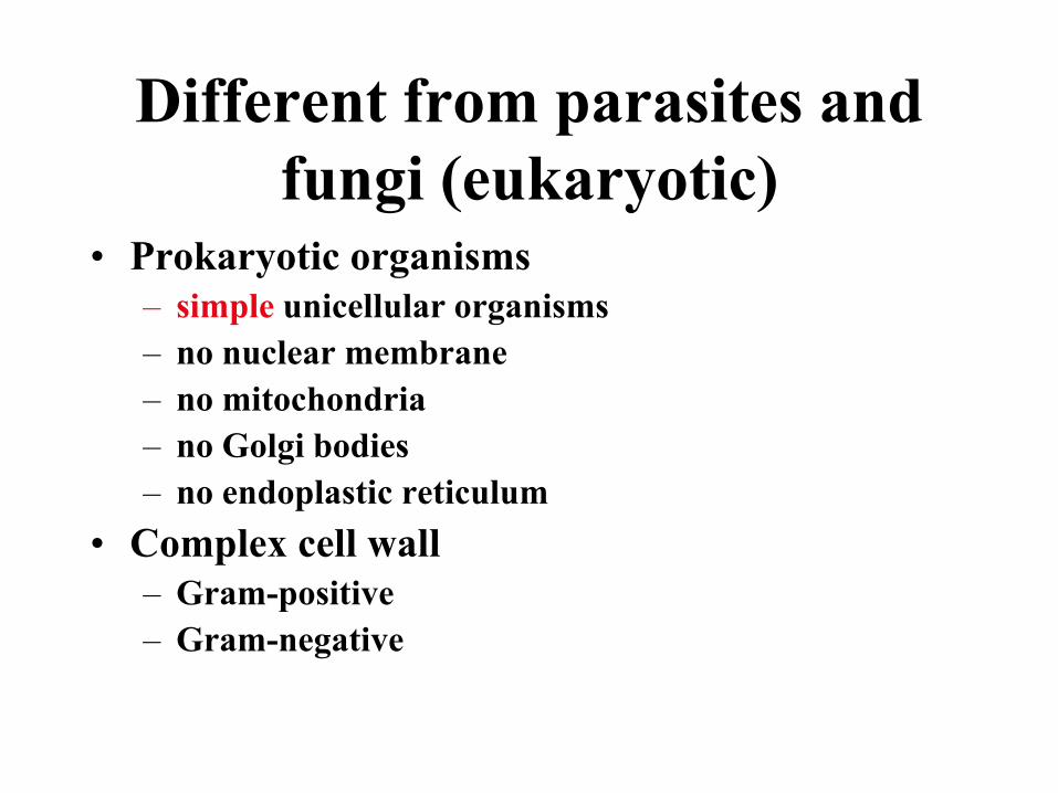
Different from parasites and
fungi (eukaryotic)• Prokaryotic organisms
– simple unicellular organisms
– no nuclear membrane
– no mitochondria
– no Golgi bodies
– no endoplastic reticulum
• Complex cell wall
– Gram-positive
– Gram-negative
Microbial Disease
• The relationship between many organisms and
their diseases is not simple.
• Most organisms do not cause a single, well-
defined disease, although some do e.g.,
Treponema pallidum--syphilis.
• More common for infections to result in many
manifestation of disease e.g., S. aureus--
endocarditis, pneumonia, skin infections, bone
infections, sepsis, food poisoning.

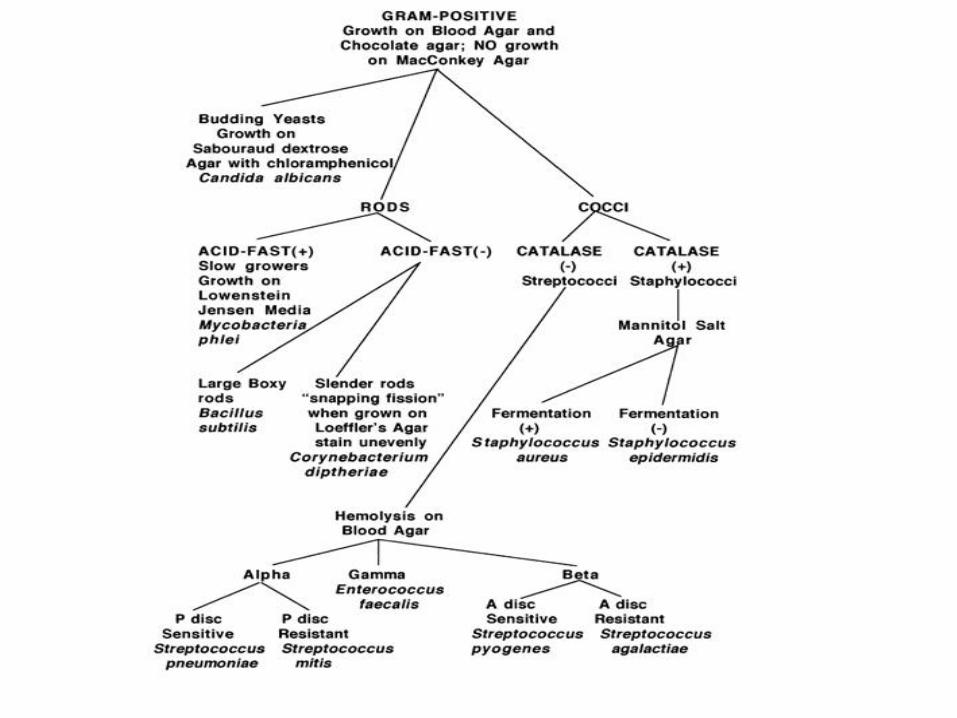
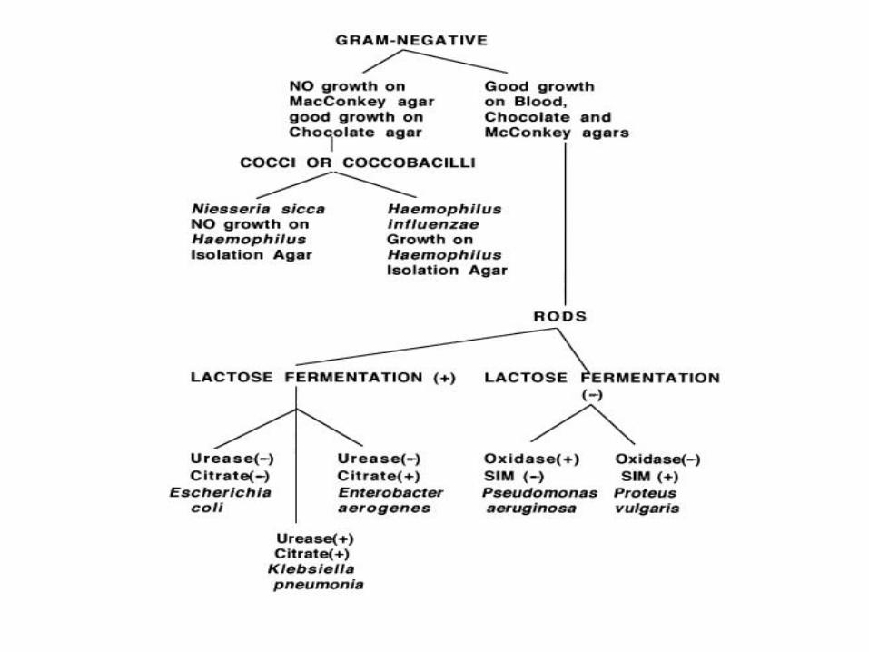
Bacterial Classification
• Phenotypic
• Analytic
• Genotypic
Phenotypic Classification• Microscopic morphology
– Gram stain, shape i.e., rods (bacillus), spheres (cocci), curved or spiral, size
• Macroscopic
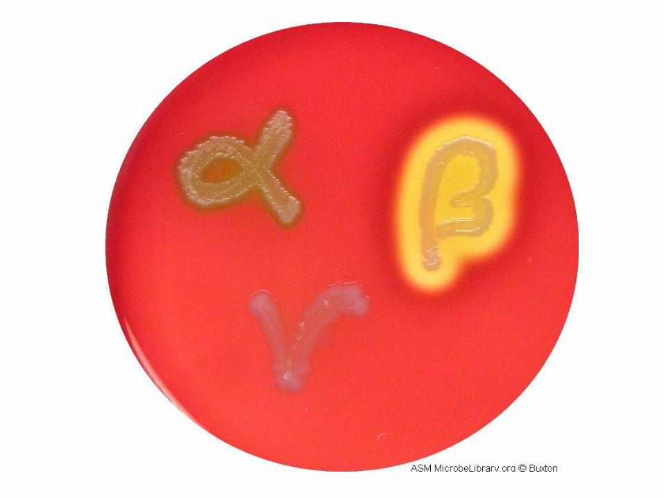
– Hemolytic properties on agar containing blood, pigmentation of the colonies, size and shape of colonies, smell and color.
• Serotyping
– Antibody reactivity to specific antigens
• Antibiogram patterns
– Susceptibility to antibiotics
• Phage typing
– Susceptibility to viruses that infect bacteria--bacteriophages
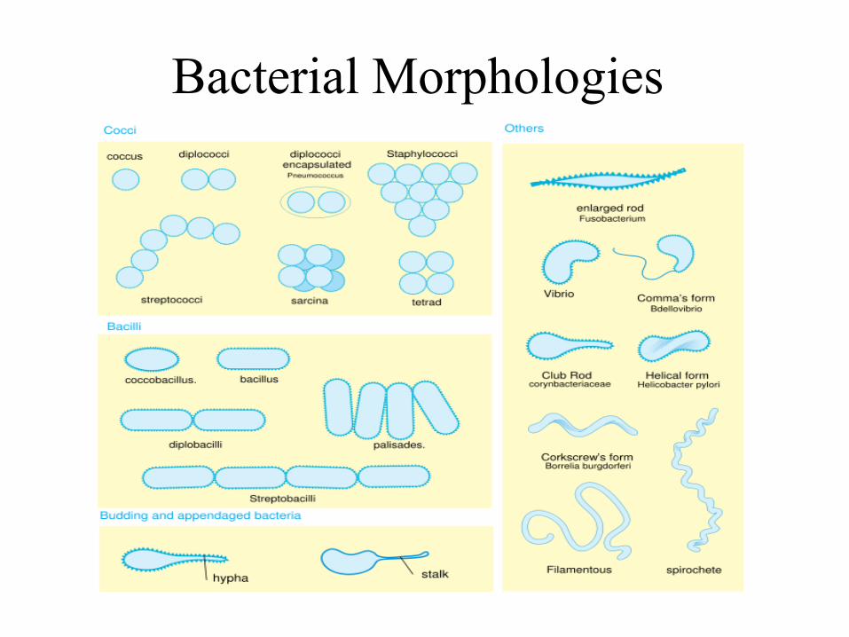
Bacterial Morphologies
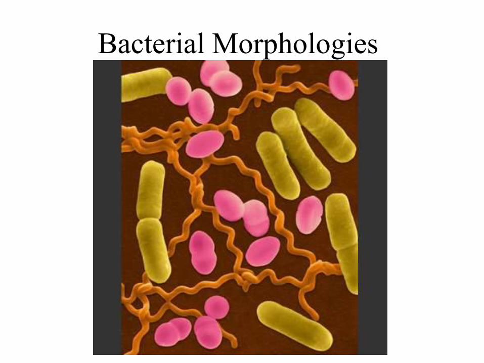
Bacterial Morphologies

Phenotypic Classification• Microscopic morphology
– Gram stain, shape i.e., rods (bacillus), spheres (cocci), curved or spiral, size
• Macroscopic
– Hemolytic properties on agar containing blood, pigmentation of the colonies, size and shape of colonies, smell and color.
• Serotyping
– Antibody reactivity to specific antigens
• Antibiogram patterns
– Susceptibility to antibiotics
• Phage typing
– Susceptibility to viruses that infect bacteria--bacteriophages
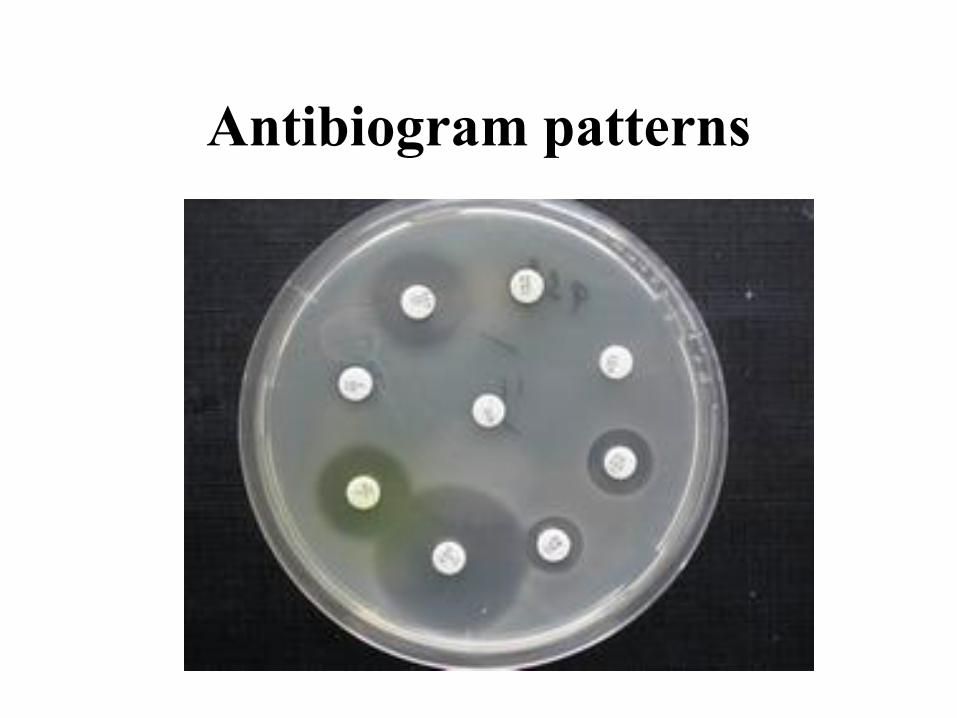
Antibiogram patterns

Phenotypic Classification• Microscopic morphology
– Gram stain, shape i.e., rods (bacillus), spheres (cocci), curved or spiral, size
• Macroscopic
– Hemolytic properties on agar containing blood, pigmentation of the colonies, size and shape of colonies, smell and color.
• Serotyping
– Antibody reactivity to specific antigens
• Antibiogram patterns
– Susceptibility to antibiotics
• Phage typing
– Susceptibility to viruses that infect bacteria--bacteriophages
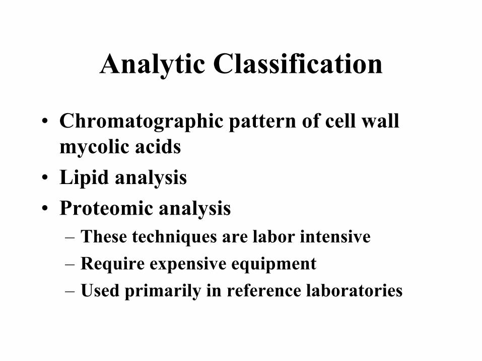
Analytic Classification
• Chromatographic pattern of cell wall
mycolic acids
• Lipid analysis
• Proteomic analysis
– These techniques are labor intensive
– Require expensive equipment
– Used primarily in reference laboratories
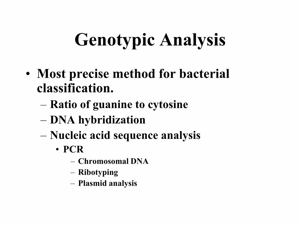
Genotypic Analysis
• Most precise method for bacterial classification.
– Ratio of guanine to cytosine
– DNA hybridization
– Nucleic acid sequence analysis
• PCR
– Chromosomal DNA
– Ribotyping
– Plasmid analysis
Bacterial Morphology and Cell
Wall Structure and Synthesis
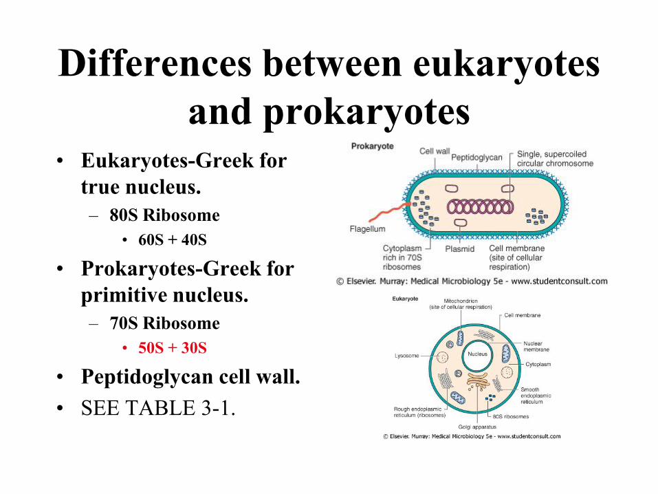
Differences between eukaryotes
and prokaryotes• Eukaryotes-Greek for
true nucleus.
– 80S Ribosome
• 60S + 40S
• Prokaryotes-Greek for
primitive nucleus.
– 70S Ribosome
• 50S + 30S
• Peptidoglycan cell wall.
• SEE TABLE 3-1.
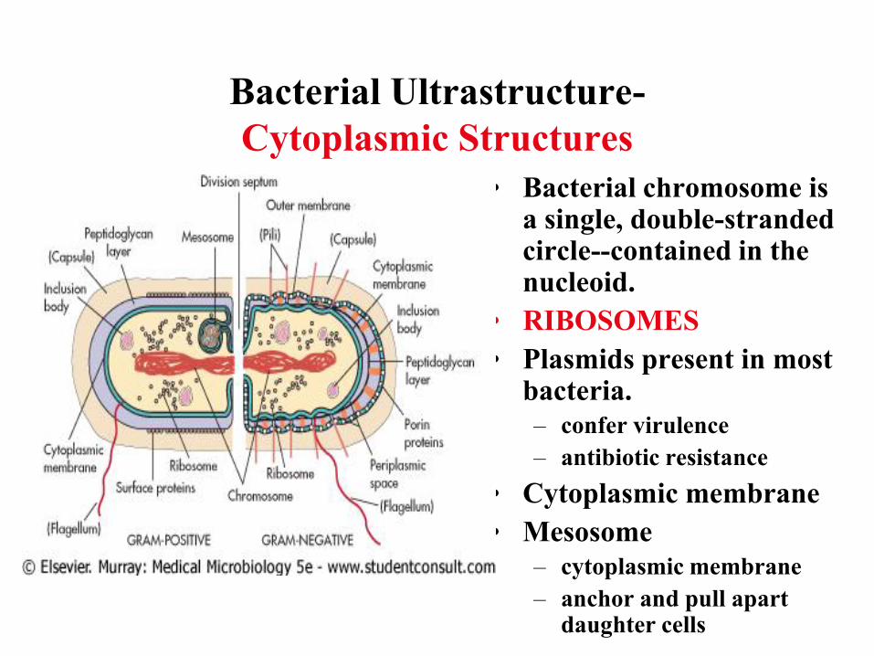
Bacterial Ultrastructure-
Cytoplasmic Structures
• Bacterial chromosome is a single, double-stranded circle--contained in the nucleoid.
• RIBOSOMES
• Plasmids present in most bacteria.
– confer virulence
– antibiotic resistance
• Cytoplasmic membrane
• Mesosome
– cytoplasmic membrane
– anchor and pull apart daughter cells
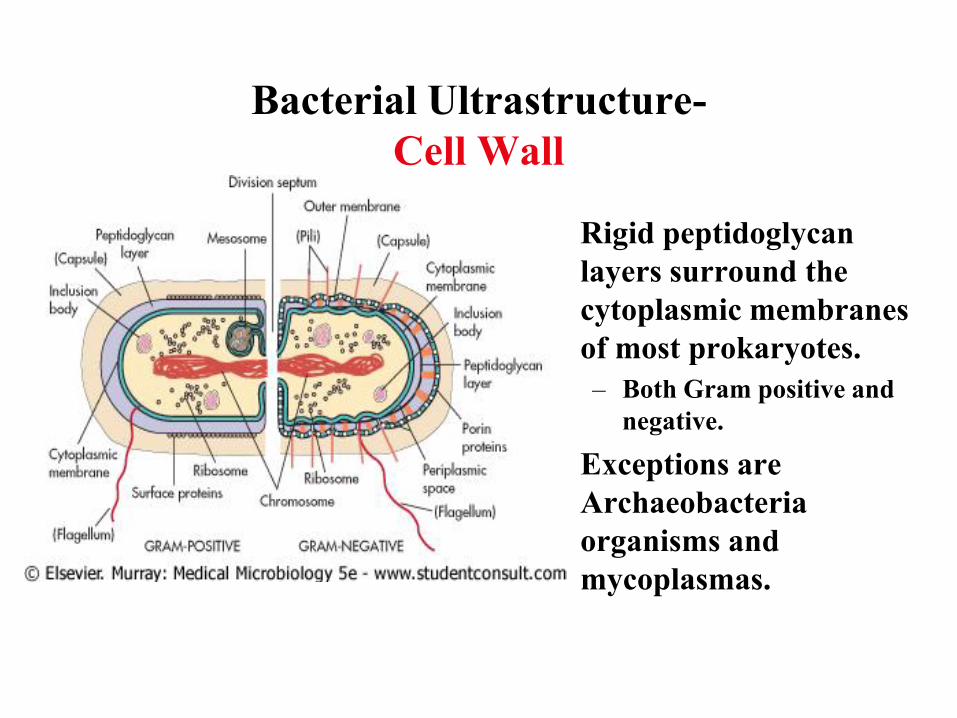
Bacterial Ultrastructure-
Cell Wall
• Rigid peptidoglycan
layers surround the
cytoplasmic membranes
of most prokaryotes.
– Both Gram positive and
negative.
• Exceptions are
Archaeobacteria
organisms and
mycoplasmas.
Differences Between
Prokaryotes--
The Gram Stain
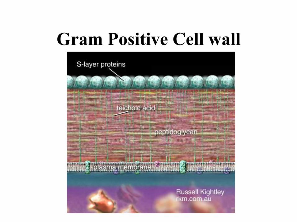
Gram Positive Cell wall
Gram-Negative Cell wall
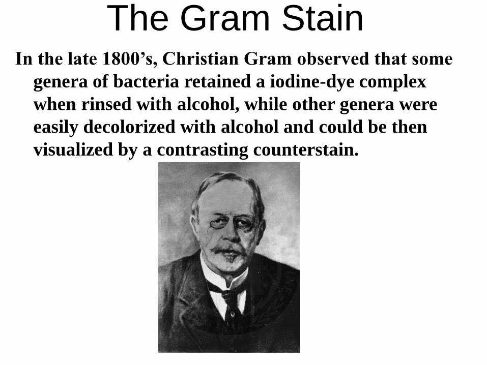
The Gram StainIn the late 1800’s, Christian Gram observed that some
genera of bacteria retained a iodine-dye complex
when rinsed with alcohol, while other genera were
easily decolorized with alcohol and could be then
visualized by a contrasting counterstain.
The Gram Stain
This staining procedure defines two bacterial groups:
those which retain the primary dyes (“Positive by
Gram’s Method” or “Gram-Positive”) and those
which are easily decolorized (“Negative by Gram’s
Method” or “Gram-Negative”). This is the starting
point for bacterial identification procedures.
The Gram StainThe difference in dye retention is dependent on such physical
properties as thickness, density, porosity, and integrity of
the bacterial cell wall, as well as, to some extent, the
chemical composition.
Gram-Positive bacteria have thick, dense, relatively non-
porous walls, while Gram-Negative bacteria have thin
walls surrounded by lipid-rich membranes.
Some non-bacterial organisms with thick cell walls (e.g.,
some yeasts) also stain Gram-Positive.
Gram-Positive bacteria which have lost wall integrity
through aging or physical or chemical damage may stain
Gram-Negative.
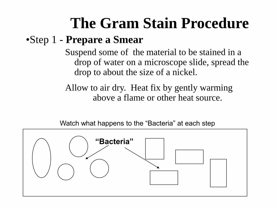
The Gram Stain Procedure•Step 1 - Prepare a Smear
Watch what happens to the “Bacteria” at each step
“Bacteria”
Suspend some of the material to be stained in a drop of water on a microscope slide, spread the drop to about the size of a nickel.
Allow to air dry. Heat fix by gently warming above a flame or other heat source.
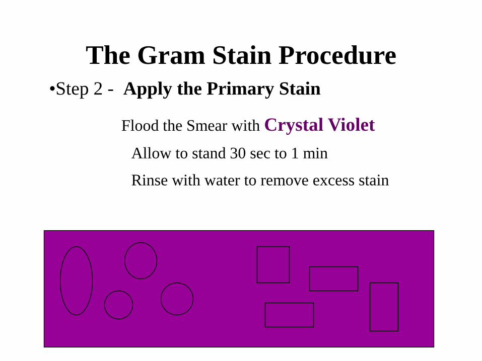
The Gram Stain Procedure
•Step 2 - Apply the Primary Stain
Flood the Smear with Crystal Violet
Allow to stand 30 sec to 1 min
Rinse with water to remove excess stain
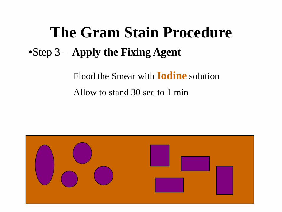
The Gram Stain Procedure
•Step 3 - Apply the Fixing Agent
Flood the Smear with Iodine solution
Allow to stand 30 sec to 1 min
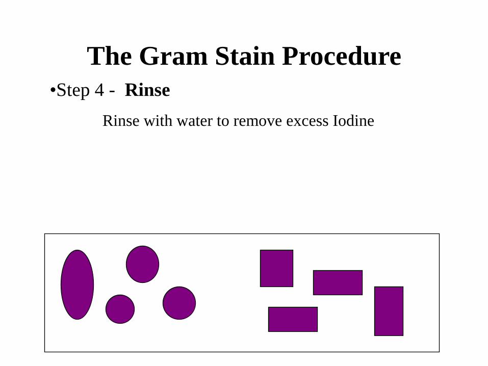
The Gram Stain Procedure
•Step 4 - Rinse
Rinse with water to remove excess Iodine
The Gram Stain Procedure
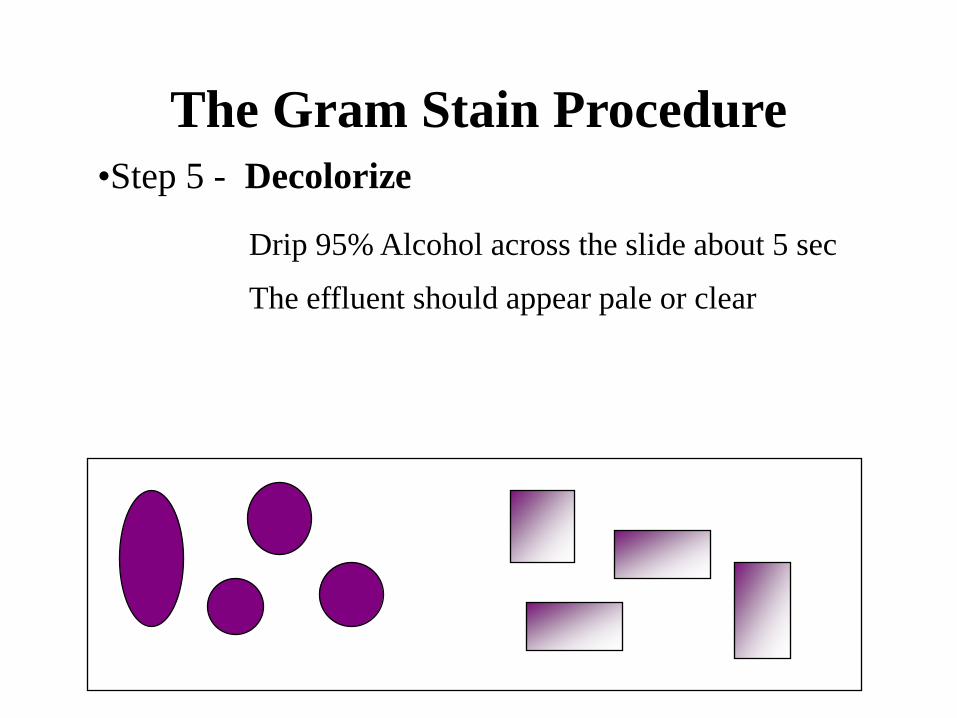
•Step 5 - Decolorize
Drip 95% Alcohol across the slide about 5 sec
The effluent should appear pale or clear
The Gram Stain Procedure
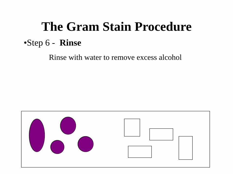
•Step 6 - Rinse
Rinse with water to remove excess alcohol
The Gram Stain Procedure
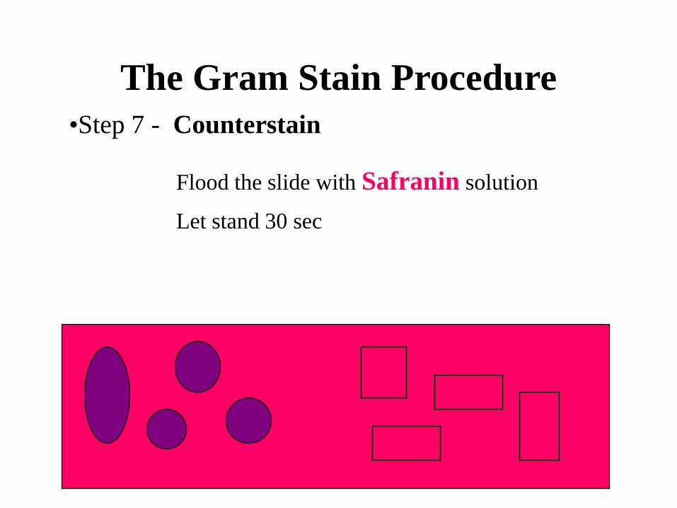
•Step 7 - Counterstain
Flood the slide with Safranin solution
Let stand 30 sec

The Gram Stain
•Step 8 - Rinse, Dry and Observe
Gram-Positive Gram-Negative
Rinse with water to remove excess stain
Blot dry
Observe under Oil Immersion
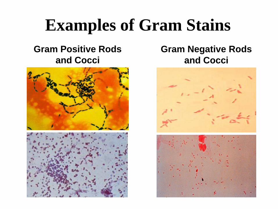
Examples of Gram Stains
Gram Positive Rods
and Cocci
Gram Negative Rods
and Cocci
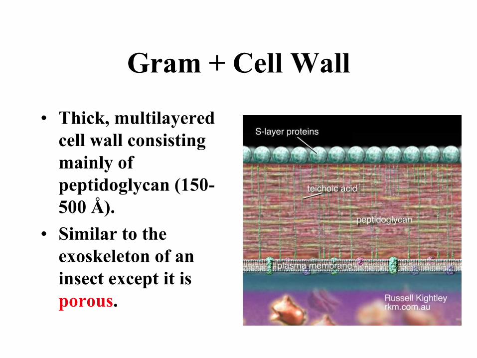
Gram + Cell Wall
• Thick, multilayered
cell wall consisting
mainly of
peptidoglycan (150-
500 Å).
• Similar to the
exoskeleton of an
insect except it is
porous.
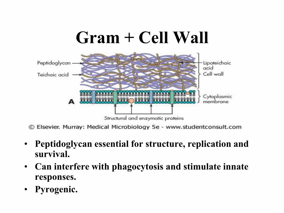
Gram + Cell Wall
• Peptidoglycan essential for structure, replication and survival.
• Can interfere with phagocytosis and stimulate innate responses.
• Pyrogenic.
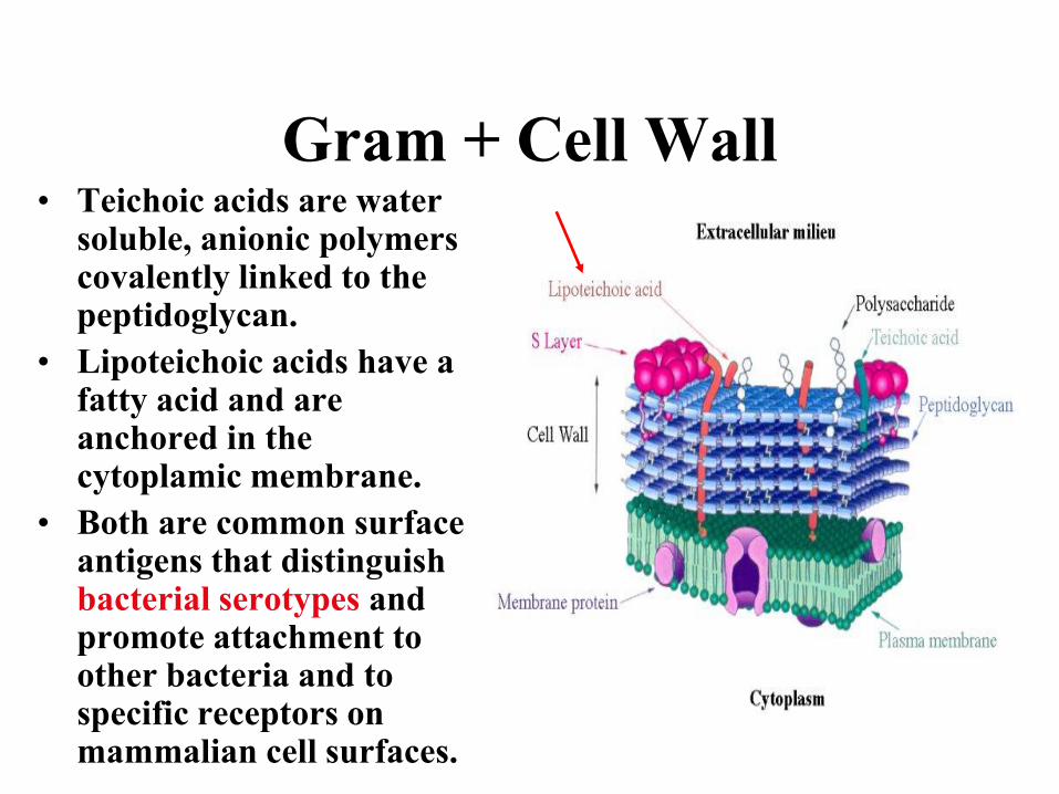
Gram + Cell Wall• Teichoic acids are water
soluble, anionic polymers covalently linked to the peptidoglycan.
• Lipoteichoic acids have a fatty acid and are anchored in the cytoplamic membrane.
• Both are common surface antigens that distinguish bacterial serotypes and promote attachment to other bacteria and to specific receptors on mammalian cell surfaces.
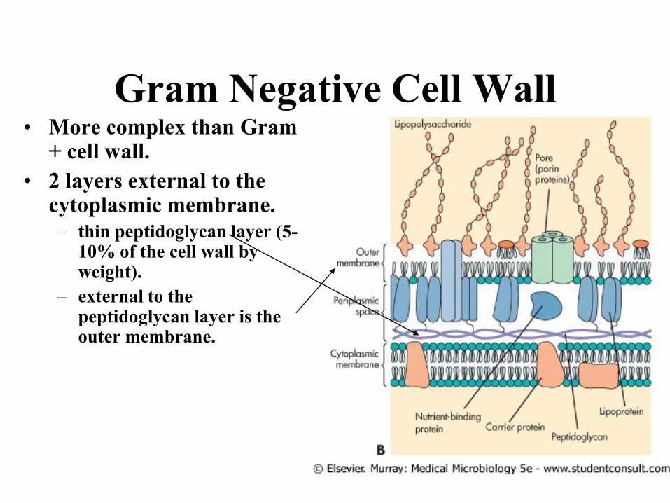
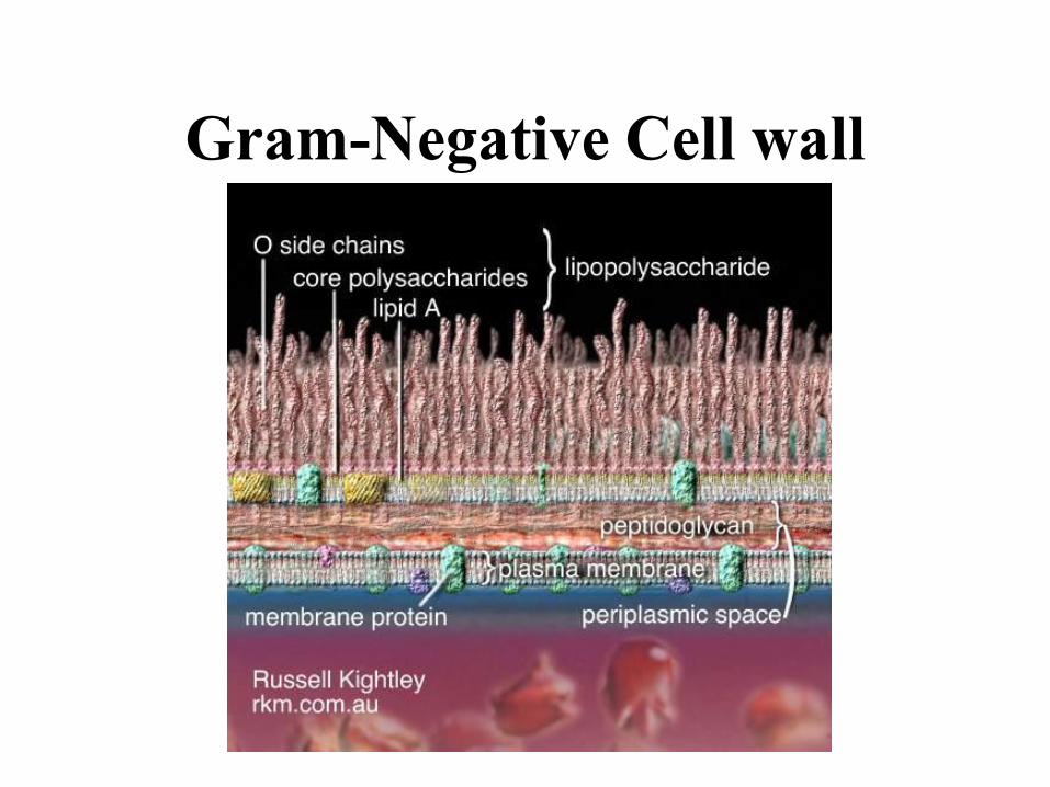
Gram Negative Cell Wall• More complex than Gram
+ cell wall.
• 2 layers external to the cytoplasmic membrane.
– thin peptidoglycan layer (5-10% of the cell wall by weight).
– external to the peptidoglycan layer is the outer membrane.

Gram Negative Cell Wall• Periplasmic space-
– The area between the external surface of the cytoplasmic membrane and the internal surface of the outer membrane.
– Contains hydrolytic enzymes important to the cell for breakdown of large macromolecules for metabolism.
– Also contains enzymes associated with pathology e.g., proteases, hyaluronidase, collagenases and b-lactamase.
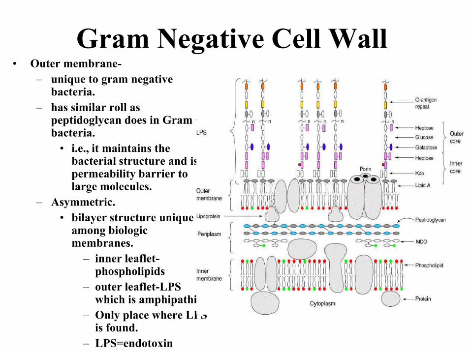
Gram Negative Cell Wall• Outer membrane-
– unique to gram negative bacteria.
– has similar roll as peptidoglycan does in Gram + bacteria.
• i.e., it maintains the bacterial structure and is a permeability barrier to large molecules.
– Asymmetric.
• bilayer structure unique among biologic membranes.
– inner leaflet-phospholipids
– outer leaflet-LPS which is amphipathic.
– Only place where LPS is found.
– LPS=endotoxin
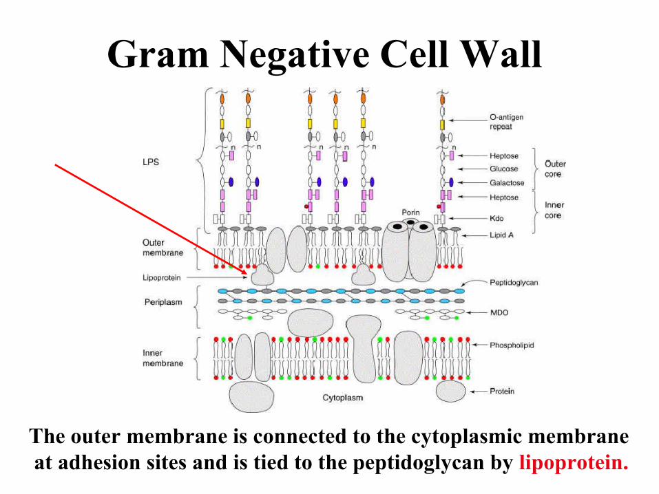
Gram Negative Cell Wall
The outer membrane is connected to the cytoplasmic membrane
at adhesion sites and is tied to the peptidoglycan by lipoprotein.
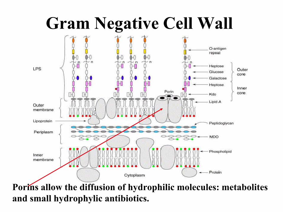
Gram Negative Cell Wall
Porins allow the diffusion of hydrophilic molecules: metabolites
and small hydrophylic antibiotics.
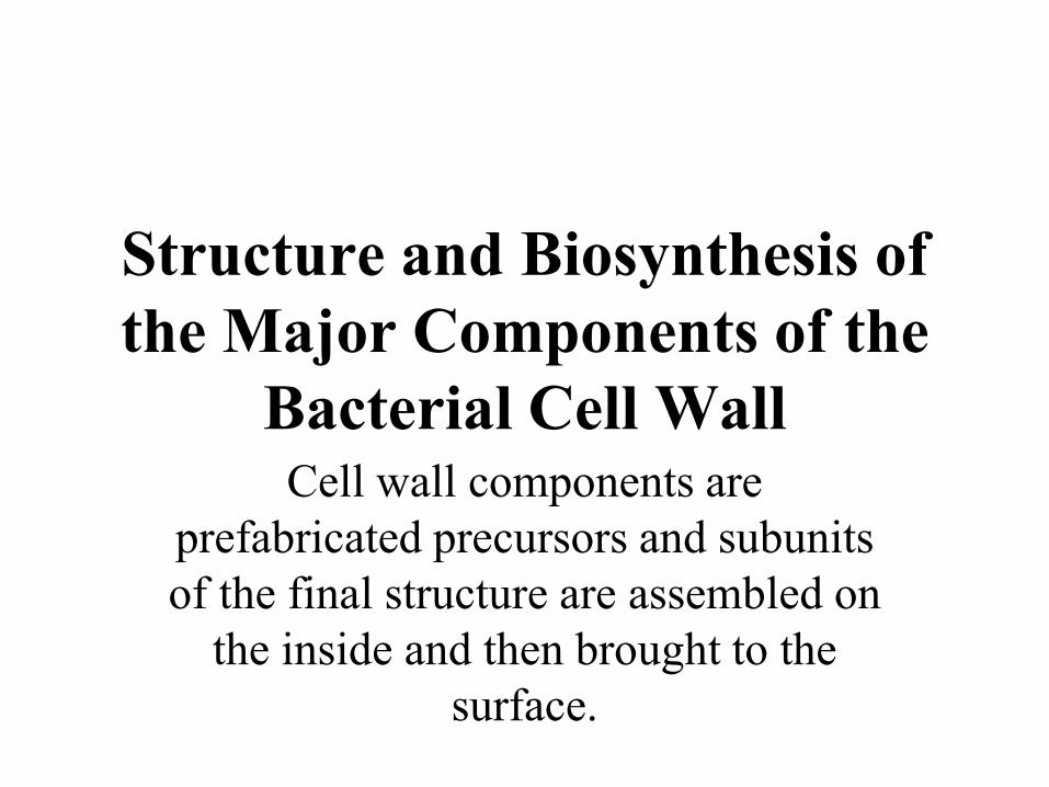
Structure and Biosynthesis of
the Major Components of the
Bacterial Cell WallCell wall components are
prefabricated precursors and subunits
of the final structure are assembled on
the inside and then brought to the
surface.
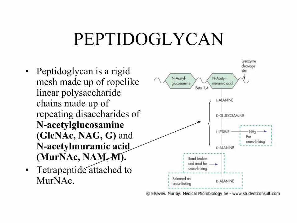
PEPTIDOGLYCAN
• Peptidoglycan is a rigid mesh made up of ropelike linear polysaccharide chains made up of repeating disaccharides of N-acetylglucosamine (GlcNAc, NAG, G) and N-acetylmuramic acid (MurNAc, NAM, M).
• Tetrapeptide attached to MurNAc.
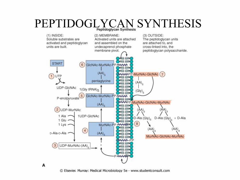
PEPTIDOGLYCAN SYNTHESIS

PEPTIDOGLYCAN SYNTHESIS
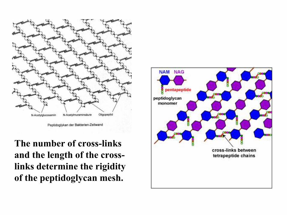
The number of cross-links
and the length of the cross-
links determine the rigidity
of the peptidoglycan mesh.
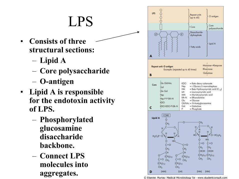
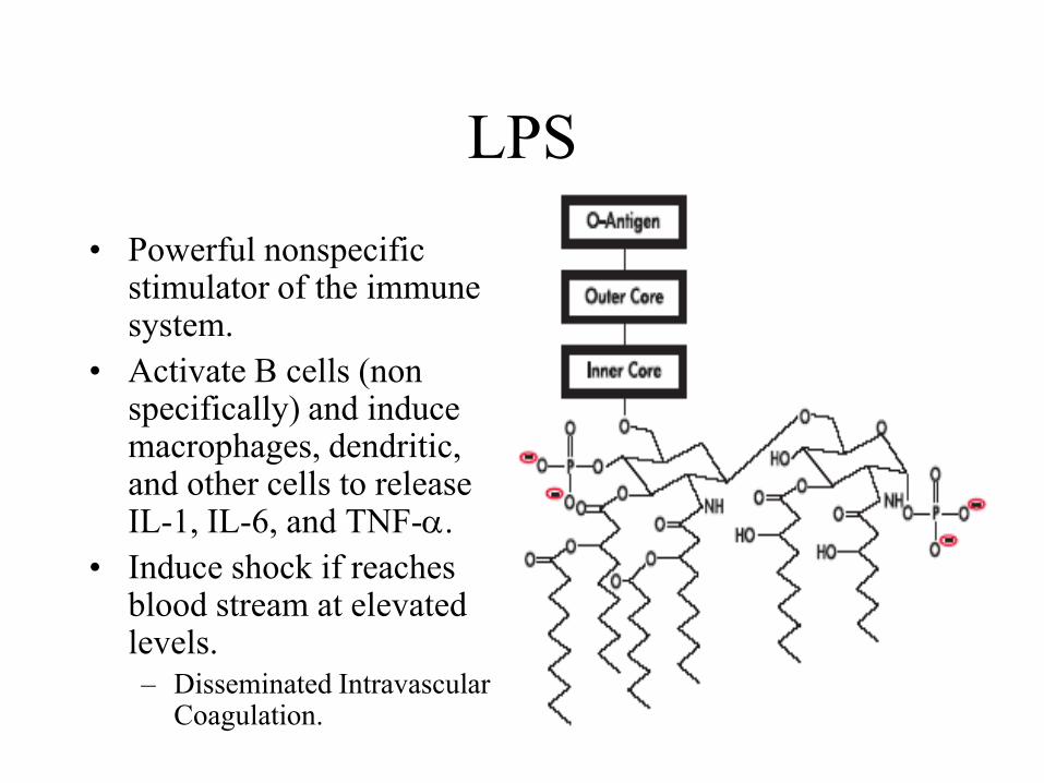
LPS
• Consists of three structural sections:
– Lipid A
– Core polysaccharide
– O-antigen
• Lipid A is responsible for the endotoxin activity of LPS.
– Phosphorylated glucosamine disaccharide backbone.
– Connect LPS molecules into aggregates.
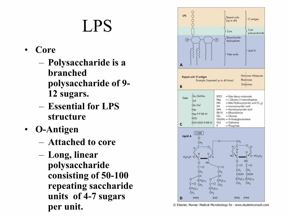
LPS
• Core
– Polysaccharide is a branched polysaccharide of 9-12 sugars.
– Essential for LPS structure
• O-Antigen
– Attached to core
– Long, linear polysaccharide consisting of 50-100 repeating saccharide units of 4-7 sugars per unit.
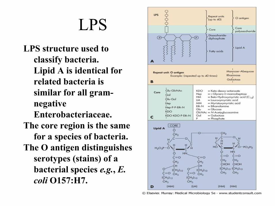
LPS
LPS structure used to
classify bacteria.
Lipid A is identical for
related bacteria is
similar for all gram-
negative
Enterobacteriaceae.
The core region is the same
for a species of bacteria.
The O antigen distinguishes
serotypes (stains) of a
bacterial species e.g., E.
coli O157:H7.
LPS
• Powerful nonspecific stimulator of the immune system.
• Activate B cells (non specifically) and induce macrophages, dendritic, and other cells to release IL-1, IL-6, and TNF-a.
• Induce shock if reaches blood stream at elevated levels.
– Disseminated Intravascular Coagulation.
Nature of Infection
• Plays a critical role in the interactions
between Acquired and Adaptive
immunity
– Intracellular pathogens
– Extracellular pathogens
– Dose
– Route
Infection-Immunity-
Pathogenicity
• Only rarely is the infectious disease the direct and invariable consequence of an encounter between host and pathogen.
• Rather, it is the eventual outcome of complex interactions between them
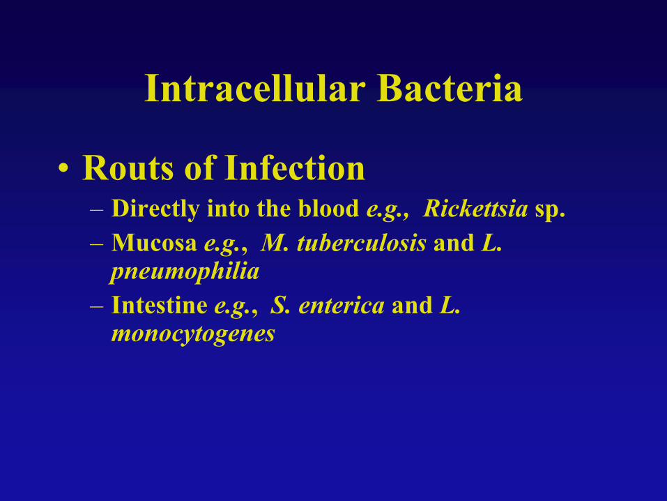
Intracellular Bacteria
• Routs of Infection– Directly into the blood e.g., Rickettsia sp.
– Mucosa e.g., M. tuberculosis and L. pneumophilia
– Intestine e.g., S. enterica and L. monocytogenes
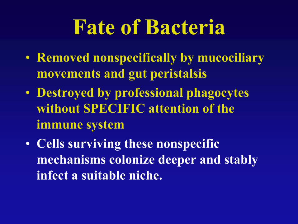
Fate of Bacteria
• Removed nonspecifically by mucociliary
movements and gut peristalsis
• Destroyed by professional phagocytes
without SPECIFIC attention of the
immune system
• Cells surviving these nonspecific
mechanisms colonize deeper and stably
infect a suitable niche.
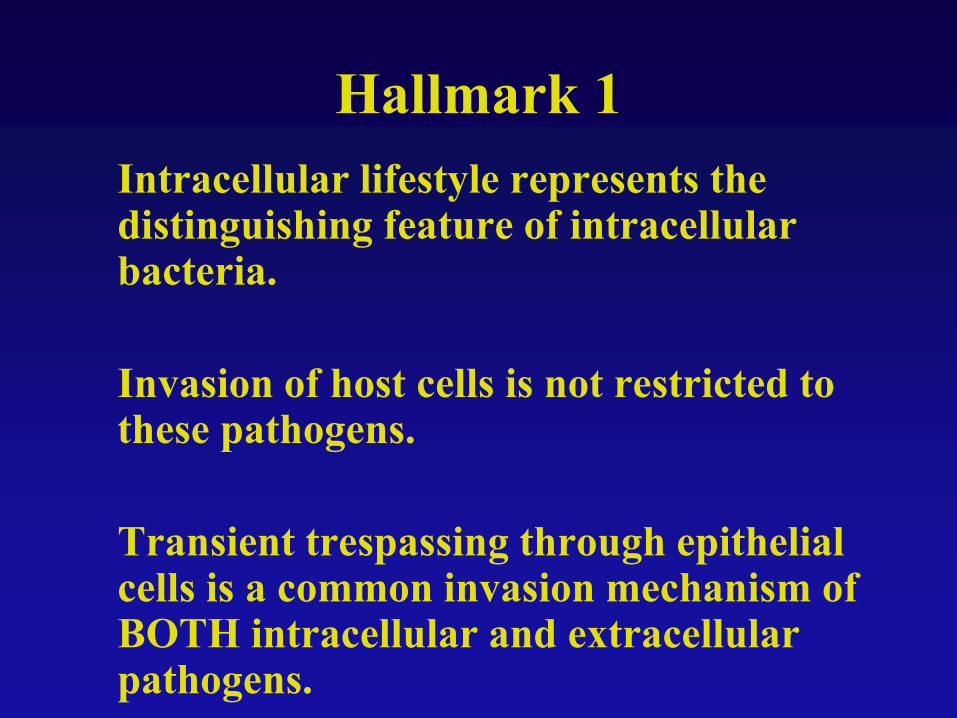
Hallmark 1
Intracellular lifestyle represents the distinguishing feature of intracellular bacteria.
Invasion of host cells is not restricted to these pathogens.
Transient trespassing through epithelial cells is a common invasion mechanism of BOTH intracellular and extracellular pathogens.
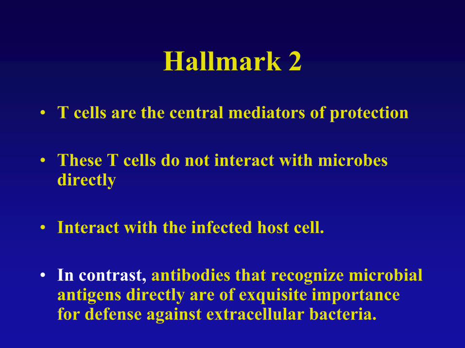
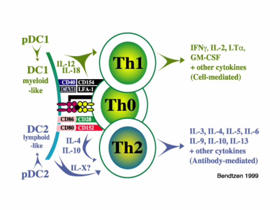
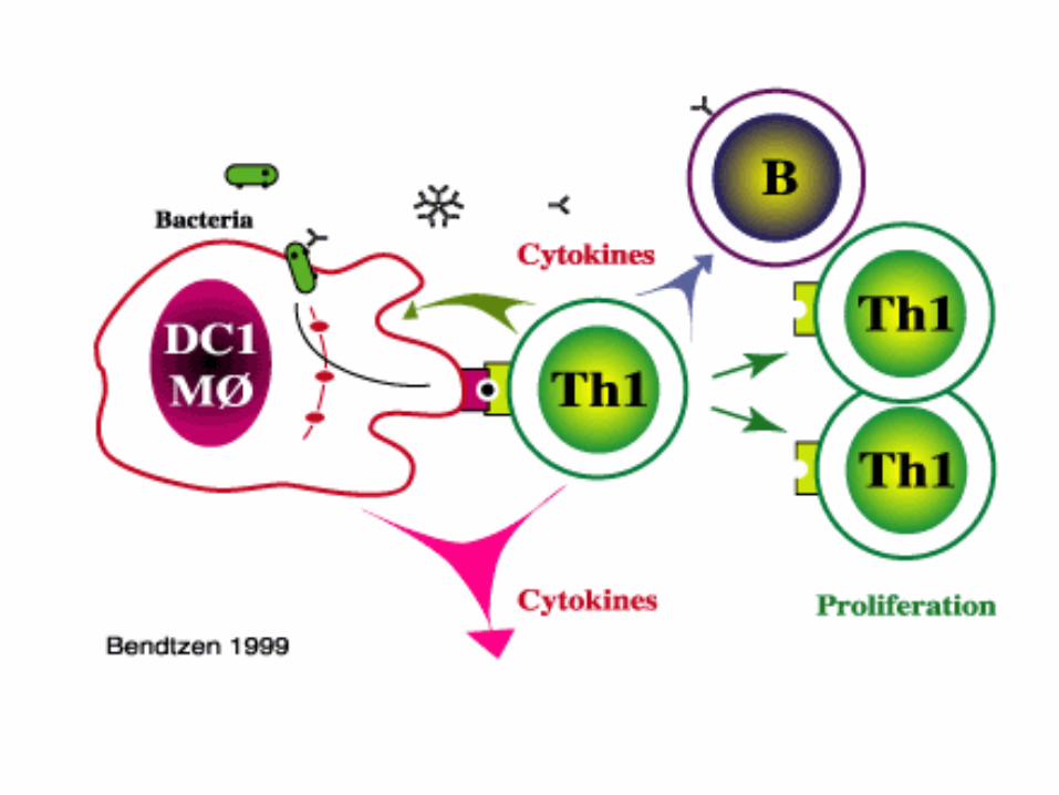
Hallmark 2
• T cells are the central mediators of protection
• These T cells do not interact with microbes directly
• Interact with the infected host cell.
• In contrast, antibodies that recognize microbial antigens directly are of exquisite importance for defense against extracellular bacteria.
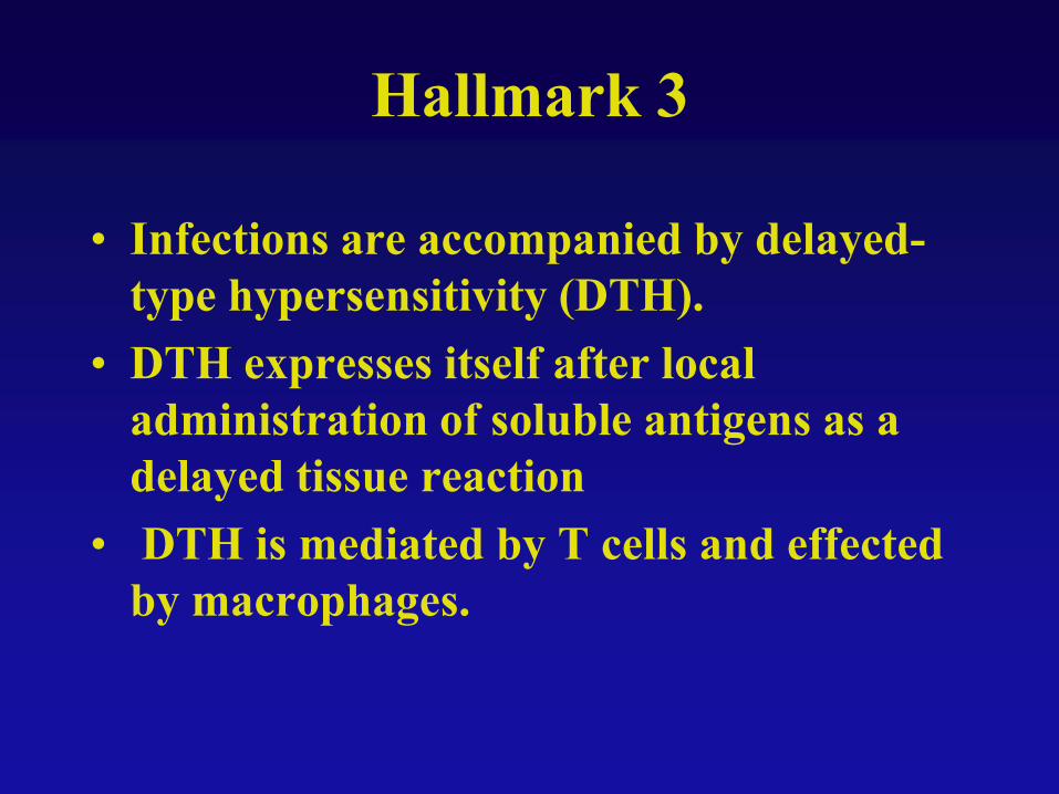
Hallmark 3
• Infections are accompanied by delayed-
type hypersensitivity (DTH).
• DTH expresses itself after local
administration of soluble antigens as a
delayed tissue reaction
• DTH is mediated by T cells and effected
by macrophages.
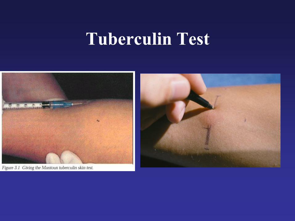
Tuberculin Test
Hallmark 4
• Tissue reactions against intracellular bacteria are granulomatous.
• Rupture of a granuloma promotes bacterial dissemination and formation of additional lesions.
• In contrast, tissue reactions against extracellular bacteria are purulent and lead to abscess formation or systemic reactions.
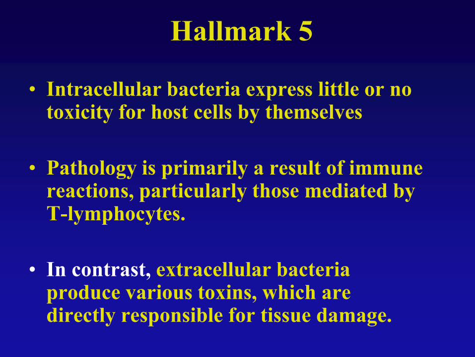
Hallmark 5
• Intracellular bacteria express little or no toxicity for host cells by themselves
• Pathology is primarily a result of immune reactions, particularly those mediated by T-lymphocytes.
• In contrast, extracellular bacteria produce various toxins, which are directly responsible for tissue damage.
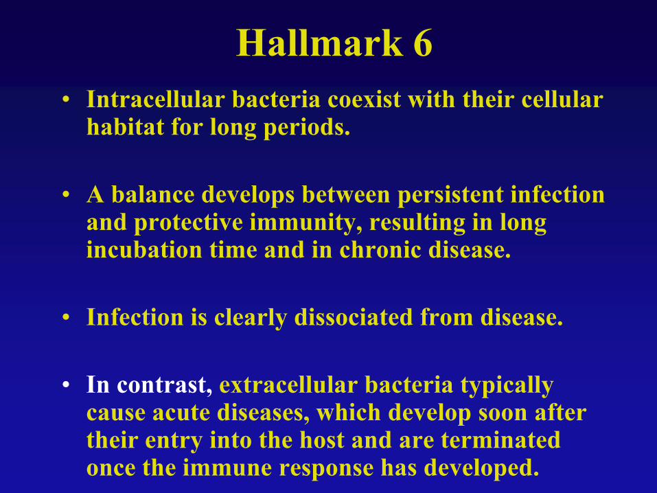
Hallmark 6
• Intracellular bacteria coexist with their cellular habitat for long periods.
• A balance develops between persistent infection and protective immunity, resulting in long incubation time and in chronic disease.
• Infection is clearly dissociated from disease.
• In contrast, extracellular bacteria typically cause acute diseases, which develop soon after their entry into the host and are terminated once the immune response has developed.
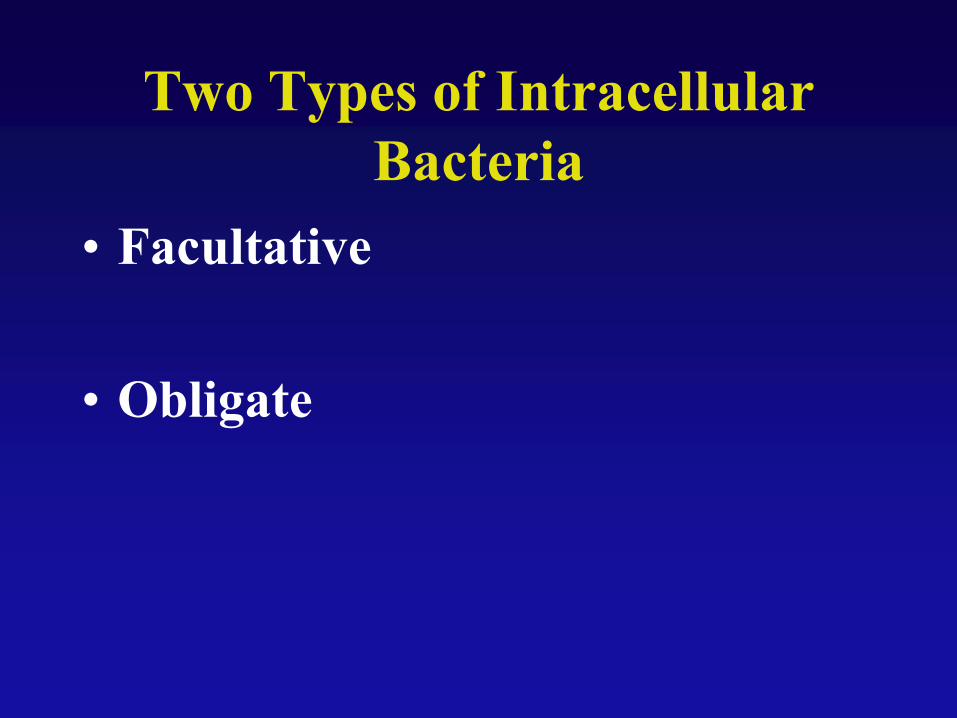
Two Types of Intracellular
Bacteria
• Facultative
• Obligate
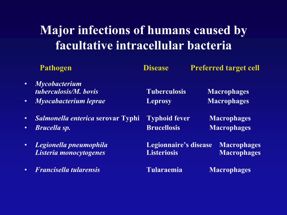
Major infections of humans caused by
facultative intracellular bacteria
Pathogen Disease Preferred target cell
• Mycobacterium tuberculosis/M. bovis Tuberculosis Macrophages
• Myocabacterium leprae Leprosy Macrophages
• Salmonella enterica serovar Typhi Typhoid fever Macrophages
• Brucella sp. Brucellosis Macrophages
• Legionella pneumophila Legionnaire’s disease Macrophages Listeria monocytogenes Listeriosis Macrophages
• Francisella tularensis Tularaemia Macrophages
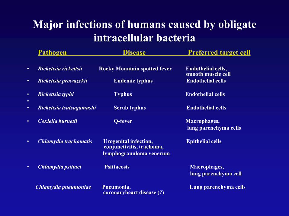
Major infections of humans caused by obligate
intracellular bacteria
Pathogen Disease Preferred target cell
• Rickettsia rickettsii Rocky Mountain spotted fever Endothelial cells, smooth muscle cell
• Rickettsia prowazekii Endemic typhus Endothelial cells
• Rickettsia typhi Typhus Endothelial cells
•
• Rickettsia tsutsugamushi Scrub typhus Endothelial cells
• Coxiella burnetii Q-fever Macrophages,
lung parenchyma cells
• Chlamydia trachomatis Urogenital infection, Epithelial cells conjunctivitis, trachoma,
lymphogranuloma venerum
• Chlamydia psittaci Psittacosis Macrophages,
lung parenchyma cell
Chlamydia pneumoniae Pneumonia, Lung parenchyma cells coronaryheart disease (?)
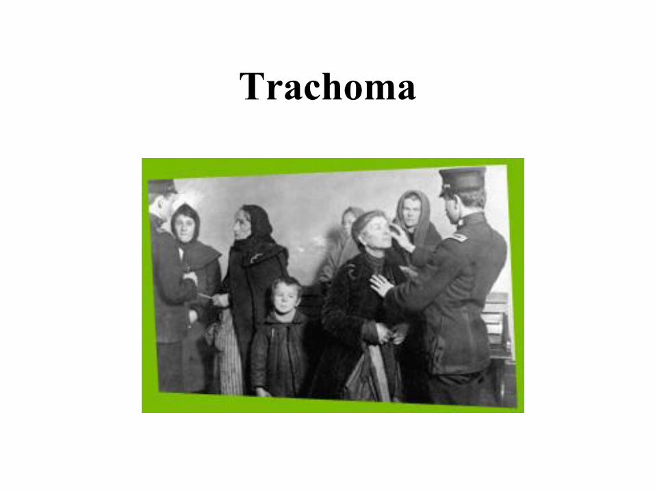
Trachoma
Mechanisms of Immune Evasion
• Easy way—avoid the immune system
entirely…how?
• MIMs (Microbial Immunomodulatory
Molecules)

Bacterial Invasion
• Invasive bacteria actively induce their own uptake by phagocytosis in normally nonphagocytic cells.
– Establish a protective niche.
– Avoid immunity.
– Multiply.
– Active process.
• Opposite to phagocytosis by phagocytes which is active.

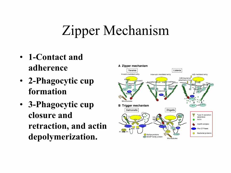
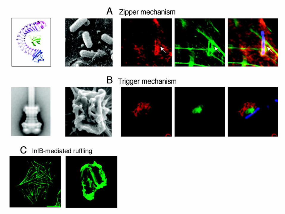
Zipper Mechanism
• 1-Contact and
adherence
• 2-Phagocytic cup
formation
• 3-Phagocytic cup
closure and
retraction, and actin
depolymerization.
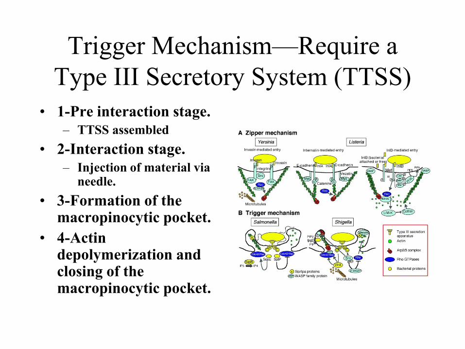
Trigger Mechanism—Require a
Type III Secretory System (TTSS)
• 1-Pre interaction stage.
– TTSS assembled
• 2-Interaction stage.
– Injection of material via needle.
• 3-Formation of the macropinocytic pocket.
• 4-Actin depolymerization and closing of the macropinocytic pocket.
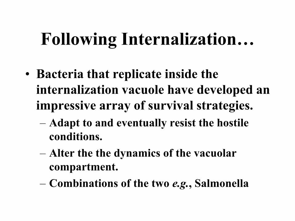
Following Internalization…
• Bacteria that replicate inside the
internalization vacuole have developed an
impressive array of survival strategies.
– Adapt to and eventually resist the hostile
conditions.
– Alter the the dynamics of the vacuolar
compartment.
– Combinations of the two e.g., Salmonella
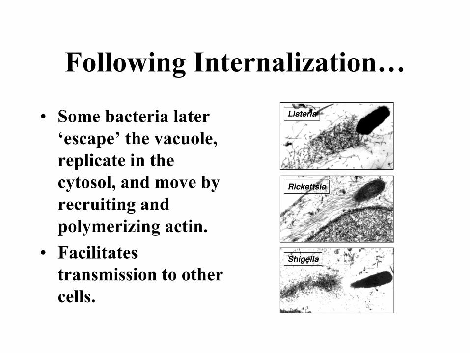
Following Internalization…
• Some bacteria later
‘escape’ the vacuole,
replicate in the
cytosol, and move by
recruiting and
polymerizing actin.
• Facilitates
transmission to other
cells.
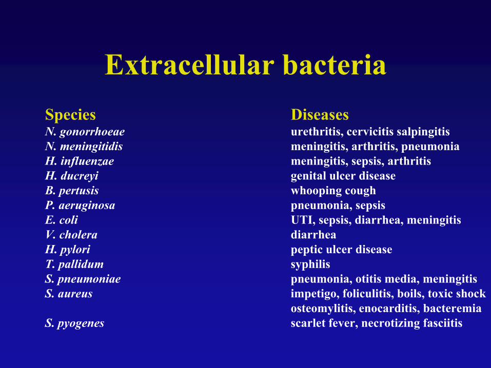
Extracellular bacteria
Species DiseasesN. gonorrhoeae urethritis, cervicitis salpingitis
N. meningitidis meningitis, arthritis, pneumonia
H. influenzae meningitis, sepsis, arthritis
H. ducreyi genital ulcer disease
B. pertusis whooping cough
P. aeruginosa pneumonia, sepsis
E. coli UTI, sepsis, diarrhea, meningitis
V. cholera diarrhea
H. pylori peptic ulcer disease
T. pallidum syphilis
S. pneumoniae pneumonia, otitis media, meningitis
S. aureus impetigo, foliculitis, boils, toxic shock
osteomylitis, enocarditis, bacteremia
S. pyogenes scarlet fever, necrotizing fasciitis
OBJECTIVES• 1. The general nature of immune
responsiveness.
– Memory
– Specificity
• Innate immunity
• Acquired Immunity
• 2 Infection and Immunity
• 3. The anatomic basis of immune responsiveness.

Where things happen
But…
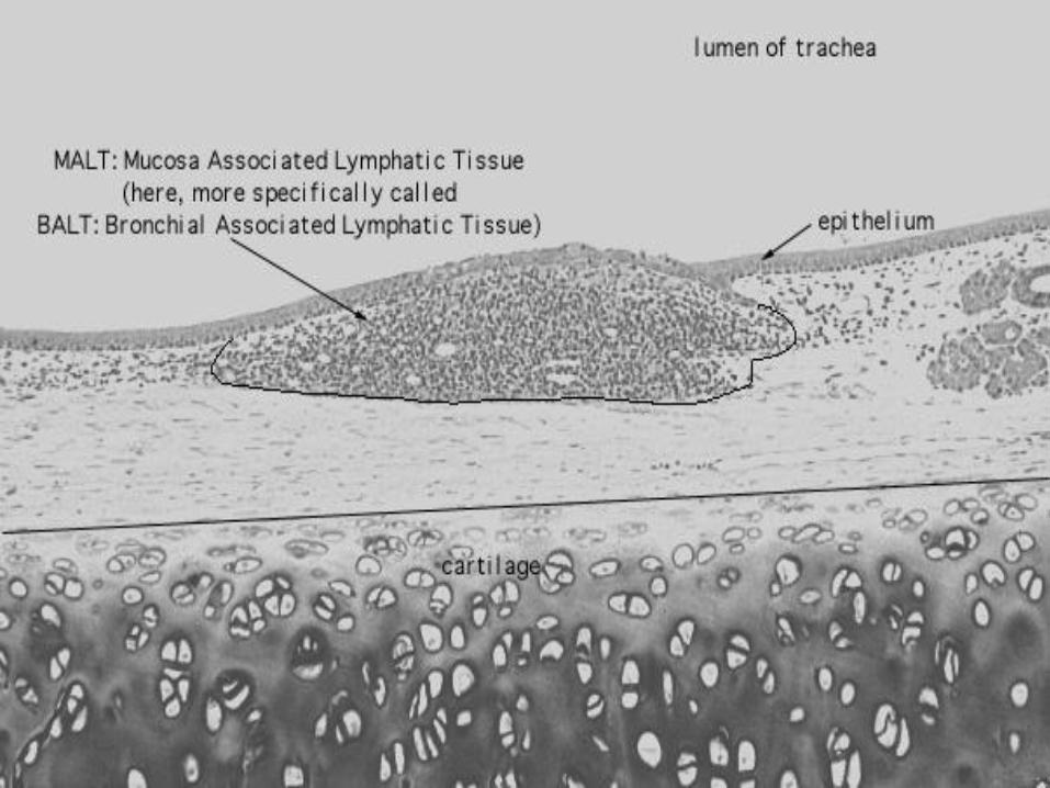

Mounting a Response
Mounting a Response

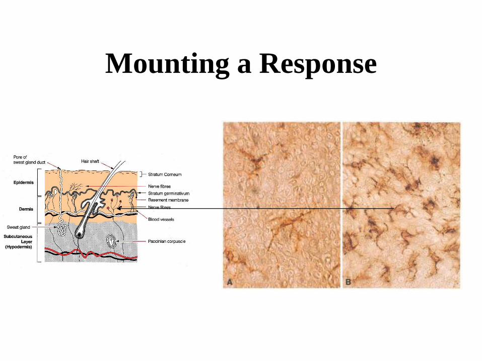
The
Largest
Immune
Organ
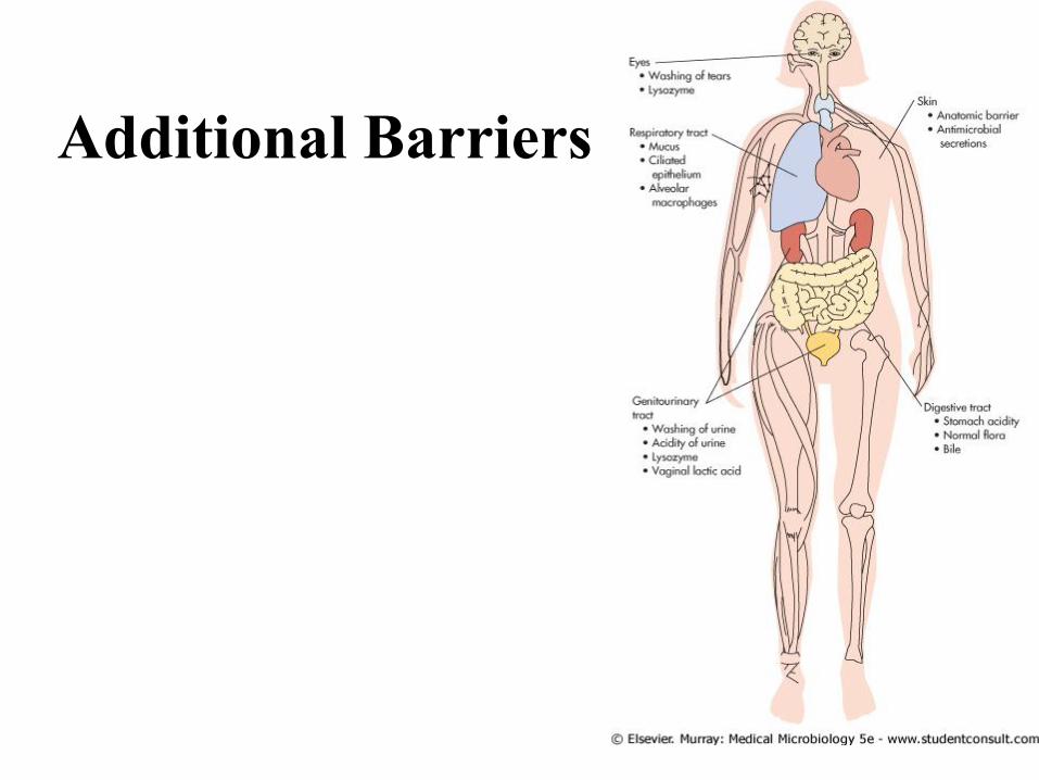
Additional Barriers
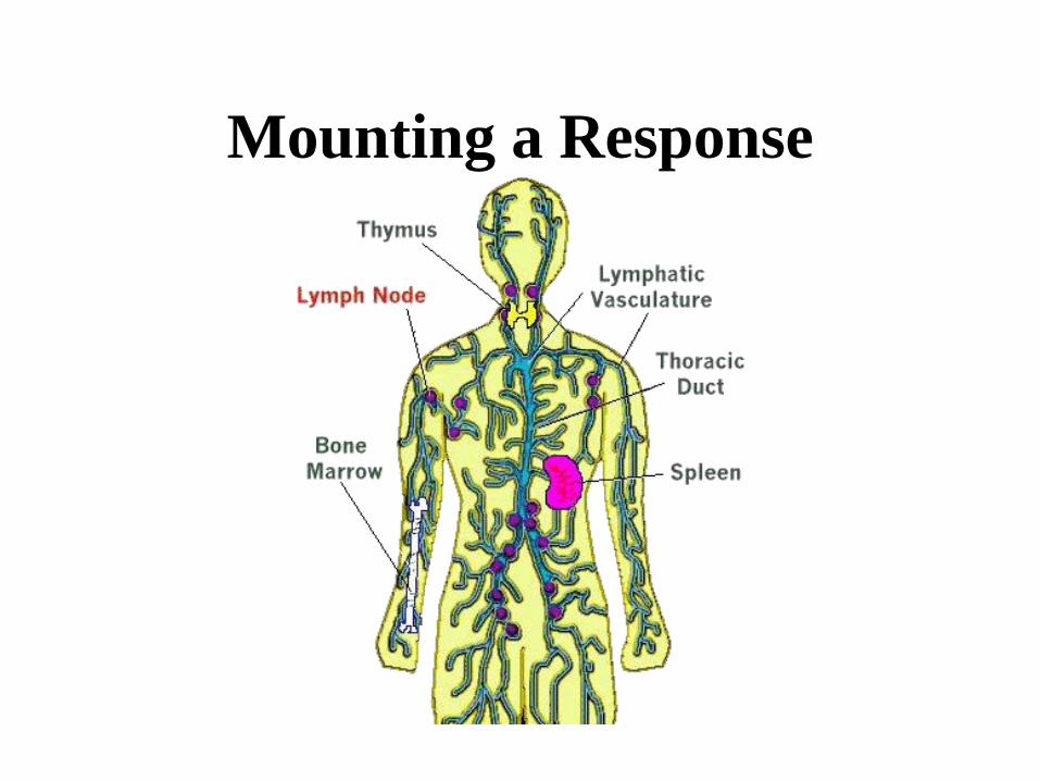
Mounting a Response
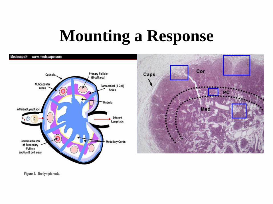
Mounting a Response
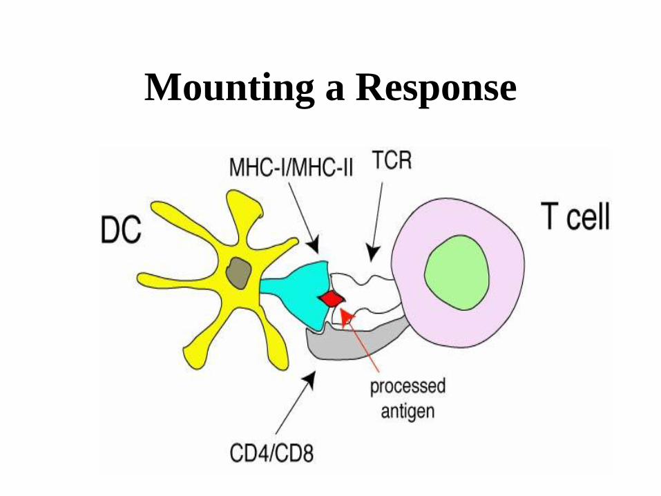
Mounting a Response
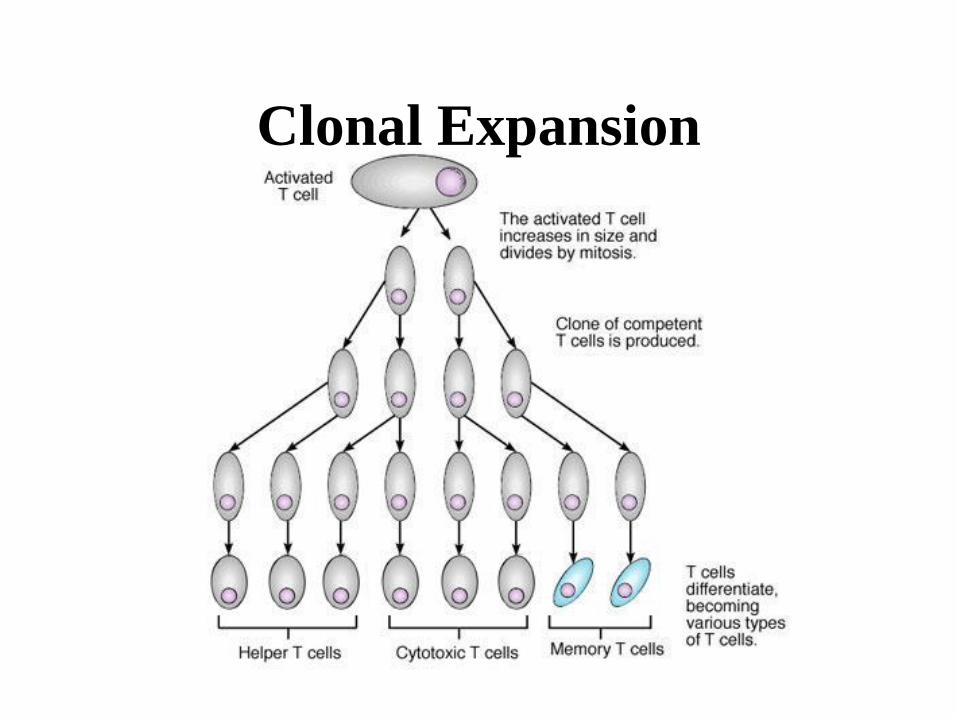
Clonal Expansion
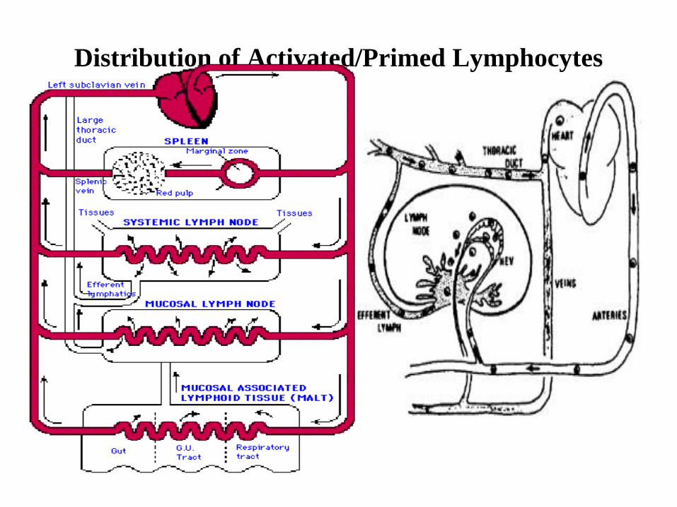
Distribution of Activated/Primed Lymphocytes