introduction to deconvolution and image preparation introduction.pdfintroduction to deconvolution...
TRANSCRIPT

Introduction to deconvolution and Image Preparation
Version 3.0
Image restoration for presentation and analysis
By courtesy
of Nicolas Fête, EPFL / SV / IBI / LDCS

What is “Deconvolution”
?
Deconvolution is
a mathematical
operation
used
in image restoration
to recover
an object
from
an image
that
is
degraded
by blurring
and noise.In fluorescence microscopy
the blurring
is
largely
due
to diffraction limited
imaging
by the instrument; the noise is
mainly
photon noise.
Usage:•
Microscopy: biomedical research, ophthalmology, medical diagnostics, nuclear imaging (CT), food processing, forensics, skin care/beauty product development, etc.
•
Astronomy: telescope imaging, satellite imaging, aerial imaging (cartography), etc.•
Industrial: Quality control for printed circuit boards and semiconductors, etc.

What can be deconvolved in bio-microscopy?
•
Image Type :
–
Widefield Imaging (fluorescence):•
2D Single layer
•
3D Volume or time-lapse•
4D Volume in time
•
xD
Volume in time with different dyes
–
Confocal Imaging (fluorescence):•
Same as Widefield for:
–
Scanning confocal–
Spinning Disk confocal
–
Multi-Photon confocal
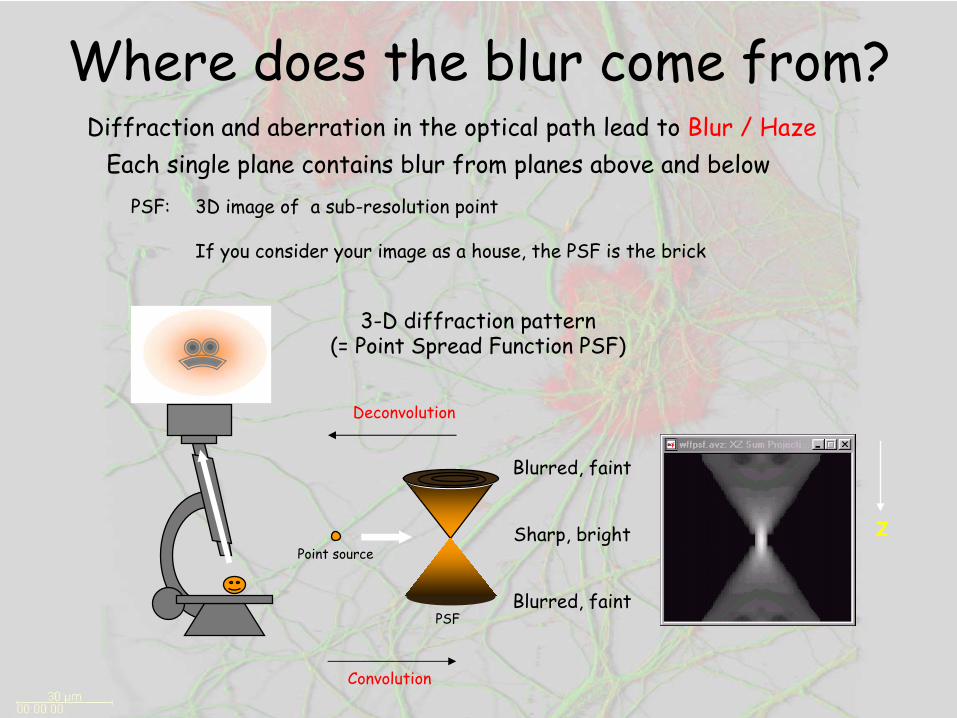
Where does the blur come from?
PSF: 3D image of a sub-resolution point
If you consider your image as a house, the PSF is the brick
Diffraction and aberration in the optical path lead to Blur / Haze
3-D diffraction pattern (= Point Spread Function PSF)
Blurred, faint
Sharp, bright
Blurred, faint
z
Each single plane contains blur from planes above and below
Point source
PSF
Convolution
Deconvolution

The Point Spread of a Microscope
•
Every element in the optical path affects the point spread function:–
Microscope type
–
Objective–
Coverslip
–
Mounting medium–
Sample (cell culture, slices)
–
External factors(T°, vibrations, etc)
•
The PSF is the geometrical signature of the microscope (and the condition of acquisition)

•
Different PSFs
Confocal PSFWidefield PSF
Out of focus light Cut
by the pinhole

•
Reattribute the out of focus light to the point source•
Raise resolution of small objects
•
Increase definition of structure in multiple dimensions•
Improve signal to noise (SNR)
•
Simplest processing for segmentation•
More accurate localization of intensity for quantification, ratiometry and colocalization
•
Only images acquired with the correct sampling parameters(Nyquist, cf. calculator) can be deconvolved
•
Bad acquisitions are not improved
What “restore”
means?
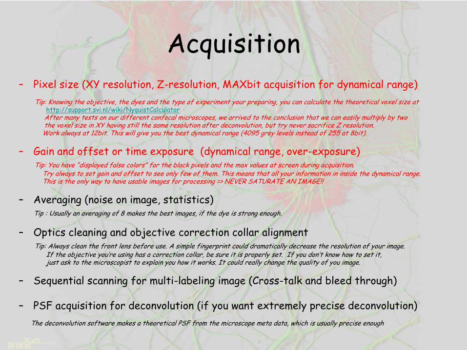
Acquisition–
Pixel size (XY resolution, Z-resolution, MAXbit acquisition for dynamical range)Tip: Knowing the objective, the dyes and the type of experiment your preparing, you can calculate the theoretical voxel size at
http://support.svi.nl/wiki/NyquistCalculatorAfter many tests on our different confocal microscopes, we arrived to the conclusion that we can easily multiply by twothe voxel size in XY having still the same resolution after deconvolution, but try never sacrifice Z resolution.
Work always at 12bit. This will give you the best dynamical range (4095 grey levels instead of 255 at 8bit).
–
Gain and offset or time exposure (dynamical range, over-exposure)Tip: You have “displayed false colors” for the black pixels and the max values at screen during acquisition.
Try always to set gain and offset to see only few of them. This means that all your information in inside the dynamical range.This is the only way to have usable images for processing => NEVER SATURATE AN IMAGE!!
–
Averaging (noise on image, statistics)Tip : Usually an averaging of 8 makes the best images, if the dye is strong enough.
–
Optics cleaning and objective correction collar alignmentTip: Always clean the front lens before use. A simple fingerprint could dramatically decrease the resolution of your image.
If the objective you’re using has a correction collar, be sure it is properly set. If you don’t know how to set it,just ask to the microscopist to explain you how it works. It could really change the quality of you image.
–
Sequential scanning for multi-labeling image (Cross-talk and bleed through)
–
PSF acquisition for deconvolution (if you want extremely precise
deconvolution)The deconvolution software makes a theoretical PSF from the microscope meta data, which is usually precise enough

(Spirogyra)
Benefits of Deconvolution
1. Haze removal More contrast Better segmentation
2. Improved resolution More detail revealed
3. Essential for 3D reconstruction
4. Produces quantitative results (non-destructive)
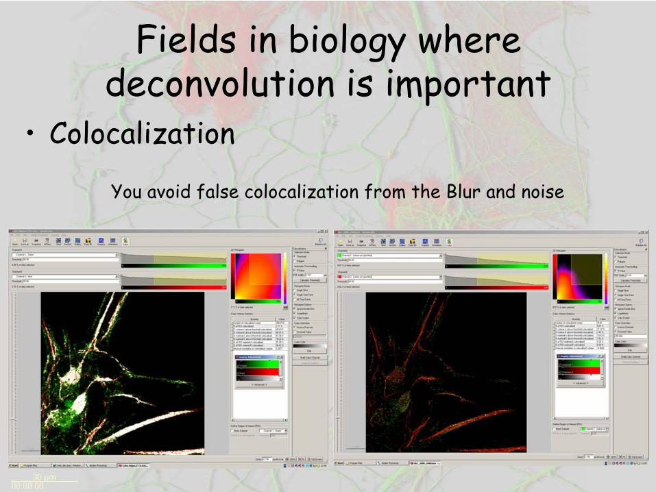
Fields in biology where deconvolution is important
•
Colocalization
You avoid false colocalization from the Blur and noise
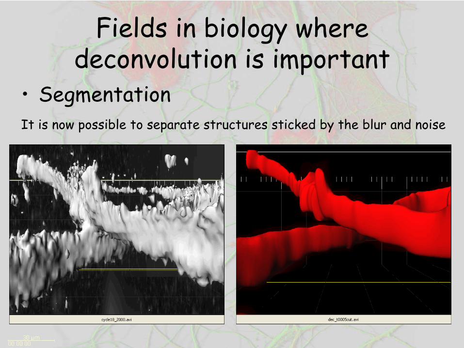
Fields in biology where deconvolution is important
•
SegmentationIt is now possible to separate structures sticked by the blur and noise

Fields in biology where deconvolution is important
•
VisualizationBetter understanding of the third dimension

How•
Software: Huygens 3 (http://www.svi.nl)
•
Current calculator:SGI PRISM from the BBP (32 processor, 320Gb RAM)
•
Web interface: http://svitsrv7.epfl.ch/hrm/login.php
•
Theory: http://www.svi.nl/support/wiki
•
Bibliography:
–
Deconvolution improves colocalization analysis of multiple fluorochromes in 3D Confocal data setsmore than filtering techniques – L. LandmannJournal of Microscopy, Vol. 208, Pt. 2 November 2002, pp. 134-147
–
A workingperson’s Guide to Deconvolution in Light Microscopy - Wes Wallace & al.BioTechechniques 31:1076-1097 (November 2001)