introduction to enzyme and coenzyme chemistry · 3.9 the involvement of protein dynamics in enzyme...
TRANSCRIPT

T.D.H. Bugg
Introduction toEnzyme andCoenzymeChemistrySECOND EDITION

Introduction to Enzyme andCoenzyme Chemistry
Bugg/Introduction to Enzyme and Coenzyme Chemistry Final Proof 22.7.2004 3:52pm page i

Bugg/Introduction to Enzyme and Coenzyme Chemistry Final Proof 22.7.2004 3:52pm page ii

Introduction to Enzyme andCoenzyme Chemistry
Second Edition
TIM BUGGProfessor of Biological Chemistry, Department of Chemistry,University of Warwick, UK
Bugg/Introduction to Enzyme and Coenzyme Chemistry Final Proof 22.7.2004 3:52pm page iii

� 1997, 2004 by Blackwell Publishing Ltd
Editorial oYces:
Blackwell Publishing Ltd, 9600 Garsington Road, Oxford OX4 2DQ, UK
Tel: þ44 (0)1865 776868
Blackwell Publishing Inc., 350 Main Street, Malden, MA 02148-5020, USA
Tel: þ1 781 388 8250
Blackwell Publishing Asia Pty Ltd, 550 Swanston Street, Carlton, Victoria 3053, Australia
Tel: þ61 (0)3 8359 1011
The right of the Author to be identiWed as the Author of this Work has been asserted in accordance with the
Copyright, Designs and Patents Act 1988.
All rights reserved. No part of this publication may be reproduced, stored in a retrieval system, or transmitted, in any
form or by any means, electronic, mechanical, photocopying, recording or otherwise, except as permitted by the UK
Copyright, Designs and Patents Act 1988, without the prior permission of the publisher.
First published 1997 by Blackwell Science
Second edition published 2004 by Blackwell Publishing
Library of Congress Cataloging-in-Publication Data
Bugg, Tim.
Introduction to enzyme and coenzyme chemistry / Tim Bugg.–2nd ed.
p. cm.
Includes bibliographical references and index.
ISBN 1-4051-1452-5 (pbk. : alk. paper)
1. Enzymes. 2. Coenzymes. I. Title.
QP601.B955 2004
5720.7–dc22
2003025117
ISBN 1-4051-1452-5
A catalogue record for this title is available from the British Library
Set in 10/13 pt Times
by Kolam Information Services Pvt. Ltd, Pondicherry, India
Printed and bound in Great Britain
by Ashford Colour Press, Gosport
The publisher’s policy is to use permanent paper from mills that operate a sustainable forestry policy, and which
has been manufactured from pulp processed using acid-free and elementary chlorine-free practices. Furthermore,
the publisher ensures that the text paper and cover board used have met acceptable environmental accreditation
standards.
For further information on Blackwell Publishing, visit our website:
www.blackwellpublishing.com
Bugg/Introduction to Enzyme and Coenzyme Chemistry Final Proof 22.7.2004 3:52pm page iv

Contents
Preface ix
Representation of Protein Three-Dimensional Structures x
1 From Jack Beans to Designer Genes 1
1.1 Introduction 1
1.2 The discovery of enzymes 1
1.3 The discovery of coenzymes 3
1.4 The commercial importance of enzymes in biosynthesis
and biotechnology 3
1.5 The importance of enzymes as targets for drug discovery 6
2 All Enzymes are Proteins 8
2.1 Introduction 8
2.2 The structures of the l -a-amino acids 8
2.3 The primary structure of polypeptides 10
2.4 Alignment of amino acid sequences 12
2.5 Secondary structures found in proteins 13
2.6 The folded tertiary structure of proteins 16
2.7 Enzyme structure and function 19
2.8 Metallo-enzymes 22
2.9 Membrane-associated enzymes 23
2.10 Glycoproteins 24
3 Enzymes are Wonderful Catalysts 29
3.1 Introduction 29
3.2 A thermodynamic model of catalysis 31
3.3 Proximity eVects 33
3.4 The importance of transition state stabilisation 36
3.5 Acid/base catalysis in enzymatic reactions 37
3.6 Nucleophilic catalysis in enzymatic reactions 41
3.7 The use of strain energy in enzyme catalysis 45
3.8 Catalytic perfection 47
3.9 The involvement of protein dynamics in enzyme catalysis 47
4 Methods for Studying Enzymatic Reactions 51
4.1 Introduction 51
4.2 Enzyme puriWcation 51
4.3 Enzyme kinetics 54
4.4 The stereochemical course of an enzymatic reaction 60
Bugg/Introduction to Enzyme and Coenzyme Chemistry Final Proof 22.7.2004 3:53pm page v
v

4.5 The existence of intermediates in enzymatic reactions 68
4.6 Analysis of transition states in enzymatic reactions 71
4.7 Determination of active site catalytic groups 75
5 Enzymatic Hydrolysis and Group Transfer Reactions 81
5.1 Introduction 81
5.2 The peptidases 82
CASE STUDY: HIV-1 protease 96
5.3 Esterases and lipases 98
5.4 Acyl transfer reactions in biosynthesis: use of coenzyme A (CoA) 99
5.5 Enzymatic phosphoryl transfer reactions 102
5.6 Adenosine 50-triphosphate 106
5.7 Enzymatic glycosyl transfer reactions 109
5.8 Methyl group transfer: use of S-adenosyl methionine and
tetrahydrofolate coenzymes for one-carbon transfers 112
6 Enzymatic Redox Chemistry 121
6.1 Introduction 121
6.2 Nicotinamide adenine dinucleotide-dependent dehydrogenases 123
6.3 Flavin-dependent dehydrogenases and oxidases 129
6.4 Flavin-dependent mono-oxygenases 134
6.5 CASE STUDY: Glutathione and trypanothione reductases 137
6.6 DeazaXavins and pterins 141
6.7 Iron–sulphur clusters 142
6.8 Metal-dependent mono-oxygenases 143
6.9 a-Ketoglutarate-dependent dioxygenases 147
6.10 Non-haem iron-dependent dioxygenases 148
7 Enzymatic Carbon–Carbon Bond Formation 156
7.1 Introduction 156
Carbon–carbon bond formation via carbanion equivalents 158
7.2 Aldolases 158
CASE STUDY: Fructose-1,6-bisphosphate aldolase 158
7.3 Claisen enzymes 164
7.4 Assembly of fatty acids and polyketides 166
7.5 Carboxylases: use of biotin 170
7.6 Ribulose bisphosphate carboxylase/oxygenase (Rubisco) 171
7.7 Vitamin K-dependent carboxylase 173
7.8 Thiamine pyrophosphate-dependent enzymes 176
Carbon–carbon bond formation via carbocation intermediates 179
7.9 Terpene cyclases 179
Carbon–carbon bond formation via radical intermediates 183
7.10 Phenolic radical couplings 184
Bugg/Introduction to Enzyme and Coenzyme Chemistry Final Proof 22.7.2004 3:53pm page vi
vi Contents

8 Enzymatic Addition/Elimination Reactions 193
8.1 Introduction 193
8.2 Hydratases and dehydratases 194
8.3 Ammonia lyases 199
8.4 Elimination of phosphate and pyrophosphate 202
8.5 CASE STUDY: 5-Enolpyruvyl-shikimate-3-phosphate (EPSP) synthase 204
9 Enzymatic Transformations of Amino Acids 210
9.1 Introduction 210
9.2 Pyridoxal 50-phosphate-dependent reactions at the a-position
of amino acids 211
9.3 CASE STUDY: Aspartate aminotransferase 215
9.4 Reactions at b- and g-positions of amino acids 218
9.5 Serine hydroxymethyltransferase 220
9.6 N-Pyruvoyl-dependent amino acid decarboxylases 222
9.7 Imines and enamines in alkaloid biosynthesis 222
10 Isomerases 227
10.1 Introduction 227
10.2 Cofactor-independent racemases and epimerases 227
10.3 Keto–enol tautomerases 230
10.4 Allylic isomerases 231
10.5 CASE STUDY: Chorismate mutase 233
11 Radicals in Enzyme Catalysis 240
11.1 Introduction 240
11.2 Vitamin B12-dependent rearrangements 240
11.3 The involvement of protein radicals in enzyme catalysis 244
11.4 S-adenosyl methionine-dependent radical reactions 246
11.5 Biotin synthase and sulphur insertion reactions 249
11.6 Oxidised amino acid cofactors and quinoproteins 250
12 Non-Enzymatic Biological Catalysis 255
12.1 Introduction 255
12.2 Catalytic RNA 255
12.3 Catalytic antibodies 259
12.4 Synthetic enzyme models 265
Appendices 272
1. Cahn–Ingold–Prelog rule for stereochemical nomenclature 272
2. Amino acid abbreviations 274
3. A simple demonstration of enzyme catalysis 275
4. Answers to problems 277
Index 285
Bugg/Introduction to Enzyme and Coenzyme Chemistry Final Proof 22.7.2004 3:53pm page vii
Contents vii

Bugg/Introduction to Enzyme and Coenzyme Chemistry Final Proof 22.7.2004 3:53pm page viii

Preface
Since the publication of the Wrst edition in 1998, the Weld of chemical biology
has, I would say, become more a part of the core research and teaching of
Chemistry Departments around the world. As the genes and proteins involved
in important biological problems are elucidated, so they become accessible for
study at the molecular level by chemists. Enzymology is a core discipline within
chemical biology, since enzymes are the biological catalysts that make things
happen within cells: they translate gene sequence into biological function.
The main feature of the Wrst edition that I wanted to improve was the
Wgures. My original intention was to have a case study in each chapter, where
I would illustrate the chemical mechanism, with curly arrows, and on the facing
page would show the three-dimensional structure of the enzyme active site, and/
or tertiary structure of the enzyme. One of the fascinations of enzymology is the
interplay between structure and function, and I wanted to try to convey this to
students. In the Wrst edition I was only able to include a set of colour plates in
the middle of the book. In the second edition, I have prepared a series of two-
colour pictures using Rasmol, to accompany the text. I hope that these pictures
convey the desired ideas.
I have also updated the Wrst edition with recent observations and references
from the literature, and have added a few new topics. I have written a new
chapter entitled Radicals in Enzyme Catalysis (Chapter 11), which includes the
discovery of protein radicals and the discovery of SAM-dependent radical
reactions. I have also mentioned the recent proposals for protein dynamics
(Chapter 3) and proton tunnelling (Chapter 4) in enzyme catalysis, in a way that
I hope will be accessible to undergraduate students.
I would like to thank my colleagues, researchers, and students at Warwick
for their encouragement, and hope that the book is useful to chemical biologists
everywhere.
Tim Bugg
University of Warwick
Bugg/Introduction to Enzyme and Coenzyme Chemistry Final Proof 22.7.2004 3:53pm page ix
ix

Representation of ProteinThree-Dimensional Structures
In the second edition I have used the program Rasmol to draw representa-
tions of protein three-dimensional structures. Rasmol was developed by
Roger Sayle (GlaxoSmithKline Pharmaceuticals), is freely available from the
internet (http://www.umass.edu/microbio/rasmol), and can be downloaded with
instructions for its use. There are several packages available for representation
of protein structures, but Rasmol is straightforward to learn and freely
available.
In order to view a protein structure, you must Wrst download the PDB Wle
from the Brookhaven Protein Database, which contains all the data for pub-
lished X-ray crystal structures and NMR structures of proteins and nucleic
acids. I have included the PDB Wlename for each of the pictures that I have
drawn, in the Wgure legend. I recommend that you try downloading a few of
these, and viewing each one on a computer screen, as you can turn the structure
around and hence get a really good feel for the three-dimensional structure of
the protein. You can download the PDB Wle from http://www.rcsb.org/pdb.
Once you have downloaded the PDB Wle, then you run the Rasmol
program, and open the PDB structure Wle to view the structure. You can view
the protein backbone in several diVerent ways: as individual atoms (wireframe),
protein backbone (backbone), strands or cartoons. In most of the pictures in
this edition I have drawn the protein backbone in cartoon format. I have then
selected certain catalytic amino acid residues, and highlighted them in red, and
selected any bound substrate analogues or coenzymes, and highlighted them in
black. In preparing Wgures for the book I used only black and red, but on a
computer screen you can use a wide range of colours and you can prepare your
own multi-colour pictures!
Further reading
R.A. Sayle & E.J. Milner-White (1995) RasMol: Biomolecular graphics for all. Trends
Biochem. Sci., 20, 374–6.
Bugg/Introduction to Enzyme and Coenzyme Chemistry Final Proof 22.7.2004 3:53pm page x
x

1 From Jack Beans toDesigner Genes
1.1 Introduction
Enzymes are giant macromolecules which catalyse biochemical reactions. They
are remarkable in many ways. Their three-dimensional structures are highly
complex, yet they are formed by spontaneous folding of a linear polypeptide
chain. Their catalytic properties are far more impressive than synthetic catalysts
which operate under more extreme conditions. Each enzyme catalyses a single
chemical reaction on a particular chemical substrate with very high enantio-
selectivity and enantiospeciWcity at rates which approach ‘catalytic perfection’.
Living cells are capable of carrying out a huge repertoire of enzyme-catalysed
chemical reactions, some of which have little or no precedent in organic
chemistry. In this book I shall seek to explain from the perspective of organic
chemistry what enzymes are, how they work, and how they catalyse many of the
major classes of enzymatic reactions.
1.2 The discovery of enzymes
Historically, biological catalysis has been used by mankind for thousands of
years, ever since fermentation was discovered as a process for brewing and
bread-making in ancient Egypt. It was not until the 19th century AD, however,
that scientists addressed the question of whether the entity responsible for
processes such as fermentation was a living species or a chemical substance.
In 1897 Eduard Buchner published the observation that cell-free extracts of
yeast containing no living cells were able to carry out the fermentation of sugar
to alcohol and carbon dioxide. He proposed that a species called ‘zymase’
found in yeast cells was responsible for fermentation. The biochemical pathway
involved in fermentation was subsequently elucidated by Embden and
Meyerhof – the Wrst pathway to be discovered.
The exquisite selectivity of enzyme catalysiswas recognised as early as 1894by
Emil Fischer, who demonstrated that the enzyme which hydrolyses sucrose,
which he called ‘invertin’, acts only upon a-d -glucosides, whereas a second
enzyme ‘emulsin’ acts only upon b-d -glucosides. He deduced that these two
enzymes must consist of ‘asymmetrically built molecules’, and that ‘the enzyme
and glucoside must Wt each other like a lock and key to be able to exert a chemical
inXuence upon each other’. Fischer’s ‘lock and key’ hypothesis remained a
powerful metaphor of enzyme action for many years. The crystallisation in
1
Bugg/Introduction to Enzyme and Coenzyme Chemistry Final Proof 22.7.2004 3:53pm page 1

1926 of the enzyme urease from Jack beans by Sumner proved beyond doubt that
biological catalysis was carried out by a chemical substance (Figure 1.1).
The recognition that biological catalysis is mediated by enzymes heralded
the growth of biochemistry as a subject, and the elucidation of the metabolic
pathways catalysed by enzymes. Each reaction taking place on a biochemical
pathway is catalysed by a speciWc enzyme. Without enzyme catalysis the unca-
talysed chemical process would be too slow to sustain life. Enzymes catalyse
reactions involved in all facets of cellular life: metabolism (the production of
cellular building blocks and energy from food sources); biosynthesis (how cells
make new molecules); detoxiWcation (the breakdown of toxic foreign chem-
icals); and information storage (the processing of deoxyribonucleic acid
(DNA) ).
In any given cell there are present several thousand diVerent enzymes, each
catalysing its speciWc reaction. How does a cell know when it needs a particular
enzyme? The production of enzymes, as we shall see in Chapter 2, is controlled
by a cell’s DNA, both in terms of the speciWc structure of a particular enzyme
and the amount that is produced. Thus diVerent cells in the same organism have
the ability to produce diVerent types of enzymes and to produce them in
diVering amounts according to the cell’s requirements.
Since the majority of the biochemical reactions involved in cellular life are
common to all organisms, a given enzyme will usually be found in many or
all organisms, albeit in diVerent forms and amounts. By looking closely at
the structures of enzymes from diVerent organisms which catalyse the same
reaction, we can in many cases see similarities between them. These similarities
are due to the evolution and diVerentiation of species by natural selection.
So by examining closely the similarities and diVerences of an enzyme from
diVerent species we can trace the course of molecular evolution, as well as
learning about the structure and function of the enzyme itself.
Recent developments in biochemistry, molecular biology and X-ray
crystallography now allow a far more detailed insight into how enzymes work
at a molecular level. We now have the ability to determine the amino acid
sequence of enzymes with relative ease, whilst the technology for solving the
three-dimensional structure of enzymes is developing apace. We also have the
ability to analyse their three-dimensional structures using molecular modelling
and then to change the enzyme structure rationally using site-directed muta-
genesis. We are now starting to enter the realms of enzyme engineering where,
by rational design, we can modify the genes encoding speciWc enzymes, creating
the ‘designer genes’ of the title. These modiWed enzymes could in future perhaps
O
NH2H2N
Jack beanurease
CO2 + 2 NH3+ H2O
Figure 1.1 Reaction catalysed by the enzyme urease.
Bugg/Introduction to Enzyme and Coenzyme Chemistry Final Proof 22.7.2004 3:53pm page 2
2 Chapter 1

be used to catalyse new types of chemical reactions, or via gene therapy used to
correct genetic defects in cellular enzymes which would otherwise lead to
human diseases.
1.3 The discovery of coenzymes
At the same time as the discovery of enzymes in the late 19th and early 20th
centuries, a class of biologically important small molecules was being discovered
which had remarkable properties to cure certain dietary disorders. These mol-
ecules were christened the vitamins, a corruption of the phrase ‘vital amines’
used to describe their dietary importance (several of the Wrst-discovered vitamins
were amines, but this is not true of all the vitamins). The vitamins were later
found to have very important cellular roles, shown in Table 1.1.
The Wrst demonstration of the importance of vitamins in the human diet
took place in 1753. A Scottish naval physician, James Lind, found that the
disease scurvy, prevalent amongst mariners at that time, could be avoided by
deliberately including green vegetables or citrus fruits in the sailors’ diets. This
discovery was used by Captain James Cook to maintain the good health of
his crew during his voyages of exploration in 1768–76. The active ingredient
was elucidated much later as vitamin C, ascorbic acid.
The Wrst vitamin to be identiWed as a chemical substance was thiamine, lack
of which causes the limb paralysis beriberi. This nutritional deWciency was Wrst
identiWed in the Japanese Navy in the late 19th century. The incidence of beriberi
in sailors was connected with their diet of polished rice by Admiral Takaki, who
eliminated the ailment in 1885 by improvement of the sailors’ diet. Subsequent
investigations by Eijkman identiWed a substance present in rice husks able to
cure beriberi. This vitamin was subsequently shown to be an essential ‘cofactor’
in cellular decarboxylation reactions, as we shall see in Chapter 7.
Over a number of years the family of vitamins shown in Table 1.1 was
identiWed and their chemical structures elucidated. Some, like vitamin C, have
simple structures, whilst others, like vitamin B12, have very complex structures.
It has taken much longer to elucidate the molecular details of their biochemical
mode of action. Many of the vitamins are in fact coenzymes: small organic
cofactors which are used by certain types of enzyme in order to carry out
particular classes of reaction. Table 1.1 gives a list of the coenzymes that we
are going to encounter in this book.
1.4 The commercial importance of enzymes in biosynthesisand biotechnology
Many plants and micro-organisms contain natural products that possess potent
biological activities. The isolation of these natural products has led to the
Bugg/Introduction to Enzyme and Coenzyme Chemistry Final Proof 22.7.2004 3:53pm page 3
From Jack Beans to Designer Genes 3

discovery of many biologically active compounds such as quinine, morphine
and penicillin (Figure 1.2) which have been fundamental to the development of
modern medicine.
N
NHO
H
MeON
SHN
OCO2H
O
PhH H
HO
HO
NMeHH
O
penicillin Gquinine morphine
Figure 1.2 Structures of quinine, morphine and penicillin G.
Table 1.1 The vitamins.
Vitamin Chemical name DeWciency disease Biochemical function Coenzyme chemistry
A Retinol Night blindness Visual pigments —
B1 Thiamine Beriberi Coenzyme (TPP) Decarboxylation of a-keto
acids
B2 RiboXavin Skin lesions Coenzyme (FAD, FMN) 1e�=2e�
redox chemistry
Niacin Nicotinamide Pellagra Coenzyme (NAD) Redox chemistry
B6 Pyridoxal Convulsions Coenzyme (PLP) Reactions of a-amino acids
B12 Cobalamine Pernicious anaemia Coenzyme Radical re-arrangements
C Ascorbic acid Scurvy Coenzyme, anti-oxidant Redox agent (collagen
formation)
D Calciferols Rickets Calcium homeostasis —
E Tocopherols Newborn haemolytic
anaemia
Anti-oxidant —
H Biotin Skin lesions Coenzyme Carboxylation
K Phylloquinone Bleeding disorders Coenzyme, anti-oxidant Carboxylation of glutamyl
peptides
Folic acid Megaloblastic anaemia Coenzyme
(tetrahydrofolate)
1-carbon transfers
Pantothenic acid Burning foot syndrome Coenzyme (CoA,
phosphopantotheine)
Acyl transfer
CoA, coenzyme A; FAD, Xavin adenine dinucleotide; FMN, Xavin mononucleotide; NAD, nicotinamide adenine
dinucleotide; PLP, pyridoxal-50-phosphate; TPP, thiamine pyrophosphate.
Bugg/Introduction to Enzyme and Coenzyme Chemistry Final Proof 22.7.2004 3:53pm page 4
4 Chapter 1

The process of natural product discovery continues today, with the recent
identiWcation of important compounds such as cyclosporin A, a potent
immunosuppressant which has dramatically reduced the rejection rate in
organ transplant operations; and taxol, an extremely potent anti-cancer drug
isolated from yew bark (Figure 1.3).
Many of these natural products are structurally so complex that it is not
feasible to synthesise them in the laboratory at an aVordable price. Nature,
however, is able to biosynthesise these molecules with apparent ease using
enzyme-catalysed biosynthetic pathways. Hence, there is considerable interest
in elucidating the biosynthetic pathways for important natural products and
using the enzymes to produce natural products in vitro. One example of this is
the industrial production of semi-synthetic penicillins using a naturally occur-
ring enzyme, penicillin acylase (Figure 1.4). Penicillin G, which is obtained from
growing Penicillium mould, has certain clinical disadvantages; enzymatic dea-
cylation and chemical re-acylation give a whole range of ‘semi-synthetic’ peni-
cillins which are clinically more useful.
The use of enzyme catalysis for commercial applications is an exciting area
of the biotechnology industry. One important application that we shall encoun-
ter is the use of enzymes in asymmetric organic synthesis. Since enzymes are
highly eYcient catalysts that work under mild conditions and are enantiospe-
ciWc, they can in many cases be used on a practical scale to resolve racemic
mixtures of chemicals into their optically active components. This is becoming
increasingly important in drug synthesis, since one enantiomer of a drug usually
MeN
NMe
MeN
NH
O
O
O
O
NMe
O
NMe
O
HO
NH
HN
NMe
HN
O
O
O
O
NMe
O
OAcO
OH
OHO
O
OAcO
O
HN
Ph OPh
HOPh
O
cyclosporin A taxol
Figure 1.3 Structures of cyclosporin A and taxol.
N
SHN
OCO2H
O
Ph
N
SH2N
OCO2H
N
SHN
OCO2H
R
O
penicillinacylase
chemicalacylation
Figure 1.4 Industrial production of a semi-synthetic penicillin using penicillin acylase.
Bugg/Introduction to Enzyme and Coenzyme Chemistry Final Proof 22.7.2004 3:53pm page 5
From Jack Beans to Designer Genes 5

has very diVerent biological properties from the other. The unwanted enantio-
mer might have detrimental side-eVects, as in the case of thalidomide, where
one enantiomer of the drug was useful in relieving morning sickness in pregnant
women, but the other enantiomer caused serious deformities in the newborn
child when the racemic drug was administered.
1.5 The importance of enzymes as targets for drug discovery
If there is an essential enzyme found uniquely in a certain class of organism or
cell type, then a selective inhibitor of that enzyme could be used for selective
toxicity against that organism or cell type. Similarly, if there is a signiWcant
diVerence between a particular enzyme found in bacteria as compared with the
same enzyme in humans, then a selective inhibitor could be developed for
the bacterial enzyme. If this inhibitor did not inhibit the human enzyme, then
it could be used as an antibacterial agent. Thus, enzyme inhibition is a basis for
drug discovery.
This principle has been used for the development of a range of pharmaceut-
ical and agrochemical agents (Table 1.2). We shall see examples of important
enzyme targets later in the book. In many cases resistance to these agents has
emerged due to mutation in the structures of the enzyme targets. This has
provided a further incentive to study the three-dimensional structures of
Table 1.2 Commercial applications of enzyme inhibitors.
Anti-bacterial agents Penicillins and cephalosporins inactivate the transpeptidase enzyme which
normally makes cross-links in the bacterial cell wall (peptidoglycan),
leading to weakened cell walls and eventual cell lysis. Streptomycin and
kanamycin inhibit protein synthesis on bacterial ribosomes, whereas
mammalian ribosomes are less aVected.
Anti-fungal agents Ketoconazole inhibits lanosterol 14a-demethylase, an enzyme involved in
the biosynthesis of an essential steroid component of fungal cell
membranes. Nikkomycin inhibits chitin synthase, an enzyme involved in
making the chitin cell walls of fungi.
Anti-viral agents AZT inhibits the reverse transcriptase enzyme required by the HIV virus
in order to replicate its own DNA.
Insecticides Organophosphorus compounds such as dimethoate derive their lethal
activity from the inhibition of the insect enzyme acetylcholinesterase
involved in the transmission of nerve impulses.
Herbicides Glyphosate inhibits the enzyme EPSP synthase which is involved in the
biosynthesis of the essential amino acids phenylalanine, tyrosine and
tryptophan (see Section 8.5).
AZT, 30-azido,30-deoxythymidine; EPSP, 5-enolpyruvyl-shikimate-3-phosphate.
Bugg/Introduction to Enzyme and Coenzyme Chemistry Final Proof 22.7.2004 3:53pm page 6
6 Chapter 1

enzyme targets, and has led to the development of powerful molecular model-
ling software for analysis of enzyme structure and de novo design of enzyme
inhibitors.
The next two chapters are ‘theory’ chapters on enzyme structure and
enzyme catalysis, followed by a ‘practical’ chapter on methods used to study
enzymatic reactions. Chapters 5–11 cover each of the major classes of enzym-
atic reactions, noting each of the coenzymes used for enzymatic reactions.
Finally, there is a brief introduction in Chapter 12 to other types of biological
catalysis. In cases where discussion is brief the interested reader will Wnd
references to further reading at the end of each chapter.
Further reading
Historical development of enzymology
T.D.H. Bugg (2001) The development of mechanistic enzymology in the 20th century.
Nat. Prod. Reports, 18, 465–93.
Enzymes in biosynthesis and biotechnology
J. Mann (1987) Secondary Metabolism, 2nd edition. Clarendon Press, Oxford.
C.H. Wong & G.M. Whitesides (1994) Enzymes in Synthetic Organic Chemistry.
Pergamon, Oxford.
Medicinal chemistry
G.L. Patrick (2001) An Introduction to Medicinal Chemistry, 2nd edition. OUP, Oxford.
R.B. Silverman (2001) The Organic Chemistry of Drug Design and Drug Action.
Academic Press, San Diego.
Bugg/Introduction to Enzyme and Coenzyme Chemistry Final Proof 22.7.2004 3:53pm page 7
From Jack Beans to Designer Genes 7

2 All Enzymes are Proteins
2.1 Introduction
Enzymes are giant molecules. Their molecular weight varies from 5000 to
5 000 000 Da, with typical values in the range 20 000–100 000 Da. At Wrst
sight this size suggests a bewildering complexity of structure, yet we shall see
that enzymes are structurally assembled in a small number of steps in a fairly
simple way.
Enzymes belong to a larger biochemical family of macromolecules known as
proteins. The common feature of proteins is that they are polypeptides: their
structure is made up of a linear sequence of a-amino acid building blocks joined
together by amide linkages. This linear polypeptide chain then ‘folds’ to give a
unique three-dimensional structure.
2.2 The structures of the L-a-amino acids
Proteins are composed of a family of 20 a-amino acid structural units whose
general structure is shown in Figure 2.1. a-Amino acids are chiral molecules:
that is, their mirror image is not superimposable upon the original molecule.
Each a-amino acid can be found as either the l- or d-isomer depending on
the conWguration at the a-carbon atom (except for glycine where R5H). All
proteins are composed only of l-amino acids, consequently enzymes are inher-
ently chiral molecules – an important point. d-amino acids are rare in biological
systems, although they are found in a number of natural products and notably
in the peptidoglycan layer of bacterial cell walls (see Chapter 9).
The a-amino acids used to make up proteins number only 20, whose struc-
tures are shown in Figure 2.2. The diVerences between these 20 lie in the nature
of the side chain R. The simplest amino acids are glycine (abbreviated Gly or
simply G), which has no side chain, and alanine (Ala or A), whose side chain is a
methyl group. A number of side chains are hydrophobic (‘water-hating’) in
character, for example the thioether of methionine (Met); the branched aliphatic
side chains of valine (Val), leucine (Leu) and isoleucine (Ile); and the aromatic
H2N CO2H
RH
H2N CO2H
HR
general structure ofa D-α-amino acid
general structure ofan L-α-amino acid
Figure 2.1 General structure of l- and
d-amino acids.
Bugg/Introduction to Enzyme and Coenzyme Chemistry Final Proof 22.7.2004 4:07pm page 8
8

side chains of phenylalanine (Phe) and tryptophan (Trp). The remainder of the
amino acid side chains are hydrophilic (‘water-loving’) in character. Aspartic
acid (Asp) and glutamic acid (Glu) contain carboxylic acid side chains, and their
corresponding primary amides are found as asparagine (Asn) and glutamine
Glutamine (Gln, Q)
Asparagine (Asn, N)
-CH2-CH2-CONH2
-CH2-CONH2
Threonine (Thr, T)
Cysteine (Cys, C)
Serine (Ser, S)
-CH2-SH
-CH2-OHPolar
Histidine (His, H)
Arginine (Arg, R)
Lysine (Lys, K)
-CH2-CH2-CO2H
-CH2-CH2-CH2-NH
-CH2-CH2-CH2-CH2-NH2Basic
Glutamate (Glu, E)
Aspartate (Asp, D)-CH2-CO2HAcidic
Aromatic
Isoleucine (Ile, I)
Leucine (Leu, L)
Valine (Val, V)
Proline (Pro, P)
Methionine (Met, M)
Alanine (Ala, A)
Glycine (Gly, G)
-CH2-CH2-S-CH3
Tryptophan (Trp, W)
Tyrosine (Tyr, Y)
Phenylalanine (Phe, F)
+
-CH3
H
H2N
CHCH3
CH3
CH2
CH2
CH CH3
CH3
CH3
CH3
CH2
CH2CH2
OH
NH
NH
NH2
N
NHCH2
OH
CH3
RH
H3N
H
Aliphatic
+
H
General structure(side chain R)
CO2−
CO2−
Figure 2.2 The side chains of the 20 a-amino acids found in proteins. Whole amino acid structure
shown for proline. Functionally important groups highlighted in red.
Bugg/Introduction to Enzyme and Coenzyme Chemistry Final Proof 22.7.2004 4:07pm page 9
All Enzymes are Proteins 9

(Gln). There are three basic side chains consisting of the e-amino group of
lysine (Lys), the guanidine group of arginine (Arg), and the imidazole ring
of histidine (His). The polar nucleophilic side chains that will assume a key
role in enzyme catalysis are the primary hydroxyl of serine (Ser), the secondary
hydroxyl of threonine (Thr), the phenolic hydroxyl group of tyrosine (Tyr) and
the thiol group of cysteine (Cys).
The nature of the side chain confers certain physical and chemical properties
upon the corresponding amino acid, and upon the polypeptide chain in which it
is located. The amino acid side chains are therefore of considerable structural
importance and, as we shall see in Chapter 3, they play key roles in the catalytic
function of enzymes.
2.3 The primary structure of polypeptides
To form the polypeptide chain found in proteins each amino acid is linked to
the next via an amide bond, forming a linear sequence of 100–1000 amino acids
– this is the primary structure of the protein. A portion of the amino-terminal
(or N-terminal) end of a polypeptide is shown in Figure 2.3, together with the
abbreviated representations for this peptide sequence.
The sequence of amino acids in the polypeptide chain is all-important. It
contains all the information to confer both the three-dimensional structure of
proteins in general and the catalytic activity of enzymes in particular. How is
this amino acid sequence controlled? It is speciWed by the nucleotide sequence
of the corresponding gene, the piece of DNA (deoxyribonucleic acid) which
encodes for that particular protein in that particular organism. To give an idea
of how this is achieved, I will give a simpliWed account of how the polypeptide
sequence is derived from the gene sequence. For a more detailed description
the reader is referred to biochemical textbooks.
Genes are composed of four deoxyribonucleotides (or ‘bases’): deoxyade-
nine (dA), deoxycytidine (dC), deoxyguanine (dG) and deoxythymidine (dT),
HN
NH
HN
NH
HN
H3N
H
SCH3
O
O
O
O
OH CH3
H
H
OH
H
+
CO2−
Met AspSerPheAla- - - - -
M A F S D
Figure 2.3 Structure of the
N-terminal portion of a poly-
peptide chain.
Bugg/Introduction to Enzyme and Coenzyme Chemistry Final Proof 22.7.2004 4:07pm page 10
10 Chapter 2

arranged in a speciWc linear sequence. To give some idea of size, a typical gene
might consist of a sequence of 1000 nucleotide bases encoding the information
for the synthesis of a protein of approximately 330 amino acids, whose molecu-
lar weight would be 35–40 kDa.
How is the sequence encoded? First the deoxyribonucleotide sequence of the
DNA strand is transcribed into messenger ribonucleic acid (mRNA) containing
the corresponding ribonucleotides adenine (A), cytidine (C), guanine (G) and
uridine (U, corresponding to dT). The RNA strand is then translated into
protein by the biosynthetic machinery known as ribosomes, as shown in Figure
2.4. The RNA sequence is translated into protein in sets of three nucleotide
bases, one set of three bases being known as a ‘triplet codon’. Each codon
encodes a single amino acid. The code deWning which amino acid is derived
from which triplet codon is the ‘universal genetic code’, shown in Figure 2.5.
This universal code is followed by the protein biosynthetic machinery of all
organisms.
As an example we shall consider in Figure 2.6 the N-terminal peptide
sequence Met–Ala–Phe–Ser–Asp illustrated in Figure 2.3. The Wrst amino
acid at the N-terminus of each protein is always methionine, whose triplet
codon is AUG. The next triplet GCC encodes alanine; UUC encodes phenyl-
alanine; UCC encodes serine; and GAC encodes aspartate. Translation then
continues in triplets until one of three ‘stop’ codons is reached; at this point
protein translation stops. Note that for most amino acids there is more than
one possible codon: thus if UUC is changed to UUU, phenylalanine is still
encoded, but if changed to UCC then serine is encoded as above.
In this way the nucleotide sequence of the gene is translated into the amino
acid sequence of the encoded protein. An important practical consequence is
that the amino acid sequence of an enzyme can be determined by nucleotide
sequencing of the corresponding gene, which is now the most convenient way to
determine a protein sequence.
DNA 5� 3�gene
RNA polymeraseribonucleoside triphosphates
promoter site
mRNA 5� 3�
ribosomesamino acyl-transfer RNAs
ribosome binding site
+Protein H3N-aa1-aa2-aa3-aa4-............-aan-1-aan-CO2
−
Figure 2.4 Pathway for protein biosynthesis.
Bugg/Introduction to Enzyme and Coenzyme Chemistry Final Proof 22.7.2004 4:07pm page 11
All Enzymes are Proteins 11

2.4 Alignment of amino acid sequences
Most biochemical reactions are found in more than one organism, in some
cases in all living cells. If the enzymes which catalyse the same reaction
in diVerent organisms are puriWed and their amino acid sequences are deter-
mined, then we often see similarity between the two sequences. The degree
of similarity is usually highest in enzymes from organisms which have
evolved recently on an evolutionary timescale. The implication of such an
observation is that the two enzymes have evolved divergently from a common
ancestor.
Over a long period of time, changes in the DNA sequence of a gene can
occur by random mutation or by several types of rare mistakes in DNA
replication. Many of these mutations will lead to a change in the encoded
protein sequence in such a way that the mutant protein is inactive. These
mutations are likely to be lethal to the cell and are hence not passed down to
the next generation. However, mutations which result in minor modiWcations
to non-essential residues in an enzyme will have little eVect on the activity of the
enzyme, and will therefore be passed onto the next generation.
So if we look at an alignment of amino acid sequences of ‘related’ enzymes
from diVerent organisms, we would expect that catalytically important amino
AAA Lys ACA Thr AGA Arg AUA IleAAG Lys ACG Thr AGG Arg AUG MetAAC Asn ACC Thr AGC Ser AUC IleAAU Asn ACU Thr AGU Ser AUU Ile
CAA Gln CCA Pro CGA Arg CUA LeuCAG Gln CCG Pro CGG Arg CUG LeuCAC His CCC Pro CGC Arg CUC LeuCAU His CCU Pro CGU Arg CUU Leu
GAA Glu GCA Ala GGA Gly GUA ValGAG Glu GCG Ala GGG Gly GUG ValGAC Asp GCC Ala GGC Gly GUC ValGAU Asp GCU Ala GGU Gly GUU Val
UAA Stop UCA Ser UGA Stop UUA LeuUAG Stop UCG Ser UGG Trp UUG LeuUAC Tyr UCC Ser UGC Cys UUC PheUAU Tyr UCU Ser UGU Cys UUU Phe Figure 2.5 The universal genetic code.
....GGATCAUGGCCUUCUCCGACUACCGGA....
AUG GCC UUC UCC GAC ...Met Ala Phe Ser Asp
startcodon
mRNA
Figure 2.6 Translation of mRNA into protein.
Bugg/Introduction to Enzyme and Coenzyme Chemistry Final Proof 22.7.2004 4:07pm page 12
12 Chapter 2

acid residues would be invariant or ‘conserved’ in all species. In this way
sequence alignments can provide clues for identifying important amino
acid residues in the absence of an X-ray crystal structure. For example, in
Figure 2.7 there is an alignment of the N-terminal portion of the amino
acid sequence of a dioxygenase enzyme MhpB from Escherichia coli with
‘related’ dioxygenase enzymes from Alcaligenes eutrophus (MpcI) and Pseudo-
monas (LigB) and another E. coli enzyme HpcB. Clearly there are a small
number of conserved residues (indicated by a *) which are very important
for activity, and a further set of residues for which similar amino acid side
chains are found (e.g. hydroxyl-containing serine and threonine, indicated
with a þ).
Furthermore, sequence similarity is sometimes observed between diVerent
enzymes which catalyse similar reactions or use the same cofactor, giving rise to
‘sequence motifs’ found in a family of enzymes. We shall meet some examples
of sequence motifs later in this book.
2.5 Secondary structures found in proteins
When the linear polypeptide sequence of the protein is formed inside cells by
ribosomes, a remarkable thing happens: the polypeptide chain spontaneously
folds to form the three-dimensional structure of the protein. All the more
remarkable is that from a sequence of 100–1000 amino acids a unique stable
three-dimensional structure is formed. It has been calculated that if the protein
folding process were to sample each of the available conformations then it
would take longer than the entire history of the universe – yet, in practice, it
takes a few seconds! The mystery of protein folding is currently a topic of
intense research, and the interested reader is referred to specialist articles on this
topic. Factors that seem to be important in the folding process are:
Alignment of N-terminal 15 amino acids of four sequences in 3-letter codes:
1 5 10 15E. coli MhpB Met His Ala Tyr Leu His Cys Leu Ser His Ser Pro Leu Val GlyA. eutrophus MpcI Met Pro Ile Gln Leu Glu Cys Leu Ser His Thr Pro Leu His GlyP. paucimobilis LigB Met Ala Arg Val Thr Thr Gly Ile Thr Ser Ser His Ile Pro Ala Leu GlyE. coli HpcB Met Gly Lys Leu Ala Leu Ala Ala Lys Ile Thr His Val Pro Ser Met Tyr
+ * *
Alignment of N-terminal 60 amino acids of two sequences in 1-letter codes:
1 11 21 31 41 51E. coli MhpB MHAYLHCLSH SPLVGYVDPA QEVLDEVNGV IASARERIAA FSPELVVLFA PDHYNGFFYDA. eutrophus MpcI MPIQLECLSH TPLHGYVDPA PEVVAEVERV QAAARDRVRA FDPELVVVFA PDHFNGFFYD
* **** +** ****** **+ ** * *+**+*+ * * *****+** ***+******
* = identically conserved residue + = functionally conserved residue
Figure 2.7 Amino acid sequence alignment.
Bugg/Introduction to Enzyme and Coenzyme Chemistry Final Proof 22.7.2004 4:07pm page 13
All Enzymes are Proteins 13

(1) packing of hydrophobic amino acid side chains and exclusion of solvent
water;
(2) formation of speciWc non-covalent interactions;
(3) formation of secondary structures.
Secondary structure is the term given to local regions (10–20 amino acids)
of stable, ordered three-dimensional structures held together by hydrogen-
bonding, that is non-covalent bonding between acidic hydrogens (O2H,
N2H) and lone pairs as shown in Figure 2.8.
There are at least three stable forms of secondary structure commonly
observed in proteins: the a-helix, the b-sheet and the b-turn. The a-helix is
a helical structure formed by a single polypeptide chain in which hydrogen
bonds are formed between the carbonyl oxygen of one amide linkage and
the N2H of the amide linkage four residues ahead in the chain, as shown in
Figure 2.9.
In this structure each of the amide linkages forms two speciWc hydrogen
bonds, making it a very stable structural unit. All of the amino acid side chains
point outwards from the pitch of the helix, consequently amino acid side chains
that are four residues apart in the primary sequence will end up close in space.
Interactions between such side chains can lead to further favourable inter-
actions within the helix, or with other secondary structures. A typical a-helix
is shown in Figure 2.10a, showing the positions of the side chains of the amino
acid residues. In Figure 2.10b, the same helix is drawn in ‘ribbon’ form, a
convenient representation that is used for drawing protein structures.
NH
O
HN
O
Figure 2.8 A hydrogen bond.
O
N H
O
N H
O
N H
O
HN
O
N HN
O
H
N HO
O
N H
ON H
C
OH
CN
O
N HN
O
H
N H
O
Figure 2.9 Structure of an a-helix. Positions of amino acid a-carbons are indicated with dots.
Bugg/Introduction to Enzyme and Coenzyme Chemistry Final Proof 22.7.2004 4:07pm page 14
14 Chapter 2

The b-sheet is a structure formed by two or more linear polypeptide strands,
held together by a series of interstrand hydrogen bonds. There are two types of
b-sheet structures: parallel b-sheets, in which the peptide strands both proceed
in the same amino-to-carboxyl direction; and anti-parallel, in which the peptide
strands proceed in opposite directions. Both types are illustrated in Figure 2.11.
Figure 2.12a shows an example of two anti-parallel b-sheets in a protein
structure, with Figure 2.12b showing the same b-sheets in ‘ribbon’ form.
Figure 2.10 Structure of an a-helix, (a) showing positions of the polypeptide chain and side chains
and (b) showing the same structure in ribbon format.
N
O
N
O
N
O
N
H
H
H
O
C
N
O
N
O
N
H
H
H
O
N H
C N
O
N
O
N
O
N
H
H
H
O
C
N
O
N
O
N
O
N
H
H
H
O
C
N
O
N
O
N
O
N
H
H
H
O
C
N
O
N
O
N
O
N
H
H
H
O
C
Parallel β-sheetAnti-parallel β-sheet
Figure 2.11 Structure of b-sheets. Positions of amino acid a-carbons are indicated with dots.
(a) (b)
Bugg/Introduction to Enzyme and Coenzyme Chemistry Final Proof 22.7.2004 4:07pm page 15
All Enzymes are Proteins 15

The b-turn is a structure often formed at the end of a b-sheet which leads to
a 1808 turn in the direction of the peptide chain. An example of a b-turn is
shown in Figure 2.13, where the role of hydrogen bonding in stabilising such
structures can be seen.
2.6 The folded tertiary structure of proteins
The three-dimensional structure of protein sub-units, known as the tertiary
structure, arises from packing together elements of secondary structure to
form a stable global conformation, which in the case of enzymes is catalytically
active. The packing of secondary structural units usually involves burying
Figure 2.12 Structure of two anti-parallel b-sheets, (a) showing positions of the polypeptide chain
and side chains and (b) showing the same structure in ribbon format.
N
O
N
O
H
N
H O
NH
C
R
R R
R
Figure 2.13 Structure of a b-turn.
(a)
(b)
Bugg/Introduction to Enzyme and Coenzyme Chemistry Final Proof 22.7.2004 4:07pm page 16
16 Chapter 2

hydrophobic amino acid side chains on the inside of the protein and positioning
hydrophilic amino acid side chains on the surface.
Although in theory the number of possible protein tertiary structures is
virtually inWnite, in practice proteins are often made up of common structural
motifs, from which the protein structure can be categorised. Common families
of protein structure are:
(1) a-helical proteins;
(2) a=b structures;
(3) anti-parallel b structures.
Members of each class are illustrated below, with a-helices and b-sheets repre-
sented in ribbon form. The a-helical proteins are made up only of a-helices
which pack onto one another to form the tertiary structure. Many of the haem-
containing cytochromes which act as electron carriers (see Chapter 6) are four-
helix ‘bundles’, illustrated in Figure 2.14 in the case of cytochrome b562. The
family of globin oxygen carriers, including haemoglobin, consist of a more
complex a-helical tertiary structure. The a=b structures consist of regular
arrays of b-sheet–a-helix–parallel b-sheet structures. The redox Xavoprotein
Xavodoxin contains Wve such parallel b-sheets, forming a twisted b-sheet sur-
face interwoven with a-helices, as shown in Figure 2.15. Anti-parallel b struc-
tures consist of regular arrays of b-sheet–b-turn–anti-parallel b-sheet. For
example, the metallo-enzyme superoxide dismutase contains a small barrel of
anti-parallel b-sheets, as shown in Figure 2.16.
Frequently, proteins consist of a number of ‘domains’, each of which
contains a region of secondary structure. Sometimes a particular domain has
a speciWc function, such as binding a substrate or cofactor. Larger proteins
often consist of more than one tertiary structure, which Wt together to form
the active ‘quaternary’ structure. In some cases a number of identical sub-units
can bind together to form a homodimer (two identical sub-units), trimer or
tetramer, or in other cases non-identical sub-units Wt together to form highly
complex quaternary structures. One familiar example is the mammalian oxygen
transport protein haemoglobin, which consists of a tetramer of identical
16-kDa sub-units.
How are protein tertiary structures determined experimentally? The most
common method for solving three-dimensional structures of proteins is to use
X-ray crystallography, which involves crystallisation of the protein, and analy-
sis of the diVraction pattern obtained from X-ray irradiation of the crystal. The
Wrst protein structure to be solved by this method was lysozyme in 1965, since
which time several hundred crystal structures have been solved. Recent ad-
vances in nuclear magnetic resonance (NMR) spectroscopy have reached the
point where the three-dimensional structures of small proteins (<15 kDa) in
solution can be solved using multi-dimensional NMR techniques.
Bugg/Introduction to Enzyme and Coenzyme Chemistry Final Proof 22.7.2004 4:07pm page 17
All Enzymes are Proteins 17

Figure 2.14 Structure of cytochrome b562 (PDB Wle 256B), a four-helix bundle protein. Haem
cofactor shown in red.
Figure 2.15 Structure of Xavodoxin (PDB Wle 1AHN), a redox carrier protein
containing Wve parallel b-sheets, each connected by an intervening a-helix. Parallel b-sheets
shown in red.
Bugg/Introduction to Enzyme and Coenzyme Chemistry Final Proof 22.7.2004 4:07pm page 18
18 Chapter 2

2.7 Enzyme structure and function
All enzymes are proteins, but not all proteins are enzymes, the diVerence being
that enzymes possess catalytic activity. The part of the enzyme tertiary structure
which is responsible for the catalytic activity is called the ‘active site’ of the
enzyme, and often makes up only 10–20% of the total volume of the enzyme.
This is where the enzyme chemistry takes place.
The active site is usually a hydrophilic cleft or cavity containing an array of
amino acid side chains which bind the substrate and carry out the enzymatic
reaction, as shown in Figure 2.17a. In some cases the enzyme active site also
binds one or more cofactors which assist in the catalysis of particular types of
enzymatic reactions, as shown in Figure 2.17b.
How does the enzyme bind the substrate? One of the hallmarks of enzyme
catalysis is its high substrate selectivity, which is due to a series of highly speciWc
non-covalent enzyme–substrate binding interactions. Since the active site is
chiral, it is naturally able to bind one enantiomer of the substrate over the
other, just as a hand Wts a glove. There are four types of enzyme–substrate
interactions used by enzymes, as follows:
Figure 2.16 Structure of superoxide dismutase (PDB Wle 1CB4), a b-barrel protein containing eight
anti-parallel b-sheets. Anti-parallel b-sheets shown in red.
Bugg/Introduction to Enzyme and Coenzyme Chemistry Final Proof 22.7.2004 4:07pm page 19
All Enzymes are Proteins 19

(1) Electrostatic interactions. Substrates containing ionisable functional groups
which are charged in aqueous solution at or near pH 7 are often bound via
electrostatic interactions to oppositely charged amino acid side chains at
the enzyme active site. Thus, for example, carboxylic acids (pKa 4–5) are
found as the negatively charged carboxylate anion at pH 7, and are often
bound to positively charged side chains such as the protonated e-amino side
chain of a lysine or the protonated guanidine side chain of arginine, shown
in Figure 2.18.
Similarly, positively charged substrate groups can be bound electrosta-
tically to negatively charged amino acid side chains of aspartate and glu-
tamate. Energetically speaking, the binding energy of a typical electrostatic
interaction is in the range 25–50 kJ mol�1, the strength of the electrostatic
interaction varying with 1=r2, where r is the distance between the two
charges.
(2) Hydrogen bonding. Hydrogen bonds can be formed between a hydrogen-
bond donor containing a lone pair of electrons and a hydrogen-bond
acceptor containing an acidic hydrogen. These interactions are widely
used for binding polar substrate functional groups. The strength of
hydrogen bonds depends upon the chemical nature and the geometrical
alignment of the interacting groups. Studies of enzymes in which hydrogen-
bonding groups have been speciWcally mutated has revealed that hydrogen
X
Y
XY
XY
XY
X
Y
X
Y+ Substrate Catalysis
Enzyme
Enzyme Enzyme Enzyme
Enzyme Enzyme
S P Enzyme+
Product
+ Cofactor Z Z S+ SubstrateCatalysis
(a)
(b)
Figure 2.17 Schematic Wgure of (a) enzyme plus substrate and (b) enzyme plus substrate plus
cofactor.
O
O
N
N
HN Arg Enz
H
H
H
H
S
Figure 2.18 Electrostatic enzyme–substrate
interaction.
Bugg/Introduction to Enzyme and Coenzyme Chemistry Final Proof 22.7.2004 4:07pm page 20
20 Chapter 2

bonds between uncharged donors/acceptors are of energy 2.0–7.5 kJ mol�1,
whilst hydrogen bonds between charged donors/acceptors are much
stronger, in the range 12.5– 25 kJ mol�1.
(3) Non-polar (Van der Waals) interactions. Van der Waals interactions arise
from interatomic contacts between the substrate and the active site. Since
the shape of the active site is usually highly complementary to the shape of
the substrate, the sum of the enzyme–substrate Van der Waals interactions
can be quite substantial (50–100 kJ mol�1), even though each individual
interaction is quite weak (6–8 kJ mol�1). Since the strength of these inter-
actions varies with 1=r6 they are only signiWcant at short range (2–4 A), so a
very good ‘Wt’ of the substrate into the active site is required in order to
realise binding energy in this way.
(4) Hydrophobic interactions. If the substrate contains a hydrophobic group or
surface, then favourable binding interactions can be realised if this is bound
in a hydrophobic part of the enzyme active site. These hydrophobic inter-
actions can be visualised in terms of the tendency for hydrophobic organic
molecules to aggregate and extract into a non-polar solvent rather than
remain in aqueous solution. These processes of aggregation and extraction
are energetically favourable due to the maximisation of inter-water hydro-
gen-bonding networks which are otherwise disrupted by the hydrophobic
molecule, as shown in Figure 2.19.
There are many examples of hydrophobic ‘pockets’ or surfaces in enzyme
active sites which interact favourably with hydrophobic groups or surfaces in
the substrate and hence exclude water from the two hydrophobic surfaces. As
mentioned above, these hydrophobic interactions may be very important for
HO
H
O
H
H
H
OH
OHH
H
OH
H
O H
O
H
H
OH
H
O
H
H
OH
H
HO
H
O
H
H
H
OH
OHH
H
OH
H
O H
O
H
H
OH
H
O
H
H
OH
H
OH
H
O
HH
Hydrophobic molecule in water Additional water−water hydrogenbonds possible if hydrophobic moleculeis excluded from water
Figure 2.19 Hydrophobic interaction.
Bugg/Introduction to Enzyme and Coenzyme Chemistry Final Proof 22.7.2004 4:07pm page 21
All Enzymes are Proteins 21

maintaining protein tertiary structure and, as we shall see below, they are
central to the behaviour of biological membranes.
Having bound the substrate, the enzyme then proceeds to catalyse its
speciWc chemical reaction using active site catalytic groups, and Wnally releases
its product back into solution. Enzyme catalysis will be discussed in the next
chapter. However, before Wnishing the discussion of enzyme structure three
special classes of enzyme structural types will be introduced.
2.8 Metallo-enzymes
Although the polypeptide backbone of proteins is made up only of the 20
common l-amino acids, many proteins bind one or more metal ions. Enzymes
which bind metal ions are known as metallo-enzymes: in these enzymes the
metal cofactor is usually found at the active site of the enzyme, where it may
have either a structural or a catalytic role.
A brief summary of the more common metal ions is given in Table 2.1.
Magnesium ions are probably the most common metal ion cofactor: they are
found in many enzymes which utilise phosphate or pyrophosphate substrates,
since magnesium ions eVectively chelate polyphosphates (see Figure 2.20).
Zinc ions are used structurally to maintain tertiary structure, for example in
the ‘zinc Wnger’ DNA-binding proteins by co-ordination with the thiolate side
chains of four cysteine residues, as shown in Figure 2.21a. In contrast, zinc is
also used in a number of enzymes as a Lewis acid to co-ordinate carbonyl
groups present in the substrate and hence activate them towards nucleophilic
attack, as shown in Figure 2.21b.
Table 2.1 Metallo-enzymes.
Metal Types of enzyme Role of metal Redox active?
Mg Kinases, phosphatases, phosphodiesterases Binding of phosphates/polyphosphates �
Zn Metalloproteases, dehydrogenases Lewis acid carbonyl activation �
Fe Oxygenases (P450, non-haem) Binding and activation of oxygen �
[FeS] Clusters Electron transport, hydratases
Cu Oxygenases Activation of oxygen �
Mn Hydrolases, hydratases Lewis acid? �
Co Vitamin B12 coenzyme Homolysis of Co–carbon bond �
Mo Nitrogenase Component of Mo/Fe cluster �
Bugg/Introduction to Enzyme and Coenzyme Chemistry Final Proof 22.7.2004 4:07pm page 22
22 Chapter 2

The other common role of metal ions is as redox reagents. Since none of
the 20 common amino acids are able to perform any useful catalytic redox
chemistry, it is not surprising that many redox enzymes employ redox-active
metal ions. We shall meet a number of examples of these redox-active metallo-
enzymes in Chapter 6. For a more detailed discussion of the role of metal ions
in biological systems the reader is referred to several excellent texts in
bio-inorganic chemistry.
2.9 Membrane-associated enzymes
Although the majority of enzymes are freely soluble in water and exist in the
aqueous cytoplasm of living cells, there is a substantial class of enzymes that are
associated with the biological membranes which encompass all cells. Biological
membranes are made up of a lipid bilayer composed of phospholipid molecules
containing a polar head group and a hydrophobic fatty acid tail. The phospho-
lipid molecules assemble spontaneously to form a stable bilayer in which the
hydrophilic head groups are exposed to solvent water and the hydrophobic tails
are packed together in a hydrophobic interior.
Enzymes that are associated with biological membranes fall into two
classes, as illustrated in Figure 2.22:
(1) extrinsic membrane proteins which are bound loosely to the surface of the
membrane, often by a non-speciWc hydrophobic interaction, or in some
cases by a non-peptide membrane ‘anchor’ which is covalently attached to
the protein;
PO
O−
O−
O
P OO
OR
Mg
−
2+
Figure 2.20 Binding of polyphosphate by magnesium.
Figure 2.21 Uses of zinc (a) in a structural role and (b) as a Lewis acid.
Bugg/Introduction to Enzyme and Coenzyme Chemistry Final Proof 22.7.2004 4:07pm page 23
All Enzymes are Proteins 23
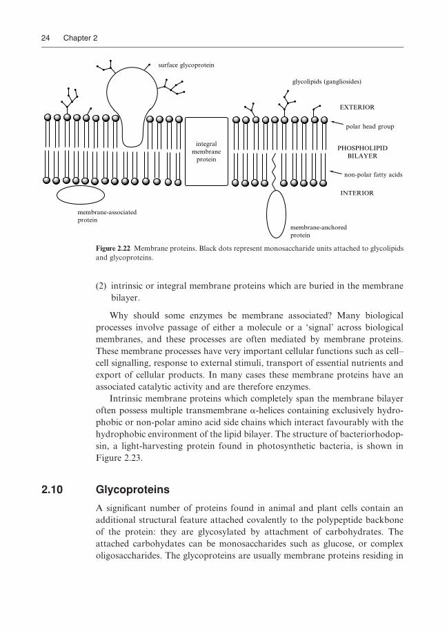
(2) intrinsic or integral membrane proteins which are buried in the membrane
bilayer.
Why should some enzymes be membrane associated? Many biological
processes involve passage of either a molecule or a ‘signal’ across biological
membranes, and these processes are often mediated by membrane proteins.
These membrane processes have very important cellular functions such as cell–
cell signalling, response to external stimuli, transport of essential nutrients and
export of cellular products. In many cases these membrane proteins have an
associated catalytic activity and are therefore enzymes.
Intrinsic membrane proteins which completely span the membrane bilayer
often possess multiple transmembrane a-helices containing exclusively hydro-
phobic or non-polar amino acid side chains which interact favourably with the
hydrophobic environment of the lipid bilayer. The structure of bacteriorhodop-
sin, a light-harvesting protein found in photosynthetic bacteria, is shown in
Figure 2.23.
2.10 Glycoproteins
A signiWcant number of proteins found in animal and plant cells contain an
additional structural feature attached covalently to the polypeptide backbone
of the protein: they are glycosylated by attachment of carbohydrates. The
attached carbohydates can be monosaccharides such as glucose, or complex
oligosaccharides. The glycoproteins are usually membrane proteins residing in
PHOSPHOLIPIDBILAYER
INTERIOR
EXTERIOR
glycolipids (gangliosides)
surface glycoprotein
integralmembrane
protein
membrane-associatedprotein
membrane-anchoredprotein
polar head group
non-polar fatty acids
Figure 2.22 Membrane proteins. Black dots represent monosaccharide units attached to glycolipids
and glycoproteins.
Bugg/Introduction to Enzyme and Coenzyme Chemistry Final Proof 22.7.2004 4:07pm page 24
24 Chapter 2

the cytoplasmic membrane of the cell, in which the sugar residues attached to
the protein are located on the exterior of the cell membrane. Since these
glycoproteins are exposed to the external environment of the cell, they are
often important for cell–cell recognition processes. In this respect they act as
a kind of ‘bar-code’ for the type of cell on which they are residing. This function
has been exploited in a sinister fashion, as a means of recognition and entry into
mammalian cells, by viruses such as inXuenza virus and human immunodeW-
ciency virus (HIV).
The carbohydrate residues are attached in one of two ways shown in Figure
2.24: either to the hydroxyl group of a serine or threonine residue (O-linked
glycosylation); or to the primary amide nitrogen of an asparagine residue
(N-linked glycosylation).
The level of glycosylation can be very substantial: in some cases up to 50%
of the molecular weight of a glycoprotein can be made up of the attached
carbohydrate residues. The pattern of glycosylation can also be highly complex,
for example highly branched mannose-containing oligosaccharides are often
found. The sugar attachments are generally not involved in the active site
catalysis, but are usually required for full activity of the protein.
Figure 2.23 Structure of bacteriorhodopsin (PDB Wle 1C3W), a seven-transmembrane helix mem-
brane protein, solved in the presence of phospholipid. Protein structure shown in red; phospholipid
and water molecules shown in monochrome (spaceWll).
Bugg/Introduction to Enzyme and Coenzyme Chemistry Final Proof 22.7.2004 4:07pm page 25
All Enzymes are Proteins 25

Problems
(1) Which of the amino acid side chains found in proteins would be
(a) positively charged or (b) negatively charged at pH 4, 7 and 10, respect-
ively?
(2) The amide bonds found in polypeptides all adopt a trans-conformation in
which the N2H bond is transcoplanar with the C5O. Why? Certain
peptides containing proline have been found to contain cis-amide bonds
involving the amine group of proline. Explain.
(3) The following segment of RNA sequence is found in the middle of a gene,
but the correct reading frame is not known. What amino acid sequences
would be encoded from each of the three reading frames? Comment on
which is the most likely encoded amino acid sequence.
50-ACGGCUGAAAACUUCGCACCAAGUCGAUAG-30
(4) You have just succeeded in purifying a new enzyme, and you have obtained
an N-terminal sequence for the protein, which reads Met–Ala–Leu–Ser–
His–Asp–Trp–Phe–Arg–Val. How many possible nucleotide sequences
might encode this amino acid sequence? If you want to design a 12-base
oligonucleotide ‘primer’ with a high chance of matching the nucleotide
sequence of the gene as well as possible, what primer sequence would you
suggest?
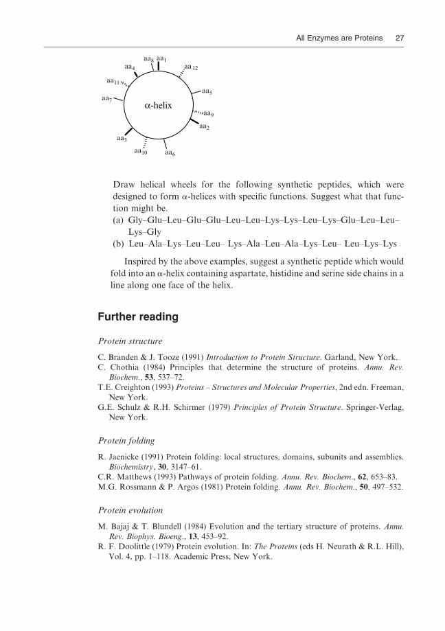
(5) a-Helices in proteins have a ‘pitch’ of approximately 3.6 amino acid resi-
dues. In order to visualise the side chain–side chain interactions in a-helices,
the structure of the helix is often represented as a ‘helical wheel’. This
representation is constructed by viewing along the length of the helix
from the N-terminal end, with the amino acid side chains protruding
from the central barrel of the helix, as shown below.
O
O
R6O
HOR3O
NH
O CH
NH
C
NH
O
H/CH3
O
OH
OHO
NH
O
CH
NH
C
NH
OHN
O
GlcNAc
O-linked glycosylation(via serine/threonine)
(Man)n
N-linked glycosylation(via asparagine)
R3 = H or Gal or GlcNAcR6 = H or NeuAc or GlcNAc
α
β
Figure 2.24 O- and N-linked glycosylation. Gal, galactose; GlcNAc, N-acetylglucosamine; Man,
mannose; NeuAc, N-acetylneuraminic acid.
Bugg/Introduction to Enzyme and Coenzyme Chemistry Final Proof 22.7.2004 4:07pm page 26
26 Chapter 2
aa1
aa2
aa3
aa4
aa5
aa6
aa7
aa8
aa9
aa10
aa11
aa12
α-helix
Draw helical wheels for the following synthetic peptides, which were
designed to form a-helices with speciWc functions. Suggest what that func-
tion might be.
(a) Gly–Glu–Leu–Glu–Glu–Leu–Leu–Lys–Lys–Leu–Lys–Glu–Leu–Leu–
Lys–Gly
(b) Leu–Ala–Lys–Leu–Leu– Lys–Ala–Leu–Ala–Lys–Leu– Leu–Lys–Lys
Inspired by the above examples, suggest a synthetic peptide which would
fold into an a-helix containing aspartate, histidine and serine side chains in a
line along one face of the helix.
Further reading
Protein structure
C. Branden & J. Tooze (1991) Introduction to Protein Structure. Garland, New York.
C. Chothia (1984) Principles that determine the structure of proteins. Annu. Rev.
Biochem., 53, 537–72.
T.E. Creighton (1993) Proteins – Structures and Molecular Properties, 2nd edn. Freeman,
New York.
G.E. Schulz & R.H. Schirmer (1979) Principles of Protein Structure. Springer-Verlag,
New York.
Protein folding
R. Jaenicke (1991) Protein folding: local structures, domains, subunits and assemblies.
Biochemistry, 30, 3147–61.
C.R. Matthews (1993) Pathways of protein folding. Annu. Rev. Biochem., 62, 653–83.
M.G. Rossmann & P. Argos (1981) Protein folding. Annu. Rev. Biochem., 50, 497–532.
Protein evolution
M. Bajaj & T. Blundell (1984) Evolution and the tertiary structure of proteins. Annu.
Rev. Biophys. Bioeng., 13, 453–92.
R. F. Doolittle (1979) Protein evolution. In: The Proteins (eds H. Neurath & R.L. Hill),
Vol. 4, pp. 1–118. Academic Press, New York.
Bugg/Introduction to Enzyme and Coenzyme Chemistry Final Proof 22.7.2004 4:07pm page 27
All Enzymes are Proteins 27

Metalloproteins
I. Bertini, H.B. Gray, S.J. Lippard & J.S. Valentine (1994) Bio-inorganic Chemistry.
University Science Books, Mill Valley, California.
Biological membranes
J.B.C. Findlay & W.H. Evans (1987) Biological Membranes – A Practical Approach. IRL
Press, Oxford.
R.G. Gennis (1989) Biomembranes: Molecular Structure and Function. Springer-Verlag,
New York.
Glycoproteins
T.W. Rademacher, R.B. Parekh & R.A. Dwek (1988) Glycobiology. Annu. Rev. Bio-
chem., 57, 785–838.
Y.C. Lee & R.T. Lee (1995) Carbohydrate-protein interactions: the basis of glycobiol-
ogy. Acc. Chem. Res., 28, 321–7.
Bugg/Introduction to Enzyme and Coenzyme Chemistry Final Proof 22.7.2004 4:07pm page 28
28 Chapter 2

3 Enzymes are WonderfulCatalysts
3.1 Introduction
The function of enzymes is to catalyse biochemical reactions. Each enzyme has
evolved over millions of years to catalyse one particular reaction, so it is
perhaps not surprising to Wnd that they are extremely good catalysts when
compared with man-made catalysts.
The hallmarks of enzyme catalysis are: speed, selectivity and speciWcity.
Enzymes are capable of catalysing reactions at rates well in excess of a mil-
lion-fold faster than the uncatalysed reaction, typical ratios of kcat=kuncat being
106---1014. Figure 3.1 shows an illustration of the speed of enzyme-catalysed
glycoside hydrolysis. The rate of acid-catalysed glycoside catalysis is accelerated
103-fold by intramolecular acid catalysis, but enzyme-catalysed glycoside hy-
drolysis is 104-fold faster still – some 107 faster than the uncatalysed reaction
carried out at pH 1.
OHOHO
OH
OH
OPh
OHOHO
OH
OH
O
O OH
O
OH
HO
OH
OH
OPh
H
OO
Enz
Rate of hydrolysis
kobs = 1.9 � 10−6 s−1 in 0.1 M HCl
H+
kuni = 1.4 � 10−3 s−1
acid-catalysedhydrolysis
intramolecularcatalysis
β-galactosidase kcat = 40 s−1
Figure 3.1 Rate acceleration of glycoside hydrolysis by intramolecular and enzyme catalysis.
Bugg/Introduction to Enzyme and Coenzyme Chemistry Final Proof 22.7.2004 4:08pm page 29
29

Enzymes are highly selective in the reactions that they catalyse. Since they
bind their substrates via a series of selective enzyme–substrate binding inter-
actions at a chiral active site, they are able to distinguish the most subtle
changes in substrate structure, and are able to distinguish between regioisomers
and between enantiomers, as shown in Figure 3.2. Finally, enzymes carry out
their reactions with near faultless precision: they are able to select a unique site
of action within the substrate, and carry out the enzymatic reaction stereospe-
ciWcally, as illustrated in Figure 3.3.
In this chapter we shall examine the factors that contribute to the remarkable
rate acceleration achieved in enzyme-catalysed reactions. Examples of enzyme
stereospeciWcity will be discussed in Chapter 4. It is worth at this point distin-
guishing between selectivity, which is the ability of the enzyme to select a certain
substrate or functional group out of many; and speciWcity, which is a property of
O
NH
NH
CO2H
R
H2N
O
CO2H
R R
OAc OH OAc
AcO
acylase+
C. cylandricealipase
+
(±) (+) (−)
C. cylandricealipase
no reaction
CO2H
Figure 3.2 Stereoselectivity in enzymatic hydrolysis reactions.
CO2Me
CO2Me
CO2H
CO2Me
AcO OAc
AcO OH
pig liveresterase
HO OAc
pig liver esterase
porcine pancreatic lipase
Figure 3.3 Stereoselectivity in enzymatic hydrolysis reactions.
Bugg/Introduction to Enzyme and Coenzyme Chemistry Final Proof 22.7.2004 4:08pm page 30
30 Chapter 3

the reaction catalysed by the enzyme, being the production of a single regio- and
stereo-isomer of the product. Both are properties which are highly prized in
synthetic reactions used in organic chemistry: enzymes are able to do both.
3.2 A thermodynamic model of catalysis
A catalyst may be deWned as a species which accelerates the rate of a chemical
reaction whilst itself remaining unchanged at the end of the catalytic reaction.
In thermodynamic terms, catalysis of a chemical reaction is achieved by redu-
cing the activation energy for that reaction, the activation energy being the
diVerence in free energy between the reagent(s) and the transition state for the
reaction. This reduction in activation energy can be achieved either by stabilisa-
tion (and hence reduction in free energy) of the transition state by the catalyst,
or by the catalyst Wnding some other lower energy pathway for the reaction.
Figure 3.4 illustrates the free energy proWle of a typical acid-catalysed
chemical reaction which converts a substrate, S, to a product, P. In this case
an intermediate chemical species SHþ is formed upon protonation of S. If
the conversion of SHþ to PHþ is ‘easier’ than the conversion of S to P, then
the activation energy for the reaction will be reduced and hence the reaction will
go faster. It is important at this point to deWne the diVerence between an
intermediate and a transition state: an intermediate is a stable (or semi-stable)
chemical species formed during the reaction and is therefore a local energy
minimum, whereas a transition state is by deWnition a local energy maximum.
The rate of a chemical reaction is related to the activation energy of the
reaction by the following equation:
k ¼ A:e(�Eact=RT)
Therefore, the rate acceleration provided by the catalysis can simply be
calculated:
kcat=kuncat ¼ e(Euncat�Ecat=RT)
If, for example, a catalyst can provide 10 kJ mol�1 of transition stabilisation
energy for a reaction at 258C a 55-fold rate acceleration will result, whereas a
20 kJ mol�1 stabilisation will give a 3000-fold acceleration and a 40 kJ mol�1
stabilisation a 107-fold acceleration! A consequence of the exponential relation-
ship between activation energy and reaction rate is that a little extra transition
state stabilisation goes a long way!
An enzyme-catalysed reaction can be analysed thermodynamically in the
same way as the acid-catalysed example, but is slightly more complicated. As
explained in Chapter 2, enzymes function by binding their substrate reversibly
at their active site, and then proceeding to catalyse the biochemical reaction
Bugg/Introduction to Enzyme and Coenzyme Chemistry Final Proof 22.7.2004 4:08pm page 31
Enzymes are Wonderful Catalysts 31

using the active site amino acid side chains. Often enzyme-catalysed reactions
are multi-step sequences involving one or more intermediates, as illustrated in
Figure 3.4. An enzyme–substrate intermediate, ES, is formed upon binding of
the substrate, which is then converted to the enzyme–product complex, EP,
either directly or via one or more further intermediates.
In both catalysed reactions shown in Figure 3.4 the overriding consideration
as far as rate acceleration is concerned is transition state stabilisation. Just as in
non-enzymatic reactions there is acid–base and nucleophilic catalysis taking
place at enzyme active sites. However, the secret to the extraordinary power of
enzyme catalysis lies in the fact that the reaction is taking place as the substrate
is bound to the enzyme active site. So, what was in the non-enzymatic case
an intermolecular reaction has eVectively become an intramolecular reaction.
The rate enhancements obtained from these types of proximity eVects can
uncatalysedreaction
S
P
SH+
PH+
acid-catalysedreaction
Freeenergy
Reaction co-ordinate
Eact(uncat)
Eact(cat)
∆G
transition states
uncatalysedreaction
S
P
ES
EPenzyme-catalysedreaction
Freeenergy
Reaction co-ordinate
Eact(uncat)
Eact(cat)
∆G
transition state
E.Int
(a)
(b)
Figure 3.4 Free energy proWles for (a) an acid-catalysed reaction and (b) an enzyme-catalysed
reaction which converts substrate S to product P.
Bugg/Introduction to Enzyme and Coenzyme Chemistry Final Proof 22.7.2004 4:08pm page 32
32 Chapter 3

be illustrated by intramolecular reactions in organic chemistry, which is where
we shall begin the discussion.
3.3 Proximity effects
There are many examples of organic reactions that are intramolecular: that is,
they involve two or more functional groups within the same molecule, rather
than functional groups in diVerent molecules. Intramolecular reactions gener-
ally proceed much more rapidly and under much milder reaction conditions
than their intermolecular counterparts, which makes sense since the two
reacting groups are already ‘in close proximity’ to one another. But how can
can we explain these eVects?
A useful concept in quantitating proximity eVects is that of eVective concen-
tration. In order to deWne the eVective concentration of a participating group
(nucleophile, base, etc.), we compare the rate of the intramolecular reaction
with the rate of the corresponding intermolecular reaction where the reagent
and the participating group are present in separate molecules. The eVective
concentration of the participating group is deWned as the concentration of
reagent present in the intermolecular reaction required to give the same rate
as the intramolecular reaction.
I will illustrate this using data for the rates of hydrolysis of a series of phenyl
esters in aqueous solution at pH 7, given in Figure 3.5. The reference reaction in
this case is the hydrolysis of phenyl acetate catalysed by sodium acetate at the
same pH. Introduction of a carboxylate group into the same molecule as the
ester leads to an enhancement of the rate of ester hydrolysis, which for phenyl
succinate (see Figure 3.5 (3) ) is 23 000-fold faster than phenyl acetate (see
Figure 3.5 (1) ). This remarkable rate acceleration is because the neighbouring
carboxylate group can attack the ester to form a cyclic anhydride intermediate,
shown in Figure 3.6. This intermediate is more reactive than the original ester
group and so hydrolyses rapidly.
Note that the rate acceleration is largest when a Wve-membered anhydride is
formed, since Wve-membered ring formation is kinetically favoured over
six-membered ring formation, which in turn is greatly favoured over three-,
four- and seven-membered ring formation. The eVective concentration can be
worked out by comparing the rates of these intramolecular reactions with the
rates of the intermolecular reaction between phenyl acetate and sodium acetate
in water. For phenyl succinate an eVective concentration of 4 000 m is found, so
the hydrolysis of phenyl succinate proceeds much faster than if phenyl acetate
was surrounded completely by acetate ions! Here we start to see the catalytic
potential of proximity eVects.
In the same series of phenyl esters, if the possible ring size of Wve is
maintained, but a cis- double bond is placed in between the reacting groups,
Bugg/Introduction to Enzyme and Coenzyme Chemistry Final Proof 22.7.2004 4:08pm page 33
Enzymes are Wonderful Catalysts 33

1010
O
OPh
O
OPh−O2C
−O2C
−O2C
−O2C
O
OPh
O
OPh
O
OPh
CO2−
CO2−
−
9
1,300
(1)
25
2 � 105
(2)
(3)
(4)
(5)
(6)
1
Effectiveconcentration
(mol l−1)
krelEster
O
OPh
O
OPh
O
OPhMe2N
Me2N
O
OPh
1
Reference Reaction
50
O
OPh
150
23,000
O
O−
1,100,000
5 � 1010
1,000,000
2,500,000
O
OPh
NaOAc
H2O
O
O−
Et3N
H2O490
4,000
(7)
(8)
(9)
+ PhO−
+ PhO−
Figure 3.5 Intramolecular catalysis of ester hydrolysis. Et3N, triethylamine; NaOAc, sodium
acetate.
O
OPh
O
O
O
O
O OPh
O
O
O
O
O
O
O
intramolecularnucleophilic attack
reactiveanhydride intermediate
FAST
H2O
Figure 3.6 Mechanism for intramolecular hydrolysis of phenyl succinate (3).
Bugg/Introduction to Enzyme and Coenzyme Chemistry Final Proof 22.7.2004 4:08pm page 34
34 Chapter 3

the observed rates of hydrolysis are even faster. Phenyl phthalate (see Figure 3.5
(5) ) has an eVective concentration of acetate ions of 2� 105m, whilst phenyl
maleate (see Figure 3.5 (6) ) has an astonishing eVective concentration of 1010m!
Yet the same molecule containing a trans- double bond has no rate acceleration
at all. So it is clear that by holding the reactive groups rigidly in close proximity
to one another remarkable rate acceleration can be achieved. Why is the
hydrolysis of phenyl maleate, in which a Wve-membered anhydride is formed,
so much faster than the hydrolysis of phenyl succinate, in which an apparently
similar Wve-membered anhydride is formed? The answer is that in phenyl
maleate the reactive groups are held in the right orientation to react, as
shown in Figure 3.7, so the probability of the desired reaction is increased.
In thermodynamic terms, the restriction of the double bond in the case of
phenyl maleate has removed rotational degrees of freedom, so that in going to
the transition state for the intramolecular reaction fewer degrees of freedom are
lost, which means that the reaction is entropically more favourable. If you think
of entropy as a measure of order in the system, then in the case of phenyl
maleate the molecule is already ordered in the right way with respect to the
reacting groups. Thus, there is a large kinetic advantage in intramolecular
chemical reactions due to the ordering of reactive groups.
The same eVect operates in enzyme active sites, and is a major factor in
enzyme catalysis. The binding of substrates and cofactors at an enzyme active
site of deWned three-dimensional structure brings the reagents into close prox-
imity to one another and to the enzyme active site functional groups. This
increases the probability of correct positioning for reaction to take place, so it
speeds up the reaction. An important factor in this analysis is that the enzyme
structure is already held rigidly (or fairly rigidly at least) in the correct con-
formation for binding and catalysis. Recent studies have argued that the
thermodynamic origin of this kinetic advantage in enzyme catalysis is primarily
enthalpic, rather than entropic. Nevertheless, the catalytic power of proximity
eVects, or ‘pre-organisation’, has been demonstrated by the synthesis of host–
guest systems which mimic enzymes by binding substrates non-covalently (see
Chapter 12).
O
OPh
O
O
O
OPh
O
O
O
OPh
O
O
(3)
unreactiveconformation
reactiveconformation
(6)
held in reactiveconformation
Figure 3.7 Intramolecular hydrolysis of phenyl succinate (3) versus phenyl maleate (6).
Bugg/Introduction to Enzyme and Coenzyme Chemistry Final Proof 22.7.2004 4:08pm page 35
Enzymes are Wonderful Catalysts 35

3.4 The importance of transition state stabilisation
All catalysts operate by reducing the activation energy of the reaction, by
stabilising the transition state for the reaction. Enzymes do the same, but the
situation is somewhat more complicated since there are usually several transi-
tion states in an enzymatic reaction. We have already seen how an enzyme binds
its substrate reversibly at the enzyme active site. One might imagine that if an
enzyme were to bind its substrate very tightly that this would lead to transition
state stabilisation. This, however, is not the case: in fact, the enzyme does not
want to bind its substrate(s) too tightly!
Shown in Figure 3.8 are free energy curves for a hypothetical enzyme-
catalysed reaction proceeding via a single rate determining transition state.
Suppose that we can somehow alter the enzyme, E, so that it binds the
substrate, S, or the transition state, TS, more tightly. In each case the starting
free energy (of E þ S) is the same. In the presence of high substrate concen-
trations the enzyme will in practice be fully saturated with substrate, so the
activation energy for the reaction will be governed by the energy diVerence
between the ES complex and the transition state. In Figure 3.8b the enzyme is
able to bind both the substrate and the transition state more tightly (and hence
lower their free energy equally). This, however, leads to no change in the
activation energy, and hence no rate acceleration. In Figure 3.8c the enzyme
ES
E + S
ES
E + S
(b) Enzyme binds S and TS more tightly
ES
E + S
(c) Enzyme binds S more tightly
ES
E + S
(d) Enzyme binds TS more tightly
(a)
Figure 3.8 The importance of transition state stabilisation – a thought experiment.
Bugg/Introduction to Enzyme and Coenzyme Chemistry Final Proof 22.7.2004 4:08pm page 36
36 Chapter 3

binds only the substrate more tightly: this generates a ‘thermodynamic pit’ for
the ES complex and hence increases the activation energy, so the reaction is
slower! However, if, as in Figure 3.8d, the enzyme can bind selectively the
transition state, then it can reduce the activation energy and hence speed up
the reaction.
The conclusion of this thought experiment is that in order to achieve
optimal catalysis, enzymes should selectively bind the transition state, rather
than the substrate. Hence, it is not advantageous for enzymes to bind their
substrates too tightly. This is evident when one looks at substrate binding
constants for enzymes (these will be discussed in more detail in Section 4.3).
Typical KM values for enzymes are in the mm–mm range (10�3---10�6m),
whereas dissociation constants for binding proteins and antibodies whose
function is to bind small molecules tightly are in the range nm–pm
(10�9---10�12m). We shall meet several examples of speciWc transition state-
stabilising interactions later in the chapter.
3.5 Acid/base catalysis in enzymatic reactions
Acid and base catalysis is involved in all enzymatic processes involving proton
transfer, so in practice there are very few enzymes that do not have acidic or
basic catalytic groups at their active sites. However, unlike organic reactions
which can be carried out under a very wide range of pH conditions to suit the
reaction, enzymes have a strict limitation that they must operate at physio-
logical pH, in the range 5–9. Given this restriction, and the fairly small range of
amino acid side chains available for participation in acid/base chemistry (shown
in Figure 3.9), a remarkably diverse range of acid/base chemistry is achieved.
General acid catalysis takes place when the substrate is protonated by a
catalytic residue which in turn gives up a proton, as shown in Figure 3.10. The
active site acidic group must, therefore, be protonated at physiological pH but
its pKa must be just above (i.e. in the range 7–10). If the pKa of a side chain was
in excess of 10 then it would become thermodynamically unfavourable to
transfer a proton.
General base catalysis takes place either when the substrate is deprotonated,
or when water is deprotonated prior to attack on the substrate, as shown in
Figure 3.11.
Enzyme active site bases must therefore be deprotonated at physiological
pH but have pKa values just below. Typical pKa ranges for amino acid side
chains in enzyme active sites are shown in Figure 3.12. They can be measured by
analysis of enzymatic reaction rate versus pH, as described in Section 4.7.
Although the pKa values given in Figure 3.12 are the typical values found in
proteins, in some cases the pKa values of active site acidic and basic groups can
be strongly inXuenced by their micro-environment. Thus, for example, the
Bugg/Introduction to Enzyme and Coenzyme Chemistry Final Proof 22.7.2004 4:08pm page 37
Enzymes are Wonderful Catalysts 37

enzyme acetoacetate decarboxylase contains an active site lysine residue which
forms an imine linkage with its substrate – its pKa value was found to be 5.9,
much less than the expected value of about 9. When an active site peptide was
obtained containing the catalytic lysine, it was found to be adjacent to another
lysine residue. So it is likely that the proximity of another positively charged
residue would make the protonated form thermodynamically less favourable,
and hence reduce the pKa. The same eVect can be observed in the pKa values of
ethylenediamine (see Figure 3.13), where the pKa for the monoprotonated form
is 10.7 as usual, but the pKa for the doubly protonated form is 7.5.
O HCH2 O−CH2
(CH2)4NH3+
+
(CH2)4 NH2
CH2 S CH2 S−
Tyrosine + H+
pKa
~10
CH2
HN
NH
Lysine
CH2
+ H+
HN
N
~9
Cysteine + H+
+ H+
+ H+
8−9
O
O
Histidine6−8
O
O−
Aspartate/glutamate4−5
H
H
Figure 3.9 Amino acid side chains used for acid/base catalysis.
X:H AEnz
XH+
−AEnz
S S
Figure 3.10 General acid catalysis.
H
O
H
HO
(1) Enz-B−
Enz-B−(2) O
Figure 3.11 General base catalysis.
Bugg/Introduction to Enzyme and Coenzyme Chemistry Final Proof 22.7.2004 4:08pm page 38
38 Chapter 3

Similarly, if a charged group was involved in a salt bridge with an oppositely
charged residue, its pKa would be altered, or if it was in a hydrophobic region
of the active site, which would destabilise the charged form of the group. So, for
example, we shall later on see protonated aspartic acid and glutamic acid
residues acting as acidic groups in some cases (i.e. with elevated pKa values of
7.0 or above). Finally, it is worth noting that histidine, whose side chain
contains an imidazole ring of pKa 6–8, can act either as an acidic or a basic
residue, depending on its particular local pKa, making it a versatile reagent for
enzymatic acid/base chemistry.
Acid/base catalysis by enzymes is made that much more eVective by the
optimal positioning of the active site acid/base groups in close proximity to
the substrate, generating a high eVective concentration of the enzyme reagent.
This can be illustrated in the case of glycoside hydrolysis using the data from
Figure 3.1. The mechanism of the non-enzymatic reaction involves protonation
of the glycosidic group by external acid to form a good leaving group, followed
by formation of an oxonium intermediate. Glycoside hydrolysis can be acceler-
ated dramatically by positioning an acidic group in close proximity to the
glycosidic leaving group, as shown in Figure 3.1. Enzymes which catalyse
3 4 5 6 7 8 9 10 11pKa
Asp/Glu
His
Cys
Lys
Tyr
= ‘normal’ range of pKa values
= unusual pKa values observed in specific cases
Figure 3.12 Range of pKa values observed for amino acid side chains in enzyme active sites.
NH3
O
NH
NH
NH2
O
H3NNH2
+ deprotonated at pH 7;pKa reduced by adjacent positive charge
+
deprotonated at pH 7.5-10(pK1 = 10.7, pK2 = 7.5)
Figure 3.13 Abnormally low lysine pKa in acetoacetate decarboxylase.
Bugg/Introduction to Enzyme and Coenzyme Chemistry Final Proof 22.7.2004 4:08pm page 39
Enzymes are Wonderful Catalysts 39

glycoside hydrolysis (see Section 5.7) also employ an acidic catalytic group to
protonate the glycosidic leaving group – but the enzymatic reaction is some
30 000-fold faster even than the intramolecular reaction, suggesting that the
enzyme is able to further stabilise the transition state for this reaction.
Enzymes also have the ability to carry out bifunctional catalysis: protona-
tion of the substrate at the same time as deprotonation in another part of
the molecule. An example of bifunctional catalysis is the enzyme ketosteroid
isomerase, whose active site (see Figure 3.14) contains two catalytic residues:
Asp-38 which acts as a catalytic base; and Tyr-14 which acts as an acidic group.
The mechanism (see Figure 3.15) involves the formation of a dienol intermedi-
ate via a concerted step involving simultaneous deprotonation of the substrate
by Asp-14 and protonation of the substrate carbonyl by Tyr-14. In the second
step the tyrosinate group acts as a base, and the substrate is re-protonated by
the protonated Asp-38.
Bifunctional catalysis is thought to make possible the deprotonation of
substrates with apparently high pKa values. Thus, in the above example depro-
tonation adjacent to a ketone in solution to form an enolate species would
involve removal of a proton of pKa 18–20, which would be impractical at pH 7.
However, simultaneous protonation to form an enol intermediate makes the
reaction thermodynamically much more favourable.
Figure 3.14 Active site of ketosteroid isomerase (PDB Wle 1E3V). Tyr-14 and Asp-38 are shown
in red.
Bugg/Introduction to Enzyme and Coenzyme Chemistry Final Proof 22.7.2004 4:08pm page 40
40 Chapter 3

Finally, enzymes which bind metal cofactors such as Zn2þ and Mg2þ can
utilise their properties as Lewis acids, i.e. electron pair acceptors. An example is
the enzyme thermolysin, whose mechanism is illustrated in Figure 3.16. In this
enzyme, Glu-143 acts as an active site base to deprotonate water for attack on
the amide carbonyl, which is at the same time polarised by co-ordination by an
active site Zn2þ ion. The protonated glutamic acid probably then acts as an
acidic group for the protonation of the departing amine.
3.6 Nucleophilic catalysis in enzymatic reactions
Nucleophilic (or covalent) catalysis is a type of catalysis seen relatively rarely
in organic reactions, but which is used quite often by enzymes. It involves
nucleophilic attack of an active site group on the substrate, forming a covalent
bond between the enzyme and the substrate, and hence a covalent intermediate
in the reaction mechanism. This is a particularly eVective strategy for enzyme-
catalysed reactions for two reasons. First, as we have seen before, an enzyme is
able to position an active site nucleophile in close proximity and correctly
aligned to attack its substrate, generating a very high eVective concentration
of nucleophile.
O
HOTyr14
H
−O O
Asp38
O
O−Tyr14
H
O O
Asp38
H
O
O−Tyr14
H
OO
Asp38
HO
HOTyr14
O−O
Asp38
H
Figure 3.15 Mechanism for ketosteroid isomerase.
Bugg/Introduction to Enzyme and Coenzyme Chemistry Final Proof 22.7.2004 4:08pm page 41
Enzymes are Wonderful Catalysts 41

Second, since enzyme active sites are often largely excluded from water
molecules, an enzyme active site nucleophile is likely to be ‘desolvated’. Thus,
a charged nucleophile in aqueous solution would be surrounded by several
layers of water molecules, which greatly reduce the polarity and eVectiveness
of the nucleophile. However, a desolvated nucleophile at a water-excluded
active site will be a much more potent nucleophile than its counterpart in
solution. This eVect can be illustrated in organic reactions carried out in dipolar
aprotic solvents such as dimethylsulphoxide (DMSO) or dimethylformamide
(DMF), in which nucleophiles are not hydrogen bonded as they would be in
aqueous solution. A consequence of this desolvation eVect is that nucleophilic
displacement reactions occur much more readily in these solvents.
Enzymes have a range of potential nucleophiles available to them, which are
shown in Table 3.1.
Probably the best nucleophile available to enzymes is the thiol side chain of
cysteine, which we shall see operating in proteases and acyl transfer enzymes.
The e-amino group of lysine is used in a number of cases to form imine linkages
with ketone groups in substrates, as in the example of acetoacetate decarbox-
ylase, shown in Figure 3.17.
This enzyme catalyses the decarboxylation of acetoacetate to acetone. Two
lines of evidence were used to show that an imine linkage is formed between the
ketone of acetoacetate and the e-amino group of an active site lysine. First,
O
HNR
O
Ph
Zn2+
OH
H
O−
−
−
OGlu143
O
HNR
O
Ph
Zn2+
HO
O
OGlu143
H
O
H2N
R
O
Ph
Zn2+
O
OGlu143
OH
Figure 3.16 Mechanism for thermolysin. R, peptide chain.
Bugg/Introduction to Enzyme and Coenzyme Chemistry Final Proof 22.7.2004 4:08pm page 42
42 Chapter 3

treatment of enzyme with substrate and sodium borohydride leads to irrevers-
ible enzyme inactivation, via in situ reduction of the enzyme-bound imine
intermediate by borohydride. Treatment of enzyme with borohydride alone
gives no inactivation. Second, incubation of acetoacetate labelled with 18O at
the ketone position leads to the rapid exchange of 18O label out of this position,
consistent with reversible imine formation. As mentioned above, the pKa of this
lysine group is abnormally low at 5.9, which is suYciently low for it to act as a
nucleophile at pH 7.
The other nitrogen nucleophile available to enzymes is the versatile
imidazole ring of histidine. This group is more often used for acid/base chemis-
try, but is occasionally used as a nucleophile in, for example, phosphotransfer
reactions. Finally, enzymes have oxygen nucleophiles available in the form of
Table 3.1 Amino acid side chains used for nucleophilic catalysis in enzymatic reactions.
Amino acid Side chain Examples
Serine 2CH22OH Serine proteases, esterases, lipases
Threonine 2CH(CH3)2OH Phosphotransferases
Cysteine 2CH22SH Cysteine proteases, acyl transferases
Aspartate, glutamate 2(CH2)n2CO2H Epoxide hydrolase, haloalkane dehalogenase
Lysine 2(CH2)42NH2 Acetoacetate decarboxylase, class I aldolases
Histidine imidazole2NH Phosphotransferases
Tyrosine Ar2OH DNA topoisomerases
Ar, aromatic group.
O−
O O O
O−
NH+ +
O
Lys
NH
H
EnzA
Lys
NH
Lys
acetoacetatedecarboxylase
+ CO2
NH2
Lys
Figure 3.17 Mechanism for acetoacetate decarboxylase.
Bugg/Introduction to Enzyme and Coenzyme Chemistry Final Proof 22.7.2004 4:08pm page 43
Enzymes are Wonderful Catalysts 43

the hydroxyl groups of serine, threonine and tyrosine, and the carboxylate
groups of aspartate and glutamate. There are examples of each of these groups
being used for nucleophilic catalysis, especially serine, which we shall see in
Chapter 5 used for the serine proteases.
An example of the use of aspartate as a nucleophile is the enzyme haloalk-
ane dehalogenase from Xanthobacter autotrophicus, which is involved in the
dechlorination of organochlorine chemicals found in industrial waste. This
35-kDa protein contains seven strands of b-sheet arranged centrally with
intervening a-helices. This type of ab structure is found in many hydrolase
enzymes such as the serine proteases discussed in Chapter 5. The catalytic
residues Asp-124 and His-289 are situated on loops at the ends of b-sheets.
The active site cavity of volume 37 A3 is lined with hydrophobic residues, which
can form favourable hydrophobic interactions with its non-polar substrates.
Shown in Figure 3.18 is a view of the protein structure, highlighting the active
site catalytic residues. The catalytic mechanism proceeds by displacement of
chloride by Asp-128 residue, to give a covalent ester intermediate, followed by
base-catalysed hydrolysis involving His-289, as shown in Figure 3.19.
Figure 3.18 Structure of haloalkane dehalogenase (PDB Wle 2DHD). Asp-124 and His-289 are
shown in red; the product ethanol is shown in black.
Bugg/Introduction to Enzyme and Coenzyme Chemistry Final Proof 22.7.2004 4:08pm page 44
44 Chapter 3

3.7 The use of strain energy in enzyme catalysis
The concept of ‘strain’ is one that is rather diYcult to explain, since it occurs
very rarely in organic reactions, and there are only a few examples of enzymatic
reactions in which there is evidence that it operates. Remember that the over-
riding factor in achieving rate acceleration in enzyme-catalysed reactions is the
diVerence in free energy between the ES complex and the transition state of the
enzymatic reaction. If the enzyme can somehow bind the substrate in a strained
conformation which is closer to the transition state than the ground state
conformation, then the diVerence in energy between the bound conformation
and the transition state will be reduced, and the reaction will be accelerated (see
Figure 3.20).
How can an enzyme bind its substrate in a strained conformation? Is that
not energetically unfavourable? The answer to these questions is that if the
substrate is of a reasonable size, the enzyme can form a number of enzyme–
substrate binding interactions, and the total enzyme–substrate binding energy
can be quite substantial. In some cases, in order to beneWt from the most
favourable overall binding interactions, the substrate must adopt an unfavour-
able conformation in a part of the molecule. That part of the substrate may
cunningly happen to be where the reaction is going to take place! In thermo-
dynamic terms, the enzyme uses its favourable binding energy in the rest of the
Asp124
O−
O
Cl
H3C
Asp124
O
O
CH3
N
HN
His289
O
H
H
Asp124
O−
O
HO
CH3
N
HN
His289
Asp124
O
O
CH3
HO
N
HN +
−
His289
H
Cl−
N
HN
His289
Figure 3.19 Mechanism for haloalkane dehalogenase.
Bugg/Introduction to Enzyme and Coenzyme Chemistry Final Proof 22.7.2004 4:08pm page 45
Enzymes are Wonderful Catalysts 45

substrate to compensate for the adoption of an unfavourable conformation in
the strained part of the molecule.
To illustrate this concept, we shall look at the example of carboxypeptidase
A, a Zn2þ-containing protease similar to thermolysin. When the X-ray crystal
structure of carboxypeptidase A was solved, it was found that in order to bind
the peptide substrate with the most favourable enzyme–substrate interactions, a
‘twist’ needed to be introduced into the scissile amide bond. In this conform-
ation the carbonyl group of the amide being hydrolysed was bound slightly out
of plane of the amide N2H bond, assisted by co-ordination to the active site
zinc ion. This has the eVect of reducing the overlap of the nitrogen lone pair of
electrons with the carbonyl p-bond, which requires the amide bond to be
planar. This makes the carbonyl much more reactive, more like a ketone
group than an amide, so it is much more susceptible to nucleophilic attack, in
this case by the carboxylate side chain of Glu-270. Figure 3.21 shows that in this
strained conformation the carbonyl oxygen has already moved some distance
towards where it will be in the transition state, so the energy diVerence between
the bound conformation and the transition state is reduced, hence we see rate
acceleration.
Thus, if an enzyme is able to bind its substrate in a less favourable but more
reactive conformation, then it is able to realise additional rate acceleration in
this way. Analysis of this type requires detailed insight from X-ray crystallog-
raphy, so it is not surprising that there are only a few well-documented
examples of this phenomenon. One other is that of lysozyme, which we shall
see in Chapter 5. However, it is possible that this strategy may be used in many
enzyme-catalysed reactions.
E + SES
ES'E + S
‘strained’ conformation
Figure 3.20 Rate acceleration from a strained ES0 complex.
ONH
O
NH
Zn2+
O
NH
Zn2+
O-Enz
normal planar conformation ‘strained’ conformation
δ−
δ−
Figure 3.21 Strained substrate conformation in carboxypeptidase A.
Bugg/Introduction to Enzyme and Coenzyme Chemistry Final Proof 22.7.2004 4:08pm page 46
46 Chapter 3

3.8 Catalytic perfection
How fast is it possible for enzyme-catalysed reactions to proceed? Is there a
limit to the rate acceleration achievable by enzymes? The answer is yes, and a
small number of enzymes have achieved it. For extremely eYcient enzymes, the
rate of reaction becomes limited by the rate at which a substrate can diVuse
onto its active site and diVuse away into solution – the so-called diVusion limit.
This diVusion limit for collision of enzyme and substrate corresponds to a
bimolecular rate constant of approximately 108m�1 s�1. We can compare this
value with the bimolecular rate constant for reaction of free enzyme with free
substrate, which is the catalytic eYciency kcat=KM (see Section 4.3).
Many enzymes have catalytic eYciencies of 106 to 107m�1 s�1, but a small
number have kcat=KM values which are at the diVusion limit – these are listed in
Table 3.2. One of these is the enzyme acetylcholinesterase, which is involved in
the propagation of nerve impulses at synaptic junctions: a process for which the
utmost speed is necessary. For these enzymes the rate-determining step has
become the diVusion of substrates onto the active site. As fast as a substrate
diVuses onto the active site it is processed by the enzyme before the next
molecule of substrate appears. These are truly wonderful catalysts.
3.9 The involvement of protein dynamics in enzyme catalysis
Our understanding of such high rates of catalysis in enzymes is still incomplete.
Chemical models for enzyme catalysts, and catalytic antibodies which function
via transition state stabilisation (see Chapter 12), are orders of magnitude less
active catalysts than the above examples. Therefore, there are likely to be other
factors that enzymes use to achieve high rates of catalysis.
Table 3.2 Catalytic eYciencies of some diVusion-limited enzymes.
Enzyme Reaction type kcat=KM(m�1 s�1)
Superoxide dismutase Redox dismutation 3:0� 109
Fumarase Hydration 2:0� 109
Cytochrome c peroxidase Redox peroxidase 6:0� 108
Triose phosphate isomerase Keto/enol isomerase 3:8� 108
Acetylcholinesterase Ester hydrolysis 1:4� 108
Ketosteroid isomerase Keto/enol isomerase 1:3� 108
b-Lactamase b-lactam hydrolysis 1:0� 108
Bugg/Introduction to Enzyme and Coenzyme Chemistry Final Proof 22.7.2004 4:08pm page 47
Enzymes are Wonderful Catalysts 47

Examination of protein structure in solution by nuclear magnetic resonance
(NMR) spectroscopy has revealed that there is a signiWcant amount of internal
motion in a protein, on a timescale of 1–10 ns. Such internal motion could
transmit kinetic energy from a distant part of the protein to the active site in
order to assist in catalysis. It has been proposed that dynamic Xuctations in the
protein structure are used by enzymes to organise the enzyme–substrate com-
plex into a reactive conformation.
The role of protein dynamics in enzyme catalysis is therefore a topic of
considerable interest. Selection of enzyme catalysts by ‘directed evolution’ has
revealed, in a number of cases, that amino acid residues distant from active sites
can have a dramatic eVect on enzyme activity. Replacement of Gly-120 to
valine in dihydrofolate reductase (see Figure 3.22) disrupts the internal motion
of a protein loop, and so interferes with the conversion of the ternary complex
to the reactive conformation, hence this step becomes partially rate-limiting,
reducing the rate of hydride transfer 500-fold. Thus, protein dynamics may
have an important role to play in enzyme catalysis.
Figure 3.22 Structure of dihydrofolate reductase (PDB Wle 1DDS), showing Gly-120 and
the loop whose internal motion is implicated in catalysis. Gly-120 and the FG loop
involving Gly-120 are shown in red; the bound inhibitor (methotrexate) at the active site is shown
in black.
Bugg/Introduction to Enzyme and Coenzyme Chemistry Final Proof 22.7.2004 4:08pm page 48
48 Chapter 3

Problems
(1) Rationalise the rate accelerations observed for the phenyl esters (8) and (9)
in Figure 3.5 which contain tertiary amine groups. What type of catalysis is
operating in this case?
(2) Explain why the hydrolysis of the substituted benzoic acid (A) at pH 4 is
1000-fold faster than the hydrolysis of the corresponding methyl ester (B)
under the same conditions.
O
O
CO2R
Ph
OH
OH
CO2R
pH 4
buffer+ PhCHO
(A) R = H(B) R = Me
(3) (a) Incubation of compound (C) under alkaline conditions leads to a
unimolecular reaction of rate constant 7:3� 10�2 s�1 to give a bicyclic
product – suggest a structure. Incubation of compound (D) with phenoxide
(PhO�) leads to a bimolecular reaction of rate constant 10�6m�1 s�1. Work
out the eVective concentration of phenoxide in (C) and comment on the rate
acceleration observed.
RO
(C) R = OH(D) R = H
(3) (b) Epoxide hydrolase catalyses the hydrolysis of a wide range of
epoxide substrates. Its active site contains an aspartate residue which is
essential for catalytic activity. Given that the enzyme is most active at pH
8–9, propose two possible mechanisms for the enzymatic reaction, and
suggest how you might distinguish them experimentally. Comparing the
kcat of 1.1 s�1 for epoxide hydrolase with the rate constant for reaction of
(C) above, suggest how the enzyme might achieve its additional rate accel-
eration.
(4) Incubation of haloalkane dehalogenase (see Figure 3.19) with substrate
under multiple turnover conditions in H182 O leads to incorporation of one
atom of 18O into the alcohol product. However, incubation of a large
quantity of enzyme with less than one equivalent of substrate in H182 O
revealed that the product contained no 18O. Explain this observation.
Bugg/Introduction to Enzyme and Coenzyme Chemistry Final Proof 22.7.2004 4:08pm page 49
Enzymes are Wonderful Catalysts 49

(Note: Similar results were obtained with this type of experiment for the
epoxide hydrolase enzyme in Problem 3).
Further reading
Intramolecular catalysis
N.S. Isaacs (1987) Physical Organic chemistry. Longman ScientiWc, Harlow.
A.J. Kirby (1980) EVective molarities for intramolecular reactions. Adv. Phys. Org.
Chem., 17, 183–278.
F.M. Menger (1985) On the source of intramolecular and enzymatic reactivity. Acc.
Chem. Res., 18, 128–34.
Enzyme catalysis
T.C. Bruice (2002) A view at the millennium: the eYciency of enzyme catalysis. Acc.
Chem. Res., 35, 139–48.
T.C. Bruice & S.J. Benkovic (2000) Chemical basis for enzyme catalysis. Biochemistry,
39, 6267–74.
A. Fersht (1985) Enzyme Structure and Mechanism, 2nd Ed. Freeman, New York.
W.P. Jencks (1969) Catalysis in Chemistry and Enzymology. McGraw-Hill, New York.
W.P. Jencks (1975) Binding energy, speciWcity and enzyme catalysis – the Circe eVect.
Adv. Enzymol., 43, 219–410.
R. Wolfenden & M.J. Snider (2001) The depth of chemical time and the power of
enzymes as catalysts. Acc. Chem. Res., 34, 938–45.
Haloalkane dehalogenase
S.M. Franken, H.J. Rozeboom, K.H. Kalk & B.W. Dijkstra (1991) Crystal structure of
haloalkane dehalogenase: an enzyme to detoxify halogenated alkanes. EMBO J., 10,
1297–302.
F. Pries, J. Kingma, M. Pentenga, G. van Pouderoyen, C.M. Jeronimus-Stratingh, A.P.
Bruins & D.B. Janssen (1994) Site-directed mutagenesis and oxygen isotope incorpor-
ation studies of the nucleophilic aspartate of haloalkane dehalogenase. Biochemistry,
33, 1242–7.
Dihydrofolate reductase
G.P. Miller & S.J. Benkovic (1998) Stretching exercises – Xexibility in dihydrofolate
reductase catalysis. Chemistry & Biology, 5, R105–13.
Bugg/Introduction to Enzyme and Coenzyme Chemistry Final Proof 22.7.2004 4:08pm page 50
50 Chapter 3

4 Methods for Studying EnzymaticReactions
4.1 Introduction
Having established the general principles of enzyme structure and enzyme
catalysis, the remaining chapters will deal with each major class of enzymes
and their associated coenzymes, and a range of enzyme mechanisms will be
discussed. In this chapter we will meet the kind of experimental methods that
are used to study enzymes and to elucidate the mechanisms that will be given
later. There will be only a brief discussion of the biochemical techniques
involved in enzyme puriWcation and characterisation, since such methods are
described in much more detail in many biochemistry texts. The chapter will
focus on those experimental techniques that provide insight into the enzymatic
reaction and active site chemistry.
4.2 Enzyme purification
If we want to study a particular enzymatic reaction, the Wrst thing we need to do
is to Wnd a source of the enzyme and purify it. In order to test the activity of the
enzyme we must Wrst of all have an assay: a quantitative method for measuring
the conversion of substrate into product (Figure 4.1). In some cases conversion
of substrate to product can be monitored directly by ultraviolet (UV) spectros-
copy, if the substrate or product has a distinctive UV absorbance. Failing this, a
chromatographic method can be used to separate substrate from product and
hence monitor conversion. In order to quantify a chromatographic assay a
radioactive label is usually required in the substrate, so that after separation
from substrate the amount of product can be quantitated by scintillation
counting. Such an assay is highly speciWc and highly sensitive, but unfortu-
nately is rather tedious for kinetic work.
A more convenient assay for kinetic purposes is to monitor consumption of
a stoichiometric cofactor or cosubstrate, for example the cofactor nicotinamide
adenine dinucleotide (NADH) by UV absorption at 340 nm, or consumption of
oxygen by an oxygenase enzyme using an oxygen electrode. In other cases a
coupled assay is used, in which the product of the reaction is immediately
consumed by a second enzyme (or set of enzymes) which can be conveniently
monitored.
Once a reliable assay has been developed, it can be used to identify a
rich source of the enzyme, which might be a plant, an animal tissue, or
Bugg/Introduction to Enzyme and Coenzyme Chemistry Final Proof 22.7.2004 4:08pm page 51
51

a micro-organism. Enzymes are generally produced in the cytoplasm contained
within the cells of the producing organism, so in order to isolate the enzyme we
must break open the cells to release the enzymes inside (Figure 4.2). If it is a
bacterial source, the bacteria can be grown in culture media and the cells
harvested by centrifugation. The bacterial cell walls are then broken by treat-
(1) Direct UV
S P (UV active at 394 nm)
+ ENZ
A394
time
slope v
S* P*
(2) Radiochemical
separate P*
scintillationcounting
time
P*(counts perminute)
(3) Indirect UV
S
ENZ ENZ
ENZP
NAD+NADH
NAD+NADH
UV active at 340 nm
+ ENZ
A340
time
slope v
(4) Coupled UV assay
S PENZ
QENZ 2excess of coupling enzyme
monitor decrease in absorbance at 340 nm
Figure 4.1 Types of enzyme assays. A340, ultraviolet (UV) absorbance at 340 nm; A394, UV absorb-
ance at 394 nm; ENZ, enzyme; NADH, nicotinamide adenine dinucleotide, P, product; Q, product
of coupling enzyme; S, substrate.
lyse cells at high pressure
extractcelldebris
high speedcentrifugation
Obtain or grow cellscontaining enzyme
enzymes
cell wall
Figure 4.2 Preparation of an enzyme extract.
Bugg/Introduction to Enzyme and Coenzyme Chemistry Final Proof 22.7.2004 4:08pm page 52
52 Chapter 4

ment in a high pressure cell. Animal cells can be readily broken by homogenisa-
tion, but plant cells sometimes require rapid freeze/thaw methods in order to
break their tough cell walls.
Using the enzyme assay mentioned above, the enzyme activity can then be
puriWed from the crude extract using precipitation methods such as ammonium
sulphate precipitation, and chromatographic methods such as ion exchange
chromatography, gel Wltration chromatography, hydrophobic interaction chro-
matography, etc. PuriWcation can be monitored at each stage by measuring
the enzyme activity, in units per ml, where 1 unit conventionally means the
activity required to convert 1 mmole of substrate per minute. Protein concen-
tration can also be measured using colorimetric assays, in mg protein per
ml. The ratio of enzyme activity to protein concentration (i.e. units per mg of
protein) is known as the speciWc activity of the enzyme, and is a measure of the
purity of the enzyme. As the enzyme is puriWed the speciWc activity of
the enzyme should increase until the protein is homogeneous and pure, which
can be demonstrated using sodium dodecyl sulphate (SDS)-polyacrylamide gel
electrophoresis. A puriWcation scheme for a hydratase enzyme from the
author’s laboratory is shown in Table 4.1. The puriWcation of the enzyme can
be seen from the increase in speciWc activity at each stage of the puriWcation. An
SDS-polyacrylamide gel containing samples of protein at each stage of the
puriWcation is shown in Figure 4.3. You can see that in the crude extract
there are hundreds of protein bands, but that as the puriWcation proceeds the
28-kDa protein becomes more and more predominant in the gel.
Why do we need pure enzyme? If we can see enzyme activity in the original
extract, why not use that? The problem with using unpuriWed enzyme for
kinetic or mechanistic studies is that there may be interference from other
enzymes in the extract that use the same substrate or cofactor. There may
also be enzymes that give rise to UV absorbance changes which might interfere
with a UV-based assay. If the enzyme can be puriWed then the turnover number
of the enzyme can be measured, which is the number of mmoles of substrate
converted per mmole of enzyme per second. The turnover number can be simply
Table 4.1 Enzyme puriWcation table (see Figure 4.3). DEAE, diethylaminoethyl.
Volume
(ml)
Enzyme
activity
(units ml�1)
Protein
concentration
(mg ml�1)
SpeciWc
activity
(units mg�1)
PuriWcation
(-fold)
Crude extract 14 13.1 62.0 0.212 1.0
DEAE sephadex pool 19 11.6 17.0 0.684 3.2
Phenyl agarose pool 11 11.8 0.085 140 662
MonoQ anion-exchange pool 6.0 29.2 0.037 787 3710
Bugg/Introduction to Enzyme and Coenzyme Chemistry Final Proof 22.7.2004 4:08pm page 53
Methods for Studying Enzymatic Reactions 53

calculated from the speciWc activity of the pure enzyme (in units per mg of
protein) and the molecular weight of the enzyme (see Problem 1). The isolation
of pure enzyme also allows active site studies to be carried out on the homoge-
neous protein, and crystallisation of the enzyme for X-ray crystallographic
analysis.
4.3 Enzyme kinetics
It is possible to learn a great deal about how an enzyme works from a detailed
kinetic study of the enzymatic reaction. For the purposes of this chapter I will
give only a brief discussion of a simple model of enzyme kinetics; a full discussion
of enzyme kinetics is given in texts such as Segel (see Further reading).
Figure 4.3 PuriWcation of 2-hydroxypentadienoic acid hydratase from Escherichia coli. The photo
shows an SDS-polyacrylamide gel of samples taken from the puriWcation of this enzyme. Lane 1, E.
coli crude extract; lane 2, DEAE sephadex pool; lane 3, phenyl agarose pool; lane 4, monoQ anion-
exchange pool; lane 5, molecular weight standards (2 � 98 kDa, 66 kDa, 43 kDa, 29 kDa, 21 kDa).
(see also Table 4.1).
Bugg/Introduction to Enzyme and Coenzyme Chemistry Final Proof 22.7.2004 4:08pm page 54
54 Chapter 4

The Michaelis–Menten model for enzyme kinetics assumes that the
following steps are involved in the enzymatic reaction: reversible formation of
the enzyme–substrate (ES) complex, followed by conversion to product:
Michaelis–Menten Model
k2
k−1
k1E + PESE + S
There are several assumptions implicit in this model: that the enzyme binds only
a single substrate; that there is only one kinetically signiWcant step between the
ES complex and product formation; and that product formation is irreversible.
Despite the fact that these assumptions are not strictly correct for most
enzymes, this proves to be a useful model for a very wide range of enzymes.
Derivation of a rate equation uses a kinetic criterion known as the steady state
approximation: that the enzymatic reaction will quickly adopt a situation of
steady state in which the concentration of the intermediate species, ES, remains
constant. Under these conditions the rate of formation of ES is equal to the rate
of consumption of ES. The other criterion that is used is that the total amount
of enzyme in the system E0, made up free enzyme, E, and enzyme–substrate
complex, ES, is constant. The derivation is as follows:
Derivation of Michaelis–Menten equation
[S]
vmax
k1 [E] [S] = k2 [ES] + k−1 [ES]
Steady state approximation: Rate of formation of ES = Rate of breakdown of ES
Total amount of enzyme [E]0 = [E] + [ES]
k1 [S] + k2 + k−1
[ES] =k1 [E]0 [S]
k1 [E]0 [S] − k1 [ES] [S] = k2 [ES] + k−1 [ES]
KM + [S]
vmax [S]
=kcat [E]0 [S]
KM + [S]+ [S]
k1
k2 [E]0 [S]
k−1 + k2
Rate of production of P = k2 [ES] =
Observed rate v =
where vmax = kcat [E0]
v
vmax/2
KM
Bugg/Introduction to Enzyme and Coenzyme Chemistry Final Proof 22.7.2004 4:08pm page 55
Methods for Studying Enzymatic Reactions 55

The two kinetic constants in the Michaelis–Menten rate equation have special
signiWcance. The kcat parameter is the turnover number mentioned above: it is a
unimolecular rate constant whose units are s�1 (or min�1 if it is a very slow
enzyme!), and it represents the number of mmoles of substrate converted per
mmole of enzyme per second. Alternatively, in molecular terms it represents the
number of molecules turned over by one molecule of enzyme per second, which
gives a good feel for how quickly the enzyme is operating. Typically values are
in the range 0:1---100 s�1.
The KM parameter is known as the Michaelis constant for the enzyme: its
units are mol l�1 or m . In practice, the KM is the concentration of substrate at
which half-maximal rate is observed. It can be taken as a rough indication of
how tightly the enzyme binds its substrate, so a substrate bound weakly by an
enzyme will have a large KM value, and a substrate bound tightly will have a
small KM. However, it must be stressed that KM is not a true dissociation
constant for the substrate, since it also depends on the rate constant k2. Values
of KM are typically in the range 1 mm–1 mm .
Values of kcat and KM can be measured for a particular enzyme by measur-
ing the rate of the enzymatic reaction at a range of diVerent substrate concen-
trations. At high substrate concentrations ([S] >> KM) the rate equation
reduces to v ¼ kcat [E0], so a maximum rate is observed however high the
substrate concentration. Under these conditions the enzyme is fully saturated
with substrate, and no free enzyme is present. So as soon as an enzyme molecule
releases a molecule of product it immediately picks up another molecule of
substrate. In other words the enzyme is working Xat out: the observed rate of
reaction is limited only by the rate of catalysis.
Under low substrate concentrations the rate equation reduces to
v ¼ kcat=KMð Þ[E][S], so the observed rate is proportional to substrate concen-
tration, and the reaction has eVectively become a bimolecular reaction between
free enzyme, E, and free substrate, S. Under these conditions the majority of
enzyme is free enzyme, and the observed rate of reaction depends on how
eYciently the enzyme can bind the substrate at that concentration. The bimo-
lecular rate constant under these conditions kcat=KM is known as the catalytic
eYciency of the enzyme, since it represents how eYciently free enzyme will react
with free substrate.
A schematic representation of the energetic proWles at high and low sub-
strate concentrations is given in Figure 4.4. At high substrate concentrations
the enzyme is fully saturated with substrate, so the activation energy for the
enzymatic reaction is the free energy diVerence between the ES complex and the
transition state. At [S] ¼ KM the enzyme is half-saturated with substrate. At
low substrate concentrations the majority of the enzyme is free of substrate, so
the activation energy for the reaction is the free energy diVerence between free
enzyme þ substrate and the transition state.
Bugg/Introduction to Enzyme and Coenzyme Chemistry Final Proof 22.7.2004 4:08pm page 56
56 Chapter 4

How do we actually determine kcat and KM? The value of kcat can be
roughly visualised from the plot of v versus [S] by estimating the rate at high
substrate concentrations. The KM value corresponds to the substrate concen-
tration at which half-maximal rate is observed. However, a more accurate way
to determine KM and kcat from the data is to use either of the Lineweaver–Burk
or Eadie–Hofstee plots, shown in Figure 4.5, both of which give straight lines
from which the kinetic constants can be determined as indicated.
Enzyme inhibition takes place if a molecule other than the substrate binds
at the active site and prevents the enzymatic reaction from taking place.
Broadly speaking, there are two types of enzyme inhibition that are commonly
observed: reversible and irreversible inhibition. Reversible inhibition occurs if
an inhibitor is bound non-covalently at the active site, preventing one of
the substrates from binding. The most common type of reversible inhibition
observed for a single substrate enzyme reaction is competitive inhibition, where
the inhibitor binds at the same site as the substrate, S, and therefore competes
for the same binding site. The kinetic scheme and associated kinetic behaviour
ES
E + S
[S] >> KM
v = kcat [E0] Enzyme saturatedwith substrate
ESE + S
[S] = KM
v = kcat [E0] Enzyme 50% saturatedwith substrate2
ES
E + S
[S] << KM
v = kcat [E0][S] Free enzyme + substrateKM
Figure 4.4 SigniWcance of KM, kcat. Plots are of free energy versus reaction co-ordinate. Red dots
indicate the population of enzyme molecules present.
Bugg/Introduction to Enzyme and Coenzyme Chemistry Final Proof 22.7.2004 4:08pm page 57
Methods for Studying Enzymatic Reactions 57

observed for competitive inhibition is shown in Figure 4.6. If the rate of the
enzyme reaction is measured at varying substrate concentrations but Wxed
inhibitor concentrations, apparent KM values ((KM)app) can be measured at
varying inhibitor concentrations. If plotted on a Lineweaver–Burk plot a series
of straight lines are obtained, intersecting on the y-axis. Thus the vmax is
unaVected by competitive inhibition, since at high substrate concentrations
the substrate can competitively displace the inhibitor.
Non-competitive reversible inhibition is observed when an inhibitor,
I, binds to another part of the enzyme active site leading to a non-productive
EIS complex. This type of inhibition, illustrated in Figure 4.6, typically occurs
in multi-substrate reactions when the inhibitor, I, binds to the binding
site of the co-substrate. For a full discussion of these and other types of inhib-
ition in multi-substrate reactions reference to texts on enzyme kinetics is
recommended.
Irreversible inhibition occurs when an inhibitor Wrst binds at the active
site, then reacts with an active site group to form a covalent bond (E2I). The
active site is then irreversibly blocked by the inhibitor and is permanently
inactivated. Irreversible inhibitors usually contain electrophilic functional
groups such as halogen substituents or epoxides. The kinetic characteristic
associated with irreversible inhibition is that it is time-dependent. This is
because as time goes by more and more enzyme will be blocked irreversibly
by conversion of the reversible EI complex to E2I. Since [S] >> [E] then in
practice this is a unimolecular reaction, so the observed kinetic behaviour
follows unimolecular reaction kinetics. Thus, if enzyme activity (i.e. vmax) is
plotted versus time, an exponential decrease of activity is observed versus time
(Figure 4.7).
1/[S]
1/v
Lineweaver−Burk Plot:
[S]1KM= +vmax vmax
1v1
slope KM/vmax
1/vmax
Eadie−Hofstee Plot:
v = vmax − KM v[S]
v
v/[S]
vmax
slope −KM
Figure 4.5 Graphical methods for determination of KM and kcat. v, reaction velocity; [S], substrate
concentration.
Bugg/Introduction to Enzyme and Coenzyme Chemistry Final Proof 22.7.2004 4:08pm page 58
58 Chapter 4

All of the above kinetic data can be obtained by steady state kinetics, which
can be observed conveniently over a 1–10-minute assay period. However, if one
is able to examine an enzymatic reaction before the attainment of steady state,
then individual enzymatic rate constants can be measured directly using pre-
steady-state kinetics. If the turnover number for an enzyme is 10 s�1 then under
saturating conditions a molecule of substrate will be converted to product in
0.1 s. Therefore, in order to examine a single catalytic cycle one must examine
the enzymatic reaction in the range 0–100 ms. This can be done using a stopped
Xow apparatus, shown in simpliWed form in Figure 4.8.
(a) Competitive inhibition
E + S ES E + P
EI
Ki + I
1/vmaxunchanged
1/[S]
1/vinc. [I]
KM(app) = KM 1 + [I]
Ki
1 + [I]
Ki
(b) Non-competitive inhibition
−1/KM unchanged
1/[S]
1/v
+ IKi
EI + S
E + PESE + S
EIS
Ki + I
inc. [I]
vmax(app) = vmax
Figure 4.6 Reversible inhibition.
Bugg/Introduction to Enzyme and Coenzyme Chemistry Final Proof 22.7.2004 4:08pm page 59
Methods for Studying Enzymatic Reactions 59

This apparatus consists of two syringes containing: (i) substrate and (ii) a
stoichiometric amount (i.e. 1–100 nmol) of enzyme, connected to a rapid mixing
device. The syringes are driven so as to Wll the observation cuvette with freshly
mixed enzyme and substrate. At this point, the reaction time of the mixture is
given by the distance from the mixer and the linear Xow rate. The syringes are
physically stopped, and the enzyme/substrate mixture is allowed to react along
the enzymatic reaction pathway. Changes in absorbance or Xuorescence in the
observation cuvette are measured on a 10–1000-ms timescale, giving a complete
time course of a single catalytic cycle of the enzymatic reaction. Several substrate
concentrations are used to enable individual rate constants to be measured. In
this way a multi-step enzymatic reaction can in principle be broken down into its
individual steps, the slowest of which (the rate-determining step) should corres-
pond to the steady state kcat value measured by steady state kinetics.
4.4 The stereochemical course of an enzymatic reaction
The vast majority of biological molecules are chiral, that is a molecule whose
mirror image is non-superimposable upon the original. In the case of carbon-
based compounds, if a carbon atom is surrounded by four diVerent groups,
+ IKi
EI
E + PESE + S
kiE-I
Act
time
vi
1/vi
1/[I]
1/ki
slope Ki/ki
Figure 4.7 Irreversible inhibition.
ENZ
S
rapid mixing device ENZ + S
observation cuvette
observe time-course of enzymatic reactionby UV or fluorescence spectroscopy
stopping syringe
Figure 4.8 Stopped Xow apparatus.
Bugg/Introduction to Enzyme and Coenzyme Chemistry Final Proof 22.7.2004 4:08pm page 60
60 Chapter 4

then it will be chiral. One simple example shown in Figure 4.9 is that of the
amino acid l-alanine, which is a substrate for several pyridoxal 50-phosphate
enzymes described in Chapter 9. Chiral centres can be designated as R or S
depending on the relative orientation of the four groups, according to the
Cahn–Ingold–Prelog rule (see Appendix 1). Thus l-alanine is more formally
written as 2S-alanine.
Enzyme-catalysed reactions are in general both stereoselective and stereo-
speciWc. Stereoselectivity is the ability to select a single enantiomer of the
substrate in the presence of other isomers. StereospeciWcity is the ability to
catalyse the production of a single enantiomer of the product via a speciWc
reaction pathway. StereospeciWcity in enzyme catalysis arises from the fact that
catalysis is taking place in an enzyme active site in which the bound substrate is
held in a deWned orientation relative to the active site groups (unnatural
substrates which can adopt more than one orientation in an enzyme active
site may be processed with less stereospeciWcity). How do we elucidate the
stereochemical course of an enzymatic reaction?
Generation of a chiral product
In many cases enzymes are able to generate a product containing one or more
chiral centres from achiral substrates – a process known as asymmetric induc-
tion. For example, the aldolase enzyme fructose-1,6-bisphosphate aldolase
(see Chapter 7) catalyses the aldol condensation of achiral substrates dihydroxy-
acetone phosphate and acetaldehyde to generate a chiral aldol product, as
shown in Figure 4.10.
If a chiral product is formed, then its absolute conWguration can be deter-
mined in the following ways:
(1) by chemical conversion to a compound of established absolute conWgura-
tion, and measurement of optical activity;
H2N CO2H
CH3H
S1 2
34
L-alanine = 2S-alanine
Figure 4.9 A chiral molecule.
O
OH
O
H
O
OH
OH
+fructose-1,6-bisphosphate
aldolase 2−O3PO2−O3PO
Figure 4.10 Production of a chiral product.
Bugg/Introduction to Enzyme and Coenzyme Chemistry Final Proof 22.7.2004 4:08pm page 61
Methods for Studying Enzymatic Reactions 61

(2) by treatment with another known chiral reagent to make a diastereomeric
derivative whose conWguration can be determined by methods such as
nuclear magnetic resonance (NMR) spectroscopy or X-ray crystallography;
(3) by treatment with an enzyme whose reaction proceeds with known stereo-
speciWcity.
Prochiral selectivity
Carbon centres which are surrounded by XXYZ groups are known as prochiral
centres. For example, the C-1 carbon atom of ethanol is prochiral since it is
attached to two hydrogens, one methyl group, and one hydroxyl group. This
is not a chiral centre, since there are two hydrogens attached, yet these two
protons can be distinguished by the enzyme alcohol dehydrogenase, which
oxidises ethanol to acetaldehyde as shown in Figure 4.11.
Prochiral hydrogens can be designated using a variation of the Cahn–
Ingold–Prelog rule. The convention is that the hydrogen to be assigned is
replaced by a deuterium atom (making the centre chiral), and the chirality of
the resulting centre determined using the Cahn–Ingold–Prelog rule (see Appen-
dix 1). If the resulting chiral centre has conWguration R, then the hydrogen
atom replaced by deuterium is labelled proR. Conversely, if the deuterium-
containing centre is S, then the hydrogen atom is labelled proS. In the case of
alcohol dehydrogenase, the enzyme removes stereospeciWcally the proR hydro-
gen. This was demonstrated by synthesising authentic samples of (1R-2H)- and
(1S-2H)-ethanol. Each sample was separately incubated with the enzyme and
the deuterium content of the product analysed in each case. In the case of the
1R substrate deuterium was removed by the enzyme, whereas with the 1S
substrate deuterium was retained in the product, as shown in Figure 4.12.
How does this enzyme achieve this remarkable selectivity? If you imagine
that the ethanol molecule is Wxed in the plane of the page with the methyl group
pointing left and the hydroxyl group pointing right, as in Figure 4.12, then one
of the two hydrogens is pointing up out of the page, and the other is pointing
down into the page. Since we can visualise this situation in three dimensions, we
can easily distinguish between these two hydrogens. Thus, two hydrogen atoms
attached to a prochiral centre can be distinguished if they are held in a Wxed
orientation in a chiral environment. Enzyme active sites satisfy both these
criteria, since they are able to bind molecules in a deWned orientation using
OH O
HH
NAD+
alcohol dehydrogenase
NADH
H
Figure 4.11 Alcohol dehydrogenase reaction.
Bugg/Introduction to Enzyme and Coenzyme Chemistry Final Proof 22.7.2004 4:08pm page 62
62 Chapter 4

speciWc enzyme–substrate binding interactions, and of course the enzyme active
site is chiral.
Examination of the X-ray crystal structure of alcohol dehydrogenase reveals
that the C-1 oxygen substituent is bound by an active site Zn2þ cofactor, and
the methyl group is bound in such a way that the proR hydrogen is pointing
directly at the NADþ cofactor, as shown in Figure 4.13. The prochiral selectiv-
ity can therefore easily be explained by the orientation adopted by the substrate
in the enzyme active site.
H3C OH
DH
OH
HRHS
H3C O
HH
OH O
DH H
1
OH
2
O
is R enantiomer
alcohol dehydrogenase
HD
34
NAD+
D
enzyme removes proR hydrogen
alcohol dehydrogenase
NAD+
Zn2+
H
Figure 4.12 Stereochemistry of alcohol dehydrogenase.
Figure 4.13 Active site of alcohol dehydrogenase (PDB Wle 1HLD), showing the positioning of a
bound pentaXuorobenzyl alcohol substrate. Zn2þ cofactor and bound substrate shown in red,
NADþ cofactor shown in black.
Bugg/Introduction to Enzyme and Coenzyme Chemistry Final Proof 22.7.2004 4:08pm page 63
Methods for Studying Enzymatic Reactions 63

In general the stereochemical course of an enzymatic reaction is
usually determined by replacement of particular atoms of the substrate by
isotopes of carbon, hydrogen, oxygen and nitrogen, whose fate at the end
of the reaction can then be monitored. In particular, enzymatic reactions
involving prochiral centres can only be studied by replacement of one of the
prochiral substituents by another isotope, thus generating a chiral centre.
The isotopes available are listed in Table 4.2, together with the methods
available for their analysis. In some cases such as 1H, 13C and 15N these nuclei
possess nuclear spin, which allows analysis by NMR spectroscopy. In other
cases, such as 3H and 14C, the nuclei are radioactive and their presence can
be detected by scintillation counting. Isotopes such as 18O which are
neither radioactive nor possess nuclear spin must be detected either by mass
spectrometry or by their eVect on neighbouring nuclei: in the case of 18O their
attachment to a neighbouring 13C shifts the 13C NMR signal upWeld by
0.01–0.05 ppm.
Enzymatic reactions which involve the substitution of one group for
another group in a deWned relative orientation can either take place with
retention or inversion of stereochemistry. Where such reactions occur at a
prochiral centre, the stereochemical course can be examined by synthesis of
a stereospeciWcally labelled substrate. For example, the haem enzyme P450cam
Table 4.2 Isotopes available for stereochemical elucidation.
Isotope
Natural
abundance (%) Method of analysis
Hydrogen 1H 99.985 NMR (I ¼ 1/2)
2H 0.015 NMR (I ¼ 1) or shift in 13C NMR
3H — Scintillation counting
Carbon 12C 98.9
13C 1.1 NMR (I ¼ 1/2)
14C — Scintillation counting
Nitrogen 14N 99.63
15N 0.37 NMR (I ¼ 1/2)
Oxygen 16O 99.8
17O 0.037 NMR (I ¼ 5/2)
18O 0.20 Mass spectrometry or shift in 13C NMR
Bugg/Introduction to Enzyme and Coenzyme Chemistry Final Proof 22.7.2004 4:08pm page 64
64 Chapter 4

which catalyses the hydroxylation of camphor was shown to proceed with
retention of stereochemistry by use of the labelled substrate illustrated in
Figure 4.14.
Interconversions of methylene (5CH2 or 2CH22) groups to methyl
(2CH3) groups require a special type of stereochemical analysis, since the
resulting methyl group contains three apparently identical hydrogen atoms
(i.e. an XXXY system). However, it is possible to analyse these methylene-
to-methyl interconversions using all three of the isotopes of hydrogen – 1H,2H
and 3H – in the form of a chiral methyl group. It is important to note that in this
analysis the 1H and 2H substituents are present in 100% abundance, whereas
only a small proportion of molecules contain 3H (since 3H is only available and
only safe to handle in relatively low abundance). Therefore the analysis of
chiral methyl groups must focus on those molecules containing 3H, by detecting
the presence or absence of 3H label.
Chiral methyl groups can be generated from enzymatic reactions by
preparing the methylene substrate in a stereospeciWcally labelled form using
two of the isotopes of hydrogen, and carrying out the enzymatic reaction in
the presence of the third isotope. If the product can be degraded to chiral
acetic acid, then the conWguration of the chiral methyl group can be determined
using a method developed independently by Cornforth and Arigoni, shown in
Figure 4.15.
The method of analysis involves conversion to chiral acetyl coenzyme
A (CoA) (see Section 5.4), followed by incubation with malate synthase
(see Section 7.3), which removes one of the hydrogens on the methyl group,
and combines with glyoxalate to form malic acid. The malate synthase reaction
has a preference for removal of 1H rather than 2H or 3H (i.e. a kinetic isotope
eVect of kH=kT ¼ 2:7), so in the majority of molecules 1H is removed. The
reaction with glyoxalate then occurs with inversion of conWguration. Therefore,
the 2S enantiomer of acetyl CoA is converted into the 2S,3R enantiomer of
malate containing 2H and 3H stereospeciWcally at C-3, as illustrated in Figure
4.15. Treatment with the enzyme fumarase then results in a stereospeciWc anti-
elimination of water, the enzyme removing only the proR hydrogen at C-3. The
conWguration of the major product at C-3 can, therefore, be deduced by
O
2H
1H
O
OH
1H
P450cam
O2, NADPH
RETENTION
Figure 4.14 Stereochemistry of P450cam-catalysed reaction.
Bugg/Introduction to Enzyme and Coenzyme Chemistry Final Proof 22.7.2004 4:08pm page 65
Methods for Studying Enzymatic Reactions 65

HD
HT
D
CO
2H
HT
D
HT
DH
TD
OSC
oA
HO
2CC
HO
DT
CO
2− CO
2−
CO
2− CO
2−C
O2−
CO
2−CO
2−
CO
2−
CO
2−
HT
HO
HD
T
ster
eosp
ecific
add
itio
nof
tritium
to
top
face
chem
ical
degr
adat
ion
Ana
lysis
of c
hira
l ac
etic
aci
d:
acet
ate
kina
se, A
TP, M
g2+
phos
phot
rans
acet
ylas
e, C
oASH
HD
T
mal
ate
synt
hase
HO
2CC
HO
mal
ate
synt
hase
OSC
oA
fum
aras
e
HO
D
maj
or p
rodu
ct TD
H
76.7
% r
eten
tion
of
3 H
acet
ate
kina
se, A
TP, M
g2+
phos
phot
rans
acet
ylas
e, C
oASH
Dfu
mar
ase
2S iso
mer
HO
2R iso
mer
HO
T
maj
or p
rodu
ct76
.5%
los
s of
3H
CO
2−
Fig
ure
4.1
5C
hiralm
ethylgro
ups.
ATP,aden
osine
trip
hosp
hate
;C
oA
SH
,co
enzy
me
A.
Bugg/Introduction to Enzyme and Coenzyme Chemistry Final Proof 22.7.2004 4:08pm page 66

monitoring the fate of the 3H, either to water or to tritiated fumaric acid.
Since the stereochemistry of the fumarase and malate synthase reactions is
known, the conWguration of the chiral methyl group can be deduced. By this
method a number of such methylene-to-methyl interconversions have been
analysed.
A similar stereochemical strategy is used to analyse the stereochemistry
of phosphoryl transfer reactions, since phosphates also contain three appar-
ently identical oxygen substituents. Three isotopes of oxygen are also available:16O, 17O and 18O. Using skilful synthetic chemistry approaches, phosphate
ester substrates can be prepared containing all three isotopes of oxygen. Incu-
bation of the chiral phosphate ester substrate with the corresponding phospho-
transferase enzyme generates a chiral phosphate ester product, as shown in
Figure 4.16. The conWguration of the chiral product reveals whether the enzym-
atic reaction proceeds with retention or inversion of conWguration. Analysis of
the conWguration of the chiral phosphate ester product is complicated, but in
essence involves chemical or enzymatic conversion to a diastereomeric deriva-
tive, followed by 31P NMR spectroscopic analysis.
The stereochemistry of reactions releasing inorganic phosphate presents
an even more diYcult problem, since there are four apparently identical
oxygens to be distinguished, but only three isotopes of oxygen. This has been
solved by incorporating one atom of sulphur as a substituent, since thiopho-
sphate ester substrates are accepted by these enzymes (Figure 4.17). Again the
conWguration of the [16O, 17O, 18O]-thiophosphate product can be deduced by
conversion to a diastereomeric derivative followed by NMR spectroscopic
analysis. For a detailed discussion of this stereochemical analysis the interested
reader is referred to the further reading at the end of the chapter.
O PR18O−
16O
17O−
18−O
16O
17O−
OP R'+ R'O− ENZ convert todiastereomericderivative
31P NMR
INVERSION
Figure 4.16 Stereochemistry of phosphoryl transfer reaction.
O PR
S
P
SENZ
H216O
RETENTION
17O−
16−O
17O−
18O− 18O−
Figure 4.17 Stereochemistry of phosphate release.
Bugg/Introduction to Enzyme and Coenzyme Chemistry Final Proof 22.7.2004 4:08pm page 67
Methods for Studying Enzymatic Reactions 67

4.5 The existence of intermediates in enzymatic reactions
Enzymatic reactions are often multi-step reactions involving a number of
transient enzyme-bound intermediates (Figure 4.18). If the enzymatic reaction
is very rapid and none of the intermediates are released from the active site, how
can we prove the existence of these transient intermediates?
Direct observation
Since the turnover numbers for most enzymes are > 1 s�1 then it is impractical
to observe the formation of intermediates directly. However, in some cases the
turnover number can be reduced by changing the temperature or pH, or by
using an unnatural substrate, to such an extent that intermediates can be
detected directly by NMR spectroscopy.
If no intermediate is detectable by these methods then a faster analytic
method can be used in the form of stopped Xow methods. Just as stopped
Xow methods can be used to study rapid enzyme kinetics, in the same way rapid
quench methods can be used to isolate intermediates. This method involves
mixing enzyme with substrate in a rapid mixing device similar to that shown in
Figure 4.8, then, after a Wxed time interval of say 100 ms, mixing with a quench
reagent such as an organic solvent or a diVerent pH solution.
This type of approach was recently used to identify a tetrahedral intermedi-
ate in the reaction of 5-enolpyruvyl-shikimate-3-phosphate (EPSP) synthase, as
shown in Figure 4.19 (also see Section 8.5). In this case the intermediate was
isolated by quenching 50 mg quantities of enzyme and substrate with neat
triethylamine, which was found to stabilise the intermediate.
Trapping
Intermediates in enzymatic reactions that possess enhanced chemical reactivity
can sometimes be trapped using a selective chemical reagent. One example
E + SES E.Int1
E.Int2E.P
E + P
freeenergy
reaction co-ordinate
Figure 4.18 Energy proWle of a multi-step enzymatic reaction.
Bugg/Introduction to Enzyme and Coenzyme Chemistry Final Proof 22.7.2004 4:08pm page 68
68 Chapter 4

already mentioned is the trapping of imine intermediates formed upon reaction
of active site lysine residues with carbonyl substrates with sodium borohydride.
Another common example is the trapping of activated carbonyl intermediates
with hydroxylamine, a potent nitrogen nucleophile. This reaction forms hydrox-
amic acid products which can be detected spectrophotometrically by treatment
with iron(III) chloride solutions, as shown in Figure 4.20.
Chemical inference
The existence of certain intermediates can be inferred by following the fate of
individual atoms in the substrate. For example, the existence of an acyl phos-
phate intermediate in the glutamine synthetase reaction was established by
incubating 18O-labelled substrate with enzyme and cofactor adenosine triphos-
phate (ATP). The inorganic phosphate product was found to contain one atom
of 18O, consistent with formation of the acyl phosphate intermediate as shown
in Figure 4.21.
OH
OH
EPSPsynthase
+
OH
O
CH3
2−O3PO
2−O3PO
OPO32−
2−O3PO
2−O3PO
OH
O CO2−
CO2−
CO2−
CO2−
CO2−
CO2−
isolated by rapid quench in 100% NEt3!
Figure 4.19 IdentiWcation of 5-endpyruvyl-shikimate-3-phosphate (EPSP) synthase reaction
intermediate. NEt3, triethylamine.
substrate
X-Enz
O
substrateNH
O
OH
substrate
HN
O
O
FeIIINH2OH FeCl3
3hydroxamic acid
Figure 4.20 Hydroxylamine trapping of an activated intermediate.
Bugg/Introduction to Enzyme and Coenzyme Chemistry Final Proof 22.7.2004 4:08pm page 69
Methods for Studying Enzymatic Reactions 69

Isotope exchange
If an enzyme reaction involves the reaction of two species to form an intermedi-
ate which is then attacked by a third species, the Wrst step or partial reaction can
be analysed in isolation using isotope exchange. Taking the glutamine synthe-
tase reaction as an example, glutamate Wrst reacts with ATP to form adenosine
diphosphate (ADP) and an intermediate g-glutamyl phosphate, which is
attacked by ammonia to form glutamine. If the enzyme is incubated with
glutamate, 14C-ATP and unlabelled ADP in the absence of ammonia, then
the enzyme cannot complete the overall reaction, but it can convert glutamate
to enzyme-bound g-glutamyl phosphate and 14C-ADP. In this case 14C-ADP
can be released from the enzyme without releasing the intermediate. The
enzyme can then convert unlabelled ADP to ATP via the reverse reaction.
Overall this process leads to the ‘exchange’ of 14C label from ATP to ADP, as
illustrated in Figure 4.22.
This method depends on the ability of the enzyme to release 14C-ADP at the
intermediate stage. Since many enzymes have well-deWned orders of binding of
their substrates, this release may be slow or even impossible in the absence of
the third substrate. A more subtle method of analysing such exchange is the
method of positional isotope exchange developed by Rose. This method is
illustrated in Figure 4.23 for the glutamine synthetase reaction.
For this method ATP is labelled with 18O at the b,g-O bridge position.
Upon formation of the reaction intermediate the g-phosphate is transferred
to glutamate, allowing the 18O label to scramble amongst the b-phosphate
oxygens whilst still bound to the enzyme. Upon reformation of the b,g-O
bridge in the reverse reaction the 18O label will be present not only in the bridge
H3N H3N H3NCO2− CO2
− CO2−
18O−
18O−
18O 18O 18O18O P
O
O−
O−
+ ATP
ENZ
+
P
O
O−
O−NH3
NH2
+
+
ADP
Figure 4.21 IdentiWcation of glutamine synthetase intermediate by chemical inference. ADP,
adenasine diphosphate; NH3, ammonia.
L-Glu
14C-ATP 14C-ADP
ADP
L-Glu-γ-PO32− NH3
L-Gln
ATP
Figure 4.22 Isotope exchange in the glutamine synthetase reaction. l-Gln, glutamine; l-Glu,
glutamate; l -Glu-�-PO2�3 , �-glutamyl phosphate.
Bugg/Introduction to Enzyme and Coenzyme Chemistry Final Proof 22.7.2004 4:08pm page 70
70 Chapter 4

position but also in the b-phosphate oxygens. Not only can such isotope
exchanges be observed, the rate of isotope exchange can be measured and
compared with the rate of the overall enzymatic reaction.
The Wnal proof that a certain species is a true intermediate in an enzymatic
reaction is by preparing the intermediate by independent chemical synthesis.
There are two criteria that must be satisWed for a potential intermediate. The
Wrst is chemical competence: the intermediate must be converted by the enzyme
to the product of the reaction (and also be a substrate for the reverse reaction if
the enzymatic reaction is reversible). The second is kinetic competence: the rate
of conversion to products must be at least as fast as the rate of the overall
enzymatic reaction.
4.6 Analysis of transition states in enzymatic reactions
Enzymatic reactions are frequently multi-step reactions. In such reactions
the overall rate of the enzymatic reaction is governed by the step having the
highest transition state energy – the rate-determining step. If we want to
determine a detailed kinetic proWle for an enzymatic reaction, it is important
to know which is the rate-determining step. Once again we can use isotopic
substitution, this time to study the existence of kinetic isotope eVects.
O O−O O
P
O−O
−O
−
−P OP P
O
O−
O
O−
O
O−O−
OAdO
O−
P O P
O
O−
OAd
O O−O O
P
O
O−
−O P P O P
O
O− −
O
O−
OAd
+
−
O PP
O
O−
OAd
ENZ
+
++
−O
H3N H3N
H3NH3N
CO2− CO2
−
CO2−CO2
−
Figure 4.23 Positional isotope exchange.
Bugg/Introduction to Enzyme and Coenzyme Chemistry Final Proof 22.7.2004 4:08pm page 71
Methods for Studying Enzymatic Reactions 71

If a reaction involves cleavage of a C2H bond in its rate-determining step,
then substitution of hydrogen for deuterium leads to a reduction in rate for that
step, and hence the overall reaction. This eVect arises because the C2D bond is
slightly stronger than the C2H bond, because the C2D bond has a lower zero
point energy, as shown in Figure 4.24.
There is a larger activation energy for cleavage of a C2D bond than for
cleavage of a C2H bond. Substitution of a hydrogen that is removed in an
enzymatic reaction with deuterium and measurement of the kcat and KM values
for the deuteriated substrate can, therefore, provide information about the
thermodynamic proWle of the reaction. Kinetic isotope eVects are commonly
observed also in organic reactions, for exactly the same reasons; however, the
analysis of isotope eVects in enzymatic reactions can be complicated in cases
where there are a number of transition states.
Four typical scenarios are shown in Figure 4.25:
(a) If the step involving C2H cleavage has a signiWcantly higher transition
state energy than the other steps, then substitution for a C2D bond will
give a substantial kinetic isotope eVect (kH=kD � 6---7).
(b) If the step involving C2H cleavage is of similar transition state energy to
the other steps, then this step is only partially rate determining, and a
kinetic isotope eVect of kH=KD � 2---3 is observed.
(c) If C2H cleavage occurs after the rate-determining step, then no kinetic
isotope eVect is observed.
(d) Finally, if a partially rate-determining C2H cleavage occurs before the
rate-determining step, then a small kinetic isotope eVect will be observed
on kcat=KM, but may not be observed on kcat. Remember that kcat=KM is
the bimolecular rate constant for the reaction of free enzyme with free
substrate, whereas kcat is the unimolecular rate constant for conversion of
freeenergy
C H bond length
C H zero point energyC D zero point energy
greater activation energy forcleavage of C D bond
Figure 4.24 Origin of deuterium kinetic isotope eVect.
Bugg/Introduction to Enzyme and Coenzyme Chemistry Final Proof 22.7.2004 4:08pm page 72
72 Chapter 4

saturated ES complex. In the latter case where the enzyme is fully saturated
with substrate an early C2H cleavage step may not be kinetically so sig-
niWcant.
In the early 1980s, measurement of kinetic isotope eVects for the yeast and
horse alcohol dehydrogenase-catalysed reactions was found to give anomously
high secondary kinetic isotope eVects (kH=kD ¼ 10:2). These values are too high
to be explained by conventional semi-classical mechanics, thus it was proposed
that quantum mechanical tunnelling of the hydrogen transferred onto NADþ
was taking place. Since the de Broglie wavelength for 1H is 0.5 A, it is quite
feasible for hydrogen tunnelling to occur during the movement of hydrogen
atoms in enzymatic transition states, and hence to ‘tunnel through’ a high
activation energy barrier, and accelerate the reaction.
Kinetic isotope eVects are also observed when the isotopic substitution does
not involve any of the bonds broken in the reaction, but does involve a change
of orbital hybridisation in a rate-determining step. Thus, if an sp3-hybridised
carbon bearing a deuterium atom changes to an sp2-hybridised carbon bearing
the deuterium atom in the rate-determining step of a reaction, a smaller second-
ary deuterium isotope eVect will be observed (kH=kD ¼ 1:1---1:4). An example is
the ketosteroid isomerase reaction we met in Chapter 3 (Figure 3.14), illustrated
in Figure 4.26. With 4R-[4-2H]-ketosteroid as a substrate the hydrogen being
C H
C H
(a) C H cleavage rate determining kH
kD
kH
kD
= 6-7
C H
(b) C H cleavage partially rate determining
ES
[S] << KM
[S] >> KM
= 2-3
C H
(d) C H cleavage before rate determining step
KIE observed on kcat/KM ([S] << KM)
(c) C H cleavage after rate determining step
no KIE observed
Figure 4.25 Expression of kinetic isotope eVects (KIE).
Bugg/Introduction to Enzyme and Coenzyme Chemistry Final Proof 22.7.2004 4:08pm page 73
Methods for Studying Enzymatic Reactions 73

abstracted is replaced by deuterium, and a primary kinetic isotope eVect of 6.2
is observed on kcat. However, with 4S-[4-2H]-ketosteroid a secondary kinetic
isotope eVect of 1.1 is observed, due to re-hybridisation of C-2 in the rate-
determining step.
If an enzymatic reaction is carried out in 2H2O rather than 1H2O a solvent
kinetic isotope eVect is observed if there is a proton transfer from water or a
water-exchangeable group in the rate-determining step. For example, in the
ketosteroid isomerase reaction there is a D2O solvent isotope eVect of 1.6 on
kcat, due to replacement of the phenolic proton of Tyr-14 by deuterium, as
shown in Figure 4.27.
D2O solvent isotope eVects also arise where there is base-catalysed attack
of water in a rate-determining step. However, D2O solvent isotope eVects can
in some cases arise for a number of reasons far removed from active site
catalysis, so they need to be interpreted with caution. Finally, kinetic isotope
eVects are not restricted only to cleavage of C2H bonds: rate-determining
cleavage of C2O, C2N and C2C bonds can be studied using 18O-, 15N- and13C-labelled substrates. In these cases the diVerence in zero point energy be-
tween heavy isotopes is very much smaller, so the observed eVects are typically
O
D H
O
D
kD
EnzB-H
SLOW
HO
EnzB-D
SLOW
kH
O
H D
O
= 6.2
HO
kH
ketosteroidisomerase
FAST
D
FAST
D
ketosteroidisomerase
kD
= 1.1
H
Figure 4.26 Substrate kinetic isotope eVects in the ketosteroid isomerase reaction.
O
DOTyr14
H
−O O
Asp38
kH2O
kD2O= 1.6
Figure 4.27 D2O solvent isotope eVect in the ketosteroid isomerase reaction.
Bugg/Introduction to Enzyme and Coenzyme Chemistry Final Proof 22.7.2004 4:08pm page 74
74 Chapter 4

< 1:1. For a discussion of heavy atom isotope eVects the reader is referred to
specialist references.
4.7 Determination of active site catalytic groups
As well as examining the molecular details of an enzymatic reaction, it is
equally important to study the groups present in the enzyme active site which
carry out the catalysis.
One convenient method for obtaining clues regarding active site catalytic
groups is to analyse the variation of enzyme activity with pH: a pH/rate proWle.
Thus, if there are acidic and basic groups involved in the enzyme mechanism,
they must be in the correct ionisation state in order for the enzyme to operate
eYciently. For example, the ketosteroid isomerase reaction illustrated above
(Figure 4.27) has the pH/rate proWle shown in Figure 4.28, from which the pKa
value of 4.7 for the active site Asp-38 was Wrst deduced.
The second method that can be used to identify active site groups is by
covalent modiWcation. There are a series of chemical reagents available which
will react in a fairly speciWc way with diVerent amino acid side chains, shown in
Table 4.3. Thus, if an enzyme is inactivated upon treatment with diethyl
pyrocarbonate, then this provides a clue that there may be an essential histidine
residue at the active site of the enzyme. However, residues identiWed by such
methods are not necessarily catalytic groups: they may simply be residues in the
vicinity of the active site which when covalently modiWed block the entrance to
the active site suYciently to inactivate the enzyme.
A related technique involving substrate analogues is known as aYnity
labelling. A substrate analogue is synthesised containing a reactive functional
group (e.g. halogen substituent, epoxide, etc.) in a part of the molecule.
The substrate analogue is recognised by the enzyme and binds to the active
site in the same way as the natural substrate, but then alkylates an essential
enzymeactivity
pH4.7
Figure 4.28 pH/rate proWle of ketosteroid isomerase.
Bugg/Introduction to Enzyme and Coenzyme Chemistry Final Proof 22.7.2004 4:08pm page 75
Methods for Studying Enzymatic Reactions 75

active site residue. The enzyme is then irreversibly inactivated, since the active
site is blocked. The advantage of this method over the group speciWc reagents
mentioned above is that aYnity labelling is more selective in its site of action,
due to the precise positioning of the substrate analogue at the enzyme active
site. One example of this method is the inactivation of the serine proteases
by chloromethyl ketone substrate analogues, which will be described in
Section 5.2.
If a successful irreversible inhibitor is found, the site of action of the aYnity
label can be determined using a radiolabelled inhibitor as shown in Figure 4.29.
The enzyme inactivated with labelled inhibitor contains a modiWed active site
E + I*
irreversibleinhibition
I*
E-I*protease
peptidesequencing
protein sequence
peptide sequence
..K F D A L H S M Y V..
D A L * S M Y
I*
HPLCpurification
Figure 4.29 IdentiWcation of an active site residue by peptide mapping. HPLC, high-performance
liquid chromatography.
Table 4.3 Group speciWc reagents for active site amino acid modiWcation.
Amino acid Group modiWed Reagent ModiWcation reaction
Cysteine S Iodoacetate, iodoacetamide Alkylation
DTNB Disulphide formation
p-Hydroxymercuribenzoate Metal complexation
Histidine N Diethyl pyrocarbonate Acylation
Lysine N Succinic anhydride Acylation
Arginine N Phenylglyoxal Heterocycle formation
Asp, Glu O EDC (water-soluble carbodi-imide) þ amine Amide formation
Tyrosine O Tetranitromethane Nitration
Tryptophan Indole N-bromosuccinimide Oxidation
DTNB, 5,50-dithiobis-(2-nitrobenzoic acid); EDC, 1-ethyl-3-(3-dimethylaminopropyl)carbodi-imide.
Bugg/Introduction to Enzyme and Coenzyme Chemistry Final Proof 22.7.2004 4:08pm page 76
76 Chapter 4

residue bearing a radioactive label. In order to identify this active site residue
the inactivated enzyme is broken down into peptide fragments using a protease
enzyme, and the peptide fragment containing the 14C label is sequenced. At the
position in the peptide sequence containing the radiolabelled inhibitor a non-
standard amino acid is found and the radioactive label is released from the
peptide. This method is known as peptide mapping.
The covalent attachment of an inhibitor or substrate to an enzyme can also
be analysed by ‘weighing’ the protein. The technique of electrospray mass
spectrometry can be used to determine accurately the molecular weight of
pure proteins of up to 50 kDa to an accuracy of �1 Da! The molecular weight
of a covalently modiWed enzyme can, therefore, reveal the molecular weight of
the attached small molecule.
Finally, with the advent of modern molecular biology techniques it has
become possible to speciWcally replace individual amino acids in an enzyme by
site-directed mutagenesis. This method involves speciWcally altering the se-
quence of the gene encoding the enzyme in such a way that the triplet codon
encoding the amino acid of interest is changed to that of a non-functional amino
acid such as alanine. The mutant enzyme can then be puriWed and tested for
enzymatic activity. In this way the precise role of individual amino acids impli-
cated by modiWcation studies or sequence alignments can be explored.
The ultimate solution to identifying active site amino acid groups is to solve
the three-dimensional structure of the whole enzyme. This is most commonly
done by X-ray crystallography, which depends on obtaining a high-quality
crystal of pure enzyme that is suitable for X-ray diVraction. Recent develop-
ments in multi-dimensional NMR spectroscopy have allowed the structure
determination of small proteins up to 20 kDa in size. Most of the detailed
examples that we shall meet in later chapters have been analysed both by
X-ray crystallography and by several of the above methods.
Problems
(1) Using the data given in Table 4.2, calculate the turnover number (kcat) for
the enzyme being puriWed. Assume that the Wnal puriWed enzyme is 100%
pure, and that the enzyme contains one active site per monomer. The
subunit molecular weight is 28 kDa.
(2) From the data below obtained from the rate of an enzyme-catalysed reac-
tion at a range of substrate concentrations, calculate the KM and vmax of the
enzyme for this substrate. If 1:65 mg of enzyme was used for each assay, and
if the molecular weight of the enzyme is 36 000, work out the turnover
number for the enzyme for this substrate. Hence calculate the catalytic
eYciency (kcat=KM) for this substrate.
Bugg/Introduction to Enzyme and Coenzyme Chemistry Final Proof 22.7.2004 4:08pm page 77
Methods for Studying Enzymatic Reactions 77

[S]
(mM)
Rate of product formation
(nmol min�1; duplicate assays)
1.8 4.75, 4.44
0.9 3.55, 3.20
0.4 2.15, 2.19
0.2 1.28, 1.32
0.1 0.74, 0.76
(3) Some enzymes are inhibited in the presence of high concentrations of
substrate. This behaviour can be rationalised by the model below, involving
the formation of a non-productive ES2 complex. Starting from this model,
use the steady state approximation for ES and ES2 to construct a rate
equation of the form given below. By considering the behaviour at low
and high substrate concentrations, sketch the expected dependence of
v versus [S].
E + S ES
ES2
E + Pk1
k-1
k-2
k3
k2
Rate =k3[E]0[S]
KM + [S] + K2[S]2where K2 = k2/k-2
(4) A carbon–phosphorus lyase activity has been found which catalyses the
reductive cleavage of ethyl phosphonate to ethane, as shown below. The
stereochemistry of this reaction was elucidated using a chiral methyl group
approach. Incubation of [1R-2H, 3H]-ethyl phosphonate with enzyme gave a3H-labelled ethane product, which was converted via halogenation and
oxidation into 3H-labelled acetic acid. Analysis using the method described
in the text revealed that the acetate derivative had predominantly the
S conWguration. Incubation of [1S-2H, 3H]-ethyl phosphonate with
enzyme and analysis by the same method gave 2R-acetate. Deduce whether
the reaction proceeds with retention or inversion of conWguration, and
comment on this result.
Bugg/Introduction to Enzyme and Coenzyme Chemistry Final Proof 22.7.2004 4:08pm page 78
78 Chapter 4

H3CPO3
2−
2H3H
H3C
1H
3H
2H−O2C
1H
3H
2HC-P lyase
1R-ethyl phosphonate 2S acetate
(5) The enzyme phosphonoacetaldehyde hydrolase catalyses the conversion of
phosphonoacetaldehyde to phosphate and acetaldehyde, as shown below.
The enzyme requires no cofactors, but is inactivated by treatment with
phosphonoacetaldehyde and sodium borohydride. Deduce which amino
acid side chain is involved in the catalysis and suggest a possible mechanism.
O
HP
O
O−O−
H
O
CH3
phosphonoacetaldehydehydrolase
+ PO43−
[17O, 18O]-Thiophosphonoacetaldehyde was prepared with the stereochem-
istry shown below, and incubated with the enzyme in H162 O. The resulting
thiophosphate was analysed and found to have the S conWguration. Deduce
whether the reaction proceeds with retention of inversion of conWguration
at the phosphorus centre. Comment on the implications for the enzyme
mechanism.
The same reaction is catalysed by aniline (PhNH2), but at a much slower
rate. Using the labelled substrate for the aniline-catalysed process, the
thiophosphate product was found to have the R conWguration. Explain
these observations.
O
HP
S
18O−
17O−
(6) How would you attempt to obtain further evidence for the intermediate
implied in Problem 5?
Further reading
Enzyme puriWcation
R.K. Scopes (1987) Protein puriWcation: Principles and Practice. Springer-Verlag, New
York.
Bugg/Introduction to Enzyme and Coenzyme Chemistry Final Proof 22.7.2004 4:08pm page 79
Methods for Studying Enzymatic Reactions 79

Enzyme kinetics
I.H. Segel (1993) Enzyme Kinetics. Wiley-Interscience, New York.
A. Fersht (1985) Enzyme Structure and Mechanism, 2nd edn. Freeman, New York.
Stereochemistry of enzymatic reactions
H.G. Floss & S. Lee (1993) Chiral methyl groups: small is beautiful. Acc. Chem. Res., 26,
116–22.
H.G. Floss & M.D. Tsai (1979) Chiral methyl groups. Adv. Enzymol., 50, 243–302.
J.A. Gerlt, J.A. Coderre & S. Mehdi (1984) Oxygen chiral phosphate esters. Adv.
Enzymol., 55, 291–380.
J.R. Knowles (1980) Enzyme-catalysed phosphoryl transfer reactions. Annu. Rev. Bio-
chem., 49, 877–920.
K.H. Overton (1979) Concerning stereochemical choice in enzymic reactions. Chem. Soc.
Rev., 8, 447–73.
C.T. Walsh (1979) Enzymatic Reaction Mechanisms. Freeman, San Francisco.
Intermediates in enzymatic reactions
K.S. Anderson & K.A. Johnson (1990) Kinetic and structural analysis of enzyme
intermediates: lessons from EPSP synthase. Chem. Rev. 90, 1131–49.
P.D. Boyer (1978) Isotope exchange probes and enzyme mechanisms. Acc. Chem. Res.,
11, 218–24.
I.A. Rose (1979) Positional isotope exchange studies on enzyme mechanisms. Adv.
Enzymol., 50, 361–96.
Isotope eVects in enzymatic reactions
N.P. Botting (1994) Isotope eVects in the elucidation of enzyme mechanisms. Nat. Prod.
Reports, 11, 337–53.
W.W. Cleland (1987) The use of isotope eVects in the detailed analysis of catalytic
mechanisms of enzymes. Bioorg. Chem., 15, 283–302.
D.B. Northrop (1981) The expression of isotope eVects on enzyme-catalysed reactions.
Annu. Rev. Biochem., 50, 103–32.
L. Xue, P. Talalay, & A.S. Mildvan (1990) Studies of the mechanism of the
D5-3-ketosteroid isomerase reaction by substrate, solvent, and combined kinetic deu-
terium isotope eVects on wild-type and mutant enzymes. Biochemistry 29, 7491–500.
Proton tunnelling in enzymatic reactions
Y. Cha, C.J. Murray, and J.P. Klinman (1989) Science, 243, 1325.
A. Kohen & J.P. Klinman (1998) Acc. Chem. Res., 31, 397.
Covalent modiWcation of enzymes
T.E. Creighton (ed.) (1989) Protein Function – a Practical Approach. IRL Press, Oxford.
Bugg/Introduction to Enzyme and Coenzyme Chemistry Final Proof 22.7.2004 4:08pm page 80
80 Chapter 4

5 Enzymatic Hydrolysis and GroupTransfer Reactions
5.1 Introduction
Hydrolysis reactions are fundamental to cellular metabolism. In order to break
down biological foodstuVs into manageable pieces that animals can utilise for
energy, they must have enzymes capable of hydrolysing biological macromol-
ecules. Thus, many of the hydrolytic enzymes that we shall meet in this chapter
are involved in digestive processes. However, that is by no means the only area
in which we shall encounter hydrolase enzymes.
We shall look in turn at enzymes that hydrolyse each of the three major
classes of biological macromolecules: polypeptides, polysaccharides and nucleic
acids. More detailed discussions of the structures and chemistry of these macro-
molecules can be found in most advanced chemistry or biochemistry texts. The
classes of enzymes that hydrolyse these molecules are shown in Table 5.1:
Table 5.1 Classes of hydrolase and group transfer enzymes.
Group
transferred Hydrolase Transferase
AcylC
O
NHR C
O
OH + RNH2
Protease
Peptidase
Amidase
Transpeptidase
C
O
OR C
O
OH + ROH
Esterase
Lipase
Acyl transferase
Glycosyl OHOH2C
OH
ORRO'HO
OHOH2C
OH
OHRO'HO
+ ROH Glycosidase Glycosyl transferase
PhosphorylP
O
O−
OR P
O
O−
OH + ROH
Phosphatase
Nuclease
Phosphodiesterase
Kinase
Phosphotransferase
Bugg/Introduction to Enzyme and Coenzyme Chemistry Final Proof 22.7.2004 4:10pm page 81
81

the polypeptides are hydrolysed by peptidase (or protease) enzymes; polysac-
charides by glycosidases; and nucleic acids by nucleases.
The group transferases are related in function to the hydrolases, but carry
out quite distinct reactions. In each of the above classes of hydrolases a group is
being cleaved and transferred to the hydroxyl group of water: for polypeptides
an acyl group; for polysaccharides a glycosyl group; and for nucleic acids a
phosphoryl group. Transferases simply transfer this group to an acceptor other
than water. For example, there are a few transpeptidases which cleave an amide
bond and transfer the acyl group to another amino group, forming a new amide
bond (see Figure 5.1). In the same way glycosyl transferases and phosphoryl
transferases transfer glycosyl and phosphoryl groups to acceptor substrates.
One Wnal piece of terminology regards the position of cleavage of a very
long polymeric biological macromolecule. Enzymes which cleave such bio-
logical polymers either cleave progressively from the end of the chain, which
is known as exo-cleavage, or they cleave at speciWc points in the middle of the
chain, which is known as endo-cleavage (see Figure 5.2). In the case of exo-
cleavage the end which is cleaved is speciWed, for example 50 ! 30-exonuclease.
This chapter will deal with each of the major classes of peptidases, glycosi-
dases, and nucleases, and we will focus on the human immunodeWciency virus
1 (HIV-1) protease as a topical example to examine in more detail. We will also
examine other examples of acyl group transfer and methyl group transfer which
are of considerable biological signiWcance.
5.2 The peptidases
Peptidases are responsible for hydrolysing the amide bonds found in the poly-
peptide structures of proteins, hence they are often known as proteases or
proteinases. They have a very important role in the digestive systems of all
C
O
NHR
C
O
OHpeptidase
H2O
C
O
NHR'
transpeptidase
R'NH2
+ RNH2
+ RNH2
=
==
Figure 5.1 Peptidase versus transpeptidase.
endo- exo-
Figure 5.2 Exo- versus endo-cleavage
Bugg/Introduction to Enzyme and Coenzyme Chemistry Final Proof 22.7.2004 4:10pm page 82
82 Chapter 5

animals for the breakdown of the protein content of food and are produced in
large quantities in the stomach and pancreas. DiVerent types of peptidases are
produced elsewhere in the body for a large assortment of hydrolytic purposes,
notably their key involvement in the blood coagulation cascade. Other prote-
ases are produced in a wide range of species, including bacteria, yeasts, and
plants, of which a selection are listed in Table 5.2. Note that there is an active
Table 5.2 Some commercially available proteases.
Name Class Exo/endo SpeciWcity Source pHopt
Bromelain Cys Endo X–X Pineapple 6.0
Carboxypeptidase A Metallo Exo (C) X–C (not Arg, Lys) Bovine pancreas 7–8
Carboxypeptidase B Metallo Exo (C) X–C (Arg/Lys) Pig pancreas 7–9
Carboxypeptidase P Ser Exo (C) X–C Penicillium 4–5
Carboxypeptidase Y Ser Exo (C) X–C Yeast 5.5–6.5
Cathepsin C (dipeptidase) Cys Exo (N) N–Gly/Pro–X Bovine spleen 4–6
Cathepsin D Asp Endo Phe/Leu–X Bovine spleen 3–5
Chymotrypsin A Ser Endo Aro–X Bovine pancreas 7.5–8.5
Clostripain Cys Endo Arg–X Clostridium 7.1–7.6
Elastase Ser Endo Neutral aa–X Porcine pancreas 7.8–8.5
Factor X Ser Endo Arg–X Bovine plasma 8.3
Leucine aminopeptidase Metallo Exo (N) N(not Arg/Lys)–X Porcine kidney 7.5–9
Papain Cys Endo Arg/Lys–X Papaya plant 6–7
Pepsin Asp Endo Hyd–X Porcine stomach 2–4
Proteinase K Ser Endo X–Aro/Hyd Tritirachium album 7.5–12
Renin Asp Endo His–Leu–X Porcine kidney 6.0
Subtilisin Carlsberg Ser Endo Neutral/acidic aa–X Bacillus subtilis 7–8
Thermolysin Metallo Endo X–Hyd B. thermoproteolyticus 7–9
Thrombin Ser Endo Arg–Gly Bovine plasma 7–8
Trypsin Ser Endo Lys/Arg–X Bovine pancreas 8.5–8.8
V8 Protease Ser Endo Asp/Glu–X Staphylococcus aureus 7.8
aa, amino acid; Aro, aromatic amino acid (Phe/Tyr/Trp); C, C-terminal amino acid; Hyd, hydrophobic amino acid
(Leu/Ile/Val/Met); N, N-terminal amino acid; X, any amino acid.
Bugg/Introduction to Enzyme and Coenzyme Chemistry Final Proof 22.7.2004 4:10pm page 83
Enzymatic Hydrolysis and Group Transfer Reactions 83

non-speciWc protease called bromelain present in fresh pineapple, which is why
fresh pineapple will attack your gums if you do not brush your teeth!
There are four main classes of peptidase enzyme, classiWed according to the
groups found at their active site which carry out catalysis. They are as follows:
the serine proteases; the cysteine proteases; the metalloproteases and the aspar-
tyl (or acid) proteases. Table 5.2 shows the more common commercially avail-
able proteases. The table shows to which mechanistic group they belong,
whether they are exo- or endo-proteases, and what is their preferred site of
cleavage. We shall now consider each class of protease in turn.
The serine proteases
These enzymes are characterised by an active site serine residue which partici-
pates covalently in catalysis. The active site serine is assisted in catalysis by a
histidine and an aspartate residue, the three residues acting in concert as a
‘catalytic triad’. The best characterised of the serine proteases is a-chymotryp-
sin, a 241-amino acid endoprotease which shows speciWcity for cleavage after
aromatic amino acids (phenylalanine, tyrosine or tryptophan). This selectivity
arises from a favourable hydrophobic interaction between the aromatic side
chain of the substrate and a hydrophobic binding pocket situated close to the
catalytic site, as illustrated in Figure 5.3.
The catalytic mechanism of chymotrypsin is shown in Figure 5.4. It is a
classic example of covalent catalysis, in which the active site Ser-195 attacks the
amide carbonyl to form a tetrahedral oxyanion intermediate. Attack of Ser-195
is made possible by base catalysis from His-57, generating an imidazolium
cation which is stabilised by the carboxylate of Asp-102. Selective stabilisation
of the high energy oxyanion intermediate takes place via formation of two
R
O
N
O
NH-peptide
R'
peptide-NH
HN
O
NHO
HN
O
NHO
- specific for L-amino acids (or D- in a few cases)- usually specific for amino acid preceding cleavage site.
H3N
α-chymotrypsin - cuts after Phe, Tyr, Trp (aromatic side chains)
O O−
trypsin- cuts after Arg, Lys(basic side chains)
+
H
Figure 5.3 SpeciWcity of endopeptidases.
Bugg/Introduction to Enzyme and Coenzyme Chemistry Final Proof 22.7.2004 4:10pm page 84
84 Chapter 5

hydrogen bonds between the tetrahedral oxyanion and the backbone amide
N2H bonds of Ser-195 and Gly-193, as shown in Figure 5.5. Note that these
interactions are speciWc for the oxyanion intermediate and are not formed with
the bound substrate, leading to reduction of the activation energy for the
overall step (since this is a non-isolable high-energy intermediate that is similar
to the transition state for the Wrst step, the transition state stabilisation logic of
Section 3.4 applies in this case).
Protonation of the departing nitrogen by His-57 gives the amine product
and leads to the formation of an acyl enzyme intermediate. There is consider-
able experimental evidence for the existence of this covalent intermediate from
kinetic studies, trapping experiments and X-ray crystallography. Histidine-57
then acts as a base to deprotonate water attacking the acyl enzyme intermedi-
ate, leading to a second tetrahedral oxyanion intermediate which is once again
stabilised by hydrogen bond formation. Protonation of the departing serine
oxygen by His-57 leads to release of the carboxylic acid product and completion
of the catalytic cycle.
The use of the hydroxyl group as a nucleophile in this reaction is quite
remarkable since an alcohol would normally have a pKa of about 16, whereas
the pKa of histidine is normally in the range 6–8. EVectively this enzyme is using
Figure 5.4 Active site of bovine trypsin (PDB Wle 1BTX), showing in red (from left to right) the
catalytic triad Asp-102, His-57 and Ser-195; the oxyanion hole formed by Gly-193; and a bound
boronic acid inhibitor in black.
Bugg/Introduction to Enzyme and Coenzyme Chemistry Final Proof 22.7.2004 4:10pm page 85
Enzymatic Hydrolysis and Group Transfer Reactions 85

a relatively weak imidazole base to generate a potent alkoxide nucleophile. This
process is made thermodynamically favourable by participation of Asp-102 in
stabilising the imidazolium cation of His-57. Mutant enzymes have been gener-
ated using site-directed mutagenesis in which Asp-102 has been replaced by
asparagine – these enzymes are 104-fold less active, showing the catalytic
importance of this interaction.
Serine proteases are speciWcally inactivated by two classes of inhibitors:
organophosphorus and chloromethyl ketone substrate analogues. Mechanisms
of inactivation are shown in Figure 5.6. Organophosphorus inhibitors are
attacked by the active site serine, generating stable tetrahedral phosphate esters
which closely resemble the tetrahedral transition state of the normal enzymatic
reaction, and hence are bound tightly by the enzyme. Chloromethyl ketone
analogues were found to modify the active site histidine base, in this case
N N
His57
HO−
O
Asp102 OHSer195
ONH
R'
N N
His57
HO
O
Asp102H O
Ser195
NHR'
O-H
H
N
N
Ser195
Gly193
N N
His57
HO−
O
Asp102 OSer195
OOH
H
N N
His57
HO−
O
Asp102H O
Ser195
HO
N
O−H
H
N
N
Ser195
Gly193
N
His57
HO−
O
Asp102 OHSer195
OHO
RNH PhRNH Ph
RNHPh
−
RNH Ph
+
+
RNH Ph
R'NH2
Figure 5.5 Mechanism for a-chymotrypsin.
Bugg/Introduction to Enzyme and Coenzyme Chemistry Final Proof 22.7.2004 4:10pm page 86
86 Chapter 5

His-57, rather than the apparently more reactive serine residue. A stereochemi-
cal analysis of this inactivation reaction has revealed that it proceeds with
retention of conWguration at the chlorine-bearing carbon, suggesting that a
double inversion is taking place. This can be rationalised by attack of the
active site serine on the carbonyl group and displacement of chloride by
the resulting oxyanion, generating an enzyme-bound epoxide. Opening of
the epoxide by the neighbouring histidine leads to covalent modiWcation
of the histidine residue.
HN N
His57
O
Ser195
H
PO
O
O
F
HN NH
His57
O
Ser195
PO
O
O
H
H
N
N
HN N
His57
O
Ser195
H
Cl
O
RNH Ph
HN NH
His57
O
Ser195
Cl
O−
RNH Ph
HN N
His57
O
Ser195
RNH Ph
OHN N
His57
O
Ser195
Organophosphorus inhibition
RNH Ph
O−
+
Chloromethyl ketone inhibition
+
+
Figure 5.6 Inhibitors of serine proteases.
Bugg/Introduction to Enzyme and Coenzyme Chemistry Final Proof 22.7.2004 4:10pm page 87
Enzymatic Hydrolysis and Group Transfer Reactions 87

Other serine proteases such as trypsin, elastase and subtilisin have very
similar active site structures (Figure 5.7) and employ the same type of mechan-
ism, even though in the case of subtilisin the primary sequence of the enzyme
bears no resemblance to that of chymotrypsin. This may be a rare example of
convergent evolution, where the same active site structure has arisen from two
evolutionary origins, suggesting that this particular three-dimensional align-
Figure 5.7 Comparison of the structures of: (a) a-chymotrypsin (PDB Wle 5CHA, catalytic triad Asp-102, His-57,
Ser-195); (b) trypsin (PDB Wle 1BTX, catalytic triad Asp-102, His-57, Ser-195); and (c) subtilisin Carlsberg (PDB Wle
1AV4, catalytic triad Asp-32, His-64, Ser-125). In (a) and (b) the speciWcity loops are shown in red. Selectivity for a
basic side chain in trypsin is provided by Asp-189, which is found in a-chymotrypsin as Ser-189.
(a)(b)
(c)
Bugg/Introduction to Enzyme and Coenzyme Chemistry Final Proof 22.7.2004 4:10pm page 88
88 Chapter 5

ment of functional groups is especially adept for this type of catalysis.
The speciWcity of trypsin is for cleavage after basic amino acids such as lysine
and arginine. This speciWcity is provided by a similar speciWcity pocket to that of
chymotrypsin inwhich there is thecarboxylate sidechainofAsp-189at thebottom
of thepocketwhich formsa favourable electrostatic interactionwith thebasic side
chains of lysine and arginine-containing substrates (see Figure 5.3).
There are several other classes of enzyme which also contain serine catalytic
triads. The ab-hydrolase family of esterase and lipase enzymes, discussed in
Section 5.3, also contain active site serine groups, and use the same type of
mechanism as chymotrypsin (which itself is capable of hydrolysing esters as well
as amides).
The cysteine proteases
This family of proteins is characterised by an active site cysteine residue whose
thiol side chain is also involved in covalent catalysis. The cysteine proteases are
less commonly used for digestive purposes and are more often found in intra-
cellular proteases used for post-translational processing of cellular proteins.
The active site thiol is prone to oxidation, which means that these enzymes must
be puriWed and handled in the presence of mild reducing agents. The active site
cysteine is easily modiWed by cysteine-directed reagents such as p-chloromer-
curibenzoate, an organomercury compound which functions by forming a
strong mercury–sulphur bond.
The best characterised member of this family is papain, a 212-amino acid
endoprotease found in papaya plants. The preferred cleavage site is following
basic amino acids such as arginine and lysine. The active site of papain contains
the nucleophilic Cys-25 and an active site base, His-159, as shown in Figure 5.8.
There is good evidence that Cys-25 acts as a nucleophile to attack the amide
bond, generating a covalent thioester intermediate. There is evidence from
X-ray crystallography and modiWcation studies that Cys-25 is deprotonated
by His-159 as it attacks the amide substrate. As in the case of the serine
proteases, a high-energy oxyanion intermediate is formed which is speciWcally
stabilised by hydrogen bonding to the backbone amide N2H bonds of Cys-25
and Gln-17. Breakdown of the thioester intermediate by base-catalysed
attack of water leads to formation of the carboxylic acid product, as shown
in Figure 5.9.
Analysis of the active site histidine residue by 1H nuclear magnetic reson-
ance (NMR) spectroscopy has revealed that in the active form of the enzyme
the imidazole ring is in fact protonated, suggesting that in this case the resting
state of the enzyme contains an imidazolium–thiolate ion pair. This is possible
in the case of the cysteine proteases since the pKa of the thiol side chain of
cysteine is only 8–9, and stabilisation of the ion pair by active site electrostatic
interactions seems likely.
Bugg/Introduction to Enzyme and Coenzyme Chemistry Final Proof 22.7.2004 4:10pm page 89
Enzymatic Hydrolysis and Group Transfer Reactions 89

Given the similarities in mechanism between the serine proteases and the
cysteine proteases, it is interesting to note that the serine proteases may have
evolved from a forerunner of the cysteine proteases. This hypothesis was put
forward by Brenner, who observed that in some cases the triplet codons
encoding the active site serine have the UCX codon, whilst others have the
AGC/AGU codon for serine. The key point is that these two sub-families of
enzymes cannot be directly related in evolutionary terms, since two simultan-
eous nucleotide base changes would be required to change from one to the
other, which is statistically improbable. However, they could both have
evolved from the UGC/UGU cysteine codon via one-base changes as shown
in Figure 5.10. So just on the basis of this one observation it seems probable
that a cysteine-containing enzyme was the forerunner of the serine-containing
enzymes. This makes a lot of sense in terms of the catalytic chemistry, since the
thiol side chain of cysteine has a lower pKa, is more nucleophilic and is a better
leaving group than the hydroxyl group of serine. So why do the serine proteases
Figure 5.8 Structure of papain (PDB Wle 1POP), showing in red the catalytic residues Cys-25 and
His-159.
Bugg/Introduction to Enzyme and Coenzyme Chemistry Final Proof 22.7.2004 4:10pm page 90
90 Chapter 5

HN NH
His159
−S
ONH
R'
NHR
Cys25
HN N
His159
S
Cys25
H
NH3
HN N
His159
SO-
NHR'
NHR
Cys25
NH3
HH
H
N
N
Cys25
Gln17
HN N
His159
S
Cys25
O
RNH
NH3
O
H
H
HN NH
His159
SO-
HO
NHR
Cys25
NH3
H
H
N
N
Cys25
+
HN NH
His159
S
Cys25
+
+
+
O
RNH
NH3
++
+
HO
++
R'NH2
Gln17
Figure 5.9 Mechanism for papain.
UGC
SO
X
R
UGCCAC
SO
X
R
HHN N
UCCCACCys
OO
X
R
H
His
HN N
Cys
AGCCAC
Ser
His Ser
His
Figure 5.10 Possible evolution of serine proteases.
Bugg/Introduction to Enzyme and Coenzyme Chemistry Final Proof 22.7.2004 4:10pm page 91
Enzymatic Hydrolysis and Group Transfer Reactions 91

now predominate? Perhaps, as mentioned above, because they are more stable
in an oxygen atmosphere.
The metalloproteases
The metalloproteases are characterised by a requirement for an active site
metal ion, usually Zn2þ, which is involved in the catalytic cycle. These enzymes
can be readily distinguished from the other classes by treatment with metal
chelating agents such as ethylene diamine tetra-acetic acid (EDTA) or
1,10-phenanthroline (see Figure 5.11), leading to removal of the metal ion
cofactor and inactivation.
The best characterised members of this family are carboxypeptidase
A, a 307-amino acid exopeptidase from bovine pancreas which cleaves the
C-terminal residue of a peptide chain (not arginine, lysine or proline); and
thermolysin, a 35-kDa endopeptidase from Bacillus thermoproteolyticus which
cleaves before hydrophobic amino acids such as leucine, isoleucine, valine or
phenylalanine. Both enzymes contain a single Zn2þ ion at their active sites,
which in the resting state of the enzyme is co-ordinated by three protein ligands
and one solvent water molecule. There is evidence to suggest that in both
enzymes when the substrate is bound the water molecule is displaced by the
carbonyl oxygen of the amide bond to be hydrolysed, which is thus activated
towards nucleophilic attack by Zn2þ Lewis acid catalysis.
The mechanism of amide bond hydrolysis has been well studied in both
enzymes, with slightly diVerent results emerging. Both enzymes contain an
active site glutamate, which in theory could either act as a nucleophile to attack
the amide carbonyl, or act as a base to deprotonate an attacking water mol-
ecule. In thermolysin the active site glutamate (Glu-143) is positioned 3.9 A
away from the amide carbonyl (see Figure 5.12a), too far away to act as
a nucleophile, but far enough to accommodate an intervening water molecule,
which is thought to be activated by co-ordination to the Zn2þ cofactor. There is
evidence from X-ray crystallography to suggest that Glu-143 acts as a base to
deprotonate an attacking water molecule, forming an oxyanion intermediate
which is stabilised by speciWc hydrogen bonds. Breakdown of this intermedi-
N N
M
Ethylene diamine tetra-acetic acid (EDTA)
N
M
OO
OO
N
OO
OO−
−−
−n+
n+
1,10-Phenanthroline
Figure 5.11 Metal chelating agents.
Bugg/Introduction to Enzyme and Coenzyme Chemistry Final Proof 22.7.2004 4:10pm page 92
92 Chapter 5

ate using Glu-143 to transfer a proton to the departing nitrogen gives the
hydrolysis products.
In the active site of carboxypeptidase A, the active site Glu-270 is positioned
somewhat closer to the substrate, 2.5 A away from the amide carbonyl (see
Figure 5.12b), close enough to act either as a nucleophile or as a base. Evidence
for the existence of an anhydride intermediate, formed by nucleophilic attack of
Glu-270 on the amide substrate, has come from the treatment of carboxypepti-
dase A with NaB(CN)3H3 in the presence of substrate leading to the incorpor-
ation of 3H label into the protein and the isolation of 3H-hydroxynorvaline in
the modiWed enzyme (see Figure 5.13). Further support for an anhydride
intermediate in the carboxypeptidase A reaction has also emerged from reson-
ance Raman spectroscopic studies of the enzyme-catalysed reaction, showing
bands at 1700---1800 cm�1 characteristic of anhydrides. Nucleophilic and base-
catalysed mechanisms for carboxypeptidase A and thermolysin, respectively,
are shown in Figure 5.14.
Thermolysin is strongly inhibited (Ki 28 nm ) by a natural product phos-
phoramidon, a monosaccharide derivative containing a phosphonamidate
functional group (Figure 5.15). The phosphonamidate group binds to the
Figure 5.12 Comparison of the active sites of: (a) thermolysin (PDB Wle 5TMN, active site Glu-143);
(b) carboxypeptidase A (PDB Wle 2CTC, active site Glu-270). In each structure the active site
glutamic acid residue is (on left hand side of each figure) highlighted in red. The Zn2þ cofactor and
its ligands are illustrated. A bound substrate analogue is drawn in black.
Glu270
O
O
O
peptide
Enz
OH3H
3H 3H3H
OH
H3N
CO2HNaB(CN)3H3 6M HCl
+
anhydride intermediate3H-norvaline
Figure 5.13 Sodium cyanoborohydride trapping of putative anhydride intermediate.
(a) (b)
Bugg/Introduction to Enzyme and Coenzyme Chemistry Final Proof 22.7.2004 4:10pm page 93
Enzymatic Hydrolysis and Group Transfer Reactions 93

(b)
Nuc
leop
hilic
(ill
ustr
ated
for
car
boxy
pept
idas
e A
)
(a)
Bas
e-ca
taly
sed
(illu
stra
ted
for
ther
mol
ysin
)
O
H NR
O Ph
Zn
OH
H
OOG
lu14
3
O
H NR
O Ph
Zn
HO
OOG
lu14
3
H
O
H2N
R
O Ph
Zn
OOG
lu14
3
OH
O
H NR
O
O−
Ph
Zn
OOG
lu27
0
O
H NR
O
O−
Ph
Zn
OOG
lu27
0
OR
Zn
OOG
lu27
0
HA
Enz
O
H
H
2+
OR Zn
2+
OOG
lu27
0
2+2+
-AE
nz
2+2+
OH
2+
Fig
ure
5.1
4Poss
ible
base
-cata
lyse
d(a
)and
nucl
eophilic
(b)m
echanism
sfo
rth
erm
oly
sin
and
carb
oxypep
tidase
A.R
,pep
tide
chain
.
Bugg/Introduction to Enzyme and Coenzyme Chemistry Final Proof 22.7.2004 4:10pm page 94

active site zinc, acting as an analogue of the oxyanion tetrahedral intermediate,
and is therefore bound extremely tightly by the enzyme. Further phosphona-
midate inhibitors have now been devised and synthesised which bind even
more tightly.
A further class of metalloproteases contain two divalent metal ions,
arranged in a binuclear metal cluster. One example of this family of enzymes
is leucine aminopeptidase. In these enzymes it is thought that one metal centre
activates the nucleophilic water molecule, while the second metal centre acti-
vates the amide carbonyl group via Lewis acid activation.
The aspartyl proteases
The aspartyl (or acid) proteases are characterised by the presence of two active
site aspartate residues whose carboxylate side chains are involved in catalysis.
They are probably the smallest class of protease enzymes, but have recently
come to prominence through the discovery that the HIV-1 virus contains an
essential aspartyl protease enzyme, which we shall examine below. The aspartyl
proteases are characteristic in their ability to operate at low pH: the enzyme
pepsin operates in the range 2–4, which makes it well suited for operating in the
acidic environment of the stomach. This pH requirement will become apparent
when we examine the mechanism of these enzymes, since one of the two
aspartate residues must be protonated for activity.
This family of enzymes are also characterised by their inhibition by
low levels (1 mgml�) of pepstatin (Figure 5.16), a modiWcation of a naturally
occurring peptide containing the unusual amino acid statine (see Figure 5.20 for
an illustration of the mechanism of inhibition by this class of inhibitor).
O
OP
O−
O
NH
O
NH-Trp
H3CHO
HOOH
Zn2+
H
OO
Glu143
Figure 5.15 Inhibition of thermolysin by
phosphoramidon.
O
HN
O
NH-Ala-StatineVal-Val-Val
OH
statine Figure 5.16 Pepstatin.
Bugg/Introduction to Enzyme and Coenzyme Chemistry Final Proof 22.7.2004 4:10pm page 95
Enzymatic Hydrolysis and Group Transfer Reactions 95

CASE STUDY: HIV-1 protease
The discovery in 1983 that human immunodeWciency virus (HIV) is the causa-
tive agent of acquired immunodeWciency syndrome (AIDS) prompted a huge
research eVort into this virus. The virus was found to contain an essential
aspartyl protease (known as ‘HIV protease’) which is required for the cleavage
of two 55-kDa and 160-kDa precursor polypeptides produced from the gag and
pol genes of HIV-1. The cleavage products of these precursor polypeptides are
the structural proteins and retroviral replication enzymes required for the
assembly of new HIV-1 virions and completion of the viral life cycle. This
enzyme, therefore, represented an immediate target for anti-HIV therapy: if
inhibitors could be devised for the HIV-1 protease, the virus would be unable to
synthesise its essential proteins and the life cycle would be blocked.
The HIV-1 protease has been overexpressed, puriWed and crystallised, and
several research groups have solved its X-ray crystal structure to high reso-
lution. The enzyme is a 99-kDa homodimer that is similar in structure to other
members of the family such as pepsin. Its active site lies at the interface of the
two sub-units, and the two active site carboxylate residues are Asp-25 from one
sub-unit and the complementary Asp-250 from the other sub-unit, as shown in
Figure 5.17. Preferred cleavage sites for the HIV-1 protease are at Aro–proline
sites, where Aro is an aromatic amino acid (tyrosine, phenylalanine, trypto-
phan). Using synthetic substrates containing a tyrosine–proline cleavage site,
enzyme-catalysed exchange of 18O from H182 O into the amide carbonyl has been
Figure 5.17 Structure of HIV-1 protease (PDB Wle 1HVR), showing the catalytic aspartic acid
residues Asp-25 and Asp-250 in red, and a bound inhibitor in black.
Bugg/Introduction to Enzyme and Coenzyme Chemistry Final Proof 22.7.2004 4:10pm page 96
96 Chapter 5

observed at up to 10% of the rate of the forward reaction. Since the reaction is
in practice irreversible, this exchange is consistent with the reversible formation
of a hydrated intermediate, as shown in Figure 5.18.
D2O solvent isotope eVects of 1.5–3.2 have been measured under a range of
conditions, consistent with base-catalysed attack of water being the Wrst step
of the mechanism. Finally, small inverse 15N isotope eVects have been measured
for the departing nitrogen atom, suggesting protonation of nitrogen in the rate-
determining step of the mechanism. The proposed mechanism is illustrated in
Figure 5.19.
According to this mechanism Asp-25 acts as a base to deprotonate an
attacking water molecule, with Asp-250 acting as a general acid, forming the
hydrated intermediate. Breakdown of this intermediate with protonation of
the departing nitrogen atom by the protonated Asp-25 completes the catalytic
cycle.
Most of the research eVort on the HIV-1 protease has been directed towards
synthesising potent inhibitors. Given the precedented inhibition of pepsin and
other aspartyl proteases by pepstatin, a range of substrate analogues containing
statine-like groups have been synthesised and found to act as potent inhibitors
for the HIV-1 protease. It is thought that the statine unit mimics the tetrahedral
intermediate formed in the reaction, as shown in Figure 5.20.
Administration of human T-lymphocyte cells that are infected with the
HIV-1 virus with such inhibitors has demonstrated that these compounds
peptide-NH
HO
O
N
Rpeptide-NH
HO
N
RH OHpeptide-NH
HO
N
R
hydrolysis products
H2
ENZ
Figure 5.18 18O exchange via a hydrate intermediate.
peptide-NH
HO
O
N
Rpeptide-NH
HO
N
RO OHOH
H−O O
Asp25
HO
O
Asp25'
H
O O
Asp25
O−
O
Asp25'
H
peptide-NH
HO
O
HN
R
−O O
Asp25
H
O
O
Asp25'
OH
Figure 5.19 Mechanism for HIV-1 protease.
Bugg/Introduction to Enzyme and Coenzyme Chemistry Final Proof 22.7.2004 4:10pm page 97
Enzymatic Hydrolysis and Group Transfer Reactions 97

have potent anti-viral properties in vivo, so this stategy represents a realistic
hope for development of anti-HIV therapy. Figure 5.17 shows the X-ray crystal
structure of the above inhibitor bound to the active site of HIV-1 protease. This
type of high-resolution data provides a good model for the development of
further inhibitors of this enzyme.
5.3 Esterases and lipases
An important part of food digestion is the breakdown of fats, oils and lipid
content in food. Lipids are largely made up of glycerol esters of long-chain fatty
acids. The digestive system of animals contains high levels of esterase and lipase
enzymes which hydrolyse the ester functional groups of fats and oils. Lipases
are often fat-soluble enzymes which are able to operate at the lipid–water
surface which would otherwise present a physical barrier for a soluble esterase
enzyme.
Several members of the family of esterase and lipase enzymes contain
active site serine groups, and proceed via the same type of mechanism as
chymotrypsin (which is capable of hydrolysing esters as well as amides). Two
notable examples are the enzymes pig liver esterase and porcine pancreatic
lipase which are commercially available in large quantities. These enzymes
have found a number of applications in organic synthesis due to their ability
to hydrolyse a wide range of ester substrates with high stereospeciWcity.
Examples of resolution reactions catalysed by these enzymes were illustrated
in Figures 3.2 and 3.3.
Since these enzymes proceed through a covalent acyl enzyme intermediate,
they are able to catalyse transesteriWcation reactions in which an acyl group is
peptide-NH
HO
N
O OHH H
Ac-Ser-Leu-Asn-NHN
HO H
O
O
Ile-Val-OMe
Transition state
Inhibitor JG-365Ki = 0.66 nM
Figure 5.20 Transition state inhibitor for HIV-1 protease.
Bugg/Introduction to Enzyme and Coenzyme Chemistry Final Proof 22.7.2004 4:10pm page 98
98 Chapter 5

transferred to an alcohol acceptor. High-yielding regio- and enantio-speciWc
acylation reactions have been developed using vinyl acetate as a solvent. For-
mation of the acetyl–enzyme intermediate is accompanied by formation of the
enol form of acetaldehyde, which rapidly tautomerises to give acetaldehyde,
making the reaction irreversible. Attack of the alcohol functional group then
gives the acetylated product. An example is illustrated in Figure 5.21 in which
an achiral meso-substrate is regioselectively acylated to give a single enantio-
meric product.
5.4 Acyl transfer reactions in biosynthesis: use ofcoenzyme A (CoA)
How do living systems synthesise the amide bonds found in proteins, or the
ester functional groups found in lipids, oils and other natural products?
The general strategy, shown in Figure 5.22, is to make an activated acyl
derivative containing a good leaving group, and then to carry out an
acyl transfer reaction.
OH
O O
O
O
OH
OH
O
HO
HO
R*OH + R*OAc
+
AcO
HO
porcine pancreatic lipase
vinyl acetateTHF, Et3N
98% enantiomeric excess52% yield
Figure 5.21 Enantioselective acylation reaction using a serine esterase. Et3N, triethylamine; THF,
tetrahydrofuran.
R
O
OH
R
O
X
R
O
Yactivation
acceptor Y
acyltransfer
good leaving group
Figure 5.22 Use of an activated acyl group for acyl transfer.
Bugg/Introduction to Enzyme and Coenzyme Chemistry Final Proof 22.7.2004 4:10pm page 99
Enzymatic Hydrolysis and Group Transfer Reactions 99

The aminoacyl group of amino acids is activated and transferred during
the assembly of the polypeptide chains of proteins by ribosomes. Amino acid
activation is carried out by adenosine triphosphate (ATP)-dependent amino
acyl transfer RNA (tRNA) synthetase enzymes. Each individual amino acid
is converted into an acyl adenylate mixed anhydride derivative, followed
by transfer of the aminoacyl group onto a speciWc tRNA molecule. The ami-
noacyl-tRNA ester is then bound to the ribosome and the free amine used to
form the next amide bond in the sequence of the protein (see Figure 5.23).
Activation and transfer of acyl groups is a common process found in fatty
acid biosynthesis, polyketide natural product biosynthesis, and the assembly of
a variety of amide and ester functional groups in biological molecules. For the
majority of these processes a special cofactor is used – coenzyme A (CoA).
The structure of CoA contains a primary thiol group which is the point of
attachment to the acyl group being transferred, forming a thioester linkage
shown in Figure 5.24.
O
O
NH3
H3C
O
NH2
H3C
O
O−tRNA O−tRNA
O−tRNANH
H3C
O
+
alanyl tRNAsynthetase
ribosome
P
O
O−
Ad
tRNA-OH
AMP
O
O−
NH3
H3C
ATP
O
peptide-NH
Rn−1
PPi
peptide-NH
Rn−1
+
Figure 5.23 Acyl transfer reactions in protein biosynthesis. PPi, inorganic pyrophosphate.
SNH
NH
OP
O
O
O O
OH O O− O
OH2−O3PO
N
NN
N
NH2
Figure 5.24 Structure of acetyl CoA.
Bugg/Introduction to Enzyme and Coenzyme Chemistry Final Proof 22.7.2004 4:10pm page 100
100 Chapter 5

Coenzyme A is well suited to carry out acyl transfer reactions, since thiols
are inherently more nucleophilic than alcohols or amines. Thiols are also better
leaving groups (pKa 8–9), which explains why the hydrolysis of thioesters under
basic conditions is more rapid than ester hydrolysis. Acetyl CoA is used by
acyltransferase enzymes to transfer its acetyl group to a variety of acceptors,
which can be alcohols, amines, carbon nucleophiles or other thiol groups (see
Figure 5.25). We shall encounter speciWc examples of the use of acetyl CoA in
later chapters.
Finally, there are a small number of transpeptidase enzymes that transfer
the acyl group of a peptide chain onto another amine acceptor. One important
example is the transpeptidase enzyme involved in the Wnal step of the
assembly of peptidoglycan – a major structural component of bacterial cell
walls. Peptidoglycan consists of a polysaccharide backbone of alternating
N-acetyl-glucosamine (GlcNAc) and N-acetyl-muramic acid (MurNAc) resi-
dues, from which extend pentapeptide chains which contain the unusual
d-amino acids d-alanine and d-glutamate. The Wnal step in peptidoglycan
assembly involves the cross-linking of these pentapeptide side chains, catalysed
by a transpeptidase enzyme which contains an active site serine residue analo-
gous to the serine proteases (see Figure 5.26). A covalent acyl enzyme inter-
mediate is formed with release of d-alanine, which can either be hydrolysed to
generate a tetrapeptide side chain, or be attacked by the e-amino side chain of a
lysine residue from another chain, generating an amide cross-link. These cross-
links add considerable rigidity to the peptidoglycan layer, enabling it to with-
stand the high osmotic stress from inside the bacterial cell.
CoA-S
O
RO
O
+ ROH
acyltransferase+ CoA-SH
Figure 5.25 Acyl transfer using acetyl CoA.
MurNAc
L-Ala
D-Glu
L-Lys
D-Ala
O
NH
D-Ala
Ser-Enz
HO
MurNAc
L-Ala
D-Glu
L-Lys
D-Ala
O
Ser-Enz
O
MurNAc
L-Ala
D-Glu
L-Lys
D-Ala
D-Ala
H2N
MurNAc
L-Ala
D-Glu
L-Lys
D-Ala
O
MurNAc
L-Ala
D-Glu
L-Lys
D-Ala
D-Ala
NH
D-Ala
Figure 5.26 Cross-linking of peptidoglycan catalysed by d ,d-transpeptidase.
Bugg/Introduction to Enzyme and Coenzyme Chemistry Final Proof 22.7.2004 4:10pm page 101
Enzymatic Hydrolysis and Group Transfer Reactions 101

5.5 Enzymatic phosphoryl transfer reactions
Phosphate esters are widespread in biological systems: phosphate mono-esters
occur as alkyl phosphates, sugar phosphates, and even phosphoproteins; whilst
phosphodiester linkages comprise the backbone of the nucleic acids RNA and
DNA, and are found in the phospholipid components of biological membranes.
Examples of phosphotriesters in biological systems are known, but they
are relatively scarce in comparison. Some examples of biologically important
phosphate esters are shown in Figure 5.27.
Phosphoryl transfer is therefore of fundamental importance to biological
systems for the biosynthesis and replication of nucleic acids, and in the transfer
of phosphate groups between small molecules and between proteins. Three
mechanisms have been observed for phosphoryl transfer reactions, as shown
in Figure 5.28. Mechanism A is a dissociative mechanism (similar to an SN1
mechanism at carbon), in which a metaphosphate intermediate is formed, prior
to attack by a nucleophile. Mechanism B is a concerted, SN2-like mechanism,
with no intermediate, but proceeding via a penta-co-ordinate transition state.
Mechanism C is an associative mechanism, in which attack of the nucleophile
occurs Wrst, to give a penta-co-ordinate phosphorane intermediate.
Studies of non-enzymatic phosphoryl transfer have shown that phosphate
mono-ester dianions usually react via a concerted mechanism (mechanism B),
and with hindered nucleophiles can access a more dissociative mechanism
(mechanism A). Phosphate mono-ester anions proceed via a mechanism involv-
H2C
CO2−
O
O
O−
O−
O
O
P
O
O−O
O
O
P
O
O−O
N
NH
HN
N
N
N
O
O
O
NH2O
H2C
CH
O
O
H2C O
P
O−
O
ONMe3
C
O
C17H35
C
O
C15H31
phosphoenolpyruvate (PEP)
phosphodiester backboneof deoxyribonucleic acid (DNA)
phosphatidylcholine - a major componentof the lipid bilayer of biological membranes
P
Figure 5.27 Examples of biologically important phosphates.
Bugg/Introduction to Enzyme and Coenzyme Chemistry Final Proof 22.7.2004 4:10pm page 102
102 Chapter 5

ing transfer of a proton onto the departing oxygen, and concerted phosphoryl
transfer. Phosphate diesters and triesters proceed via progressively more asso-
ciative mechanisms, either concerted (mechanism B) in the presence of a good
leaving group, or fully associative (mechanism C) in the absence of a
good leaving group.
Enzymatic hydrolysis of phosphate mono-esters is carried out by a family of
phosphatase enzymes. Of these enzymes the bovine alkaline phosphatase has
been best studied, due to its availability and its broad substrate speciWcity: it
will hydrolyse a very wide range of phosphate mono-esters. Analysis of the
stereochemistry of the alkaline phosphatase reaction using the chiral phosphate
methodology described in Section 4.4 revealed that this reaction proceeds with
retention of stereochemistry at phosphorus. Rapid kinetic analysis has revealed
that a stoichiometric amount of the alcohol product ROH is released prior to
phosphate release, and that the kcat for a range of substrates is independent of
the nature of ROH. These data suggest the existence of a phospho–enzyme
intermediate, whose breakdown is rate-determining. Incubation of enzyme with
RO32PO2�3 gave enzyme labelled with 32P, which upon tryptic digestion and
sequencing was found to be localised on a unique serine residue, Ser-102. The
active site of alkaline phosphatase, shown in Figure 5.29, contains two Zn2þ
ions, with a separation of 3.9 A. One zinc centre is used to bind the phosphate
mono-ester substrate, the other to activate Ser-102 for nucleophilic attack, as
shown in Figure 5.30. The phosphate oxygens are co-ordinated by Arg-166,
which provides transition state stabilisation for the phosphotransfer reaction.
P
O
RO
−O O−
O P
O−O
O−
OR'
P
O
RO
O−
O− P
O−
RO
−O OH
OR'
P
O
RO
O−
O−
+ R'OH ROH +
P
O
RO
O−
O− P
O
−O O
Mechanism A − dissociative
Mechanism C − associative
+ R'OH ROH +
Mechanism B − concerted
metaphosphate
RO− + P
O
OR'
−O
−O
P
O−O
O−
OR'
R'
H
R'OHROH +
Figure 5.28 Mechanisms for phosphoryl transfer.
Bugg/Introduction to Enzyme and Coenzyme Chemistry Final Proof 22.7.2004 4:10pm page 103
Enzymatic Hydrolysis and Group Transfer Reactions 103

Protein tyrosine phosphatases, which catalyse the dephosphorylation of
phosphotyrosine peptides, also contain an active site arginine residue which
provides transition state stabilisation for phosphoryl transfer. 15N kinetic iso-
tope eVects measured for these enzymes are consistent with extensive bond
cleavage to the leaving group in the transition state, consistent with a dissocia-
tive transition state (mechanism A, Figure 5.28). Therefore, the active site
arginine residue appears to stabilise the metaphosphate intermediate ‘in Xight’.
Enzymes that cleave the phosphodiester backbone of RNA and DNA are
known as nucleases. Some nucleases are relatively non-speciWc, for example an
30 ! 50 exonuclease enzyme found in snake venom which will digest single-
stranded DNA from the 30 end successively. However, the endonuclease
enzymes usually have highly speciWc cleavage sites, for example the restriction
endonuclease EcoR1 (produced by Escherichia coli) cleaves at a speciWc six-base
sequence 50-GAATTC-30, making cuts on both strands of the DNA as shown in
Figure 5.31.
The best characterised nuclease enzyme is ribonuclease A, a 14-kDa
protein which was the Wrst protein to be reversibly unfolded and re-
folded in the absence of other proteins. This enzyme cleaves RNA at
sites immediately following pyrimidine bases (cytidine or uridine), leaving a
Figure 5.29 Structure of alkaline phosphatase (PDB Wle 1B8J), in which the catalytic Ser-102 has
been derivatised with vanadate, representing the phospho–serine intermediate. ModiWed Ser-102
and Arg-166 are highlighted in red. The two active site zinc ions are shown in black.
Bugg/Introduction to Enzyme and Coenzyme Chemistry Final Proof 22.7.2004 4:10pm page 104
104 Chapter 5

OR
P
S
O17 18O
OH
Ser102
Zn2+Zn2+
H2N NH2
HNArg166
OR
P
S
18O
17O
OH
Ser102
Zn2+Zn2+
H2N NH2
HNArg166
OHP
S
18O
17O
O
Ser102
Zn2+Zn2+
H2N NH2
HNArg166
HO P
S
18O
17O
OH
Ser102
Zn2+Zn2+
H2N NH2
HNArg166
Figure 5.30 Mechanism and stereochemistry of alkaline phosphatase.
(1) Ribonuclease A - cleavage of RNA after pyrimidine (cytidine or uridine)
5'-....X-p-Y-p-C-p-Z-p....-3'RNase A
EcoR1 5' N N G A A T T C N N 3'
3' N N C T T A A G N N 5'
5' N N C C C G G G N N 3'
3' N N G G G C C C N N 5'
+ 5'-HO-Z-p....3'
5' N N G G A T C C N N 3'
3' N N C C T A G G N N 5'
5'....X-p-Y-p-C-3' -OPO32-
BamH1
Sma1
(2) Restriction endonucleases - cleavage of dsDNA at specific 6-base recognition site
Figure 5.31 SpeciWcity of phosphodiesterases.
Bugg/Introduction to Enzyme and Coenzyme Chemistry Final Proof 22.7.2004 4:10pm page 105
Enzymatic Hydrolysis and Group Transfer Reactions 105

pyrimidine 30-phosphate product. Covalent modiWcation studies using iodoa-
cetate and iodoacetamide implicated two histidine residues, His-12 and His-
119, as being involved in catalysis. When the X-ray crystal structure of the
enzyme was solved, these residues were found on opposite sides of the active
site, with the side chain of Lys-41 also occupying a prominent position (see
Figure 5.32). The currently accepted mechanism for ribonuclease A is shown in
Figure 5.33. The mechanism involves participation of the 20-hydroxyl of the
pyrimidine residue, which is deprotonated by His-112 and attacks the phos-
phodiester via an associative mechanism to form a divalent transition state
stabilised by Lys-41. Breakdown of this transition state using His-119 as an acid
leads to a cyclic phosphodiester intermediate which, due to internal strain, is
much more reactive than the substrate. Acid–base catalysis by the two histidine
groups completes the mechanism via a second divalent transition state.
5.6 Adenosine 50-triphosphate
Enzymatic phosphoryl transfer reactions usually involve the transfer of phos-
phoryl groups from a ‘high-energy’ phosphoric anhydride species to an acceptor
which can be an alcohol, a carboxylic acid or another phosphate. The most
common source of phosphoryl groups for such transfer reactions is the coen-
zyme adenosine 50-triphosphate or ATP. This is the nucleoside triphosphate
derivative of adenosine, which is one of the components of RNA. However, in
addition to its role in RNA it is used as a coenzyme by a wide range of enzymes.
Figure 5.32 Structure of ribonuclease A (PDB Wle 1AFL), showing the catalytic residues His-12,
His-119 and Lys-41 in red, and a bound inhibitor in black.
Bugg/Introduction to Enzyme and Coenzyme Chemistry Final Proof 22.7.2004 4:10pm page 106
106 Chapter 5

OO
C
OO
PO −O
H
OO
B
OO
HNN
HHis
12
Lys
41 N
H3
HN
N
His
119
H
OO
C
OO
P−O −O
H
OO
B
OO
H
NN
HHis
12
Lys
41 N
H3 N
HN
His
119
H
OO
C
OO
PO
−O
H
OH
OB
O
NN
HHis
12
Lys
41 N
H3
HN
N
His
119
OO
C
O PO
−O
NN
HHis
12
Lys
41 N
H3
HN
N
His
119
H
OH
OO
C
OO
PO
−O
HN
NHHis
12
Lys
41 N
H3
HN
N
+
+
+
+
+
OO
C
OO
P−O −O
+
+
+H
OH
NN
HHis
12
Lys
41 N
H3
HN
N
His
119
H+
+
+
H
OH
+
+
+
OH
His
119
OH
Fig
ure
5.3
3M
echanism
for
ribonucl
ease
A.
Bugg/Introduction to Enzyme and Coenzyme Chemistry Final Proof 22.7.2004 4:10pm page 107

Adenosine 50-triphosphate is a thermodynamically unstable molecule, since
the hydrolysis of its phosphoric anhydride linkages is thermodynamically highly
favourable, hence its designation as a ‘high-energy’ source of phosphate. Yet
ATP is reasonably stable in aqueous solution – why? The explanation is that
although the hydrolysis of ATP is thermodynamically favourable it is kinetically
unfavourable, particularly the hydrolysis of the phosphodiester groups.
The triphosphate group of ATP can be cleaved at a number of diVerent
points, shown in Figure 5.34, leading to the transfer of either a phosphoryl
group, a pyrophosphoryl group or an adenosine phosphoryl group. The most
common transfer is of a single phosphoryl unit, leaving behind adenosine
50-diphosphate (ADP). This process is used by kinase enzymes in the phosphor-
ylation of alcohols, and is used by a number of ligase enzymes to activate
carboxyl groups as acyl phosphate intermediates (see Figure 5.35).
O
HO OH
OP
O−
O
OP
O−
O
OP
O−
−O
O
N
NN
N
NH2
R-OPO32− + ADP
R-OPP + AMP
R-OAMP + PPi
Figure 5.34 Structure of ATP and three common modes of phosphoryl transfer.
CO2−
HOOH
OH
CO2−
2−O3POOH
OH
O
O−
O
OPO32−
O
SCoA
NH
OH
O
NH-peptide
shikimate kinase
ATP
O
peptide
ADP
acetate kinase
ATP ADP
NH
OPO32−
O
NH-peptide
Ser/ Thr proteinkinase
CoASH
O
peptide
ATP ADP
phosphotransacetylase
Figure 5.35 Phosphorylation of alcohols and carboxylates by ATP.
Bugg/Introduction to Enzyme and Coenzyme Chemistry Final Proof 22.7.2004 4:10pm page 108
108 Chapter 5

One example of the use of ATP to activate a carboxylic acid derivative is in
the biosynthesis of acetyl CoA. This is carried out in bacteria by the action of
two enzymes: acetate kinase, which activates acetate as acetyl phosphate using
ATP as a cofactor; and phosphotransacetylase, which transfers the acetyl group
onto coenzyme A (see Figure 5.35). Phosphorylation of serine and tyrosine
residues on the surface of proteins is a very important reaction in cell signalling
pathways. These reactions are catalysed by the serine–threonine and tyrosine–
protein kinases, which also use ATP as coenzyme.
5.7 Enzymatic glycosyl transfer reactions
Carbohydrates fulWl many important roles in biological systems: polysacchar-
ides are important structural components of plant and bacterial cell walls, and
constitute an important part of the human diet; mammalian polysaccharides,
such as glycogen, are used as short-term cellular energy stores; whilst the
attachment of carbohydrates to mammalian glycoproteins has an important
role to play in cell–cell recognition processes.
Hydrolysis of polysaccharides can be achieved in the laboratory using acid
hydrolysis, as shown in Figure 5.36, since the glycosidic linkage is an acetal
OCH2OH
RO
OHHO
OR'
OCH2OH
RO
OHHO
OR'
OCH2OH
RO
OHHO OH
OCH2OH
RO
OHHO
OH
OCH2OH
RO
OH
HOOR'
OCH2OH
RO
OHHO
OO+
CH2OH
RO
OHHO
H
R'
+ R'OHb-glycosidase
OCH2OH
RO
OH
HOOH2
+
a-glycosidase+ R'OH
H+/H2O +
OCH2OH
RO
OH
HO
OH
oxonium ion
Figure 5.36 Non-enzymatic and enzymatic glycoside hydrolysis.
Bugg/Introduction to Enzyme and Coenzyme Chemistry Final Proof 22.7.2004 4:10pm page 109
Enzymatic Hydrolysis and Group Transfer Reactions 109

functional group. Glycosyl transfer enzymes also employ acid catalysis for just
the same reason. It will not surprise you to learn that glycosidase enzymes are
highly speciWc: they are speciWc for cleavage at the glycosidic bond of a particu-
lar monosaccharide, and they are also speciWc for cleavage of either an a- or a
b-glycosidic linkage (see Figure 5.36).
The best studied glycosidase enzyme is lysozyme, a mammalian protein
found in such diverse sources as egg whites and human tears. It functions as
a mild anti-bacterial agent by hydrolysing the glycosidic linkage between
MurNAc and GlcNAc residues in the peptidoglycan layer of bacterial cell
walls. The active site contains two carboxylic acid groups which are involved
in catalysis: Glu-35 and Asp-52, shown in Figure 5.37. The mechanism is
illustrated in Figure 5.38.
Upon binding of substrate, Glu-35 donates a proton to the departing
GlcNAc C-4 oxygen, promoting cleavage of the glycosidic bond and formation
of an oxonium ion intermediate. There has been much debate in the literature
over the precise role of Asp-52: whether it stabilises the oxonium ion via an ion
pair electrostatic interaction, or whether it becomes covalently attached to the
oxonium ion. There is evidence to suggest that covalent attachment does take
place reversibly, and upon attack of water on the subsequent oxonium ion
intermediate the product is formed with retention of stereochemistry at the
glycosidic centre.
When the X-ray crystal structure of lysozyme was solved, it was found that
the MurNAc residue whose glycosidic linkage was attacked could not be
modelled into the active site of the enzyme – the lowest energy conformation
Figure 5.37 Structure of chicken egg lysozyme (PDB Wle 193L), showing the catalytic residues
Glu-35 and Asp-52 in red.
Bugg/Introduction to Enzyme and Coenzyme Chemistry Final Proof 22.7.2004 4:10pm page 110
110 Chapter 5

of the substrate simply did not Wt! However, if the pyranose ring of this residue
adopts a somewhat Xattened structure, it is able to bind to the active site,
suggesting that the enzyme binds this residue in a strained but more reactive
conformation. This is thought to be another example of the use of strain in
enzyme catalysis mentioned in Section 3.7. Thus the enzyme uses the binding
energy of other residues in the polysaccharide chain to oVset the binding of the
strained ring, which is then closer in structure and free energy to the Xattened
oxonium ion intermediate.
There are also glycosidase enzymes which proceed via inversion of stereo-
chemistry at the glycosidic centre. It is thought in these cases that there is no
covalent intermediate, and that there is attack directly on the oxonium inter-
mediate on the opposite face of the molecule. This is not strictly an SN2 displace-
ment, since acetals are not susceptible to nucleophilic attack. However, it may be
viewed as a displacement occurring via a dissociative transition state, as shown
in Figure 5.39. These enzymes also contain two active site carboxyl groups, but
they are situated 10–11 A apart, far enough to accommodate the nuclophilic
water molecule (whereas the two carboxyl groups in lysozyme are 5.5 A apart).
Glycosyl transferases are involved in the assembly of polysaccharides and
oligosaccharides. Glycosidic bonds are assembled through use of an activated
glycosyl unit, namely a glycosyl phosphate or glycosyl diphospho-uridine
O
OH
O
NHAcRO
OO
OH
HO O
Glu35
O O
H
−O O
Asp52
O
OH
O
NHAcRO
O
OH
HO
NHAcHO
O
Glu35
O O−
−O O
Asp52
O
OH
O
AcNHRO
O
OH
O
NHAc
RO
Glu35
O O−
−O
O O
Asp52O
Asp52
H
OHO
OH
O
NHAcRO
Glu35
O O
−O O
Asp52
OH
H +
+
NHAc
Figure 5.38 Mechanism for lysozyme.
Bugg/Introduction to Enzyme and Coenzyme Chemistry Final Proof 22.7.2004 4:10pm page 111
Enzymatic Hydrolysis and Group Transfer Reactions 111

derivative. For example, the disaccharide sucrose that we know as table sugar is
synthesised by the enzyme sucrose phosphorylase from glucose-1-phosphate
and fructose, as shown in Figure 5.40.
The overall reaction involves displacement of phosphate by fructose with
retention of stereochemistry at the glycosyl centre, which by analogy with the
above glycosidase enzymes would suggest that this is a double displacement reac-
tion involving a covalent intermediate. This has been conWrmed by incubation of
the enzymewith sucrose labelledwith 14CatC-1of theglucose residue,whereupon
in theabsenceof inorganicphosphate the 14C label becomes covalently attached to
the enzyme. Addition of phosphate converts the 14C–glucosyl–enzyme intermedi-
ate into 14C-glucose-1-phosphate. Other glycosyl transferases utilise glycosyl
diphospho-uridine activatedderivatives,whichuponglycosylation releaseuridine
diphosphate (UDP).Theseglycosyl transfer reactionsusuallyoccurwith inversion
of stereochemistry, via a one-step displacement reaction.
5.8 Methyl group transfer: use of S-adenosyl methionine andtetrahydrofolate coenzymes for one-carbon transfers
The Wnal example of group transfer reaction that we shall meet is that of methyl
group transfer, and more generally the transfer of one-carbon units. Many
biologically important molecules contain methyl (2CH3) groups attached to
oxygen, nitrogen and carbon substituents which have arisen by transfer of a
OCH2OH
RO
OHHO
OR'
OH H
H
O
OCH2OH
RO
OHHO
OR'
OH H
H
O
OCH2OH
RO
OHHO
R'OH
O
OH
O
O−O
O
O
O− O
OO
H
+
Figure 5.39 Mechanism of an inverting glycosidase.
OHOH2C
HOHO
OHO-fructose
OHOH2C
HOHO
OHOPO3
2−
OHOH2C
HOHO
OH
Enz
P
O−
O
O−
fructose-OH
O−
Figure 5.40 Sucrose phosphorylase. Black dot indicates the position of 14C label.
Bugg/Introduction to Enzyme and Coenzyme Chemistry Final Proof 22.7.2004 4:10pm page 112
112 Chapter 5

methyl group from Nature’s methyl group donor – S-adenosyl methionine
(SAM). The methyl group to be transferred is attached to a positively charged
sulphur atom, which is a very good leaving group for such a methylation
reaction. Analysis of such methyltransferase reactions using the chiral methyl
group approach detailed in Section 4.4 has revealed that they proceed with
inversion of stereochemistry, implying that the reaction is a straightforward
SN2 displacement reaction (see Figure 5.41).
As well as oxygen and nitrogen nucleophiles, SAM-dependent methyltrans-
ferases also operate on stabilised carbon nucleophiles, providing many of the
methyl groups found in polyketide natural products, and in the structure of
vitamin B12 (see Section 11.2). The by-product of methyltransferase enzymes is
S-adenosyl homocysteine, which is recycled to SAM via hydrolysis to adenosine
and homocysteine. How is the structure of S-adenosyl methionine assembled? It
is synthesised from methionine and ATP by a very unusual displacement of
triphosphate, which is subsequently hydrolysed to phosphate and pyropho-
sphate as shown in Figure 5.42.
As well as transferring methyl groups, Nature is able to transfer methylene
(2CH22) groups and even methyne (2CH5) groups using another cofactor –
tetrahydrofolate. Tetrahydrofolate is biosynthesised from folic acid, which is an
essential element of the human diet. The active part of the molecule as far as
one-carbon transfer is concerned are the two nitrogen atoms N5 and N10. It is
O
HO OH
S N
NN
N
NH2
H3C
CO2−
H3N
R
H
O
HO OH
S N
NN
N
NH2
CO2−
H3N++
+ methyltransferase
S-adenosyl methionine S-adenosyl homocysteine
OR O CH3
Figure 5.41 Methyl group transfer from S-adenosyl methionine.
O
HO OH
OP
O
OP
O
OP
O− O− O−
−O
O
N
NN
N
NH2SCH3
H3N
CO2−
O
HO OH
O−POPOP
O− O− O−
−O
O O O
O−P
O
OP
O− O−
−O
O
O−P
O−
−O
O
N
NN
N
NH2
S+ CH3
H3N
CO2−
S-adenosyl methioninesynthase
+
+
+
+
Figure 5.42 Biosynthesis of S-adenosyl methionine.
Bugg/Introduction to Enzyme and Coenzyme Chemistry Final Proof 22.7.2004 4:10pm page 113
Enzymatic Hydrolysis and Group Transfer Reactions 113

to these atoms that the one-carbon unit is attached, and there are several
diVerent forms of the cofactor, illustrated in Figure 5.43.
The mechanism for transfer of a methylene or methyne involves nucleophi-
lic attack on the imine formed between the one-carbon unit and either N5 or
N10, followed by breakdown of the substrate–coenzyme intermediate. An
example is the formation of 5-hydroxymethylcytidine from cytidine, which is
illustrated in Figure 5.44.
How is methylene-tetrahydrofolate synthesised in the cell? Not from free
formaldehyde, which is toxic to biological systems. Conversion of tetrahydro-
folate to methylene-tetrahydrofolate is carried out by serine hydroxymethyl-
transferase, a pyridoxal phosphate-dependent enzyme which we shall meet in
Section 9.5. SuYce it to say that it is the hydroxymethyl group of serine which is
transferred, generating glycine as a by-product. Methyne-tetrahydrofolate can
be synthesised either by the NADP-dependent oxidation of methylene-tetrahy-
drofolate, or it can be synthesised by a synthetase enzyme which uses formyl
phosphate as an activated one-carbon equivalent, generated from formate by
ATP (see Figure 5.45).
HN
N
NH
HNH2N
O HN10
O
HN
CO2−
O
O−
NH
HN
N
HN
N
N
N
N
5
CH3
n
CH2 CH
N5-Methyl-FH4FH4
N
HN
N5,N10-Methylene-FH4 N5,N10-Methyne-FH4
CH2
+
+
Figure 5.43 Tetrahydrofolate and its one-carbon adducts.
Bugg/Introduction to Enzyme and Coenzyme Chemistry Final Proof 22.7.2004 4:10pm page 114
114 Chapter 5

Thus, we have seen how enzymes are able to transfer a wide variety of
carbon- and phosphorus-base groups in biological systems. These reactions are
equally important for the breakdown and assembly of biological materials, and
form the cornerstone of the family of enzymatic reactions found in living
systems.
N
NCH2
N
HNCH2
N
HNCH2
NH
HN
N
N
NH2
O
R
N
N
NH2
O
R
NH
HNCH2
N
N
NH2
O
R
H
CH2N
N
NH2
O
R
+
CH2OHN
N
NH2
O
R
:OH2
H+
+
+
+
Figure 5.44 Methylene transfer to cytidine.
H
O
O−H
O
OPO32−
NH
NH
O
N
N
ATP ADP
FH4
+
Figure 5.45 Biosynthesis of methyne-tetrahydrofolate.
Bugg/Introduction to Enzyme and Coenzyme Chemistry Final Proof 22.7.2004 4:10pm page 115
Enzymatic Hydrolysis and Group Transfer Reactions 115

Problems
(1) Using para-nitrophenyl acetate as a substrate for chymotrypsin, a rapid
‘burst’ of p-nitrophenol is observed, followed by a slower steady state
release of p-nitrophenol. The amount of product released in the initial
burst is 1 mmol of p-nitrophenol per mmol of enzyme. Explain.
O2N O
O
O2N OH−O
O
+
(2) Acetylcholinesterase is an esterase enzyme which catalyses the hydrolysis of
acetylcholine (a neurotransmitter) to choline and acetate at nerve synaptic
junctions. Inhibition of acetylcholinesterase is catastrophic and usually
fatal. Given that the enzyme contains a serine catalytic triad, suggest a
mechanism for this enzyme.
O
ONMe3
+ +O
O−HO
NMe3
acetylcholine
acetylcholinesterase+
Nerve gas sarin and insecticide parathion both act on this enzyme –
suggest a common mechanism of action. Suggest reasons why parathion is
not so toxic to humans (LD50 6800 mg kg�) as sarin.
H3C
H3C
OP
O
H3CF
PO
EtO
EtO NO2
sarin parathion
S
Pyridine aldoximine methiodide is an eVective antidote for organophos-
phorus poisonings (used at the time of the Tokyo subway incident in March
1995). Suggest a mechanism for how it works.
N
CH3
NOH
pyridine aldoximinemethiodide (PAM)
I− +
Bugg/Introduction to Enzyme and Coenzyme Chemistry Final Proof 22.7.2004 4:10pm page 116
116 Chapter 5

(3) Cysteine proteases are eVectively inhibited by substrate analogues contain-
ing an aldehyde functional group in place of the amide targetted by the
enzyme. Suggest a possible mechanism of inactivation.
(4) Acetyl CoA is biosynthesised from acetate by diVerent pathways in bacteria
versus higher organisms, as shown below. In the case of the bacterial
pathway incubation of 18O-labelled acetate yields one atom of 18O in the
phosphate product, whereas in higher organisms the same experiment
yields one atom of 18O in the adenosine monophosphate (AMP) product.
Suggest intermediates and mechanisms for the two pathways.
CH3COSCoA + ADP + PiCH3CO2− + ATP + CoASH
Bacteria: (1) acetate kinase(2) phosphotransacetylase
acetate thiokinaseCH3CO2
− + ATP + CoASH
Higher organisms:
CH3COSCoA + AMP + PPi
(5) Glycogen is a mammalian polysaccharide consisting of repeating
a-1,4-linked d-glucose units. It is stored in liver and muscle and is used as
a carbohydrate energy source by the body, being converted to d-glucose
when required by the pathway shown below. Suggest mechanisms for each
of the enzymes on the pathway. What would be the medical consequences
of a genetic defect in muscle glycogen phosphorylase?
OOHO
OH
OH
OOHO
OHO
OHOHO
HOOH
OH
OPO32−
OHOHO
OH
OPO32−
OH
OHOHO
OH
OH
OH
n
glycogenphosphorylase
Na2HPO4
phosphoglucoseisomerase
glucose6-phosphatase
Pi
glycogen
D-glucose
Bugg/Introduction to Enzyme and Coenzyme Chemistry Final Proof 22.7.2004 4:10pm page 117
Enzymatic Hydrolysis and Group Transfer Reactions 117

(6) Retaining glycosidases (like lysozyme) are irreversibly inhibited by sub-
strate analogues containing a 20-Xuorine substituent, leading to covalent
modiWcation of the enzyme active site. Write a mechanism for this process,
and explain why the covalent intermediate is not hydrolysed by the normal
reaction pathway of the enzyme.
OHOHO
F
OH
OAr2'-fluoro substrate analogue
Further reading
General
R.H. Abeles, P.A. Frey & W.P. Jencks (1992) Biochemistry. Jones & Bartlett, Boston.
C.T. Walsh (1979) Enzymatic Reaction Mechanisms. Freeman, San Francisco.
C.H. Wong & G.M. Whitesides (1994) Enzymes in Synthetic Organic Chemistry.
Pergamon, Oxford.
Serine proteases
S. Brenner (1988) The molecular evolution of genes and proteins: a tale of two serines.
Nature, 334, 528–30.
C.S. Craik, S. Roczniak, C. Largman & W.J. Rutter (1987) The catalytic role of the
active site aspartic acid in serine proteases. Science, 237, 909–13.
J. Kraut (1977) Serine proteases: structure and mechanism of catalysis. Annu. Rev.
Biochem., 46, 331–58.
K. Kreutter, A.C.U. Steinmetz, T.C. Liang, M. Prorok, R.H. Abeles & D. Ringe
(1994) Three-dimensional structure of chymotrypsin inactivated with (2S)-N-acetyl-
L-Ala-L-Phe a-chloroethane: implication for the mechanism of inactivation of serine
proteases by chloroketones. Biochemistry, 33, 13792–800.
M. Prorok, A. Albeck, B.M. Foxman & R.H. Abeles (1994) Chloroketone hydrolysis by
chymotrypsin and N-MeHis57-chymotrypsin: implications for the mechanism of
chymotrypsin inactivation by chloroketones. Biochemistry, 33, 9784–90.
S. Sprang, T. Standing, R.J. Fletterick, R.M. Stroud, J. Finer-Moore, N.H. Xuong,
R. Hamlin, W.J. Rutter, & C.S. Craik (1987) The three-dimensional structure of
Asn102 mutant of trypsin: role of Asp102 in serine protease catalysis. Science, 237,
905–9.
Cysteine proteases
F.A. Johnson, S.D. Lewis & J.A. Shafer (1981) Determination of a low pKa for histi-
dine-159 in the S-methylthio derivative of papain by proton NMR spectroscopy.
Biochemistry, 20, 44–8.
S.D. Lewis, F.A. Johnson & J.A. Schafer (1981) EVect of cysteine-25 on the ionization of
histidine-159 in papain as determined by proton NMR spectroscopy. Biochemistry,
20, 48–51.
Bugg/Introduction to Enzyme and Coenzyme Chemistry Final Proof 22.7.2004 4:10pm page 118
118 Chapter 5

J.P.G. Malthouse, M.P. Gamesik, A.S.F. Boyd, N.E. Mackenzie & A.I. Scott (1982)
Cryoenzymology of proteases: NMR detection of a productive thioacyl derivative of
papain at subzero temperature. J. Am. Chem. Soc., 104, 6811–13.
E. Shaw (1990) Cysteinyl proteases and their selective inactivation. Adv. Enzymol., 63,
271–348.
T. Vernet, D.C. Tessier, J. Chatellier, C. PlouVe, T.S. Lee, D.Y. Thomas, A.C. Storer &
R. Menard (1995) Structural and functional roles of asparagine-175 in the cysteine
protease papain. J. Biol. Chem., 270, 16645–52.
Metalloproteases
P.A. Bartlett & C.K. Marlowe (1987) Possible role for water dissociation in the slow
binding of phosphorus-containing transition-state-analogue inhibitors of thermoly-
sin. Biochemistry, 26, 8553–61.
B.M. Britt & W.L. Peticolas (1992) Raman spectral evidence for an anhydride
intermediate in the catalysis of ester hydrolysis by carboxypeptidase A. J. Am.
Chem. Soc., 114, 5295–303.
D.W. Christianson & W.N. Lipscomb (1989) Carboxypeptidase A. Acc. Chem. Res.,
22, 62–9.
H.M. Holden, D.E. Tronrud, A.F. Monzingo, L.M. Weaver & B.W. Matthews (1987)
Slow- and fast-binding inhibitors of thermolysin display diVerent modes of binding:
crystallographic analysis of extended phosphonamidate transition state analogues.
Biochemistry, 26, 8542–53.
B.W. Matthews (1988) Structural basis of the action of thermolysin and related zinc
proteases. Acc. Chem. Res., 21, 333–40.
M.E. Sander & H. Witzel (1985) Direct chemical evidence for the mixed anhydride
intermediate of carboxypeptidase A in ester and peptide hydrolysis. Biochem. Bio-
phys. Res. Commun., 132, 681–7.
HIV protease
J. Erickson, D.J. Neidhart, J. VanDrie, D.J. Kempf, X.C. Wang, D.W. Norbeck, J.J.
Plattner, J.W. Rittenhouse, M. Turon, N. Wideburg, W.E. Kohlbrenner, R. Simmer,
R. Helfrich, D.A. Paul & M. Knigge (1990) Design, activity, and 2.8 A crystal structure
of a C2 symmetric inhibitor complexed to HIV-1 protease. Science, 249, 527–33.
L.J. Hyland, T.A. Tomaszek, Jr., G.D. Roberts, S.A. Carr, V.W. Magaard, H.L. Bryan,
S.A. Fakhoury, M.L. Moore, M.G. Minnich, J.S. Culp, R.L. DesJarlais & T.D.
Meek (1991) Human immunodeWciency virus-1 protease. 1. Initial velocity studies
and kinetic characterization of reaction intermediates by 18O isotope exchange.
Biochemistry, 30, 8441–53.
M. Jaskolski, A.G. Tomasselli, T.K. Sawyer, D.G. Staples, R.L. Heinrikson,
J. Schneider, S.B.H. Kent & A. Wlodawer (1991) Structure at 2.5 A resolution of
chemically synthesised HIV type 1 protease complexed with a hydroxyethylene-based
inhibitor. Biochemistry, 30, 1600–609.
A. Wlodawer & J.W. Erickson (1993) Structure-based inhibitors of HIV-1 protease.
Annu. Rev. Biochem., 62, 543–86.
Phosphoryl transfer
A.C. Hengge (2002) Isotope eVects in the study of phosphoryl and sulfuryl transfer
reactions. Acc. Chem. Res., 35, 105–12.
Bugg/Introduction to Enzyme and Coenzyme Chemistry Final Proof 22.7.2004 4:10pm page 119
Enzymatic Hydrolysis and Group Transfer Reactions 119

J.R. Knowles (1980) Enzyme-catalysed phosphoryl transfer reactions. Annu. Rev.
Biochem., 49, 877–920.
Glycosyl transfer
M.L. Sinnott (1990) Catalytic mechanisms of enzymic glycosyl transfer. Chem. Rev.,
90, 1171–202.
D.J. Vocadlo, G.J. Davies, R. Laine & S.G. Withers (2001) Catalysis by hen egg-white
lysozyme proceeds via a covalent intermediate. Nature, 412, 835–8.
D.L. Zechel & S.G. Withers (2000) Glycosidase mechanisms: anatomy of a Wnely tuned
catalyst. Acc. Chem. Res., 33, 11–18.
One-carbon transfers
R.G. Matthews & J.T. Drummond (1990) Providing one-carbon units for biological
methylations: mechanistic studies on serine hydroxymethyltransferase, methylene
FH4 reductase and methylene FH4-homocysteine methyltransferase. Chem. Rev.,
90, 1275–90.
Bugg/Introduction to Enzyme and Coenzyme Chemistry Final Proof 22.7.2004 4:10pm page 120
120 Chapter 5

6 Enzymatic Redox Chemistry
6.1 Introduction
Oxidation and reduction involves the transfer of electrons between one
chemical species and another. Electron transfer processes are widespread in
biological systems and underpin the production of biochemical energy in all
cells. The ultimate source of energy for all life on Earth is sunlight, which is
utilised by plants for the process of photosynthesis. Photosynthesis involves a
series of high-energy electron transfer processes, converting sunlight energy into
high-energy reducing equivalents, which are then used to drive biochemical
processes. These biochemical processes lead ultimately to the Wxation of carbon
dioxide and the production of oxygen. Oxygen in turn serves a vital role for
mammalian cellular metabolism as an electron acceptor.
Cells contain a number of intermediate electron carriers which serve as
cofactors for enzymatic redox processes. In this chapter we shall examine
some of these redox cofactors, and we shall also examine metallo-enzymes
which utilise redox-active metal ions to harness the oxidising power of molecu-
lar oxygen.
First of all we need to deWne a scale to measure the eVectiveness of these
diVerent electron carriers as oxidising or reducing agents. How do we measure
quantitatively whether something is a strong or weak oxidising/reducing agent?
We can measure the strength of an oxidising agent electrochemically by dis-
solving it in water and measuring the voltage required to reduce it (i.e. add
electrons) to a stable reduced form. The voltage measured under standard
conditions with respect to a reference half-cell is known as the redox potential.
The reference half-cell is the reaction 2Hþ þ 2e� ! H2, whose redox potential
is �0.42 V at pH 7.0. A strong oxidising agent will be reduced very readily,
corresponding to a strongly positive redox potential; whilst a weak oxidising
agent will be reduced much less readily, corresponding to a less positive or a
negative redox potential. A scale showing the redox potential of some import-
ant biological electron carriers is shown in Figure 6.1. One obvious point is that
the strongest available oxidising agent is oxygen, hence the large number of
enzymes which use molecular oxygen either as a substrate (oxygenases) or as an
electron acceptor (oxidases).
We can use redox potentials to work out whether a particular redox reaction
will be thermodynamically favourable. For example, the enzyme lactate dehy-
drogenase catalyses the reduction of pyruvate to lactate, using nicotinamide
adenine dinucleotide (NADH) as a redox cofactor. NADþ (the oxidised form
of NADH) has a redox potential of �0.32 V, whereas pyruvate has a redox
Bugg/Introduction to Enzyme and Coenzyme Chemistry Final Proof 22.7.2004 4:10pm page 121
121

Redoxpotential(Volts)
+1.0
OSCoA
+ 2H+ + 2e−
+0.5
0
−0.5
OSCoA
−1.0
+0.82 O2 + 4H+ + 4e− 2H2O
+0.55 cyta3.Fe3+ + 1e− cyta3.Fe2+
CO2− + 2H+ + 2e−
+0.30 O2 + 2H2O + 4e− 2H2O2
+0.25 cytc.Fe3+ + 1e−
−O2CCO2
−
+0.19
cytc.Fe2+
+0.06 (ascorbate)ox + 2H+ + 2e− (ascorbate)red+0.03
−0.16 CH3CHO + 2H+ + 2e− CH3CH2OH−0.18 FAD + 2H+ + 2e− FADH2−0.19 CH3COCO2
− + 2H+ + 2e− CH3CH(OH)CO2−
−0.29 (lipoate)ox + 2H+ + 2e− (lipoate)red−0.32 NAD(P)+ + H+ + 2e− NAD(P)H−0.36 (F420)ox + 2H+ + 2e− (F420)red−0.42 2H+ + 2e− H2
−O2CCO2
−
(ferredoxin)red−0.43 (ferredoxin)ox + 1e−
−0.60 CH3CO2− + 3H+ + 2e−
−O2C
CH3CHO + H2O O
CO2−−0.67 + CO2 + 2H+ + 2e− + H2O
−0.70 CH3CO2− + CO2 + 2H+ + 2e− CH3COCO2
− + H2O
flavoproteins
ferredoxins,iron-sulphurproteins
−O2C
Figure 6.1 Some biologically important redox potentials.
Bugg/Introduction to Enzyme and Coenzyme Chemistry Final Proof 22.7.2004 4:10pm page 122
122 Chapter 6

potential of �0.19 V. We simply subtract the redox potentials as shown in
Figure 6.2, giving an overall redox potential diVerence for the pyruvate-
to-lactate reaction of þ0.13 V. If the redox potential diVerence is above zero,
then the reaction is thermodynamically favourable. The more positive the redox
potential diVerence is, the more favourable the reaction is and the greater the
equilibrium constant for the reaction (the equilibrium constant can be calcu-
lated from the redox potential diVerence using the Nernst equation – see
physical chemistry texts). Remember that a highly positive redox potential
means a strong oxidising agent, whilst a highly negative redox potential
means a strong reducing agent.
One Wnal point is that an enzyme cannot change the equilibrium constant of a
chemical reactionthat is thermodynamicallyunfavourable,however it canmodify
the redox potential of a cofactor bound at its active site by selectively stabilising
either the oxidised or the reduced form of the cofactor. For example, the redox
potential for free oxidised riboXavin is �0.20 V, whereas redox potentials of
between �0.45 and þ0.15 V have been measured for Xavo-enzymes in general.
Justaswithaminoacid sidechainpKa values, enzymesareable tomodifychemical
reactivity at their active sites using subtle changes in micro-environment.
6.2 Nicotinamide adenine dinucleotide-dependentdehydrogenases
The Wrst class of redox enzymes that we shall meet are the dehydrogenases
(Table 6.1). These enzymes transfer two hydrogen atoms from a reduced
substrate, which is usually an alcohol, to an electron acceptor. The electron
acceptor in these enzymes is the coenzyme NAD, whose oxidised and reduced
forms are shown in Figure 6.3.
The redox-active part of this coenzyme is the nicotinamide heterocyclic ring.
In the oxidised form NADþ this is a pyridinium salt, which is reduced to a 1,4-
dihydro-pyridine in the reduced form NADH. A phosphorylated version of the
cofactor is also found, bearing a 20-phosphate on the adenosine portion of
the structure, which is written as NADPþ in the oxidised form and NADPH
O
CO2−
CO2−
OHHL-lactate dehydrogenase
NADH NAD+
NAD+ + H+ + 2e−
CH3COCO2H + 2H+ + 2e−
(1)
(2)
NADH
CH3CH(OH)CO2H
CH3COCO2H + NADH CH3CH(OH)CO2H + NAD+(2) - (1)
− 0.32 V
− 0.19 V
+ 0.13 V
Figure 6.2 Redox potentials in the lactate dehydrogenase reaction.
Bugg/Introduction to Enzyme and Coenzyme Chemistry Final Proof 22.7.2004 4:10pm page 123
Enzymatic Redox Chemistry 123

Table
6.1
Cla
sses
ofre
dox
enzy
mes
.
Tra
nsf
orm
ation
Enzy
me
class
Red
ox
cofa
ctors
CH
XH
CX
+
2H+
+ 2
e−D
e hydro
gen
ase
s
Oxid
ase
s
(1)N
ADþ(X¼
O,N
H)
(2)Xavin
/1e�
acc
epto
rs(X¼
O,N
H,C
HR
)
Fla
vin
/O
2(X¼
NH
)
CH
COH
Mono-o
xygen
ase
s,
hydro
xyla
ses
Aro
matic
C2
H:(1
)Fla
vin
/O2/N
AD
PH
(2)Pte
rin/O
2/N
AD
PH
Aliphatic
or
aro
matic
C2
H:(1
)P
450=O
2=N
AD
H
(2)N
on-h
aem
met
al/O
2=e�
donor
(3)Fe2þ=O
2=
a-k
etoglu
tara
te
CC
CC
HO
OH
CC
OO
Dio
xygen
ase
s(d
ihydro
xyla
ting)
Dio
xygen
ase
s(C
2C
clea
vin
g)
Fe2þ=O
2=flavin=N
AD
H
Fe2þ
or
Fe3þ=O
2
AH H
A +
2H
+ +
2e
−Per
oxid
ase
s(1
)P
450=H
2O
2
(2)N
on-h
aem
met
al/H
2O
2
HH
2H+ +
2e
−H
ydro
gen
ase
sN
i2þ/fact
or
F420
Bugg/Introduction to Enzyme and Coenzyme Chemistry Final Proof 22.7.2004 4:10pm page 124
124 Chapter 6

in the reduced form. Dehydrogenase enzymes which utilise this cofactor are
usually speciWc either for NADþ=NADH or for NADPþ=NADPH. NADþ and
NADH tend to be utilised for catabolic processes (which break down cellular
metabolites into smaller pieces), whereas NADPþand NADPH tend to be used
for biosynthetic processes.
The redox potential for NADþ is� 0.32 V, consequently NADH is the most
powerful of the commonly available biological reducing agents. NADþ is,
therefore, a relatively weak oxidising agent. NADH is most commonly used
for the reduction of ketones to alcohols, although it is also sometimes used for
reduction of the carbon–carbon double bond of an a,b-unsaturated carbonyl
compound, as shown in Figure 6.4.
N
NN
N
NH2
P OO
O
O− O
HO
PO
O
O−OHHO
OH
N
CONH2
HRHS
O
N
CONH2
HH
N
CONH2
R R
(OPO32− for NADPH)
+
+ 2e−/H+ (or H−)
− 2e−/H+
NAD+
(oxidised form)NADH
(reduced form)
NADH
Figure 6.3 Structures of NADþ and NADH.
OH
HRHS
O
O
SCoAR
O
SCoAR
alcohol dehydrogenase
NAD+NADH
NADH NAD+
enoyl reductase
H
Figure 6.4 Examples of NADþ-dependent dehydrogenases.
Bugg/Introduction to Enzyme and Coenzyme Chemistry Final Proof 22.7.2004 4:10pm page 125
Enzymatic Redox Chemistry 125

An important point is that NADH is a stoichiometric reagent which is
bound non-covalently by the enzyme at the start of the reduction reaction
and released as NADþ at the end of the reaction. The concentration of
NADH in solution, therefore, decreases as an enzyme-catalysed reduction
proceeds, which allows the reaction to be monitored by UV spectroscopy,
since NADH has a strong absorption at 340 nm (e ¼ 6:3� 103m�1 cm�1).
We have already met the example of horse liver alcohol dehydrogenase,
which catalyses the NADþ-dependent oxidation of ethanol to acetaldehyde (see
Section 4.4). In a classic series of experiments by Westheimer, this enzyme was
shown to be entirely stereospeciWc for the removal of prochiral hydrogen atoms
in both the substrate and the cofactor. These experiments are illustrated in
Figure 6.5.
Incubation of enzyme with CH3CD2OH followed by re-isolation of oxidised
NADþ revealed no deuterium incorporation into the oxidised cofactor, dem-
onstrating that the deuterium atom transferred to the cofactor was stereospe-
ciWcally removed in the reverse reaction. Isolation of the deuterium-containing
reduced cofactor (NADD) followed by chemical conversion to a substance of
established conWguration revealed that the reduced cofactor had the 4R stereo-
chemistry. Incubation of acetaldehyde and [4R-2H]-NADD with enzyme for
prolonged reaction times gave no exchange of the deuterium label into acetalde-
hyde, indicating that the enzyme was also stereoselective for the removal of a
hydrogen atom from C-1 of ethanol.
Incubation with speciWcally deuteriated substrates subsequently revealed
that the enzyme removes speciWcally the proR hydrogen, which is transferred
to the C-4 proR position of NADþ. The mechanism is thought to proceed via
direct hydride transfer, as shown in Figure 6.6. The active site of horse liver
alcohol dehydrogenase contains a Zn2þ cofactor which activates the substrate
for nucleophilic attack. Protonation of the reduced carbonyl group is carried
out by a dyad of Ser-48 and His-51, as illustrated in Figure 6.7.
OH
DD D
O
H
OOH
DH
N
CONH2
R
N
R
CONH2
DH
N
R
CONH2
DH
4R-2H-NADD
ENZ
NAD+
ENZ
No D incorporationinto re-isolated NAD+
N
CONH2
+
R
No D incorporationinto re-isolatedCH3CHO
+
+
H
+
1R-2H-ethanol
Figure 6.5 StereospeciWcity of alcohol dehydrogenase. NADD, deuterium-containing reduced
NADH.
Bugg/Introduction to Enzyme and Coenzyme Chemistry Final Proof 22.7.2004 4:10pm page 126
126 Chapter 6

Although hydride transfer might at Wrst sight seem unlikely in aqueous
solution, note that this transfer is occurring at close proximity within the
conWnes of an enzyme active site. There is also precedent for organic reactions
involving hydride transfer occurring in aqueous solution, such as the Canniz-
zaro reaction shown in Figure 6.8.
Deuterium-labelling studies carried out on alcohol dehydrogenase have
established that the same hydrogen atom abstracted from ethanol is transferred
to the nicotinamide ring. It has also been established, using kinetic isotope
eVect studies, that there is a single transition state for the enzyme-bound
N
HRHS
R
CONH2
O
CH3
H
Zn2+
H OH
N
NH
N+
R
CONH2
CH3
HS
Zn2+
O
N
NH
+H HR
O
H
H
His51
Ser48
His51
Ser48
Figure 6.6 Mechanism for alcohol dehydrogenase.
Figure 6.7 Active site of horse liver alcohol dehydrogenase (PDB Wle 1HLD). Zn2þ cofactor and
pentaXuorobenzyl alcohol substrate shown in black; NADþ cofactor, Ser-48, and His-51 shown
in red.
Bugg/Introduction to Enzyme and Coenzyme Chemistry Final Proof 22.7.2004 4:10pm page 127
Enzymatic Redox Chemistry 127

reaction. Although radical intermediates are in theory possible, attempts to
identify such intermediates have been unsuccessful, so hydride transfer via a
single transition state is the accepted mechanism.
Dehydrogenase enzymes are each speciWc for one of the enantiotopic C-4
protons of NADH: some are speciWc for the 4R hydrogen; others are speciWc for
the 4S hydrogen. StereospeciWcity of an NAD-dependent dehydrogenase can be
readily determined as shown in Figure 6.9. [1-2H2]-Ethanol is incubated with
alcohol dehydrogenase and the [4R-2H]-NADD product isolated. This is then
incubated with the dehydrogenase of unknown stereospeciWcity, and the in-
corporation of 1H or 2H into the reduced product is monitored. The experiment
can be done more sensitively using a 3H label in a similar way.
While this stereospeciWcity is largely due to the orientation of the cofactor in
the enzyme active site, there are indications that a further stereoelectronic eVect
may also be important. Model studies using dihydropyridine rings contained in
molecules in which the two C-4 hydrogens are diastereotopic have shown that
the environments of the two hydrogens are quite diVerent (see Figure 6.10). This
O H−O
OHH
OH
O O−OHH
H
NaOHhydridetransfer
+
−OH
Figure 6.8 The Cannizzaro reaction.
DD
OHN
DH
CONH2
R
alcoholdehydrogenase
NAD+
dehydrogenase
R2C=OR2CHOH orR2CDOH ?
NAD+ +
Figure 6.9 Determination of dehydrogenase speciWcity.
N
NH HN
OO
Ph
H H N
H
HR
1H NMR
δH 1.43, 2.12 ppm
σ* orbital of thisC H bond alignedwith p-orbitals, promotesC H cleavage
Figure 6.10 Puckering of the dihydropyridine ring of NADH.
Bugg/Introduction to Enzyme and Coenzyme Chemistry Final Proof 22.7.2004 4:10pm page 128
128 Chapter 6

can be explained by a puckering of the dihydropyridine ring, causing one of the
two hydrogens to adopt a pseudo-axial orientation. This C2H bond is aligned
more favourably with the p-orbitals of the dihydropyridine ring and is, therefore,
stereoelectronically better aligned for hydride transfer. It seems likely that
dehydrogenase enzymes also use this stereoelectronic eVect to assist the reaction.
6.3 Flavin-dependent dehydrogenases and oxidases
RiboXavin was Wrst isolated from egg white in 1933 as a vitamin whose
deWciency causes skin lesions and dermatitis. The most striking property of
riboXavin is its strong yellow–green Xuorescence, a property conveyed onto
those enzymes which bind this cofactor. The structure of riboXavin consists of
an isoalloxidine heterocyclic ring system, which is responsible for its redox
activity. Attached to N-10 is a ribitol side chain, which can be phosphorylated
in the case of Xavin mononucleotide (FMN) or attached through a diphosphate
linkage to adenosine in Xavin adenine dinucleotide (FAD), as shown in
Figure 6.11.
The Wrst important diVerence between NAD and Xavin is that enzymes
which use Xavin bind it very tightly, sometimes covalently (attached to Cys or
His through C-8a), such that it is not released during the enzymatic reaction but
remains bound to the enzyme throughout. Consequently the active form of the
cofactor must be regenerated at the end of each catalytic cycle by external redox
reagents. The next important diVerence is that Xavin can exist either as oxidised
FAD, or reduced FADH2, or as an intermediate semiquinone radical species
FADH., as shown in Figure 6.12. Therefore Xavin is able to carry out
2
1
34a5N
N
NH
N
O
N
N
NH
N O
O
OR
OH
HOOH
P
O
O
O−P
O
O−OAd
4
67
88a 9
10
R10a
R = H riboflavin
R = PO32− FMN
NumberingScheme
R = FAD
- occasionally covalently attached to Cys or His via C-8a.
O
Figure 6.11 Structures of Xavin redox cofactors.
N
N
NH
N O
ONH
NH
N O
ONH
HN
NH
O
Ooxidised flavin (FAD) flavin semiquinone reduced flavin (FADH2)
+1e−+1e−
R
N
R
N
R
+ H++ H+
Figure 6.12 Redox states of riboXavin.
Bugg/Introduction to Enzyme and Coenzyme Chemistry Final Proof 22.7.2004 4:10pm page 129
Enzymatic Redox Chemistry 129

one-electron transfer reactions, whereas NAD is restricted to two-electron
hydride transfers. This seemingly minor point has far-reaching consequences,
since it allows Xavin to react with the most powerful oxidising agent in bio-
logical systems: molecular oxygen.
In the reactions of Xavin-dependent dehydrogenases and oxidases, a pair of
hydrogen atoms is transferred from the substrate to the Xavin nucleus, generat-
ing reduced FADH2 (or FMNH2). A few examples of reactions catalysed by
these enzymes are shown in Figure 6.13. Since oxidised FAD is required for the
next catalytic cycle, the enzyme-bound FADH2 must be oxidised in situ. In
the Xavin-dependent dehydrogenases this is done by external oxidants, which
in vivo are electron carriers such as cytochromes. In vitro the reduced Xavin can
be oxidised by chemical oxidants such as benzoquinone.
In the case of the Xavin-dependent oxidases the regeneration of oxidised
Xavin is carried out by molecular oxygen, which is reduced to hydrogen peroxide.
Since the ground state of molecular oxygen contains two unpaired electrons (in
its p2px,y molecular orbitals) it is spin-forbidden to react with species containing
paired electrons. However, reduced Xavin is able to transfer a single electron to
dioxygen to give superoxide and Xavin semiquinone. Recombination of super-
oxide with the Xavin semiquinone followed by fragmentation of the peroxy
adduct generates oxidised Xavin and hydrogen peroxide as shown in Figure 6.14.
How does the Xavin nucleus carry out the dehydrogenation reaction?
A number of possible mechanisms have been proposed for Xavin-catalysed
dehydrogenation, which have been much debated in the literature. The four
main possibilities are illustrated in Figure 6.15: hydride transfer from the
R
O
SCoA R
O
SCoA
CO2−
CO2−
CO2−
CO2−
−O2C−O2C
NH3+
acyl CoA dehydrogenase
FAD
R
FADH2
FADH2FAD
R
O
succinate dehydrogenase
NH3+
R
D-amino acid oxidase+ NH4
+
+ NH4+
OR
monoamine oxidase
O2 H2O2
O2 H2O2
FAD
FAD
+ H2O
+ H2O
Figure 6.13 Flavin-dependent dehydrogenases and oxidases.
Bugg/Introduction to Enzyme and Coenzyme Chemistry Final Proof 22.7.2004 4:10pm page 130
130 Chapter 6

substrate to oxidised Xavin (a); nucleophilic attack by the substrate on the Xavin
nucleus, either by a heteroatom at C-4a (b), or by a substrate carbanion at N-5
(c); radical mechanisms involving single electron transfer (d).
Nucleophilic attack on the Xavin nucleus (Figure 6.15b) is well precedented
in Xavin model systems, but there is no Wrm evidence for the existence of
covalent substrate–Xavin adducts in Xavin-dependent dehydrogenases. We
shall examine the available evidence for the enzymes acyl coenzyme A (CoA)
dehydrogenase and monoamine oxidase.
There is good evidence in the reaction of acyl CoA dehydrogenase that the
Wrst step of the mechanism involves deprotonation of the substrate adjacent to
the thioester carbonyl, since the enzyme catalyses the exchange of a Ca proton
with 2H2O. However, there is also evidence for an a-radical intermediate, since
the enzyme is inactivated by a substrate analogue containing a b-cyclopropyl
group. This inhibitor is a metabolite of hypoglycin A, which is the causative
agent of Jamaican vomiting sickness. The proposed mechanism of inactivation
is shown in Figure 6.16. Upon formation of the a-radical a rapid opening of the
strained cyclopropyl ring takes place, giving a rearranged radical intermediate
which recombines with the Xavin semiquinone irreversibly.
A possible mechanism for acyl CoA dehydrogenase consistent with both
observations is shown in Figure 6.17. Deprotonation to form an a-carbanion is
followed by a one-electron transfer from the substrate to FAD, generating a
substrate radical and the Xavin semiquinone. The reaction can be completed by
abstraction of H. at the b-position by the Xavin radical, generating reduced
FADH2. In support of the Wnal proposed step in the mechanism there are
isolated examples of Xavo-enzymes which catalyse hydrogen atom transfer
from FADH2 to the b-position of a,b-unsaturated carbonyl substrates (see
Problem 4).
NH
N
NH
N
O
NH
NN
NH
O
O
RR
N
N
NH
N
O
R
O
HO
N
N
NH
N
O
R
1-electron transfer to O2 O2
HO2−
H
H
recombination
NH
NN
NH
O
O
R H+
O2
O
OO
Figure 6.14 Regeneration of oxidised Xavin by molecular oxygen.
Bugg/Introduction to Enzyme and Coenzyme Chemistry Final Proof 22.7.2004 4:10pm page 131
Enzymatic Redox Chemistry 131

The monoamine oxidase reaction is a very important medicinal target for
the mammalian nervous system, since it is responsible for the deamination of
several neuro-active primary amines. Inhibitors of monoamine oxidase there-
fore have potential clinical applications as antidepressants. Monoamine oxidase
is rapidly inactivated by trans-2-phenylcyclopropylamine (marketed as the
antidepressant Tranylcypromine) via a radical mechanism shown in Figure
6.18. This behaviour indicates that an amine radical cation intermediate is
N
NRN
NH
O
O
XH
H
N
NRN
NH
O
O
R'
XH
H
NH
HN
RN
NH
O
O
X'R'R
NH
NRN
NH
O
O
R'
X
H
N
NRN
NH
O
O
XH'R
N
HN
RN
NH
O
O'R XH
N
HN
RN
NH
O
O
XH'R
(b)
H+(a)
H+
H+
H+
NH
HN
RN
NH
O
O
X'R
(c)
H+
(d)
H−
transfer
attack of Xat C-4a
attack ofcarbanion
at N-5
1e− transfer
1e−, H+
ORH transfer
Figure 6.15 Possible mechanisms for Xavin-catalysed dehydrogenation. (a) Hydride transfer from
the substrate to oxidised Xavin; nucleophilic attack by the substrate on the Xavin nucleus by a
heteroatom at C-4a (b); by a substrate carbanion at N-5 (c); (d) radical mechanism involving single
electron transfer.
Bugg/Introduction to Enzyme and Coenzyme Chemistry Final Proof 22.7.2004 4:10pm page 132
132 Chapter 6

formed in the mechanism via single electron transfer to FAD. This amine
radical cation undergoes a further one-electron oxidation to give an iminium
ion, which is then hydrolysed to give the corresponding aldehyde, as shown in
Figure 6.18. The second one-electron transfer could proceed either by hydrogen
atom transfer (i.e. loss of H. from the a-position, route a) or by loss of a proton
from the a-carbon followed by single electron transfer (route b). There is
evidence from further ‘radical trap’ inhibitors for the existence of an a-radical
intermediate, consistent with route b.
SCoA
H
O
SCoA
H
O
SCoA
O
SCoA
O
FAD
ENZ
recombinationwith FADH
FADH2 FADH
Figure 6.16 Inactivation of acyl CoA dehydrogenase by hypoglycin A.
N
NN
NH
O
O
R
O
SCoAR
H
N
NN
NH
O
O
R
O
SCoAR
N
HNN
NH
O
O
R
O
SCoAR
H
NH
HNN
NH
O
O
R
O
SCoAR
1-electron transferto flavin
2 x 1e−
externaloxidants
HAEnz
EnzB−
1e− transfer
substratedeprotonation
H atom transferat β position
αβ
Figure 6.17 Probable mechanism for acyl CoA dehydrogenase.
Bugg/Introduction to Enzyme and Coenzyme Chemistry Final Proof 22.7.2004 4:10pm page 133
Enzymatic Redox Chemistry 133

Thus, both the Xavin-dependent dehydrogenases and oxidases appear to
follow radical mechanisms involving single electron transfers to Xavin. In the
case of the amine oxidases, there is good chemical precedent for this type of
mechanism in the form of electrochemical single electron oxidation of amines to
the corresponding imines. Similar radical mechanisms can be written for suc-
cinate dehydrogenase and d-amino acid oxidase. Note that in acyl CoA dehy-
drogenase deprotonation occurs adjacent to a thioester carbonyl to form a
stabilised enolate anion, whereas in succinate dehydrogenase deprotonation
would occur adjacent to a carboxylate anion. As discussed in Section 7.3, a
proton adjacent to an ester or a carboxylic acid is much less acidic than
a proton adjacent to a ketone or thioester. The mechanism by which this
problem is resolved is not well understood, but may involve protonation of
the carboxylate and/or electrostatic stabilisation of the carbanion intermediate.
This issue is also relevant to the cofactor-independent amino acid racemases
which will be discussed in Section 10.2.
6.4 Flavin-dependent mono-oxygenases
The ability of Xavin to react with molecular oxygen, seen above with the
oxidases, also makes possible a number of mono-oxygenase reactions, in
which one atom from molecular oxygen is incorporated into the product. The
most common Xavin-dependent mono-oxygenases are the phenolic hydroxy-
lases, in which a hydroxyl group is inserted into the ortho- or para- position of a
phenol (or aniline). As in the Xavin-dependent oxidases, oxygen reacts with
RNH2
RNH2
RNH2
+R
O
deprotonation FADH2
FADH + H+ 1e− transferroute b
RNH2
RNH2
route a
NH2
Ph
FAD+ H+
FADH2
NH2+
+ +
+
+
Ph
+ NH4+
NH2Ph
1e− transfer H transfer
H SEnz
iminehydrolysis
Inactivation by tranylcypromine
FAD
NH2Ph
FADH
FADH FADH
SEnzSEnz
FADH2
Possible reaction mechanisms
Figure 6.18 Monoamine oxidase inactivation and reaction mechanisms.
Bugg/Introduction to Enzyme and Coenzyme Chemistry Final Proof 22.7.2004 4:10pm page 134
134 Chapter 6

reduced FADH2 to form a peroxy Xavin adduct, which then acts as an electro-
philic species for attack of the phenol. The mechanism for para-hydroxybenzo-
ate hydroxylase is shown in Figure 6.19. The structure of p-hydroxybenzoate
hydroxylase is shown in Figure 6.20.
In this case FADH2 is generated from FAD by reduction with NADPH.
Presumably the mechanism for this reduction involves hydride transfer to either
N-1 or N-5 of the Xavin nucleus. Thus Xavin has the ability to accept two
electrons from NADPH, but then transfer them via two one-electron transfers.
It is apparent from this mechanism why hydroxylation occurs ortho- or para- to
a phenol, because the phenolic hydroxyl group activates the ortho- and para-
positions for nucleophilic attack.
There is also at least one example of an enzyme in which the Xavin hydro-
peroxide intermediate can act as a nucleophile, rather than an electrophile. This
is the enzyme cyclohexanone mono-oxygenase, which catalyses the oxidation of
cyclohexanone to the corresponding seven-membered lactone. This reaction is
analogous to the well-studied Baeyer–Villiger oxidation which uses a peracid to
achieve the same transformation. A likely mechanism for this reaction is shown
in Figure 6.21. Again NADPH is used to generate reduced Xavin for reaction
with oxygen.
NH
N
NH
N
ONH
HNN
NH
O
O
RR
NH
N
NH
N
O
R
O
HO
N
N
NH
N
O
R
O
H
−O2C
NH
N
NH
N
O
R
OH
O
−O2C HOH
H BEnz
OH
−O2C OH
H+
1-electrontransfer
O2
O2
−BEnz
N
N
NH
N
O
R
H+
N
CONH2
R'
HH
NADPH
NADP+
H+
NADPH
H+
O O
O O O
Figure 6.19 Mechanism for p-hydroxybenzoate hydroxylase.
Bugg/Introduction to Enzyme and Coenzyme Chemistry Final Proof 22.7.2004 4:10pm page 135
Enzymatic Redox Chemistry 135

Figure 6.20 Structure of p-hydroxybenzoate hydroxylase (PDB Wle 1IUT). FAD cofactor shown in
red; p-aminobenzoate substrate analogue shown in black.
NH
N
NH
N
OO
OO
H
R
NH
N
NH
N
OO
O
R
−O
NH
N
NH
N
OOH
R
O
O
N
N
NH
N
O
R
O2
H+
NADPH
NADP+
H BEnz
FADH2
−BEnz
O
OO
O
Figure 6.21 Mechanism for cyclohexanone mono-oxygenase.
Bugg/Introduction to Enzyme and Coenzyme Chemistry Final Proof 22.7.2004 4:10pm page 136
136 Chapter 6

Cyclohexanone mono-oxygenase is the enzyme used by Pseudomonas for
the biodegradation of aliphatic hydrocarbons containing cyclohexane rings.
The lactone product is subsequently hydrolysed and broken down into small
fragments which can be utilised for growth.
6.5 CASE STUDY: Glutathione and trypanothione reductases
Glutathione is a tripeptide l-Glu-g-l-Cys-Gly which is found at concentrations
of about 1mm in all mammalian cells. Its function is to protect the cell against
‘oxidative stress’, or the presence of activated oxygen species which would
otherwise have harmful eVects upon the cell. In the course of its reaction with
these activated oxygen species, the thiol side chain of glutathione (GSH) is
converted into an oxidised disulphide (GSSG). Reduced glutathione is regener-
ated in the cell by the enzyme glutathione reductase (see Figure 6.22), which
uses NADPH as reducing equivalent and contains one equivalent of tightly
bound FAD.
Glutathione reductase is a dimer of identical 50-kDa protein sub-units.
Each sub-unit contains FAD- and NADPH-binding domains which are com-
posed of a babab secondary structural motif common to many nucleotide-
binding proteins. The active site of the enzyme lies at the interface of the two
protein sub-units. On one face of the Xavin cofactor is the NADPH cofactor
responsible for generation of reduced FADH2, as shown in Figure 6.23a. On
the other face of the Xavin cofactor are two active site cysteine residues (Cys-46
and Cys-41) which form an active site disulphide at the start of the catalytic
cycle.
+
H3N+
+
HN
NH
CO2−
CO2−
OS
O
H3N NH
HN CO2
−
CO2−
OS
O
H3N
HN N
HCO2
−
CO2−
OHS
O
NADP+
glutathionereductase
2
NADPH
FAD
oxidised glutathione
reduced glutathione
Figure 6.22 Glutathione reductase.
Bugg/Introduction to Enzyme and Coenzyme Chemistry Final Proof 22.7.2004 4:10pm page 137
Enzymatic Redox Chemistry 137

Figure 6.23 Structure of human glutathione reductase (PDB Wle 1GRA). (a) Overall structure;
shown in black are reduced glutathione substrate (top), FAD cofactor (centre) and nicotinamide
diphosphate (bottom); Cys-41 and Cys-46 shown in red. (b) Binding of 20-phosphate of NADP
(black) by Arg-218, His-219 and Arg-224 (shown in red).
(a)
(b)
Bugg/Introduction to Enzyme and Coenzyme Chemistry Final Proof 22.7.2004 4:10pm page 138
138 Chapter 6

The mechanism of the glutathione reductase catalytic cycle is shown in
Figure 6.24. NADPH reduces the bound Xavin to FADH2, which in turn
reduces the active site disulphide into two reduced cysteine residues. Attack
of a free cysteine thiol onto the disulphide linkage of oxidised glutathione
generates one equivalent of reduced glutathione. Attack of the second free
cysteine thiol generates a second equivalent of reduced glutathione and regener-
ates the active site disulphide.
A high-resolution X-ray crystal structure for the human glutathione reduc-
tase has made possible extensive protein engineering studies on the glutathione
reductase active site. Close examination of the NADPH binding site reveals that
the 20-phosphate of NADPH is bound by three positively charged residues: Arg-
218, His-219 and Arg-224 (see Figure 6.23b). Arg-218 and Arg-224 are strictly
conserved in the amino acid sequences of other Xavoprotein disulphide oxido-
reductases that use NADPH. However, the amino acid sequence of NADH-
speciWc lipoamide dehydrogenase contains methionine and proline, respectively,
at these positions, which are incapable of forming electrostatic interactions.
Mutation of either of these two arginine residues in the Escherichia coli
glutathione reductase enzyme (to methionine and leucine, respectively) gave
mutant enzymes whose kcat=KM values for NADPH were reduced by approxi-
mately 100-fold, whilst a mutant enzyme containing both mutations had a 500-
fold reduced kcat=KM. Mutation of four additional residues identiWed in the
NADPH binding site to the corresponding residues in the NADH-speciWc
lipoamide dehydrogenase gave a mutant enzyme with an eight-fold preference
for NADH over NADPH. The wild type enzyme in contrast has a 2000-fold
preference for NADPH over NADH. This type of study shows that enzyme
characteristics such as cofactor speciWcity can in principle be rationally mod-
iWed using protein engineering.
A closely related enzyme trypanothione reductase (TR) has been found in
Trypanosoma and Leishmania parasites which cause human diseases such as
sleeping sickness and Chagas’ disease. These parasites use a modiWed form of
S S S S
SH S−
SSG
G
FAD
S− S
NADP+NADPH
FAD
S
G
FADH2
FAD
GSH
GSH
Figure 6.24 Mechanism for glutathione reductase.
Bugg/Introduction to Enzyme and Coenzyme Chemistry Final Proof 22.7.2004 4:10pm page 139
Enzymatic Redox Chemistry 139

glutathione called trypanothione in which the C-terminal glycine carboxylates
are connected by a spermidine linker, as shown in Figure 6.25.
Examination of the amino acid sequence of the Trypanosoma congolense
enzyme revealed that three of the amino acid residues involved in substrate
binding in human glutathione reductase are modiWed in the parasite enzyme:
Arg-347 (found as alanine in TR), Ala-34 (found as glutamine in TR) and Arg-
37 (found as tryptophan in TR). The location of these residues is shown
in Figure 6.26. Mutation of these three residues in the parasite TR to the
H3N+
+ +
+
+ +
HN
NH
CO2−
OS
O
H3N NH
HN
CO2−
OS
O
O
HN
O
NH
H2N
H3N
HN
NH
CO2−
OHS
O
H3N NH
HN
CO2−
OSH
O
O
HN
trypanothionereductase
O
NH
NADPH NADP+
H2N
FAD
oxidised trypanothione reduced trypanothione
Figure 6.25 Trypanothione reductase.
Figure 6.26 Location of Arg-347, Ala-34 and Arg-37 (red, right hand side) in glutathione reductase
active site, in relation to reduced glutathione (black), FAD (black, top) and Cys 41/46 (red, ball-
and-stick). Picture prepared using RASMOL.
Bugg/Introduction to Enzyme and Coenzyme Chemistry Final Proof 22.7.2004 4:10pm page 140
140 Chapter 6

corresponding residues in the human glutathione reductase gave a mutant
enzyme with 103-fold lower trypanothione reductase activity and 104-fold
higher glutathione reductase activity. The speciWcity of the parasite enzyme
for trypanothione over glutathione oVers the potential for selective anti-para-
site activity via inhibition of trypanothione reductase.
6.6 Deazaflavins and pterins
Nicotinamide and riboXavin are by far the most common carbon-based redox
cofactors used by enzymes, but there are several other heterocyclic redox
cofactors used by particular enzymes.
Factor F420 was isolated in 1978 from methanogenic bacteria (strictly an-
aerobic bacteria which ferment acetate to methane and carbon dioxide) in yields
of up to 100 mg per kg of cells. It was found to have a structure similar to that
of riboXavin, except that N-5 is replaced by a carbon atom. The discovery of
this deazaXavin prompted an investigation into the properties of other deaza-
analogues of riboXavin, which are shown in Figure 6.27. Both factor F420 and
5-deazaXavin have much lower redox potentials than riboXavin itself, but
2e− only−360 mV
Factor F420
H
H
1e− and 2e−
2e− only
1e− and 2e−
Redox chemistry
−280 mV
−310 mV
−210 mV
E0R
R
R
Flavin
5-Deazaflavin
1-Deazaflavin
N
N
NH
N
O
O
O
N
NH
N
O
N
N
NH
O
O
N
NH
NHO O
O
O
OH
HO
OH
P
O
O−
O
O
N
CO2−
CO2−O
N CO2−
Figure 6.27 Structures and redox potentials of natural and synthetic deazaXavins.
Bugg/Introduction to Enzyme and Coenzyme Chemistry Final Proof 22.7.2004 4:10pm page 141
Enzymatic Redox Chemistry 141

neither has a stable semiquinone form, so are capable of only two-electron
transfers. In these respects their chemistry is more similar to nicotinamide than
riboXavin. 1-DeazaXavin on the other hand behaves more like riboXavin in terms
of its redox potential and its ability to carry out one-electron redox chemistry.
Factor F420 is used as a cofactor in a Ni2þ-dependent hydrogenase enzyme
found in methanogenic bacteria which uses hydrogen gas to reduce carbon
dioxide to methane. Its role appears to be as one component of a complex
chain of electron carriers in this multi-enzyme complex. The redox potential
of F420 is ideally suited for its role in this enzyme, since at �0:36 V it is higher
than the redox potential for hydrogen (�0:42 V) but lower than the redox
potentials of other redox cofactors such as NADH and Xavin. Therefore F420 is
able to accept electrons from hydrogen and transfer them to NADþ or FAD.
The pterin cofactor is used in a number of redox enzymes, in particular a
small family of mono-oxygenase enzymes which hydroxylate aromatic rings.
Phenylalanine hydroxylase catalyses the conversion of l-phenylalanine to
l-tyrosine, using tetrahydropterin as a cofactor. The enzyme incorporates one
atom of oxygen from dioxygen into the product, similar to the Xavin-dependent
mono-oxygenases. However, unlike the Xavin-dependent mono-oxygenases,
there is no hydroxyl group present in the ortho- or para- positions of the
substrate.
Conversion of phenylalanine labelled with deuterium at C-4 of the ring by
phenylalanine hydroxylase gives 3-2H-tyrosine, indicating that a 1,2-shift
(known historically as the ‘NIH shift’, since this startling result was discovered
at the National Institute of Health research laboratories) is taking place during
the reaction. It is likely that a pterin hydroperoxide is formed upon reaction with
dioxygen, as found with Xavin. The mammalian phenylalanine hydroxylase
requires iron(II) for activity, and it is believed that a high-valent iron-oxo species
is formed which carries out substrate hydroxylation. The NIH shift could result
upon formation either of an epoxide intermediate or a carbonium ion intermedi-
ate, shown in Figure 6.28. Although rearrangement is observed with the [4-2H]
substrate, there is no kinetic isotope eVect observed with this substrate, implying
that the rearrangement occurs after the rate-determining step of the reaction.
6.7 Iron–sulphur clusters
We have seen in the case of Xavin how single electron transfer is an important
process in biological systems. The most common type of one-electron carrier
found in biological systems is the family of iron–sulphur clusters. They are
inorganic clusters of general formula (FeS)n, where n is commonly 2 or 4. The
[2Fe2S] and [4Fe4S] clusters shown in Figure 6.29 are commonly found in
biological electron carriers known as ferredoxins, and are also found in a
number of redox enzymes. They have the ability to accept a single electron
from a single electron donor such as Xavin, and transfer the single electron to
Bugg/Introduction to Enzyme and Coenzyme Chemistry Final Proof 22.7.2004 4:10pm page 142
142 Chapter 6

another electron carrier or to the active site of a redox enzyme. We shall meet
one such example in Section 6.10. Redox potentials for ferredoxins are typically
in the range �0:2 to �0:6V (see Figure 6.1), implying that these are strongly
reducing biological redox agents.
6.8 Metal-dependent mono-oxygenases
Enzymatic hydroxylation of organic molecules is one of the remarkable
examples of enzymatic reactions for which there is little precedent in organic
chemistry. We have seen above how Xavin is able to utilise dioxygen to carry out
the hydroxylation of a phenolic aromatic ring. However, more remarkably there
is a class of mammalian enzymes that are able to catalyse the speciWc hydroxyla-
tion of unactivated alkanes. These enzymes are known as the P450 mono-
oxygenases, due to the presence of a haem cofactor which upon treatment with
carbon monoxide gives a characteristic UV absorbance at 450 nm. At the centre
NH
NHN
NH
O
NH2
RN
NHN
NH
O
NH2
RO
O
−O2C
NH3+
2H
N
NHN
NH
O
NH2
ROH
R
2H
FeIIFeII
O
FeIV
OFeII
R
OFeII
1H2H
R
OH2H
++
O2
Figure 6.28 Mechanism for phenylalanine hydroxylase involving iron(IV)-oxo species.
Fe Fe
S
S
S-EnzEnz-S Fe
S
S
Fe
S Fe
SFe
EnzS
SEnz
SEnz
EnzS
[2Fe2S] [4Fe4S]
Figure 6.29 Iron–sulphur clusters.
Bugg/Introduction to Enzyme and Coenzyme Chemistry Final Proof 22.7.2004 4:10pm page 143
Enzymatic Redox Chemistry 143

of the haem cofactor is an iron centre which in the resting enzyme is in the þ3
oxidation state, but which is reduced to the þ2 oxidation state upon substrate
binding. This reduction is carried out by a reductase sub-unit of the enzyme
which contains a Xavin cofactor, itself reduced by NADPH.
The mechanism of hydroxylation by P450 enzymes shown in Figure 6.30 is
explained as follows. Just as reduced Xavin is able to donate a single electron to
dioxygen in the case of the Xavin-dependent mono-oxygenases, so the reduced
iron(II) is able to donate a single electron to dioxygen, forming iron(III)-super-
oxide. Transfer of a second electron from theXavin reductase sub-unit via the iron
centre generates iron(III)-peroxide. Protonation of the peroxide generates a good
leaving group for cleavage of the O2O bond, giving formally an iron(V) oxo
species,which is believed to exist as an iron(IV) oxo/porphyrin radical cation.The
mechanism of substrate hydroxylation by this reactive intermediate has been
much debated in the literature. One possible mechanism, shown in Figure 6.30,
is the abstractionofH. from the substrate, forming a substrate radical species and
iron(IV)-hydroxide. Homolytic cleavage of the Fe2O bond and transfer of HO.
to the substrate radical gives the hydroxylated product and regenerates iron(III).
The lack of evidence for a substrate radical intermediate from radical trapping
experiments has led to the more recent proposal of a concerted mechanism
involving oxygen insertion into the C2H bond, illustrated in Figure 6.31.
Fe
N N
N N
Cys-S
CO2−
CO2−
OH2
N N
NN
FeIIIFeII
FeIII
FeIII
FeVFeIVFeIII
FeIII
OH2
Cys-S
N N
NN
C H
N N
NN
O
Cys-S
N N
NN
O
Cys-S
Cys-S
O
OH
N N
NN
O
Cys-S
N N++
NN
O
Cys-S
N N
NN
OH
Cys-S
R-H
H+
N N
NN
Cys-S
III
O2
C H
C H
C HC H
H2O
C
1e−
1e−
C OH
Figure 6.30 Mechanism for P450-dependent hydroxylation.
Bugg/Introduction to Enzyme and Coenzyme Chemistry Final Proof 22.7.2004 4:10pm page 144
144 Chapter 6

The best characterised enzyme of this class is P450cam, which catalyses the
stereospeciWc hydroxylation of camphor. This reaction has been shown by
deuterium labelling to proceed with retention of stereochemistry at the position
of hydroxylation, as shown in Figure 6.32. Retention of stereochemistry is
generally found in other P450-dependent hydroxylases. The structure of
P450cam is shown in Figure 6.33.
P450-dependent enzymes also catalyse other types of oxidative reactions: one
common example is demethylation. This reaction can be rationalised as shown in
Figure 6.34 by hydroxylation of the terminal methyl group as above, generating a
labilehemi-acetalwhichbreaksdown, liberatingthefreealcoholandformaldehyde.
Other haem-dependent enzymes utilise hydrogen peroxide instead of dioxygen
to access the iron(III)-peroxide intermediate directly – these enzymes are known as
peroxidases.An important function of peroxidases in plants, where they arewidely
found, is to initiate lignin formation by abstraction of H., as will be discussed in
Section 7.10. Finally, the chloroperoxidases are a family of haem-dependent
enzymes found in marine organisms which catalyse the formation of carbon–
chlorine bonds in organic molecules. These enzymes also use hydrogen peroxide
to access the iron(III)-peroxide intermediate, which reacts with a chloride ion to
generateanelectrophilichypochlorite species (i.e. a ‘Clþ’ equivalent).Formationof
a carbon–chlorine bond with a phenol substrate is shown in Figure 6.35.
C
C
C OH
C
N N
NNFeIV
O N N
NNFeIII
OH
N N
NNFeIII
FeIII
O
N N
NN
concertedinsertion of O
oxygenrebound
H
H
Figure 6.31 Mechanisms for oxygen insertion in P450 mono-oxygenases.
O
2H
1H
O
OH
1H
P450cam
O2, NADPH
RETENTION
Figure 6.32 Stereochemistry of P450cam.
Bugg/Introduction to Enzyme and Coenzyme Chemistry Final Proof 22.7.2004 4:11pm page 145
Enzymatic Redox Chemistry 145

Figure 6.33 Active site of P450cam (PDB Wle 1AKD). Haem cofactor shown in red; camphor
substrate shown in black (superposition of several bound conformations).
R CH3R CH2
OHR OH
O
CH2
demethylase
O2, NADPH
H+
+OO
Figure 6.34 P450 demethylation reactions.
N N
NN
FeIV
O
Cys-S
H+
N N
NN
FeIII
O
Cys-S
Cl
OH
N N
NN
FeIII
OH
Cys-S
O
Cl
H
OH
Cl
Cl
H+
Figure 6.35 Mechanism for chloroperoxidase.
Bugg/Introduction to Enzyme and Coenzyme Chemistry Final Proof 22.7.2004 4:11pm page 146
146 Chapter 6

Iron is the only metal utilised in haem-dependent mono-oxygenases.
However, metal-dependent mono-oxygenases are found which involve other
redox-active metal ions such as copper, which can access the þ1 and þ2 oxida-
tion states, and hence also has the ability to interact with molecular oxygen.
6.9 a-Ketoglutarate-dependent dioxygenases
Dioxygenases are enzymes that incorporate both atoms of dioxygen into the
product(s) of their enzymatic reactions. The Wrst class of dioxygenases that we
shall consider catalyse hydroxylation reactions that at Wrst glance seem similar
to the P450-dependent mono-oxygenases. However, there are two diVerences:
(1) these enzymes use a-ketoglutarate as a co-substrate, which they convert
into succinate and carbon dioxide;
(2) they use non-haem iron rather than the haem cofactor.
As in the haem enzymes, the non-haem iron(II) can donate one electron to
oxygen, forming iron(III)-superoxide which can react with the carbonyl group
of a-ketoglutarate, forming a cyclic peroxy intermediate. Decarboxylation of
this intermediate releases succinate and produces an iron(IV)-oxo species. This
species can carry out the hydroxylation reaction via a radical mechanism shown
in Figure 6.36, which is similar to the P450 mono-oxygenase mechanism.
An important member of this family of enzymes is prolyl hydroxylase,
which catalyses the hydroxylation of proline amino acid residues in developing
–O2C
–O2C
–O2C
–O2C
–O2C –O2C
–O2C
O
CO2– CO2
–
O2
FeIIO
OO
FeIIIO
O OFeIV
O−O
O−O
R
O
FeIV
OH
FeIIIR OH +
R
Mechanism
+ CO2
CO2
O2, Fe2+
General reaction
R H + R
FeII
dioxygenase
O
OO−
FeIV
OH +
H
Figure 6.36 a-Ketoglutarate-dependent iron(II) dioxygenases.
Bugg/Introduction to Enzyme and Coenzyme Chemistry Final Proof 22.7.2004 4:11pm page 147
Enzymatic Redox Chemistry 147

collagen Wbres to 4-hydroxyproline. Collagen is a very important structural
protein found in skin, teeth, nail and hair, and the presence of hydroxyproline is
necessary for maintenance of the tertiary structure of the protein. This enzyme
also uses ascorbic acid – vitamin C – as a cofactor, and this is one of the major
functions of vitamin C in the body. The overall reaction does not apparently
require any further redox equivalents, but ascorbic acid is required in non-
stoichiometric amounts for full activity. The probable role of ascorbic acid is to
maintain the iron cofactor in the reduced iron(II) state in cases where the
catalytic cycle is not completed. For example, if superoxide is released from
the enzyme active site an inactive iron(III) form of the enzyme is generated.
Ascorbic acid has the ability to reduce iron(III) to iron(II), generating a stable
ascorbate radical shown in Figure 6.37. Thus in the absence of vitamin C the
body’s machinery for making collagen is impaired, leading to the symptoms of
the dietary deWciency disease scurvy.
6.10 Non-haem iron-dependent dioxygenases
There are a number of further non-haem iron-dependent dioxygenase enzymes,
of which I shall introduce two: the catechol dioxygenases and the aromatic
OO
OH−O
HOOH
OO
OHO
HOOH
OO
OO
HOOH
N
O
HN
O
HN
N
O
HN
O
HN
Fe3+
HO
Fe2+ Fe3+ Fe2+
prolyl hydroxylase
Fe2+, O2, αKG(ascorbate)red
reduced ascorbate ascorbate radical oxidised ascorbate
Figure 6.37 Prolyl hydroxylase and the role of ascorbate as a reducing agent.
R R
OH
OH
ROH
OH
R
CO2−
CO2−
CO2−
R
O
OH
dihydroxylatingdioxygenase
O2, Fe2+,NADH
dihydrodioldehydrogenase
NAD+
intradioldioxygenaseO2, Fe3+
extradioldioxygenaseO2, Fe2+
Figure 6.38 Aromatic hydroxylation/cleavage reactions.
Bugg/Introduction to Enzyme and Coenzyme Chemistry Final Proof 22.7.2004 4:11pm page 148
148 Chapter 6

R
R
HO OH
NAD+
NADH FAD
FADH2[FeS]ox
[FeS]red
+ O2
Fe2+
flavoproteinreductase ferredoxin
dioxygenase
Figure 6.39 Electron transfer in dihydroxylating dioxygenases.
dihydroxylating dioxygenases. Both these classes of enzyme are involved in the
bacterial degradation of aromatic compounds in the environment. As shown in
Figure 6.38, the dihydroxylating dioxygenases catalyse the oxidative conversion
of an aromatic substrate into the corresponding cis-dihydrodiol. The cis-diol is
then oxidised by a class of NADþ-dependent dehydrogenases into the corres-
ponding aromatic diol, or catechol. Oxidative cleavage of the aromatic ring is
then carried out in one of two ways. Cleavage of the carbon–carbon bond
between the two hydroxyl groups and insertion of both atoms of dioxygen is
catalysed by non-haem iron(III)-dependent intradiol dioxygenases. Alterna-
tively, cleavage of a carbon–carbon bond adjacent to the two hydroxyl groups
and insertion of two oxygen atoms can be catalysed by non-haem iron(II)-
dependent extradiol dioxygenases.
The dihydroxylating dioxygenases are multi-component enzymes whose
sub-units are involved in electron transfer. Two electrons are transferred by a
Xavoprotein reductase sub-unit from NADH to FADH2. The two electrons are
then transferred singly to iron–sulphur clusters contained in a ferredoxin sub-
unit. These electrons are then transferred to the active site of the dioxygenase
sub-unit, as shown in Figure 6.39.
The mechanisms of these non-haem iron-dependent dioxygenases are not
well understood, but all presumably involve activation of dioxygen by electron
transfer from the iron cofactor. In the case of the dihydroxylating dioxygenases,
formation of a four-membered dioxetane intermediate has been proposed,
followed by reduction to generate the cis-diol product, as shown in Figure
6.40. This mechanism would explain the cis-stereochemistry of the product
and the incorporation of 18O into both hydroxyl groups from 18O2. However,
the formation of dioxetane intermediates from ground state oxygen is endo-
thermic and therefore apparently unlikely.
Bugg/Introduction to Enzyme and Coenzyme Chemistry Final Proof 22.7.2004 4:11pm page 149
Enzymatic Redox Chemistry 149

R R
O
O
R
OH
OHO2, Fe2+ [2H]
Figure 6.40 Dioxetane mechanism for dihydroxylating dioxygenases.
O−
−
−
−
−−
OFen+
OO
O
O
Fen+
OO
O
O
O
O
O
O
FeII
FeIII
OH
OH
OH
HOO
O−
O−
O−
Fe2+
alkenylmigration
OO
Fe3+
acylmigration
extradiol
intradiol
Figure 6.41 Possible mechanisms for catechol dioxygenases.
Figure 6.42 Active site of protocatechuate 4,5-dioxygenase (PDB Wle 1B4U), showing the tridentate
His, His, Glu motif responsible for binding the non-haem iron(II) cofactor. This motif is found in
many non-haem iron-dependent oxygenases. The bound substrate is shown in black. Picture
prepared using RASMOL.
Bugg/Introduction to Enzyme and Coenzyme Chemistry Final Proof 22.7.2004 4:11pm page 150
150 Chapter 6

Dioxetane intermediates were also proposed for the intradiol and extradiol
catechol dioxygenases, based on 18O2 incorporation experiments. However,
recent evidence suggests that 1,2-rearrangements (similar to the Baeyer–Villiger
oxidation of ketones) of peroxy intermediates are taking place in these reactions
leading to anhydride and lactone intermediates respectively, as shown in Figure
6.41. The non-haem iron(II) centre of the extradiol catechol dioxygenases, the
dihydroxylating dioxygenases, and the a-ketoglutarate dioxygenases, is ligated
by a common motif of two histidine residues and one carboxylate group from a
glutamic acid or aspartic acid residue. This tridentate motif seems to be espe-
cially used in Nature for activation of dioxygen and organic substrates by non-
haem iron(II). It is illustrated in Figure 6.42 in the case of protocatechuate
4,5-dioxygenase.
Many of the redox reactions in the second half of this chapter have little
precedent in organic chemistry, although in some cases inorganic model com-
plexes have been prepared which mimic the action of metallo-enzymes. Thus,
utilising only a small selection of organic redox coenzymes and metal redox
cofactors as electron carriers, an extraordinary range of enzyme-catalysed
oxidation/reduction chemistry is possible.
Problems
(1) Work out the redox potential diVerences for the following enzymatic reac-
tions, using the data in Figure 6.1. In example (c) what can you deduce
about the redox potential of the enzyme-bound Xavin?
OHO
O
SCoAR
O
SCoAR
O
SCoAR
O
SCoAR
alcohol dehydrogenase
(a)
NAD+
(b)
flavin, cytcox
(c)
NAD+
NADH
enoyl reductase
NADH
acyl CoA dehydrogenase
(2) In the enoyl reductase reaction illustrated in Problem 1(b), incubation of
4S-2H-NADPD with enzyme and crotonyl CoA gives no incorporation
of deuterium in the butyryl CoA product. Incubation of 4R-2H-NADPD
with enzyme and crotonyl CoA gives 3R-2H-butyryl CoA. Incubation
of enzyme, NADH and crotonyl CoA in 2H2O gives 2S-2H-butyryl
Bugg/Introduction to Enzyme and Coenzyme Chemistry Final Proof 22.7.2004 4:11pm page 151
Enzymatic Redox Chemistry 151

CoA. Explain these results, and write a mechanism for the enzymatic
reaction.
(3) The enzyme which catalyses the conversion below has been puriWed
and requires a catalytic amount of NADþ for activity. Given that H�
is transferred intact as shown, suggest a mechanism for the enzymatic
reaction.
O
HO
*H
HO
OH
OHOCDP
O
*H
OH
OHOCDP
Ocat. NAD+
ENZ
(4) The reductase enzyme which catalyses the reaction shown below contains a
stoichiometric amount of tightly bound FAD, which is reducible during
catalytic turnover, and utilises NADPH as a reducing equivalent. Incuba-
tion of 4S-2H-NADPD with enzyme and substrate gives product contain-
ing one atom of deuterium b to the carboxylate. Incubation with unlabelled
NADPH in 2H2O gives product containing one atom of deuterium a to the
carboxylate. Suggest a mechanism for the enzymatic reaction consistent
with these data.
O
OH
HOO
CO2− CO2
−AcNH
OUDP
O
OH
HOO
AcNHOUDP
HαHβ
ENZ
NADPH NADP+
FAD
(5) A Xavo-enzyme has been puriWed which catalyses the conversion of
para-nitrophenol into hydroquinone and nitrite. The enzyme contains
tightly bound FAD, and each catalytic cycle consumes one equivalent of
dioxygen and two equivalents of NADH. Suggest a mechanism.
NO2
OH
OH
OH
FAD, O2,2 x NADH
+ NO2−
ENZ
Bugg/Introduction to Enzyme and Coenzyme Chemistry Final Proof 22.7.2004 4:11pm page 152
152 Chapter 6

(6) Thymine hydroxylase is an a-ketoglutarate-dependent iron(II) dioxygenase
which catalyses three successive oxidations of thymine, as shown below.
(a) Write a mechanism for the Wrst oxidation.
(b) 5-Ethynyluracil is a potent inhibitor of this enzyme. Inactivation results
in covalent modiWcation of the enzyme with a stoichiometry of one
adduct/enzyme sub-unit, and also generates a byproduct 5-carboxyur-
acil. Suggest a possible mechanism of inactivation which would account
for these observations.
HN
NH
O
O
HN
NH
O
O
HN
NH
O
O
CHOHN
NH
O
O
CO2H
OH
HN
NH
O
O
H*
O2, Fe2+
α-KG
HN
NH
O
O
O2, Fe2+
O2, Fe2+
O2, Fe2+
α-KG α-KG
CO2H
H*H
α-KG1:1 enzyme adduct +
succinate+ CO2
Further reading
General
R.H. Abeles, P.A. Frey & W.P. Jencks (1992) Biochemistry. Jones & Bartlett, Boston.
I. Bertini, H.B. Gray, S.J. Lippard, & J.S. Valentine (1994) Bio-inorganic Chemistry.
University Science Books, Mill Valley, California.
C.T. Walsh (1979) Enzymatic Reaction Mechanisms. Freeman, San Francisco.
C.H. Wong & G.M. Whitesides (1994) Applications for Organic Synthesis: Enzymes in
Synthetic Organic Chemistry. Pergamon, Oxford.
NAD-dependent dehydrogenases
J. Everse & N.O. Kaplan (1973) Lactate dehydrogenases: structure and function. Adv.
Enzymol., 37, 61–134.
F.A. Loewus, F.H. Westheimer & B. Vennesland (1953) Enzymatic synthesis of the
enantiomorphs of ethanol-1-d. J. Am. Chem. Soc., 75, 5018-23.
F.H. Westheimer, H.F. Fisher, E.E. Conn & B. Vennesland (1951) The enzymatic
transfer of hydrogen from alcohol to DPN. J. Am. Chem. Soc., 73, 2403.
Bugg/Introduction to Enzyme and Coenzyme Chemistry Final Proof 22.7.2004 4:11pm page 153
Enzymatic Redox Chemistry 153

NADH models
O. Almarsson, R. Karaman & T.C. Bruice (1992) Kinetic importance of conformations
of NAD in the reactions of dehydrogenase enzymes. J. Am. Chem. Soc., 114, 8702–4.
F. Rob, H.J. van Ramesdonk, W. von Gerresheim, P. Bosma, J.J. Scheele & J.W.
Verhoeven (1984) Diastereo-diVerentiating hydride transfer in bridged NAD(H)
models. J. Am. Chem. Soc., 106, 3826–32.
Flavin-dependent enzymes
T. Bruice (1980) Mechanisms of Xavin catalysis. Acc. Chem. Res., 13, 256–62.
P.F. Fitzpatrick (2001) Substrate dehydrogenation by Xavoproteins. Acc. Chem. Res.,
34, 299–307.
S. Ghisla, C. Thorpe & V. Massey (1984) Mechanistic studies with general acyl-CoA
dehydrogenase and butyryl-CoA dehydrogenase: evidence for the transfer of the
b-hydrogen to the Xavin N(5)-position as a hydride. Biochemistry, 23, 3154–61.
M. Lai, D. Li, E. Oh & H. Liu (1993) Inactivation of medium-chain acyl-CoA dehy-
drogenase by a metabolite of hypoglycine. J. Am. Chem. Soc., 115, 1619–28.
V. Massey (1994) Activation of molecular oxygen by Xavins and Xavoproteins. J. Biol.
Chem., 269, 22459–62.
G.E. Schulz, R.H. Schirmer, W. Sachsenheimer & E.F. Pai (1978) The structure of the
Xavoenzyme glutathione reductase. Nature 273, 120–24.
N.S. Scrutton, A. Berry, & R.N. Perham (1990) Redesign of the coenzyme speciWcity of a
dehydrogenase by protein engineering. Nature, 343, 38–43.
R.B. Silverman (1995) Radical ideas about monoamine oxidase. Acc. Chem. Res., 28,
335–42.
F.X. Sullivan, S.B. Sobolov, M. Bradley & C.T. Walsh (1991) Mutational analysis of
parasite trypanothione reductase: acquisition of glutathione reductase activity in a
triple mutant. Biochemistry, 30, 2761–7.
C. Walsh, (1980) Flavin coenzymes: at the crossroads of biological redox chemistry. Acc.
Chem. Res., 13, 148–55.
C.T. Walsh (1986) Naturally occurring 5-deazaXavin coenzymes: biological redox roles.
Acc. Chem. Res., 19, 216–21.
Pterin-dependent mono-oxygenases
S.J. Benkovic (1980) On the mechanism of action of folate- and biopterin-requiring
enzymes. Annu. Rev. Biochem., 49, 227–52.
G.R. Moran, A. Derecskei-Kovacs, P.J. Hillas, and P.F. Fitzpatrick (2000) On the
catalytic mechanism of tryptophan hydroxylase. J. Am. Chem. Soc., 122, 4535–41.
Iron–sulphur clusters
R.H. Holm, S. Ciurli & J.A. Weigel (1990) Subsite-speciWc structures and reactions in
native and synthetic [4Fe-4S] cubane-type clusters. Prog. Inorg. Chem., 38, 1–74.
W.V. Sweeney & J.C. Rabinowitz, (1980) Proteins containing [4Fe-4S] clusters. Annu.
Rev. Biochem., 49, 139–62.
Bugg/Introduction to Enzyme and Coenzyme Chemistry Final Proof 22.7.2004 4:11pm page 154
154 Chapter 6

Haem-dependent oxygenases
M. Akhtar & J.N. Wright (1991) A uniWed mechanistic view of oxidative reactions
catalysed by P450 and related iron-containing enzymes. Nat. Prod. Reports, 8, 527–52.
M. Newcomb & P.H. Toy (2000) Hypersensitive radical probes and the mechanisms of
cytochrome P450-catalysed hydroxylation reactions. Acc. Chem. Res., 33, 449–55.
M. Sono, M.P. Roach, E.D. Coulter & J.H. Dawson (1996) Heme-containing oxyge-
nases. Chem. Rev., 96, 2841–88.
Non-haem iron-dependent oxygenases
G.J. Cardinale & S. Udenfriend (1974) Prolyl hydroxylase. Adv. Enzymol., 41, 245-300.
A.L. Feig & S.J. Lippard (1994) Reactions of non-heme iron (II) centers with dioxygen
in biology and chemistry. Chem. Rev., 94, 759–805.
K.I. Kivirikko, R. Myllyla & T. Pihlajaniemi (1989) Protein hydroxylation: prolyl
4-hydroxylase, an enzyme with four cosubstrates and a multifunctional subunit.
FASEB J., 3, 1609–17.
L. Que Jr & R.Y.N. Ho (1996) Dioxygen activation by enzymes with mononuclear non-
heme iron active sites. Chem. Rev., 96, 2607–24.
J. Sanvoisin, G.J. Langley & T.D.H. Bugg (1995) Mechanism of extradiol catechol
dioxygenases: evidence for a lactone intermediate in the 2,3-dihydroxyphenylpropio-
nate 1,2-dioxygenase reaction. J. Am. Chem. Soc., 117, 7836–7.
T.D.H. Bugg (2003) Dioxygenase enzymes: catalytic mechanisms and chemical models.
Tetrahedron, 59, 7075–7101.
Bugg/Introduction to Enzyme and Coenzyme Chemistry Final Proof 22.7.2004 4:11pm page 155
Enzymatic Redox Chemistry 155

7 Enzymatic Carbon–CarbonBond Formation
7.1 Introduction
The formation of carbon–carbon bonds is central to biosynthesis, which is the
assembly of carbon-based compounds within living cells. Most of these com-
pounds are primary metabolites – molecules such as amino acids, carbohydrates
and nucleic acids which are necessary to support life. Many organisms also
produce secondary metabolites – molecules whose presence is not essential for
the survival of the cell, but which often have other biological properties such as
defence against micro-organisms or communication with other organisms. In
this chapter we shall analyse the types of enzymatic reactions used in the
assembly of the carbon skeletons of primary and secondary metabolites, and
also carbon–carbon cleavage reactions involved in their degradation.
A carbon–carbon bond consists of a pair of electrons contained within a
Wlled molecular orbital. Formation of a carbon–carbon bond can be achieved
either by donation of a pair of electrons from one carbon atom to an empty
orbital on another carbon atom, or by the combination of two single electron
species. Examples of these processes are illustrated in Figure 7.1, involving:
(1) nucleophilic attack of a carbanion onto an electron-accepting carbonyl
group;
(2) electrophilic attack of a carbocation onto an electron-rich alkene;
(3) recombination of two phenoxy radicals.
We shall meet examples of each of these types of carbon–carbon formation
reactions in this chapter.
One general point to note is that carbanions and carbocations are high-
energy species which can usually only be generated under strenuous reaction
conditions in organic chemistry. How then are they generated by enzymes that
work at neutral pH with relatively weak acidic and basic groups? The answer is
that carbanion and carbocation intermediates in enzyme-catalysed reactions
must be highly stabilised, either by neighbouring groups in the substrate mol-
ecule, or by the enzyme active site, using the type of enzyme–substrate inter-
actions mentioned in Section 2.7. This stabilisation will be explained where
possible in the following examples; however, in some cases the means by which
high-energy intermediates are stabilised is not fully understood.
Bugg/Introduction to Enzyme and Coenzyme Chemistry Final Proof 22.7.2004 4:11pm page 156
156

O
R
O−
R
O
R'
O
R
O−
R'
O
R
OH
R'
RR
OPP
RO
PP
HH
RO
PP
(1)
Via
car
bani
on e
quiv
alen
ts
O
−
O
H+
O
(2)
Via
car
boni
um ion
s
O
O
++
HO
HO
+
(3)
Via
rad
ical
int
erm
edia
tes
Fig
ure
7.1
Form
ation
ofca
rbon–ca
rbon
bonds.
Bugg/Introduction to Enzyme and Coenzyme Chemistry Final Proof 22.7.2004 4:11pm page 157

Carbon–carbon bond formation via carbanionequivalents
7.2 Aldolases
The aldol reaction involves the condensation of two carbonyl compounds via
an enolate intermediate. The reaction is illustrated in Figure 7.2 for the case
of the self-condensation of acetone in alkaline aqueous solution. A wide range
of aldol reactions occur in biological systems that are used for the formation of
carbon–carbon bonds. Cleavage of carbon–carbon bonds by the reverse reac-
tion is also found.
The enzymes which catalyse these aldol reactions are known as aldolases,
and they are divided into two families based on their mechanism of action. The
class I aldolases function by formation of an imine linkage between one
carbonyl reagent and the e-amino group of an active site lysine residue,
followed by deprotonation of the adjacent carbon to generate an enamine
intermediate. Enamines are well-known enolate equivalents in synthetic organic
chemistry: they can be formed under mild conditions by condensation of the
carbonyl compound with a primary or secondary amine, and they react with
carbonyl compounds also under mild conditions.
The class II aldolases do not proceed through enamine intermediates but
instead use a metal ion to assist catalysis. The metal ion is usually a divalent
metal ion such as Mg2þ, Mn2þ or Zn2þ. The two classes are exempliWed by the
enzyme fructose-1,6-bisphosphate aldolase, which is found in mammals as a
class I enzyme, and in bacteria as a class II enzyme.
CASE STUDY: Fructose-1,6-bisphosphate aldolase
Fructose-1,6-bisphosphate aldolase catalyses the reversible reaction of dihy-
droxyacetone phosphate (DHAP) with glyceraldehyde 3-phosphate (G3P) to
give fructose-1,6-bisphosphate, shown in Figure 7.3.
O
H
O−
O
O O− O OHH OH
−OH
Figure 7.2 Aldol reaction.
2−O3PO 2−O3POOPO3
2− OPO32−
O
OH
OH
OH
O
OH
O
OH
fructose-1,6-bisphosphatealdolase
fructose-1,6-bisphosphate
+
dihydroxyacetone-1-phosphate glyceraldehyde-3-phosphate
Figure 7.3 Reaction catalysed by fructose-1,6-bisphosphate aldolase.
Bugg/Introduction to Enzyme and Coenzyme Chemistry Final Proof 22.7.2004 4:11pm page 158
158 Chapter 7

The reaction catalysed by the class I enzyme from rabbit muscle has been
shown to proceed via formation of an imine linkage between the e-amino group
of an active site lysine residue and the C-2 carbonyl of DHAP. The imine
linkage can be reduced by sodium borohydride to give an irreversibly inacti-
vated secondary amine. Incubation of enzyme with radiolabelled G3P and
sodium borohydride followed by proteolytic digestion of the inactivated
enzyme has identiWed the imine-forming active site residue as Lys-229.
Incubation of enzyme and DHAP in 2H2O (in the absence of G3P) leads to
stereospeciWc 2H exchange at C-3 to give 3S-[3-2H]-DHAP. This indicates that
the proS proton at C-3 is speciWcally removed following imine formation, giving
an enamine intermediate. In order to generate the observed stereochemistry at C-
3 and C-4 of the product the enamine intermediate must react from its si-face
with the si-face of the aldehyde group of G3P (see Figure 7.5 later). The resulting
imine intermediate is hydrolysed to yield the product fructose-1,6-bisphosphate.
An X-ray crystal structure was determined for rabbit muscle fructose-
1,6-bisphosphate aldolase in 1987. The tertiary structure of the protein sub-
unit is an ab barrel consisting of 9 parallel b-sheets interconnected by a-helices.
The active site lies in the centre of the barrel formed by the b-sheets, as shown in
Figure 7.4. Close to the imine-forming Lys-229 in the centre of the active site
cavity are three active site residues which participate in catalysis: Glu-187,
Lys-146 and Asp-33. Site-directed mutagenesis has been used to replace
each of these residues with a non-functional side chain. Replacement of
Glu-187 by alanine dramatically lowers the rate of SchiV base formation,
indicating that Glu-187 participates in acid–base catalysis during SchiV base
formation. Replacement of Asp-33 by alanine dramatically reduces the rate of
C2C bond formation, consistent with a role as a catalytic base in C2C bond
cleavage. Mutation of Lys-146 to alanine gives a mutant enzyme which is
> 106-fold less active, whilst a histidine mutant enzyme is only 2000-fold less
active than the wild type enzyme, suggesting that Lys-146 fulWls an acid–base
function.
The most plausible catalytic mechanism is shown in Figure 7.5. SchiV base
formation with Lys-229 is assisted by nearby Glu-187 and Lys-146. Asp-33 acts
as the base for enamine formation, and protonates the carbonyl oxygen of G3P
on the same face.
The fructose-1,6-bisphosphate aldolase enzyme in yeast and bacteria is
a class II enzyme, dependent upon Zn2þ for activity. Studies of the yeast
aldolase by infrared spectroscopy have shown that the carbonyl stretching
frequency of G3P is shifted from 1730 cm�1 in aqueous solution to 1706 cm�1
when bound to the enzyme, whereas the stretching frequency of the DHAP is
unchanged upon binding. These data indicate that the aldehyde of G3P is
polarised when bound to the active site. The X-ray crystal structure of the
Escherichia coli aldolase has been solved, revealing that the ketone and hy-
droxyl groups of DHAP are chelated to the Zn2þ cofactor, which is ligated by
Bugg/Introduction to Enzyme and Coenzyme Chemistry Final Proof 22.7.2004 4:11pm page 159
Enzymatic Carbon–Carbon Bond Formation 159

three histidine residues, as shown in Figure 7.6. The proS hydrogen is removed
by an active site base, which appears to be Glu-182, to give a dienolate
intermediate, as shown in Figure 7.7. Reaction of the si-face of the enolate
with the si-face of G3P forms the C2C bond, with Asp-109 protonating
the aldehyde carbonyl of G3P.
Fructose-1,6-bisphosphate aldolase has been used for enantioselective
carbon–carbon bond formation in organic synthesis. The class I enzyme is
highly selective for the DHAP substrate, but can react with a wide range
of aldehyde substrates. Reaction with racemic 2-hydroxy-3-azido-propionalde-
hyde on a 1–10 mmol scale yields a single enantiomer of the aldol product
containing three chiral centres. Biotransformation of the same substrates
with aldolase enzymes of diVerent stereospeciWcity yields diastereomeric
products as shown in Figure 7.8. Dephosphorylation of the products followed
by reduction of the azido group generates the corresponding amines, which
undergo stereospeciWc reductive amination reactions with the free keto
group. The cyclic amine products can be readily converted into a range
of aza-sugar analogues which are of considerable interest as glycosidase
inhibitors.
Figure 7.4 Structure of rabbit muscle fructose-1,6-bisphosphate aldolase (PDB Wle 1ADO), a class
I aldolase. Catalytic residues Lys-229, Glu-187, Lys-146 and Asp-33 shown in red. Bound substrate
analogue shown in black. Picture prepared using RASMOL.
Bugg/Introduction to Enzyme and Coenzyme Chemistry Final Proof 22.7.2004 4:11pm page 160
160 Chapter 7

HN
OH
HS
HR
HO
H
NH
Lys
229
NH
Lys
229
OPO
32−O
PO
32−O
PO
32−O
PO
32−
OPO
32−
OPO
32−O
PO
32−
OPO
32−
2−O
3PO
2−O
3PO
2−O
3PO
O
OH
H2N
H2N
Lys
229
Lys
229
Lys
229
Lys
229
Lys
229
O
HO
HO
OH
O
OH
2 H1 H
OH
H2
N
OH
OH
H N
O O
Glu
187
Glu
187
Glu
187
Glu
187
H− O O
OH
O O
H
H3N
H3N
H3N
Lys
146
Lys
146
Lys
146
O
HH
O
H
HO
HO
OH
H
+
O O
HO−
O
Asp
33
+
O O
H
2 H2O
O−
OA
sp33
Asp
33no
G3P
Fig
ure
7.5
Mec
hanism
for
rabbit
musc
lecl
ass
Ifr
uct
ose
-1,6
-bisphosp
hate
ald
ola
se.
Bugg/Introduction to Enzyme and Coenzyme Chemistry Final Proof 22.7.2004 4:11pm page 161

Figure 7.6 Active site of Escherichia coli fructose-1,6-bisphosphate aldolase (PDB Wle 1B57), a class
II aldolase. Bound substrate analogue phosphoglycolohydroxamate and Zn2þ cofactor shown in
black. Catalytic residues Asp-109 (bottom) and Glu-182 (top left), and Zn2þ ligands shown in red.
OPO32−
OPO32−
OPO32−
2−O3PO
2−O3PO
O
OH
ZnHS
HR
NHis
NHis
NHis
O−
OH
Zn
NHis
NHis
NHis
O−
OGlu182 H
H
OHH
OH
O
OAsp109
Asp109
O
OH
Zn
NHis
NHis
NHis
2+
HH
OHH
2+
HOO−
O
2+
Figure 7.7 Mechanism for Eschericha coli class II fructose-1,6-biphosphate aldolase.
Bugg/Introduction to Enzyme and Coenzyme Chemistry Final Proof 22.7.2004 4:11pm page 162
162 Chapter 7

2−O
3PO
2−O
3PO
2−O
3PO
2−O
3PO
O
OH
O
OH
N3
O
OH
OH
OH
N3
O
OH
OH
OH
N3
+
fruc
tose
1,6
-P2
aldo
lase
O
OH
OH
OH
N3
fucu
lose
-1-P
aldo
lase
1) a
lkal
ine
phos
phat
ase
2) H
2/Pd/
C
NH
OH
HO H
OO
H
NH
OH
OH
HO
HO
1) a
lkal
ine
phos
phat
ase
2) H
2/Pd/
C
1) a
lkal
ine
phos
phat
ase
2) H
2/Pd/
C
rham
nulo
se-1
-Pal
dola
se
NH
HO
HO H
O
OH
Fig
ure
7.8
Use
ofald
ola
seen
zym
esfo
rorg
anic
synth
esis.
Bugg/Introduction to Enzyme and Coenzyme Chemistry Final Proof 22.7.2004 4:11pm page 163

7.3 Claisen enzymes
In the presence of alkoxide ions, carboxylic esters react via an ester enolate
intermediate to form b-keto-esters. This reaction, illustrated in Figure 7.9 for
ethyl butyrate, is known as the Claisen ester condensation. The Claisen ester
condensation requires more vigorous reaction conditions than the aldol
condensation, since the C2H proton adjacent to an ester is signiWcantly less
acidic than the C2H proton adjacent to a ketone.
Claisen reactions also occur in biological systems, using thioesters rather
than oxygen esters. Since the sulphur atom of a thioester is larger than oxygen
and utilises 3p valence electrons rather than 2p electrons, there is consequently
much less overlap between the sulphur lone pair of electrons and the carbonyl
group than in the oxygen equivalent. Thus the carbonyl group of a thioester
behaves more like a ketone in terms of reactivity than an ester. Shown in Figure
7.10 are pKa values for formation of enolate or enolate equivalents for a
selection of ketone and ester systems. The Wrst set of data highlight the dramatic
increase in acidity of an iminium salt compared to a ketone, which rationalises
the use of enamine intermediates in the class I aldolases. From the second set of
data we can see that the pKa of a b-keto-thioester is similar to that of a
b-diketone, and signiWcantly less than for a b-keto-ester.
The thioester used for biological Claisen ester reactions is the coenzyme
A (CoA) ester that we encountered in Chapter 5 as an acyl transfer
coenzyme. Acetyl Co A is the substrate for several types of carbon–carbon
forming reactions, shown in Figure 7.11.
O
OEt
H
O−
OEt
O OEt
CO2Et
O
NaOEt
EtO−
Figure 7.9 Example of a Claisen reaction.
R
O
HR
O−
R
NEt2H
R
NEt2
RO
O
HRO
O−
+ H+
pKa
+ H+
+ H+
OO
OO
OO
MeO
S
24-25
pKa
8.4
10.5
+
19-20
AcNH10-12 8.5
Figure 7.10 pKa values for enolate formation.
Bugg/Introduction to Enzyme and Coenzyme Chemistry Final Proof 22.7.2004 4:11pm page 164
164 Chapter 7

Mevalonic acid is an important cellular precursor to terpene and steroid
natural products. The biosynthesis of mevalonic acid involves two reactions in
which carbon–carbon bonds are formed from the a-position of acetyl CoA. The
Wrst reaction is a condensation reaction with a second molecule of acetyl CoA
to form 3-ketobutyryl CoA. Note that the good leaving group properties of the
thiol group of CoA are also signiWcant in this reaction. Reaction of the b-keto
group with a further equivalent of acetyl CoA generates hydroxymethyl-
glutaryl CoA, which is reduced by a nicotinamide adenine dinucleotide
(NADPH)-dependent reductase to give mevalonic acid. Two further examples
shown in Figure 7.11 are malate synthase and citrate synthase, which catalyse
important metabolic reactions of acetyl CoA.
The mechanism of these ‘Claisen enzymes’ which react through the
a-position of acetyl CoA could either proceed via formation of an a-carbanion
intermediate, or by a concerted deprotonation/bond formation step. Incubation
of acetyl CoA with malate synthase, or citrate synthase in the presence of 3H2O
followed by re-isolation of substrate, gives no exchange of 3H into acetyl CoA,
ruling out the reversible formation of a carbanion intermediate. Stereochemical
studies using chiral [2-2H, 3H]-labelled acetyl CoA illustrated in Figure 7.12,
have established that the malate synthase reaction proceeds with overall inver-
sion of stereochemistry, and with a kinetic isotope eVect (kH=kT ¼ 2:7).
These results suggest that there is a transient thioester enolate intermediate
formed in the malate synthase reaction, which then reacts with the aldehyde
carbonyl of glyoxylate. Formation of the thioester enolate intermediate would
be thermodynamically unfavourable due to the high pKa of the a-proton;
O
CoAS CH3
O OH
R
O
H3C H3CSCoA
O
SCoA
+
OO
SCoA
O
CoAS CH3
O
SCoA
OH
OH
+
OH
+ CoASH
−O2C −O2C
CO2−
CO2−R
−O2C
β-ketoacylthiolase
hydroxymethylglutarylCoA synthase
HMG CoAreductase
2 � NADPHmevalonic acid
malate synthase R = Hcitrate synthase R = CH2CO2H
Figure 7.11 Reactions catalysed by Claisen enzymes, involving acetyl CoA.
Bugg/Introduction to Enzyme and Coenzyme Chemistry Final Proof 22.7.2004 4:11pm page 165
Enzymatic Carbon–Carbon Bond Formation 165

however, the enzymatic reaction is made eVectively irreversible by the subse-
quent hydrolysis of malyl CoA to malic acid. It is not known exactly how the
formation of the thioester enolate (or enol) is made possible at the enzyme
active site, but there must be signiWcant stabilisation of this intermediate in situ
by the enzyme.
7.4 Assembly of fatty acids and polyketides
Acetyl CoA is also used in the formation of the carbon chains of fatty acids and
polyketide natural products in biological systems. Fatty acids are long-chain
carboxylic acids, such as stearic acid (C17H35CO2H), which are found widely as
triglyceride esters in the lipid component of living cells. Polyketide natural
products are assembled from a polyketide precursor containing ketone func-
tional groups on alternate carbon atoms. In many cases these polyketide
precursors are cyclised to form aromatic compounds, such as orsellinic acid
shown in Figure 7.13.
There are many similarities between fatty acid biosynthesis and polyketide
biosynthesis: they are both assembled from acyl CoA thioesters via a series of
Claisen-type reactions by multi-enzyme synthase complexes (fatty acid synthase
or polyketide synthase). The assembly is carried out via stepwise addition of
two-carbon units onto a growing acyl chain which is attached to an acyl carrier
protein (ACP) via a thioester linkage.
The Wrst acetyl unit is transferred from acetyl CoA onto the acyl carrier
protein, but thereafter the substrate for carbon–carbon bond formation is
malonyl CoA, formed by carboxylation of acetyl CoA (described in Section
7.5). The malonyl group is transferred from malonyl CoA onto the acyl carrier
protein by an acyltransferase (AT) activity (see Figure 7.14). Carbon–carbon
bond formation is achieved by attack of the a-carbon of the malonyl-thioester
O
CoAS
OH
D TD
O
CoAS
DT
O CO2−
CO2−
−O2C−O2C
CO2−CO2
−
H
OH
D T
O
CoASH
TD
fumarase(anti-elimination)
major productcorrespondingto removal of H
H2O
kH/kT = 2.7
76% loss of 3H
inversion of configurationEnzB−
Figure 7.12 Stereochemistry of the malate synthase reaction.
Bugg/Introduction to Enzyme and Coenzyme Chemistry Final Proof 22.7.2004 4:11pm page 166
166 Chapter 7
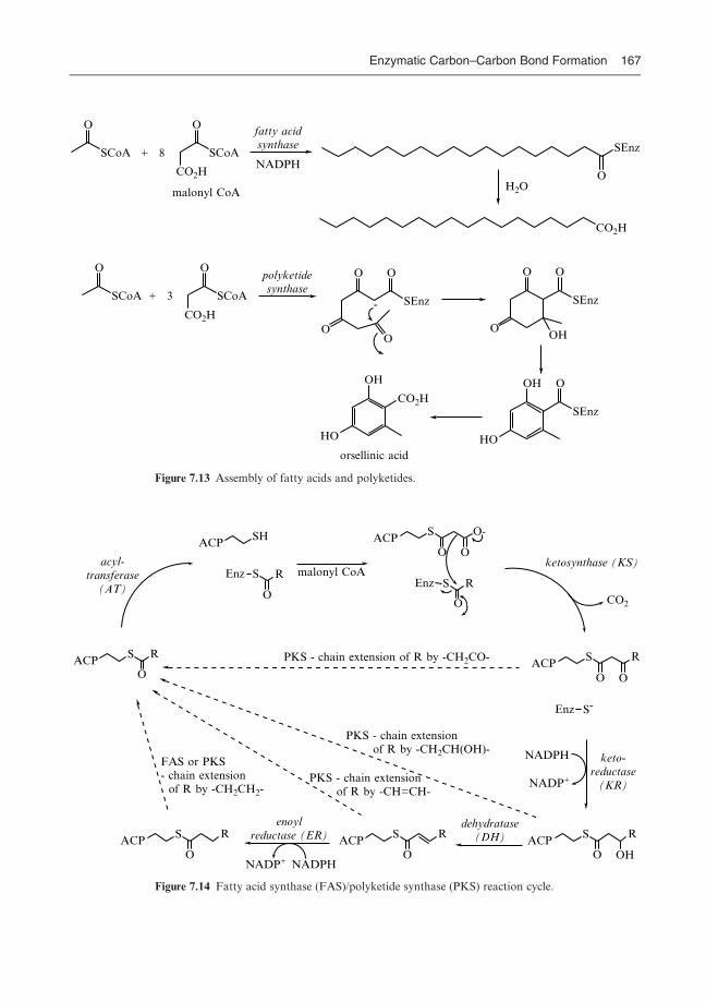
O
SCoA
O
SCoA
CO2H
CO2H
SEnz
O
CO2H
O
SCoA
O
SCoA SEnz
OO O O
OO
SEnz
OHO
HO
OH O
SEnz
+ 8
fatty acidsynthase
HO
OH
CO2H
+ 3
NADPH
-
polyketidesynthase
H2Omalonyl CoA
orsellinic acid
Figure 7.13 Assembly of fatty acids and polyketides.
acyl-transferase
(AT)
malonyl CoAketosynthase (KS)
CO2
NADPH
NADP+
keto-reductase
(KR)
dehydratase(DH)
enoylreductase (ER)
NADP+ NADPH
ACPS R
O
ACPSH
Enz S
O
R
ACPS
O O
O-
Enz S
O
R
ACPS
O O
R
Enz S-
ACPS
O OH
RACP
S
O
RACP
S
O
R
PKS - chain extension of R by -CH2CO-
PKS - chain extension of R by -CH2CH(OH)-
PKS - chain extension of R by -CH=CH-
FAS or PKS- chain extension of R by -CH2CH2-
Figure 7.14 Fatty acid synthase (FAS)/polyketide synthase (PKS) reaction cycle.
Bugg/Introduction to Enzyme and Coenzyme Chemistry Final Proof 22.7.2004 4:11pm page 167
Enzymatic Carbon–Carbon Bond Formation 167

onto the acetyl-thioester intermediate, with decarboxylation of the malonyl
group, by a ketosynthase (KS) activity. This reaction, producing a
b-keto-thioester, is similar to that of the Claisen enzymes above, except that
decarboxylation occurs at the same point.
The question which then arises is whether decarboxylation occurs before,
after, or at the same time as carbon–carbon bond formation? StereospeciWc
labelling studies shown in Figure 7.15 have demonstrated that this reaction
occurs with inversion of conWguration at C-2 of the malonyl unit and with little
or no hydrogen exchange at C-2. This implies that carbon–carbon bond forma-
tion and decarboxylation are, in fact, concerted.
In the case of fatty acid biosynthesis, the new b-keto-thioester is reduced to
a b-hydroxy-thioester, eliminated to give an a,b-unsaturated thioester, and
then further reduced to give a two-carbon-extended acyl chain, as shown in
Figure 7.14. In the case of polyketide biosynthesis, each two-carbon unit can
be processed as either the b-keto-thioester, the b-hydroxy-thioester, the
a,b-unsaturated thioester or as the fully reduced thioester. Assembly of each
polyketide is therefore controlled by the arrangement of processing enzyme
activitites on the polyketide synthase multi-enzyme complex. How is this done?
Information regarding the molecular structure and organisation of polyke-
tide synthases is now emerging from the cloning and sequencing of genes which
encode these enzymes. The genes responsible for the biosynthesis of the
polyketide antibiotic erythromycin have been identiWed and their nucleotide
sequences determined. They encode three huge multi-functional polypeptides of
size 300–500 kDa, illustrated in Figure 7.16. The enzyme activities responsible
for processing of the growing polyketide chain have been identiWed by amino
acid sequence alignments, and are found sequentially along the polypeptide
chains. Remarkably, the arrangement of processing enzyme activities on the
polyketide synthases matches the order of chemical steps required for biosyn-
thesis of the polyketide precursor. It, therefore, appears that these multi-
enzyme complexes function as molecular production lines built up of ‘modules’
of enzyme activities.
O
SCoAH
D T
O
SCoA−O2C
−O2C
D T
O
S-ACP
T D
O
R
O
S-ACP
T D
R
OHHO
S-ACPR
T
O
S-ACPR
T
acetyl CoAcarboxylase
biotinCO2ATP
keto-synthase
keto-reductase
O
S-ACP
D T
NADPH
dehydratase
O
R S-Enz
enoylreductase
NADPH
fatty acidsynthase
RCO-S-ACP
Figure 7.15 Stereochemistry of fatty acid biosynthesis.
Bugg/Introduction to Enzyme and Coenzyme Chemistry Final Proof 22.7.2004 4:11pm page 168
168 Chapter 7

SO
SO O
H
SO O
H
OH
SO
SO
OH
OH
O
OH
OH
O
SO
SO
OH
OH
O
OH
OH
O
OO
OO
H
HO
OO
H
OH
O
O OH
OO
-sug
ar
O-s
ugar
O
O OH
OH
HO
OH
OH
OH
OH
HO
AT ACP KS AT KR ACP KS AT KR ACP
KS AT ACP KS AT DH ER KR ACP
KS AT KR ACP KS AT KR ACP TE
Mod
ule
1:K
R r
educ
es to
R-a
lcoh
ol
Mod
ule
2:K
R r
educ
es to
S-al
coho
l
Mod
ule
3:no
mod
ific
atio
nof
β-k
eto-
thio
este
r
Mod
ule
4:K
R,D
H,E
R g
ive
satu
rate
d th
ioes
ter
Mod
ule
5:K
R r
educ
es to
S-al
coho
l
Mod
ule
6:K
R r
educ
es to
S-al
coho
l
CO
2−
OC
oAS
lact
onisat
ion
via
serine
acy
l en
zym
ein
term
edia
te
2 �
P45
0 m
ono-
oxyg
enas
es2
� g
lyco
syl tr
ansf
eras
es
eryt
hrom
ycin
A
+ 6 Fig
ure
7.1
6A
ssem
bly
ofer
yth
rom
yci
nA
via
multi-en
zym
epoly
ket
ide
synth
ase
s.
Bugg/Introduction to Enzyme and Coenzyme Chemistry Final Proof 22.7.2004 4:11pm page 169

In the case of erythromycin the polyketide is assembled from the three-
carbon unit of propionyl CoA, which is carboxylated to give methylmalonyl
CoA. Acyl transfer and carbon–carbon bond formation takes place through
the a-carbon of a methylmalonyl-thioester, in the same way as is shown in
Figure 7.14, giving a-methyl-b-keto-thioesters at each stage. Each ‘module’
of enzyme activities contains the enzymes required for the assembly of a
new b-keto-thioester and its subsequent modiWcation. For example, the Wrst
module contains ketosynthase (KS) and acyltransferase (AT) activities to make
the new b-keto-thioester, and a ketoreductase (KR) activity to reduce the
b-keto-thioester to a b-R-hydroxy-thioester, and so on. Each of the multi-
functional polyketide synthases contains two such ‘modules’ of enzymatic activ-
ities. At the end of the third polyketide synthase is a thioesterase (TE) activity
which catalyses the intramolecular lactonisation via a serine acyl enzyme inter-
mediate. Subsequent modiWcation of the polyketide precursor to erythromycin
A occurs by separate P450 mono-oxygenase and glycosyl transferase enzymes.
7.5 Carboxylases: use of biotin
We have already seen examples of nucleophilic attack of a carbanion equivalent
onto aldehyde and ester electrophiles. There are Wnally a number of examples of
nucleophilic attack of carbanion equivalents onto carbon dioxide to generate
carboxylic acid products.
We have just seen that the carboxylation of acetyl CoA to give malonyl
CoA is an important step in fatty acid and polyketide natural product biosyn-
thesis. This step is catalysed by acetyl CoA carboxylase. This enzyme uses acetyl
CoA and bicarbonate as substrates, but also requires adenosine triphosphate
(ATP), which is converted to adenosine diphosphate (ADP) and inorganic
phosphate (Pi), and the cofactor biotin. Biotin was Wrst isolated from egg
yolk in 1936, and was found to act as a vitamin whose deWciency causes
dermatitis. Its structure is a bicyclic ring system containing a substituted urea
functional group which is involved in its catalytic function. The biotin
cofactor is covalently attached to the e-amino side chain of an active site lysine
residue.
How does such an apparently unreactive chemical structure serve to activate
carbon dioxide for these carboxylation reactions, and what is the role of ATP in
the reaction? These questions were answered by a series of experiments with
isotopically labelled bicarbonate substrates. Bicarbonate is rapidly formed from
carbon dioxide in aqueous solution and is the substrate for biotin-dependent
carboxylases. Incubation of biotin-dependent b-methylcrotonyl-CoA carboxy-
lase with 14C-bicarbonate and ATP gave an intermediate 14C-labelled enzyme
species. Methylation with diazomethane followed by degradation of the
enzyme structure revealed that the 14C-label was covalently attached to the biotin
cofactor, in the form of a carbon dioxide adduct onto N�1 of the cofactor. This
Bugg/Introduction to Enzyme and Coenzyme Chemistry Final Proof 22.7.2004 4:11pm page 170
170 Chapter 7

species N1-carboxy-biotin, illustrated in Figure 7.17, has been shown to be chem-
ically and kinetically competent as an intermediate in the carboxylation reaction.
Incubation of enzyme with 18O-labelled bicarbonate led to the isolation of
product containing two atoms of 18O and inorganic phosphate containing one
atom of 18O. The transfer of 18O to inorganic phosphate implies that ATP is used
to activate bicarbonate by formation of an acyl phosphate intermediate, known
as carboxyphosphate. It is thought that carboxyphosphate is then attacked by
N-1 of the biotin cofactor. Since the NH group of amides and urea is normally a
very unreactive nucleophile, it is thought that N-1 is deprotonated prior to
attack on carboxyphosphate. The carboxy-biotin intermediate thus formed is
then attacked by a deprotonated substrate, probably by initial decarboxylation
to generate carbon dioxide, to form the carboxylated product and regenerate the
biotin cofactor, as shown in Figure 7.18. It has been shown that the carboxyla-
tion of pyruvate to oxaloacetate catalysed by pyruvate carboxylase proceeds
with retention of conWguration at C-3, as shown in Figure 7.19. Other biotin-
dependent carboxylases also proceed with retention of conWguration.
7.6 Ribulose bisphosphate carboxylase/oxygenase (Rubisco)
All of the carbon-based moleules found in living systems are ultimately derived
from the Wxation of gaseous carbon dioxide by green plants during photosyn-
thesis. Carbon dioxide is then regenerated from respiration of living organisms,
from the decomposition of carbon-based material and dead organisms, and
from the combustion of wood and fossil fuels. This global cycle of processes is
known as the carbon cycle. The enzyme which is responsible for the Wxation of
carbon dioxide by green plants is an enzyme of primary importance to life on
earth. This enzyme is ribulose bisphosphate carboxylate/oxygenase, commonly
known as Rubisco.
NHHN
S
O
O
N Lys-EnzH H
CO2H
OO
CO2H
SCoA
O
HO2C
HO2C
O
SCoA
NHN
S
O
O
N Lys-EnzH H
HO
O
+ CO2pyruvate carboxylase
biotin, ATP
H
+ CO2
biotin
biotin, ATP
acetyl CoA carboxylase
H
N1-carboxy-biotin1 3
Figure 7.17 Biotin-dependent enzymes.
Bugg/Introduction to Enzyme and Coenzyme Chemistry Final Proof 22.7.2004 4:11pm page 171
Enzymatic Carbon–Carbon Bond Formation 171

The reaction catalysed by Rubisco is the carboxylation and concomitant
fragmentation of ribulose-1,5-bisphosphate to generate two molecules of
3-phosphoglycerate (see Figure 7.20). This is a step on the Calvin cycle of
green plants, which transforms 3-phosphoglycerate via a series of aldolase
and transketolase enzymes once again into ribulose-1,5-bisphosphate. Thus
each cycle incorporates one equivalent of carbon dioxide into cellular carbon.
The enzymatic reaction has been shown to commence by deprotonation at
C-3 of ribulose-1,5-bisphosphate, and enolisation of the C-2 ketone, since
[3-3H]-ribulose-1,5-bisphosphate rapidly exchanges 3H with solvent in the pres-
ence of enzyme. The resulting enediol intermediate reacts with carbon dioxide
to generate a carboxylated intermediate. Carbon–carbon bond cleavage is
thought to occur by hydration of the C-3 ketone, followed by fragmentation
of the C-2,C-3 bond to generate a carbanion product, which protonates to give
O
HO O− O
HO OPO32−
NHHN
S
O
H HEnz
NHN
S
O−
H HEnz
NHN
S
O
H HEnz
O
−O
O
CoASH
−B Enz−O
CoAS
O
CoAS
O
O−
ATPADP
C
O
O
Figure 7.18 Mechanism for biotin-dependent carboxylation.
O
H
DT
O
DT
OH
DT
pyruvatecarboxylase
biotin, ATP, CO2
CO2−
CO2−
CO2−
−O2C
−O2C
−O2C
−O2C
NADH
fumarase
D
malatedehydrogenase
3H2O
Figure 7.19 Stereochemistry of the pyruvate carboxylase reaction.
Bugg/Introduction to Enzyme and Coenzyme Chemistry Final Proof 22.7.2004 4:11pm page 172
172 Chapter 7

the second molecule of 3-phosphoglycerate. The proposed mechanism is shown
in Figure 7.21.
7.7 Vitamin K-dependent carboxylase
One other carboxylase enzyme worthy of note is the vitamin K-dependent
carboxylase responsible for the activation of prothrombin via carboxylation
of a series of glutamate amino acid residues. This step is involved in the
calcium-dependent activation of platelets during the blood clotting response.
Vitamin K is a fat-soluble naphthoquinone which can exist in either oxidised
(quinone form) or reduced (hydroquinone form) states. The carboxylase
enzyme is also an integral membrane protein which has only recently been
puriWed. The carboxylation of prothrombin requires reduced vitamin K, carbon
dioxide, oxygen and a carboxylation substrate (see Figure 7.22). Reduced
CH2OPO32−
CH2OPO32−
CH2OPO32−
CH2OPO32−
OOH
HOHO
CO2−
CO2−
HO
Rubisco
Mg2+, Cu2+
CO2
+
ribulose-1,5-bisphosphate2 � phosphoglycerate
Figure 7.20 Reaction catalysed by ribulose-1,5-bisphosphate carboxylase.
O
OHH
OH
OH
O
OH
C
O
O OH
O
OH
−O2C
−O2C−O2C
−O2C OH
O−
OH
HO
OH
O
OH
HO
OH3H
OH
HB−Enz
−3H2O
H AEnzH
EnzB−
−AEnz
CH2OPO32−
CH2OPO32−
CH2OPO32−
CH2OPO32−
CH2OPO32−
CH2OPO32−
CH2OPO32−
CH2OPO32−
CH2OPO32−
CH2OPO32−
CH2OPO32−
Figure 7.21 Mechanism for the ribulose-1,5-bisphosphate carboxylase reaction.
Bugg/Introduction to Enzyme and Coenzyme Chemistry Final Proof 22.7.2004 4:11pm page 173
Enzymatic Carbon–Carbon Bond Formation 173

vitamin K is converted stoichiometrically into vitamin K epoxide, which is
recycled via separate reductase enzymes back to reduced vitamin K.
A major clue to the role of vitamin K in the enzymatic reaction came from a
chemical model reaction shown in Figure 7.23. In the presence of molecular
oxygen and a crown ether, a naphthol analogue of reduced vitamin K was
NH
O
CO2−
NH
O
OO
O−
O
Ca2+
OH
OH
O
O
R
O
O
R
O
vitamin K-dependentcarboxylase
vitamin K, CO2, O2
−
reduced vitamin K
3
carboxylase
CO2, O2prothrombin
reductasereductase
NADHNADH
Figure 7.22 The vitamin K-dependent carboxylase reaction.
O−K+ CO2EtCO2Et
CO2Et
CO2Et
O
OH
O O
O
O O− OO
O−O
O−
O
O2+
H
O
EtO
18-crown-6THF
+
O2
Figure 7.23 Model reaction for role of vitamin K.
Bugg/Introduction to Enzyme and Coenzyme Chemistry Final Proof 22.7.2004 4:11pm page 174
174 Chapter 7

found to act as a catalyst for the Dieckmann condensation of diethyl adipate,
generating an epoxide byproduct. A mechanism was proposed for this reaction
in which the naphthoxide salt and dioxygen react via a dioxetane intermediate
to form a tertiary alkoxide species which acts as a base for the ester condensa-
tion reaction. Note that the naphthoxide salt itself is not a strong enough base
to catalyse the reaction, but is converted into a much stronger base via reaction
with oxygen.
In order to investigate the mechanism of the enzyme-catalysed carboxyla-
tion reaction, reduced vitamin K was incubated with carbon dioxide and
enzyme in the presence of 18O2. Complete incorporation of 18O into the epoxide
oxygen of vitamin K epoxide was observed, together with partial incorporation
of 18O (approx. 20%) into the ketone carbonyl, consistent with the dioxetane
mechanism. It has been proposed that the hydrate alkoxide ion released from
fragmentation of the dioxetane intermediate acts as a base to deprotonate the
g-H of a glutamyl substrate, followed by reaction with carbon dioxide, as
shown in Figure 7.24. It has been shown that the C-4 proS hydrogen is
abstracted, and subsequent reaction with carbon dioxide occurs with inversion
of conWguration. Note that it is remarkable that the hydrate alkoxide ion does
not simply collapse to form the corresponding ketone: presumably the micro-
environment of the enzyme active site disfavours the loss of hydroxide ion from
the lower face of vitamin K.
O−
R
OH
O
HO O O− OO
O−
R
HO
O
R
HO O−
O
NH
O
CO2−
CO2−
CO2−
HS
O
R
HO OH
O
NH
O
O
C
O
O
R
O
NH
O
O
HR
O2
−O2C
−
H
R
Figure 7.24 Mechanism for the vitamin K-dependent carboxylase.
Bugg/Introduction to Enzyme and Coenzyme Chemistry Final Proof 22.7.2004 4:11pm page 175
Enzymatic Carbon–Carbon Bond Formation 175

7.8 Thiamine pyrophosphate-dependent enzymes
Decarboxylation reactions are also widely found in biological chemistry. In
Chapter 3 we saw an example of the decarboxylation of a b-keto acid aceto-
acetate by the action of acetoacetate decarboxylase, via an imine linkage. In
general terms b-keto acids can be fairly readily decarboxylated since the b-keto
group provides an electron sink for the decarboxylation reaction. No such
electron sink exists for a-keto acids; however, Nature has found a way of
decarboxylating a-keto acids, and in many cases forming a carbon–carbon
bond at the same time, using the coenzyme thiamine pyrophosphate (TPP).
Thiamine pyrophosphate is formed by phosphorylation of the vitamin
thiamine, lack of which causes the deWciency disease beri-beri. The structure
of TPP consists of two heterocyclic rings: a substituted pyrimidine ring and a
substituted thiazolium ring (see Figure 7.25). The thiazolium ring is responsible
for the catalytic chemistry carried out by this coenzyme, due to two chemical
properties:
(1) the acidity of the proton attached to the thiazolium ring;
(2) the presence of a carbon–nitrogen double bond that can act as an electron
sink for decarboxylation.
The decarboxylation of pyruvic acid is carried out by several TPP-dependent
enzymes, yielding acetaldehyde, acetic acid, acetyl CoA or a-hydroxyacetyl
compounds. Each of these products is formed via closely related mechanisms.
CO2−
O
H
O
O−
O
SCoA
O
O
R
OH
N
N
N S
OPP
H
SS N Lys-Enz
O
N-4'
TPP TPP, RCHO
TPPFAD,O2
TPPlipoamide, CoASH
H
+
Thiamine pyrophosphate (TPP)
C-2 exchanges with D2O at pH 7
NH2
Lipoamide
Figure 7.25 Thiamine pyrophosphate-dependent reactions of pyruvate.
Bugg/Introduction to Enzyme and Coenzyme Chemistry Final Proof 22.7.2004 4:11pm page 176
176 Chapter 7

The Wrst step in each of the TPP-dependent reactions is deprotonation of the
thiazolium ring to generate a carbanion ylid at C-2. In chemical models the pKa
of this proton is 17–19, yet when bound to the enzyme it is able to exchange
with 2H2O, therefore the enzyme active site increases the acidity of this position
dramatically. When bound to the active site, the TPP cofactor adopts a ‘V’
conformation (shown for pyruvate decarboxylase in Figure 7.26), which brings
N-40 of the aminopyrimidine ring in close proximity to C-2 of the thiazolium
ring. It is thought therefore that N-40 of an imine tautomer of the aminopyr-
imidine ring deprotonates C-2, to generate the C-2 carbanion.
The ylid carbanion then attacks the keto group of pyruvate to generate a
covalently attached lactyl adduct (see Figure 7.27). Decarboxylation of the
lactyl adduct then takes place, using the carbon–nitrogen double bond as an
electron sink, forming an enamine intermediate. Protonation of this intermedi-
ate and fragmentation of the linkage with the coenzyme yields acetaldehyde and
regenerates the coenzyme ylid. Alternatively, the enamine intermediate can
react with an electrophile (Eþ), generating after fragmentation of the linkage
with the coenzyme, an acetyl-E species.
An important reaction in the cellular breakdown of d-glucose is the
conversion of pyruvate to acetyl CoA, which is catalysed by the pyruvate
Figure 7.26 Active site of yeast pyruvate decarboxylase (PDB Wle 1QPB), showing the ‘V’ conform-
ation of the bound TPP cofactor, which is shown in black. Three active site residues, Asp-28, Glu-51
and Ile-415, are shown in red.
Bugg/Introduction to Enzyme and Coenzyme Chemistry Final Proof 22.7.2004 4:11pm page 177
Enzymatic Carbon–Carbon Bond Formation 177

dehydrogenase complex. This reaction follows the mechanism shown in Figure
7.27, but in this case the electrophile is a second cofactor present in the pyruvate
dehydrogenase complex: lipoamide. The structure of lipoamide shown in
Figure 7.28 is simply a Wve-membered ring containing a disulphide linkage
attached via an acyl chain to the e-amino group of a lysine residue. Upon
reaction of the thiamine pyrophosphate–enamine intermediate with oxidised
lipoamide and fragmentation of the linkage with thiamine pyrophosphate, an
acetyl-lipoamide thioester is formed, as shown in Figure 7.28. Transfer of the
acyl group to the thiol group of CoA generates acetyl CoA and reduced
RN S
OPP
RN S
OPP
HOO−
O
RN S
OPP
OH
RN S
OPP
OE
O
E
O
CO2−
H
+ +
E+
+
HN
N
N S
OPP
H
(E+ = H+ or electrophile)
NH
+
Figure 7.27 Mechanism for pyruvate decarboxylation.
RN S
OPP
OH
RN S
OPP
O
H
SS Enz
S
SH
Enz
S
SH
Enz
O
HS
SH
Enz
O
SCoA
+
CoAS−
FAD,NAD+
lipoamidedehydrogenase
pyruvatedecarboxylase
Figure 7.28 TPP-dependent production of acetyl CoA using lipoamide.
Bugg/Introduction to Enzyme and Coenzyme Chemistry Final Proof 22.7.2004 4:11pm page 178
178 Chapter 7

lipoamide, which is recycled to oxidised lipoamide by Xavin-dependent lipo-
amide dehydrogenase (see Section 6.5).
Thiamine pyrophosphate is also used by several enzymes for carbon–carbon
bond formation. Illustrated in Figure 7.29 is one example of the family of TPP-
dependent transketolase enzymes which carry out a range of carbohydrate
2-hydroxyacetyl transfer reactions. A similar mechanism can be written for
these reactions initiated by attack of the thiazolium ylid on the keto group,
followed in this case not by decarboxylation but by carbon–carbon bond
cleavage. The ability of these enzymes to form carbon–carbon bonds enantio-
selectively is also being exploited for novel biotransformation reactions that are
of use in organic synthesis.
Carbon–carbon bond formation via carbocationintermediates
Carbon–carbon bond formation via carbocation (or carbonium ion) intermedi-
ates is less widely found than via carbanion equivalents. However, there is one
large class of biological reactions that involve highly stabilised carbocation ion
intermediates: the conversion of allylic pyrophosphate metabolites into terpe-
noid natural products.
7.9 Terpene cyclases
Terpenes are a major class of natural products found widely in plants, but also
including the steroid lipids and hormones found in animals. The common
structural feature of the terpene natural products is the Wve-carbon isoprene
unit. Some common examples shown in Figure 7.30 are plant natural products
menthol, camphor and geraniol.
The biosynthesis of terpenoid natural products proceeds from a family of
allylic pyrophosphates containing multiples of Wve carbon atoms. Two Wve-
carbon units are joined together by the enzyme geranyl pyrophosphate
synthase as shown in Figure 7.31. Loss of pyrophosphate from dimethylallyl
CH2OH
O
HHO
OHH
CH2OPO32− CH2OPO3
2−CH2OPO3
2−
CH2OPO32−
CHO
OHH
OHH
OHH
CH2OH
O
CHO
OHH + OHH
OHH
OHH
+transketolase
TPP
HO H
Figure 7.29 Example of a transketolase reaction.
Bugg/Introduction to Enzyme and Coenzyme Chemistry Final Proof 22.7.2004 4:11pm page 179
Enzymatic Carbon–Carbon Bond Formation 179

pyrophosphate (DMAPP) generates a stabilised allylic carbonium ion. Attack
of the p-bond of isopentenyl pyrophosphate (IPP) generates a tertiary carbo-
cation. StereospeciWc loss of a proton generates the product geranyl pyrophos-
phate. Similar reactions with further units of IPP generate the C15 building
block farnesyl pyrophosphate, the C20 geranylgeranyl pyrophosphate, and
so on.
Geranyl pyrophosphate is converted to the family of C10 monoterpenes
by the monoterpene cyclases. A 96-kDa cyclase enzyme responsible for the
assembly of (þ)-a-pinene has been puriWed from sage leaves, whilst a separate
55-kDa enzyme from the same source catalyses the production of the opposite
enantiomer (�)-a-pinene. The 55-kDa cyclase II produces several other
isomeric products. As well as (�)-a-pinene (26%) the enzyme produces
(�)-b-pinene (21%), (�)-camphene (28%), myrcene (9%) and (�)-limonene
(8%). These products can be rationalised by the mechanism shown in Figure
7.32. It has been shown that the Wrst step in the assembly of these monoterpenes
is a 1,3-migration of pyrophosphate to give linalyl pyrophosphate, which for
cyclase II has the 3S stereochemistry. Loss of pyrophosphate gives an allylic
cation, which is attacked by the second alkene to give a six-membered ring and
a tertiary carbocation intermediate. Further ring closure to generate a four-
membered ring gives another tertiary carbocation, which can deprotonate in
either of two ways to give a-pinene or b-pinene. Limonene can be formed by
OH
O
OH
menthol
camphor geraniol
Figure 7.30 Terpenoid natural products.
OPP OPP OPP
H HDMAPP
+
OPP
+
+
IPP
OPP IPP
geranyl PPfarnesyl PP
−BEnz
Figure 7.31 Biosynthesis of farnesyl pyrophosphate.
Bugg/Introduction to Enzyme and Coenzyme Chemistry Final Proof 22.7.2004 4:11pm page 180
180 Chapter 7

elimination from the monocyclic carbocation, whilst camphene is formed
by two consecutive 1,2-alkyl migrations, followed by elimination.
Most enzymes are speciWc for the production of a single isomeric product,
so in this case how can we be sure that all of these products are from one
enzyme? Apart from demonstrating that there is only one homogeneous protein
present, a chemical test can be made. If there is a common precursor to two or
more products, then by introducing deuterium atoms at speciWc points in the
substrate, the ratio of products should be aVected by the presence of a deuter-
ium isotope eVect, which should favour one pathway over another. This test
was applied to the (�)-pinene synthase reaction by incubation of the speciWcally
deuterated substrate shown in Figure 7.33. At the point of divergence to a-
pinene or b-pinene the presence of a terminal 2CD3 group would be expected
to favour formation of a-pinene. This was indeed observed: the proportion of
a-pinene in the product mixture rose from 26% to 38%, whilst the proportion
of b-pinene dropped from 21% to 13%. The proportion of myrcene product
also dropped signiWcantly from 9% to 4%, also consistent with an isotope eVect
operating on the allylic carbocation intermediate.
Farnesyl pyrophosphate is cyclised to form the large family of sesquiterpene
natural products by a further class of terpene cyclase enzymes, several of which
have been puriWed to homogeneity. Pentalenene synthase catalyses the cyclisa-
tion of farnesyl pyrophosphate to give pentalenene, which is further processed
in Streptomyces UC5319 to give the pentalenolactone family of antibiotics, as
shown in Figure 7.34. The enzyme is a 41-kDa protein with a KM of 0.3�m for
PPO
OPP OPP PPO−
H
H
−H+
−H+−H+
−H+
−H+
myrcene (9%)
H
H
(−) limonene (8%)
=
(−)-β-pinene (21%)
(−)-α-pinene (26%)
1,2-shift
H1,2-shift
(−) camphene (28%)
geranyl PP 3S-linalyl PP
Figure 7.32 Mechanism for (�)-a-pinene synthase.
Bugg/Introduction to Enzyme and Coenzyme Chemistry Final Proof 22.7.2004 4:11pm page 181
Enzymatic Carbon–Carbon Bond Formation 181

farnesyl pyrophosphate and a kcat of 0:3 s�1, and an absolute requirement for
Mg2þ ions. A series of isotopic labelling experiments have been carried out to
support the mechanism of cyclisation shown in Figure 7.34. Cyclisation of
farnesyl pyrophosphate is proposed to form an 11-membered intermediate,
humulene, which is followed by a Wve-membered ring closure to form a bicyclic
tertiary carbocation. 1,2-Hydride migration followed by a further Wve-
membered ring closure gives a tricyclic carbocation, which upon elimination
gives pentalenene.
Examination of the inferred amino acid sequence of pentalenene synthase
revealed an amino acid sequence motif Asp–Asp–X–X–Asp (DDXXD) found
in other pyrophosphate-utilising enzymes. It is believed that the Asp–Asp pair
are involved in chelating the essential Mg2þ ion, which in turn chelates the
pyrophosphate ion as shown in Figure 7.35. An indication of the tight binding
of pyrophosphate is that this product inhibits the enzymatic reaction strongly
(Ki3�m ).
H
β-pinene H* = H 21%
CH3*
CH3*
CH2*
*H3C
OPP
α-pinene H* = H 26%
−H+
−H*+
(−)pinenesynthase
Mg2+
H* = D 13%
H* = D 38%
Figure 7.33 Kinetic isotope eVect in the (�)-pinene synthase reaction.
PPO
H HBEnz
HH
H
HHH
CO2H
H
O OO
humulene
−BEnz
−BEnz
pentalenenepentalenolactone
Figure 7.34 Mechanism for pentalenene synthase.
Bugg/Introduction to Enzyme and Coenzyme Chemistry Final Proof 22.7.2004 4:11pm page 182
182 Chapter 7

How do these cyclase enzymes control the precise regiochemistry and
stereochemistry of these multi-step cyclisations? In 1997 the crystal structure
of pentalenene synthase was solved. The active site consists of a conical hydro-
phobic cleft, Xanked by six a-helices, lined with aromatic and non-polar resi-
dues. The DDXXD motif is located near the upper edge of the cavity, as shown
in Figure 7.36. The identity of the catalytic base has not yet been identiWed, but
replacement of Phe-76 or Phe-77 by alanine gave at least a 10-fold reduction in
activity, suggesting that they may stabilise carbocationic intermediates through
�-cation interactions. The presence of aromatic residues in key positions of the
wall of terpene cyclase active sites suggests that the �-cation interaction, shown
in Figure 7.37, may have an important role in the speciWc stabilisation of the
carbocation intermediates.
Carbon–carbon bond formation via radicalintermediates
In Chapter 6 we have seen how redox enzymes are able to generate radical
intermediates in enzyme-catalysed reactions. In certain instances these
radical intermediates are used for carbon–carbon bond formation reactions.
The two examples which we shall examine are phenol radical couplings used in
natural product biosynthesis and in lignin biosynthesis.
R OP
OP
O−
−O O−
O O
Mg2+
O− −OO
O
NH
O
HN
O
O
H
H
Pentalenene synthase F L D D F L DTrichodiene synthase V L D D S K DAristolochene synthase L I D D V L ECasbene synthase L I D D T I DLimonene synthase V I D D I Y DFarnesyl PP synthase V Q D D I L DGeranylgeranyl PP synthase I A D D Y H N
Consensus D D X X(D)
Figure 7.35 Mg2þ-pyrophosphate binding motif.
Bugg/Introduction to Enzyme and Coenzyme Chemistry Final Proof 22.7.2004 4:11pm page 183
Enzymatic Carbon–Carbon Bond Formation 183

7.10 Phenolic radical couplings
Certain aromatic natural products are formed by the radical coupling of two
phenol precursors in an enzyme-catalysed process. In some cases the enzymes
are blue copper proteins (the characteristic colour due to the metal cofactor).
For example, the phenol radical coupling of sulochrin shown in Figure 7.38 is
catalysed by a 157-kDa oxidase enzyme containing six atoms of copper. The
Figure 7.36 Structure of pentalanene synthase (PDB Wle 1PS1), showing the active site residues Asp-
80, Asp-81, Asp-84 (DDXXD motif, top), Phe-76 and Phe-77 in red.
Phe
C+
Figure 7.37 p-Cation interaction.
Bugg/Introduction to Enzyme and Coenzyme Chemistry Final Proof 22.7.2004 4:11pm page 184
184 Chapter 7

enzyme exhibits a deep blue colour (lmax 605 nm) which disappears upon
reduction with ascorbate under nitrogen. These observations suggest that
there are active site Cu2þ metal ions which accept one electron from the phenol
substrates to generate Cuþ intermediates. The phenoxy radicals thus generated
are relatively stable, and react together as shown in Figure 7.38.
Oxidative phenol coupling is frequently found in pathways responsible
for the biosynthesis of isoquinoline and indole alkaloids in plants. In several
cases the enzymes responsible for the oxidative phenol coupling have been
found to be cytochrome P450 enzymes (described in Section 6.8). One example
is the enzyme salutaridine synthase, shown in Figure 7.39, involved in
the biosynthesis of morphine in Papaver somniferum. In other cases, the
enzymes responsible for oxidative phenol couplings have been found to be
a-ketoglutarate-dependent dioxygenases (described in Section 6.9), or Xavopro-
tein oxidases (described in Section 6.3).
Lignin is a complex aromatic polymeric material which is a major structural
component of woody tissues in plants. In most trees that require a great deal of
structural rigidity lignin constitutes 30–40% of the dry weight of the wood. It is
highly cross-linked and exceptionally resistant to chemical degradation, making
elucidation of its chemical structure very diYcult. However, it is now known to
be heterogeneous in structure, composed of phenylpropanoid (aromatic ring þthree-carbon alkyl chain) units linked together by a variety of types of carbon–
carbon linkage. Shown in Figure 7.40 are a selection of structural components
found in lignin. Although structurally diverse, the common feature of these
components is that they can all be formed via radical couplings.
HO
OMe O OH
HOCO2Me
O
OMe O OH
OCO2Me
OO
CO2Me
MeO O OH
2Cu2+ 2Cu+
sulochrin
Figure 7.38 Reaction catalysed by sulochrin oxidase.
MeO
HO
MeO
OH
NMeH
MeO
HO
MeO
O
NMeH
reticuline salutaridine
P450NADPH,O2
salutaridinesynthase
Figure 7.39 Reaction catalysed by salutaridine synthase.
Bugg/Introduction to Enzyme and Coenzyme Chemistry Final Proof 22.7.2004 4:11pm page 185
Enzymatic Carbon–Carbon Bond Formation 185

The biosynthesis of lignin starts from three cinnamyl alcohol precursors:
p-hydroxycinnamyl alcohol, coniferyl alcohol and sinapyl alcohol, shown in
Figure 7.40. The ratio of these precursors used for lignin assembly is species-
dependent. They are each activated by formation of a phenoxy radical at the
C-4 hydroxyl group by a family of peroxidase, phenolase and tyrosinase
enzymes widely found in plants. As shown in Figure 7.41, the phenoxy radical
can exist in a number of resonance structures in which radical character is
found, for example, on oxygen, at the C-5 position and at the b-position of the
side chain. Carbon–carbon bond formation can then take place with the a,b-
double bond of another molecule, generating a new radical species which can
HOCH
OH
OCH3
HC O
HOCH2 OCH3
C
C
C
HC
OH
OCH3
HC
O
HOCH2
OCH3
C
C
C
HC
OH
OCH3
HC
H2C CH
O
OCH3
CH
CH2O
O
OH
OCH3
C
C
C
OH
CH3O
C
C
C
O
OCH3
C
C
C
OH
CH3O
C
C
C
CH2OH
OH
CH2OH
OH
CH2OH
OH
OCH3 H3CO OCH3
p-hydroxycinnamylalcohol
coniferylalcohol
sinapylalcohol
Figure 7.40 Structural components of lignin.
Bugg/Introduction to Enzyme and Coenzyme Chemistry Final Proof 22.7.2004 4:11pm page 186
186 Chapter 7

form a further carbon–carbon bond. Thus the formation of a phenoxy radical
initiates a radical polymerisation reaction which forms a highly heterogeneous
polymer. Evidence that the polymerisation is a chemical reaction and not an
enzyme-catalysed reaction comes from the observation that lignin is not optic-
ally active, despite containing many chiral centres.
There are also examples of carbon–carbon forming reactions involving the
coenzyme vitamin B12 which proceed via radical mechanisms, which will be
described in Section 11.1.
Problems
(1) N-acetylneuraminic acid aldolase catalyses the reaction shown below.
Given that the enzyme requires no cofactors, suggest a mechanism (Hint:
use the open chain forms of the monosaccharides).
CH2OH
OH
OCH3
CH2OH
O
OCH3
O
CH2OH
OCH3
O
CH2OH
OCH3
OH
CH2OH
OCH3
O
CH2OH
OCH3
OH
OCH3
HOH
O
CH2OH
OCH3
HC
OH
OCH3
HC
O
HOCH2
OCH3
CH2OH
O
CH2OH
OCH3
O
OCH3
HO
peroxidaseenzyme
CH2OH
OCH3
OH
OCH3
HO
OHradical
polymerisation
− H
OH
OCH3
HO
Figure 7.41 Lignin formation via radical coupling.
Bugg/Introduction to Enzyme and Coenzyme Chemistry Final Proof 22.7.2004 4:11pm page 187
Enzymatic Carbon–Carbon Bond Formation 187

OHO
HO
NHAc
OH
AcOO
CO2HO
AcHNHO
OH
CO2H
AcOOH
HO+
(2) The two plant enzymes chalcone synthase and resveratrol synthase catalyse
the reactions shown below. Suggest mechanisms for the two enzymatic
reactions. Comment on the observation that when these enzymes were
sequenced, they were found to share 70–75% amino acid sequence identity.
COSCoA
OH
HO
O
HO
OH
OH
HO OH
OH
chalconesynthase
3 � malonyl CoA
resveratrolsynthase
3 � malonyl CoA
(3) PEP carboxylase catalyses the carboxylation of phosphoenolpyruvate
(PEP) using Mn2þ as a cofactor. When incubated with HC18O�3 two
atoms of 18O were found in the oxaloacetate product, and one atom of18O in inorganic phosphate. Suggest a mechanism.
OPO32−
CO2−
CO2−
O
+ HCO3− −O2C
PEPcarboxylase
Mn2++ PO4
3−
(4) Transcarboxylase catalyses the simultaneous carboxylation of propionyl
CoA to methylmalonyl CoA and decarboxylation of oxaloacetate to
pyruvate, as shown below. The enzyme requires biotin as a cofactor, but
does not require ATP. Suggest a mechanism.
CO2−
CO2−CO2
−
O
SCoA
O O
SCoA
O
−O2C+
trans-carboxylase
biotin+
Bugg/Introduction to Enzyme and Coenzyme Chemistry Final Proof 22.7.2004 4:11pm page 188
188 Chapter 7

(5) Suggest a mechanism for the TPP-dependent transketolase reaction
illustrated in Figure 7.25.
(6) Pyruvate oxidase catalyses the oxidative decarboxylation of pyruvate to
acetate (not acetyl CoA). The enzyme requires TPP and oxidised Xavin as
cofactors, and utilises oxygen as an electron acceptor (i.e. it is converted to
hydrogen peroxide). Suggest a mechanism.
CO2−
O−
O Opyruvateoxidase
TPP, FAD,O2
+ CO2
(7) The conversion of geranyl pyrophosphate into bornyl pyrophosphate is
catalysed by bornyl pyrophosphate synthetase. Write a mechanism for the
enzymatic reaction. When the enzyme was incubated with geranyl pyrophos-
phate speciWcally labelled with 18O attached to C-1, the product was found
to contain 18O only in the oxygen attached to carbon, and not in any of the
other six oxygens. Comment on this remarkable result.
O
O P O P
O O
O−
bornyl PPsynthetase
Mg2+
P O P
O
O−O−
O−
O− O−O
(8) Kaurene synthetase catalyses the cyclisation of geranylgeranyl pyrophos-
phate (GGPP) to kaurene. An intermediate in the reaction is thought to
be copalyl pyrophosphate. Suggest mechanisms for conversion of GGPP
into copalyl PP and thence to kaurene. How would you attempt to prove
that copalyl pyrophosphate is an intermediate in this reaction?
OPP
H H
HHGGPP(C20)
copalyl PP kaurene
Bugg/Introduction to Enzyme and Coenzyme Chemistry Final Proof 22.7.2004 4:11pm page 189
Enzymatic Carbon–Carbon Bond Formation 189

(9) Usnic acid is a natural product thought to be biosynthesised from two
molecules of an aromatic precursor. Suggest a biosynthetic pathway to
the aromatic precursor from acetyl CoA, and a mechanism for the conver-
sion to usnic acid.
O
OHHO
OH
O
O
HO
OH OH3C
H3CH3C
OH
O
Acetyl CoA
derived fromS-adenosyl methionine
usnic acid
Further reading
General
R.H. Abeles, P.A. Frey & W.P. Jencks (1992) Biochemistry. Jones & Bartlett, Boston.
J. Mann (1987) Secondary Metabolism, 2nd edn. Clarendon Press, Oxford.
J.S. Staunton (1978) Primary Metabolism: A Mechanistic Approach. Clarendon Press,
Oxford.
C.T. Walsh (1979) Enzymatic Reaction Mechanisms. Freeman, San Francisco.
Aldolases
J.G. Belasco & J.R. Knowles (1983) Polarization of substrate carbonyl groups by yeast
aldolase: investigation by Fourier transform infrared spectroscopy. Biochemistry, 22,
122–9.
K.H. Choi, J. Shi, C.E. Hopkins, D.R. Tolan & K.N. Allen (2001) Snapshots of
catalysis: the structure of fructose-1,6-bisphosphate aldolase covalently bound to
the substrate dihydroxyacetone phosphate. Biochemistry, 40, 13868–75.
D.R. Hall, G.A. Leonard, C.D. Reed, C.I. Watt, A. Berry & W.N. Hunter (1999) The
crystal structure of Escherichia coli class II fructose-1,6-bisphosphate aldolase
in complex with phosphoglycolohydroxamate reveals details of mechanism and
speciWcity. J. Mol. Biol., 287, 383–94.
J.B. Jones (1986) Enzymes in organic synthesis. Tetrahedron, 42, 3341–403.
A.J. Morris & D.R. Tolan (1994) Lysine-146 of rabbit muscle aldolase is essential
for cleavage and condensation of the C3–C4 bond of fructose 1,6-bisphosphate.
Biochemistry, 33, 12291–7.
J. Sygusch, D. Beaudry & M. Allaire (1987) Molecular architecture of rabbit skeletal
muscle aldolase at 2.7 A resolution. Proc. Natl. Acad. Sci. USA, 84, 7846–50.
E.J. Toone, E.S. Simon, M.D. Bednarski & G.M. Whitesides (1989) Enzyme-catalysed
synthesis of carbohydrates. Tetrahedron, 45, 5365–422.
G.M. Whitesides & C.H. Wong (1985) Enzymes as catalysts in synthetic organic chem-
istry. Angew. Chem. Int. Ed. Engl., 24, 617–38.
Bugg/Introduction to Enzyme and Coenzyme Chemistry Final Proof 22.7.2004 4:11pm page 190
190 Chapter 7

C.H. Wong & G.M. Whitesides (1994) Enzymes in Synthetic Organic Chemistry. Perga-
mon, Oxford.
Claisen enzymes
S.I. Chang & G.G. Hammes (1990) Structure and mechanism of action of a multifunc-
tional enzyme: fatty acid synthase. Acc. Chem. Res., 23, 363–9.
J.D. Clark, S.J. O’Keefe & J.R. Knowles (1988) Malate synthase: proof of a stepwise
Claisen condensation using the double-isotope fractionation test. Biochemistry,
27, 5961–71.
J.S. Staunton (1991) The extraordinary enzymes involved in erythromycin biosynthesis.
Angew. Chem. Intl. Ed. Engl., 30, 1302–6.
Carboxylases
P.V. Attwood & J.C. Wallace (2002) Chemical and catalytic mechanisms of carboxyl
transfer reactions in biotin-dependent enzymes. Acc. Chem. Res., 35, 113–20.
P. Dowd, R. Hershline, S.W. Ham & S. Naganathan (1994) Mechanism of action of
vitamin K. Nat. Prod. Reports, 11, 251–64.
F.C. Hartman & M.R. Harpel (1994) Chemical and genetic probes of the active site of d -
ribulose-1,5-bisphosphate carboxylase-oxygenase: a retrospective based on the three-
dimensional structure. Adv. Enzymol., 66, 1–76.
J.R. Knowles (1989) The mechanism of biotin-dependent enzymes. Annu. Rev. Biochem.,
58, 195–222.
N.F.B. Phillips, M.A. Shoswell, A. Chapman-Smith, D.B. Keech & J.C. Wallace (1992)
Isolation of a carboxyphosphate intermediate and the locus of acetyl CoA action in
the pyruvate carboxylase reaction. Biochemistry, 31, 9445–50.
J.W. Suttie (1985) Vitamin K-dependent carboxylase. Annu. Rev. Biochem.,
54, 459–78.
Thiamine pyrophosphate
F. Jordan (2003) Current mechanistic understanding of thiamin diphosphate-dependent
enzymatic reactions. Nat. Prod. Reports, 20, 184–201.
R. Kluger, J. Chin, & T. Smyth (1981) Thiamin-catalyzed decarboxylation of pyruvate.
Synthesis and reactivity analysis of the central, elusive intermediate, a-lactylthiamin.
J. Am. Chem. Soc., 103, 884–8.
Terpene cyclases
D.E. Cane (1990) Enzymatic formation of sesquiterpenes. Chem. Rev., 90, 1089–103.
R.B. Croteau, C.J. Wheeler, D.E. Cane, R. Ebert & H.J. Ha (1987) Isotopically sensitive
branching in the formation of cyclic monoterpenes: proof that (� )a-pinene and
(�)b-pinene are synthesised by the same monoterpene cyclase via deprotonation of a
common intermediate. Biochemistry, 26, 5383–9.
C.A. Lesburg, G. Zhai, D.E. Cane, and D.W. Christianson (1997) Crystal structure of
pentalenene synthase: mechanistic insights on terpenoid cyclization reactions in
biology. Science, 277, 1820–24.
M. Seemann, G. Zhai, J.W. de Kraker, C.M. Paschall, D.W. Christianson & D.E. Cane
(2002) Pentalenene synthase. Analysis of active site residues by site-directed
mutagenesis. J. Am. Chem. Soc., 124, 7681–9.
Bugg/Introduction to Enzyme and Coenzyme Chemistry Final Proof 22.7.2004 4:11pm page 191
Enzymatic Carbon–Carbon Bond Formation 191

Radical couplings
W.M. Chou & T.M. Kutchan (1998) Enzymatic oxidations in the biosynthesis of
complex alkaloids. Plant. J., 15, 289–300.
K. Freudenberg (1965) Lignin: its constitution and formation from p-hydroxycinnamyl
alcohols. Science, 148, 595–600.
T. Higuchi (1971) Formation and biological degradation of lignins. Adv. Enzymol.,
34, 207–83.
H. Nordlov & S. Gatenbeck (1982) Enzymatic synthesis of (þ) and (�)-
bisdechlorogeodin with sulochrin oxidase from Penicillium frequentans and Oospora
sulphurea-ochracea. Arch. Microbiol. 131, 208–11.
Bugg/Introduction to Enzyme and Coenzyme Chemistry Final Proof 22.7.2004 4:11pm page 192
192 Chapter 7

8 Enzymatic Addition/EliminationReactions
8.1 Introduction
The addition and elimination of the elements of water is a common process
in biochemical pathways. Particularly common is the dehydration of b-
hydroxy-ketones or b-hydroxy-carboxylic acids, which can be constructed by
aldolase and Claisen enzymes. In this chapter we shall examine the mechanisms
employed by these hydratase/dehydratase enzymes, and the related ammonia
lyases.
In particular we shall discuss several enzymes on the shikimate pathway,
which is depicted in Figure 8.1. This pathway is responsible for the biosynthesis
of the aromatic amino acids l-phenylalanine, l-tyrosine and l-tryptophan in
plants and micro-organisms. Nature synthesises the aromatic amino acids
starting from d-glucose via a pathway involving a series of interesting elimin-
ation reactions. This is an important pathway for plants, since it is also respon-
sible for the biosynthesis of the precursors to the structural polymer lignin
encountered in Section 7.10. Animals, which do not utilise this pathway, have
to consume the aromatic amino acids as part of their diet.
The elimination of the elements of water involves cleavage of a C2H bond
and cleavage of a C2O bond. The timing of C2H versus C2O cleavage
chorismatesynthase
PEP
EPSPsynthase
ADP ATP
shikimatekinase
shikimatedehydrogenase
NADP+
NADPH
3-dehydroquinatesynthaseDAHP synthase
PEP
3-dehydroquinatedehydrataseCH2OPO3
2-
2-O3POCH2
2-O3PO2-O3PO
CHOHO
OH
OH
O
CO2−
CO2− CO2
− CO2−
CO2−CO2
−
CO2−CO2
−
CO2−
OH
OH OH
HO
OH
O O
OH
OH
HO
OH
OH
OH
OH
OH
O
OH
O
(1) (2) (3)(4)
(5)(6)(7)(8)
Figure 8.1 The shikimate pathway. (1) erythrose-4-phosphate; (2) 3-deoxy-d-arabinoheptulosonic
acid 7-phosphate (DAHP); (3) 3-dehydroquinate; (4) 3-dehydroshikimate; (5) shikimate;
(6) shikimate-3-phosphate; (7) 5-enolpyruvyl-shikimate-3-phosphate (EPSP); (8) chorismate.
Bugg/Introduction to Enzyme and Coenzyme Chemistry Final Proof 22.7.2004 4:12pm page 193
193

determines the type of elimination mechanism involved, as illustrated in Figure
8.2. If C2O cleavage takes place Wrst, a carbonium ion is generated which
eliminates via cleavage of an adjacent C2H bond. This E1 mechanism is found
in organic reactions, but is rare in biological systems. If C2H bond cleavage
occurs Wrst a carbanion intermediate is generated, which eliminates via subse-
quent C2O bond cleavage. This E1cb mechanism is quite common in biological
chemistry in cases where the intermediate carbanion is stabilised, as in the
elimination of b-hydroxy-ketones or b-hydroxy-thioesters. If C2O and C2H
bond cleavage are concerted then a single-step E2 mechanism is followed.
The stereochemical course of enzymatic elimination reactions is strongly
dependent upon the mechanism of elimination. If an E2 mechanism is operating
then an anti-elimination will ensue. This is generally observed in the enzyme-
catalysed dehydrations of b-hydroxy-carboxylic acids. However, if an E1cb
elimination mechanism is operating then the stereochemistry of the reaction
can either be syn- or anti- depending upon the positioning of the catalytic
groups at the enzyme active site. Commonly the syn- stereochemistry is ob-
served for enzyme-catalysed elimination of b-hydroxy-ketones, although this is
not true in all cases.
8.2 Hydratases and dehydratases
Enzymes that catalyse the elimination of water are usually known as dehydra-
tases. However, since addition/elimination reactions are reversible, sometimes
the biologically relevant reaction is the hydration of an alkene, in which case the
enzyme would be known as a hydratase. We shall consider in turn enzymes
which catalyse the elimination of b-hydroxy-ketones, b-hydroxy-thioesters and
b-hydroxy-carboxylic acids.
The dehydration of 3-dehydroquinic acid to 3-dehydroshikimic acid shown
in Figure 8.3 is a well-studied example of an enzyme-catalysed elimination of a
H
OH
H
OH
C-Ocleavage
E1
H
OH
C-Hcleavage E1cb
concertedelimination
B−
E2
Figure 8.2 Mechanisms for elimination of water.
Bugg/Introduction to Enzyme and Coenzyme Chemistry Final Proof 22.7.2004 4:12pm page 194
194 Chapter 8

b-hydroxy-ketone. This reaction is catalysed by 3-dehydroquinate dehydratase,
the third enzyme on the shikimate pathway.
The enzymatic reaction is reversible, although the equilibrium constant of
16 lies in favour of the forward reaction. The stereochemical course of the
enzymatic reaction in Escherichia coli was shown, using stereospeciWcally
labelled substrates, to be a syn-elimination of the equatorial C-2 proR hydrogen
and the C-1 hydroxyl group. The stereochemistry of the reaction is remarkable,
since the axial C-2 proS hydrogen is much more acidic in basic solution than the
proR hydrogen, due to favourable overlap with the C2O p bond.
A major clue to the enzyme mechanism is that the enzyme is inactivated
irreversibly by treatment with substrate and sodium borohydride. This indi-
cates that an imine linkage is formed between the C-3 ketone and the e-amino
group of an active site lysine residue. Peptide mapping studies have established
that this residue is Lys-170.
A mechanism for enzymatic reaction is shown in Figure 8.4. Upon forma-
tion of the imine linkage at C-3, a conformational change is thought to take
place, giving a twist boat structure. In this conformation the C2H bond of the
proR hydrogen lies parallel to the orbital axes of the adjacent C2N p bond,
favouring removal of this hydrogen. Proton abstraction by an active site base
−O2C−O2C
HOO
OH
OHHS
HR O
OH
OH3-dehydroquinate
dehydratase
+ H2O
Figure 8.3 Reaction catalysed by 3-dehydroquinate dehydratase.
−O2C
−O2C −O2C
−O2C −O2C
HOO
OH
OH
*HHO
NHEnzOH
OH
H*HO
NHEnzOH
OH
H*
EnzB
NHEnzOH
OH
OHH
O
OH
OH
EnzB−
EnzB−
Enz-NH2
twist-boat conformation
Figure 8.4 Mechanism for reaction catalysed by Escherichia coli 3-dehydroquinate dehydratase.
Bugg/Introduction to Enzyme and Coenzyme Chemistry Final Proof 22.7.2004 4:12pm page 195
Enzymatic Addition/Elimination Reactions 195

leads to the formation of a planar enamine intermediate. This intermediate acts
as a stabilised carbanion intermediate for an E1cb elimination mechanism.
Extrusion of water, presumably assisted by general acid catalysis, is followed
by hydrolysis of the iminium salt linkage to give 3-dehydroshikimic acid.
The elimination of b-hydroxyacyl coenzyme A (CoA) thioesters is one of
the reactions involved in the fatty acid synthase cycle described in Section 7.4.
b-Hydroxydecanoyl thioester dehydratase catalyses the reversible dehydration
of b-hydroxydecanoyl thioesters to give trans-2-decenoyl thioesters, which are
subsequently isomerised to give cis-3-decenoyl thioesters. This reaction
sequence represents a branch point in the biosynthesis of saturated and unsat-
urated fatty acids, as shown in Figure 8.5.
b-Hydroxydecanoyl thioester dehydratase from E. coli is a dimer of
sub-unit molecular weight 18 kDa and requires no cofactors for activity.
Stereochemical studies have revealed that the proS hydrogen is abstracted at
C-2, so elimination of the 3R alcohol results in an overall syn-elimination of
water. The subsequent isomerisation involves abstraction of the proR hydrogen
at C-4. There is considerable evidence for a histidine active site base, which has
been identiWed by peptide mapping studies as His-70. It is thought that His-70
also mediates the isomerisation reaction, as shown in Figure 8.6. However,
there is no intramolecular transfer of deuterium from C-4 to C-2, suggesting
that the protonated His-70 exchanges readily with solvent water.
This enzyme is speciWcally inactivated by a substrate analogue 3-decy-
noyl-N-acetyl-cysteamine. The a-proton of the inhibitor is abstracted by
His-70 in the normal fashion, but protonation of the alkyne at the g-position
generates a highly reactive allene intermediate. This intermediate is then
attacked by the nearby His-70 resulting in irreversible inactivation, as shown
in Figure 8.7. Since the inhibitor is activated by deprotonation at C-2 the
enzyme brings about its own inactivation by way of the enzyme mechanism.
This class of mechanism-based inhibitors are, therefore, often known as ‘suicide
inhibitors’.
The dehydration of b-hydroxy-carboxylic acids is a reaction which occurs
quite frequently in biochemical pathways. Two dehydrations which occur on
the citric acid cycle are shown in Figure 8.8: the dehydration of citrate and
rehydration of cis-aconitate to give isocitrate, catalysed by the single enzyme
SR
OOH
SR
O
SR
O
dehydratase
unsaturatedfatty acids
isomerase
saturatedfatty acids
Figure 8.5 Reactions catalysed by b-hydroxydecanoyl thioester dehydratase.
Bugg/Introduction to Enzyme and Coenzyme Chemistry Final Proof 22.7.2004 4:12pm page 196
196 Chapter 8

aconitase; and the hydration of fumarate to malate catalysed by the enzyme
fumarase. Both these enzymes were found at an early stage to be dependent
upon iron for activity. When the enzymes were puriWed and characterised, both
enzymes were found to contain [4Fe4S] iron–sulphur clusters at their active
sites. The usual biological function of iron–sulphur clusters, as explained in
SR
O
C6H13
HO H
HS HR
NHN
His70
H AEnz
SR
O
C6H13
HO H
NHN
His70
−AEnzH
H
SR
O
C6H13
NHN
His70
H AEnz
HR
HS
SR
O
C6H13
NHN
His70
H AEnz
SR
O
C6H13
NHN
His70
−AEnzH
H
H2O+
Figure 8.6 Mechanism for b-hydroxydecanoyl thioester dehydratase.
S
O
H
NAcS
O
NAcH
H
N
N
S
O−
NAcS
O
NAc
H
EnzB:
H+
H
N
N
alleneintermediate
N
N
covalently modified enzyme
Figure 8.7 Irreversible inhibition of b-hydroxydecanoyl thioester dehydratase.
Bugg/Introduction to Enzyme and Coenzyme Chemistry Final Proof 22.7.2004 4:12pm page 197
Enzymatic Addition/Elimination Reactions 197

Section 6.7, is single electron transport. What then is the role of an iron–sulphur
cluster in a hydratase enzyme?
Determination of an X-ray crystal structure for mitochondrial aconitase
revealed that one of the iron atoms in the cluster co-ordinates the hydroxyl
group of isocitrate, as shown in Figure 8.9. This observation suggests that the
CO2−
CO2−
CO2−
CO2−
CO2−
CO2−
CO2−
CO2−
−O2C −O2C
−O2C−O2C
−O2C
−O2C
OH
H*H
H*
HHO
SCoAO
OHH
*HH
aconitase aconitase
fumarase
citrate cis-aconitate
H
iso-citrate
H
H
fumaratemalate
Figure 8.8 Reactions catalysed by aconitase and fumarase.
Figure 8.9 Active site of aconitase (PDB Wle 1B0J). Bound isocitrate shown in black. Catalytic
residue Ser-642 (on left) shown in red. 4Fe4S cluster: Fe atoms shown in red; S atoms shown in
black; cysteine ligands shown in black.
Bugg/Introduction to Enzyme and Coenzyme Chemistry Final Proof 22.7.2004 4:12pm page 198
198 Chapter 8

role of the iron–sulphur cluster is to function as a Lewis acid group to facilitate
C2O cleavage during the catalytic mechanism. Subsequently, the activation of
water as iron(II) hydroxide could provide a reactive nucleophile for water
addition. Examination of the enzyme crystal structure indicated that Ser-642 is
well positioned as the base for proton abstraction of isocitrate. The stereochem-
ical course of both citrate and isocitrate dehydrations has been shown to be anti-,
thus an E2 mechanism is likely as shown in Figure 8.10. It has also been shown
that the proton abstracted by Ser-642 is returned upon hydration of cis-aconitate
(although the hydroxyl group removed is exchanged). This implies both that Ser-
642 is somehow shielded from solvent water and that the intermediate cis-
aconitate is Xipped over in the active site before rehydration.
This brief discussion covers by no means all of the types of hydratase
enzymes found in biological systems, but illustrates a few of the most common
examples, and the types of mechanistic and stereochemical methods used in
other cases.
8.3 Ammonia lyases
The enzymatic elimination of ammonia is carried out on several l-amino acids
in biological systems: l-phenylalanine, l-tyrosine, l-histidine and l-aspartic
acid, as shown in Figure 8.11. In each of these cases the Cb2H bond being
CO2−
CO2−
CO2−
CO2−
CO2−
CO2−
CO2−
CO2−
CO2−
−O2C −O2C
CO2−
H*
HO
FeS
SFe
SFe
S Fe
EnzS
EnzSSEnz
H
O
H
Ser642
OHFeS
SFe
SFe
S Fe
EnzS
EnzSSEnz
O*H
Ser642
OHFeS
SFe
SFe
S Fe
EnzS
EnzSSEnz
O*H
Ser642
HO
FeS
SFe
SFe
S Fe
EnzS
EnzSSEnz
O
H
Ser642H*H
iso-citratecis-aconitate
‘flipping’ ofcis-aconitate
citrate
HBEnz
−BEnz
Figure 8.10 Mechanism for aconitase.
Bugg/Introduction to Enzyme and Coenzyme Chemistry Final Proof 22.7.2004 4:12pm page 199
Enzymatic Addition/Elimination Reactions 199

broken lies adjacent to an activating group in the b-position of the amino acid
side chain.
The histidine and phenylalanine ammonia lyases utilise a novel mechanism
to assist the leaving group properties of ammonia, which would normally be a
poor leaving group. These enzymes are readily inactivated by treatment with
nucleophilic reagents, implying that there is an electrophilic group present at the
enzyme active site. Examination of puriWed enzyme revealed that the electro-
philic cofactor has a characteristic ultraviolet (UV) absorption at 340 nm.
Inactivation of phenylalanine ammonia lyase with NaB3H4 followed
by acidic hydrolysis of the protein gave 3H-alanine, suggesting that the electro-
philic group might be a dehydroalanine amino acid residue, formed by dehydra-
tion of a serine residue. However, determination of the X-ray crystal structure
of Pseudomonas putida histidine ammonia lyase in 1999 revealed that the
electrophilic prosthetic group is 3,5-dihydro-5-methylene-imidazolone, formed
by dehydration of Ser-143, and cyclisation of the amide nitrogen of Gly-144
with the amide carbonyl of Ala-142, as shown in Figure 8.12. Substitution of
Ser-143 for threonine using site-directed mutagenesis gives an inactive enzyme;
however, a Cys-143 mutant enzyme is able to form the dehydroalanine cofactor
by elimination of hydrogen sulphide.
Two possible mechanisms for the phenylalanine ammonia lyase reaction are
illustrated in Figure 8.13. Attack of the a-amino group of the substrate upon
the methylene group of the cofactor (path A), followed by 1,3-prototropic shift,
generates a secondary amine intermediate, in which the amine is activated as an
enamide leaving group. Anti-elimination of this intermediate leaves the amine
covalently attached to the cofactor. Prototropic shift, followed by elimination
of ammonia, regenerates the methylene–imidazolone cofactor.
RCO2
−CO2
−
NH3+
NH4+
H
H
R
*H
phenylalanine ammonia lyasehistidine ammonia lyaseaspartase
+
R = PhR = imidazoleR = CO2H
Figure 8.11 Reactions catalysed by some ammonia lyases.
142144
143 N
N
O
O
NH
NH
HN
HN
O
O
NH
NH
OH
O
Figure 8.12 Methylene-imidazolone cofactor.
Bugg/Introduction to Enzyme and Coenzyme Chemistry Final Proof 22.7.2004 4:12pm page 200
200 Chapter 8

N
N
O
NH
2
H*
H
N
N
O−
NH
CO
2−C
O2−
CO
2−
CO
2−
CO
2−C
O2−
CO
2−
H*
H
N
N
O
NH
H*
H
H
HN
N
O
NH
H*
HH
HN
N
O−
NH
H
N
N
OH
NH
3
H*
H
N
N
OH
HH
N
N
O
HN
N
O
NH
2
H
N
N
O
NH
2
H
NH
3
cinn
amic
acid
phen
ylal
anin
e am
mon
ia lya
se
HAE
nz
nucl
eoph
ilic
atta
ck o
f N
Pat
h B
HE
nzA
Pat
h A
elec
trop
hilic
atta
ck o
f A
r ring
H
Enz
B−
anti-
elim
inat
ion
Fig
ure
8.1
3N
ucl
eophilic
(path
A)and
Fried
l-C
rafts
type
(path
B)m
echanism
sfo
rphen
yla
lanin
eam
monia
lyase
.
Bugg/Introduction to Enzyme and Coenzyme Chemistry Final Proof 22.7.2004 4:12pm page 201

One would expect to see a primary kinetic isotope eVect for b-2H-labelled
substrates, which is not observed for phenylalanine ammonia lyase, although a
kinetic isotope eVect is observed for histidine ammonia lyase. An alternative
mechanism (path B) involving C2C bond formation with the aryl ring of
phenylalanine has recently been proposed for phenylalanine ammonia lyase,
analogous to a Friedel–Crafts reaction.
An important mechanistic question is whether the elimination of the sub-
strate is concerted or stepwise? This question has been addressed using kinetic
isotope eVects in the reaction of methylaspartase, shown in Figure 8.14. This
enzyme catalyses the anti-elimination of threo-b-methyl-aspartic acid, also
utilising a dehydroalanine cofactor. Measurement of the rate of the enzymatic
reaction using the [3-2H] substrate revealed a primary kinetic isotope eVect of
1.7, indicating that C2H bond cleavage is partially rate-determining. However,
there was also found to be a 15N isotope eVect of 1.025 upon the [2-15N] labelled
substrate. If the reaction is concerted, then both of these kinetic isotope eVects
are operating on the same step, in which case the eVects should be additive. So a
[3-2H, 2-15N] substrate was prepared and a kinetic isotope eVect of 1.042 was
observed, indicating that the isotope eVects are additive and that the elimin-
ation is indeed concerted.
Interestingly, the enzyme aspartase, which catalyses the elimination of as-
partic acid, does not contain the methylene-imidazolone cofactor, thus Nature is
able to catalyse the elimination of ammonia without the assistance of covalent
catalysis.
8.4 Elimination of phosphate and pyrophosphate
In all elimination reactions an important determinant of reaction rate and
mechanism is whether a good leaving group is available. Elimination of water
is hindered by the fact that the hydroxyl group is a poor leaving group, since the
pKa of the conjugate acid water is 15.7. In the dehydratase enzymes this
problem is alleviated by acid or Lewis acid catalysis. However, another strategy
found in biochemical pathways for provision of an eYcient leaving group is
phosphorylation of the leaving group.
The pKa values for the three dissociation equilibria of phosphoric acid, as
shown in Figure 8.15, are 2.1, 7.2 and 12.3. At neutral pH a phosphate mono-
15NH3+ 15NH2
+
15NH3
2HCO2
−2H
CO2−
H
Me−O2C
−O2CCO2
−
−O2CH
Me
Enz
EnzB−
H
Me
methylaspartase+
Figure 8.14 Concerted C2H and C2N cleavage in the methylaspartase reaction.
Bugg/Introduction to Enzyme and Coenzyme Chemistry Final Proof 22.7.2004 4:12pm page 202
202 Chapter 8

ester would be singly deprotonated, thus for departure of a phosphate leaving
group the pKa of its conjugate acid (H2PO�4 ) would be 7.2 – thus phosphate is a
much better leaving group than a hydroxide ion.
Two important examples of the elimination of phosphate are shown in
Figure 8.16. The Wrst is the formation of isopentenyl pyrophosphate by an
eliminative decarboxylation. The enzyme catalysing this reaction has been
puriWed from baker’s yeast, and has been shown to be strongly inhibited by a
tertiary amine substrate analogue (Ki ¼ 0:8 mm ). At neutral pH the tertiary
amine will be protonated, thus the potent inhibition could be explained by
this analogue mimicking a tertiary carbonium ion intermediate in the mechan-
ism of this enzyme, as shown in Figure 8.17.
Chorismate synthase catalyses the 1,4-elimination of phosphate from
5-enolpyruvyl-shikimate-3-phosphate (EPSP) to give chorismic acid (see Figure
8.1). The stereochemistry of the enzymatic elimination in E. coli has been shown
to be anti-. The highly unusual feature of this enzymatic reaction is that the
enzyme requires reduced Xavin as a cofactor, although the overall reaction
P
O
OHHO
OH
P
O
OHHO
O−
P
O
O−HO
O−
P
O
O−−O
O−
−H+
+H+
−H+
+H+
−H+
+H+
pK1 = 2.1 pK2 = 7.2 pK3 = 12.3
Figure 8.15 pKa values for phosphate anions.
−O2C
CO2−
CO2−
CO2−
CO2−OO
OPP
H3C OPO32−
HOPO32−
HOPO32−
2−O3PO
OPP
OH
HR
OH
+
+
chorismatesynthase
FMNH2
+ CO2
Figure 8.16 Enzymatic eliminations of phosphate. FMNH2, Xavir mononuclectide (reduced).
OPP
N
H3C
H3COPP
−O
−O
−O OO
HPO3
2−
Inhibitor Transition state
Ki 0.8 µM
Figure 8.17 Transition state inhibitor for mevalonate pyrophosphate decarboxylase.
Bugg/Introduction to Enzyme and Coenzyme Chemistry Final Proof 22.7.2004 4:12pm page 203
Enzymatic Addition/Elimination Reactions 203

involves no change in redox level. Recent experiments have indicated that the
Xavin cofactor is involved somehow in the elimination reaction, since incuba-
tion of the 6R-6-Xuoro analogue (in which the proton removed is substituted
with Xuorine) with enzyme leads to the observation of a Xavin semiquinone
species. It, therefore, appears that one-electron transfers may be involved in the
mechanism employed by this enzyme.
8.5 CASE STUDY: 5-Enolpyruvyl-shikimate-3-phosphate(EPSP) synthase
There are a number of enzymes that catalyse multi-step addition-elimination
reactions, of which the best characterised is the enzyme EPSP synthase. This
enzyme catalyses the sixth step on the shikimate pathway, namely the transfer
of an enolpyruvyl moeity from phosphoenol pyruvate (PEP) to shikimate-
3-phosphate (see Figure 8.1).
Early experiments on the mechanism of this enzyme examined 3H exchange
processes. Incubation of [3-3H]-PEP with enzyme and shikimate-3-phosphate
led to the release of 3H label into solvent, consistent with the existence of an
intermediate containing a rotatable methyl group, as shown in Figure 8.18.
Subsequent overexpression of this enzyme allowed a detailed examination
of the reaction using stopped Xow kinetics and rapid quench methods. Analysis
of a single turnover of the enzymatic reaction by stopped Xow methods revealed
the existence of an intermediate formed from shikimate-3-phosphate and PEP
after 5ms and eventually consumed after 50 ms. Incubation of large amounts of
enzyme with shikimate-3-phosphate and PEP followed by rapid quench of the
enzymatic reaction into 100% triethylamine led to the isolation of small
amounts of the desired intermediate. This substance was a good substrate for
both the forward and reverse reactions of EPSP synthase, satisfying the criteria
for establishment of an intermediate in an enzymatic reaction.
Large-scale rapid quench studies using 500 mg of pure enzyme gave 300 mg
of intermediate, which was characterised by 1H, 13C and 31P nuclear magnetic
OH
CO2−
CO2−
CO2−
CO2−
CO2−
CO2−
CO2−
2−O3PO 2−O3PO 2−O3PO
2−O3PO
2−O3PO
OPO32−
OH
OH
OH
O
3HHH
+
OH
O
CO2−O
+3H+
3H
3H
+ Pi
+ H+ + Pi
loss of H+
FAST
loss of 3H+
SLOW
release of 3Hinto solvent
Figure 8.18 3H exchange in the EPSP synthase reaction.
Bugg/Introduction to Enzyme and Coenzyme Chemistry Final Proof 22.7.2004 4:12pm page 204
204 Chapter 8

resonance (NMR) spectroscopy, verifying the structure of the tetrahedral inter-
mediate previously suspected. This intermediate was subsequently observed
transiently at the active site of the enzyme by NMR spectroscopy. Synthetic
phosphonate analogues of the tetrahedral intermediate have been synthesised
and were found to be potent inhibitors of the enzyme, as shown in Figure 8.19.
The X-ray crystal structure of EPSP synthase is shown in Figure 8.20.
Examination of the structure reveals that the enzyme contains two domains
connected by a Xexible hinge, suggesting that there is a conformational change
of the protein occurring during catalytic turnover.
EPSP synthase is of signiWcant commercial interest, since it is the target for
a herbicide called glyphosate, or phosphonomethyl-glycine. Inhibition of the
shikimate pathway in plants is catastrophic, since the plant can no longer
synthesise the aromatic amino acids required for metabolism and for the con-
struction of the structural polymer lignin. However, glyphosate is non-toxic to
CO2−
CO2−
CO2−
CO2−
OH
O
CH3
OPO32−
PO32−
2−O3PO 2−O3PO
OH
O
CH3
Intermediate Phosphonate analogue
Figure 8.19 Tetrahedral intermediate in the EPSP synthase reaction.
Figure 8.20 Structure of EPSP synthase (PDB Wle 1G6S), complexed with glyphosate (shown in red)
and shikimate-3-phosphate (shown in black). Situated beneath the active site is a ‘hinge’ region,
which mediates a protein conformational change.
Bugg/Introduction to Enzyme and Coenzyme Chemistry Final Proof 22.7.2004 4:12pm page 205
Enzymatic Addition/Elimination Reactions 205

animals as they do not utilise the shikimate pathway and hence have to consume
the aromatic amino acids as part of their diet. Since glyphosate is non-toxic and
remarkably bio-degradable (by enzymes which cleave carbon–phosphorus
bonds!), it represents an ‘environmentally friendly’ herbicide. How does it inhibit
EPSP synthase? It is thought that the protonated nitrogen of glyphosate mimics
the transition state for attack of shikimate-3-phosphate upon PEP. It is likely
that signiWcant positive charge accumulates in this transition state on C-2 of
PEP, as shown in Figure 8.21.
There is only one other example of an enolpyruvyl transfer reaction, the
conversion of UDP-N-acetyl-glucosamine to enolpyruvyl-UDP-N-acetyl-
glucosamine, involved in bacterial cell wall peptidoglycan biosynthesis. The
transferase enzyme responsible for this transformation has been puriWed, over-
expressed, and analysed by similar approaches. A tetrahedral intermediate has
been found for this enzyme also, although there is evidence that PEP forms a
reversible adduct with an active site cysteine residue. A mechanism for this
enzyme is shown in Figure 8.22.
N
PO32−
PO32−
CO2−
CO2−
O
HH
H
O
H
R
EnzB
δ−
Transition stateGlyphosate
δ+
HH
Figure 8.21 Inhibition of EPSP synthase by glyphosate.
O
OUDPNHAc
HOHO
OH
2−O3PO
2−O3PO
OPO32−
CO2−
CO2−
CO2−
CO2−
CH3
EnzS
O
OUDPNHAc
OHO
OH
H3C
O
OUDPNHAc
OHO
OH
EnzS−
+
reversiblePEP adduct UDP-N-acetyl-glucosamine
tetrahedral intermediate
Figure 8.22 Reaction catalysed by UDPGlcNAc enolpyruvyl transferase.
Bugg/Introduction to Enzyme and Coenzyme Chemistry Final Proof 22.7.2004 4:12pm page 206
206 Chapter 8

Problems
(1) In the 3-dehydroquinate dehydratase reaction, how would you identify the
active site lysine residue which forms an imine linkage with the substrate?
(2) The following intramolecular addition reaction is catalysed by a cyclo-
isomerase enzyme from Pseudomonas putida. When the cycloisomerase
reaction was carried out in 3H2O, product was found to contain tritium
at C-5 and was found to have the 4S, 5R stereochemistry. Deduce whether
the addition reaction occurs with syn- or anti- stereochemistry, and suggest
a mechanism for the reaction.
muconatecycloisomerase
CO2−
−O2C −O2CO
O45
(3) Dihydroxy acid dehydratase is an iron–sulphur cluster-containing hydra-
tase enzyme which catalyses the interconversion of ab-dihydroxy acids with
a-keto acids. Suggest possible mechanisms for this enzyme, and predict
what would be observed for each mechanism if an a-2H dihydroxy acid
substrate was incubated with the enzyme.
OH
HO
R
O
R
dihydroxy aciddehydratase
[FeS]+ H2OCO2
−CO2−
(4) S-Adenosyl homocysteine hydrolase catalyses the conversion of S-adenosyl
homocysteine to adenosine and homocysteine. Given the following data,
suggest a mechanism for this enzyme: (i) the enzyme contains a catalytic
amount (1 mole per mole enzyme sub-unit) of tightly bound NADþ (ii) the
enzyme catalyses the exchange of the C-40 hydrogen (H�) with 2H2O.
O
HO OH
S N
NN
N
NH2
H3N H3N*H
SHO
HO OH
N
NN
N
NH2
HO
+
S-adenosyl-homocysteinehydrolase
NAD++ H2O
S-adenosylhomocysteine
+ +
CO2−CO2
−
Bugg/Introduction to Enzyme and Coenzyme Chemistry Final Proof 22.7.2004 4:12pm page 207
Enzymatic Addition/Elimination Reactions 207

(5) Chorismic acid (see Figure 8.1) is a substrate for the three enzymes shown
below: anthranilate synthase, para-aminobenzoate synthase and isochoris-
mate synthase. The amino acid sequences of the three enzymes are similar,
suggesting that they may follow a similar mechanistic course. Suggest
intermediates and possible mechanisms for these three reactions.
OH
O
O
OH
NH2
NH2
isochorismatesynthase
anthranilatesynthase
NH3
p-amino-benzoate synthase
NH3
CO2−
CO2−
CO2−
CO2−
CO2−
CO2−
Further reading
General
R.H. Abeles, P.A. Frey & W.P. Jencks (1992) Biochemistry. Jones & Bartlett, Boston.
C. T. Walsh (1979) Enzymatic Reaction Mechanisms. Freeman, San Francisco.
Dehydratases
J.R. Butler, W.L. Alworth, & M.J. Nugent (1974) Mechanism of dehydroquinase
catalyzed dehydration. I. Formation of a SchiV base intermediate. J. Am. Chem.
Soc., 96, 1617–18.
H. Lauble, M.C. Kennedy, H. Beinert & C.D. Stout (1992) Crystal structures of
aconitase with isocitrate and nitroisocitrate bound. Biochemistry, 31, 2735–48.
J.M. Schwab & B.S. Henderson (1990) Enzyme-catalysed allylic rearrangements. Chem.
Rev., 90, 1203–45.
M.J. Turner, B.W. Smith & E. Haslam (1975) The shikimate pathway. Part IV. The
stereochemistry of the 3-dehydroquinate dehydratase reaction and observations on 3-
dehydroquinate synthetase. J. Chem. Soc. Perkin Trans., 1, 52–5.
Bugg/Introduction to Enzyme and Coenzyme Chemistry Final Proof 22.7.2004 4:12pm page 208
208 Chapter 8

Ammonia lyases
N.P. Botting, A.A. Jackson & D. Gani (1989) 15N-isotope and double isotope fraction-
ation studies of the mechanism of 3-methylaspartase: concerted elimination of am-
monia from (2S,3S)-3-methylaspartic acid. J. Chem. Soc. Chem. Commun., 1583–5.
L. Poppe (2001) Methylidene-imidazolone: a novel electrophile for substrate activation.
Curr. Opin. Chem. Biol., 5, 512–24.
T.F. Schwede, J. Retey & G.E. Schulz (1999) Crystal structure of histidine ammonia
lyase revealing a novel polypeptide modiWcation as the catalytic electrophile.
Biochemistry, 38, 5355–61.
Elimination of phosphates
K.S. Anderson & K.A. Johnson (1990) Kinetic and structural analysis of enzyme
intermediates: lessons from EPSP synthase. Chem. Rev., 90, 1131–49.
S. Dhe-Paganon, J. Magrath & R.H. Abeles (1994) Mechanism of mevalonate
pyrophosphate decarboxylase: evidence for a carbocationic transition state.
Biochemistry, 33, 13355–62.
M.N. Ramjee, S. Balasubramanian, C. Abell, J.R. Coggins, G.M. Davies, T.R. Hawkes,
D.J. Lowe & R.N.F. Thorneley (1992) Reaction of (6R)-6-F-EPSP with recombinant
E. coli chorismate synthase generates a stable Xavin mononucleotide semiquinone
radical. J. Am. Chem. Soc., 114, 3151–3.
M.N. Ramjee, J.R. Coggins, T.R. Hawkes, D.J. Lowe & R.N.F. Thorneley (1991)
Spectrophotometric detection of a modiWed Xavin mononucleotide (FMN) intermedi-
ate formed during the catalytic cycle of chorismate synthase. J. Am. Chem. Soc., 113,
8566–7.
W.C. Stallings, S.S. Abdel-Meguid, L.W. Lim, H.S. Shieh, H.E. Dayringer,
N.K. Leimgruber, R.A. Stegeman, K.S. Anderson, J.A. Sikorski, S.R. Padgette
& G.M. Kishore (1991) Structure and topological symmetry of the glyphosate target
5-enolpyruvylshikimate-3-phosphate synthase: a distinctive protein fold. Proc. Natl.
Acad. Sci., USA, 88, 5046–50.
D.R. Welch, K.W. Cole & F.H. Gaertner (1994) Chorismate synthase of Neurospora
crassa: a Xavoprotein. Arch. Biochem. Biophys., 165, 505–18.
Bugg/Introduction to Enzyme and Coenzyme Chemistry Final Proof 22.7.2004 4:12pm page 209
Enzymatic Addition/Elimination Reactions 209

9 Enzymatic Transformationsof Amino Acids
9.1 Introduction
a-Amino acids are primary cellular metabolites that are required for the assem-
bly of proteins, as discussed in Chapter 2. They are also used for the biosyn-
thesis of alkaloids – a class of nitrogen-containing natural products produced
by many organisms. There is, therefore, a sizeable group of enzymatic reactions
involved in the biosynthesis, breakdown and transformation of a-amino acids.
In this chapter we shall discuss the common enzymatic transformations of
a-amino acids, focusing in particular on the role of the coenzyme pyridoxal
50-phosphate (PLP).
Illustrated in Figure 9.1 are the general types of transformations found for
a-amino acids. As discussed in Chapter 2, the amino acids used for protein
NH3
CO2−
H
Rγ
NH3
HCO2
−
CO2−
R
NH3
HH
R
NH2
CO2−
R
NH3
CO2−
H
R'
O
R
racemases,epimerases
PLP
decarboxylasesPLP orN-pyruvoyl
transaminases
reactions atβ or γ positions
PLP
dehydrogenases
NAD+
oxidasesFAD + O2 H2O
PLP PMP
NH3
α
β
Figure 9.1 Types of enzymatic transformations of a-amino acids. FAD, Xavin adenine dinucleotide;
NAD, nicotinamide adenine dinucleotide; PLP, pyroxidal 5’-phosphate; PMP, pyridoxamine
50-phosphate.
Bugg/Introduction to Enzyme and Coenzyme Chemistry Final Proof 22.7.2004 4:12pm page 210
210

biosynthesis have exclusively the l-conWguration. However, there are a number
of racemase and epimerase enzymes producing d-amino acids that are used for
a small number of speciWc purposes in biological systems. a-Amino acid dec-
arboxylases produce the corresponding primary amines, some of which have
important bodily functions in mammals, and others which are used in the
biosynthesis of alkaloids in plants. Oxidation of a-amino acids has been men-
tioned in Chapter 6: there are several NADþ- and Xavin-dependent dehydro-
genase and oxidase enzymes that oxidise amino acids via the corresponding
iminium salt to the a-keto acid. Imine intermediates then make possible a
number of further transformations as we shall see later in the chapter.
9.2 Pyridoxal 50-phosphate-dependent reactions atthe a-position of amino acids
Pyridoxal 50-phosphate is a coenzyme derived from vitamin B6 (pyridoxine).
Pyridoxine is oxidised and phosphorylated in the body to give the active form
of the coenzyme, as shown in Figure 9.2. This vitamin was Wrst isolated from
rice bran in 1938, and was found to be active against the deWciency disease
pellagra.
A wide range of reaction types are catalysed by PLP-dependent enzymes;
however, in general the substrates for these enzymes are a-amino acids. The
structural features of the coenzyme which make this chemistry possible are a
pyridine ring that acts as an electron sink, and an aldehyde substituent at the
C-4 position through which the coenzyme becomes covalently attached to the
amino acid substrate. The structure of the coenzyme is shown in Figure 9.2.
Enzymes which utilise PLP bind the cofactor through an imine linkage
between the aldehyde group of PLP and the e-amino group of an active site
lysine residue. At neutral pH this imine linkage is protonated to form a more
electrophilic iminium ion. Upon binding of the a-amino acid substrate, the a-
amino group attacks the iminium ion, displacing the lysine residue and forming
an imine linkage itself with the pyridoxal cofactor. This aldimine intermediate,
shown in Figure 9.2, is the starting point for each of the mechanisms that we
shall meet in the following sections. Although for sake of simplicity I shall write
the phenolic hydroxyl group of PLP in protonated form, there is evidence that
it is deprotonated when bound to the enzyme, and that the phenolate anion
forms a hydrogen bond to the protonated iminium ion, as shown in Figure 9.2.
Formation of the aldimine adduct dramatically increases the acidity of the
amino acid a-proton. This activation is utilised by a family of racemase and
epimerase enzymes which utilise PLP as a cofactor. In these enzymes formation
of the aldimine intermediate is followed by abstraction of the a-proton of the
amino acid utilising the pyridine ring as an electron sink and generating a
quinonoid species. Delivery of a proton from the opposite face of the molecule
Bugg/Introduction to Enzyme and Coenzyme Chemistry Final Proof 22.7.2004 4:12pm page 211
Enzymatic Transformations of Amino Acids 211

N
OH
NH
+
+
N
2−O
3PO
2−O
3PO
OH
O
N H
OH
NH
+
H
Lys
NH
2
RH
H+
N H
OH
NH
Lys
HN
RH
HRC
O2−
CO
2−C
O2−
H
+
pyrido
xal 5'
-pho
spha
te
+
− H
+
+
+
PL
P-a
min
o ac
id a
dduc
t
N
2−O
3PO
2−O
3PO
2−O
3PO
2−O
3PO
O−
N
RC
O2−
H
H
N H
OH
NH
+
Lys
enzy
me-
boun
d fo
rm o
f co
fact
or
N
HO
OH
CH
2OH
pyrido
xine
(vi
tam
in B
6)
+
Fig
ure
9.2
Pyridoxal50 -phosp
hate
and
its
attach
men
tto
PLP-d
epen
den
ten
zym
e s.
Bugg/Introduction to Enzyme and Coenzyme Chemistry Final Proof 22.7.2004 4:12pm page 212

results in inversion of conWguration at the a-centre, as shown in Figure 9.3.
Detachment of the product from the coenzyme is carried out by attack of the
active site lysine residue.
In some racemases reprotonation is carried out by a second active site
residue (the ‘two-base’ mechanism). In other cases deprotonation and reproto-
nation are carried out by a single active site base that is able to access both faces
of the ketimine adduct. The latter ‘one-base’ mechanism can be demonstrated
in a single turnover experiment by incubating a 2-2H-l-amino acid substrate
with a stoichiometric amount of enzyme. Isolation of d-amino acid product
containing deuterium at the a-position implies intramolecular atom transfer by
a single active site base.
One important example of a PLP-dependent racemase is alanine racemase,
which is used by bacteria to produce d-alanine. d-alanine is then incorporated
into the peptidoglycan layer of bacterial cell walls in the form of a d-Ala-d-Ala
dipeptide. Inhibition of alanine racemase is, therefore, lethal to bacteria, since
without peptidoglycan the cell walls are too weak to withstand the high osmotic
pressure, and the bacteria lyse. One inhibitor of alanine racemase that has
antibacterial properties is b-chloro-d-alanine, which inhibits the enzyme via
an interesting mechanism shown in Figure 9.4. b-Chloro-d-alanine is accepted
as a substrate by the enzyme, which proceeds to bind the inhibitor covalently
to its PLP cofactor. However, once in 800 turnovers deprotonation at the
a-position is followed by loss of chloride, generating a PLP-bound enamine
intermediate. This is detached from the PLP cofactor by attack of the lysine
e-amino group; however, the liberated free enamine reacts with the carbon
centre of the PLP-enzyme imine, generating an irreversibly inactivated species.
Further examples of such mechanism-based inhibitors will be given in the
Problems section. Note also that there is a family of cofactor-independent
racemase/epimerase enzymes which will be discussed in Section 10.2.
Amino acid decarboxylases proceed from the PLP-amino acid adduct
shown in Figure 9.2, this time using the PLP structure as an electron sink for
decarboxylation of this adduct. Reprotonation at (what was) the a-position,
followed by detachment of the product from the PLP cofactor, generates the
N
2−O3PO 2−O3POOH
NH+
HHH
+ +
aldimine intermediate
N
2−O3POOH
NH+
R CO2−H
CO2−R
N
OH
NH+
R HCO2
−
B1− B2H B1 B2
H HB1H B2
−
NH3+
RH
CO2−
quinonoid intermediate
Figure 9.3 Mechanism for PLP-dependent racemases (two-base mechanism illustrated).
Bugg/Introduction to Enzyme and Coenzyme Chemistry Final Proof 22.7.2004 4:12pm page 213
Enzymatic Transformations of Amino Acids 213

corresponding primary amine. Reprotonation usually takes place with reten-
tion of conWguration at the a-position, as shown in Figure 9.5. One important
example of a PLP-dependent decarboxylase is the mammalian 3,4-dihydroxy-
phenylalanine (dopa) decarboxylase, which catalyses the decarboxylation of
3,4-dihydroxyphenylalanine to dopamine. Dopamine is a precursor to the
neurotransmitters epinephrine and norepinephrine. This enzyme also catalyses
the decarboxylation of 5-hydroxytryptophan to give another neurotransmitter
serotonin, as shown in Figure 9.6.
The third transformation carried out at the a-position of amino acids by
PLP-dependent enzymes is transamination: conversion of the a-amino acid to
an a-keto acid. This class of enzymes will be illustrated by the case study of
aspartate aminotransferase.
N
2−O3PO 2−O3PO 2−O3PO
2−O3PO2−O3PO
2−O3PO
OH
NH+
HCO2
−
Cl
N
OH
NH+
CO2−
Cl
N
OH
NH+
CO2−
N
OH
NH+
−O2C−O2CLys
NH2NH2
–BEnz
H
+Cl−
H
N
OH
NH
Lys
displacementby Lys-NH2
H+
H
+HN
OH
NH
+
+
−O2C
H
Lys
O
+
H2O
Figure 9.4 Mechanism for inhibition of alanine racemase by b-chloro-d-alanine.
N
2−O3PO 2−O3PO 2−O3POOH
NH+
R H
N
OH
NH+NH+
N
OH
O−
OHR
DAEnz
NH3++
HH
+
H
−AEnz
D2O
R HD
R HD
Figure 9.5 Mechanism for PLP-dependent a-amino acid decarboxylases.
Bugg/Introduction to Enzyme and Coenzyme Chemistry Final Proof 22.7.2004 4:12pm page 214
214 Chapter 9

9.3 CASE STUDY: Aspartate aminotransferase
Aspartate aminotransferase catalyses the transamination of l-aspartic acid into
oxaloacetate, at the same time converting a-ketoglutarate into l-glutamic acid.
The enzymatic reaction, therefore, consists of two half-reactions, shown in
Figure 9.7.
Mammals contain two forms of aspartate aminotransferase, a cytosolic
form and a mitochondrial form, both of which have been puriWed and studied
extensively. The bacterial enzyme from Escherichia coli has also been puriWed,
overexpressed and crystallised, allowing a detailed study of its mechanism of
action, which is depicted in Figure 9.8. The resting state of the enzyme in the
absence of substrate contains the Lys-258–PLP–aldimine adduct, which
absorbs at 430 nm. Upon binding of l-aspartate, the PLP–aldimine intermedi-
ate is formed, which also absorbs at 430 nm. The e-amino group of Lys-258,
released from binding the PLP cofactor, acts as a base for deprotonation of the
a-hydrogen. This forms the quinonoid intermediate (visible at 490 nm by
stopped Xow kinetics) also found in the PLP-dependent racemases. However,
in this case the quinonoid intermediate is reprotonated adjacent to the hetero-
cyclic ring, generating a ketimine intermediate which can be observed at 340 nm
by stopped Xow kinetics. Hydrolysis of the ketimine intermediate releases the
product oxaloacetate, and generates a modiWed form of the cofactor known as
pyridoxamine 50-phosphate (PMP), visible at 330 nm.
NH3HO
HO CO2HH
NH3
HO
HO
NH3
CO2HH
NH
HO
L-dopadecarboxylase
NH3
PLP
NHserotonin
HO
PLP
L-dopadecarboxylase
dopamine
+ CO2
+ CO2
Figure 9.6 Reactions catalysed by l-dopa decarboxylase.
NH3
NH3
−O2C
−O2C
−O2C
−O2C
CO2−
CO2−
CO2−
CO2−
H
O
O
HPLP PMP
aspartateaminotransferase
Figure 9.7 Two half-reactions catalysed by aspartate aminotransferase.
Bugg/Introduction to Enzyme and Coenzyme Chemistry Final Proof 22.7.2004 4:12pm page 215
Enzymatic Transformations of Amino Acids 215

The reaction is completed by carrying out the reverse transamination on the
other a-keto acid substrate for this enzyme. a-Ketoglutarate is bound via a
ketimine linkage, which is isomerised as before to the aldimine intermediate.
Displacement of the aldimine linkage by Lys-258 releases l-glutamic acid and
regenerates the PLP form of the cofactor.
Examination of the X-ray crystal structure of aspartate aminotransferase,
illustrated in Figure 9.9, reveals that Lys-258 is suitably positioned to act as an
intramolecular base for proton transfer. Replacement of Lys-258 for alanine by
site-directed mutagenesis gives a completely inactive mutant enzyme, as
expected, since there is no point of attachment or active site base. A Cys-258
mutant enzyme is similarly inactive. However, if this mutant is alkylated with
2-bromoethylamine an active enzyme is obtained which contains a thioether
analogue of lysine at its active site, as shown in Figure 9.10. This enzyme has
7% of the activity of the wild-type enzyme with a slightly shifted pH/rate proWle
of enzymatic activity, since the thioether-containing lysine analogue is slightly
less basic than lysine.
Examination of the active site of aspartate aminotransferase, shown in
Figure 9.9, reveals that both carboxylate groups of l-aspartate are bound by
electrostatic interactions to active site arginine residues: the a-carboxylate
N
2−O3PO
2−O3PO
2−O3PO
2−O3PO
OH
NH+NH+
NH+
N
OH
N
OH
N
OH
*H
O
CO2−
CO2−
CO2−
CO2−
H*
*HH
−O2C −O2C
−O2C−O2C
NH2NH2+
NH2
NH2
Lys258 Lys258
Lys258
Arg386 Arg386
Arg292 Arg292
H2O
+
H
H H
+
H
+
430 nm 490 nm
340 nm330 nm
Figure 9.8 Mechanism for aspartate aminotransferase half-reaction.
Bugg/Introduction to Enzyme and Coenzyme Chemistry Final Proof 22.7.2004 4:12pm page 216
216 Chapter 9

by Arg-386 and the b-carboxylate by Arg-292. In principle, the substrate spe-
ciWcity of this enzyme could be changed by replacing Arg-292 by other amino
acids. Mutation of Arg-292 to an aspartate residue gave an enzyme whose
catalytic eYciency for l-aspartate had dropped from 34 500 m�1s�1 to 0.07
m�1s�1. However, the mutant enzyme was found to be capable of processing
l-amino acid substrates containing positively charged side chains which could
interact favourably with Asp-292, illustrated in Figure 9.11. So l-arginine,
l-lysine and l-ornithine (one carbon shorter side chain than lysine) were all
processed by the mutant enzyme, the best substrate being l-arginine with a
kcat=KM of 0:43 m�1s�1. This is an example of how the method of site-directed
Figure 9.9 Active site of asparate aminotransferase (PDB Wle 1AJS). l-Aspartic acid bound to
pyridoxal 50-phosphate coenzyme shown in black. Arg-292 (left), Arg-386 (top right) and catalytic
base Lys-258 (right) shown in red.
HN
OH
SH
BrNH3
+NH3
+HN
OH
S
inactive 7% activity
Figure 9.10 Cys-258 mutant of aspartate aminotransferase.
Bugg/Introduction to Enzyme and Coenzyme Chemistry Final Proof 22.7.2004 4:12pm page 217
Enzymatic Transformations of Amino Acids 217

mutagenesis can be used to ‘engineer’ the substrate speciWcity of enzyme active
sites.
9.4 Reactions at the b- and g-positions of amino acids
There is a smaller group of enzymatic reactions that take place at the b- and
g-positions of a-amino acids which are also dependent upon PLP. These
reactions also make use of the PLP cofactor as an electron sink, but we shall
see that there are examples in this class in which PLP acts as a four-electron sink
rather than a two-electron sink. I shall illustrate one example of a reaction at
the b-position, threonine dehydratase, and one at the g-position, methionine
g-lyase (see Figure 9.12).
Threonine dehydratase catalyses the conversion of l-threonine into a-
ketobutyrate and ammonia. The enzymatic reaction starts from the aldimine
N
2−O3PO 2−O3POOH
NH+ NH+
H
Arg386 Arg386
Arg292 Asp292
O
O
O−
−
O
H2 H2N
NH2
NH2+
NH2
H2N
H2N
NH2H
N
HN
N
OH
H
H
+
O
O
NHN
Binding of L-arginine to R292D mutant
NH
H
O−
O
+
Binding of L-aspartate to native enzyme
Figure 9.11 Interaction of l-arginine substrate with Asp-292 of R292D mutant enzyme.
NH3
NH3
CO2−
CO2−
H
HS
OH
H3C
O
CO2−
CO2−
O
threoninedehydratase
methionineγ-lyase
PLP+ NH4
+ CH3SH
+ NH4+
PLP
+ H2O +
Figure 9.12 Reactions at the b- and g-positions of a-amino acids.
Bugg/Introduction to Enzyme and Coenzyme Chemistry Final Proof 22.7.2004 4:12pm page 218
218 Chapter 9

adduct of PLP with l-threonine, which is deprotonated as above to generate the
familiar quinonoid species. In this case the hydroxyl substituent at the b-pos-
ition acts as a leaving group, presumably with acid catalysis, and an a,b-
elimination reaction ensues. Displacement of the imine linkage by the active
site lysine residue releases the enamine equivalent of a-ketobutyrate. Hydrolysis
of the enamine generates a-ketobutyrate and ammonia, and regenerates the
PLP cofactor. The mechanism is depicted in Figure 9.13.
Methionine g-lyase catalyses the conversion of l-methionine into
a-ketobutyrate, ammonia and methanethiol (a particularly smelly enzyme to
work with!). The mechanism of this enzyme starts from the aldimine adduct of
PLP with l-methionine, which is deprotonated to generate the quinonoid
intermediate. However, at this point a second deprotonation takes place at
the b-position, utilising the adjacent iminium species as a second electron sink
to stabilise the b-carbanion. Elimination of the g-substituent can then take
place, followed by reprotonation at the g-position, to generate the enamine
intermediate seen above. The enamine equivalent of a-ketobutyrate is released,
which after hydrolysis generates a-ketobutyrate and ammonia. The mechanism
is depicted in Figure 9.14.
There are a number of other enzymes in this class, most of which also
employ a second deprotonation step, eVectively utilising the PLP-amino acid
iminium salt as a four-electron sink. The cellular role of these enzymes is often
for the degradation of the respective amino acids and recycling of their nitrogen
content.
displacementby Lys-NH2
H
N
2−O3PO 2−O3PO 2−O3PO
OH
NH+
N
OH
NH+NH+
N
OH
+
+
CO2−
HOH
CO2−
CO2−
CO2−CO2
−CO2−
OH HBEnz
NH2
H
+
NH2
H
O
−BEnz
H2ONH3 +
Figure 9.13 Mechanism for threonine dehydratase.
Bugg/Introduction to Enzyme and Coenzyme Chemistry Final Proof 22.7.2004 4:12pm page 219
Enzymatic Transformations of Amino Acids 219

9.5 Serine hydroxymethyltransferase
Serine hydroxymethyltransferase catalyses the interconversion of glycine and
l-serine, using PLP and tetrahydrofolate as cofactors. This enzyme is unusual
in that it utilises the PLP coenzyme for carbon–carbon bond formation.
The mechanism for this reaction is depicted in Figure 9.15, in the glycine-
to-serine direction (the reaction is freely reversible). Following attachment of
glycine to the PLP cofactor, deprotonation generates a quinonoid intermediate
as seen above. However, this intermediate now reacts with N5-methylene tetra-
hydrofolate, forming the carbon–carbon bond. A second deprotonation at the
a-position allows the elimination of the tetrahydrofolate cofactor, generating a
PLP–enamine adduct. This intermediate is attacked by water to generate the
hydroxymethyl side chain of l-serine.
This is an important cellular reaction, since in the reverse direction it can
be used to generate N5-methylene tetrahydrofolate from l-serine, and this
enzyme is largely responsible for the provision of cellular one-carbon methylene
equivalents.
−B1Enz EnzB2−
EnzB2
H HN
2−O3PO 2−O3PO
2−O3PO 2−O3PO
2−O3POOH
NH+
NH+NH+
N
OH
NH+
N
OH
NH
N
OH
N
OH
S CO2−
CO2− CO2
−CO2
−CO2
−
HCO2
−CO2
−S
H
S
H
H
H
+
NH2 NH2PLP
CO2−
O H
+
+
+ NH3
displacementby Lys-NH2
H2O
Figure 9.14 Mechanism for methionine g-lyase.
Bugg/Introduction to Enzyme and Coenzyme Chemistry Final Proof 22.7.2004 4:12pm page 220
220 Chapter 9

H
+
+
HH+
+
+
H H
N
2−O
3PO
2−O
3PO
2−O
3PO
2−O
3PO
2−O
3PO
2−O
3PO
2−O
3PO
OH
NH
+N
H+
N
OH
NH
+
NH
+N
H+
NH
+
NH
+N
N
OH
CH
2
N
OH
N
OH
N
OH
N
OH
H− O
2C− O
2C− O
2C− O
2C
HN H
N
N HH
N
N H
N HN H
CO
2−C
O2−
CO
2−O
H
HO
H+
H
HO
H
NH
3CO
2−H
HO
+
H + H
disp
lace
men
tby
Lys
-NH
2
Enz
B−
Enz
B–
Enz
B−
H
H
Enz
B
Fig
ure
9.1
5M
echanism
for
serine
hydro
xym
ethyltra
nsf
erase
.
Bugg/Introduction to Enzyme and Coenzyme Chemistry Final Proof 22.7.2004 4:12pm page 221

9.6 N-Pyruvoyl-dependent amino acid decarboxylases
A small number of amino acid decarboxylases have been found which show no
requirement for PLP. Historically, the Wrst of these enzymes to be discovered
was histidine decarboxylase from Lactobacillus. The enzyme can, however, be
inactivated by treatment with either sodium borohydride or phenylhydrazine,
suggesting the presence of an electrophilic carbonyl cofactor. This cofactor has
been shown to be an N-terminal pyruvoyl group, which forms an imine linkage
with the a-amino group of l-histidine. The amide carbonyl group then acts as
an electron sink for decarboxylation, as shown in Figure 9.16.
Other examples of N-pyruvoyl-dependent decarboxylases include an
S-adenosyl methionine decarboxylase and an aspartate decarboxylase from
E. coli.
9.7 Imines and enamines in alkaloid biosynthesis
Certain a-amino acids are used as biosynthetic precursors for the assembly of
alkaloid natural products. Examples of alkaloid natural products include nico-
tine, morphine and strychnine, shown in Figure 9.17.
The complex carbocyclic skeletons of these alkaloid natural products are
assembled by intricate biosynthetic pathways. Many of the carbon–carbon
bond-forming steps involve imines and enamines derived from a-amino acids.
For example, lupinine is a quinolizidine alkaloid found in Lupinus plants which
is assembled from two molecules of l-lysine as shown in Figure 9.18. The amino
acid is Wrst decarboxylated by a PLP-dependent decarboxylase enzyme to give a
NH2
N
NH
CO2−
H
O
O
NHEnz
NH+
N
NH
H
O
NHEnz
O−
O
NH+
N
NH
O−
NHEnz
H
NH+
N
NH
H
O
NHEnz
H
NH3+
N
NH O
O
NHEnz
H
AEnz
Figure 9.16 Mechanism for N-pyruvoyl-dependent histidine decarboxylase.
Bugg/Introduction to Enzyme and Coenzyme Chemistry Final Proof 22.7.2004 4:12pm page 222
222 Chapter 9

symmetrical diamine. Oxidative deamination of one amino group, probably by
a Xavin-dependent oxidase, generates an amino-aldehyde. This condenses to
give a cyclic imine, which in turn isomerises to give a cyclic enamine. Reaction
of one equivalent of the enamine with another equivalent of the imine forms a
carbon–carbon bond between the two lysine-derived species. Hydrolysis of the
resulting imine, followed by formation of a second imine linkage, leads to the
natural product lupinine as shown in Figure 9.18.
NMe
NH
HO
HO
O
NMeHHN
N
O
H
H
O
morphine strychninenicotine
Figure 9.17 Alkaloid natural products.
NH3+
CO2−
HNH2
NH
NH
hydrolysis
L-lysine
FAD,O2 or PMP
H3N H2N
NH2O
NH
HN
NH
HN
NH
CHO
NH2
NH
CHO
O
N
CHO
N
OH
PLP
oxidase
reduction
H
2 � NADH
lysinedecarboxylase
Figure 9.18 Biosynthesis of lupinine.
Bugg/Introduction to Enzyme and Coenzyme Chemistry Final Proof 22.7.2004 4:12pm page 223
Enzymatic Transformations of Amino Acids 223

The biosyntheses of nicotine (from l-lysine and l-ornithine), morphine
(from two molecules of l-tyrosine) and strychnine (from l-tryptophan) also
involve intermediate imine and enamine species, as well as examples of phenolic
radical couplings encountered in Section 7.10. Readers interested further in
alkaloid biosynthesis should consult the Further reading below.
Problems
(1) Suggest a mechanism for the reaction catalysed by threonine b-epimerase, a
PLP-dependent enzyme.
CO2−
NH3+
OH
CO2−
NH3+
OHH H
threonineβ-epimerase
PLP
(2) Aspartate aminotransferase is inactivated in time-dependent fashion by
l-vinyl-glycine. Suggest a mechanism of inactivation.
CO2−
NH3+
H
L-vinyl-glycine
(3) g-Aminobutyrate transaminase has an important function in the human
brain, where it catalyses the conversion of g-aminobutyrate (a neurotrans-
mitter) to succinate semialdehyde. This enzyme is inactivated (Ki 0:6 mm )
by gabaculine. Suggest a mechanism of inactivation.
NH3+
NH3+
NH3+
CO2−
CO2−
CO2−
OO
H+
γ-aminobutyratetransaminase
PLP+
gabaculine
−O2C −O2C
Bugg/Introduction to Enzyme and Coenzyme Chemistry Final Proof 22.7.2004 4:12pm page 224
224 Chapter 9

(4) The enzyme threonine synthase catalyses the conversion of l-homoserine
phosphate to l-threonine using PLP as a cofactor. Write a mechanism for
the enzymatic reaction that is consistent with the following observations:
(i) carrying out the enzymatic reaction in H182 O leads to the incorporation of
18O into the b-hydroxyl group of l-threonine; (ii) incubation of 3S-[3-3H]-
homoserine phosphate with the enzyme leads to loss of the 3H label in the
product, whilst incubation of the 3R-[3-3H] isomer leads to retention of
the 3H label.
NH3+
NH3+
CO2−
CO2−
HOH
H2−O3POthreoninesynthase
PLP+ PO4
3−
(5) Kynureninase is a PLP-dependent enzyme that catalyses the hydrolytic
cleavage of l-kynurenine to give anthranilic acid and l-alanine. Given
that the enzyme catalyses the exchange of the Ca hydrogen with solvent,
suggest a mechanism for this enzyme. A sample of stereospeciWcally labelled
l-kynurenine (see below) was incubated with the enzyme in 2H2O, and the
labelled l-alanine product was converted to labelled acetic acid. The
resulting chiral methyl group was found to have the S conWguration.
Deduce whether the enzymatic reaction proceeds with retention or inver-
sion of conWguration at the labelled centre.
O NH3+
O NH3+
NH3+CO2
−
CO2−
CO2−
CO2−
NH2
NH2
NH2
Hkynureninase
TH
+
enzyme
PLP
D2O[2H, 3H]-L-alanine S-[2H, 3H]-acetate
L-kynurenine anthranilic acid
Bugg/Introduction to Enzyme and Coenzyme Chemistry Final Proof 22.7.2004 4:12pm page 225
Enzymatic Transformations of Amino Acids 225

Further reading
General
C.T. Walsh (1979) Enzymatic Reaction Mechanisms. Freeman, San Francisco.
PLP-dependent enzymes
A.E. Braunstein & E.V. Goryachenkova (1984) The b-replacement-speciWc pyridoxal-P-
dependent lyases. Adv. Enzymol., 56, 1–90.
C.N. Cronin & J.F. Kirsch (1988) Role of arginine-292 in the substrate speciWcity of
aspartate aminotransferase as examined by site-directed mutagenesis. Biochemistry,
27, 4572–9.
H. Hayashi, H. Wada, T. Yoshimura, N. Esaki & K. Soda (1990) Recent topics in
pyridoxal 50-phosphate enzyme studies. Annu. Rev. Biochem., 59, 87–110.
J. Jager, M. Moser, U. Sauder & J.N. Jansonius (1994) Crystal structures of Escherichia
coli aspartate aminotransferase in two conformations. J. Mol. Biol., 239, 285–305.
A.E. Martell (1982) Reaction pathways and mechanisms of pyridoxal catalysis. Adv.
Enzymol., 53, 163–200.
A. Planas & J.F. Kirsch (1991) Re-engineering the catalytic lysine of aspartate amino-
transferase by chemical elaboration of a genetically introduced cysteine. Biochemis-
try, 30, 8268–76.
L. Schirch (1982) Serine hydroxymethyltransferase. Adv. Enzymol., 53, 83–112.
J.C. Vederas & H.G. Floss (1980) Stereochemistry of pyridoxal phosphate-catalysed
enzyme reactions. Acc. Chem. Res., 13, 455–63.
C.T. Walsh (1982) Suicide substrates: mechanism-based enzyme inactivators. Tetrahe-
dron, 38, 871–909.
Pyruvoyl-dependent enzymes
P.A. Recsei & E.E. Snell (1984) Pyruvoyl enzymes Annu. Rev. Biochem., 53, 357–88.
P.D. van Poelje & E.E. Snell (1990) Pyruvoyl-dependent enzymes. Annu. Rev. Biochem.,
59, 29–60.
Alkaloid biosynthesis
J. Mann (1987) Secondary Metabolism, 2nd edn., Clarendon Press, Oxford.
T. Hashimoto & Y. Yamada (1994) Alkaloid biosynthesis: molecular aspects. Annu. Rev.
Plant Physiol. Plant Mol. Biol., 45, 257–85.
R.B. Herbert (1989) The Biosynthesis of Secondary Metabolites, 2nd edn., Chapman &
Hall, London.
Bugg/Introduction to Enzyme and Coenzyme Chemistry Final Proof 22.7.2004 4:12pm page 226
226 Chapter 9

10 Isomerases
10.1 Introduction
The Wnal group of enzymatic transformations that we shall meet are the
isomerisation reactions: the interconversion of two isomeric forms of a mol-
ecule. The interconversion of enantiomers is catalysed by racemase enzymes, as
discussed in Section 9.2. In this chapter we shall meet a second family of
racemases that require no cofactors. We shall also meet enzymatic proton
transfer reactions which involve the interconversion of tautomeric forms of
ketones, and the interconversion of positional isomers of allylic compounds.
These isomerisation reactions are summarised in Figure 10.1.
10.2 Cofactor-independent racemases and epimerases
In Section 9.2 we met the family of a-amino acid racemase enzymes which
utilise the coenzyme pyridoxal 50-phosphate (PLP). In addition there is a family
of racemase and epimerase enzymes which require no cofactors at all.
A separate section is being devoted to these enzymes because of a single
important mechanistic issue: how do these enzymes achieve the deprotonation
of the a-proton of an a-amino acid? In the PLP-dependent racemases the
formation of an imine linkage with the a-amino group dramatically increases
the acidity of the a-proton. If these enzymes contain no PLP then no such
assistance is possible, so they must have some alternative way of carrying out
this deprotonation. This question is of wider signiWcance, since there is evidence
for enzymatic deprotonation adjacent to carboxylic acids in other enzymes such
as the Xavin-dependent amino acid oxidase enzymes (see Section 6.3).
Examples of cofactor-independent a-amino acid racemases are glutamate
racemase from Lactobacillus fermenti and aspartate racemase from Streptococ-
X
H
YZ
X
H
YZ
H
X X
H
XX
Racemases, epimerases
Allylic isomerases X = CKeto/enol tautomerases X = O
Pyrophosphate isomerases X = OPP
Figure 10.1 Summary of enzyme-catalysed isomerisation reactions.
Bugg/Introduction to Enzyme and Coenzyme Chemistry Final Proof 22.7.2004 4:13pm page 227
227

cus thermophilus. There is evidence in both these enzyme-catalysed reactions for
a two-base mechanism of racemisation, as shown in Figure 10.2. In this mech-
anism an a-proton is removed from one face of the amino acid by an active
site base and a proton delivered onto the other face by a protonated base on
the opposite side of the enzyme active site. In this mechanism the unstable
a-carbanion would exist only Xeetingly.
How is experimental evidence obtained for such a two-base mechanism?
One such method is illustrated in Figure 10.3. In this experiment a stoichiomet-
ric amount of enzyme is incubated with substrate for a short period of time in
tritiated water, and the distribution of 3H label examined in the products. In a
one-base active site the a-hydrogen is removed from the l-enantiomer and
returned to the opposite face by the same base, giving the d-enantiomer.
Hence any 3H label incorporated by exchange of the active site base with3H2O would be delivered equally to both l- and d-enantiomers. However, in
a single catalytic cycle of a two-base active site one would expect the a-proton
of the l-enantiomer to be abstracted by one base and a 3H label to be delivered
from the opposite face by the other base, resulting in increased incorporation of3H label into the d-enantiomer. Thus, starting with l-glutamate, glutamate
racemase catalyses the preferential incorporation of 3H from 3H2O into
d-glutamic acid.
This approach has established the likelihood of a two-base mechanism for a
number of cofactor-independent racemases and epimerases. However, this does
not explain how the intermediate carbanion, however Xeeting, is stabilised.
H
R
NH3+ +
+
CO2−
H B2B1– H
R
H3N CO2−
H B2B1
H
R
H3N
−O2C
H –B2B1
Figure 10.2 Two-base mechanism for cofactor-independent racemases.
H
CO2−
CO2−
NH3+
3H B2B1– H
−O2C
−O2C
H3N+
3H –B2B1
glutamateracemase
3H2O
single turnover
1H-L-glutamic acid 3H-D-glutamic acid
Figure 10.3 Experimental evidence in favour of a two-base mechanism.
Bugg/Introduction to Enzyme and Coenzyme Chemistry Final Proof 22.7.2004 4:13pm page 228
228 Chapter 10

Recent studies on mandelate racemase have provided an insight into this
problem. Mandelate racemase is a cofactor-independent enzyme that catalyses
the interconversion of R- and S-mandelic acid. A two-base mechanism has been
implicated for this enzyme, and is supported by an X-ray crystal structure of the
enzyme in which active site residues Lys-166 and His-297 are suitably pos-
itioned to act as the two bases (see Figure 10.4). Replacement of His-297 by
glutamine using site-directed mutagenesis gives a mutant enzyme that is unable
to catalyse the racemisation reaction. This mutant enzyme (still containing
Lys-166) is able to catalyse the exchange of the a-proton of S-mandelic acid
with 2H2O to give 2H-S-mandelic acid, but does not catalyse exchange with
R-mandelic acid. This result implies that Lys-166 deprotonates the a-proton of
S-mandelic acid, forming a carbanion intermediate. This intermediate is suY-
ciently stable to exchange with the e-NHDþ2 of Lys-166.
How is the carbanion intermediate stabilised? It was evident from the X-ray
crystal structure that the carboxylate group of the substrate forms hydrogen
bonds with a protonated Lys-164 residue and a protonated Glu-317 residue.
Replacement of Glu-317 by a glutamine residue using site-directed mutagenesis
gives a mutant enzyme with 104-fold reduced catalytic eYciency. It has been
Figure 10.4 Structure of mandelate racemase (PDB Wle 2MNR), showing the a,b-barrel structure of
the protein. Catalytic bases Lys-166 and His-297, and active site residues Lys-164 and Glu-317, are
shown in red. Bound SO2�4 ion shown in black.
Bugg/Introduction to Enzyme and Coenzyme Chemistry Final Proof 22.7.2004 4:13pm page 229
Isomerases 229

proposed that Glu-317 forms a strong ‘low barrier’ hydrogen bond with the
substrate carboxylate which stabilises the formation of the a-carbanion, as
shown in Figure 10.5. It may be that similar methods of stabilisation are
employed in other cases of enzymatic deprotonation adjacent to carboxylate
groups.
10.3 Keto–enol tautomerases
The enolisation of ketones is a well-known reaction in organic chemistry that is
utilised as an intermediate process in many enzyme-catalysed reactions, notably
the aldolases encountered in Section 7.2. In most cases enols and enolate anions
are thermodynamically unstable species that are not isolable. However, in a few
cases enol tautomers of ketones are suYciently stabilised to be isolable, and
there are several enzymes which catalyse the interconversion of keto and enol
tautomers.
One simple example is that of phenylpyruvate tautomerase, which catalyses
the interconversion of phenylpyruvic acid with its stabilised enol form, as
shown in Figure 10.6. The enzyme catalyses the stereospeciWc exchange of the
proR hydrogen with 2H2O via the E enol isomer, using acid/base active site
chemistry.
O
O–HO
H
NH2Lys166
NH+HN
His297
Mg2+
NH3+
Lys164
H
O
O
Glu317
O–
O–HO
NH3+Lys166
N+
HN
His297Mg2+
NH3+
Lys164
H
O
OGlu317
H
O
O–HO
H
NH3+
Lys166
NHN
His297
Mg2+
‘low-barrier’hydrogen bond
NH3+
Lys164
H O
OGlu317
Figure 10.5 Mechanism for mandelate racemase.
Bugg/Introduction to Enzyme and Coenzyme Chemistry Final Proof 22.7.2004 4:13pm page 230
230 Chapter 10

Two-step keto–enol isomerisations are involved in the mechanisms of a
number of isomerase enzymes. Ketosteroid isomerase (see Section 3.5) catalyses
the isomerisation of D-2-ketosteroid to D-3-ketosteroid through a dienol inter-
mediate, as shown in Figure 10.7. This reaction involves deprotonation by
an active site base Asp-38 and concerted protonation by Tyr-14 to generate
the dienol intermediate. Asp-38 then returns the abstracted proton to the
g-position to complete the isomerisation reaction.
There is also a family of aldose–ketose isomerases that catalyse the inter-
conversion of aldose sugars containing a C-1 aldehyde with ketose sugars
containing a C-2 ketone. Phosphoglucose isomerase catalyses the interconver-
sion of glucose-6-phosphate with fructose-6-phosphate, as shown in Figure
10.7. This reaction proceeds by abstraction of the C-2 proton to generate an
enediol intermediate, followed by return of the proton speciWcally at the proR
position of C-1. Intramolecular transfer of H� is observed in both directions,
indicating that a single active site base is responsible.
10.4 Allylic isomerases
We have already met a few examples of allylic (or 1,3-) migrations: the
ketosteroid isomerase reaction illustrated in the previous section is eVectively
CO2− CO2
−
O OH
HR*HS
phenylpyruvatetautomerase
H
Figure 10.6 Phenylpyruvate tautomerase.
O HO O
OH
OH*H
HHO
OHH
OHH
OH
OH
HHO
OHH
OHH
HS
O
HHO
OHH
OHH
CH2OPO32−CH2OPO3
2−CH2OPO32−
OH*HR
ketosteroidisomerase
H* H H*H
phosphoglucoseisomerase
H
glucose-6-phosphate fructose-6-phosphate
Figure 10.7 Ketosteroid isomerase and phosphoglucose isomerase.
Bugg/Introduction to Enzyme and Coenzyme Chemistry Final Proof 22.7.2004 4:13pm page 231
Isomerases 231

a 1,3-hydrogen migration; and we have encountered 1,3-migrations of allylic
pyrophosphates in the terpene cyclase reactions of Section 7.9. There are many
allylic isomerases which operate in many diVerent areas of biological chemistry:
I will brieXy mention two examples that are important for the cellular assembly
of fatty acids and terpenoid natural products, respectively.
In Section 8.2 we met the enzyme b-hydroxydecanoyl thioester dehydratase,
which catalyses the elimination of b-hydroxy-thioesters in fatty acid assembly.
This enzyme also catalyses the isomerisation of the trans-2,3-alkenyl thioester
to the cis-3,4-alkenyl thioester, as shown in Figure 10.8. The dehydration
reaction catalysed by this enzyme utilises an active site histidine base, His-70
(see Section 8.2). This base is also thought to be involved in the isomerisation
reaction. The reaction involves the transfer of the C-4 proR hydrogen to the C-2
proS position. This is deWned as a 1,3-suprafacial hydrogen shift, since the
hydrogen is transferred across the same face of the molecule. It is thought
that His-70 mediates this transfer via an enediol intermediate as shown in
Figure 10.8. However, it is worthy of note that no intramolecular hydrogen
transfer is observed in this case, unlike examples such as ketosteroid isomerase,
suggesting that the protonated histidine exchanges readily with solvent water.
Isopentenyl pyrophosphate isomerase catalyses the interconversion of iso-
pentenyl pyrophosphate (IPP) and dimethylallyl pyrophosphate (DMAPP), the
two Wve-carbon building blocks for the assembly of terpene natural products
(also see Section 7.9). The enzymatic reaction involves stereospeciWc removal of
the proR hydrogen at C-2, and delivery of a hydrogen onto the re-face of the
4,5-double bond. The stereochemistry was established by conversion of a
stereospeciWcally labelled substrate in 2H2O (as shown in Figure 10.9), generat-
ing a chiral methyl group of R conWguration at C-4. This reaction is deWned as a
SR
O
C6H13
NHN
His70
HR
HS
SR
O
C6H13
NHN
His70
H AEnz
SR
O
C6H13
NHN
His70
−AEnzH
H
HAEnz
*H H
Figure 10.8 Mechanism for b-hydroxydecanoyl thioester isomerase.
OPPH
T HSHR
OPPH
D T
isopentenyl PPisomerase
D2O
D+
Figure 10.9 Stereochemistry of isopentenyl pyrophosphate isomerase.
Bugg/Introduction to Enzyme and Coenzyme Chemistry Final Proof 22.7.2004 4:13pm page 232
232 Chapter 10

1,3-antarafacial hydrogen shift, since the hydrogen is re-inserted on the oppos-
ite face of the molecule.
The antarafacial stereochemistry of hydrogen transfer suggests the involve-
ment of two active site groups in the mechanism. This was conWrmed by
subsequent chemical modiWcation studies. Site-directed mutagenesis experi-
ments have implicated Cys-139 as an acidic group which protonates the
4,5-double bond, generating a tertiary carbonium ion intermediate. Stereospe-
ciWc deprotonation is then carried out by Glu-207, as shown in Figure 10.10.
The existence of the carbocation intermediate is supported by the potent inhib-
ition of this enzyme (Ki 1:4� 10�11m) by a tertiary amine substrate analogue.
At neutral pH this amine is protonated, and the ammonium cation mimics the
carbocation intermediate which is bound tightly by the enzyme.
10.5 CASE STUDY: Chorismate mutase
Pericyclic reactions are commonly used in organic synthesis; the Diels–Alder
reaction being an important synthetic reaction for the stereoselective synthesis
of cyclohexane rings. However, only one enzyme-catalysed pericyclic reaction
has been well characterised, which is the reaction catalysed by chorismate
mutase.
Chorismate mutase catalyses the Claisen rearrangement of chorismic acid
into prephenic acid. This reaction is important for plants and micro-organisms,
HS
OPP
HSHR HR
H
S
Cys139
OPP
−S
−S
Cys139
H
–O O
Glu207
OPP
Cys139
O O
Glu207
H
N+
+
OPPH
analogue ofcarbonium ionintermediate
Ki 1.4 x 10⁻¹¹ M
Figure 10.10 Mechanism for isopentenyl pyrophosphate isomerase.
Bugg/Introduction to Enzyme and Coenzyme Chemistry Final Proof 22.7.2004 4:13pm page 233
Isomerases 233

which utilise prephenic acid as a precursor to the amino acids l-phenylalanine
and l-tyrosine (see Figure 10.11). This reaction occurs non-enzymatically at a
signiWcant rate, contributing to the singular instability of the important metab-
olite chorismic acid. However, the reaction is accelerated 106-fold by the
enzyme chorismate mutase.
The stereochemistry of the enzyme-catalysed and uncatalysed reactions has
been investigated, revealing that both reactions proceed through a chair-like
transition state involving the trans-diaxial conformer of the substrate, as shown
in Figure 10.12. Tritium substitution at C-5 or C-9 gives no secondary kinetic
isotope eVect on the enzyme-catalysed reaction, whereas such isotope eVects are
observed on the uncatalysed reaction. This suggests that the rate-determining
step of the enzyme-catalysed reaction is substrate binding, hence isotope eVects
on subsequent steps would not be observed. This situation, in which there is a
high preference for catalytic processing of a bound substrate, rather than
dissociation, is termed a ‘high commitment to catalysis’.
How does the enzyme achieve the 106-fold rate acceleration? If the mechan-
isms of the uncatalysed and enzyme-catalysed reactions are similar, does the
CO2−
CO2−
CO2−
CO2−
OH
O
OH
−O2C
O
O
CO2− CO2
−
CO2−
H
H
NH3
NH3
O
chorismic acid
chorismatemutase
prephenic acid
PLPPMP
OH OH
PLPPMP
prephenatedehydratase
prephenatedehydrogenase
NAD+
aminotransferase
aminotransferase
Figure 10.11 Reaction catalysed by chorismate mutase.
O
OH
CO2– CO2
– CO2– CO2
–
OHO
–O2C
–O2C–O2C
–O2C
O
OH
O
OH
di-equatorial conformerdi-axial conformer
5
9
Figure 10.12 Mechanism for chorismate mutase.
Bugg/Introduction to Enzyme and Coenzyme Chemistry Final Proof 22.7.2004 4:13pm page 234
234 Chapter 10

rate acceleration come from transition state stabilisation? A bicyclic transition
state analogue has been synthesised for the chorismate mutase reaction, as
shown in Figure 10.13. This analogue inhibits the enzymatic reaction strongly
(Ki 3 mm), suggesting that the enzyme does selectively bind the transition state
of the reaction. This analogue has been used to prepare catalytic antibodies (see
Section 12.3) capable of catalysing the chorismate mutase reaction to the extent
of 104-fold over the uncatalysed reaction.
The chorismate mutase enzyme from Bacillus subtilis has been crystallised in
the presence of the transition state analogue, and the X-ray crystal structure
solved (see Figure 10.14). This enzyme is a trimer of identical 127-amino acid
CO2−O
OH
−O2C
chair-like transition state
CO2−
−O2CO
OH
H
transition state analogue
CO2−O
OH
OO
N H
H2N
HN
Arg90
H
transition state stabilisation by Arg-90
Figure 10.13 Transition state analogue for chorismate mutase.
Figure 10.14 Structure of Bacillus subtilis chorismate mutase (PDB Wle 2CHT) trimer, with bound
transition state analogue shown in black. Arg-90 is shown in red. Each active site is at the interface
of the protein sub-units.
Bugg/Introduction to Enzyme and Coenzyme Chemistry Final Proof 22.7.2004 4:13pm page 235
Isomerases 235

sub-units. Each sub-unit contains Wve strands of b-sheet and two a-helices. The
b-strands of each sub-unit pack together to form the trimer structure, with
active sites at the interface of each pair of sub-units.
Examination of the active site structure has revealed that the ether oxygen of
the analogue lieswithinhydrogen-bondingdistanceof the guanidiniumside chain
of Arg-90, as shown in Figure 10.13. Molecular modelling studies on the enzyme–
substrate and enzyme–transition state complexes have revealed that Arg-90 can
form a favourable electrostatic/hydrogen-bonding interaction with the transition
state. This interaction and other transition-state binding appear to provide suY-
cient stabilisation to account for the 106-fold rate acceleration. This type of
electrophilic catalysis is precedented in synthetic organic chemistry in the form
of Lewis acid catalysis of pericyclic reactions such as the Claisen rearrangement.
There is one additional class of isomerisation reactions, the vitamin B12-
dependent skeletal rearrangements, which will be described in Section 11.2,
since they involve radical chemistry.
Problems
(1) 2,6-Diaminopimelic acid is a naturally occurring amino acid involved
in the biosynthesis of l-lysine. SS-Diaminopimelate is converted to RS-
diaminopimelate by a cofactor-independent epimerase enzyme, and
RS-diaminopimelate is then decarboxylated to l-lysine by a PLP-dependent
decarboxylase. What product would you expect if these two reactions were
carried out consecutively in 2H2O?
HO2C CO2H
NH2 NH2
2,6-diaminopimelic acid
(2) The following reactions are part of a pathway for degradation of phenol in
Pseudomonas putida. Suggest a mechanism for the isomerase reaction. This
organism is found to degrade 2-chlorophenol quite readily. What do you
think the fate of the additional chlorine atom is?
OH OH
OH
CO2−
CO2−
CO2−
CO2− CO2
− CO2− CO2
−
CO2−
CO2−
OO
OO
OCH3
+
isomerase
Bugg/Introduction to Enzyme and Coenzyme Chemistry Final Proof 22.7.2004 4:13pm page 236
236 Chapter 10

(3) Suggest a mechanism for the hydrolase enzyme below, which is involved in
an aromatic degradation pathway in Escherichia coli.
CO2−
CO2−
CO2−
O
OH−O2C
−O2C OH
+ H2Ohydrolase
+
(4) Peptidyl–proline amide bonds are sometimes found in a kinetically stable
cis-conformation rather than the thermodynamically more favourable
trans-conformation. An enzyme activity has been found that is capable of
catalysing the cis–trans isomerisation of such peptidyl–proline amide
bonds, as shown below (Note: This enzyme is strongly inhibited by the
immunosuppressant cyclosporin A shown in Figure 1.3.) Suggest possible
mechanisms for this isomerase reaction.
O
N
O
O
N
Opeptidyl-prolinecis-trans isomerase
(5) Suggest step-wise and concerted mechanisms for the phosphoenol pyruvate
(PEP) mutase reaction shown below. How might you distinguish between
these mechanisms?
−O2C−O2C
2−O3PO O PO32−
PEPmutase
Bugg/Introduction to Enzyme and Coenzyme Chemistry Final Proof 22.7.2004 4:13pm page 237
Isomerases 237

Further reading
General
C.T. Walsh (1979) Enzymatic Reaction Mechanisms. Freeman, San Francisco.
Racemases
E. Adams (1976) Enzymatic racemization. Adv. Enzymol., 44, 69–138.
K.A. Gallo, M.E. Tanner & J.R. Knowles (1993) Mechanism of the reaction catalysed
by glutamate racemase. Biochemistry, 32, 3991–7.
G.L. Kenyon, J.A. Gerlt, G.A. Petsko & J.W. Kozarich (1995) Mandelate racemase:
structure-function studies of a pseudosymmetric enzyme. Acc. Chem. Res., 28,
178–86.
J.S. Wiseman & J.S. Nichols (1984) PuriWcation and properties of diaminopimelic acid
epimerase from Escherichia coli. J. Biol. Chem., 259, 8907–14.
M.E. Tanner (2002) Understanding Nature’s strategies for enzyme-catalysed racemiza-
tion and epimerization. Acc. Chem. Res., 35, 237–46.
Allylic isomerases
A. Kuliopulos, A.S. Mildvan, D. Shortle & P. Talalay (1989) Kinetic and ultraviolet
spectroscopic studies of active-site mutants of D5-3-ketosteroid isomerase. Biochem-
istry, 28, 149–59.
J.E. Reardon & R.H. Abeles (1985) Time-dependent inhibition of isopentenyl pyropho-
sphate isomerase by 2-(dimethylamino)ethyl pyrophosphate. J. Am. Chem. Soc., 105,
4078–9.
J.E. Reardon & R.H. Abeles (1986) Mechanism of action of isopentenyl pyrophosphate
isomerase: evidence for a carbonium ion intermediate. Biochemistry, 25, 5609–16.
I.A. Rose (1975) Mechanism of the aldose-ketose isomerase reactions. Adv. Enzymol.,
43, 491–518.
J.M. Schwab & B.S. Henderson (1990) Enzyme-catalysed allylic rearrangements. Chem.
Rev., 90, 1203–45.
I.P. Street, H.R. CoVman, J.A. Baker & C.D. Poulter (1994) IdentiWcation of cysteine-
139 and gluatamate-207 as catalytically important groups in the active site of
isopentenyl pyrophosphate/dimethylallyl pyrophosphate isomerase. Biochemistry,
33, 4212–17.
L. Xue, P. Talalay & A.S. Mildvan (1990) Studies of the mechanism of the
D5-3-ketosteroid isomerase reaction by substrate, solvent, and combined kinetic
deuterium isotope eVects on wild-type and mutant enzymes. Biochemistry, 29,
7491–500.
Chorismate mutase
Y.M. Chook, H. Ke & W.N. Lipscomb (1993) Crystal structures of the monofunctional
chorismate mutase from Bacillus subtilis and its complex with a transition state
analog. Proc. Natl. Acad. Sci. USA, 90, 8600–603.
S.D. Copley & J.R. Knowles (1985) The uncatalysed Claisen rearrangement of chor-
ismate to prephenate prefers a transition state of chairlike geometry. J. Am. Chem.
Soc., 107, 5306–8.
Bugg/Introduction to Enzyme and Coenzyme Chemistry Final Proof 22.7.2004 4:13pm page 238
238 Chapter 10

M.N. Davidson, I.R. Gould & I.H. Hillier (1995) Contribution of transition-state
binding to the catalytic activity of Bacillus subtilis chorismate mutase. J. Chem.
Soc. Chem. Commun., 63–4.
S.G. Sogo, T.S. Widlanski, J.H. Hoare, C.E. Grimshaw, G.A. Berchtold & J.R. Knowles
(1984) Stereochemistry of the rearrangement of chorismate to prephenate: chorismate
mutase involves a chair transition state. J. Am. Chem. Soc., 106, 2701–3.
Bugg/Introduction to Enzyme and Coenzyme Chemistry Final Proof 22.7.2004 4:13pm page 239
Isomerases 239

11 Radicals in Enzyme Catalysis
11.1 Introduction
A special chapter is being devoted to the topic of radical chemistry in enzyme
catalysis because of a series of remarkable discoveries in this area that have
occurred since 1990. Before then, it was known that certain enzymes could
generate free radical intermediates, such as cytochrome P450 mono-oxygenases
(Section 6.8), certain metallo-enzymes (Sections 6.9, 6.10, 7.10), and certain
Xavo-enzymes (Section 6.3), but that such intermediates were generally short-
lived reaction intermediates, generated Xeetingly in special circumstances. It has
now emerged that enzymes can generate a variety of radical species, some of
which are long-lived, using several diVerent strategies.
11.2 Vitamin B12-dependent rearrangements
Vitamin B12 has the most complex structure of all of the vitamins. The X-ray
crystal structure of vitamin B12 was solved in 1961 by Hodgkin. The structure,
shown in Figure 11.1, consists of an extensively modiWed porphyrin ring
system, containing a central Co3þ ion. The two axial ligands are a benzimida-
zole nucleotide and an adenosyl group. The cobalt–carbon bond formed with
the adenosyl ligand is weak and susceptible to homolysis, and this is the
initiation step for the radical-mediated vitamin B12-dependent reactions.
We shall consider three vitamin B12-dependent rearrangements: propane-
diol dehydrase, methylmalonyl coenzyme A (CoA) mutase, and glutamate
mutase. Both reactions involve the 1,2-migration of a hydrogen atom, and the
corresponding 1,2-migration of another substituent, either 2OH or 2CO2H, as
shown in Figure 11.2.
Propanediol dehydrase catalyses the rearrangement of propane-1,2-diol to
propionaldehyde. There is no incorporation of solvent hydrogens during the
reaction, implying that there is an intramolecular hydrogen transfer. Stereo-
speciWc labelling studies have shown that the reaction involves the removal of
the proS hydrogen at C-1. This hydrogen atom is transferred speciWcally to the
proS position at C-2, giving an inversion of conWguration at C-2.
Tritium labelling of the C-1 proS hydrogen gives rise to exchange of 3H into
the adenosyl 50-position. This implies that there is an adenosyl 50-CH3 inter-
mediate in the enzyme mechanism formed by homolysis of the adenosyl–cobalt
bond and hydrogen atom transfer from the substrate. Homolysis of the
adenosyl–cobalt bond is further supported by the detection of Co2þ intermedi-
Bugg/Introduction to Enzyme and Coenzyme Chemistry Final Proof 22.7.2004 4:20pm page 240
240

Co
N N
N N
N
CH3
CH2
CoIII
CH2
H3C
H3C
H CONH2H3CH3C
H
CH3
HCH3
H3C
ON
HO OH
N
N
N
NH2
N
OHO
O
OH
P
O
O−O
HNO
Ad
O
O
H2N
H2N
O
NH2
NH2
O
HO
H2N
=H
III
Figure 11.1 Structure of vitamin B12 cofactor.
OH
H*H
OHH
O
H*H
C C
R
H
C C
H
R
O
SCoA
O
SCoA−O2C
−O2C−O2C
*H
H*
NH3
propanedioldehydrasevit B12
+ H2O
NH3
vitamin B12
methylmalonyl CoAmutase
CO2−
CO2−
CO2−
vit B12
glutamatemutase
vit B12
Figure 11.2 Vitamin B12-dependent reactions.
Bugg/Introduction to Enzyme and Coenzyme Chemistry Final Proof 22.7.2004 4:20pm page 241
Radicals in Enzyme Catalysis 241

ates in the reaction by stopped Xow electron spin resonance (ESR) spectroscopy
studies.
The proposed mechanism for propanediol dehydrase is shown in Figure
11.3. Initiation of the reaction by homolysis of the carbon–cobalt bond gener-
ates an adenosyl radical, which abstracts the C-1 proS hydrogen, to give a
substrate-derived radical. Migration of the OH group then occurs, from C-2 to
C-1, probably through a cyclic transition state, to give a product radical. The
reaction is completed by abstraction of H. from the 50-deoxy-adenosyl group,
and regeneration of the B12 cofactor. The hydrate product is then dehydrated
stereospeciWcally, losing the migrating hydroxyl group to give the product
aldehyde.
The methylmalonyl CoA mutase reaction is also initiated by formation of
the 50-deoxy-adenosyl radical. This abstracts a hydrogen atom from the sub-
strate methyl group as shown in Figure 11.4. Intramolecular ring closure of this
primary radical onto the thioester carbonyl forms a cyclopropyl-oxy radical
intermediate. Fragmentation of this strained intermediate by cleavage of a
diVerent C2C bond generates a more stable secondary radical adjacent to the
carboxylate group. Attachment of the abstracted hydrogen atom gives the
succinyl CoA product, and regenerates the vitamin B12 cofactor. There is
convincing precedent for this mechanism from organic reactions that perform
this type of rearrangement via free radical intermediates. The X-ray crystal
structure of methylmalonyl CoA mutase has been determined, and is shown in
Figure 11.5.
There are a small number of other vitamin B12-dependent rearrangements
that can be also rationalised by the cyclisation of a radical intermediate onto an
sp2 carbon centre. The reaction catalysed by glutamate mutase, however,
cannot be rationalised in this way, since the migrating group is an sp3 centre,
the a-carbon of glutamic acid. An alternative radical mechanism has been
CoIII
CH2 Ad
OH
H*H
OHH
CoII
CH2 Ad
OHOHH
CoII
CH2 Ad
H*
CoII
CH2 Ad*H
HOH
OH
CoII
CH2 Ad
HOH
OH
H*
CoII
CH2 Ad
HO
OH
H*
HO
*H H
OHOH
H
CoII
CH2 Ad
H*
Figure 11.3 Mechanism for propanediol dehydrase.
Bugg/Introduction to Enzyme and Coenzyme Chemistry Final Proof 22.7.2004 4:20pm page 242
242 Chapter 11

CO2−
O
CoAS
CoIII
CH2 Ad
CoII
CH2 Ad
CoII CoII
CoIICoII
CH2 Ad
H*H*
CO2−
O
CoASCH2 Ad
H*
CO2−
O
CoAS
CH2 Ad
H*O
CoAS CO2−
CH2 AdO
CoAS CO2−
H*
CoIII
CH2 AdO
CoAS CO2−
H*
Figure 11.4 Mechanism for methylmalonyl CoA mutase.
Figure 11.5 Structure of methylmalonyl CoA mutase (PDB Wle 4REQ), showing the vitamin B12
cofactor in red, and bound methylmalonyl CoA in black. The adenosyl ligand can be seen above the
porphyrin ring of the cofactor. The protein structure is shown in strands, since the active site is
buried in the centre of the structure.
Bugg/Introduction to Enzyme and Coenzyme Chemistry Final Proof 22.7.2004 4:20pm page 243
Radicals in Enzyme Catalysis 243

proposed for this enzyme, involving the C2C fragmentation of the substrate
radical to give acrylic acid and a glycyl radical intermediate, which reattaches to
C-2 of acrylic acid to give the product radical. The mechanism is shown in
Figure 11.6.
The elucidation of X-ray crystal structures for vitamin B12-dependent
rearrangements is now permitting detailed studies of many unanswered ques-
tions: how is the Co2C homolysis initiated? How does the enzyme accelerate
radical-based rearrangements?
11.3 The involvement of protein radicals in enzyme catalysis
The enzyme ribonucleotide reductase catalyses the conversion of ribonucleo-
tides (used for RNA biosynthesis) to 20-deoxyribonucleotides (used for DNA
biosynthesis), shown in Figure 11.7. In 1973 it was discovered that incubation
of the R2 sub-unit of Escherichia coli ribonucleotide reductase with iron(II)
salts, oxygen and ascorbate led to the formation of a stable, long-lived radical
species. Detailed studies using electron paramagnetic resonance (EPR) spec-
troscopy established that this species is a tyrosyl radical, formed in close
proximity to a binuclear iron cluster. This was the Wrst example of a stable
protein radical.
Reduction of the tyrosyl radical was shown to lead to the loss of enzyme
activity, thus it is required for catalysis; however, structural studies showed that
Tyr-122 bearing the protein radical was located approximately 35 A away from
the active site of the enzyme, which is located on the R1 sub-unit of the enzyme.
It is now known that single electron transfers within the protein lead to the
formation of a cysteine radical on Cys-439 in the active site, which then
abstracts the C-30 hydrogen to initiate a rather complicated radical reaction,
CoIII
CH2 Ad
CoII CoII CoII
CoIICoII
CH2 AdCH2 Ad
H*CH2 Ad
H*
CH2 Ad*HCH2 Ad
CoIII
CH2 Ad
−O2C
NH3++
+
+++
CO2− CO2
− CO2−
CO2−CO2
−CO2−
NH3
H*
−O2C −O2C
−O2C−O2C
−O2C
NH3 NH3
NH3
H*
NH3
H*
Figure 11.6 Mechanism for glutamate mutase.
Bugg/Introduction to Enzyme and Coenzyme Chemistry Final Proof 22.7.2004 4:20pm page 244
244 Chapter 11

shown in Figure 11.8. Protonation of the C-20 hydroxyl group by Cys-462, and
one-electron transfer from C-30 to C-20, generates a radical at C-20. Abstraction
of a hydrogen atom from a second cysteine, Cys-225, gives the reduced centre at
C-20, and a Cys–Cys disulphide radical anion, which then transfers one electron
to C-30 to generate a further radical at C-30. Abstraction of a hydrogen atom
from Cys-439 regenerates the cysteinyl radical and completes the catalytic cycle.
OFe Fe
O O
Glu115
NO
H2O
N
NN His241
O
O
His118
OAsp84
O
Glu204
O Glu238
H2O
OTyr122
O
HO OH
BPPO
S
SH
Cys462
SH
Cys225
CO2−
Glu441
5.3 Å
R2 sub-unit
Cys439
R1 sub-unit
long range electron transfer ~35 Å
Figure 11.7 Tyrosyl and cysteinyl radicals in ribonucleotide reductase.
O
HO OH
BPPO
S
Cys439
H
SH
Cys462
SH
Cys225
CO2−
Glu441
O
O OH
BPPO
SH
Cys439
S
Cys462
SH
Cys225
CO2−
Glu441
HH
O
O H
BPPO
SH
Cys439
S−
Cys462
S
Cys225
CO2H
Glu441
O
O H
BPPO
SH
Cys439
S
Cys462
S
Cys225
Glu441
HO
HO H
BPPO
S
Cys439
S
Cys462
S
Cys225
CO2−
Glu441
HO
HO H
BPPO
S
Cys439
H
S
Cys462
S
Cys225
CO2−
Glu441
H
O
O
H
H
1e− transferfrom C-3� to C-2�
H
1e−
H2O
Figure 11.8 Mechanism for ribonucleotide reductase.
Bugg/Introduction to Enzyme and Coenzyme Chemistry Final Proof 22.7.2004 4:20pm page 245
Radicals in Enzyme Catalysis 245

The second protein radical to be discovered was in the enzyme pyruvate
formate lyase, which catalyses the reversible interconversion of pyruvate and
CoA with formate and acetyl CoA, as shown in Figure 11.9. This is a key step in
the anaerobic fermentation of glucose, converting pyruvate into acetyl CoA. In
1989 a stable radical species was detected in E. coli pyruvate formate lyase,
which was subsequently shown to be located on the a-carbon of a glycine
residue, at Gly-734. The glycine radical is generated by a separate activating
enzyme.
The glycine radical is present at the active site of the enzyme, which also
contains two cysteine residues, Cys-418 and Cys-419. The catalytic mechanism
is thought to involve the formation of a thiyl radical at Cys-419, followed by
attack on the keto group of pyruvate. C2C fragmentation then generates a
formate radical, which abstracts a hydrogen atom from Gly-734 to complete
the catalytic cycle, as shown in Figure 11.10.
Several other examples of protein radicals in enzyme-catalysed reactions
have since been discovered. A stable tyrosyl radical is involved in the reaction
catalysed by prostaglandin H synthase, and a glycine radical is implicated in the
anaerobic ribonucleotide reductase. A stable tryptophan radical has been im-
plicated in cytochrome C peroxidase, in proximity to a haem cofactor. Thus, it
has been Wrmly established that protein radicals can be involved directly in
enzyme catalysis.
11.4 S-adenosyl methionine-dependent radical reactions
Shortly after the discovery of stable protein radicals described above, several
observations emerged that implicated the coenzyme S-adenosyl methionine
(SAM) in radical-mediated enzyme reactions. The activating enzyme for pyru-
vate formate lyase was found to require SAM for activity (see Figure 11.9).
Similarly, the activating enzyme for anaerobic ribonucleotide reductase
HN
O
NH
HN
O
NH
HH
H3C
O
O
O−H3C
O
SCoA
PFL activatingenzyme
O
O−H
[4Fe4S]redS-adenosyl Met
734
+ CoASH +pyruvate formate lyase
734
Figure 11.9 Pyruvate formate lyase.
Bugg/Introduction to Enzyme and Coenzyme Chemistry Final Proof 22.7.2004 4:20pm page 246
246 Chapter 11

required SAM for activity. Until this point, the only known role for SAM was
for methyl group transfer (see Section 5.8), thus it was surprising to Wnd it
involved in redox chemistry.
The molecular details of the involvement of SAM in radical chemistry
were Wrst established for the enzyme lysine 2,3-aminomutase, from Clostridium
subterminale. This enzyme catalyses the interconversion of l -lysine with
b-lysine (see Figure 11.11), a 1,2-rearrangement similar in kind to the vitamin
B12-dependent reactions described in Section 11.2. The enzyme did not, how-
ever, utilise vitamin B12 as a coenzyme, but instead required pyridoxal
50-phosphate (PLP) and SAM for activity, and a reduced [4Fe4S] cluster.
Detailed studies of this enzyme by rapid-quench EPR spectroscopy have
established the existence of radical intermediates in the catalytic mechanism.
Following the attachment of l-lysine to the PLP cofactor (see Section 9.2),
hydrogen atom abstraction at the b-position generates a C-3 radical, which
attacks the imine linkage to give a three-membered aziridine intermediate.
S
Cys419
SH
Cys418
HN
O
NH
H
S
Cys419
SH
Cys418
HN
O
NHHH
O
CO2−
S
Cys419
SH
Cys418
HN
O
NHHH
H3C
O
CO2−
S
Cys419
SH
Cys418
HN
O
NHHH
H3C O O−
O
S
Cys419
SH
Cys418
HN
O
NH
H3C O O−
OH
S
Cys419
SH
Cys418
HN
O
NH
H3C O
−SCoA
HCO2−
CH3COSCoA
Figure 11.10 Mechanism for pyruvate formate lyase.
NH3
CO2−
HH3N
H*H
H
CO2−
*H
H3N+
+
+
+H3N Hlysine
2,3-aminomutase
PLPSAM, [4Fe4S]red
Figure 11.11 Reaction catalysed by lysine 2,3-aminomutase.
Bugg/Introduction to Enzyme and Coenzyme Chemistry Final Proof 22.7.2004 4:20pm page 247
Radicals in Enzyme Catalysis 247

Fragmentation of the other C2N bond then gives a C-2 radical, which re-
acquires a hydrogen atom to give the b-lysine-PLP adduct, as shown in Figure
11.12. What species is responsible for hydrogen atom abstraction? By analogy
with the vitamin B12-dependent reactions, the reactive species is thought to be a
50-deoxyadenosyl radical, formed by reductive cleavage C2S bond cleavage of
SAM. Generation of this radical species must be carried out by the [4Fe4S]
cluster, but the precise mechanism is of interest, since the C2S bond is much
stronger than the C2Co bond of vitamin B12. It is known that the a-amino and
carboxyl substituents of SAM are co-ordinated to the [4Fe4S] cluster, thus a
mechanism for activation can be drawn, as shown in Figure 11.13.
N
2−O3PO 2−O3PO2−O3PO
2−O3PO2−O3PO
OH
N
CO2−
HH3N
H*H
Ado CH2
N
OH
N
CO2−
HH3N
Ado CH3
N
OH
N
CO2−
H
Ado CH3
H3N
N
OH
NCO2
−
H
Ado CH2
H3N
H
N
OH
N
H
CO2−
H
+
H
+
+ ++
++
+
+
Ado CH2
H
H3N
*H
+
NH3
CO2−
H
+
H
H3N
H
*H
H+
Figure 11.12 Catalytic mechanism for lysine 2,3-aminomutase.
OFeS
SFe
SFe
S Fe
EnzS
EnzS SEnzO
H2N H
SH3C
O
OHOH
Ad
OFeS
SFe
SFe
S Fe
EnzS
EnzS SEnzO
H2N H
SH3C
H2C
O
OHOH
Ad
++
2+
Figure 11.13 Generation of 50-deoxyadenosyl radical from SAM by [4Fe4S] cluster.
Bugg/Introduction to Enzyme and Coenzyme Chemistry Final Proof 22.7.2004 4:20pm page 248
248 Chapter 11

The same combination of SAM and an [4Fe4S] cluster is found in the
activating enzymes for pyruvate formate lyase and anaerobic ribonucleotide
reductase, and, as we shall see in the next section, in the enzyme biotin synthase.
Thus, although at Wrst it was termed ‘the poor man’s vitamin B12’, the involve-
ment of SAM in radical chemistry is proving to be at least as great as its more
elaborate cousin.
11.5 Biotin synthase and sulphur insertion reactions
The coenzyme biotin is an essential cofactor in enzymes such as acetyl CoA
carboxylase responsible for a-carboxylation reactions, described in Section 7.5.
Mammals must consume biotin in their diet, but bacteria are able to biosynthe-
sise biotin using a remarkable pathway. The Wnal step in the pathway, catalysed
by biotin synthase, is a sulphur insertion reaction shown in Figure 11.14. This
type of reaction has little chemical precedent, but is also used for the biosyn-
thesis of the cofactor lipoic acid, as shown in Figure 11.14.
The mechanism of biotin synthase is still a topic of active research, but
several parallels have emerged with the reactions described in Section 11.4.
The enzyme requires SAM for activity, and a reduced [4Fe4S] cluster, which is
assembled from two [2Fe2S] clusters. Enzyme activity also requires several
other redox proteins and redox cofactors for activation, whose precise role
has yet to be established; however, it seems likely that, as described in Section
11.4, the [4Fe4S] is somehow able to activate SAM to generate a 50-deoxyade-
nosine radical. It is thought that this radical abstracts a hydrogen atom at C-9
to generate a radical intermediate, as shown in Figure 11.15, which abstracts a
sulphur atom from the [4Fe4S] cluster. Abstraction of a second hydrogen atom
at C-6 generates a second radical intermediate, which cyclises to form the Wve-
membered ring. Abstraction of a sulphur atom from the [4Fe4S] cluster means
NHHN
O
CO2HH3C
NHHN
O
CO2H
S H
HH
CO2H
COSCoA
biotin synthase
S-adenosyl Met[4Fe4S]red
COSCoA
9
SS
6
lipoate synthase H
S-adenosyl Met[4Fe4S]red
Figure 11.14 Enzymatic sulphur insertion reactions.
Bugg/Introduction to Enzyme and Coenzyme Chemistry Final Proof 22.7.2004 4:20pm page 249
Radicals in Enzyme Catalysis 249

that the enzyme eVectively self-destructs in order to complete one catalytic
cycle, which is one of several remarkable features of this enzyme.
Sulphur insertion reactions have also appeared in the biosynthesis of coen-
zymes thiamine and molybdopterin, and in the modiWed RNA base thio-uri-
dine. This area of enzymology is therefore expanding rapidly at present, and the
emergence of X-ray crystal structures of these enzymes is eagerly awaited.
11.6 Oxidised amino acid cofactors and quinoproteins
At the same time that protein radicals were being discovered in enzyme active
sites, several unusual amino acid cofactors were being studied. In the active site
of galactose oxidase from Fusarium spp., a copper metallo-enzyme, a modiWed
NHHN
O
CO2HCH2
NHHN
O
CO2H
S H
HH
O
HO OH
S AdH3C
CO2−
H3N
O
HO OH
H2CAd
H
NHHN
O
CO2HH2C
O
HO OH
H3CAd
NHHN
O
CO2HH2C
9
S
[FeS]NHHN
O
CO2H
[4Fe4S]red
6
H2C
[4Fe4S]ox
L-Met
S
[FeS]
AdoMet
CH2
9
6AdoMet
CH3
S-adenosyl Met
6
[4Fe4S]
+
+
Figure 11.15 Mechanism for biotin synthase.
Bugg/Introduction to Enzyme and Coenzyme Chemistry Final Proof 22.7.2004 4:20pm page 250
250 Chapter 11

tyrosine residue, was observed in the X-ray crystal structure of the enzyme, in
which Tyr-272 is cross-linked via a thioether linkage to the side chain of Cys-
228 (see Figure 11.16). Mechanistic studies have indicated that this modiWed
tyrosine cofactor is involved in the enzyme’s catalytic mechanism, which
appears to proceed via a radical mechanism.
A number of oxidases and dehydrogenases have been discovered that con-
tain novel cofactors containing ortho-quinone oxidised aromatic rings. Collect-
ively this family of proteins are known as quinoproteins, and the cofactors are
illustrated in Figure 11.16. The cofactor pyrroloquinone (PQQ) is found in
methanol dehydrogenase from Methylobacterium extorquens, which catalyses
the oxidation of methanol to formaldehyde. Methylamine dehydrogenase from
M. extorquens, contains the cofactor tryptophylquinone (TTQ), formed by
oxidative cyclisation of Trp-55 and Trp-106, also in the vicinity of a copper
centre. Finally, the copper-containing amine oxidases, found in bacteria and
yeast, contain 2,4,5-trihydroxyphenylalanine quinone (TPQ), formed by oxida-
tion of an active site tyrosine residue by the copper centre. Thus, there is a small
family of cofactors in which tyrosine and tryptophan amino acids have been
oxidised to form ortho-quinones by a nearby metal centre. In each case, the
N
HN
HO2C
O
O
CO2HHO2C
NH
HN
CNH
CHN
O
O
O
O
ONH
CO
HO
O
NH
C
S
NH
C
OCuHis496
OH2His581
Tyr495
PQQO
O
TTQ
TPQ
272
228
galactose oxidase
Figure 11.16 Structures of ortho-quinone and oxidised amino acid cofactors.
Bugg/Introduction to Enzyme and Coenzyme Chemistry Final Proof 22.7.2004 4:20pm page 251
Radicals in Enzyme Catalysis 251

cofactor is involved in the catalytic mechanism, and in each case the formation
of the novel cofactor is likely to involve oxidative radical chemistry.
Problems
(1) The enzyme methyleneglutarate mutase requires vitamin B12 as a cofactor.
(a) Write a mechanism for this reaction involving cyclisation of a substrate
radical intermediate. (b) Also suggest an alternative mechanism involving
C2C fragmentation of a radical intermediate.
−O2C −O2CCO2
−
CO2−
vit B12
methyleneglutaratemutase
(2) The plant alkaloid hyoscyamine is generated by an enzyme-catalysed intra-
molecular rearrangement of littorine, as shown below. The stereochemistry
of the conversion is also shown. It was previously thought that this reaction
involved vitamin B12 as a cofactor; however, plants are thought not to
contain vitamin B12. (a) Suggest a radical mediated mechanism for this
transformation. (b) If vitamin B12 is not involved, how else might a radical
intermediate be generated?
O
O
MeN
H H*
HO T OH
H*T
H
OO
MeN
littorine hyoscyamine
(3) The enzyme benzylsuccinate synthase catalyses the addition of toluene to
succinate, as shown below. The enzyme is thought to contain a glycyl
radical at its active site. Suggest a catalytic mechanism involving the
glycyl radical.
Bugg/Introduction to Enzyme and Coenzyme Chemistry Final Proof 22.7.2004 4:20pm page 252
252 Chapter 11

CH3
+
−O2C
−O2C
CO2−
CO2−
benzylsuccinatesynthase
Gly
(4) Suggest a radical mechanism for the formation of the modiWed tyrosine
cofactor of galactose oxidase, shown in Figure 11.16, from the native
structures of Tyr-122 and Cys-228, employing the nearby copper centre.
You can assume that the copper centre can access Cu(I) and Cu(II) oxida-
tion states, and that air oxidation is involved.
(5) Suggest a catalytic mechanism for the quinoprotein methanol dehydrogen-
ase from Methylobacterium extorquens, which catalyses the oxidation of
methanol to formaldehyde, involving formation of a covalent linkage with
the pyrroloquinone (PQQ) cofactor.
N
HN
O
O
CO2HHO2C HO2C
HO2CHO2C
(PQQ)ox
CH3OH H2C=ON
HN
OH
OH
CO2H
(PQQ)red
Further reading
Vitamin B12-dependent enzymes
R.H. Abeles & D. Dolphin (1976) The vitamin B12 cofactor. Acc. Chem. Res., 9, 114–20.
R. Banerjee (2003) Radical carbon skeletal rearrangements: catalysis by coenzyme
B12-dependent mutases. Chem. Rev., 103, 2083–94.
W. Buckel & B.T. Golding (1996) Glutamate and 2-methyleneglutarate mutase: from
microbial curiosities to paradigms for coenzyme B12-dependent enzymes. Chem. Soc.
Rev., 26, 329–37.
E.N.G. Marsh & C.L. Drennan (2001) Adenosylcobalamin-dependent isomerases: new
insights into structure and mechanism. Curr. Opin. Chem. Biol., 5, 499–505.
T. Toraya (2003) Radical catalysis in coenzyme B12-dependent isomerization (eliminat-
ing) reactions. Chem. Rev., 103, 2095–127.
Bugg/Introduction to Enzyme and Coenzyme Chemistry Final Proof 22.7.2004 4:20pm page 253
Radicals in Enzyme Catalysis 253

Protein Radicals
J. Stubbe & W.A. van der Donk (1998) Protein radicals in enzyme catalysis. Chem. Rev.,
98, 705–62.
S-Adenosylmethionine-dependent radical processes
M. Fontecave, E. Mulliez & S. Ollagnier de Choudens (2001) Adenosylmethionine as a
source of 50-deoxyadenosyl radicals. Curr. Opin. Chem. Biol., 5, 506–11.
P.A. Frey & O.T. Magnusson (2003) S-adenosylmethionine: a wolf in sheep’s clothing,
or a rich man’s adenosylcobalamin? Chem. Rev., 103, 2129–48.
A. Marquet (2001) Enzymology of carbon–sulfur bond formation. Curr. Opin. Chem.
Biol., 5, 541–9.
Quinoproteins
C. Anthony (1996) Quinoprotein-catalysed reactions. Biochem. J., 320, 697–711.
Bugg/Introduction to Enzyme and Coenzyme Chemistry Final Proof 22.7.2004 4:20pm page 254
254 Chapter 11

12 Non-Enzymatic BiologicalCatalysis
12.1 Introduction
In the Wnal chapter I wish to address the question: are enzymes unique in their
ability to catalyse biochemical reactions? The discovery of enzymes and the
elucidation of their function provides a basis for understanding how cellular
biological chemistry can be catalysed at rates suYcient to sustain life. Many of
the reactions catalysed by enzymes are highly complex, yet they can be ration-
alised in terms of selective enzyme–substrate interactions and well-precedented
chemical reactions. Could such selective catalysis be carried out by other
biological macromolecules? The answer is yes: we shall meet examples of
naturally occurring ribonucleic acid (RNA) molecules capable of catalysing
selective self-splicing reactions, and we shall see how the immune system can
be used to produce catalytic antibodies.
Finally, a challenging problem for the biological chemist is: if you think you
understand how enzymes work, can you design synthetic molecules capable of
enzyme-like catalytic properties? This is an exciting area of current research,
and we shall see a few ingenious examples of how this problem has been
addressed.
12.2 Catalytic RNA
Ribonucleic acid is an important cellular material involved in various aspects of
protein biosynthesis. There are three types of RNA which are found in all cells:
messenger RNA (mRNA) into which the deoxyribonucleic acid (DNA) se-
quence of a gene is transcribed before being translated into protein; transfer
RNA (tRNA) which is used to activate amino acids for protein biosynthesis;
and ribosomal RNA (rRNA) which comprises the ribosome protein assembly
apparatus. Thus, information transfer and cellular structures were thought in
the 1970s to be the only functions of RNA.
In the early 1980s the groups of Cech and Altman reported the startling
discovery that certain RNA molecules were capable of catalysing chemical
reactions without the assistance of proteins. The Wrst catalytic RNA (or ‘ribo-
zyme’) to be identiWed was found in the large sub-unit ribosomal RNA of a
ciliated protozoan Tetrahymena thermophila. This RNA molecule has the re-
markable ability to cut itself out of a larger piece of RNA, leaving a shortened
RNA template which is subsequently used for protein biosynthesis. This
Bugg/Introduction to Enzyme and Coenzyme Chemistry Final Proof 22.7.2004 4:21pm page 255
255

self-splicing reaction requires no protein-based molecules, but requires two
cofactors: a divalent metal ion such as Mg2þ; and a guanosine monomer,
which can either be phosphorylated or bear a free 50-hydroxyl group.
The mechanism of this self-splicing reaction has been elucidated, and is
shown in Figure 12.1. The RNA precursor is able to bind the guanosine cofactor
in the presence of Mg2þ ions. The 30-hydroxyl group of the guanosine co-
factor then attacks the phosphodiester linkage at the 50 cleavage site, displacing
a free 30-OH and becoming covalently attached to the RNA molecule. It is
thought that the Mg2þ acts as a Lewis acid to assist the leaving group properties
of the departing 30-OH. The cleaved RNA strand remains non-covalently bound
to the ribozyme through a six-base complementary ‘guide sequence’, whilst a
conformational change brings G414 into the guanosine binding site. The free
uridine 30-OH then attacks the G414–U phosphodiester bond, forming the new
phosphodiester bond of the shortened RNA, and releasing the ribozyme which
terminates in G414.
How eVective is this ‘ribozyme’ as a catalyst? The site of cleavage and
sequence selectivity of the Tetrahymena ribozyme is very high, and the rate
constant calculated for the transesteriWcation step is an impressive 350 min�1,
some 1011-fold greater than the rate of the corresponding uncatalysed reaction.
N
NHN
N
O
NH2OHO
HOOH
N
NHN
N
O
NH2OO
O OH
P
G414-U
OO O−
3'
U
N
NHN
N
O
NH2OO
HO OH
C-U-C-U-C-U
G-G-G-A-G-G-A
ligation
3'
conformationalchange
5'
3'5'
new phosphodiester bond
C-U-C-U-C-U-U
G-G-G-A-G-G-A-G
3'G414-U
O
O
5'
P
C-U-C-U-C-U
G-G-G-A-G-G-A-G
OO−
C-U-C-U-C-U-OH
G-G-G-A-G-G-A-G
OH
5'
strandcleavage
Figure 12.1 Mechanism of self-splicing reaction of Tetrahymena ribozyme.
Bugg/Introduction to Enzyme and Coenzyme Chemistry Final Proof 22.7.2004 4:21pm page 256
256 Chapter 12

The rate of reaction is in fact limited by formation of the starting complex
rather than the phosphotransfer step, so the Tetrahymena ribozyme is highly
eYcient at catalysing its own self-splicing. However, it is debatable whether the
Tetrahymena ribozyme can be classiWed as a true catalyst, since it is modiWed in
structure by the reaction.
There are now a number of examples of such catalytic RNA species, whose
mechanisms of catalysis can be studied. The hairpin ribozyme is a 92-nucleotide
catalytic RNA which catalyses the reversible, site-speciWc cleavage of the
phosphodiester backbone of RNA through transesteriWcation. Although this
catalytic RNA requires Mg2þ for activity (a common feature of catalytic
RNA), replacement with [Co(NH3)6]3þ retains activity, implying that Mg2þ
ions are not directly involved in catalysis. Determination of the X-ray crystal
structure of the hairpin ribozyme in complex with a pentavalent vanadate
transition state analogue (shown in Figure 12.2) has shown that a rigid active
site makes additional hydrogen bonds to the transition state. These hydrogen
bonds are formed by nitrogen atoms of active site RNA bases, as shown in
Figure 12.3. These observations suggest that RNA catalysis can employ transi-
tion state stabilisation by RNA bases, in a similar fashion to protein-based
enzymes.
It has long been established that the ribosome, the cellular machinery
responsible for protein biosynthesis, contains RNA. Biochemical studies of
the peptidyl transferase activity of the ribosome have indicated that RNA is
responsible for catalysis, which was proved in dramatic fashion in 2000 by the
determination of the X-ray crystal structure of the large ribosomal sub-unit of
the Haloarcula marismortui ribosome. Co-crystallisation with substrate ana-
logues has shown that the catalytic site is composed entirely of RNA, with N-3
of A2486 apparently involved in acid–base catalysis during peptide bond for-
mation, as shown in Figure 12.4.
The fact that RNA can catalyse chemical reactions and carry genetic infor-
mation oVers the possibility that it might have been the information storage
system of primitive pre-cellular life. This would solve a paradox, that whilst
DNA is a superb carrier of genetic information, it requires protein in order for
the information to be expressed; whereas protein is highly proWcient in cataly-
sis, but it requires DNA to encode its sequence. There is, therefore, considerable
interest in the study of catalytic RNA as an evolutionary forerunner of DNA-
based information storage and protein catalysis. In this respect it is interesting
to note that many of the coenzymes that are used today in enzymatic reactions
contain ribonucleotides: adenosine triphosphate (ATP), nicotinamide adenine
dinucleotide (NAD), Xavin adenine dinucleotide (FAD), S-adenosyl methio-
nine (SAM) and vitamin B12. Maybe these molecules (or their ancestors) were
key players in pre-biotic chemistry.
Bugg/Introduction to Enzyme and Coenzyme Chemistry Final Proof 22.7.2004 4:21pm page 257
Non-Enzymatic Biological Catalysis 257

Figure 12.2 Structure of hairpin ribozyme, complexed with vanadate transition state analogue
(PDB Wle 1M5O). The structure consists of two ribonucleotides: chain A (in black), which is the
substrate for phosphodiester cleavage; and RNA chain B (in red), which is the catalytic chain. The
vanadate transition state analogue, incorporated in chain A, is shown in bold.
OO A
O O
P O
O−
CH3
O
OG
O OH
NN
NH
N
N
A38
N
NNH
N
H
O
N
N
G8
N
N N
N
NH H
A9
(a) Interactions in ground state
H
OO A
O O
V−O
O
OG
O OH
(b) Interactions in transition state
NN
NH
N
N
A38
N
NNH
N
H
O
N
N
G8
N
N N
N
NH H
A9
−OH
H
Figure 12.3 Transition state stabilisation in hairpin ribozyme.
Bugg/Introduction to Enzyme and Coenzyme Chemistry Final Proof 22.7.2004 4:21pm page 258
258 Chapter 12

12.3 Catalytic antibodies
The immune system acts as a major line of defence against foreign substances,
be they toxins, proteins, or invading micro-organisms. Upon detection of a
foreign antigen the immune system generates a ‘library’ of up to 109 antibodies,
some of which bind tightly and speciWcally to the antigen, allowing it to be
targeted and destroyed by the body’s ‘killer’ T-cells. The structure of antibodies
is illustrated in Figure 12.5. They are protein molecules consisting of four
polypeptide chains, two heavy (H) chains and two light (L) chains, linked
together by disulphide bridges. At the two ends of each Y-shaped antibody
are the ‘variable’ regions of the antibody in which variation in sequence is found
between antibodies, and at the extreme ends of the Y are the ‘hypervariable’
regions which make up the antigen combining sites. The three-dimensional
structure of an antibody is shown in Figure 12.6.
Antibodies bind their antigen target very tightly, typical Kd values being
10�9---10�11m, and very selectively. This selectivity of binding is reminiscent of
the selectivity of enzyme active sites for their substrates. So they satisfy one
of the criteria for enzyme catalysis: selective substrate recognition. Could
antibodies also catalyse chemical reactions? This question was answered by
the groups of Lerner and Schultz in 1986. They recognised that the major factor
underlying enzymatic catalysis is transition state stabilisation. If an antibody
could be generated that speciWcally recognises the transition state of a chemical
reaction, and also binds less tightly the substrate and product of the reaction,
then it should catalyse the reaction.
Insight into the mechanisms of chemical reactions has provided us with
good models for the structures of their transition states. In many cases this has
permitted the synthesis of transition state analogues. We have seen earlier how
such molecules can act as potent inhibitors of enzyme-catalysed reactions,
precisely because enzymes bind the transition state more tightly than either
the substrate or product of the reaction. Using a synthetic transition state
NN
NH
HN
N
A2486
N
H
H
R
O
O-tRNApep
OOtRNA
N+
N
NH
HN
N
A2486
NH
HR
O
O-tRNApep
OOtRNA
NN
NH
HN
N
A2486
NH
R
O
O-tRNApep
OOHtRNA−
Figure 12.4 Mechanism for peptide bond formation in the catalytic site of the ribosome.
Bugg/Introduction to Enzyme and Coenzyme Chemistry Final Proof 22.7.2004 4:21pm page 259
Non-Enzymatic Biological Catalysis 259

SS
SSSS
antigen combining site
VL
light chain
heavy chain
VH
Figure 12.5 Generalised structure of antibodies.
Figure 12.6 Structure of Fc fragment of a catalytic antibody (PDB Wle 1EAP), showing the antigen
binding site, formed by three hypervariable loops (shown in red). The bound hapten ligand is shown
in black.
Bugg/Introduction to Enzyme and Coenzyme Chemistry Final Proof 22.7.2004 4:21pm page 260
260 Chapter 12

analogue for a chemical reaction, a protocol was devised for the generation
of monoclonal (i.e. single, homogeneous) catalytic antibodies, as shown in
Figure 12.7.
First of all the transition state analogue is attached to a protein such as
bovine serum albumen in order to generate a sizeable immune response. The
‘hapten’ thus formed is injected into a mouse or rabbit and the immune
response triggered. The antibody-secreting cells are then isolated, and these
cells are immortalised by fusing them with myeloma (cancer) cells. The immor-
talised antibody-secreting cells are then diluted and screened for catalytic
activity, usually by a colorimetric assay. The most active cell lines are
then selected, allowing the monoclonal antibodies to be puriWed and analysed
kinetically.
The Wrst catalytic antibodies were generated using phosphonate ester tran-
sition state analogues for the tetrahedral intermediate involved in ester hydroly-
sis reactions. For example, the phosphonate ester shown in Figure 12.8 acts as a
transition state analogue for the hydrolysis of the corresponding ester. Haptens
based on this transition state analogue elicited antibodies capable of catalysing
the hydrolysis of this ester at rates 103---105-fold greater than the rate of
uncatalysed ester hydrolysis.
Subsequently haptens have been designed for the more challenging amide
hydrolysis reaction. Using a phosphonamidate analogue shown in Figure 12.9,
a catalytic antibody 43C9 has been elicited that is capable of catalysing the
amide hydrolysis reaction shown. Antibody 43C9 accelerates the hydrolysis of
this amide by a factor of 106 at pH 9.0, and the Michaelis–Menten kinetic
Reaction S P
S'
[S]
Transition state analogue S' S'
hapten
immuneresponse
Isolate antibody-secreting cells
Fuse withmyeloma cells
culture & dilute cell mixturescreen forcatalytic activity
S
P
Figure 12.7 Isolation of catalytic antibodies.
Bugg/Introduction to Enzyme and Coenzyme Chemistry Final Proof 22.7.2004 4:21pm page 261
Non-Enzymatic Biological Catalysis 261

HNF3C
O
PO
−O O
NHAc
NH
O
N
R
O
OR'
R
OR'
OHO−
R
O
O−
R P
O−
O
X
HNF3C
O
O
O
NHAc
+ R'OH
Hapten
OH−
Transition stateanalogue (X = O/CH2)
antibody catalyses hydrolysis
O
NH-protein
R'
Figure 12.8 Catalytic antibodies for ester hydrolysis.
O2N
O2N
NH
P
−O O
HN
O O
NH-proteinHapten
NH
OHN
O
CO2H
antibody catalyses amide hydrolysis
Figure 12.9 A catalytic antibody for amide hydrolysis.
Bugg/Introduction to Enzyme and Coenzyme Chemistry Final Proof 22.7.2004 4:21pm page 262
262 Chapter 12

parameters KM and kcat can be measured as 0.56 mm and 0:08 min�1, respect-
ively. These catalytic properties are slow by the standards of enzyme-catalysed
reactions, but they validate the idea that catalytic antibodies can, in principle,
be designed and generated.
How are hydrolysis reactions catalysed at the antigen combining sites of
these antibodies? The amide and ester hydrolysis reactions catalysed by anti-
body 43C9 have been analysed in some detail, and evidence has been obtained
for the existence of a covalent intermediate in the reaction formed by nucleo-
philic attack of a histidine side chain. Determination of the amino acid se-
quence of the antibody, together with molecular modelling studies, suggested
that His-L91 and Arg-L96 might be involved in antibody catalysis. Mutation of
either residue to glutamine gave catalytically inactive antibodies, conWrming
this suggestion. A mechanism proposed for the ester hydrolysis reaction is
shown in Figure 12.10. In this mechanism His-L91 acts as a nucleophile and
Arg-L96 stabilises the tetrahedral transition state in the reaction. Not surpris-
ingly, this antibody is strongly inhibited by the original phosphonamidate
analogue.
Catalytic antibodies have now been generated for many types of reactions,
and the interested reader is referred to several reviews at the end of the chapter.
The Wnal example was mentioned in Section 10.5. Transition state analogues
O
O
NO2NH
O
H2N NH2+
NH
ArgL96
N
HNHisL91
R O–
O
NO2NH
O
R
N N+
NH
ArgL96
N
NHisL91
O
NO2NH
O
N
NHisL91
R
–OO
NH
O
N
HNHisL91
R
OH
H H
H H
H2O
Figure 12.10 Proposed mechanism for the antibody 43C9-catalysed ester hydrolysis reaction.
Bugg/Introduction to Enzyme and Coenzyme Chemistry Final Proof 22.7.2004 4:21pm page 263
Non-Enzymatic Biological Catalysis 263

have been synthesised for the chorismate mutase-catalysed rearrangement of
chorismate to prephenate shown in Figure 12.11. One such analogue has
been used to generate catalytic antibodies capable of accelerating the rearrange-
ment by 104-fold (as compared to 106-fold for the enzyme-catalysed reaction).
The kinetic parameters for this catalytic antibody are KM 260 mm and kcat
2:7min�1.
The ability of catalytic antibodies to catalyse reactions which are diYcult to
achieve chemically has been demonstrated by the examples shown in Figure
12.12. Antibodies raised against a cyclic N-oxide were found to catalyse the
intramolecular ring opening of a structurally related epoxide, via a disfavoured
6-endo-tet transition state, to give only the six-membered ring product. Anti-
bodies capable of catalysing cationic cyclisation reactions have also been isol-
ated: using cyclic N-oxide and ammonium salt haptens, antibodies capable of
cyclisation of the corresponding sulphonate were found. Unexpectedly, it was
found that cyclisation of the substituted alkene substrate gave a cyclopropane
product, reminiscent of the monoterpene cyclase-catalysed reactions.
The fact that catalytic antibodies can be generated supports the idea that
enzymes achieve much of their rate acceleration through transition state stabil-
isation. However, the observation that most catalytic antibodies are much
slower than their enzyme counterparts raises the question: what other factors
do enzymes utilise to achieve their additional rate acceleration? Perhaps the
answer is that enzymes have had millions of years to perfect their catalytic
abilities through such subtle ploys as use of strain, bifunctional catalysis, or
protein dynamics. The other common limitation of catalytic antibodies is that
they suVer from product inhibition, whereas enzymes usually bind their sub-
strates and products relatively weakly (as discussed in Section 3.4 it is thermo-
dynamically unfavourable for enzymes to bind their substrates strongly).
CO2−
CO2−
CO2−
CO2−
CO2−
OH
O
O
−O2C
−O2C
−O2C
OH OH
O
O
OHN
O
NH-protein
Hapten
Figure 12.11 A catalytic antibody for the chorismate mutase reaction.
Bugg/Introduction to Enzyme and Coenzyme Chemistry Final Proof 22.7.2004 4:21pm page 264
264 Chapter 12

12.4 Synthetic enzyme models
One of the Wnal frontiers in biological chemistry is the design of synthetic
models for biological catalysis. Can we design and synthesise small or
medium-sized molecules which mimic enzymes?
First of all we need to deWne what requirements we need of such a model, as
follows:
N+
+
+
+
+
−O
R HO
O
R OR
HO
SiMe2Ph
SiMe2Ph SiMe2Ph
O
SO O
Ar
NPhMe2Si
R O
HN
NH
O O
NH-proteinR = CH3 or O−
CMe2Ph
O
SO O
Ar
H
HCH3
CMe2Ph
Hapten
Antibody catalyses expected cyclisation of silyl-substituted sulphonate substrate....
OH
PhMe2C CH3
H
90% 10%
....and the unexpected cyclopropanation of an alkyl-substituted substrate:
H
H
Hapten Antibody catalyses 6-endo-tet epoxide cyclisation:
Figure 12.12 Antibody catalysis of cyclisation reactions.
Bugg/Introduction to Enzyme and Coenzyme Chemistry Final Proof 22.7.2004 4:21pm page 265
Non-Enzymatic Biological Catalysis 265

(1) Selective substrate binding. The catalyst must be able to bind its substrate
eVectively and speciWcally via a combination of electrostatic, hydrogen-
bonding and hydrophobic interactions.
(2) Pre-organisation. The catalyst must have a rigid, well-deWned three-
dimensional structure, so that when it binds its substrate there is little loss
of entropy in going to the transition state of the reaction.
(3) Catalytic groups. There must be suitably positioned catalytic groups ar-
ranged convergently (i.e. pointing inwards towards the substrate), implying
that quite a large cavity is required.
(4) Physical properties. If the catalyst is to be useful in aqueous solution, it
should be water soluble, be able to bind its substrate in water, and be active
at close to pH 7.
These requirements turn out to be extremely demanding in practice, so there are
only a fairly small number of synthetic enzyme-like catalysts which have been
developed to date. However, this is a rapidly emerging area of research, so I will
give a few examples of current models and advise the interested reader to watch
this space!
Historically, the Wrst type of enzyme models developed by Cram used
cationic binding sites provided by ‘crown ethers’, a family of cyclic polyether
molecules with a high aYnity for metal ions. As well as binding metal ions,
crown ethers could bind substituted ammonium cations, which was exploited in
the design of the model shown in Figure 12.13. This model contains pendant
thiol groups that can act as catalytic groups for ester hydrolysis, analogous to
the cysteine proteases. This catalyst was found to accelerate the hydrolysis of
amino acid p-nitrophenyl esters in ethanol by 102---103-fold compared with an
acyclic version, showing the importance of pre-organisation of the catalyst.
This system also showed some enantioselectivity, being selective for d-amino
acid esters by 5–10-fold.
More recently, binding sites have been developed for anionic substrates.
One example developed by Hamilton is a small molecule containing two
gaunidinium side chains which is able to bind phosphodiesters via electrostatic
and hydrogen-bonding interactions. This system was able to accelerate the rate
of an intramolecular phosphodiester hydrolysis reaction shown in Figure 12.14
by 700-fold, presumably by a combination of transition state stabilisation and
protonation of the leaving group.
The most versatile family of enzyme models in current use are the cyclodex-
trins developed by Breslow. These are a family of cyclic oligosaccharide mol-
ecules which form a bucket-shaped cavity capable of forming tight complexes
with aromatic molecules. These systems oVer the advantages that the size of the
central cavity is quite large, the binding is eVective, they are water soluble, and
they contain pendant hydroxyl groups around the rim of the ‘bucket’ which can
be functionalised with catalytic groups.
Bugg/Introduction to Enzyme and Coenzyme Chemistry Final Proof 22.7.2004 4:21pm page 266
266 Chapter 12

By attaching a blocking agent to a hydroxyl group on one face of the
bucket, a more hydrophobic cavity is obtained that is more eVective at binding
aromatic molecules. One such blocked cavity accelerated the hydrolysis of a
meta-substituted phenyl acetate, shown in Figure 12.15, by 3300-fold over the
rate of uncatalysed hydrolysis. This can be rationalised, as shown in Figure
12.15, by the participation of the free hydroxyl groups on the rim of the cavity,
which come into close proximity with the meta-substituent.
O O
O O
O
O
SH
SH
O
O
SH
OO
SH
RCHCOO
OO
NH3
NO2
RCHCO2Et
NH3
NO2HO
O
O
S−
OO
SH
OO
H H
HH R
OO
Ar
catalyses
+EtOH
+ +
selectivity for D-amino acidsexplained by positioning of Rgroup away from aromatic spacer
Figure 12.13 Crown ether catalytic binding site.
O
NH+H2N +H2N
NH2 NH2
O
HN NH2+ NH2
+
H2N H2N
O
PO−O
O
O2NOH
O
NH
O
HN
O
PO−O
O
Figure 12.14 Anionic binding site.
Bugg/Introduction to Enzyme and Coenzyme Chemistry Final Proof 22.7.2004 4:21pm page 267
Non-Enzymatic Biological Catalysis 267

Functionalisation of the free hydroxyl groups on the rim of the cavity opens
up new possibilities for the design of enzyme models. One example is the
cyclodextrin shown in Figure 12.16, functionalised with two imidazole side
chains. This derivative was found to catalyse the regioselective hydrolysis of a
cyclic phosphodiester substrate, with a rate constant of 3� 10�4 s�1. The pH/
rate proWle of this catalyst showed that maximum activity was obtained at pH
6.3, at which point one of the imidazole groups is protonated and the other
deprotonated. The proposed mechanism shown in Figure 12.16 involves bifunc-
6
3300-fold acceleratedbase-catalysed hydrolysis of
OO
HO
HOOH
O
OHOH
OH
OH
OH
HO
O
O
HOAcO
Figure 12.15 Cyclodextrin-catalysed ester hydrolysis.
vmax 3 � 10−4 s−1
catalyses phosphate ester hydrolysis:N
NH
N
HN
O
PO
O
O−
OPO32−
OH
OPO
OO−
OH H
H
N NNN
walls of cyclodextrin`
Figure 12.16 Functionalised cyclodextrin model of ribonuclease A.
Bugg/Introduction to Enzyme and Coenzyme Chemistry Final Proof 22.7.2004 4:21pm page 268
268 Chapter 12

tional acid/base catalysis by the two imidazole groups. The regiospeciWcity of
phosphodiester hydrolysis can be rationalised by the orientation of the aro-
matic substrate in the activity, as shown. This system is a very elegant model for
the mechanism of action of ribonuclease A (see Section 5.5).
Many other types of host–guest systems have been developed in recent
years, for example using metal ions to co-ordinate substrate and catalyst
functional groups. One alternative approach with which to Wnish is the use of
polymers to generate chiral cavities containing catalytic groups. The idea of this
approach is to use a small molecule as a template for the co-polymerisation of
regular cross-linked polymer. After polymerisation the template is removed,
leaving a complementary cavity inside the polymer which can bind molecules of
related structure. For example, co-polymerisation of an aromatic dicarboxylic
acid with an amino-acrylamide derivative gives a polymer containing pendant
amino groups contained within cavities inside the polymer, as shown in Figure
12.17. After removal of the template, this polymer was found to catalyse
the b-elimination reaction of a related substrate with enzyme-like Michaelis–
Menten kinetics (KM 27 mm, kcat 1:1� 10�2min�1).
In summary, chemists are using rational design and the ability to synthesise
unnatural three-dimensional structures to try to mimic the remarkable catalytic
properties of enzymes, and perhaps to generate new types of catalysts. Such
catalysts might, for example, be able to catalyse new reactions or operate in
organic solvents or at high temperatures. Our current eVorts seem crude com-
pared with the biological counterparts, but the future holds great promise.
CO2−
CO2−
+H3N
+H3N
+H3N
+H3N
+
NH
O
NH
O
CO2−
CO2−
H
H2N
O
F
OH3C H3C H3N
radicalpolymerisation
diffusion of‘substrate’
Figure 12.17 Catalytic polymers.
Bugg/Introduction to Enzyme and Coenzyme Chemistry Final Proof 22.7.2004 4:21pm page 269
Non-Enzymatic Biological Catalysis 269

Problems
(1) How reversible do you think the Tetrahymena ribozyme-catalysed self-
splicing reaction is? What is the potential signiWcance of the reverse
reaction?
(2) Suggest a hapten that could be used to induce catalytic antibodies for the
lactonisation reaction below.
OHR
O
O
NO2
O
O
R
NO2
HO
+
(3) Explain why the hapten below elicited antibodies capable of catalysing the
elimination reaction shown.
O2N
NCONH-protein
O2N
OF
O2N
Oreaction
hapten
(4) What type of catalytic properties would you expect from a cyclodextrin
covalently modiWed with: (i) a thiol group; (ii) a riboXavin group (Note:
reduced riboXavin is rapidly oxidised by dioxygen in aerobic solutions)?
Further reading
Catalytic RNA
T.R. Cech & B.L. Bass (1986) Biological catalysis by RNA. Annu. Rev. Biochem., 55,
599–630.
T.R. Cech, D. Herschlag, J.A. Piccirilli & A.M. Pyle (1992) RNA catalysis by a group
I ribozyme. J. Biol. Chem., 267, 17479–82.
P. Nissen, J. Hansen, N. Ban, P.B. Moore & T.A. Steitz (2000) The structural basis of
ribosome activity in peptide bond synthesis. Science, 289, 920–30.
Bugg/Introduction to Enzyme and Coenzyme Chemistry Final Proof 22.7.2004 4:21pm page 270
270 Chapter 12

R.B. Rupert, A.P. Massey, S.T. Sigurdsson & A.R. Ferre-D’Amare (2002) Transition
state stabilization by a catalytic RNA. Science, 298, 1421–4.
Catalytic antibodies
S.J. Benkovic (1992) Catalytic antibodies. Annu. Rev. Biochem., 61, 29–54.
T. Li, R.A. Lerner & K.D. Janda (1997) Antibody-catalyzed cationic reactions: rerout-
ing of chemical transformations via antibody catalysis. Acc. Chem. Res., 30, 115–21.
P.G. Schultz (1989) Catalytic antibodies. Acc. Chem. Res., 22, 287–94.
P.G. Schultz & R.A. Lerner (1993) Antibody catalysis of diYcult chemical transform-
ations. Acc. Chem. Res., 26, 391–5.
J.P. Stevenson & N.R. Thomas (2000) Catalytic antibodies and other biomimetic cata-
lysts. Nat. Prod. Reports, 17, 535–77.
J.D. Stewart, L.J. Liotta & S.J. Benkovic (1993) Reaction mechanisms displayed by
catalytic antibodies. Acc. Chem. Res., 26, 396–404.
Enzyme models
R. Breslow (1986) ArtiWcial enzymes and enzyme models. Adv. Enzymol., 58, 1–60.
R. Breslow (1995) Biomimetic chemistry and artiWcial enzymes: catalysis by design. Acc.
Chem. Res., 28, 146–53.
H. Dugas (1996) Bioorganic Chemistry – A Chemical Approach to Enzyme Action, 3rd
edn. Springer-Verlag, New York.
W.B. Motherwell, M.J. Bingham & Y. Six (2001) Recent progress in the design and
synthesis of artiWcial enzymes. Tetrahedron, 57, 4663–86.
Catalytic polymers
G. WulV (2002) Enzyme-like catalysis by molecularly imprinted polymers. Chem. Rev.,
102, 1–27.
Bugg/Introduction to Enzyme and Coenzyme Chemistry Final Proof 22.7.2004 4:21pm page 271
Non-Enzymatic Biological Catalysis 271

Appendix 1: Cahn–Ingold–PrelogRule for StereochemicalNomenclature
In order to establish the conWguration of a chiral centre containing four diVerent
substituents, the four substituents are ranked according to their atomicmass. The
substituent with highest atomic mass is labelled ‘1’, the next highest ‘2’, etc. (i.e.
O > N > C > H). The molecule is then drawn with substituent ‘4’ pointing away
from you, and the substituents 1, 2 and 3 connected in the direction 1! 2! 3. If
the arrows form a clockwise right-handed screw, then the centre has the R
conWguration. If the arrows form an anti-clockwise left-handed screw, then the
centre has the S conWguration. For example, the speciWcally deuterated sample of
ethanol below has four diVerent substituents (O > C > D > H), and the C-
1 centre is designated as R. This molecule is then written as 1R-[1-2H]-ethanol.
H3C OH
DH
12
4 3
1R-[1-2H]-ethanol
Commonly, two or more of the a-substituents have the same atomic
mass. In order to prioritise two such a-substituents the atomic mass of their
respective b-substituents is analysed in the same way, and the one with higher-
ranking b-substituents is given the higher ranking. For example, l -alanine
below has two carbon substituents, but the carboxyl group has oxygen
b-substituents whereas the methyl group has hydrogen b-substituents. If
an a-substituent is attached by a double bond to an atom X, then for the
purposes of this analysis the a-substituent has two X b-substituents. l-Alanine
is therefore written as 2S-alanine.
H2N CO2H
CH3H
S1 2
34
L-alanine = 2S-alanine
Bugg/Introduction to Enzyme and Coenzyme Chemistry Final Proof 22.7.2004 4:21pm page 272
272

Prochiral conWguration is obtained by replacing the substituent of interest
with the next highest available isotope. Commonly, prochiral hydrogens are of
interest in enzyme-catalysed reactions, in which case the hydrogen is replaced
with a deuterium atom, and the conWguration of the resulting chiral centre
analysed as above. Thus, in the labelled ethanol molecule above the substituent
replaced with deuterium is the proR hydrogen.
Finally, the direction of attack onto a double bond can be deWned in similar
fashion, by designating the three substituents around the sp2 carbon of interest
as 1,2 and 3 as above. Look down onto one face of the double bond, and if
the substituents are arranged in clockwise fashion then you are looking at the
re-face. If they are arranged in anti-clockwise fashion then you are looking at
the si-face. For example, the two faces of the carbonyl group of glyceraldehyde-
3-phosphate are indicated below.
O
H OPO32−
OH
1O
H R
2
3
looking at the re-faceof the C-1 carbonyl ofglyceraldehyde-3-phosphate
attack onre-face
attack onsi-face
Bugg/Introduction to Enzyme and Coenzyme Chemistry Final Proof 22.7.2004 4:21pm page 273
Appendix 1 273

Appendix 2: Amino AcidAbbreviations
Abbreviation Amino acid Side chain
A Ala Alanine 2CH3
C Cys Cysteine 2CH2SH
D Asp Aspartic acid 2CH2CO2H
E Glu Glutamic acid 2(CH2)2CO2H
F Phe Phenylalanine 2CH2Ph
G Gly Glycine 2H
H His Histidine 2CH2-imidazole
I Ile Isoleucine 2CH(CH3)CH2CH3
K Lys Lysine 2(CH2)4NH2
L Leu Leucine 2CH2CH(CH3)2M Met Methionine 2(CH2)2SCH3
N Asn Asparagine 2CH2CONH2
P Pro Proline 2(CH3)32Na (cyclic)
Q Gln Glutamine 2(CH2)2CONH2
R Arg Arginine 2(CH2)3NHC(NH2)þ2
S Ser Serine 2CH2OH
T Thr Threonine 2CH(CH3)OH
V Val Valine 2CH(CH3)2W Trp Tryptophan 2CH2-indole
Y Tyr Tyrosine 2CH22C6H42OH
Bugg/Introduction to Enzyme and Coenzyme Chemistry Final Proof 22.7.2004 4:21pm page 274
274

Appendix 3: A SimpleDemonstration of EnzymeCatalysis
An important (and enjoyable) part of chemistry is to be able to demonstrate
chemical principles by experiment. Many of the experiments described in this
book are highly technical, but in principle enzymes can be easily isolated from a
variety of natural sources. For readers who would like to observe enzyme
catalysis at Wrst hand, I include a brief procedure for a second year experiment
that I set up at Southampton University in 1996, which involves an esterase
activity easily isolated from orange peel!
The experiment involves the regioselective enzymatic hydrolysis of hydro-
xybenzoic acid diester derivatives to either the hydroxy-ester or the acyl-acid
product (see Figure A3.1). The para-substituted diester methyl 4-acetoxybenzo-
ate is commercially available (Aldrich). Alternatively, diester derivatives of
para-, meta- or ortho-hydroxybenzoic acid can be readily synthesised by acid-
catalysed esteriWcation of the carboxyl group, followed by pyridine-catalysed
acylation of the phenolic hydroxyl group (see Figure A3.2; see Vogel for
standard procedures). The enzymatic hydrolysis of each diester derivative is
then assayed against orange peel esterase (as a crude extract) and commercially
available (Sigma) pig liver esterase (PLE) and porcine pancreatic lipase (PPL),
using thin-layer chromatography to monitor the appearance of one or other
hydrolysis product and to measure the rate of hydrolysis.
CO2Me
OCOCH3
CO2Me
OH
CO2H
OCOCH3
esteraseor ?
Figure A3.1
(4)(2)(1)(3)
CO2H CO2H CO2R
OCOR' OH OH
CO2R
OCOR'
ROH
H2SO4
R'COCl
NaOH/H2O
R'COCl
pyridine
Figure A3.2
Bugg/Introduction to Enzyme and Coenzyme Chemistry Final Proof 22.7.2004 4:21pm page 275
275

Preparation of orange peel extract
Peel the outer layer (the ‘zest’) of one or two oranges using a knife. Add the peel
to 50–60 ml of 50 mm sodium citrate buVer (pH 5.5) containing 2.3% NaCl, and
homogenise in a blender for 2 min until homogeneous. If you have access to a
centrifuge, then spin at 12 000 g for 10 min, and decant the clear orange super-
natant into a beaker for use in the enzyme assays. The extract is stable for at
least 24 h if kept on ice.
Assays for enzyme-catalysed hydrolysis
Devise a suitable thin-layer chromatography system to separate the diester (4)
from hydroxy-ester (2) and acylated acid (3). Typically 1:1 ethyl acetate/petrol-
eum ether (60–808 fraction); dichloromethane; or dichloromethane/10% metha-
nol are useful eluents. Dissolve 0.1 g of your diester (4) in 5 ml acetone, and set
up the following incubations in screw-topped vials:
(a) 0.1 ml diester (4) solution þ 0.9 ml 50mm sodium citrate buVer (pH 5.5);
(b) 0.1 ml diester (4) solution þ 0.9 ml orange peel extract;
(c) 0.1 ml diester (4) solution þ 0.9 ml pig liver esterase stock (1.0 unit ml�1 in
50 mm potassium phosphate buVer (pH 7.0) );
(d) 0.1 ml diester (4) solution þ 0.9 ml porcine pancreatic lipase stock (1.0 unit
ml�1 in 50 mm potassium phosphate buVer (pH 7.0) )
Analyse the four incubations by thin-layer chromatography at 30 min, 1 h,
2 h and 24 h time points, and you should see enzymatic hydrolysis to (2) or (3).
In all cases that we have examined some enzymatic hydrolysis was observed
with at least one of the three enzymes, and in nearly all cases the enzymatic
hydrolysis was speciWc for production of either hydroxy-ester (2) or acyl
acid (3).
Further reading
T.D.H. Bugg, A.M. Lewin & E.R. Catlin (1997) RegiospeciWc ester hydrolysis by orange
peel esterase – an undergraduate experiment. J. Chem. Educ., 74, 105–7.
A.I. Vogel (1989) Vogel’s Textbook of Practical Organic Chemistry, 5th edn. Longman,
Harlow.
Bugg/Introduction to Enzyme and Coenzyme Chemistry Final Proof 22.7.2004 4:21pm page 276
276 Appendix 3

Appendix 4: Answers toProblems
Chapter 2
(1) pH4. (a) Arginine, lysine, histidine. (b) None.
pH7. (a) Arginine, lysine, histidine (partially). (b) Aspartate, glutamate.
pH10. (a) Arginine. (b) Aspartate, glutamate, cysteine, tyrosine (partially).
(2) Donation of nitrogen lone pair into C5O bond requires nitrogen lone pair
to be parallel with p bond. In fact there is some double bond character in
the C2N bond, so the amide bond is planar and rigid. Trans-conformation
more favourable due to steric repulsions between a-carbons in cis-
conformation. Proline forms a secondary amide linkage in which there is
a much smaller diVerence in energy between cis- and trans-conformations.
(3) 1st reading frame: Thr–Ala–Glu–Asn–Phe–Ala–Pro–Ser–Arg–Stop
2nd reading frame: Arg–Leu–Lys–Thr–Ser–His–Gln–Val–Asp–
3rd reading frame: Gly–Stop (–Lys–Leu–Arg–Thr–Lys–Ser–Ile)
Stop codon is impossible in the middle of a gene, so second reading frame
appears to be the right one.
(4) 1(Met)� 4(Ala)� 6(Leu)� 6(Ser) � 2(His)� 2(Asp)� 1(Trp)� 2(Phe)�6(Arg)� 4(Val) ¼ 27 648.
A primer based on the His–Asp–Trp–Phe sequence would have a 1 in 8
chance of being correct.
(5) (a) This a�helix has 6 leucines on one face, forming a very hydrophobic
surface. This leads to self-aggregation in water to form a four-helix
bundle with hydrophobic side chains on the inside and hydrophilic side
chains on the surface. There are also favourable Glu4-Lys8 and
Glu5-Lys9 electrostatic interactions which stabilise the helix.
(b) This a-helix has Wve lysines on one face, designed to mimic the active
site of acetoacetate decarboxylase, where the proximity of a second
lysine residue leads to a lower pKa value and a more nucleophilic lysine.
This helix showed some catalytic activity as an oxaloacetate decarbox-
ylase.
Chapter 3
(1) Intramolecular base catalysis, with the internal tertiary amine acting as a
base to deprotonate an attacking water molecule. Tertiary amines are good
bases and poor nucleophiles, so nucleophilic catalysis is not feasible.
Bugg/Introduction to Enzyme and Coenzyme Chemistry Final Proof 22.7.2004 4:21pm page 277
277

(2) Intramolecular acid catalysis (carboxylic acid will be protonated at pH 4).
(3) (a) Phenoxide ion attacks to form Wve-membered ring. EVective concen-
tration 7:3� 104m. Large rate acceleration due to intramolecular
nucleophilic attack, Wve-membered ring.
(b) Either (i) general base-catalysed attack of water or (ii) nucleophilic
attack by active site aspartate to give covalent ester intermediate (simi-
lar to haloalkane dehalogenase). To examine mechanism (ii) could try
to detect covalent intermediate (e.g. by stopped Xow methods). Rate
enhancement through transition state stabilisation, bifunctional cataly-
sis, etc.
(4) In the Wrst catalytic cycle, the oxygen atom introduced into the product
comes from the aspartate nucleophile, via hydrolysis of the ester intermedi-
ate. In subsequent cycles 18O label becomes incorporated into the active site
aspartate and is transferred to product.
Chapter 4
(1) 787 unitsmg�1� 28 mg mmol�1 ¼ 28 mmol product min�1 mmol�1 enzyme.
So kcat ¼ 0:47 s�1.
(2) Use Lineweaver–Burk or Eadie–Hofstee plot. vmax ¼ 6:0 nmol min�1,
KM ¼ 0:71m m: kcat ¼ 2:0 s�1, kcat=KM ¼ 2, 800 m�1s�1.
(4) Retention of stereochemistry. Not consistent with an SN2-type
displacement.
(5) Imine linkage formed between aldehyde group and e-amino group of
an active site lysine residue. Enzymatic reaction goes with retention of
conWguration at phosphorus, whereas non-enzymatic reaction goes with
inversion. Suggests that non-enzymatic reaction is a single displacement,
whereas enzymatic reaction is probably a double displacement reaction
proceeding via a phosphoenzyme intermediate.
(6) Could try to detect phosphoenzyme intermediate using 32P-labelled sub-
strate. In D2O should see 2H incorporation into acetaldehyde.
Chapter 5
(1) Hydrolysis of acyl enzyme intermediate is rate-limiting step in this case.
Rapid formation of acyl enzyme intermediate, releasing a stoichiometric
amount of p-nitrophenol, followed by a slower hydrolysis step.
(2) Mechanism as for chymotrypsin, via acyl enzyme intermediate. Phosphor-
ylation of active site serine by organophosphorus inhibitors gives a tetrahe-
dral adduct resembling the tetrahedral intermediate in the mechanism,
which is hydrolysed very slowly. DiVerences in toxicity due to: (i) presence
of sulphur on parathion, which de-activates the phosphate ester (in insects
this is rapidly oxidised to the phosphate ester, which then kills the
Bugg/Introduction to Enzyme and Coenzyme Chemistry Final Proof 22.7.2004 4:21pm page 278
278 Appendix 4

insect!); (ii) diVerences in active site structure between human and insect
enzymes.
Antidote binds to choline site through positively-charged pyridinium
group. Hydroxylamine group is a potent nucleophile which attacks the
neighbouring tetrahedral phosphate ester. Thus the rate of hydrolysis of
the tetrahedral phosphate adduct is rapidly accelerated.
(3) Aldehyde is attacked by active site cysteine, generating a thio-hemiacetal
intermediate which mimics the tetrahedral intermediate of the normal
enzymatic reaction, and is hence bound tightly by the enzyme.
(4) Acetate kinase gives acyl phosphate intermediate, acetate thiokinase in-
volves acyl adenylate (RCO.AMP) intermediate.
(5) Glycogen phosphorylase cleaves glucose units successively from end of
chain. Reaction proceeds with retention of conWguration at the anomeric
position, so a covalent intermediate is probably formed (cf. lysozyme)
by attack of an active site carboxylate. Displacement by phosphate gives
a-d-glucose-1-phosphate. Phosphoglucose isomerase contains phosphoryl-
ated enzyme species which transfers phosphate to C-6 to give 1,6-dipho-
spho-glucose. Dephosphorylation at C-1 regenerates phosphoenzyme
species. Glucose-6-phosphatase is straightforward phosphate monoester
hydrolysis. Defect in glycogen phosphorylase leads to inability to utilise
glycogen, so unable to maintain periods of physical exercise.
(6) Processed by enzyme to give covalent 20-Xuoro glycosyl enzyme intermedi-
ate. 20-Fluoro substituent is electron-withdrawing, destabilises oxonium
ion, therefore slows down the hydrolysis of the covalent intermediate.
Chapter 6
(1) Alcohol dehydrogenase: (� 0:16 (CH3CHO))� (� 0:32( NADþ))¼þ0:16V:
Enoyl reductase: (þ 0:19 (enoyl CoA))� (� 0:32 (NADþ))¼ þ0:49V:
Acyl CoA dehydrogenase: (þ0:25 (cytcox))� (þ 0:19 (enoyl CoA))¼þ0:06V:
In acyl CoA dehydrogenase the redox potential for the intermediate FAD
must be close to þ0:19V if electron transfer is to be thermodynamically
favourable. This is right at the top end of the redox potential range for Xavin.
(2) Enzyme transfers proR hydrogen of NADPH onto C-3 position of sub-
strate. Overall syn-addition of hydrogens from NADPH and water.
(3) Transfer of H� onto enzyme-bound NADþ. Resulting C-4 ketone assists
the E1cb elimination of C-6 hydroxyl group to give unsaturated ketone
intermediate. Transfer of H� from cofactor to C-6 gives product.
(4) Transfer of proS hydrogen of NADPH to FAD. Then reverse of acyl CoA
dehydrogenase mechanism: transfer of H� onto b-position giving a-radical;
electron transfer from Xavin semiquinone to give a-carbanion; protonation
Bugg/Introduction to Enzyme and Coenzyme Chemistry Final Proof 22.7.2004 4:21pm page 279
Appendix 4 279

from water at a-position. Note that the same hydrogen transferred from
NADPH to FAD is then transferred to substrate.
(5) Formation of Xavin hydroperoxide intermediate from FADH2 and O2.
Attack on Xavin hydroperoxide para to phenolic hydroxyl group, followed
by elimination of nitrite to give quinone. Quinone then reduced to hydro-
quinone by second equivalent of NADH.
(6) Mechanism of hydroxylation as for general mechanism, via iron(IV)-oxo
species. Triple bond of inhibitor is epoxidised to give reactive alkene epox-
ide intermediate, which rearranges with 1,2-shift of H� to give a ketene
intermediate. This is attacked either by water, giving the by-product, or by
an active site nucleophile, leading to covalent modiWcation.
HN
NH
O
O
H*
HN
NH
O
O
CO2H
H*H
HN
NH
O
O
H*
O
HN
NH
O
O
O*H
HN
NH
O
O
H*H
O
XEnz
α-KG
O2, Fe2+
H2O
EnzX−
Chapter 7
(1) Opening of monosaccharide at C-1 reveals aldehyde substrate. Since
enzyme requires no cofactors it presumably proceeds through imine linkage
at C-2 of pyruvate, followed by deprotonation at C-3 to give enamine
intermediate. Carbon–carbon bond formation between enamine and alde-
hyde, followed by hydrolysis of resulting imine linkage.
(2) Sequential addition of three malonyl CoA units as for fatty acid synthase
gives a tetraketide intermediate. Formation of carbanion between Wrst and
second ketone groups, followed by reaction with thioester terminus, leads
to formation of chalcone. Formation of carbanion adjacent to thioester
terminus, followed by reaction with Wrst ketone group, leads after decar-
boxylation (b-keto-acid) to resveratrol. Very similar reactions, so similar
active sites, but diVerences in position of carbanion formation and carbon–
carbon bond formation.
(3) Attack of bicarbonate onto phosphate monoester gives enol intermediate
and carboxyphosphate. Attack of enol at C-3 onto carboxyphosphate gives
carboxylated product.
Bugg/Introduction to Enzyme and Coenzyme Chemistry Final Proof 22.7.2004 4:21pm page 280
280 Appendix 4

(4) Reverse of normal biotin mechanism. Attack of N-1 of biotin cofactor onto
oxaloacetate gives pyruvate and carboxy-biotin intermediate, which carb-
oxylates propionyl CoA to give methylmalonyl CoA.
(5) Attack of TPP anion onto keto group gives tetrahedral adduct. Cleavage of
a,b-bond using TPP as electron sink gives enamine intermediate. This
reacts with aldehyde of second substrate to give another tetrahedral adduct.
Detachment from cofactor regenerates TPP anion.
(6) Attack of TPP anion onto ketone gives tetrahedral adduct. Decarboxyla-
tion gives enamine intermediate as in normal mechanism. At this point the
enamine intermediate is oxidised by FAD to give acetyl adduct (probably
via H� transfer followed by single electron transfer). Hydrolysis by attack of
water gives acetate product, and regenerates TPP anion. FADH2 re-
oxidised to FAD by O2.
(7) 1,3-migration of pyrophosphate to give linalyl PP. Attack at C-1 to form
six-membered ring gives tertiary carbonium ion. Formation of second ring
with concerted attack of pyrophosphate gives product. No positional iso-
tope exchange in pyrophosphate means that pyrophosphate is bound very
tightly (to Mg2þ) by enzyme, and is not even free to rotate.
OPP
OPP
OPP −OPP
(8) Reaction to copalyl PP commenced by protonation of terminal alkene to
give tertiary carbonium ion. Two ring closures followed by loss of proton to
form C5C. Loss of pyrophosphate followed by formation of six-membered
ring. Closure to form Wnal Wve-membered ring followed by 1,2-alkyl shift
and elimination.
OPP
H H
HH
OPP
H
H
H+
H
H+
(9) Aromatic precursor made from tetraketide intermediate which is methyl-
ated by SAM: carbanion formation between Wrst and second ketones is
followed by attack on terminal thioester carbonyl. Formation of phenoxy
radical para to methyl group. Carbon–carbon bond formation via radical
coupling. Re-aromatisation of left-hand ring followed by attack of phen-
oxide onto ab-unsaturated ketone of right-hand ring.
Bugg/Introduction to Enzyme and Coenzyme Chemistry Final Proof 22.7.2004 4:21pm page 281
Appendix 4 281

Chapter 8
(1) Treat with substrate and NaB3H4 (or 14C-substrate þ NaBH4), degrade
labelled enzyme by protease digestion, purify labelled peptide and sequence.
(2) Overall syn-addition. Attack of carboxylate onto ab-unsaturated carbox-
ylic acid to give enolate intermediate, which protonates on same face.
(3) Possibilities are: (i) elimination of water to give enol intermediate, which
protonates in b-position; (ii) 1,2-hydride shift from a- to b-position (cf.
Pinacol re-arrangement). In mechanism (ii) an a-2H substituent would be
transferred to b-position, in mechanism (i) probably not (unless single base
responsible for proton transfer). No intramolecular proton transfer
observed in practice, so probably mechanism (i).
(4) Oxidation of C-30 hydroxyl group by enzyme-bound NADþ. Formation of
C-30 ketone assists elimination of methionine by E1cb mechanism, giving
ab-unsaturated ketone intermediate. Addition of water at C-50, followed by
reduction of C-30 ketone, regenerating enzyme-bound NADþ.
(5) Isochorismate synthase could be 1,5-addition of water, but all three reac-
tions can be rationalised by attack of an enzyme active site nucleophile at C-2
and allylic displacement of water, giving a covalent intermediate. Attack
of water at C-6 gives isochorismate. Attack of ammonia at C-6 or C-4,
followed by elimination of pyruvate, gives anthranilate or p-aminobenzoate,
respectively.
CO2−
CO2−
CO2−
CO2−O O
OHH+
EnzX−EnzX
OH2 or NH3
NH3
Chapter 9
(1) Use of PLP as a four-electron sink. Formation of threonine-PLP adduct,
followed by deprotonation in a-position, gives ketimine intermediate.
Deprotonation at b-position possible using imine as electron sink. Either
one-base or two-base mechanisms possible for epimerisation process.
(2) Formation of aldimine adduct followed by a-deprotonation gives ab-
unsaturated ketimine intermediate. This can be attacked by an active site
nucleophile at g-position to covalently modify enzyme.
(3) Formation of aldimine adduct with inhibitor followed by a-deprotonation
gives a ketimine intermediate which is a tautomeric form of an aromatic
amine. Rapid aromatisation gives a covalently modiWed PLP adduct.
Bugg/Introduction to Enzyme and Coenzyme Chemistry Final Proof 22.7.2004 4:21pm page 282
282 Appendix 4

(4) Use of PLP as a four-electron sink. Formation of amino acid-PLP adduct
followed by a-deprotonation. Removal of C-3 proS hydrogen using imine
as electron sink followed by elimination of phosphate gives bg-unsaturated
intermediate. Protonation at g-position, followed by attack of water at b-
position, and reprotonation at a-position.
(5) Attachment of PLP onto a-amino group followed by a-deprotonation gives
ketiminine intermediate. Hydration of g-ketone group is followed by cleav-
age of bg-bond, using imine as an electron sink. C–C cleavage and repro-
tonation proceeds with overall retention of stereochemistry.
Chapter 10
(1) One S centre is epimerised by the epimerase enzyme with introduction of an
a-2H, but if left to equilibrate both S centres would undergo enzyme-
catalysed exchange with 2H2O. R centre is decarboxylated, with replace-
ment by 2H, so in principle three atoms of 2H would be found in the
l -lysine product.
(2) Deprotonation at g-position to form dienol intermediate is followed by
reprotonation at a-position. 2-Chlorophenol is processed to give d-chloro
intermediate. Upon deprotonation at the g-position loss of Cl� gives a gd-
unsaturated lactone. This is processed by opening of the lactone, and
reduction of the ab-bouble bond by an NADH-dependent reductase.
(3) Protonation of the dienol at C-5 is followed by attack of water (or an active
site nucleophile) at the C-6 ketone. Cleavage of the C-5,C-6 bond is then
facilitated by the presence of an ab-unsaturated ketone group which can
act as an electron sink.
(4) Possibilities are: (i) reversible attack of an active site nucleophile at the
amide carbonyl, allowing free rotation of the tetrahedral intermediate; (ii) a
‘strain’ mechanism in which the enzyme binds the substrate in a strained
conformation close to the transition state for rotation of the amide bond.
Available evidence points to mechanism (ii), and it is thought that cyclo-
sporin A acts as a mimic of this strained intermediate. (Note: the immuno-
suppressant activity is due to the complex formed between cyclosporin
A and this protein, which is called cyclophilin.)
(5) Concerted four-electron pericyclic reaction is a disfavoured process. Step-
wise reaction possible by transfer of phosphate group to active site group,
followed by reaction of enol intermediate with phosphoenzyme species.
Chapter 11
(1) (a) Abstraction of hydrogen atom by Ado2CH2 radical at C-2, followed by
cyclisation onto C-4 to give a cyclopropyl radical; fragmentation of
other C2C bond in cyclopropane ring to give product radical.
Bugg/Introduction to Enzyme and Coenzyme Chemistry Final Proof 22.7.2004 4:21pm page 283
Appendix 4 283

(b) Formation of radical at C-2, followed by fragmentation of C-3–C-4
bond to give alkenyl radical, which re-attacks acrylic acid at C-2 to give
product radical.
(2) (a) Abstraction of H* to give radical intermediate; cyclisation onto ester
carbonyl to give cyclopropyl intermediate; fragmentation to give prod-
uct radical.
(b) Possibly SAM-dependent radical reaction, or generation by protein
radical or haem Fe5O.
(3) Abstraction of hydrogen atom from methyl group of toluene; attack
of radical on C5C of succinate to give product radical; abstraction of
hydrogen atom from glycine a-CH2 to give product, and regenerate glycyl
radical.
(4) Generation of tyrosyl radical and cysteine radical (one-electron oxidations
by copper centre); radical coupling to form C2S bond, followed by re-
aromatisation.
(5) Attack of methanol at quinone C5O to form hemiketal intermediate,
followed by abstraction of adjacent hydrogen by hemiketal O� and elimin-
ation of formaldehyde (using the other C5O as electron sink), to give
reduced PQQ.
Chapter 12
(1) In principle the reaction is reversible, which would allow the ribozyme to
integrate itself into a piece of RNA (reminiscent of the life cycle of some
viruses).
(2) A cyclic phosphonate ester would be a good transition state analogue. Such
an analogue was used to elicit antibodies capable of catalysing this lacto-
nisation reaction with an enantiomeric excess of 94%.
(3) At neutral pH the tertiary amine will be protonated. When exposed to the
immune response, this will elicit a complementary antibody containing a
negatively-charged carboxylate group in close proximity. This group func-
tions as a base for the elimination reaction. This ‘bait and switch’ trick has
been used in other cases also.
(4) (a) Might expect to mimic cysteine proteases, hydrolysing aromatic ester
(and possibly amide) substrates.
(b) Might expect to mimic FAD-containing oxidases, oxidising aromatic
amines and aromatic thioethers (to sulphoxides).
Bugg/Introduction to Enzyme and Coenzyme Chemistry Final Proof 22.7.2004 4:21pm page 284
284 Appendix 4

Index
acetoacetate decarboxylase 38–9, 42, 43
acetyl CoA carboxylase 170
acetyl coenzyme A (CoA) 65, 100–101, 109
Claisen enzymes 164–5
fatty acid and polyketide biosynthesis 166–70
acetylcholinesterase 47, 116
acid/base catalysis 37–41
aconitase 197–9, 198
activation energy 31, 36
active site 32, 75–81
aconitase 198
alcohol dehydrogenase 127
aspartame aminotransferase 217
bovine trypsin 85
carboxypeptidase A 93
defined 19
determination of catalytic groups 75–9
ketosteroid isomerse 40
thermolysin 93
acyl coenzyme A (CoA) dehydrogenase 131–4
acyl phosphate intermediate 69–70
acyl transfer reactions 99–101
addition and elimination reactions 193–206
ammonia lyases 199–202
EPSP synthase 204–6
hydratases and dehydratases 194–9
phosphate and pyrophospate 202–4
adenosine 50-diphosphate (ADP) 108
adenosine 50-triphosphate (ATP) 70, 102, 170–71
as coenzyme 106–9
phosphoryl transfer 106, 108–9
affinity labelling 75–6
AIDS 96
alanine 8
alanine racemase 213
alcohol dehydrogenase 62–3, 63, 73, 127
stereospecificity 125–6
alcohols, phosphorylation 108
aldol reactions 158
aldolases 158–63
in organic synthesis 160, 163
aldose–ketose isomerases 231
alkaline phosphotase 104, 105
alkaloid biosynthesis 222–4
allylic isomerases 231–3
a-amino acids see amino acids
a-chymotrypsin 84–6, 88, 89
a-helix 14, 15, 17
a-ketoglutarate-dependent dioxygenases 147–8,
185
(�)-a-pinene synthase 181, 182
a=b-structures 17, 44
amine oxidases 251
amino acid decarboxylases 213–14, 215
amino acids
abbreviations 274
active site modification 75, 76
a-, general structure of 8
aromatic 193
b- and g-position reactions 218–21
N-pyruvoyl-dependent decarboxylases 222
oxidised cofactors 250–52
pyridoxal 50-phosphate-dependent
reactions 210, 211–15
sequences in polypeptides 10–13
side chains 8–10
transformation reactions 210–24
ammonia lyases 199–202
anaerobic ribonucleotide reductase 249
antibodies, catalytic 259–65, 260
anti-parallel b-structures 17, 19
aromatic amino acids 193
aromatic dihydroxylating dioxygenases 148–49
ascorbic acid 3, 147–8
aspartate aminotransferase 215–18,
224
aspartate racemase 227–8
aspartyl proteases 95–8
assay of enzymes 51–3
asymmetric induction 61
ATP see adenosine triphosphate
AZT 6
bacteriorhodopsin 25
benzylsuccinate synthase 252
b-chloro-d-alanine 213–14
b-hydroxycarboxylic acids, dehydration of
196–9
Bugg/Introduction to Enzyme and Coenzyme Chemistry Final Proof 22.7.2004 4:21pm page 285
285

b-hydroxydecanoyl thioester dehydratase 196,
197
b-hydroxydecanoyl thioester isomerase 232
b-sheet 14–17
flavodoxin 18
superoxide dismutase 19
b-turn 14, 16
bifunctional catalysis 40
biosynthesis
acyl transfer reactions 99
alkaloids 222
biotechnology industry 5–6
importance of enzymes 3–7
biotin 170–71, 172
biotin synthase insertion reactions 249–50
bovine trypsin 85
Cahn–Ingold–Prelog rule 61, 272–3
camphor 179–80
camphor, stereospecific hydroxylation of 65,
145
Cannizzaro reaction 127, 128
carbanions and carbocations see under
carbon–carbon bond
carbohydrates 24–5, 109
carbon cycle 171
carbon–carbon bond formation 156–87
via carbanion equivalents 156, 158–79
via carbocation (carbonium ion)
intermediates 156, 179–83
via radical intermediates 183–7
carbonium ions see under carbon–carbon bond
carboxylases
biotin dependent 170–71, 172
Rubisco 171–3
vitamin K-dependent 173–5
carboxypeptidase A 46, 93, 94
carboxyphosphate 171
case studies
aspartate aminotransferase 215–18
chorismate mutase 233–6
EPSP synthase 204–6
fructose-1,6-bisphosphate aldolase 158–63
glutathione and trypanothione
reductases 137–41
HIV-1 protease 96–8
catalysis, enzymatic 2
acid/base 37–41
active site group determination 75–9
bifunctional 40
demonstration of 275
efficiencies 47, 56
nucleophilic 41–5
and protein dynamics 47
radicals in 240–52
strain energy 45, 111
thermodynamic model 31
catalysis, non-enzymatic biological 255–70
catalytic antibodies 259–65
catalytic polymers 269
catalytic RNA 255–9
synthetic enzyme models 265–9
catalytic efficiency 46, 56
catechol dioxygenases 148, 150, 151
chalcone synthase 188
chelating agents 92
chemical competence 71
chiral methyl groups 65–7, 113
chiral molecules 8
chiral products 61–2
nomenclature 61, 272–3
and stereochemical course of reaction 60–67
chloromethyl ketone inhibitors 86
chloroperoxidases 145
chorismate mutase 233–6
chorismate synthase 203
chorismic acid 203, 208
chymotrypsin 84–6, 88, 89, 116
citrate synthase 165
citric acid cycle 196, 198
Claisen enzymes 164–70
Claisen ester condensation 164, 166
codons 11–12
coenzyme A (CoA)
in acyl transfer 99–101
see also acetyl CoA
coenzymes 3–4
cofactors 20, 250–51
collagen 148
coniferyl alcohol 186
convergent evolution 88–9
copper ions 147, 184–5
covalent modification 75, 76
crown ethers 266, 267
cyclisation reactions 264–5
cyclodextrins 266–9
cyclohexanone mono-oxygenase 136, 137
cyclosporin A 5
cysteine proteases 89–92, 117
cysteinyl radical 244–5
Bugg/Introduction to Enzyme and Coenzyme Chemistry Final Proof 22.7.2004 4:21pm page 286
286 Index

cytidine 114, 115
cytochrome 17
deazaflavins 141–2
decarboxylation 42–3, 168, 222
a-keto acids 176–9
amino acids 213–14
dehydratases 194–9, 207
dehydrogenases 123–34, 251
flavin-dependent 129–34
NAD-dependent 123–9
3-dehydroquinate dehydratase 194–6
demethylation reactions 145, 146
50-deoxyadenosyl radical 242, 248
deuterium oxide isotope effect 74–5
2,6-diaminopimelic acid 236
diffusion limit 47
dihydrofolate reductase 48
dihydroxy acid dehydratase 207
3,4-dihydroxyphenylalanine
decarboxylase 214–15
dioxetane 149–50
dioxygenases
a-ketoglutarate-dependent 147
dihydroxylating 149
non-haem iron-dependent 148–51
discovery of enzymes and coenzymes 1–3
DNA 10, 12, 104
l-dopa decarboxylase 214–15
drugs, synthesis of 5–6
EDTA 94
effective concentration 33
elastase 88
electron transfer
in biological systems 121
dihydroxylating dioxygenases 149
flavin and NAD compared 129–30
iron-sulphur clusters 142–3
electrophoresis 53
electrospray mass spectrometry 77
electrostatic interactions 20
elimination reactions see addition and
elimination
enamines
in aldolases 158
in alkaloid biosythesis 222–4
endo-cleavage 82
endopeptidases, specificity of 84
enolpyruvyl transferase 206
5-enolpyruvyl-shikimate-3-phosphate (EPSP)
6, 203
5-enolpyruvyl-shikimate-3-phosphate (EPSP)
synthase 68–9, 204–6
enzyme models, synthetic 265–70
epimerases, cofactor-independent 227–30
epoxide hydrolase 49
EPSP 6, 203
EPSP synthase 68–9, 204–6
erythromycin 168–70
esterases 30, 98–9
ethylene diamine tetra-acetic acid (EDTA) 92
ethylenediamine 38–9
exo-cleavage 74
extraction and purification of enzymes 51–4
factor F420 141–2
FAD 129–30
farnesyl pyrophosphate 181–2
fatty acids, biosynthesis of 166–70
ferrodoxins 122, 149
flavin adenine dinucleotide (FAD) 129–30
flavin-dependent dehydrogenases 129–34
flavin-dependent mono-oxygenases 134–7
flavin-dependent oxidases 129–34
flavodoxin 17
folding, protein 13–14
free energy profiles 31–2, 36, 68
fructose-1,6-biphosphate aldolase 61, 158–62,
159, 162
galactose oxidase 250–51
g-aminobutyrate transaminase 224
genes 10–11
geraniol 179–80
geranyl pyrophosphate 180, 189
glutamate mutase 240, 241, 242, 244
glutamate racemase 227–8
l -glutamic acid 215
glutamine synthetase 69, 70
glutathione 137
glutathione reductase 137–41
glycine 8
glycine radical 246
glycogen 117
glycoproteins 24–6, 109
glycosidase enzymes 109–10, 117
glycoside hydrolysis 39–40, 109
rate acceleration 29
glycosyl transfer reactions 109–12
Bugg/Introduction to Enzyme and Coenzyme Chemistry Final Proof 22.7.2004 4:21pm page 287
Index 287

glycosylation 26–7
glysophate 205–6
group transfer reactions see transfer reactions
haem cofactor 143–4
haem-dependent enzymes 145
haemoglobin 17
hairpin ribozyme 257–8
haloalkane dehalogenase 44–5, 49
haptens 261–3
helical wheel 26–7
histidine 39
histidine ammonia lyase 200
histidine decarboxylase 222
history and discovery of enzymes 1–3
HIV virus 25
HIV-1 protease 82, 95, 96–8
hydratases 194–9
hydride transfer 132, 137
hydrogen bonding
enzyme-substrate interactions 20–21
and protein folding 14, 15
hydrogen tunnelling 73
hydrolases, classes of 81–2
hydrolysis
and catalytic antibodies 261–3
cyclodextrin-catalysed 267–8
and group transfer reactions 81–115
intramolecular 34–5
phosphate esters 102–3
rates 29, 33–5
stereoselectivity 30
hydrophilic side chains 8–10, 17, 24
hydrophobic interactions 21–2
hydrophobic side chains 8–10, 17, 24
hydroxylamine 69
hydroxylation reactions 135–6, 143–8
2-hydroxypentadienoic acid hydratase 54
hyoscymine 252
hypoglycin A 131, 133
imines in alkaloid biosynthesis 222–4
influenza virus 25
inhibition, enzyme 57–9
acetylcholinesterase 116
acyl CoA dehydrogenase 131, 133
alanine racemase 213
aspartyl proteases 95
and drug design 6–7
EPSP by glyphosate 205–6
HIV-protease 97–8
irreversible 76–7
mevalonate pyrophosphate decarboxylase 203
monoamine oxidase 132–4
thermolysin 93, 95
insertion reactions 249–50
intermediates in enzymatic reactions 68–71
themodynamic model 31–2
iron-sulphur clusters 142–3, 197–8, 207
isomerases 227–36
allylic 231–3
chorismate mutase 233–6
epimerases and racemases, cofactor-
independent 227–30
keto-enol tautomerases 230–31
racemases, PLP-dependent 213–14
isopentyl pyrophosphate 203
isopentyl pyrophosphate isomerase 232–3
isotopes
exchange 70–71
and intermediate analysis 72–5, 97
for stereochemical study 63–4
kaurene synthetase 189
keto-enol tautomerases 230–31
ketosteroid isomerase 40–41, 231
and kinetic isotope effect 74–5
kinase enzymes 108
kinetic competence 71
kinetic isotope effects 72–4
kinetics
of enzymatic reactions 51–60
and stereochemistry 60
kynureninase 225
lactate dehydrogenase 121, 123
Lewis acids 22, 41, 92, 95
ligase enzymes 108
lignin biosynthesis 185–7, 205
Lineweaver–Burk plot 58
lipases 30, 98–9
lipoamide 178–9
lupinine 222–3
lysine 236
lysine 2,3-aminomutase 247–9
lysozyme 110, 111
magnesium ion 22–3, 41, 183
malate synthase 65–6, 165–6
mandelate racemase 229–30
Bugg/Introduction to Enzyme and Coenzyme Chemistry Final Proof 22.7.2004 4:21pm page 288
288 Index

membrane-associated enzymes 23–4
menthol 179–80
metal chelating agents 92–3
metal-dependent mono-oxygenases 143–7
metallo-enzymes 22–3
metalloproteases 92–5
methionine g-lyase 219–20
methyl group transfer 112–15
methylamine dehydrogenase 251
methylaspartase 202
methylene group transfer 113–14
methylene-imidazalone cofactor 200, 202
methyleneglutarate mutase 252
methylmalonyl coenzyme A (CoA)
mutase 240–43
methyne group transfer 113–14
methyne-tetrahydrofolate 114–15
mevalonate pyrophosphate decarboxylase 203
mevalonic acid 165
Michaelis constant 56–8
Michaelis–Menten model 55–9
molybdopterin 250
monoamine oxidase 132–4
mono-oxygenases
flavin-dependent 134–7
metal-dependent 143
and pterins 142
monoterpenes 180–81
morphine 4, 222–4
mRNA 11
mutagenesis, site-directed 2, 77
N-acetylneuraminic acid 187
NAD see nicotimamide adenine dinucleotide
NAD-dependent dehydrogenases 123–9
nickel ion 142
nicotinamide 141
nicotinamide adenine dinucleotide (NAD) 121,
123, 129
oxidised form (NADþ) 121, 123–5, 126
phosphorylated (NADP) 123–5, 135–7, 139, 144
reduced form (NADH) 51, 123–5, 127, 139, 149
nicotinamide adenine dinucleotide-dependent
dehydrogenases 123–9
nicotine 222, 223, 224
NIH shift 142
NMR spectroscopy
intermediate detection 68
and protein structure 18, 77
and stereochemical elucidation 63–4, 67
non-haem iron-dependent dioxygenases 147,
148–51
non-polar interactions 21
N-pyruvoyl-dependent amino acid
decarboxylases 222
nucleases 104–5
nucleophilic catalysis 41–5
oligosaccharides 111–12
one-carbon transfers 112
organophosphorus inhibitors 86
orsellinic acid 166–7
oxaloacetate 215
oxidases 129–34, 185, 251
defined 121
oxidation see redox
oxidised amino acid cofactors 250–52
oxygenases 171
a-ketoglutarate-dependent dioxygenases 147–8
defined 121
flavin-dependent mono-oxygenases 134–7
metal-dependent mono-oxygenases 143–7
non-haem iron-dependent dioxygenases 148–51
p-chloromercuribenzoate 89
p-hydroxybenzoate hydroxylase 135, 136
p-hydroxycinnamyl alcohol 186
P450 mono-oxygenases 143–7, 185
P450cam 145, 146
papain 89, 90, 91
parathion 116
penicillin acylase 5
penicillin G 4, 5
pentalene synthase 181–3, 184
PEP carboxylase 188
pepstatin 95
peptidases 81, 82–98
commercially available 83
major classes 84
peptide mapping 77
peptidoglycan 101
peptidyl-proline amide bonds 237
pericyclic reactions 236
peroxidases 145
1,10-phenanthroline 92
phenolic hydroxylases 134
phenolic radical couplings 184–7
phenyl esters, intramolecular hydrolysis of 33–5
phenyl maleate 35
phenyl succinate 34–5
Bugg/Introduction to Enzyme and Coenzyme Chemistry Final Proof 22.7.2004 4:21pm page 289
Index 289

phenylalanine ammonia lyase 200–202
phenylalanine hydroxylase 142, 143
phenylpyruvate tautomerase 230
phosphate, elimination of 202–4
phosphate esters 102–3
phosphodiesterases 104–5
phosphoenol pyruvate 204
phosphoglucose isomerase 231
phosphonoacetaldehyde hydrolase 79
phosphonomethyl-glycine 205
phosphoramidon 93, 95
phosphorenolpyruvate 102
phosphoryl transfer reactions 102–6
and ATP 106–7
stereochemistry 67
photosynthesis 171
pKa values
and catalysis 37–40, 43
phosphate anions 202–3
PLP-dependent reactions 211–15, 220
polyketides, biosynthesis of 166–70
polymers, catalytic 269
polypeptides
hydrolysis 81–3
structure 10–13
see also protein
polysaccharides 109, 111–12
hydrolysis 81, 109–11
positional isotope exchange 70
PQQ 251–2
Prelog rule 61, 272–3
prephenic acid 233–4
primary metabolites 156
prochiral selectivity 62–7
prolyl hydroxylase 147–8
propanediol dehydrase 240–42
proteases 82–98
commercially available 83
protein dynamics 47–8
protein radicals 244–6
proteins
biosynthesis 11, 100
membrane 23–5
secondary structures 13–16
tertiary structures 16
prothrombrin 173
proximity effects 32–6
pterins 141–2
purification and extraction of enzymes 51–4
pyridine aldoximine methiodide 116
pyridoxal 50-phosphate-dependent
reactions 211–15, 220
pyridoxamine 50-phosphate (PMP) 215
pyridoxine 211–12
pyrophosphates 179–83
elimination reactions 202–4
pyrroloquinone (PQQ) 251–2
pyruvate carboxylase reaction 172
pyruvate decarboxylase 177
pyruvate formate lyase 246–7, 249
pyruvate oxidase 189
quinine 4
quinoproteins 250–52
racemases
cofactor-independent 227–30
PLP-dependent 212, 213
racemic mixtures, resolving 5
radical coupling 184–7
radicals in enzyme catalysis 240–52
biotin synthase and sulphur insertion 249–50
oxidised amino acid cofactors 250–52
protein radicals 244–6
quinoproteins 250–52
S-adenosyl methionine-dependent
reactions 246–9
vitamin B12-dependent rearrangements 240–44
radiochemical assay 51–2
rates of reaction 29–32
diffusion limit 47
rate determining step 71–4
reaction mechanism, study of 51–75
redox chemistry 23, 121–53
a-ketoglutarate dioxygenases 147–8
classes of enzymes 130
deazaflavins and pterins 141–2
electron transfer processes 121
flavin-dependent dehydrogenases and
oxidases 129–34
flavin-dependent mono-oxygenases 134–41
iron-sulphur clusters 142–3
metal dependant mono-oxygenases 143–7
NAD-dependent dehydrogenases 123–9
non-haem iron-dependent dioxygenases 148–51
redox potentials 121–3, 141
reductases 136
reduction see redox
resveratrol synthase 188
reversible inhibition 57–9
Bugg/Introduction to Enzyme and Coenzyme Chemistry Final Proof 22.7.2004 4:21pm page 290
290 Index

riboflavin 123, 129, 141
ribonuclease A 104–7
ribonucleic acid see RNA
ribonucleotide reductase 244–5
ribosomes 11, 258–9
ribozymes 255–9
ribulose biphosphate carboxylase/oxygenase
(Rubisco) 171–3
RNA
catalytic 255–9
cleavage 104–6
types 11, 255
Rubisco 171–3
S-adenosyl homocysteine 113
S-adenosyl homocysteine hydrolase 207
S-adenosyl methionine (SAM) 112–13,
246
S-adenosyl methionine (SAM)-dependent radical
reactions 246–9
salutaridine sythase 185
sarin 116
scurvy 148
secondary deuterium isotope effect 73–4
secondary metabolites 156
secondary structures, proteins 13–16
selectivity
defined 30
enzyme-substrate interactions 19–22
prochiral 62–7
self-splicing reaction 255–7
sequence alignment 12–13
serine hydroxymethyltransferase 114–15, 215,
220–21
serine proteases 84–9
evolution 90–91
inhibition 86–7
serotonin 214–15
shikimate pathway 193
side chains 8–10
nucleophilic catalysis 43
sinapyl alcohol 186
site-directed mutagenesis 2, 77
specific activity 53
specificity
defined 30
glycosidase enzymes 110
phosphodiesterases 104–5
steady state approximation 55
stereochemical course of reactions 60–68
at glycosyl centre 111–12
chiral products 61–2
of phosphate release 67
prochiral selectivity 62–7
pyruvate carboxylase reaction 172
stereochemical nomenclature 272–3
stereoselectivity
defined 61
hydrolysis 30
stereospecificity, defined 51
stopped flow 59–60, 68
strain energy and catalysis 45–6, 111
structure and function of enzymes 19–26
strychnine 222, 223, 224
subtilisin 88
sucrose phosphorylase 112
sulochrin oxidase 185
sulphur insertion reactions 249–50
superoxide dismutase 19
synthetic enzyme models 265–70
tautomerases, keto-enol 230–31
taxol 5
terpene cyclases 179–83
terpenes 179–80
tertiary structures, protein 16–19
tetrahydrofolate 113–14, 220
Tetrahymena thermophila ribozyme 255–7
thalidomide 6
thermodynamic model of catalysis 31
thermolysin 42, 92, 93, 94
thiamine 3, 250
thiamine pyrophoshate (TPP) 176, 178, 179
thiamine pyrophosphate-dependent
enzymes 176–9
thio-uridine 250
threonine b-epimerase 224
threonine dehydratase 218–19
thymine hydroxylase 153
TPQ 251
trans-2-phenylcylcopropylamine 132
transamination of amino acids 214, 215–18
transcarboxylase 188
transfer reactions
acyl group 99–102
glycosyl group 109
methyl group 112–13
methylene and methyne groups 113–15
phosphoryl group 102–6
transketolase enzymes 179
Bugg/Introduction to Enzyme and Coenzyme Chemistry Final Proof 22.7.2004 4:21pm page 291
Index 291

transition states 31–2
analysis of 71–5
in enzymatic reactions 71–5
stabilisation 32, 36–7, 259
transpeptidase enzymes 101
tranylcypromine 132
2,4,5-trihydroxyphenylalanine quinone
(TPQ) 251
trypanothione reductase (TR)
139–41
trypsin 85, 88
tryptophylquinone (TTQ) 251–2
tunnelling, hydrogen 73
turnover number 53–4, 56, 59
tyrosyl radical 244–5, 246
UDP-N-acetyl-glucosamine 206
universal genetic code 11, 12
urease 2
uridine diphosphate (UDP) 112
usnic acid 190
UV assay 51–3
Van der Waals interactions 21
vitamin B6 211–12
vitamin B12 113, 241, 252
radical rearrangements 240–44
vitamin C 3, 148
vitamin K-dependent carboxylase 173–6
vitamins and coenzymes 3, 4
water, addition and elimination of 193–9
X-ray crystallography 17, 77, 85
zinc ion 22–3
and alcohol dehydrogenase 63
and alkaline phosphotase 103–4
and Lewis acids 41–2
and metalloproteases 92, 95
Bugg/Introduction to Enzyme and Coenzyme Chemistry Final Proof 22.7.2004 4:21pm page 292
292 Index