introduction to occlusion and - university of maryland...
TRANSCRIPT

Introduction to Occlusion and Mechanics of Mandibular
Movement
Dr. Pauline Hayes Garrett Department of Endodontics, Prosthodontics,
and Operative Dentistry University of Maryland, Baltimore

Assigned reading and lecture sources:
Wheeler’s Dental Anatomy, Physiology and Occlusion, Ash, Saunders, Text, Chapter 15, and CD-Rom.
Okeson, J.P. Management of Temporomandibular Disorders and Occlusion
St. Louis, MO: Mosby, Chapters 1 and 4

• Name the components of the dentition, supportive structures and skeletal components
-Describe the temporomandibular joints
-Name the muscles and their function
• Explain the relationship of the jaws relative to occlusion
• Describe the two types of movements of the Temporomandibular Joint (TMJ)
• Explain the five movements associated with the condyles during mandibular movement
• Name and describe the border movements of the mandible
• Name and describe the functional movements of the TMJ during mandibular movement
Objectives:
Functional Anatomy
• Dentition
– 32 permanent teeth
–Crown
–Root
• Supportive Structures
– Gingival tissue
– Alveolar bone
– Periodontal ligament
• Maxilla
• Mandible
– Condyle
– Ascending Ramus
– Coronoid Process
– Body of the mandible
• Temporal bone
Skeletal Anatomy: Masticatory System
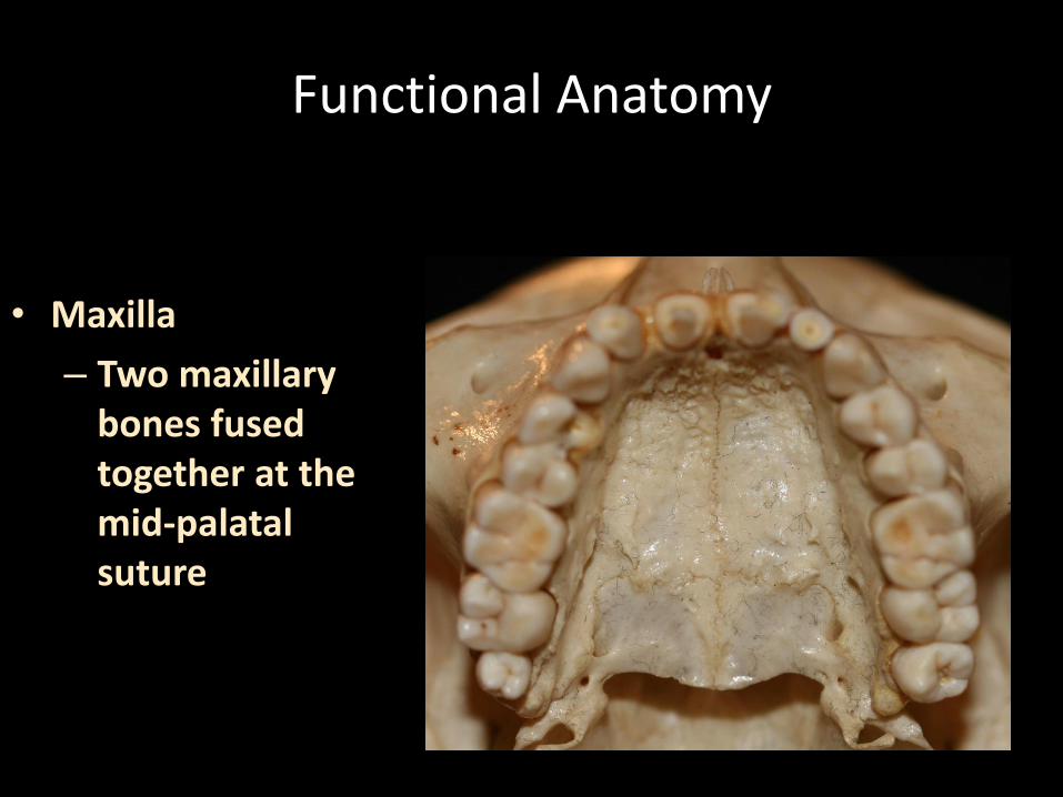
Functional Anatomy
• Maxilla
– Two maxillary bones fused together at the mid-palatal suture
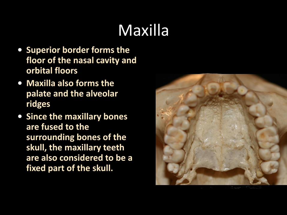
• Superior border forms the floor of the nasal cavity and orbital floors
• Maxilla also forms the palate and the alveolar ridges
• Since the maxillary bones are fused to the surrounding bones of the skull, the maxillary teeth are also considered to be a fixed part of the skull.
Maxilla

Functional Anatomy
• Mandible
– Is a U-shaped bone that supports the lower teeth
– It has no bony attachment to the skull
– It is suspended below the maxilla by muscles and ligaments.
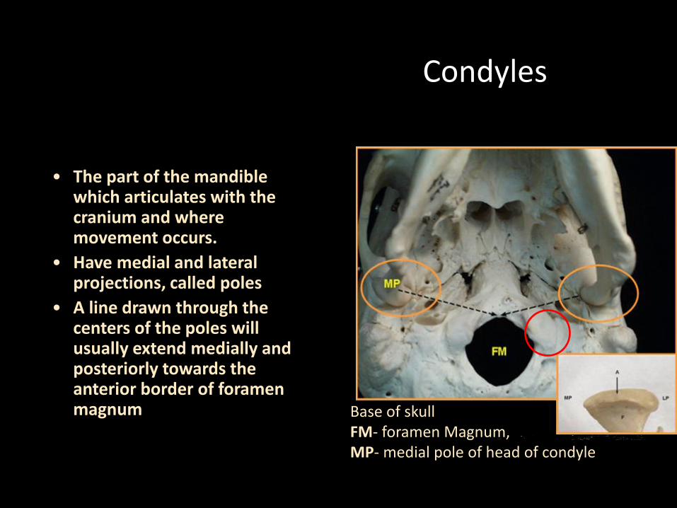
Condyles
• The part of the mandible which articulates with the cranium and where movement occurs.
• Have medial and lateral projections, called poles
• A line drawn through the centers of the poles will usually extend medially and posteriorly towards the anterior border of foramen magnum Base of skull
FM- foramen Magnum, MP- medial pole of head of condyle
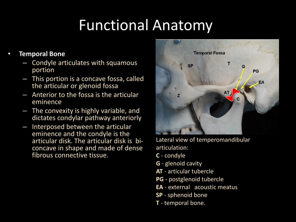
Functional Anatomy
• Temporal Bone – Condyle articulates with squamous
portion – This portion is a concave fossa, called
the articular or glenoid fossa – Anterior to the fossa is the articular
eminence – The convexity is highly variable, and
dictates condylar pathway anteriorly – Interposed between the articular
eminence and the condyle is the articular disk. The articular disk is bi- concave in shape and made of dense fibrous connective tissue.
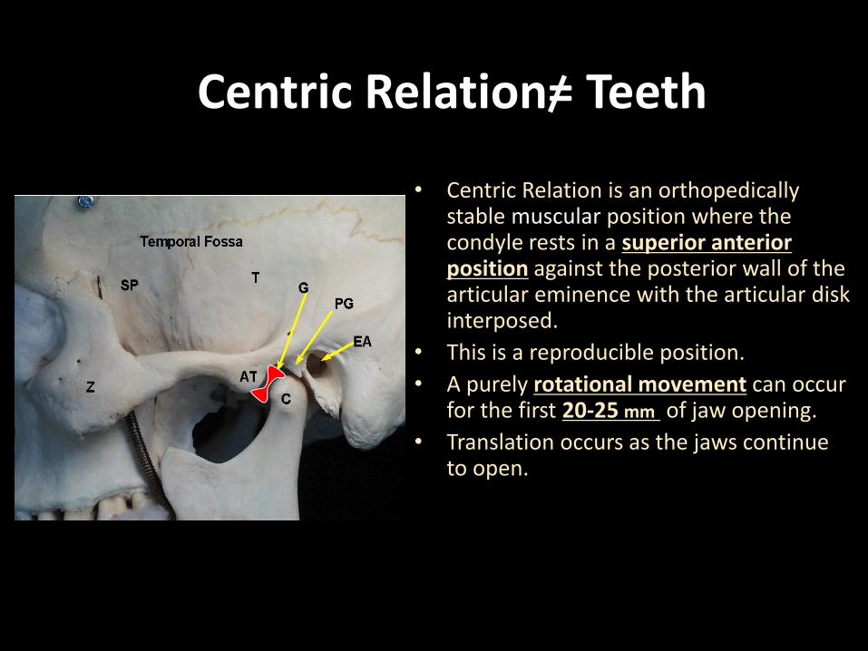
Lateral view of temperomandibular articulation: C - condyle G - glenoid cavity AT - articular tubercle PG - postglenoid tubercle EA - external acoustic meatus SP - sphenoid bone T - temporal bone.
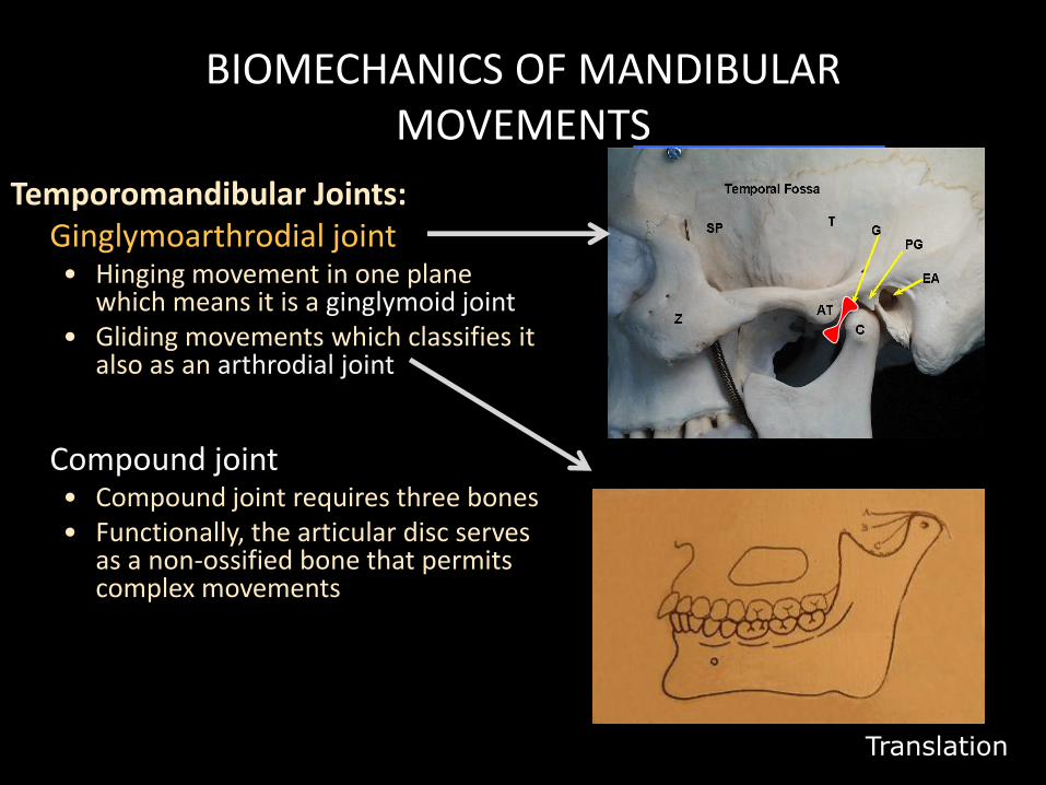
BIOMECHANICS OF MANDIBULAR MOVEMENTS
Temporomandibular Joints: Ginglymoarthrodial joint
• Hinging movement in one plane which means it is a ginglymoid joint
• Gliding movements which classifies it also as an arthrodial joint
Compound joint • Compound joint requires three bones • Functionally, the articular disc serves
as a non-ossified bone that permits complex movements
Rotation around a Horizontal Axis
Translation
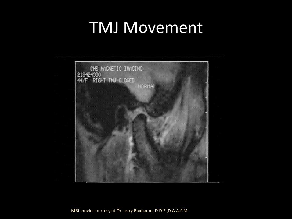
TMJ Movement
MRI movie courtesy of Dr. Jerry Buxbaum, D.D.S.,D.A.A.P.M.
Stability of the TMJ: Determined by Muscles pulling across the
joint to prevent dislocation
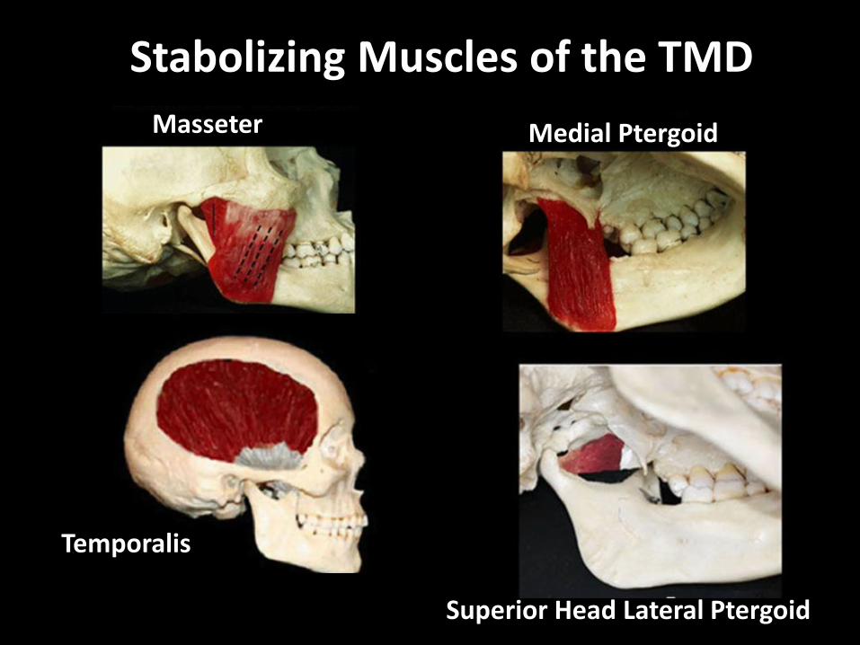
Masseter Medial Ptergoid
Temporalis
Superior Head Lateral Ptergoid
Stabolizing Muscles of the TMD
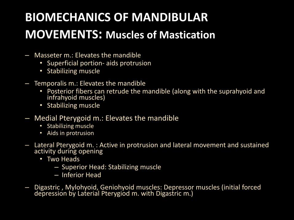
BIOMECHANICS OF MANDIBULAR
MOVEMENTS: Muscles of Mastication
– Masseter m.: Elevates the mandible • Superficial portion- aids protrusion • Stabilizing muscle
– Temporalis m.: Elevates the mandible • Posterior fibers can retrude the mandible (along with the suprahyoid and
infrahyoid muscles) • Stabilizing muscle
– Medial Pterygoid m.: Elevates the mandible • Stabilizing muscle • Aids in protrusion
– Lateral Pterygoid m. : Active in protrusion and lateral movement and sustained activity during opening • Two Heads
– Superior Head: Stabilizing muscle – Inferior Head
– Digastric , Mylohyoid, Geniohyoid muscles: Depressor muscles (initial forced depression by Laterial Pterygiod m. with Digastric m.)
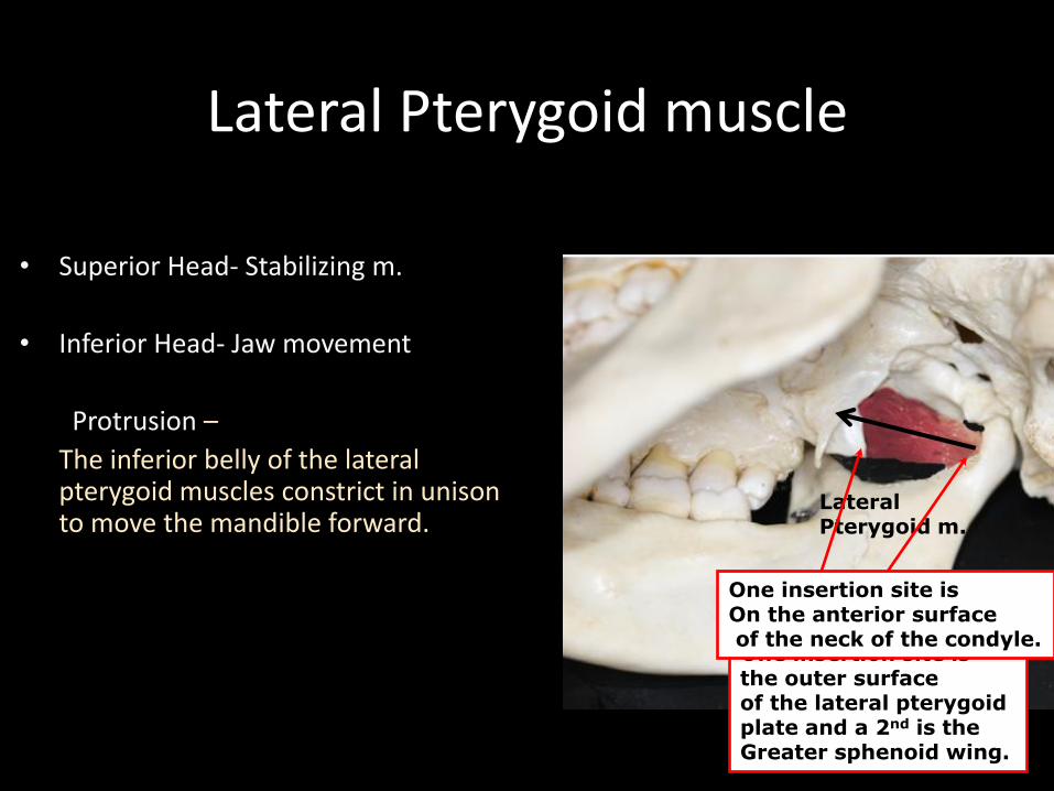
Lateral Pterygoid muscle
• Superior Head- Stabilizing m.
• Inferior Head- Jaw movement
Protrusion –
The inferior belly of the lateral pterygoid muscles constrict in unison to move the mandible forward.
Lateral Pterygoid m.
On the other side, One insertion site is the outer surface of the lateral pterygoid plate and a 2nd is the Greater sphenoid wing.
One insertion site is On the anterior surface of the neck of the condyle.

Lateral Pterygoid (cont.)
Lateral movement -
The contra lateral inferior belly of the lateral pterygoid muscle constricts with superficial portion of the masseter m.
Centric Relation= Teeth
• Centric Relation is an orthopedically stable muscular position where the condyle rests in a superior anterior position against the posterior wall of the articular eminence with the articular disk interposed.
• This is a reproducible position.
• A purely rotational movement can occur for the first 20-25 mm of jaw opening.
• Translation occurs as the jaws continue to open.

Centric Occlusion vs. Maximum Intercuspation
• Centric Occlusion (CO) is the tooth position when the TMJ is in Centric Relation (CR) and the teeth first touch on closing.
• Maximum intercuspation (MI) is the position of the teeth when there is maximum contact of teeth in occlusion.
• CO may or may not equal MI. • In 90% of the population CO does
not equal MI • Most people have a slide from Co
to MI. – Suprahyoid m.: slight activity of this
muscle may allow mandible to open to allow the cusps to glide over one another to allow a centric slide.

Anatomical Planes of Mandibular Movement
• Frontal view – Front view of the
jaws
• Sagittal view – Lateral view of the
jaws
• Horizontal view – Superior view of
the jaws
Border Movements
• Mandibular movement is limited by the ligaments, muscles of the TMJ, articular surfaces and morphology and alignment of teeth.
• The outer limits of the mandible’s range of motion are called border movements. These outer ranges of motion are reproducible and describable for each of the three reference planes. They are limited by the ligaments of the TMJ. Gothic Arch Tracing
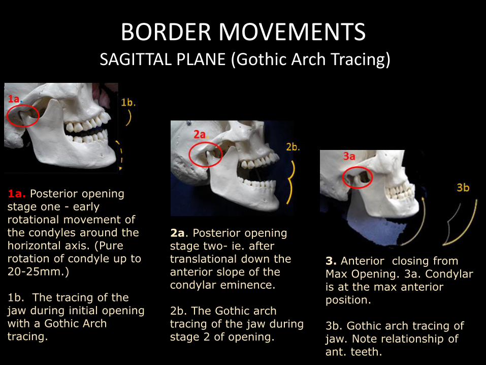
BORDER MOVEMENTS SAGITTAL PLANE (Gothic Arch Tracing)
2a. Posterior opening stage two- ie. after translational down the anterior slope of the condylar eminence. 2b. The Gothic arch tracing of the jaw during stage 2 of opening.
3. Anterior closing from Max Opening. 3a. Condylar is at the max anterior position. 3b. Gothic arch tracing of jaw. Note relationship of ant. teeth.
1a. Posterior opening stage one - early rotational movement of the condyles around the horizontal axis. (Pure rotation of condyle up to 20-25mm.) 1b. The tracing of the jaw during initial opening with a Gothic Arch tracing.

BORDER AND FUNCTIONAL MOVEMENTS (SAGITTAL PLANE)
Video Clips Ash, Nelson.(2003) Wheeler’s Dental Anatomy
, Physiology, and Occlusion 8th ED. St. Louis,MO: Saunders CD **Movement is limited by ligaments

BORDER MOVEMENTS SAGITTAL PLANE
1. In centric occlusion it is possible to have contact on only a few teeth.
- CO is considered initial tooth contact when the TMJ is in CR (the terminal hinge)
2. The application of a muscular force will then create a superor anterior movement or slide. (centric slide is the line from CO to MI on the diagram to the left.)
3. The slide from CO (centric occlusion) to MI(ICP) is present in 90% of the population, and averages 1.25 ± 1mm.
1.
3.
2.
slide

SUPERIOR CONTACT BORDER MOVEMENTS
FACTORS INFLUENCING THE DITANCE BETWEEN CO AND MI (ICP)
• Steepness of cuspal inclines of the posterior teeth.
• Amount of vertical and horizontal overlap of anterior teeth.
• Lingual morphology of maxillary anterior teeth
• General inter arch relationships of the teeth
The upper border Represents the portion of the diagram which is tooth born!

BORDER MOVEMENTS SAGITTAL PLANE
• When there is no discrepancy between CO and MI the initial description of the superior contact border movement is altered
VS.
vs.

BORDER MOVEMENTS HORIZONTAL PLANE
• A Gothic arch tracer is used to trace mandibular border movements in the horizontal plane
• While the mandible moves, the stylus attached to the mandibular teeth generates a pathway on the recording table attached to the maxillary teeth
• 1. Left lateral; 2. Continued left lateral
with protrusion; 3. Right lateral; 4. Continued Right lateral with protrusion. CO-centric occlusion; ICP-intercuspal position

BORDER MOVEMENTS HORIZONTAL PLANE
Video Clips Ash, Nelson.(2003) Wheeler’s Dental Anatomy, Physiology, and Occlusion 8th ED. St. Louis,MO: Saunders CD

FRONTAL PLANE BORDER MOVEMENTS
(Shield Shape) 1. Postural Position or Rest Position is the most superior non- occluding position in the envelope. = red Dot 2. The Right and left Lateral noted on the frontal plane is the farthest lateral movement the muscles and ligaments allow.
. Left Lateral
Right Lateral

Frontal (Vertical) border and functional movements
• Shield shaped outline
• Four components
– Right lateral superior border
– Left lateral superior border
– Left lateral opening border
– Right lateral opening border
Video Clips Ash, Nelson.(2003) Wheeler’s Dental Anatomy, Physiology, and Occlusion 8th ED. St. Louis,MO: Saunders CD

FUNCTIONAL MOVEMENTS
• Functional movements are not considered border movements since they are not determined by an outer range of motions but are determined by the conditional responses of the neuromuscular system.
• Functional Movement occur within the Border Movements/ Tracings.

FUNCTIONAL MOVEMENTS
• Functional movements occur during functional activity of the mandible (like chewing).
• Considered free movements because they occur WITHIN the limits of border movements
• Usually require maximum intercuspation (ICP) so typically being at or below that point
• When the mandible is at rest, it is typically 2-4mm below ICP, a position referred to as clinical rest position or postural position.
See Occlusion Video Clips!!!
#4 is Area of Functional Movement

FUNCTIONAL MOVEMENTS
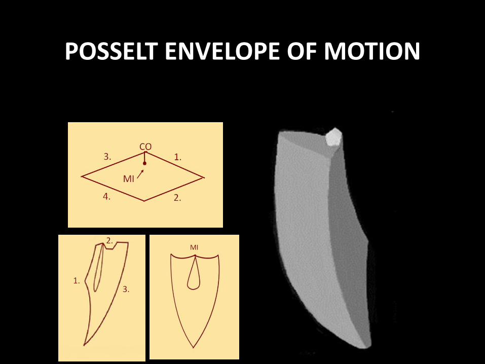
• 1, 2 and 3 comprise the
• “Envelope of Motion” in the sagittal plane
• The motion illustrated by 4 depicts functional movements

FUNCTIONAL MOVEMENTS
• As in other planes, functional movements in the frontal plane begin and end at ICP.
• During chewing, the mandible drops inferiorly until the desired opening is achieved (determined by bolus size).
• The mandible shift to the side on which the bolus (bunch of food) is placed, rising up and down until smaller and smaller particles are produced, ultimately returning to ICP.

POSSELT ENVELOPE OF MOTION
Three Dimensional Border movement with function occurring within the borders
POSSELT ENVELOPE OF MOTION
The End