introductiontobasicophthalmology
TRANSCRIPT

7/27/2019 Introductiontobasicophthalmology
http://slidepdf.com/reader/full/introductiontobasicophthalmology 1/41
Introduction to
ophthalmologyBy
Dr.Bakhtiar Q. Jaf

7/27/2019 Introductiontobasicophthalmology
http://slidepdf.com/reader/full/introductiontobasicophthalmology 2/41
Objective of the lecture ►To give a simple introduction to clinical
anatomy, physiology & embryology ofthe eye
TO recognize clinical approach to the eyecomplaints

7/27/2019 Introductiontobasicophthalmology
http://slidepdf.com/reader/full/introductiontobasicophthalmology 3/41
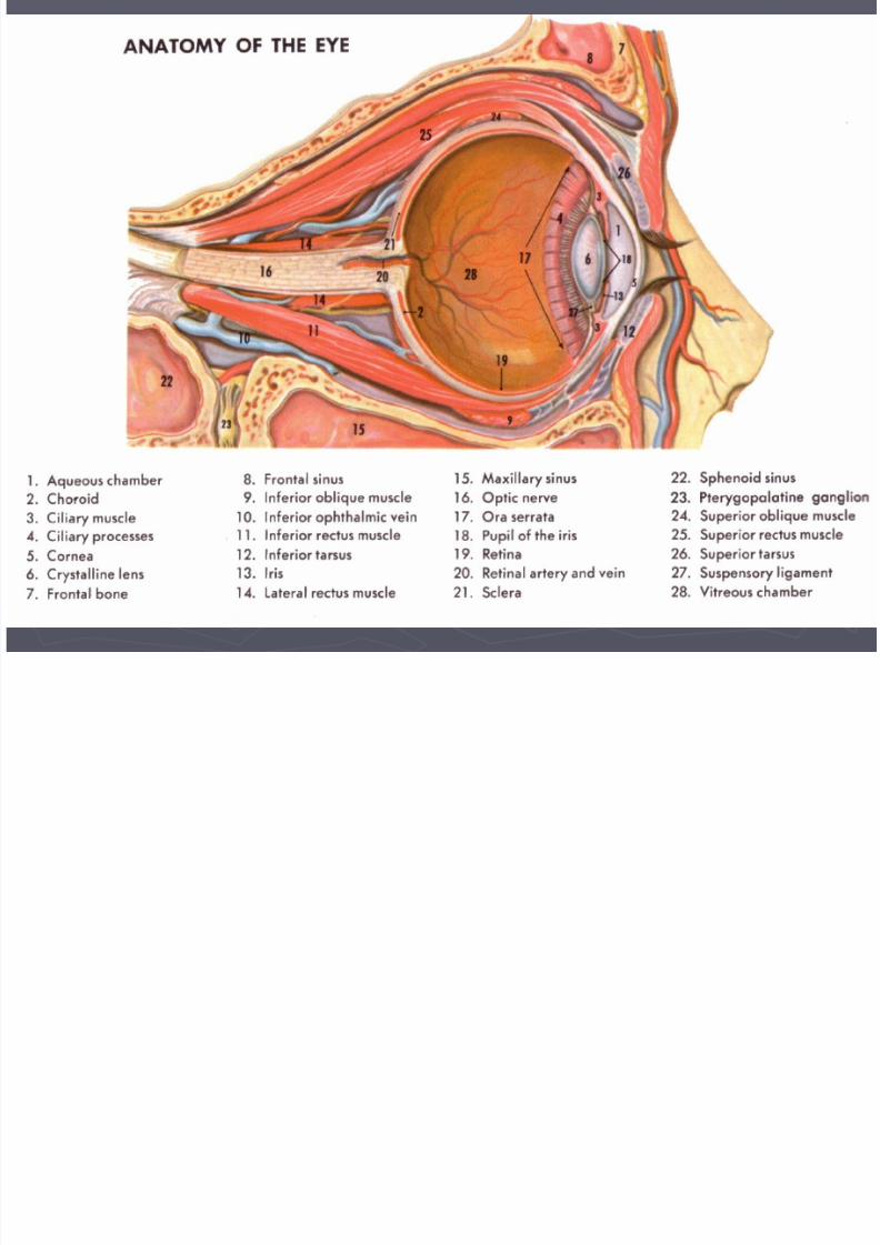
na omy o e eyeThe eyeball, or globe, sits in a protective bonystructure known as the orbit. Lined with muscle,
connective and adipose tissues.the orbit is about 4 cm in height, width, anddepth and is shaped roughly like a four-sided
pyramidsurrounded on three sides by the sinuses: Theethmoid (medially), the frontal (superiorly), and
the maxillary (inferiorly). The optic nerve and the ophthalmic artery enter the orbit at its apex through the optic
foramen.

7/27/2019 Introductiontobasicophthalmology
http://slidepdf.com/reader/full/introductiontobasicophthalmology 4/41
7/27/2019 Introductiontobasicophthalmology
http://slidepdf.com/reader/full/introductiontobasicophthalmology 5/41

7/27/2019 Introductiontobasicophthalmology
http://slidepdf.com/reader/full/introductiontobasicophthalmology 6/41
Eyelids protect the anterior portion of theeye , composed of thin elastic skin that
covers striated and smooth muscles & thetarsal plates.Tears are vitally important to the health ofthe anterior segment of the eye. They areformed by the main lacrimal gland and the
accessory lacrimal glands. The conjunctiva, a mucous membrane,provides a barrier to the external
environment and nourishes the eye.

7/27/2019 Introductiontobasicophthalmology
http://slidepdf.com/reader/full/introductiontobasicophthalmology 7/41

7/27/2019 Introductiontobasicophthalmology
http://slidepdf.com/reader/full/introductiontobasicophthalmology 8/41
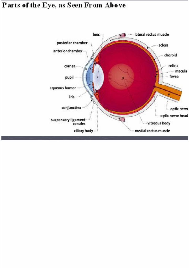
The sclera , commonly known as the "white of the
eye," is a dense fibrous structure that composesthe posterior five sixths of the eyeThe cornea a transparent avascular domelikestructure, forms the most anterior portion of theeyeball and is the main refracting surface of theeye. Behind the cornea lies the anterior chamber ,filled with a continually replenished supply of
clear aqueous humor, which nourishes thecornea.
7/27/2019 Introductiontobasicophthalmology
http://slidepdf.com/reader/full/introductiontobasicophthalmology 9/41

7/27/2019 Introductiontobasicophthalmology
http://slidepdf.com/reader/full/introductiontobasicophthalmology 10/41
The uvea consists of the iris, the ciliarybody, and the choroid. The iris, orcolored part of the eye, is a highly
vascularized, pigmented collection offibers surrounding the pupil. The pupil is a space that dilates and constricts in
response to light.
7/27/2019 Introductiontobasicophthalmology
http://slidepdf.com/reader/full/introductiontobasicophthalmology 11/41
Directly behind the pupil and iris liesthe crystalline lens, a colorless and
almost completely transparent biconvexstructure held in position by zonularfibers. It is avascular and has no nerveor pain fibers. The lens is suspended behind the iris
by the zonules and is connected to theciliary body. The ciliary body controlsaccommodation through the zonular
fibers and the ciliary muscles.

7/27/2019 Introductiontobasicophthalmology
http://slidepdf.com/reader/full/introductiontobasicophthalmology 12/41
The posterior chamber is a small space
between the vitreous and the iris. Aqueous fluid is
manufactured in the posterior chamber by theciliary body
The choroid is layered between the retina and
the sclera and is a highly vascularised tissue,supplying blood to the adjacent outer portion of
the retina.
The ocular fundus is the largest chamber of theeye and contains the vitreous humor, a clear
gelatinous substance, mostly water, encapsulated
by a hyaloid membrane, the vitreous humor .

7/27/2019 Introductiontobasicophthalmology
http://slidepdf.com/reader/full/introductiontobasicophthalmology 13/41
visual pathway Good vision is not dependent solely
on a healthy functioning eyeball butalso on an intact visual pathway.This pathway is made up of theretina, optic nerve, optic chiasma,optic tracts,
lateral geniculate bodies, opticradiations &the visual cortex of the occipital
lobe.
The pathway is an extension of thecentral nervous system. The opticnerve is the second cranial nerve.Its function is to transmit visualimpulses from the retina to thehigher centers in the brain.

7/27/2019 Introductiontobasicophthalmology
http://slidepdf.com/reader/full/introductiontobasicophthalmology 14/41
eyetheof Embryology
andectodermThe eye is formed from both
that is derivedneuroectoderm. Themesenchymefrom the neural tube gives rise to (the retina, thefibers of the optic nerve, and the smooth muscle
on the side ofsurface ectodermof the iris). Thethe head forms( the corneal and conjunctivalepithelium, the lens, and the lacrimal and tarsal
forms( the cornealmesenchymeglands). The
stroma, the sclera, the choroid, the iris, theciliary musculature, part of the vitreous body,and the cells lining the anterior chamber).

7/27/2019 Introductiontobasicophthalmology
http://slidepdf.com/reader/full/introductiontobasicophthalmology 15/41
The rudimentary eyeball develops as anectodermal diverticulum from the lateral aspectof the forebrain. The diverticulum grows out
laterally toward the side of the head, and the endbecomes slightly dilated to form the optic vesicle ,while the proximal portion becomes constricted
to form the optic stalk .At the same time, a small area of surfaceectoderm overlying the optic vesicle thickens to
form the lens placode . The lens placodeinvaginates and sinks below the surface ectodermto become the lens vesicle , the optic vesiclebecomes invaginated to form the double-layeredoptic cup .

7/27/2019 Introductiontobasicophthalmology
http://slidepdf.com/reader/full/introductiontobasicophthalmology 16/41

7/27/2019 Introductiontobasicophthalmology
http://slidepdf.com/reader/full/introductiontobasicophthalmology 17/41
► At end of 4th week of pregnancy; Optic Vesiclecontact with surface ectoderm & invaginate toform Optic cup & the lens vesicle

7/27/2019 Introductiontobasicophthalmology
http://slidepdf.com/reader/full/introductiontobasicophthalmology 18/41

7/27/2019 Introductiontobasicophthalmology
http://slidepdf.com/reader/full/introductiontobasicophthalmology 19/41

7/27/2019 Introductiontobasicophthalmology
http://slidepdf.com/reader/full/introductiontobasicophthalmology 20/41
7/27/2019 Introductiontobasicophthalmology
http://slidepdf.com/reader/full/introductiontobasicophthalmology 21/41
The optic nerveThe ganglion cells of the retina develop
axons that converge to a point wherethe optic stalk leaves the posteriorsurface of the optic cup. This site willlater become the optic disc .The axons now pass among the cells
that form the inner layer of the stalk.Gradually, the inner layer encroacheson the cavity of the stalk until the inner
and outer layers fuse.

7/27/2019 Introductiontobasicophthalmology
http://slidepdf.com/reader/full/introductiontobasicophthalmology 22/41

7/27/2019 Introductiontobasicophthalmology
http://slidepdf.com/reader/full/introductiontobasicophthalmology 23/41
7/27/2019 Introductiontobasicophthalmology
http://slidepdf.com/reader/full/introductiontobasicophthalmology 24/41
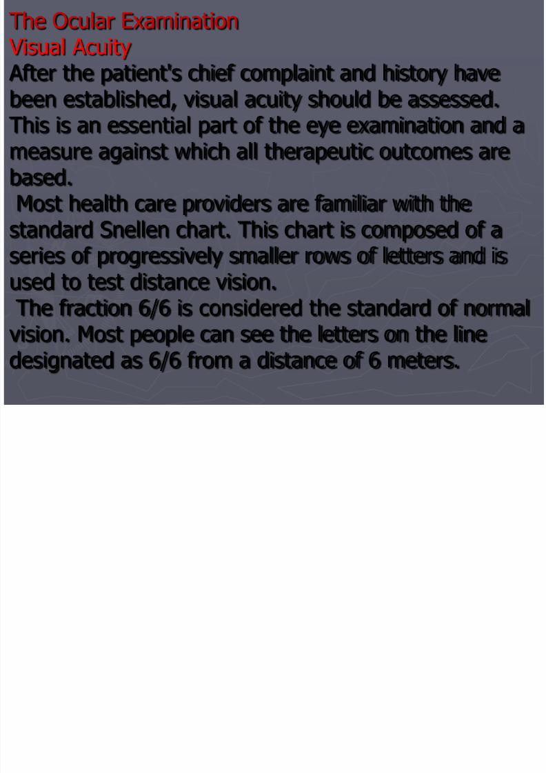
The Ocular Examination Visual AcuityAfter the patient's chief complaint and history have
been established, visual acuity should be assessed.This is an essential part of the eye examination and ameasure against which all therapeutic outcomes arebased. Most health care providers are familiar with thestandard Snellen chart. This chart is composed of aseries of progressively smaller rows of letters and is
used to test distance vision. The fraction 6/6 is considered the standard of normalvision. Most people can see the letters on the linedesignated as 6/6 from a distance of 6 meters.

7/27/2019 Introductiontobasicophthalmology
http://slidepdf.com/reader/full/introductiontobasicophthalmology 25/41
The External Eye Examination After the visual acuity has beenrecorded, an external eyeexamination is performedDIAGNOSTIC EVALUATIONDIRECT OPHTHALMOSCOPY Is a hand-held instrument with
varying plus and minus lenses.
The lenses can be rotated intoplace, enabling the examiner tobring the cornea, lens, and retinainto focus

7/27/2019 Introductiontobasicophthalmology
http://slidepdf.com/reader/full/introductiontobasicophthalmology 26/41
Indirect Ophthalmoscopy
Is an instrument commonly used by theophthalmologist. It produces a bright andintense light. The light source is fixed with apair of binocular lenses, which are mounted onthe examiner's head. The ophthalmoscope isused in conjunction with a hand-held 20-dioptcrlens. This instrument enables the examiner to
see larger areas of the retina, although in anunmagnified state.

7/27/2019 Introductiontobasicophthalmology
http://slidepdf.com/reader/full/introductiontobasicophthalmology 27/41
Slit-Lamp ExaminationIs a binocular microscope mounted on a table. This instrument enabls theuser to examine the eye with magnification of 10 to 40 times the real
image.Color Vision TestingBecause alteration in color vision is sometimes indicativeof optic nerve problems, color vision testing is oftenperformed in a neuro-ophthalmologic workup.
UltrasonographyLesions in the globe or the orbit may not be directlyvisible and are evaluated by ultrasound.Color Fundus Photography
Fundus photography is a technique used to detect anddocument retinal lesions. The patient's pupils are widelydilated during the procedure, and visual acuity isdiminished for about 30 minutes due to retinal"bleaching" by the intense flashing lights.

7/27/2019 Introductiontobasicophthalmology
http://slidepdf.com/reader/full/introductiontobasicophthalmology 28/41
Tonometry
Tonometry is used to measure IOP by determining the amount of
force necessary to indent or flatten (applanate) a small anteriorarea of the globe of the eye.
Gonioscopy
Gonioscopy is used to visualize the angle of the anterior chamber
to identify abnormalities in appearance and measurements.
Perimetry Testing
Perimetry testing is used to evaluate the field of vision. A
visual field is the area or extent of physical space visible
to an eye in a given position. Its average extent is 65degrees upward, 75 degrees downward, 60 degrees
inward, and 95 degrees outward when the eye is in the
primary gaze.

7/27/2019 Introductiontobasicophthalmology
http://slidepdf.com/reader/full/introductiontobasicophthalmology 29/41
IMPAIRED VISION REFRACTIVE ERRORS
In refractive errors, vision is impairedbecause a shortened or elongated eyeballprevents light rays from focusing sharply on
the retina. Blurred vision due to refractiveerror can be corrected with eyeglasses andcontact lenses.

7/27/2019 Introductiontobasicophthalmology
http://slidepdf.com/reader/full/introductiontobasicophthalmology 30/41
Those patients for whom the visual image focuses
precisely on the macula and who do not need eye glasses
or contact lenses are said to have emmetropia.
7/27/2019 Introductiontobasicophthalmology
http://slidepdf.com/reader/full/introductiontobasicophthalmology 31/41
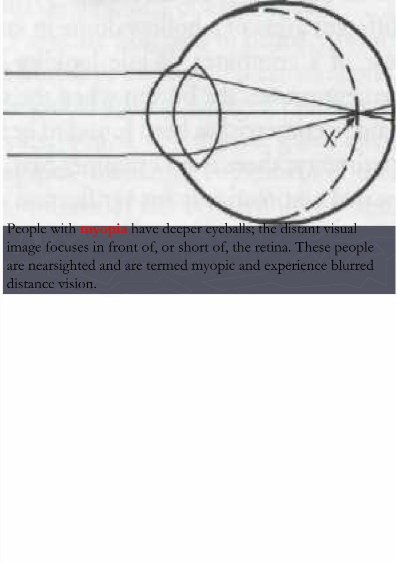
People with myopia have deeper eyeballs; the distant visual
image focuses in front of, or short of, the retina. These people
are nearsighted and are termed myopic and experience blurred
distance vision.

7/27/2019 Introductiontobasicophthalmology
http://slidepdf.com/reader/full/introductiontobasicophthalmology 32/41
When people have a shorter depth to their eyes, the visual
image focuses beyond the retina; the eyes are more shallow and
are termed hyperopic. People who have hyperopia are
farsighted.

7/27/2019 Introductiontobasicophthalmology
http://slidepdf.com/reader/full/introductiontobasicophthalmology 33/41
GLAUCOMA Glaucoma is the term used to describe a group of
ocular conditions characterized by optic nervedamage, high intraocular pressure and visualfield deffect. CLASSIFICATION OF GLAUCOMA
Glaucoma can be open angle or angle closure,depending on which mechanisms cause theimpairment of the aqueous outflow. Glaucoma
can also be primary or secondary, depending onwhether associated factors contribute to the risein IOP.

7/27/2019 Introductiontobasicophthalmology
http://slidepdf.com/reader/full/introductiontobasicophthalmology 34/41
CATARCTA cataract is a lens opacity orcloudiness
Clinical Manifestations Painless blurring of vision ischaracteristic of cataracts.
C l U d O l M di i

7/27/2019 Introductiontobasicophthalmology
http://slidepdf.com/reader/full/introductiontobasicophthalmology 35/41
Commonly Used Ocular MedicationsTopical AnestheticsOne to two drops of proparacaine hydrochlorideand tetracaine hydrochlorid are instilled before
diagnostic procedures such as tonometry andgonioscopy and in minor ocular procedures suchas removal of sutures or conjunctival or cornealscrapings.Mydriatics and Cycloplegics
Mydriasis, or pupil dilation, is the main objectiveof the administration of mydriatics andcycloplegics

7/27/2019 Introductiontobasicophthalmology
http://slidepdf.com/reader/full/introductiontobasicophthalmology 36/41
Anti-lnfectivesAnti-infective medications include
antibiotics, antifungals, and antivirals.Most are available as drops, ointments, orsubconjunctival or intravitreal injections.
Antibiotics include penicillin, thecephalosporins, aminoglycosides, andfluoroquinolones. The main antifungalagent is amphotericin B. Antivirals includeacyclovir and ganciclovir.

7/27/2019 Introductiontobasicophthalmology
http://slidepdf.com/reader/full/introductiontobasicophthalmology 37/41
Corticosteroids and Nonsteroidal Anti-
Inflammatory Drugs The topical preparations of corticosteroids
are commonly used in inflammatory
conditions of the eyelids, conjunctiva,cornea, anterior chamber, lens, and uvea. In
posterior segment diseases that involve the
posterior sclera, retina, and optic nerve, thetopical agents are less effective; hence, the
parenteral and oral routes are preferred.

7/27/2019 Introductiontobasicophthalmology
http://slidepdf.com/reader/full/introductiontobasicophthalmology 38/41

7/27/2019 Introductiontobasicophthalmology
http://slidepdf.com/reader/full/introductiontobasicophthalmology 39/41

7/27/2019 Introductiontobasicophthalmology
http://slidepdf.com/reader/full/introductiontobasicophthalmology 40/41
References1. Lecture notes in
ophthalmology2. Parson’s disease
of the eye

7/27/2019 Introductiontobasicophthalmology
http://slidepdf.com/reader/full/introductiontobasicophthalmology 41/41