investigating children´s conceptions of the brain: first steps - eric
TRANSCRIPT
fgjkl
International Journal of Environmental & Science Education
Vol. 3 , No. 3 , July 2008, xx-xx
Investigating children´s conceptions of the brain:
First steps
Amauri Betini Bartoszeck Flavio Kulevicz Bartoszeck
Received 24 November 2010; Accepted 11 March 2011
This paper reports data, part of a cross-sectional study about the use of pupil´s drawings
as a means of probing the development of 195 Brazilian pre-school children (4 to 6 year-
olds) and 681 primary school pupils 1st Grade through 4
th Grade (7 to 10 years of age)
conceptions of the human brain. The aims of the present study is to analyze how the con-
ception of the brain develops, how they represent their brains, and whether it is based on
historical models or current scientific knowledge, in their interaction with school and so-
ciety at large. The methodology involved the presentation of a contour of the head and
neck drawn on the blackboard in the classroom, and children were asked to draw what
they think they have inside their heads. After the drawings were collected some pupils
were interviewed to explain their drawings. Classification of the collected drawings were
interpreted on the perspective of historical models of the brain and scored following a 6
level rating scale depicting degrees of neuroanatomical resemblance. Gender and age
were taken into consideration. The results show that younger pupils are adepts of mental
ideas in their representations of the brain, i. e. what the brain does, but progressively as
they get older, start to develop a more morphological representation of the brain.
Knowledge obstacles for learning about the nervous system and elementary neuroscience
implications are discussed.
Keywords: biological conceptions, mental images, historical brain representations, children,
kindergarten and primary school.
Introduction
Basic Biology
All living systems share a common ancestry of organic structure (El-Hani & Videira, 2000). Or-
ganisms basically orient their existence at absorbing nutrients to secure survival, develop mecha-
nisms controlled by genes to evolve and to avoid predators or environmental perils. Thus, a
communication program evolved randomly inside organisms to detect and integrate incoming
information to face a changing world, which may be observed, for example, in very simple ani-
mals as the protozoa Paramecium sp. (Capra, 1996). Its cell membrane possesses ion channels
that allow one to register a membrane potential close to that as is obtained in a common neuron
(Hille, 1994; Loewenstein, 1999; Cotterill, 2001). Eventually cells gathered together to develop
an information network subjected to evolution pressures which in the end originated an elemen-
International Journal of Environmental & Science Education
Vol. 7, No. 1, January 2012, 123-139
ISSN 1306-3065
Copyright © 2012 IJESE
http://www.ijese.com/

124 Bartoszeck & Bartoszeck
tary neuronal net, later a complex nervous structure in other more advanced animals, and ulti-
mately the brain of vertebrates capable of dynamic adaptive behavioral responses to a variety of
environmental stimuli ( Allman, 2000; McCrone, 2002).
Historical brain representations
The creationist model of the world (the religious belief that life, the planet Earth and the universe
are the creation of a supernatural being) started to be replaced by a new hypothesis stating that the
universe is often changing and does not follow a pre-established plan due in part to science de-
velopment in the 18th-19
th Century period (Scott, 2004). Knowledge in organisms started to be
studied on the approach that mind and brain function are related and work together. The Cartesian
dualism which was a current explanation of the brain, involving the complex “mind-soul and
body”, was put aside as a valid model of investigation (Edelman, 1992). Another tenet is that the
brain is the product of neural Darwinian evolution which selected the living systems according to
their ability to adapt to the environment where they are supposed to live (Edelman, 1987). The
brain by itself favors the capacity of adaptation of the organism, turning it more independent of
genetics and more subject to experience and learning opportunities. In spite of these scientific
advances and findings amongst pupils and teachers still persists the Cartesian dualism reflected
by means of historical models of the brain instead of an understanding of biological mechanism
patterns responsible by thinking and learning (Clément & Mein, 1987).
Thus, historically the organ of the brain (cerebrum) went through a long path to find its
way as a structure responsible for human thinking and behavior. From Ancient times to the Re-
naissance the problem was to specify and to locate the site of the psychic phenomena. For in-
stance, in the Greek thinking the heart was the place for the sensations, passion and intelligence
asserted by Aristotle in the 5th Century before Christ, according to the cardiocentric thesis
(Frampton, 1991). However, even before Aristotle, Democritus had proposed a cephalocentric
thesis for a material basis by means of a circulating liquid involved with sensations and thinking
(Changeux. 1991; Dolan, 2007). The theme of the three ventricles advanced to the 18th Century
on the concept of a “hydraulic model” with circulating fluids and even Descartes accepted this
model for muscle contraction and the “seat of the soul”. More recently, the brain was represented
by a model of phrenology in the 19th Century by Gall. Nowadays, contributions by Ramon y Cajal
on the theory of neurone and contemporary studies by Donald Hebb, (1949/2002) Sperry & Sper-
ry, (1983) and John Eccles, (1994) on synapses and technical advances in brain imaging brought
new light for a better understanding of the brain ( Brazier, 1959; Johnson, 2004; Parent, 2009).
Organ and organ systems in school
Therefore, when the human body is being taught concerning organs and organs systems, teachers
make use of analogies between the human body and machines expecting that the comparison will
help in the understanding of its organization and function. Generally speaking analogies simplify
the processes and in the end might turn into obstacles to scientific learning (Duit, 1991; Andrade
et al., 2002; Giraldi & Souza, 2006; Glynn, 2007). As an alternative to the use of analogies inves-
tigations over the spontaneous conceptions of the students by means of collecting drawings and
interviews may bring the developing of efficient teaching strategies (Steward & Furuya, 1982;
MacPhail & Kinchin, 2004; Hall, 2007) . Probing the ideas which pupils have about brain repre-
sentations may reveal what are the common sense ideas that act as hindrances to an understand-
ing of scientific concept, in this case the form and functioning of the brain.
Several studies have investigated the spontaneous development of students´ conceptions
of the human organism. Conceptions may be considered as structures of knowledge through

Conceptions of the brain 125
which one constructs new information that later, after some reflection, might be converted in
knowledge (Pines & West, 1986). Some pioneering studies were carried by Nagy (1953), who
looked at what Hungary children from Budapest, 4 to 11 year-olds thought as revealed by draw-
ings and interviews, was the functioning of the brain, lungs and stomach. Her findings indicated
that children from her sample, had difficulties to tell the mechanism of the functioning of the
organs, but rather defined the role of the organ, for instance “the brain is for thinking”, lungs are
for air”. On the other hand, Tait and Ascher (1955), asked adults and a few sixth-graders “draw
the inside the body, including all the organs” and got as a response from the students that the “the
brain is inside the head”. However, it was a limited sample (N=22).
Adults agree the brain is crucial for life, whereas children recognize Gottfried et al.,
(1999) that it is an important biological organ (Crider, 1981; Gellert, 1962; Johnson & Wellman,
1982; Nagy, 1953). Several studies have investigated the spontaneous development of students´
conceptions about the human organism (Amann-Gainotti & Antenore, 1990; Fleer, 1994, Reiss &
Tunnicliffe, 2001; Reiss & Tunnicliffe et al., 2002; Manokore & Reiss, 2003; Frändberg et al.,
2004). A general trend emerged from these findings i. e. very young children (aged 4 to 5 and 5
to 6 years old) have little knowledge about the human organism including the brain (Mintzes,
1984; Carey, 1985; Osborne et al., 1992).
According to Carey (1985), when referring to the nervous system and the brain, 2nd
grad-
ers know that thought is needed for different kinds of activities, for instance to start walking. Up-
per elementary school students attribute to nerves the function of conducting messages and con-
trolling activity of the body (Gellert, 1962). However, 5th graders appear not to understand that
the brain controls involuntary activities, like breathing and heart beat (Johnson & Wellman,
1982).Learning how human organs and organ systems of the human body are structured and func-
tion which seems to be a mere descriptive issue is full of difficulties as noticed for Brazilian chil-
dren (Rabello, 1994).
Teaching how the human body works, especially the nervous system involving the brain
and thoughts demands a detailed planning. Society is facing a huge growth of neuroscience re-
search and an explosion of information through the Internet and other popular media, influencing
the school and in a way, how students learn in the classroom (Goswami & Szücs, 2011; Battro et
al., 2011).
Children´s conceptions of the brain were examined under the approach of explanations
of mental phenomena, for example, remembering, thinking, dreaming as located in the head, in
the brain (Johnson and Wellman, 1982; Johnson, 1990; Watson et al., 1998), brain imaging in
medical situations Wilf et al., (1983), understanding of cerebral computed tomography scan pro-
cedure (Hellier et al., 1986), perception of organ and function before a pre-operative event (van
Koot, 1990), usage of drawings to diagnose headaches (Stafstrom et al., 2002), and safety
measures adopted when wearing helmets (Cynkar & Rutledge-Gorman, 2004). However, fewer
studies looked at what children think is inside their heads, expressing their opinions by means of
drawings, thus revealing their thoughts, dreams, and potential models of what they imagine the
brain is and does (Savy and Clément, 2002, 2003; Bartoszeck and Bartoszeck, 2006).Thus, it is
the goal of biology education researchers to identify the starting points for teaching from children
and then find ways to add new knowledge to that which the pupil already possesses (Tunnicliffe,
2006).
The objective of the present study is to analyze qualitatively and quantitatively how the
conception of the brain develops, whether there is a historical representation of the brain from
pre-school pupils up to 4th graders, in their interaction with school and society at large ( Lawson,
1988) in a Brazilian sample. This exploratory study, by means of drawings (Cox, 1992, 1997),
aims to capture what the pupils thinks he/she has inside his/her head and the amount of

126 Bartoszeck & Bartoszeck
knowledge related to the brain and mental images or thoughts children have. The authors are un-
aware of any other study carried out with the same focus in Brazil.
Theoretical background
The concept of “science literacy” seems to include a view that a close observation of the natural
world and what neuroscientists do is poorly known to the general public. One of the goals of
Biology education, by means of neuroscience education, is to provide ways to teach how the
brain works and what it does from very early, starting in the pre-school years (Zardetto-Smith et
al., 2000; 2002). Children recognize that the brain is a very important biological organ (Gottfried
et al., 1999).
However, very few studies looked at what children think is inside their heads (Savy and
Clément, 2002, 2003), or inside themselves (Reiss et al., 2002). Thus, drawing is a tool that
might reveal what is the mental model of the brain they may have on the perspective of “intellec-
tual realism” (Luquet, 1927/1969; Cox, 1992). In this study, drawing of the contour of the head
was adopted to probe the mental model of the brain which is hard to explore. Instead, a expressed
model is expected, would bring to the surface, the mental models the pupil has of this biological
organ (Gilbert & Boulter, 2000). Therefore, researchers could identify the starting points for
teaching early year children, avoiding misconceptions on what the brain looks like and does,
and devise ways to add new knowledge to that which the student already possesses (Tunnicliffe,
2006). Biological concept in this study is considered as a mental construct created by the learner
in the cognitive structure of his mind and is supported by ideas, images, and tentative explana-
tions to cope with a new problem in his environment (Giordan & de Vecchi, 1996; Clément,
1998; Reiss & Tunnicliffe, 1999).
Key objectives and research questions
- Investigate the structure & function of the brain ( as a mental model) pupils may have
manifested by means of a drawing depicted as the expressed model;
- Evaluate whether historical models of the brain are still depicted;
- Indicate if there is a development understanding of the brain by age and gender;
- Interview a few pupils to explain what they drew and how they think brain works
Participants, methods and data sources
The participants are grouped on kindergarten II (4 to 5 year-olds), kindergarten III (5 to 6 year-
olds) [106 boys and 89 girls] pre-school, and as primary school, 1st through 4st graders [331 boys
and 350 girls] (Table 1). The fieldwork was carried out at 8 Schools of Infancy Education (6 non
fee paying and 2 private schools) and 8 Primary Schools (6 state funded and 2 private). The
schools which agreed to participate were located downtown and in suburban areas in the cities of
Curitiba and Foz do Iguaçu, Paraná State, Brazil as to reflect the availability of resources and
social and cultural strata of the population sampled in southern Brazil. All subjects from this
study were attending schools in classes corresponding to their ages.
Procedure

Conceptions of the brain 127
The researchers told the pupils that today they would perform a different kind of activity. They
were asked to write down their first names, age on the top of a blank A4 sheet of paper with a
black pencil. Next, pupils were asked to draw in pencil “what they think they have inside their
head”. An outline of the head and a portion of the neck were drawn on the blackboard of the
classroom to serve as a model. Alternatively, some pupils preferred to use the profile of a head,
which was also accepted. The drawings were created under normal classroom conditions and
pupils sat in desks no so close to each other, and asked not to copy from the drawings of the
neighbors sitting nearby.
Table 1. Number (n) of pupils whose drawings representing the brain was collected in
this sample
Pupils were given 10 to 15 minutes to complete their drawing. Many pupils spontaneous-
ly labeled the internal biological structures on the outlines. The teacher wrote labels to the
younger pupils (4 to 6 years old) for the biological structures when requested, but only the exact
words in places on the outline pointed by the children. The field work was conducted in whole
class settings.
Drawing analysis
A total of 1151 drawings were collected. Pupils´ distribution by grade level is indicated on Table
1. Classification of the drawings was done independently by the authors adapting the method of
Savy and Clément, (2002) and depicted on Table 2. The authors by analyzing the drawings de-
veloped a classification, that in their view, there are representations of mental images, hydraulic,
dog bone, “enteroid”, epithelial, “callote”, as well as neuroanatomical models. Few disagree-
ments occurred in classifying the drawings according to models which were resolved between the
authors before data processing.

128 Bartoszeck & Bartoszeck
Table 2. Kinds of models of the brain descriptive characteristics
Results
Our data revealed that children from pre-school through 4th graders are adepts of historical mod-
els of the brain, i. e. depicted as hydraulic, “dog bone”, enteric, epithelial, and finally a few
neuro-anatomical models on their drawings (Fig. 1).
Figure 1. Examples of children´s conceptions of the brain corresponding to historical
models (after Brazier, 1959; Edelman, 1992; Parent, 2009)
Not many students scored level 6 as the rubric used (Table 3). Most of them did not
achieve higher levels (Table 4).

Conceptions of the brain 129
Table 3. The rubric used to allocating a grade to the drawings.
Table 4. Frequency in percentage achieved by boys and girls from the rubric used in Ta-
ble 3. Notice that higher levels are not depicted as they reached very low frequencies.
(Legend reads: kindergarten II,III=pre-school II [5 year olds], preschool III [6 year olds];
F=first, S=second, T=third, Fo=forth grades)
Some of them do not make a clear distinction between the biological structure of the
brain and mental images i. e. their imagination as depicted in Fig.2.

130 Bartoszeck & Bartoszeck
Figure 2. A drawing by a 10 years-old boy, 4th
grade representing a mental image (imag-
ination, memory) model of the brain (caption reads: uma vaca=a cow).
During interviews to explain the children drawings and what the brain they think it does,
younger pupils mention that: it is a place with pipes where blood and ideas circulate; where hap-
piness lives; a place (organ) that helps see, sleep, dream and sometimes aches. Only in the higher
grades (7 to 10 year olds) children drew pictures resembling the brain structures and how it
works, as for example, they said in the interview, ideas are recorded and move in the inner lines;
feel pain; gives will power; remember things, make eyes and ears, arms and legs work; control
hunger and thirst, helps pay attention and think (Fig. 3).
Brazilian children´s results from the present sample are very close to those found by
(Savy & Clément, 2002) and (Savy, 2005) from 800 French pupils 6 to 11 year-olds. A low per-
centage of the students scored level 5 in the rating scale (1: lines all over the skull, to 6: cerebral
hemispheres or cerebral circumvolutions at approximate position (see Table 4). Pupils from this
sample do not make a clear distinction between the biological structures of the brain from
thoughts represented as mental images of everyday experiences, their memories. When prompted
to say something about their drawings, they mentioned that ideas circulate inside the brain and
skull, the road is the tubing system. Progressively pupils draw pictures trying to explain what the
organ is and its function, but still as with the French sample, the pupils do not separate thoughts
completely from the biological structure that creates them. Organizing curricula contemplating
nervous system issues would bring meaningful improvement in the learning of elementary neuro-
science.

Conceptions of the brain 131
Figure 3. A drawing by a 7 years-old boy, 1st grade representing a hydraulic model of the
brain
Evolution in drawings from per-school to 1st grade
Mental images defined here as an experience that , on most occasions, resembles the actual expe-
rience of perceiving some object, event or scene that occurs even when not present to the senses ,
tend to be reduced in percentage of the total drawings in this sample from pre-school to 4th
grade. Apparently children do not separate memories, recalling of situations or mental images
from the biological structure that generate them. The “ hydraulic model” (Fig. 3) still persists at
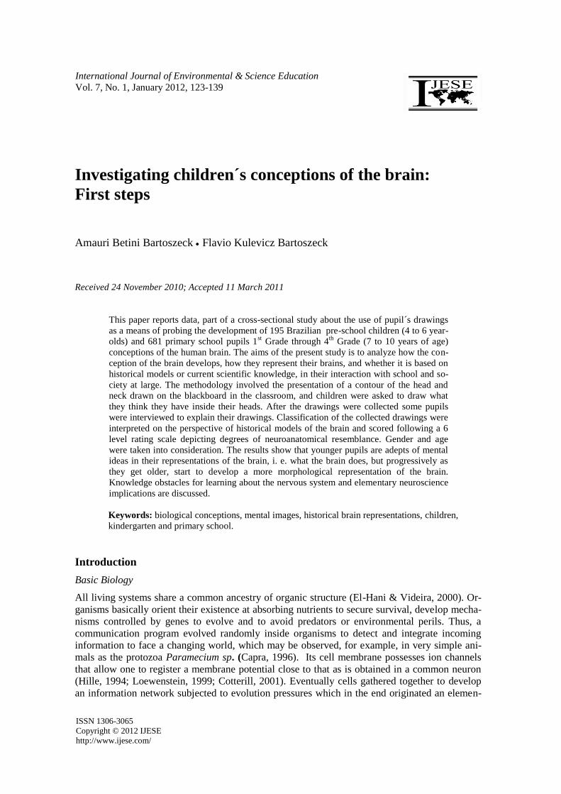
the 1st grade but the “dog bone model” as depicted in Fig.4, which is an assembly the bones
forming the skull is reduced as children get older, although about 50% of the drawings were con-
sidered unidentifiable (scribbling) according to the rubric used to allocating a grade to the draw-
ings (Table 3).
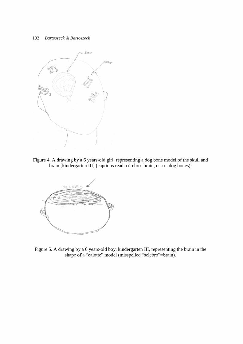
“However, there is a progressive complexity improvement in the representation of the
brain shaped as an epithelial, calotte and hemispheres (neuroanatomical) models as illustrated on
Table 2.
Evolution in drawings from 2nd
to the 4th
grade
Although mental image drawings still persist in this range of grades, we observed a drop on the
percentage for the hydraulic model, for the bone model and a meaningful increase in the elabora-
tion of the brain shaped as a calotte located on the top of the head. Another point is that there is a
decrease in the proportion of the unidentifiable drawings from 54 % at the kindergarten III pupils
to 18.3% at the 4th grade. However, even at the 4
th grade the brain is poorly represented (only
7.8% of the total sample) as distinguishable brain hemispheres (Fig. 6).
132 Bartoszeck & Bartoszeck
Figure 4. A drawing by a 6 years-old girl, representing a dog bone model of the skull and
brain [kindergarten III] (captions read: cérebro=brain, osso= dog bones).
Figure 5. A drawing by a 6 years-old boy, kindergarten III, representing the brain in the
shape of a “calotte” model (misspelled “selebro”=brain).

Conceptions of the brain 133
Figure 6. A drawing by a 10 years-old girl, 4th
grade representing the brain hemispheres
model and sensory organs (eyes)
Discussion and Educational Implications
There is a scarcity of studies concerning the development of the conception of brain in children
and adult human beings (Clément, 1984, 1994, 1997; Mein & Clément, 1988; Kochkar et al.,
2002). Young children build and developed their own models of the world and they become
frameworks as how the world is progressively understood. Pupils from the early years try to un-
derstand and explain the brain´s function and seem to succeed, but have a poor idea of its shape.
Thus, they use their limited experience in other domains seeking for meaning, and only around
10 years old, similar to the French children sample, the brain hemispheres are more realistic de-
picted, but even so, evoking conceptions of historical models (Fig. 1). It seems from our data that
children do not make a clear distinction between the biological structure of the human brain from
thoughts represented as mental images, i. e. pictures of their thoughts as happy memories of vaca-
tions, parties and members of the family. Concerning the hydraulic model it may suggest for the
youngest children, that thoughts circulate inside the skull and brain, the callote model may refer
to the position of the brain on the top of the skull and may otherwise be shaped as an enteric gas-
tric tubing, or as type epithelial tissue, where ideas circulate. These depictions may be viewed as
representatives of the stage of “realism intellectual” (Luquet, 1927/1996; Bourassa,
1999).Organizing “hands-on” practices modeling the brain, spinal cord and sense organs activi-
ties would bring meaningful improvement in the learning of elementary neuroscience earlier
starting in the pre-school years (Moreno et al., 1997; Gelman et al., 2010).
Implications
Pupils should be subjected to practical classes on the following topics:
Create models depicting the human brain with clay or styro-foam;

134 Bartoszeck & Bartoszeck
Practice in assembling brain parts into a skull plastic case;
Use sandpaper pieces and wooden shaped small objects to identify tactile stimuli;
Use flowers, perfume, and kitchen seasonings ( e. g. cinnamon, clover) to identify scent
stimuli;
Visits to Natural Science Museum to watch and manipulate with care preserved fish, rat,
bird, toad, primate and human brains in jars.
Acknowledgements
The authors acknowledge comments and suggestions by Dr. Pierre Clément, Université Claude
Bernard, Lyon 1, Villeurbanne, France, during the writing of this manuscript.
References
Allman, J. M. (2000). Evolving brains. New York: Scientific American Library.
Amann-Gainotti, M., Antenore, C. (1990). Development of internal body image from Childhood
to early adolescent. Perceptual and Motor Skill, 71:287-293.
Andrade, B. L., Zylbersztayn, A. & Ferrari, N. (2002). As analogias e metáforas no ensino de
ciências à luz da epistemologia de Gastón Bachelard. ENSAIO- Pesquisa em Educação
em Ciências, 2(2):1-11, (Analogies and metaphors in the teaching of science on the
perspective of Gastón Bachelard´s epistemology).
Bartoszeck, A. B., & Bartoszeck, F. K. (2006, August). O modelo expresso do que o aluno pensa
que tem dentro da cabeça. Poster presented at the 58th Annual Meeting of the Brazilian
Society for the Advancement of Science, Florianópolis, SC (The express model of what
the student thinks he has inside his head).
Bourassa, M. (1997). Le dessin, mieux comprendre pour mieux intervener. Canadian Psycholo-
gy/Psychologie Canadienne, 38(2):111-121.
Brazier, M. A. B. (1959). The historical development of neurophysiology. In J. Field,
H. W. Magoun, V. E. Hall ( Eds.) Handbook of Physiology. Neurophysiology, 1 (pp.1-58).
Washington, D.C.: American Physiological Society.
Capra, F. (1996). The web of life: A new scientific understanding of living systems. New York:
Anchor Books.
Carey, S. (1985). Conceptual change in childhood. Cambridge, MA: MIT Press.
Changeux, J-P. (1991). O homem neuronal. Alfragide: Dom Quixote.[The neuronal man].
Clément, P. (1984). Didactique et représentations des comportements humains: sans supports
neurobiologiques? Bulletin S.F.E.C.A., 1: 75-77.
Clément, P. (1994).La difficile évolution des conceptions sur les rapports entre cerveau, idées et
âme. In A Giordan, Y. Girault, P. Clément (Eds.), Conceptions et connaissances (pp. 73-
91). Berne: Éd. Peter Lang.
Clément, P. (1997). Cerveaux d'hommes et de femmes: l'idéologie était déjà dans la revue Natu-
re. Actes JIES, XIX: 267-272
Clément, P., Cottancin, D. (1999, September). Interactions between knowledge and opinions: the
conceptions of beginner teachers (in secondary school) on the brain and its plasticity.

Conceptions of the brain 135
Paper presented at the meeting of Second International Conference of the European Sci-
ence Education Research Association, Kiel:Germany.
Clément, P., Mein, M.Th. (1987). Modèles cérébraux et comportementaux approche historique
et relations avec les modes d'apprentissage. Actes J.I.E.S., 9:151-168.
Clément, P., Savy, C. (2001) Le cerveau des hommes et des femmes: conceptions d'universitaires
algériens. Didactique de la Biologie : recherches, innovations, formations, ANEP, 3 :
151-163.
Clément, P. (1998). La biologie et as didactique. Dix ans de recherches. Aster, 27:57-93.
Cotterill, R. M. J. (2001). Cooperation of the basal ganglia, cerebellum, sensory cerebrum and
hippocampus: possible implications for cognition, consciousness, intelligence and crea-
tivity. Progress in Neurobiology, 64:1-33.
Cox, M. (1992). Children´s drawings. Harmondsworth: Penguin Books.
Cox, M. (1997). Drawings of people by the under-5s. London: Falmer Press
Crider, C. (1981). Children´s conceptions of the body interior. In R. Bibace & M. Walsh (Eds.),
Children´s conceptions of health, illness, and bodily functions (pp. 49-65). San Francis-
co: Jossey-Bass.
Cynkar, D., Rutledge-Gorman, M. (2004). Of brains and safety: neuroscience for the 5-year-olds.
Primary Science Review, 84:17-20.
Dolan, B. (2007). Soul searching: a brief history of mind/body debate in the neurosciences. Fo-
cus, 23(1):1-7.
Duit, R. (1991). On the role of analogies and metaphors in learning science. Science Education,
75(6):649-672.
Eccles, J. C. (1994). How the self controls the brain. New York: Springer-Verlag.
Edelman, G. M. (1987). Neural darwinism: The theory of neuronal group selection. New York:
Basic Books.
Edelman, G. M. (1992). Bright air, brilliant fire: On the matter of the mind. New York: Basic
Books.
El-Hani, C. N., & Videira, A. A. P. (2000). O que é vida? – para entender a Biologia do século
XXI. Rio de Janeiro: Relume-Dumará. (What is life? –understanding Biology in the XXI
Century).
Fleer, M. (1994). An investigation into children ´s understanding of their body. Journal of Aus-
tralian Research in Early Childhood Education, 1:64-75.
Frampton, M. F. (1991). Aristotle´s cardiocentric model of animal locomotion. Journal of the
History of Biology, 24(2):291-330.
Frändberg, B., Aldman, G., & Hjorth, A. (2004, September). Children´s understanding of the
human body: a review. Paper presented at the meeting of 5th European Researchers in
Didactics of Biology, Patras, Greece
Gellert, E. (1962). Children´s conceptions of the content and functions of the human body. Ge-
netic Psychology Monograph, 65: 293-405.
Gelman, R., Breenneman, K., Macdonald, G., & Román, M. (2010). Preschool pathways to sci-
ence: facilitating scientific ways of thinking, talking, doing and understanding. Balti-
more: Paul H. Brookes Publishers.
Gilbert, J. K., Boulter, C. J. (2000). Developing models in science education. Dordrecht,: Kluwer
Publishers.
Giordan, A., de Vecchi, G., (1996). As origens do saber: das concepções dos aprendentes aos
conceitos científicos. Porto Alegre: Artes Médicas (Les orígenes du savoir: de
conceptions dês apprendants aux concepts scientifiques. Neuchâtel: Delachaux & Niestlé.

136 Bartoszeck & Bartoszeck
Giraldi, P. M., Souza, S. C. (2006). O funcionamento de analogias em textos didáticos de
Biologia: questões de linguagem. Ciência & Ensino, 1(1): 9-17, (The functioning of
analogies in Biology textbooks: a question of language).
Glynn, S. M. (2007). The teaching-with-analogies: build conceptual bridges with mental models.
Science and Children, 44(8):52-55.
Gowswami, U., & Szücs, D. (2011). Educational neuroscience, developmental mechanisms: to-
wards a conceptual framework. Neuroimage, 57:651-658.
Gottfried, G. M., Gelman, S. A., & Schultz, J. (1999). Children´s understanding of the brain:
from early essentialism to biological theory. Cognitive Development, 14: 147-174.
Hall. E. (2007, September). Mixed messages: the role & value of drawing in early education.
Paper presented at the meeting of the British Educational Research Association Confer-
ence, London, UK.
Hebb, D. O. (1949/2002). The organization of behavior: A neuropsychological theory. Mahwah:
Lawrence Erlbaum.
Hellier, A., Ptak, H., and Cerreto, M. (1986). CATS inside my brain: children´ s understanding
of the cerebral computed tomography scan procedure. CHC, 14(4):211-217.
Hille, B. (1994). Ionic channels of excitable membranes. Sunderland: Sinauer Assoc.
Johnson, C. A. (1990). If you had my brain, where would I be? Children´s understanding of the
brain and identity. Child Development, 61:962-972.
Johnson, C., and Wellman, H. (1982). Children´ s developing conceptions of the mind and brain.
Child Development, 51(1): 222-234.
Johnson, S. (2004). Mind wide open: your brain and the neuroscience of everyday life. New
York: Scribner.
Kochkar, M., Mouelhi, L., Abou Tayeh, P., Clément P. (2002). Les différences hommes –
femmes : l'argument "grosses têtes" est plus utilisé en Tunisie et au Liban qu'en France
Actes JIES, 24: 317-322.
Lawson, A. E. (1988). The acquisition of biological knowledge during childhood: cognitive con-
flict or tabula rasa? Journal of Research in Science Teaching, 25:185-199.
Loewenstein, W. R. (1999). The touchstone of life: molecular information, cell communication,
and the foundations of life. New York: Oxford University Press.
Luquet, G-H. (1927/1969). O desenho infantil. Porto, PT: Ed. Minho. [Le Dessin enfantin].
MacPhail, A., Kinchin, G. (2004). The use of drawings as an evaluative tool: students´ experi-
ences of sport education. Physical Education and Sport Pedagogy, 9(1):87-108.
Manokore, V., Reiss, M. J. (2003). Pupils´ drawings of what is inside themselves: a case study in
Zimbabwe. Zimbabwe Journal of Educational Research, 15:28-43.
McCrone, J. (2002). How the brain works. London: Dorling Kindersley.
Mein M.Th., Clément P., 1988 - Comment se représente-t-on aujourd'hui notre cerveau ? Actes
J.I.E.S., 10: 243-252.
Mintzes, J. J. (1984). Naïve theories in biology: children´ s concepts of the human body. School
Science and Mathematics, 84: 548-555.
Moreno, N., Miller, L., Tharp, B., Taber, K., Kabnick, K., & Dresden, J. (1997). Brain Link
Activities. Sensory Signals, Motor Highways. Houston: Wow Publications.
Nagy, M. H. (1953). Children´s conceptions of some bodily functions. Journal of Genetic Psy-
chology, 83:199-216.
Ndiaye V., Clément P., 1998 - Analyse des conceptions d'élèves-professeurs au Sénégal, sur le
cerveau : prégnance du dualisme cartésien ? ENS, 1: 3-15.
Osborne, J., Wadsworth, P., & Black, P. (1992). Primary SPACE research reports: Process of
Life: Liverpool: Liverpool University Press.

Conceptions of the brain 137
Parent, A. (2009). Historie du cerveau:de lántiquité aux neurosciences. Québec: Les Presses de
l´Université Laval.
Pines, A. L., West, L. H. (1986). Conceptual understanding and science learning: an interpreta-
tion of research within a sources of knowledge framework. Science Education,
70(5):583-604.
Rabello, S. H. S. (1994). A criança, seu corpo, suas ideias. Ensino em Re-vista, 3(1):15-29. (The
child, her body and her ideas).
Reiss, M. J., Tunnicliffe, S. D. (1999). Conceptual development. Journal of Biological Educa-
tion, 34(1):13-16.
Reiss, M. J., Tunnicliffe, S. D., Anderson, A., Bartoszeck, A., Carvalho, G., Chen, S., Jarman, R.,
Jonsson, S., Manokore, V., Marchenko, N., Mulemwa, J., Novikova, T., Otuka, J., Teppa,
S. and Rooy, W. (2002). An international study of young peoples´ drawings of what is
inside themselves. Journal of Biological Education, 36(2):58-64.
Reiss, M.J., Tunnicliffe, S. D. (2001). Students´ understanding of human organs and organ sys-
tems. Research in Science Education, 31:383-399.
Savy, C., Abou Tayeh. P., Clément, P. (2001) Conceptions d'étudiants et d'enseignants algériens,
libanais et français sur le cerveau et les comportements humains. Didactique de la
Biologie : recherches, innovations, formations, ANEP, 3: 127-150.
Savy, C. (2005). Comment des enfants de 5 à 11 ans dessinent ce que´ ils on dans leur tetê. Leurs
conceptions retrouvent-elles des modèles historiques? (Thèse Doctorat Universitè Lyon
1, France.
Savy, C., Clément, P. (2002). Dessine ce qu `l y dans ta tête!: conception sur le cerveau d´élèves
de maternelle et du primaire. Actes JIES, 24:509-514.
Savy, C., Clément, P. (2003, Janvier). “Dessine que tu as dans ta tetê”. La conceptualisation des
os par des enfants de 5 a 11 ans. Paper presented at the meeting of the Association pour
La Recherche en Didactique dês Sciences et de Techniques, Toulouse, France.
Scott, E. C. (2004). Evolution vs. Creationism: an introduction. Los Angeles: University of Cali-
fornia Press.
Sperry, P. A., Sperry, R. W. (1983). Science and moral priority; merging mind, brain and human
value. New York: Columbia University Press.
Stafstrom, C. E., Rostasy, K., & Minster, A. (2002). The usefulness of children´ s drawings in the
diagnosis of headache. Pediatrics, 109(3), 460-472.
Steward, M. S., Furuya, T. (1982). Japanese and American children drawings of the outside and
inside of their bodies. Journal of Cross-Cultural Psychology, 13, 87-104.
Tunnicliffe, S. D. (2006). The importance of research to biological education. Biological Journal
of Education, 40(3), 99-100.
van Koot, B. J. (1990). Introducing Mr. Mud-children´s perceptions of the brain. Axone, 12(2),
49-53.
Watson, J. K., Gelman, S. A., & Wellman, H. M. (1998). Young children ´s understanding of the
non-physical nature of thoughts and the physical nature of the brain. British Journal of
Developmental Psychology, 16, 321-335.
Wilf, R. Tyano, S., Munitz, H., & Wijsenbeek, H. (1983). Internal body image of the brain. Psy-
chotherapy & Psychomatics, 39:129-135.
Zardetto-Smith, A. M., Ahmad, M. K. O., & Royeen, C. (2000). A model program for bringing
neuroscience to children; an informal neuroscience education program bridges a gap. The
Neuroscientist, 6(3), 159-168.
Zardetto-Smith, A. Mu, K., Phelps, C. L., Houtz, L. E., & Royeen, C. B. (2002). Brains Rule!
=learning=neuroscience literacy. The Neuroscientist, 8(5), 396-404.

138 Bartoszeck & Bartoszeck
Authors
Amauri Betini Bartoszeck, Associate Professor of Physiology, is a Senior Researcher in the
neurophysiology of invertebrates (earthworm neural circuits and behavior), a Fellow in Medical
Education-ECFMG (Physiology). Currently works as a lecturer in Educational Neuroscience for
primary and secondary pre-service and in-service teachers. His research also focus in introducing
emergent science to public kindergartens in southern Brazil. Correspondence: Department of
Physiology, Laboratory of Neuroscience & Education, The University of Paraná P. O. Box 2276,
80011-970 Curitiba, PR-Brazil. E-mail: [email protected], [email protected]
Flavio Kulevicz Bartoszecck graduated in Philosophy looking to conciliate Science reflexive thinking in particular with the area of Philosophy ofMind. Furthermore began investing in the
Neuroscience itself byasserting Philosophical conclusions based in the empirical realm ofthe
Neurophysiology. Clarifying thoughts and curiosity towards theunimagined aspects of the mind
iniciated by ancient and modern philosophers suchas Aristotle and Kant. Current researches in
Psychologicalarea related with the Perception of Risk. Currently works as a Lecturer (Philosophy
and Sociology Department) in CEEBJA Paulo Freire, Community College, for adults only.
E-mail: [email protected]

Conceptions of the brain 139
Çocukların beyine ilişkin kavramalarını araştırma: İlk adımlar
Bu makale insan beyni kavramı ile ilgili olarak, birinci sınıftan dördüncü sınıfa kadar olan
7-10 yaş aralığında 681 ilköğretim öğrencisi ve 195 Brezilyalı okul öncesi çocuklarını (4-6
yaş aralığı) çizimlerinin kullanımını içeren değişimli bir çalışmayı rapor etmektedir. Bu ça-
lışmada beyin kavramının nasıl geliştiğini, çocukların beyinlerini nasıl temsil ettiklerini,
bunun tarihsel modellere mi? yoksa günümüzdeki bilimsel bilgiye mi? dayandığını, bu bağ-
lamda okul ve toplumun etkileşimini analiz etmek amaçlanmıştır. Metot, baş ve boyun çev-
resinin sınıftaki tahtada çizimini içermektedir ve çocuklara başlarının içinde ne bulunduğu
sorulmuştur. Çizimden sonra bazı öğrencilerin yaptıkları çizimleri açıklamak amacı ile gö-
rüşmeler yapılmıştır. Toplanan çizimlerin sınıflandırması için beynin tarihsel modellerinin
perspektifi yorumlamış, nöroanatomik benzerlik derecesindeki tanımlanan altılı bir likert ti-
pi ölçek ile puanlanmıştır. Cinsiyet ve yaş dikkate alınmıştır. Sonuçlar, daha genç öğrenci-
lerin beyinlerinin betimlemesinde zihinsel fikirlerin uzmanı olduklarını, başka bir ifade ile
beyin ne yapar? Fakat yaşları arttıkça beynin daha morfolojik bir betimlemesini geliştirme-
ye başlarlar. Erken nörobilim ve sinir sistemi ile ilgili olarak öğrenme için bilgi engelleri
olası etkileri tartışılır.
Anahtar kavramlar: biyolojik kavramlar, zihinsel imajlar, tarihsel beyin betimlemeleri,
çokuklar, okul öncesi&ilköğretim okulu