investigating the spectrum of biological activity of ring
TRANSCRIPT
Munster Technological University Munster Technological University
SWORD - South West Open Research SWORD - South West Open Research
Deposit Deposit
Department of Biological Sciences Publications Biological Sciences
2010-11-10
Investigating the Spectrum of Biological Activity of Ring-Investigating the Spectrum of Biological Activity of Ring-
Substituted Salicylanilides and Carbamoylphenylcarbamates Substituted Salicylanilides and Carbamoylphenylcarbamates
Jiahui Guo Department of Biological Sciences, Cork Institute of Technology
Aidan Coffey Department of Biological Sciences, Cork Institute of Technology, Bishopstown, Cork, Ireland, [email protected]
Et. al.
Follow this and additional works at: https://sword.cit.ie/dptbiosciart
Part of the Biology Commons
Recommended Citation Recommended Citation Otevrel, J. et al., 2010. Investigating the Spectrum of Biological Activity of Ring-Substituted Salicylanilides and Carbamoylphenylcarbamates. Molecules, 15(11), pp.8122–8142. Available at: http://dx.doi.org/10.3390/molecules15118122.
This Article is brought to you for free and open access by the Biological Sciences at SWORD - South West Open Research Deposit. It has been accepted for inclusion in Department of Biological Sciences Publications by an authorized administrator of SWORD - South West Open Research Deposit. For more information, please contact [email protected].
Molecules 2010, 15, 8122-8142; doi:10.3390/molecules15118122
molecules ISSN 1420-3049
www.mdpi.com/journal/molecules
Article
Investigating the Spectrum of Biological Activity of Ring-Substituted Salicylanilides and Carbamoylphenylcarbamates
Jan Otevrel 1, Zuzana Mandelova 1,2, Matus Pesko 3, Jiahui Guo 4, Katarina Kralova 5,
Frantisek Sersen 5, Marcela Vejsova 6, Danuta S. Kalinowski 7, Zaklina Kovacevic 7,
Aidan Coffey 4, Jozef Csollei 1, Des R. Richardson 7 and Josef Jampilek 1,2,*
1 Department of Chemical Drugs, Faculty of Pharmacy, University of Veterinary and Pharmaceutical
Sciences, Palackeho 1/3, 61242 Brno, Czech Republic 2 Zentiva k.s., U kabelovny 130, 102 37 Prague, Czech Republic 3 Department of Ecosozology and Physiotactics, Faculty of Natural Sciences, Comenius University,
Mlynska dolina Ch-2, 842 15 Bratislava, Slovakia 4 Department of Biological Sciences, Cork Institute of Technology, Bishopstown, Cork, Ireland 5 Institute of Chemistry, Faculty of Natural Sciences, Comenius University, Mlynska dolina Ch-2,
842 15 Bratislava, Slovakia 6 Department of Biological and Medical Sciences, Faculty of Pharmacy in Hradec Kralove, Charles
University in Prague, Heyrovskeho 1203, 500 05 Hradec Kralove, Czech Republic 7 Department of Pathology and Bosch Institute, University of Sydney, Sydney, New South Wales
2006, Australia
* Author to whom correspondence should be addressed; E-Mail: [email protected];
Tel.: +420267243695; Fax: +420272701331.
Received: 13 August 2010; in revised form: 5 November 2010 / Accepted: 9 November 2010 /
Published: 10 November 2010
Abstract: In this study, a series of twelve ring-substituted salicylanilides and
carbamoylphenylcarbamates were prepared and characterized. The compounds were
analyzed using RP-HPLC to determine lipophilicity. They were tested for their activity
related to the inhibition of photosynthetic electron transport (PET) in spinach (Spinacia
oleracea L.) chloroplasts. Moreover, their site of action in the photosynthetic apparatus
was determined. Primary in vitro screening of the synthesized compounds was also
performed against mycobacterial, bacterial and fungal strains. Several compounds showed
biological activity comparable with or higher than the standards 3-(3,4-dichlorophenyl)-
1,1-dimethylurea, isoniazid, penicillin G, ciprofloxacin or fluconazole. The most active
OPEN ACCESS

Molecules 2010, 15
8123
compounds showed minimal anti-proliferative activity against human cells in culture,
indicating they would have low cytotoxicity. For all compounds, the relationships between
lipophilicity and the chemical structure are discussed.
Keywords: salicylanilides; lipophilicity; EPR study; photosynthetic electron transport
inhibition; spinach chloroplasts; in vitro anti-fungal activity; in vitro anti-bacterial activity;
in vitro anti-mycobacterial activity; anti-proliferative activity
1. Introduction
Salicylanilides are an important class of aromatic compounds with a wide range of pharmacological
activities. A number of them show anti-bacterial [1-4], anti-mycobacterial [5,6], anti-fungal [5,7] and
anti-protozoal/molluscicidal [8] as well as anti-inflammatory [9,10] or anti-neoplastic activities
[11-13]. Recently, a number of organic carbamates have been found to be potential anti-bacterial,
anti-mycobacterial and anti-viral agents [14,15]. The carbamate residue present in these new molecules
contributes as a core component [16] or, incorporated into a known molecule, contributes to the
improvement of its pharmacodynamic and pharmacokinetic properties [17]. In particular, the
carbamate group was successfully used to protect phenolic drugs [18].
The presence of an amide or thioamide (-NHCO- or -NHCS-) group is characteristic of a number of
herbicides acting as photosynthesis inhibitors (acylanilides, thioacylanilides, phenylcarbamates, ureas,
etc.), e.g., [19-22]. The wide spectrum of biological effects of salicylanilides includes herbicidal
activity [23,24] and they were also found to be uncouplers of photosynthetic phosphorylation [25].
The inhibitory activity of the R' substituted salicylanilides related to the inhibition of oxygen
evolution in spinach chloroplasts was correlated with the hydrophobic parameter π- and the Hammett
constant (σ) of the R' substituents [19-24]. These compounds were additionally found to interact with
the intermediate D (TyrD) in the photosynthetic apparatus of spinach chloroplasts [23]. Intermediate
D is situated at the 161st position in the D2 protein occurring at the donor side of photosystem (PS) 2.
In the presence of R' substituted salicylanilides, the decreased intensity of the fluorescence emission
band at 686 nm (belonging to the chlorophyll-protein complexes mainly in PS 2 [26]) suggested PS 2
as the site of action of the studied inhibitors.
Upon addition of 1,5-diphenylcarbazide (DPC, an artificial electron donor of PS 2 with a known
site of action in the intermediate Z/D on the donor side of PS 2 [27]) to chloroplasts treated with
R' substituted salicylanilides, the inhibition of the oxygen evolution rate was completely restored. This
indicated that the core of PS 2 (P 680) and a part of the electron transport chain, at least up to
plastoquinone, remained intact. These results are in accordance with those obtained for 3-bromo- and
3,5-dibromosalicylanilides [28] and 3-methylsalicylanilides [24]. However, it should be stressed that
3,5-dibromosalicylanilides with R' = 3-F and R = 3-Cl interacted not only with the intermediate D
(TyrD), but also with the Z intermediate (TyrZ), which is situated in the 161st position of the D1
protein occurring at the donor side of PS 2 [28].
The R'-substituted salicylanilides and their 5-nitro, 5-fluoro and 5-bromo analogues also reduced
chlorophyll production in suspensions of Chlorella vulgaris. Indeed, the anti-algal activity of the
Molecules 2010, 15
8124
studied compounds depended on their lipophilicity and the electronic parameter σ of the R' substituent
[29]. Reduction of the chlorophyll content in Chlorella vulgaris was also observed in the presence of
3-methylsalicylanilides [30].
Following on from our previous investigations, [31-38], we examined the syntheses and in
particular the herbicidal activity of various aromatic ring-substituted like-carboxamide derivatives.
The compounds were tested for their photosynthesis-inhibiting activity (the inhibition of
photosynthetic electron transport) in spinach chloroplasts (Spinacia oleracea L.) and their site of
action in the photosynthetic apparatus was determined. Relationships between the structure of the new
compounds and their in vitro anti-microbial activities or/and inhibitory activity of photosynthetic
electron transport (PET) in spinach chloroplasts are discussed. The compounds were also assessed for
activity against various bacterial, mycobacterial and fungal strains. Salicylic derivatives are known as
strong chelators of the essential nutrient iron that is needed for cellular proliferation [39,40].
Therefore, the compounds were evaluated as potential iron chelating agents with possible anti-
proliferative activity against neoplastic cells.
2. Results and Discussion
2.1. Chemistry
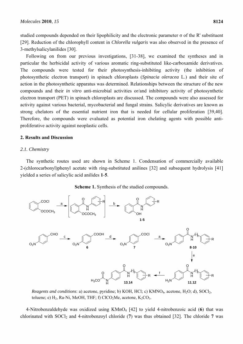
The synthetic routes used are shown in Scheme 1. Condensation of commercially available
2-(chlorocarbonyl)phenyl acetate with ring-substituted anilines [32] and subsequent hydrolysis [41]
yielded a series of salicylic acid anilides 1-5.
Scheme 1. Synthesis of the studied compounds.
COCl
OCOCH3
a b
OCOCH3
NH
OR
OH
NH
OR
1-5
CHOc
O2N
COOH
O2N
dCOCl
O2N
a NH
O
R
8-10O2N
( )n
e
NH
O
R
11,12H2N
( )nfN
H
O
R
13,14NH
( )n
H3CO
O
76
Reagents and conditions: a) acetone, pyridine; b) KOH, HCl; c) KMNO4, acetone, H2O; d), SOCl2, toluene; e) H2, Ra-Ni, MeOH, THF; f) ClCO2Me, acetone, K2CO3.
4-Nitrobenzaldehyde was oxidized using KMnO4 [42] to yield 4-nitrobenzoic acid (6) that was
chlorinated with SOCl2 and 4-nitrobenzoyl chloride (7) was thus obtained [32]. The chloride 7 was

Molecules 2010, 15
8125
condensed with ring-substituted amines, as described previously [32], to afford amides 8-10.
Nitrobenzamides 9 and 10 were hydrogenated over activated Raney Ni (Ra-Ni) [43,44] to give
aminobenzamides 11 and 12. Methylphenylcarbamates 13 and 14 were generated by means of the
reaction of aminobenzamides 11 and 12 with methyl chloroformiate [45].
2.2. Lipophilicity
Many low molecular weight drugs cross biological membranes through passive transport, which
strongly depends on their lipophilicity. Lipophilicity is a property that has a major effect on
absorption, distribution, metabolism, excretion, and toxicity (ADME/Tox) properties as well as
pharmacological activity. Lipophilicity has been studied and applied as an important drug property for
decades [46].
Hydrophobicities (log P/Clog P) of the compounds 1-5 and 8-14 were calculated using two
commercially available programs (ChemDraw Ultra 10.0 and ACD/LogP) and also measured by
means of the RP-HPLC determination of capacity factors k with subsequent calculation of log k. The
procedure was performed under isocratic conditions with methanol as an organic modifier in the
mobile phase using an end-capped non-polar C18 stationary RP column. The results are shown in Table
1 and illustrated in Figure 1.
The results obtained with all the compounds show that the experimentally-determined
lipophilicities (log k) of compounds 1-5 and 8-11, 13, 14 are lower than those indicated by the
calculated
log P/Clog P, as shown in Figure 1. The results indicate that experimentally-determined log k values
correlate relatively poorly with the calculated values (see Table 1 or Figure 1), which is probably
caused by intramolecular interactions of these highly functionalized molecules.
As expected, aminobenzamide 11 showed the lowest lipophilicity (log k), while compounds 5 and
10 exhibited the highest log k values. Generally, nitro compounds 8-10 showed relatively high
lipophilicity in comparison with amino derivatives, e.g. 11 < 13 < 9. Within the series of ring-
substituted salicylanilides 1-5, the determined log k values corresponded to the expected lipophilicity
increase 1 (2-CH3) < 3 (2-OCH3) < 2 (2,6-CH3) < 4 (2-OC2H5) < 5 (2-OC3H7). This dependence is
approximately linear and thus, it can be assumed that log k values specify lipophilicity within the
individual series of the studied compounds more precisely than calculated log P/Clog P data.
2.3. Biological activities
The compounds under investigation could be divided into three groups based on their chemical
structure: Group 1 included salicylanilides 1-5; Group 2 contained nitro derivatives 8-10; and Group 3
was composed of amino derivatives or carbamates 11-14. The compounds showed a wide range of
biological activities and some interesting structure-activity relationships were observed. All the results
are shown in Table 2 (compound 12 did not show any biological activity in any of the assays, therefore
it is not included in Table 2).

Molecules 2010, 15
8126
Table 1. Comparison of the calculated lipophilicities (log P/Clog P) with the determined
log k values, Hammett's parameter (σ) and bulk parameter (MR, reflecting bulkiness).
NH
R2O
R1
Comp. R1 R2 log k log P/Clog P ChemOffice
log P ACD/LogP
σ [47] MR [47]
1 2-OH H3C
0.6661 2.94 / 3.1212 3.73 ± 0.37 0.10 4.7
2 2-OH
H3C
CH3 0.6807 3.42 / 2.9702 4.19 ± 0.38 0.20 9.4
3 2-OH H3CO
0.6770 2.32 / 2.7576 3.17 ± 0.39 0.00 6.5
4 2-OH C2H5O
0.7515 2.66 / 3.2866 3.70 ± 0.39 0.02 11.3
5 2-OH C3H7O
0.8401 3.15 / 3.8156 4.23 ± 0.39 NF 15.9
8 4-NO2 Cl
0.6782 3.15 / 2.7782 3.19 ± 0.34 0.67 4.7
9 4-NO2 Cl
0.6966 2.82 / 3.7170 3.19 ± 0.35 0.67 4.7
10 4-NO2 Cl
CF3 0.8269 4.13 / 3.7726 4.94 ± 0.43 1.10 8.7
11 4-NH2 Cl
0.5886 2.66 / 2.6350 2.47 ± 0.39 0.67 4.7
12 4-NH2 Cl
CF3 ND 3.52 / 3.0299 4.12 ± 0.42 1.10 8.7
13 4-CH3OCONH Cl
0.6345 2.96 / 3.2510 3.21 ± 0.43 0.67 4.7
14 4-CH3OCONH Cl
CF3 0.7782 3.81 / 3.6459 4.86 ± 0.50 1.10 8.7
NF = not found in literature; ND = not determined/analyzed.
2.3.1. Inhibition of photosynthetic electron transport (PET) in spinach chloroplasts
The majority of the studied compounds inhibited PET in spinach chloroplasts, as shown in Table 2.
Four compounds showed high inhibitory IC50 values: 1.0 µmol/L (14), 1.6 µmol/L (13), 1.6 µmol/L (3)
and 2.7 µmol/L (1), which was comparable with the standard DCMU (IC50 = 1.9 µmol/L). The activity
of the rest of the studied compounds was moderate or low relative to the standard. PET inhibition by 8
or 9 could not be determined due to precipitation of the compounds during the experiments. With
respect to these small but closed specifically substituted groups of compounds some structure-activity
relationships (SAR) can be proposed.

Molecules 2010, 15
8127
Figure 1. Comparison of the log P data calculated using the two programs with the
experimentally found log k values. The compounds are arranged in the ascending manner
according to the experimental log k values.
0.0
1.0
2.0
3.0
4.0
5.0
6.0
11 13 1 3 8 2 9 4 14 10 5Compounds
Lip
op
hil
icit
y
log k log P [ChemOffice] Clog P [ChemOffice] log P [ACD/LogP]
Within Group 1 (salicylanilides; compounds 1-5), the highest PET-inhibiting activity was shown by
compounds 3 (2-OMe) and 1 (2-Me). Within the series of 2-alkoxy substituted compounds (3-5), their
activity decreased with increasing lipophilicity (log k) and higher substituent bulkiness (substituent
bulkiness (MR) and Hammett's parameter (σ) are described in Table 1 [47]). It is probable that σ
(especially the electron-donating effect) is also important for explaining biological activity and
searching for structure-activity relationships within a series of compounds [47]. The inhibitory activity
of compound 2 was two orders lower (IC50 = 331.4 μmol/L) than the activity of compounds 1 and 3,
with comparable log k values (0.6661 and 0.6770, respectively). Therefore, when compounds 1 and 2
are considered, similar statements can be made: higher lipophilicity and substituent bulkiness
decreases the inhibition of PET.
Generally, Group 2 (4-nitrobenzencarboxamides) showed only slight PET inhibition caused by the
low solubility of compounds 8 and 9 in the testing medium. Compound 10 alone showed medium PET
inhibiting activity.
Group 3 (4-aminobenzencarboxamide derivatives) showed very interesting PET activity.
Compound 11 with a primary amino moiety possessed very low activity, while substitution of
hydrogen in the 4-NH2 group of (11) by methyl acetate generating compounds 13 and 14
(carbamoylphenylcarbamates), resulted in a strong increase in the inhibitory activity of PET. This
finding underlined the importance of the amide group, which can interact with amino acid residues or
peptide bonds of proteins situated in photosystems, which can result in PET inhibition. It can be stated
that an increase in lipophilicity positively influences PET-inhibiting activity, contrary to Group 1
compounds. Thus, it can be concluded that PET-inhibiting activity is increased by the electron-
withdrawing effect and bulkiness of substituents in the anilide part of the molecule.
Molecules 2010, 15
8128
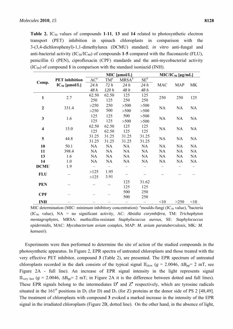
Table 2. IC50 values of compounds 1-11, 13 and 14 related to photosynthetic electron
transport (PET) inhibition in spinach chloroplasts in comparison with the
3-(3,4-dichlorophenyl)-1,1-dimethylurea (DCMU) standard; in vitro anti-fungal and
anti-bacterial activity (IC50/IC90) of compounds 1-5 compared with the fluconazole (FLU),
penicillin G (PEN), ciprofloxacin (CPF) standards and the anti-mycobacterial activity
(IC90) of compound 1 in comparison with the standard isoniazid (INH).
Comp. PET inhibition IC50 [μmol/L]
MIC [µmol/L] MIC/IC90 [µg/mL] ACa TMa MRSAb SEb
MAC MAP MK 24 h 48 h
72 h 120 h
24 h 48 h
24 h 48 h
1 2.7 62.50 250
62.50 125
125 250
125 250
250 250 125
2 331.4 250 250
250 500
500 500
500 500
NA NA NA
3 1.6 125 125
125 125
500 500
500 500
NA NA NA
4 15.0 62.50 125
62.50 62.50
125 125
125 125
NA NA NA
5 44.8 31.25 31.25
31.25 31.25
31.25 31.25
31.25 31.25
NA NA NA
10 50.1 NA NA NA NA NA NA NA 11 398.4 NA NA NA NA NA NA NA 13 1.6 NA NA NA NA NA NA NA 14 1.0 NA NA NA NA NA NA NA
DCMU 1.9 – – – – – – –
FLU – 125 125
1.95 3.91
– – – – –
PEN – – – 125 125
31.62 125
CPF – – – 500 500
250 250
INH – – – – – <10 >250 <10 MIC determination (MIC: minimum inhibitory concentration): amoulds-fungi (IC50 value), bbacteria (IC90 value), NA = no significant activity, AC: Absidia corymbifera, TM: Trichophyton mentagrophytes, MRSA: methicillin-resistant Staphylococcus aureus, SE: Staphylococcus epidermidis, MAC: Mycobacterium avium complex, MAP: M. avium paratuberculosis, MK: M. kansasii).
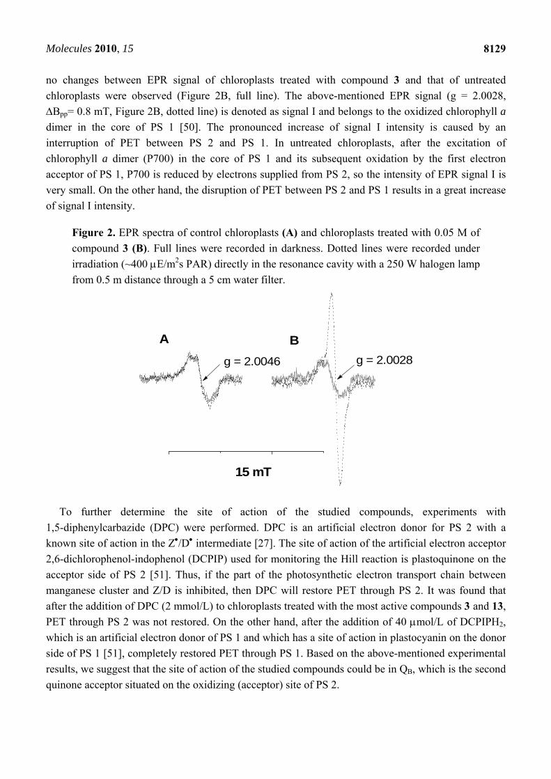
Experiments were then performed to determine the site of action of the studied compounds in the
photosynthetic apparatus. In Figure 2, EPR spectra of untreated chloroplasts and those treated with the
very effective PET inhibitor, compound 3 (Table 2), are presented. The EPR spectrum of untreated
chloroplasts recorded in the dark consists of the typical signal IIslow (g = 2.0046, Bpp= 2 mT, see
Figure 2A - full line). An increase of EPR signal intensity in the light represents signal
IIvery fast (g = 2.0046, Bpp= 2 mT; in Figure 2A it is the difference between dotted and full lines).
These EPR signals belong to the intermediates D and Z respectively, which are tyrosine radicals
situated in the 161st positions in D2 (for D) and D1 (for Z) proteins at the donor side of PS 2 [48,49].
The treatment of chloroplasts with compound 3 evoked a marked increase in the intensity of the EPR
signal in the irradiated chloroplasts (Figure 2B, dotted line). On the other hand, in the absence of light,
Molecules 2010, 15
8129
no changes between EPR signal of chloroplasts treated with compound 3 and that of untreated
chloroplasts were observed (Figure 2B, full line). The above-mentioned EPR signal (g = 2.0028,
Bpp= 0.8 mT, Figure 2B, dotted line) is denoted as signal I and belongs to the oxidized chlorophyll a
dimer in the core of PS 1 [50]. The pronounced increase of signal I intensity is caused by an
interruption of PET between PS 2 and PS 1. In untreated chloroplasts, after the excitation of
chlorophyll a dimer (P700) in the core of PS 1 and its subsequent oxidation by the first electron
acceptor of PS 1, P700 is reduced by electrons supplied from PS 2, so the intensity of EPR signal I is
very small. On the other hand, the disruption of PET between PS 2 and PS 1 results in a great increase
of signal I intensity.
Figure 2. EPR spectra of control chloroplasts (A) and chloroplasts treated with 0.05 M of
compound 3 (B). Full lines were recorded in darkness. Dotted lines were recorded under
irradiation (~400 E/m2s PAR) directly in the resonance cavity with a 250 W halogen lamp
from 0.5 m distance through a 5 cm water filter.
A B
15 mT
g = 2.0028g = 2.0046
To further determine the site of action of the studied compounds, experiments with
1,5-diphenylcarbazide (DPC) were performed. DPC is an artificial electron donor for PS 2 with a
known site of action in the Z/D intermediate [27]. The site of action of the artificial electron acceptor
2,6-dichlorophenol-indophenol (DCPIP) used for monitoring the Hill reaction is plastoquinone on the
acceptor side of PS 2 [51]. Thus, if the part of the photosynthetic electron transport chain between
manganese cluster and Z/D is inhibited, then DPC will restore PET through PS 2. It was found that
after the addition of DPC (2 mmol/L) to chloroplasts treated with the most active compounds 3 and 13,
PET through PS 2 was not restored. On the other hand, after the addition of 40 mol/L of DCPIPH2,
which is an artificial electron donor of PS 1 and which has a site of action in plastocyanin on the donor
side of PS 1 [51], completely restored PET through PS 1. Based on the above-mentioned experimental
results, we suggest that the site of action of the studied compounds could be in QB, which is the second
quinone acceptor situated on the oxidizing (acceptor) site of PS 2.
Molecules 2010, 15
8130
2.3.2. In vitro anti-fungal and anti-bacterial susceptibility testing
All discussed above, compounds were tested for their in vitro anti-fungal and anti-bacterial activity,
although only the salicylanilides 1-5 (Group 1), showed some broad spectrum activity. The rest of the
compounds did not show any significant activity, potentially due to low solubility in the testing
medium and their precipitation during the incubation period. The results are shown in Table 2. Among
a number of fungi strains, Absidia corymbifera, as a common human pathogen causing pulmonary,
rhinocerebral, disseminated, CNS or cutaneous infections and Trichophyton mentagrophytes as an
example of a dermatophyte carrying a variety of cutaneous infections were selected.
All the compounds in Group 1 showed similar structure-activity relationships with respect to anti-
fungal and anti-bacterial activity. Compounds 2 (2,6-CH3), 3 (2-OCH3) and 1 (2-CH3) exhibited low
biological activity. Compounds with the longer alkoxy chain 4 (2-OC2H5) and 5 (2-OC3H7) showed
higher anti-fungal and anti-bacterial activity. With respect to these specific alkyl/alkoxy substituted
compounds it can be assumed that a lipophilicity increase and a bulkier substituent seem to be
important factors for increasing both of these previously described activities. 2-Hydroxy-N-(2-
propoxyphenyl)benzamide (5) showed similar chemotherapeutic activity against all tested fungal and
bacterial strains. Notably, compound 5 demonstrated much higher activity against Absidia corymbifera
than the standard fluconazole and also higher activity against methicillin resistant Staphylococcus
aureus, and Staphylococcus epidermidis than the common clinically used antibiotics penicillin G
and ciprofloxacin.
2.3.3. In vitro antimycobacterial evaluation
Although all the compounds discussed above were evaluated for their in vitro anti-mycobacterial
activity against atypical mycobacterial strains, only compound 1 showed moderate activity, therefore
no thorough structure-activity relationships could be established. According to the results presented in
Table 2, it can be concluded that compound 1 exhibited activity against M. avium paratuberculosis and
that its efficacy was comparable with that of the standard, isoniazid.
2.3.4. In vitro anti-proliferative activity
Carbonyl-containing moieties are potential iron chelating agents, with many demonstrating anti-
proliferative activity due to their ability to bind cellular iron, which is required for proliferation [52].
Therefore, the anti-proliferative activity of compounds 1-5 was examined against the human SK-N-
MC neuroepithelioma cell line to determine if there were any undesired cytotoxic side-effects. The
SK-N-MC cell line was chosen as the effect of iron chelators on the proliferation of these cells has
been extensively examined [53,54]. The anti-proliferative activity of the evaluated compounds was
assessed in comparison to the well-known clinically used iron chelator, desferrioxamine (DFO), and
the highly cytotoxic chelator, di-2-pyridylketone 4,4-dimethyl-3-thiosemicarbazone (Dp44mT) [52].
All tested compounds 1-5 demonstrated poor anti-proliferative effects against the SK-N-MC cell
line, with IC50 values >6.25 µmol/L. As expected from our previous studies [53,54], the iron chelator,
DFO, demonstrated poor anti-cancer activity, with an IC50 value of 17.07 ± 3.77 µmol/L, while the
cytotoxic chelator, Dp44mT (IC50 = 0.01 ± 0.01 µmol/L), showed potent anti-proliferative effects.
Molecules 2010, 15
8131
These results suggest that the poor anti-proliferative activity of the anilides 1-5 will lead to limited
cytotoxicity if ever used as anti-fungal, anti-bacterial or anti-mycobacterial agents.
3. Experimental
3.1. General
All reagents were purchased from Aldrich. Kieselgel 60, 0.040-0.063 mm (Merck, Darmstadt,
Germany) was used for column chromatography. TLC experiments were performed on alumina-
backed silica gel 40 F254 plates (Merck, Darmstadt, Germany). The plates were illuminated under UV
(254 nm) and evaluated in iodine vapour. The melting points were determined on Boetius PHMK 05
(VEB Kombinat Nagema, Radebeul, Germany) and are uncorrected. The purity of the final compounds
was checked by a HPLC separation module (Waters Alliance 2695 XE, Waters Corp., Milford, MA,
USA). The detection wavelength of 210 nm was chosen. The peaks in the chromatogram of the solvent
(blank) were deducted from the peaks in the chromatogram of the sample solution. The purity of
individual compounds was determined from the area peaks in the chromatogram of the sample
solution. UV spectra (λ, nm) were determined on a Waters Photodiode Array Detector 2996 (Waters
Corp., Milford, MA, USA) in ca. 6 × 10⁻⁴ M methanolic solution and log ε (the logarithm of molar
absorption coefficient ε) was calculated for the absolute maximum λmax of individual target
compounds. Infrared (IR) spectra were recorded on a Smart MIRacle™ ATR ZnSe for Nicolet™
Impact 410 FT-IR Spectrometer (Thermo Scientific, USA). The spectra were obtained by
accumulation of 256 scans with 2 cm-1 resolution in the region of 4000-600 cm-1. All 1H and 13C NMR
spectra were recorded on a Bruker Avance-500 FT-NMR spectrometer (500 MHz for 1H and 125 MHz
for 13C, Bruker Comp., Karlsruhe, Germany). Chemicals shifts are reported in ppm () using internal
Si(CH3)4 as the reference, with diffuse, easily exchangeable signals being omitted. Mass spectra were
measured using a LTQ Orbitrap Hybrid Mass Spectrometer (Thermo Electron Corporation, USA) with
direct injection into an APCI source (400 °C) in the positive mode.
3.2. Synthesis
3.2.1. General procedure for synthesis of carboxamide derivatives 1-5
2-(Chlorocarbonyl)phenyl acetate (0.014 mol) dissolved in dry acetone (50 mL) was added drop-
wise to a stirred solution of the corresponding substituted aniline (0.02 mmol) in dry pyridine (50 mL)
kept at room temperature. After the addition was completed, stirring was continued for another 1 h.
The reaction mixture was then poured into cold water (100 mL). The mixture was extracted with Et2O,
dried over anhydrous MgSO4, filtered and the solvent removed in vacuo. The crude
acetylsalicylanilide was dissolved in a 1 M solution of KOH stirred at 50 °C until clarification. The
mixture was then neutralized using 1 M HCl. The crude amide was collected and recrystallized from
aqueous ethanol and dried.
2-Hydroxy-N-(2-methylphenyl)benzamide (1). Yield 35%; m.p. 153-155 °C (m.p. 145-147°C [55]);
HPLC purity 99.41%; UV (nm), λmax/log ε: 271.1/3.42; IR (Zn/Se ATR, cm-1): 3324 (N-H), 1611
Molecules 2010, 15
8132
(C=O), 3741 (O-H), 1542 (C=C); 1H-NMR (DMSO-d6), δ: 12.07 (bs, 1H, OH), 10.31 (bs, 1H, NH),
8.04 (dd, 1H, J = 7.9 Hz, J = 1.8 Hz, Ar), 7.80 (d, 1H, J = 7.9 Hz, Ar), 7.50-7.40 (m, 1H, Ar), 7.28 (d,
1H, J = 7.4 Hz, Ar), 7.23 (ddd, 1H, J = 7.6 Hz, J = 7.6 Hz, J = 1.5 Hz, Ar), 7.13 (ddd, 1H, J = 7.6 Hz,
J = 7.6 Hz, J = 1.5 Hz, Ar), 7.06-6.94 (m, 2H, Ar), 2.28 (s, 3H, CH3); 13C-NMR (DMSO-d6), δ:
165.62, 158.08, 136.16, 133.65, 130.66, 130.30, 129.51, 126.25, 125.14, 123.99, 119.26, 117.20,
117.15, 17.71; HR-MS: for C14H14O2N [M+H]+ calculated 228.098 m/z, found 228.0892 m/z.
N-(2,6-Dimethylphenyl)-2-hydroxybenzamide (2). Yield 37%; m.p. 137-140 °C; HPLC purity 99.73%;
UV (nm), λmax/log ε: 237.9/3.45; IR (Zn/Se ATR, cm-1): 3278 (N-H), 1602 (C=O), 1538 (C=C); 1H-NMR (DMSO-d6), δ: 12.34 (bs, 1H, OH), 10.07 (s, 1H, NH), 8.05 (dd, 1H, J = 8.2 Hz, J = 1.8 Hz,
Ar), 7.51-7.43 (m, 1H, Ar), 7.22-7.10 (m, 3H, Ar), 7.02-6.92 (m, 2H, Ar), 2.19 (s, 6H, CH3); 13C-NMR
(DMSO-d6), δ: 167.68, 160.06, 135.48, 134.28, 133.95, 128.14, 127.79, 127.00, 118.81, 117.45,
115.38, 17.97; HR-MS: for C15H16O2N [M+H]+ calculated 242.118 m/z, found 242.1084 m/z.
2-Hydroxy-N-(2-methoxyphenyl)benzamide (3). Yield 39%; m.p. 112-115 °C (m.p. 115-117 °C [55]);
HPLC purity 99.92%; UV (nm), λmax/log ε: 298.4/3.49; IR (Zn/Se ATR, cm-1): 3288 (N-H), 1600
(C=O), 1228 (C-O), 1547 (C=C); 1H-NMR (DMSO-d6), δ: 11.70 (bs, 1H, OH), 10.81 (bs, 1H, NH),
8.43-8.36 (m, 1H, Ar), 8.03 (dd, 1H, J = 7.9 Hz, J = 1.8 Hz, Ar), 7.45-8.38 (m, 1H, Ar), 7.12-7.07 (m,
2H, Ar), 7.02 (dd, 1H, J = 8.2 Hz, J = 0.9 Hz, Ar), 7.01-6.93 (m, 2H, Ar), 3.89 (s, 1H, CH3); 13C-NMR
(DMSO-d6), δ: 163.43, 156.28, 148.69, 133.32, 130.67, 127.84, 123.89, 120.57, 120.30, 119.59,
118.74, 116.90, 110.96, 55.99; HR-MS: for C14H14O3N [M+H]+ calculated 244.098 m/z, found
244.0980 m/z.
N-(2-Ethoxyphenyl)-2-hydroxybenzamide (4). Yield 35%; m.p. 105-107 °C; HPLC purity 99.94%; UV
(nm), λmax/log ε: 299.6/3.48; IR (Zn/Se ATR, cm-1): 3418 (N-H), 1604 (C=O), 3736 (O-H), 1216
(C-O); 1H-NMR (DMSO-d6), δ: 11.54 (bs, 1H, OH), 10.93 (bs, 1H, NH), 8.50-8.42 (m, 1H, Ar), 8.04
(dd, 1H, J = 7.9 Hz, J = 1.8 Hz, Ar), 7.45-8.39 (m, 1H, Ar), 7.10-7.02 (m, 3H, Ar), 7.01-6.97 (m, 1H,
Ar), 6.97-6.92 (m, 1H, Ar), 4.12 (q, 2H, J = 6.9 Hz, CH2), 1.44 (t, 3H, J = 6.9 Hz, CH3); 13C-NMR
(DMSO-d6), δ: 163.01, 156.02, 147.65, 133.25, 130.87, 128.32, 123.57, 120.54, 119.70, 119.65,
118.98, 116.85, 111.79, 64.08, 14.75; HR-MS: for C15H16O3N [M+H]+ calculated 258.118 m/z, found
258.0889 m/z.
2-Hydroxy-N-(2-propoxyphenyl)benzamide (5). Yield 31%; m.p. 92-95 °C; HPLC purity 99.98%; UV
(nm), λmax/log ε: 299.6/3.39; IR (Zn/Se ATR, cm-1): 3281 (N-H), 1598 (C=O), 1544 (C=C), 1241
(C-O); 1H-NMR (DMSO-d6), δ: 11.49 (bs, 1H, OH), 10.86 (bs, 1H, NH), 8.49-8.44 (m, 1H, Ar), 8.04
(dd, 1H, J = 7.9 Hz, J = 1.8 Hz, Ar), 7.44-7.37 (m, 1H, Ar), 7.11-7.02 (m, 3H, Ar), 7.02-6.96 (m, 1H,
Ar), 6.96-6.92 (m, 1H, Ar), 4.02 (t, 2H, J = 6.5 Hz, CH2CH2), 1.84 (q, 2H, J = 7.0 Hz CH2CH2), 1.04
(t, 3H, J = 7.3 Hz, CH3); 13C-NMR (DMSO-d6), δ: 163.07, 156.06, 147.79, 133.25, 130.88, 128.17,
123.64, 120.41, 119.87, 119.64, 118.95, 116.86, 111.60, 69.82, 22.16, 10.51; HR-MS: for C16H18O3N
[M+H]+ calculated 272.128 m/z, found 272.1280 m/z.
Molecules 2010, 15
8133
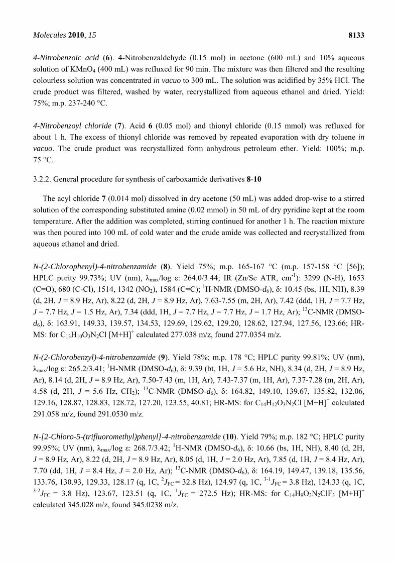
4-Nitrobenzoic acid (6). 4-Nitrobenzaldehyde (0.15 mol) in acetone (600 mL) and 10% aqueous
solution of KMnO4 (400 mL) was refluxed for 90 min. The mixture was then filtered and the resulting
colourless solution was concentrated in vacuo to 300 mL. The solution was acidified by 35% HCl. The
crude product was filtered, washed by water, recrystallized from aqueous ethanol and dried. Yield:
75%; m.p. 237-240 °C.
4-Nitrobenzoyl chloride (7). Acid 6 (0.05 mol) and thionyl chloride (0.15 mmol) was refluxed for
about 1 h. The excess of thionyl chloride was removed by repeated evaporation with dry toluene in
vacuo. The crude product was recrystallized form anhydrous petroleum ether. Yield: 100%; m.p.
75 °C.
3.2.2. General procedure for synthesis of carboxamide derivatives 8-10
The acyl chloride 7 (0.014 mol) dissolved in dry acetone (50 mL) was added drop-wise to a stirred
solution of the corresponding substituted amine (0.02 mmol) in 50 mL of dry pyridine kept at the room
temperature. After the addition was completed, stirring continued for another 1 h. The reaction mixture
was then poured into 100 mL of cold water and the crude amide was collected and recrystallized from
aqueous ethanol and dried.
N-(2-Chlorophenyl)-4-nitrobenzamide (8). Yield 75%; m.p. 165-167 °C (m.p. 157-158 °C [56]);
HPLC purity 99.73%; UV (nm), λmax/log ε: 264.0/3.44; IR (Zn/Se ATR, cm-1): 3299 (N-H), 1653
(C=O), 680 (C-Cl), 1514, 1342 (NO2), 1584 (C=C); 1H-NMR (DMSO-d6), δ: 10.45 (bs, 1H, NH), 8.39
(d, 2H, J = 8.9 Hz, Ar), 8.22 (d, 2H, J = 8.9 Hz, Ar), 7.63-7.55 (m, 2H, Ar), 7.42 (ddd, 1H, J = 7.7 Hz,
J = 7.7 Hz, J = 1.5 Hz, Ar), 7.34 (ddd, 1H, J = 7.7 Hz, J = 7.7 Hz, J = 1.7 Hz, Ar); 13C-NMR (DMSO-
d6), δ: 163.91, 149.33, 139.57, 134.53, 129.69, 129.62, 129.20, 128.62, 127.94, 127.56, 123.66; HR-
MS: for C13H10O3N2Cl [M+H]+ calculated 277.038 m/z, found 277.0354 m/z.
N-(2-Chlorobenzyl)-4-nitrobenzamide (9). Yield 78%; m.p. 178 °C; HPLC purity 99.81%; UV (nm),
λmax/log ε: 265.2/3.41; 1H-NMR (DMSO-d6), δ: 9.39 (bt, 1H, J = 5.6 Hz, NH), 8.34 (d, 2H, J = 8.9 Hz,
Ar), 8.14 (d, 2H, J = 8.9 Hz, Ar), 7.50-7.43 (m, 1H, Ar), 7.43-7.37 (m, 1H, Ar), 7.37-7.28 (m, 2H, Ar),
4.58 (d, 2H, J = 5.6 Hz, CH2); 13C-NMR (DMSO-d6), δ: 164.82, 149.10, 139.67, 135.82, 132.06,
129.16, 128.87, 128.83, 128.72, 127.20, 123.55, 40.81; HR-MS: for C14H12O3N2Cl [M+H]+ calculated
291.058 m/z, found 291.0530 m/z.
N-[2-Chloro-5-(trifluoromethyl)phenyl]-4-nitrobenzamide (10). Yield 79%; m.p. 182 °C; HPLC purity
99.95%; UV (nm), λmax/log ε: 268.7/3.42; 1H-NMR (DMSO-d6), δ: 10.66 (bs, 1H, NH), 8.40 (d, 2H,
J = 8.9 Hz, Ar), 8.22 (d, 2H, J = 8.9 Hz, Ar), 8.05 (d, 1H, J = 2.0 Hz, Ar), 7.85 (d, 1H, J = 8.4 Hz, Ar),
7.70 (dd, 1H, J = 8.4 Hz, J = 2.0 Hz, Ar); 13C-NMR (DMSO-d6), δ: 164.19, 149.47, 139.18, 135.56,
133.76, 130.93, 129.33, 128.17 (q, 1C, 2JFC = 32.8 Hz), 124.97 (q, 1C, 3-1JFC = 3.8 Hz), 124.33 (q, 1C, 3-2JFC = 3.8 Hz), 123.67, 123.51 (q, 1C, 1JFC = 272.5 Hz); HR-MS: for C14H9O3N2ClF3 [M+H]+
calculated 345.028 m/z, found 345.0238 m/z.
Molecules 2010, 15
8134
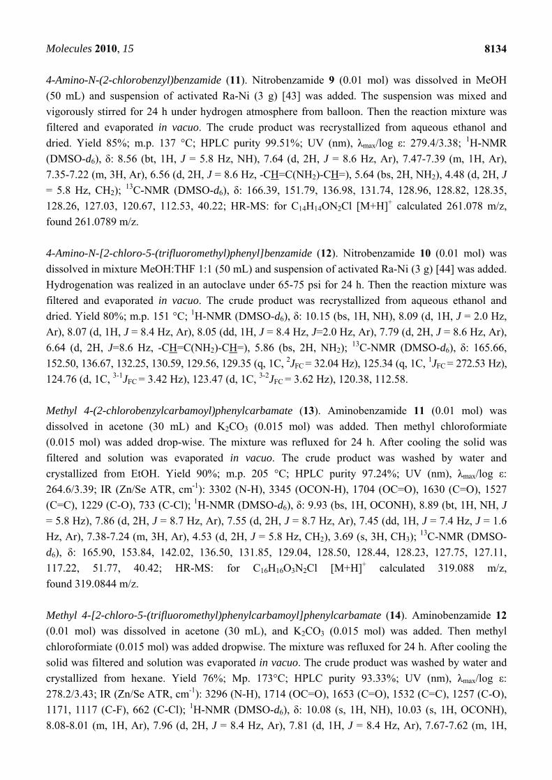
4-Amino-N-(2-chlorobenzyl)benzamide (11). Nitrobenzamide 9 (0.01 mol) was dissolved in MeOH
(50 mL) and suspension of activated Ra-Ni (3 g) [43] was added. The suspension was mixed and
vigorously stirred for 24 h under hydrogen atmosphere from balloon. Then the reaction mixture was
filtered and evaporated in vacuo. The crude product was recrystallized from aqueous ethanol and
dried. Yield 85%; m.p. 137 °C; HPLC purity 99.51%; UV (nm), λmax/log ε: 279.4/3.38; 1H-NMR
(DMSO-d6), δ: 8.56 (bt, 1H, J = 5.8 Hz, NH), 7.64 (d, 2H, J = 8.6 Hz, Ar), 7.47-7.39 (m, 1H, Ar),
7.35-7.22 (m, 3H, Ar), 6.56 (d, 2H, J = 8.6 Hz, -CH=C(NH2)-CH=), 5.64 (bs, 2H, NH2), 4.48 (d, 2H, J
= 5.8 Hz, CH2); 13C-NMR (DMSO-d6), δ: 166.39, 151.79, 136.98, 131.74, 128.96, 128.82, 128.35,
128.26, 127.03, 120.67, 112.53, 40.22; HR-MS: for C14H14ON2Cl [M+H]+ calculated 261.078 m/z,
found 261.0789 m/z.
4-Amino-N-[2-chloro-5-(trifluoromethyl)phenyl]benzamide (12). Nitrobenzamide 10 (0.01 mol) was
dissolved in mixture MeOH:THF 1:1 (50 mL) and suspension of activated Ra-Ni (3 g) [44] was added.
Hydrogenation was realized in an autoclave under 65-75 psi for 24 h. Then the reaction mixture was
filtered and evaporated in vacuo. The crude product was recrystallized from aqueous ethanol and
dried. Yield 80%; m.p. 151 °C; 1H-NMR (DMSO-d6), δ: 10.15 (bs, 1H, NH), 8.09 (d, 1H, J = 2.0 Hz,
Ar), 8.07 (d, 1H, J = 8.4 Hz, Ar), 8.05 (dd, 1H, J = 8.4 Hz, J=2.0 Hz, Ar), 7.79 (d, 2H, J = 8.6 Hz, Ar),
6.64 (d, 2H, J=8.6 Hz, -CH=C(NH2)-CH=), 5.86 (bs, 2H, NH2); 13C-NMR (DMSO-d6), δ: 165.66,
152.50, 136.67, 132.25, 130.59, 129.56, 129.35 (q, 1C, 2JFC = 32.04 Hz), 125.34 (q, 1C, 1JFC = 272.53 Hz),
124.76 (d, 1C, 3-1JFC = 3.42 Hz), 123.47 (d, 1C, 3-2JFC = 3.62 Hz), 120.38, 112.58.
Methyl 4-(2-chlorobenzylcarbamoyl)phenylcarbamate (13). Aminobenzamide 11 (0.01 mol) was
dissolved in acetone (30 mL) and K2CO3 (0.015 mol) was added. Then methyl chloroformiate
(0.015 mol) was added drop-wise. The mixture was refluxed for 24 h. After cooling the solid was
filtered and solution was evaporated in vacuo. The crude product was washed by water and
crystallized from EtOH. Yield 90%; m.p. 205 °C; HPLC purity 97.24%; UV (nm), λmax/log ε:
264.6/3.39; IR (Zn/Se ATR, cm-1): 3302 (N-H), 3345 (OCON-H), 1704 (OC=O), 1630 (C=O), 1527
(C=C), 1229 (C-O), 733 (C-Cl); 1H-NMR (DMSO-d6), δ: 9.93 (bs, 1H, OCONH), 8.89 (bt, 1H, NH, J
= 5.8 Hz), 7.86 (d, 2H, J = 8.7 Hz, Ar), 7.55 (d, 2H, J = 8.7 Hz, Ar), 7.45 (dd, 1H, J = 7.4 Hz, J = 1.6
Hz, Ar), 7.38-7.24 (m, 3H, Ar), 4.53 (d, 2H, J = 5.8 Hz, CH2), 3.69 (s, 3H, CH3); 13C-NMR (DMSO-
d6), δ: 165.90, 153.84, 142.02, 136.50, 131.85, 129.04, 128.50, 128.44, 128.23, 127.75, 127.11,
117.22, 51.77, 40.42; HR-MS: for C16H16O3N2Cl [M+H]+ calculated 319.088 m/z,
found 319.0844 m/z.
Methyl 4-[2-chloro-5-(trifluoromethyl)phenylcarbamoyl]phenylcarbamate (14). Aminobenzamide 12
(0.01 mol) was dissolved in acetone (30 mL), and K2CO3 (0.015 mol) was added. Then methyl
chloroformiate (0.015 mol) was added dropwise. The mixture was refluxed for 24 h. After cooling the
solid was filtered and solution was evaporated in vacuo. The crude product was washed by water and
crystallized from hexane. Yield 76%; Mp. 173°C; HPLC purity 93.33%; UV (nm), λmax/log ε:
278.2/3.43; IR (Zn/Se ATR, cm-1): 3296 (N-H), 1714 (OC=O), 1653 (C=O), 1532 (C=C), 1257 (C-O),
1171, 1117 (C-F), 662 (C-Cl); 1H-NMR (DMSO-d6), δ: 10.08 (s, 1H, NH), 10.03 (s, 1H, OCONH),
8.08-8.01 (m, 1H, Ar), 7.96 (d, 2H, J = 8.4 Hz, Ar), 7.81 (d, 1H, J = 8.4 Hz, Ar), 7.67-7.62 (m, 1H,

Molecules 2010, 15
8135
Ar), 7.62 (d, 2H, J = 8.8 Hz, Ar), 3.71 (s, 3H, CH3); 13C-NMR (DMSO-d6), δ: 164.93, 153.84, 142.78,
136.18, 133.14, 130.76, 128.87, 128.04 (q, 1C, 2JFC = 32.6 Hz), 126.99, 124.27 (q, 1C, 3-1JFC = 3.8 Hz),
123.63 (q, 1C, 1JFC = 272.48 Hz), 123.49 (q, 1C, 3-2JFC = 3.8 Hz), 117.30, 51.85; HR-MS: for
C16H13O3N2ClF3 [M+H]+ calculated 373.738 m/z, found 373.0562 m/z.
3.3. Lipophilicity determination by HPLC (capacity factor k/calculated log k)
A Waters Alliance 2695 XE HPLC separation module and a Waters Photodiode Array Detector
2996 (Waters Corp., Milford, MA, USA) were used. A Symmetry® C18 5 μm, 4.6 250 mm, Part No.
WAT054275 (Waters Corp., Milford, MA, USA) chromatographic column was used. The HPLC
separation process was monitored by Empower™ 2 Chromatography Data Software, Waters 2009
(Waters Corp., Milford, MA, USA). A mixture of MeOH p.a. (70%) and H2O-HPLC – Mili-Q Grade
(30%) was used as a mobile phase. The total flow of the column was 1.0 mL/min, injection volume
30 μL, column temperature 30 °C and sample temperature 10°C. The detection wavelength of 210 nm
was chosen. The KI methanolic solution was used for the dead time (tD) determination. Retention times
(tR) were measured in minutes. The capacity factors k were calculated using the Empower™ 2
Chromatography Data Software according to formula k = (tR - tD)/tD, where tR is the retention time of
the solute, whereas tD denotes the dead time obtained using an unretained analyte. Log k, calculated
from the capacity factor k, is used as the lipophilicity index converted to log P scale. The log k values
of the individual compounds are shown in Table 1.
3.4. Lipophilicity calculations
Log P, i.e. the logarithm of the partition coefficient for n-octanol/water, was calculated using the
programs CS ChemOffice Ultra ver. 10.0 (CambridgeSoft, Cambridge, MA, USA) and ACD/LogP ver.
1.0 (Advanced Chemistry Development Inc., Toronto, Canada). Clog P values (the logarithm of
n-octanol/water partition coefficient based on established chemical interactions) were generated by
means of CS ChemOffice Ultra ver. 10.0 (CambridgeSoft, Cambridge, MA, USA) software. The
results are shown in Table 1.
3.5. Study of inhibition photosynthetic electron transport (PET) in spinach chloroplasts
Chloroplasts were prepared from spinach (Spinacia oleracea L.) according to Masarovicova and
Kralova [57]. The inhibition of photosynthetic electron transport (PET) in spinach chloroplasts was
determined spectrophotometrically (Genesys 6, Thermo Scientific, USA), using an artificial electron
acceptor 2,6-dichlorophenol-indophenol (DCIPP) according to Kralova et al. [58], and the rate of
photosynthetic electron transport was monitored as a photoreduction of DCPIP. The measurements
were carried out in phosphate buffer (0.02 mol/L, pH 7.2) containing sucrose (0.4 mol/L), MgCl2
(0.005 mol/L) and NaCl (0.015 mol/L). The chlorophyll content was 30 mg/L in these experiments and
the samples were irradiated (~100 W/m2) from 10 cm distance with a halogen lamp (250 W) using
a 4 cm water filter to prevent warming of the samples (suspension temperature 22 °C). The studied
compounds were dissolved in DMSO due to their limited water solubility. The applied DMSO
concentration (up to 4%) did not affect the photochemical activity in spinach chloroplasts. The
Molecules 2010, 15
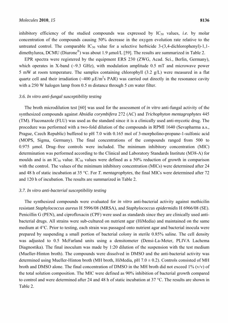
8136
inhibitory efficiency of the studied compounds was expressed by IC50 values, i.e. by molar
concentration of the compounds causing 50% decrease in the oxygen evolution rate relative to the
untreated control. The comparable IC50 value for a selective herbicide 3-(3,4-dichlorophenyl)-1,1-
dimethylurea, DCMU (Diurone®) was about 1.9 μmol/L [59]. The results are summarized in Table 2.
EPR spectra were registered by the equipment ERS 230 (ZWG, Acad. Sci., Berlin, Germany),
which operates in X-band (~9.3 GHz), with modulation amplitude 0.5 mT and microwave power
5 mW at room temperature. The samples containing chlorophyll (3.2 g/L) were measured in a flat
quartz cell and their irradiation (~400 E/m2s PAR) was carried out directly in the resonance cavity
with a 250 W halogen lamp from 0.5 m distance through 5 cm water filter.
3.6. In vitro anti-fungal susceptibility testing
The broth microdilution test [60] was used for the assessment of in vitro anti-fungal activity of the
synthesized compounds against Absidia corymbifera 272 (AC) and Trichophyton mentagrophytes 445
(TM). Fluconazole (FLU) was used as the standard since it is a clinically used anti-mycotic drug. The
procedure was performed with a two-fold dilution of the compounds in RPMI 1640 (Sevapharma a.s.,
Prague, Czech Republic) buffered to pH 7.0 with 0.165 mol of 3-morpholino-propane-1-sulfonic acid
(MOPS, Sigma, Germany). The final concentrations of the compounds ranged from 500 to
0.975 μmol. Drug–free controls were included. The minimum inhibitory concentration (MIC)
determination was performed according to the Clinical and Laboratory Standards Institute (M38-A) for
moulds and is an IC50 value. IC50 values were defined as a 50% reduction of growth in comparison with the control. The values of the minimum inhibitory concentration (MICs) were determined after 24
and 48 h of static incubation at 35 °C. For T. mentagrophytes, the final MICs were determined after 72
and 120 h of incubation. The results are summarized in Table 2.
3.7. In vitro anti-bacterial susceptibility testing
The synthesized compounds were evaluated for in vitro anti-bacterial activity against methicilin
resistant Staphylococcus aureus H 5996/08 (MRSA), and Staphylococcus epidermidis H 6966/08 (SE).
Penicillin G (PEN), and ciprofloxacin (CPF) were used as standards since they are clinically used anti-
bacterial drugs. All strains were sub-cultured on nutrient agar (HiMedia) and maintained on the same
medium at 4°C. Prior to testing, each strain was passaged onto nutrient agar and bacterial inocula were
prepared by suspending a small portion of bacterial colony in sterile 0.85% saline. The cell density
was adjusted to 0.5 McFarland units using a densitometer (Densi-La-Meter, PLIVA Lachema
Diagnostika). The final inoculum was made by 1:20 dilution of the suspension with the test medium
(Mueller-Hinton broth). The compounds were dissolved in DMSO and the anti-bacterial activity was
determined using Mueller-Hinton broth (MH broth, HiMedia, pH 7.0 ± 0.2). Controls consisted of MH
broth and DMSO alone. The final concentration of DMSO in the MH broth did not exceed 1% (v/v) of
the total solution composition. The MIC were defined as 90% inhibition of bacterial growth compared
to control and were determined after 24 and 48 h of static incubation at 37 °C. The results are shown in
Table 2.
Molecules 2010, 15
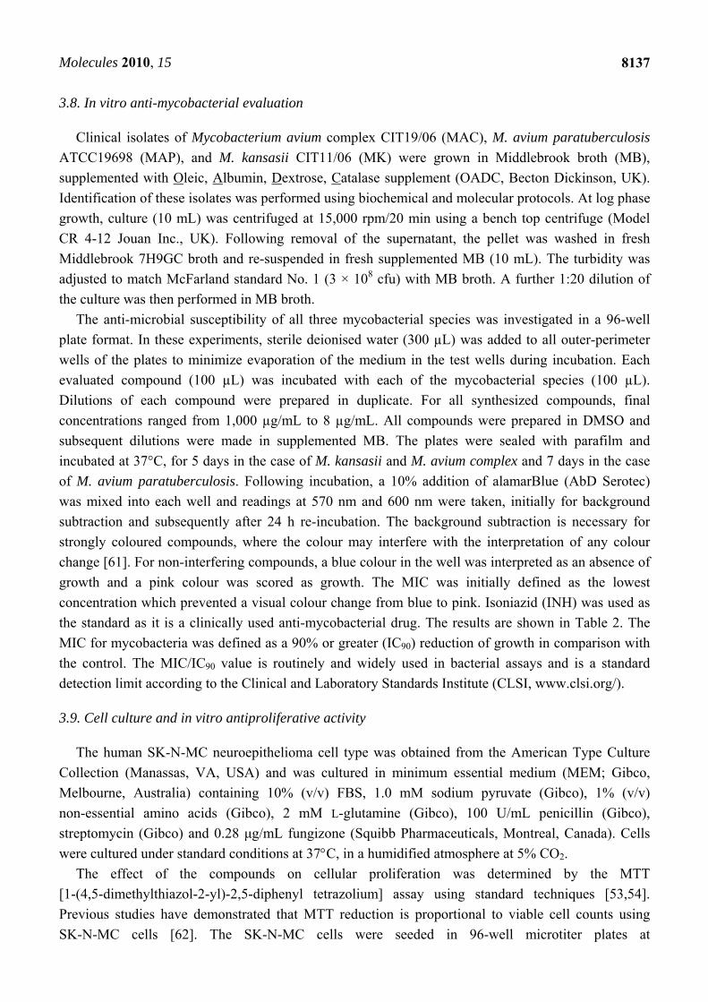
8137
3.8. In vitro anti-mycobacterial evaluation
Clinical isolates of Mycobacterium avium complex CIT19/06 (MAC), M. avium paratuberculosis
ATCC19698 (MAP), and M. kansasii CIT11/06 (MK) were grown in Middlebrook broth (MB),
supplemented with Oleic, Albumin, Dextrose, Catalase supplement (OADC, Becton Dickinson, UK).
Identification of these isolates was performed using biochemical and molecular protocols. At log phase
growth, culture (10 mL) was centrifuged at 15,000 rpm/20 min using a bench top centrifuge (Model
CR 4-12 Jouan Inc., UK). Following removal of the supernatant, the pellet was washed in fresh
Middlebrook 7H9GC broth and re-suspended in fresh supplemented MB (10 mL). The turbidity was
adjusted to match McFarland standard No. 1 (3 × 10⁸ cfu) with MB broth. A further 1:20 dilution of
the culture was then performed in MB broth.
The anti-microbial susceptibility of all three mycobacterial species was investigated in a 96-well
plate format. In these experiments, sterile deionised water (300 µL) was added to all outer-perimeter
wells of the plates to minimize evaporation of the medium in the test wells during incubation. Each
evaluated compound (100 µL) was incubated with each of the mycobacterial species (100 µL).
Dilutions of each compound were prepared in duplicate. For all synthesized compounds, final
concentrations ranged from 1,000 µg/mL to 8 µg/mL. All compounds were prepared in DMSO and
subsequent dilutions were made in supplemented MB. The plates were sealed with parafilm and
incubated at 37°C, for 5 days in the case of M. kansasii and M. avium complex and 7 days in the case
of M. avium paratuberculosis. Following incubation, a 10% addition of alamarBlue (AbD Serotec)
was mixed into each well and readings at 570 nm and 600 nm were taken, initially for background
subtraction and subsequently after 24 h re-incubation. The background subtraction is necessary for
strongly coloured compounds, where the colour may interfere with the interpretation of any colour
change [61]. For non-interfering compounds, a blue colour in the well was interpreted as an absence of
growth and a pink colour was scored as growth. The MIC was initially defined as the lowest
concentration which prevented a visual colour change from blue to pink. Isoniazid (INH) was used as
the standard as it is a clinically used anti-mycobacterial drug. The results are shown in Table 2. The
MIC for mycobacteria was defined as a 90% or greater (IC90) reduction of growth in comparison with
the control. The MIC/IC90 value is routinely and widely used in bacterial assays and is a standard
detection limit according to the Clinical and Laboratory Standards Institute (CLSI, www.clsi.org/).
3.9. Cell culture and in vitro antiproliferative activity
The human SK-N-MC neuroepithelioma cell type was obtained from the American Type Culture
Collection (Manassas, VA, USA) and was cultured in minimum essential medium (MEM; Gibco,
Melbourne, Australia) containing 10% (v/v) FBS, 1.0 mM sodium pyruvate (Gibco), 1% (v/v)
non-essential amino acids (Gibco), 2 mM L-glutamine (Gibco), 100 U/mL penicillin (Gibco),
streptomycin (Gibco) and 0.28 μg/mL fungizone (Squibb Pharmaceuticals, Montreal, Canada). Cells
were cultured under standard conditions at 37C, in a humidified atmosphere at 5% CO2.
The effect of the compounds on cellular proliferation was determined by the MTT
[1-(4,5-dimethylthiazol-2-yl)-2,5-diphenyl tetrazolium] assay using standard techniques [53,54].
Previous studies have demonstrated that MTT reduction is proportional to viable cell counts using
SK-N-MC cells [62]. The SK-N-MC cells were seeded in 96-well microtiter plates at
Molecules 2010, 15
8138
1.5 104 cells/well in medium containing human diferric transferrin (Tf) at 1.25 μmol/L
([Fe] = 2.5 μmol/L) and compounds at a range of concentrations (0-25 μmol/L). Control samples
contained medium with Fe-transferrin (1.25 μmol/L) without the compounds. The chelators, DFO and
Dp44mT, were also included as internal controls, as their effects are well characterized in this cell line
[53,54]. The cells were incubated at 37°C in a humidified atmosphere containing 5% CO2 and 95% air
for 72 h. After 72 h, 10 µL of MTT solution (stock solution: 5 mg/mL) was added to each well and
incubated for 2 h at 37°C. After solubilization of the cells with 100 μL of 10% SDS-50% isobutanol in
0.01 M HCl, the plates were read at 570 nm using a scanning multi-well spectrophotometer. The
inhibitory concentration (IC50) was defined as the compound concentration necessary to reduce the
absorbance to 50% of the untreated control.
4. Conclusions
A series of twelve ring-substituted salicylanilides and carbamoylphenylcarbamates were prepared
and characterized. Their lipophilicity was determined using a well established RP-HPLC method. The
prepared compounds were tested for their ability to inhibit photosynthetic electron transport (PET) in
spinach chloroplasts (Spinacia oleracea L.) and for their anti-fungal, anti-bacterial, anti-mycobacterial
and anti-tumor activity. Four compounds, methyl 4-[2-chloro-5-(trifluoromethyl) phenylcarbamoyl]-
phenylcarbamate (14), methyl 4-(2-chlorobenzylcarbamoyl)phenylcarbamate (13), 2-hydroxy-N-(2-
methoxyphenyl)benzamide (3), and 2-hydroxy-N-(2-methylphenyl)benzamide (1) showed PET
inhibition comparable with or higher than the standard DCMU. The site responsible for the inhibitory
action of the studied compounds in the photosynthetic apparatus could be QB, the second quinone
acceptor situated on the oxidizing (acceptor) site of PS 2.
Interestingly, 2-hydroxy-N-(2-propoxyphenyl)benzamide (5) demonstrated higher anti-fungal
activity against Absidia corymbifera than the standard fluconazole and also higher activity against
methicillin-resistant Staphylococcus aureus and Staphylococcus epidermidis than penicillin G and
ciprofloxacin. Compound 1 exhibited activity against Mycobacterium avium paratuberculosis that was
comparable with the standard isoniazid. None of the agents showed effective anti-tumor activity
in vitro.
Acknowledgements
This study was supported by the Ministry of Education of the Czech Republic MSM 6215712403,
by the Irish Department of Education and Science TSR Strand1-06/CR08 and by Sanofi-Aventis
Pharma Slovakia. D.R.R. was supported by a NHMRC Senior Principal Research Fellowship and
Project grants and an ARC Discovery Grant. D.S.K. was supported by a Cancer Institute New South
Wales Early Career Development Fellowship and Innovation Grant.
References
1. Vinsova, J.; Imramovsky, A. Salicylanilides: Still a topical potential antibacterially active group.
Ces. Slov. Farm. 2004, 53, 294–299.

Molecules 2010, 15
8139
2. De la Fuente, R.; Sonawane, N.D.; Arumainayagam, D.; Verkman, A.S. Small molecules with
antimicrobial activity against E. coli and P. aeruginosa identified by high-throughput screening.
Br. J. Pharmacol. 2006, 149, 551–559.
3. Dahlgren, M.K.; Kauppi, A.M.; Olsson, I.M.; Linusson, A.; Elofsson, M. Design, synthesis, and
multivariate quantitative structure–activity relationship of salicylanilidess–potent inhibitors of
type III secretion in Yersinia. J. Med. Chem. 2007, 50, 6177–6188.
4. Stephenson, K.; Yamaguchi, Y.; Hoch, J.A. The mechanism of action of inhibitors of bacterial
two-component signal transduction systems. J. Biol. Chem. 2000, 275, 38900–38904.
5. Vinsova, J.; Imramovsky, A.; Buchta, V.; Ceckova, M.; Dolezal, M.; Staud, F.; Jampilek, J.;
Kaustova, J. Salicylanilide acetates: Synthesis and antibacterial evaluation. Molecules 2007, 12,
1–12.
6. Imramovsky, A.; Vinsova, J.; Ferriz, J.M.; Dolezal, R.; Jampilek, J.; Kaustova, J.; Kunc, F. New
antituberculotics originated from salicylanilides with promising in vitro activity against atypical
mycobacterial strains. Bioorg. Med. Chem. 2009, 17, 3572–3579.
7. Imramovsky, A.; Vinsova, J.; Ferriz, J.M.; Buchta, V.; Jampilek, J. Salicylanilide esters of
N-protected amino acids as novel antimicrobial agents. Bioorg. Med. Chem. Lett. 2009, 19,
348–351.
8. Hassan, G.S.; Hegazy, G.H.; Safwat, H.M. Synthesis of Furo-salicylanilides and their heterocyclic
derivatives with anticipated molluscicidal activity. Arch. Pharm. Chem. Life Sci. 2006, 339,
448–455.
9. Daidone, G.; Raffa, D.; Plescia, S.; Matera, M.; Caruso, A.; Leone, V.; Amico-Roxas, M.
Synthesis and evaluation of the analgesic and antiinflammatory activities of N-substituted
salicylamides. Farmaco 1989, 44, 465–473.
10. Brown, M.E.; Fitzner, J.N.; Stevens, T.; Chin, W.; Wright, C.D.; Boyce, J.P. Salicylanilides:
Selective inhibitors of interleukin-12p40 production. Bioorg. Med. Chem. 2008, 16, 8760–8764.
11. Liechti, C.; Sequin, U.; Bold, G.; Furet, P.; Meyer, T.; Traxler, P. Salicylanilides as inhibitors of
the protein tyrosine kinase epidermal growth factor receptor. Eur. J. Med. Chem. 2004, 39, 11–26.
12. Deng, W.; Guo, Z.; Guo, Y.; Feng, Z.; Jiang, Y.; Chu, F. Acryolylamino-salicylanilides as EGFR
PTK inhibitors. Bioorg. Med. Chem. Lett. 2006, 16, 469–472.
13. Kamath S.; Buolamwini, J.K. Targeting EGFR and HER-2 receptor tyrosine kinases for cancer
drug discovery and development. Med. Res. Rev. 2006, 26, 569–594.
14. Ray, S.; Pathak, S.R.; Chaturvedi, D. Organic carbamates in drug development. Part II:
antimicrobial agents - Recent reports. Drugs Future 2005, 30, 161–180.
15. Ferriz, J.M.; Vavrova, K.; Kunc, F.; Imramovsky, A.; Stolarikova, J.; Vavrikova, E.; Vinsova, J.
Salicylanilide carbamates: Antitubercular agents active against multidrug-resistant
Mycobacterium tuberculosis strains. Bioorg. Med. Chem. 2010, 18, 1054–1061.
16. Agouridas, C.; Denis, A.; Auger, J.M.; Benedetti, Y.; Bonnefoy, A.; Bretin, F.; Chantot, J.F.;
Dussarat, A.; Fromentin, C.; D‘Ambrieres, S.G.; Lachaud, S.; Laurin, P.; Le Martret, O.; Loyau,
V.; Tessot, N. Synthesis and antibacterial activity of ketolides (6-O-methyl-3-oxoerythromycin
derivatives): A new class of antibacterials highly potent against macrolide-resistant and -
susceptible respiratory pathogens. J. Med. Chem. 1998, 41, 4080–4100.
Molecules 2010, 15
8140
17. Meng, Q.; Luo, H.; Liu, Y.; Li, W.; Zhang, W.; Yao, Q. Synthesis and evaluation of carbamate
prodrugs of SQ109 as antituberculosis agents. Bioorg. Med. Chem. 2009, 19, 2808–2810.
18. Thorberg, S.; Berg, S.; Lundstrom, L.; Pettersson, B.; Wijkstrom, A.; Sanchez, D.; Lindberg, P.;
Nilsson, J.G. Carbamate ester derivatives as potential prodrugs of the presynaptic dopamine
autoreceptor agonist (-)-3-(3-hydroxyphenyl)-N-propylpiperidine. J. Med. Chem. 1987, 30,
2008–2012.
19. Good, N.E. Inhibitors of the Hill reaction. Plant Physiol. 1961, 36, 788–803.
20. Kralova, K.; Sersen, F.; Cizmarik, J. Inhibitory effect of piperidinoethylesters of
alkoxyphenylcarbamic acids on photosynthesis. Gen. Physiol. Biophys. 1992, 11, 261–267.
21. Kralova, K.; Sersen, F.; Kubicova, L.; Waisser, K. Inhibitory effects of substituted benzanilides
on photosynthetic electron transport in spinach chloroplasts. Chem. Pap. 1999, 53, 328–331.
22. Kralova, K.; Sersen, F.; Kubicova, L.; Waisser, K. Inhibition of photosynthetic electron transport
in spinach chloroplasts by 3- and 4-halogeno substituted benzanilides and thiobenzanilides. J.
Trace Microprobe Technol. 2000, 18, 251–256.
23. Kubicova, L.; Kralova, K.; Sersen, F.; Gregor, J.; Waisser, K. Effects of substituted salicylanilides
on the photosynthetic apparatus of spinach chloroplasts. Folia Pharm. Univ. Carol. 2000, 25,
89–96.
24. Pravda, M.; Hrnciarova, D.; Kralova, K. 3-Methylthiosalicylanilides – inhibitors of Hill reaction.
Chem. Listy 2003, 97, 1122–1123.
25. Black, C.C. Photosynthetic phosphorylation and associated reactions in the presence of a new
group of uncouplers: Salicylanilides. Biochim. Biophys. Acta 1968, 162, 294–296.
26. Govindjee, S. Sixty-three years since Kautsky: Chlorophyll a fluorescence. Aust. J. Plant Physiol.
1995, 22, 131–160.
27. Jegerschold, C.; Styring, S. Fast oxygen-independent degradation of D1 reaction center protein in
photosystem II. FEBS Lett. 1991, 280, 87–90.
28. Kralova, K.; Kubicova, L.; Sersen, F.; Waisser, K. Inhibition of Hill reaction in spinach
chloroplasts by 5-bromo- and 3,5-dibromosalicylanilides. In Proceedings of 51st Congress of
Chemical Societies, Nitra, Slovakia, 6-9 September 1999.
29. Kubicova, L.; Kissova, K.; Waisser, K. Inhibition of chlorophyll production in Chlorella vulgaris
by substituted salicylanilides. Folia Pharm. Univ. Carol. 2000, 25, 67–72.
30. Pravda, M.; Sustr, M.; Hrnciarova, D.; Kubicova, L.; Kralova, K. Effects of
3-methylthiosalicylanilides on chlorophyll content in freshwater alga Chlorella vulgaris. In
Proceedings of ECOpole’03, Opole, Poland, 16-18 October 2003; Society of Ecological
Chemistry and Engineering: Opole, Poland, 2003; pp. 105–108.
31. Jampilek, J.; Dolezal, M.; Kunes, J.; Buchta, V.; Kralova, K. Quinaldine derivatives: Preparation
and biological activity. Med. Chem. 2005, 1, 591–599.
32. Dolezal, M.; Palek, L.; Vinsova, J.; Buchta, V.; Jampilek, J.; Kralova, K. Substituted
pyrazinecarboxamides: Synthesis and biological evaluation. Molecules 2006, 11, 242–256.
33. Musiol, R.; Jampilek, J.; Kralova, K.; Richardson, D.R.; Kalinowski, D.; Podeszwa, B.; Finster,
J.; Niedbala, H.; Palka, A.; Polanski, J. Investigating biological activity spectrum for novel
quinoline analogues. Bioorg. Med. Chem. 2007, 15, 1280–1288.
Molecules 2010, 15
8141
34. Musiol, R.; Tabak, D.; Niedbala, H.; Podeszwa, B.; Jampilek, J.; Kralova, K.; Dohnal, J.; Finster,
J.; Mencel, A.; Polanski, J. Investigating biological activity spectrum for novel quinoline
analogues 2: Hydroxyquinolinecarboxamides with photosynthesis inhibiting activity. Bioorg.
Med. Chem. 2008, 16, 4490–4499.
35. Dolezal, M.; Cmedlova, P.; Palek, L.; Vinsova, J.; Kunes, J.; Buchta, V.; Jampilek, J.; Kralova, K.
Synthesis and antimycobacterial evaluation of substituted pyrazinecarboxamides. Eur. J. Med.
Chem. 2008, 43, 1105–1113.
36. Jampilek, J.; Musiol, R.; Pesko, M.; Kralova, K.; Vejsova, M.; Carroll, J.; Coffey, A.; Finster, J.;
Tabak, D.; Niedbala, H.; Kozik, V.; Polanski, J.; Csollei, J.; Dohnal, J. Ring-substituted
4-hydroxy-1H-quinolin-2-ones: Preparation and biological activity. Molecules 2009, 14,
1145–1159.
37. Jampilek, J.; Musiol, R.; Finster, J.; Pesko, M.; Carroll, J.; Kralova, K.; Vejsova, M.; O'Mahony,
J.; Coffey, A.; Dohnal, J.; Polanski, J. Investigating biological activity spectrum for novel
styrylquinazoline analogues. Molecules 2009, 14, 4246–4265.
38. Musiol, R.; Jampilek, J.; Nycz, J.E.; Pesko, M.; Carroll, J.; Kralova, K.; Vejsova, M.; O'Mahony,
J.; Coffey, A.; Mrozek, A.; Polanski, J. Investigating the activity spectrum for ring-substituted
8-hydroxyquinolines. Molecules 2010, 15, 288–304.
39. Josuu, R.M.; Patel, M.M. Chelation ion-exchange properties of salicylic acid-urea-formaldehyde
copolymers. J. Chem. Sci. 1982, 91, 351–358.
40. Mahmoud, M.E.; Soliman, E.M. Study of the selective extraction of iron (III) by silica-
immobilized 5-Formyl-3-Arylazo-salicylic acid derivatives. Talanta 1997, 44, 1063–1071.
41. Rho, H.S.; Baek, H.S.; You, J.W.; Kim, S.J.; Kim, M.K.; Kim, D.H.; Chang, I.S. Biological
activities of 3,5-dihydroxy-N-(4-hydroxyphenyl)benzamide: A mimic compound of trans-
resveratrol. Bull. Korean Chem. Soc. 2007, 28, 837–839.
42. Althuis, T.H.; Hess, H.J. Synthesis and identification of the major metabolites of prazosin formed
in dog and rat. J. Med. Chem. 1977, 20, 146–149.
43. Brown, R.K.; Nelson, N.A. 6-Aminoindole. J. Am. Chem. Soc. 1954, 76, 5149–5150.
44. Fellows, I.M.; Kaelin, D.E.; Martin, S.F. Application of ring-closing metathesis to the formal total
synthesis of (+)−FR900482. J. Am. Chem. Soc. 2000, 122, 10781–10787.
45. Hiraj, K.; Yano, T.; Matsukawa, T.; Ugai, S.; Nagato, S.; Hori, M. Synthesis and herbicidal
activity of new oxazolidinedione derivates. J. Pestic. Sci. 1999, 24, 2, 156–169.
46. Kerns, E.H.; Li, D. Drug-like Properties: Concept, Structure Design and Methods; Elsevier: San
Diego, CA, USA, 2008.
47. Norrington, F.E.; Hyde, R.M.; Williams, S.G.; Wotton, R. Physicochemical-activity relations in
practice. 1. Rational and self-consistent data bank. J. Med. Chem. 1975, 18, 604–607.
48. Svensson, B.; Vass, I.; Styring S. Sequence analysis of D1 and D2 reaction center proteins of
photosystem II. Z. Naturforsch C. 1991, 46c, 765776.
49. Noren, G.H.; Barry, B.A. The YF161D1 mutant of synechocystis 6803 exhibits an EPR signal
from a light-induced photosystem II radical. Biochemistry 1992, 31, 33353342.
50. Hoff, A.J. Application of ESR in photosynthesis. Phys. Rep. 1979, 54, 75200.
51. Izawa, S. Acceptors and Donors for Chloroplast Electron Transport; San Pietro, A., Ed.;
Academic Press: London, UK, 1980; Volume 69, pp. 413–434.
Molecules 2010, 15
8142
52. Kalinowski, D.S.; Richardson, D.R. The evolution of iron chelators for the treatment of iron
overload disease and cancer. Pharmacol. Rev. 2005, 57, 547–583.
53. Richardson, D.R.; Sharpe, P.C.; Lovejoy, D.B.; Senaratne, D.; Kalinowski, D.S.; Islam, M.;
Bernhardt P.V. Dipyridyl thiosemicarbazone chelators with potent and selective antitumor activity
form iron complexes with redox activity. J. Med. Chem. 2006, 49, 6510–6521.
54. Kalinowski, D.S.; Yu, Y.; Sharpe, P.C.; Islam, M.; Liao, Y.T.; Lovejoy, D.B.; Kumar, N.;
Bernhardt P.V.; Richardson, D.R. Design, synthesis, and characterization of novel iron chelators:
Structure−activity relationships of the 2-benzoylpyridine thiosemicarbazone series and their
3-nitrobenzoyl analogues as potent antitumor agents. J. Med. Chem. 2007, 50, 3716–3729.
55. Wagner, G.; Singer, D.; Weuffen, W. Untersuchungen uber 2-hydroxythiobenzamide und
2-hydroxythiobenzanilide. Pharmazie 1966, 21, 161–166.
56. Bahrami, K.; Khodaei, M.M.; Farrokhi, A. H2O2/SOCl2: A useful reagent system for the
conversion of thiocarbonyls to carbonyl compounds. Tetrahedron 2009, 65, 7658–7661.
57. Masarovicova, E.; Kralova, K. Approaches to measuring plant photosynthesis activity. In
Handbook of Photosynthesis, 2nd ed.; Pessarakli, M., Ed.; Taylor & Francis Group: Boca Raton,
FL, USA, 2005; pp. 617–656.
58. Kralova, K.; Sersen, F.; Sidoova, E. Photosynthesis inhibition produced by 2-alkylthio-6-R-
benzothiazoles. Chem. Pap. 1992, 46, 348–350.
59. Fedke, C. Biochemistry and Physiology of Herbicide Action; Springer Verlag: New York, NY,
USA, 1982.
60. National Committee for Clinical Laboratory Standards. Method for Antifungal Disk Diffusion
Susceptibility Testing of Yeasts: Approved Guideline M44-A; National Committee for Clinical
Laboratory Standards: Wayne, PA, USA, 2004.
61. Carroll, J.; Douarre, P.; Coffey, A.; Buckley, J.; Cashman, B.; O'Farrell, K.; O'Mahony, J.
Optimization of a rapid viability assay for Mycobacterium avium paratuberculosis by using
alamarBlue. Appl. Environ. Microbiol. 2009, 75, 7870–7872.
62. Richardson, D.R.; Tran, E.H.; Ponka, P. The potential of iron chelators of the pyridoxal
isonicotinoyl hydrazone class as effective antiproliferative agents. Blood 1995, 86, 4295–4306.
Sample Availability: Samples of the compounds are available from the authors.
© 2010 by the authors; licensee MDPI, Basel, Switzerland. This article is an open access article
distributed under the terms and conditions of the Creative Commons Attribution license
(http://creativecommons.org/licenses/by/3.0/).