investigating trehalose synthesis genes after cold ... · osmoregulation within the pseudocoelomic...
TRANSCRIPT

© 2017. Published by The Company of Biologists Ltd.
This is an Open Access article distributed under the terms of the Creative Commons Attribution License (http://creativecommons.org/licenses/by/3.0), which permits unrestricted use, distribution and reproduction
in any medium provided that the original work is properly attributed.
Investigating trehalose synthesis genes after cold acclimation in the
Antarctic nematode Panagrolaimus sp. DAW1
Anna C. Seybold1, David A. Wharton2, Michael A. S. Thorne3 and Craig J. Marshall1,4*
1Department of Biochemistry, University of Otago, Dunedin, New Zealand
2Department of Zoology, University of Otago, Dunedin, New Zealand
3British Antarctic Survey, Natural Environment Research Council, Cambridge, United Kingdom
4Genetics Otago, University of Otago, Dunedin, New Zealand
*Corresponding author: [email protected]
key words: Panagrolaimus davidi, Panagrolaimus sp. DAW1, trehalose, tps-2, cryoprotective dehydration,
freezing tolerance
Bio
logy
Ope
n •
Acc
epte
d m
anus
crip
t
by guest on May 1, 2020http://bio.biologists.org/Downloaded from

Summary statement
Functional genomics was used to investigate trehalose synthesis genes after cold acclimation in
Panagrolaimus sp. DAW1, an Antarctic nematode with the ability to survive intracellular freezing.
Abstract
Panagrolaimus sp. DAW1 is a freeze-tolerant Antarctic nematode which survives extensive intracellular ice
formation. The molecular mechanisms of this extreme adaptation are still poorly understood. We recently
showed that desiccation-enhanced RNAi-soaking can be used - in conjunction with qPCR - to screen for
phenotypes associated with reduced expression of candidate genes in Panagrolaimus sp. DAW1. Here we
present the use of this approach to investigate the role of trehalose synthesis genes in this remarkable
organism.
Previous studies have showed that acclimating Panagrolaimus sp. DAW1 at 5 °C before freezing or
desiccation substantially enhances survival. In this study the expression of tps-2 and other genes
associated with trehalose metabolism, as well as lea-1, hsp-70 and gpx-1 in cold-acclimated and non-
acclimated nematodes was analyzed using qPCR. Pd-tps-2 and Pd-lea-1 were significantly up-regulated
after cold-acclimation, indicating an inducible expression in the cold-adaptation of Panagrolaimus sp.
DAW1. The role of trehalose synthesis genes in Panagrolaimus sp. DAW1 was further investigated by RNAi.
Compared to the controls, Pd-tps-2a(RNAi)-treated and cold-acclimated nematodes showed a significant
decrease in mRNA, but no change in trehalose content or freezing survival. The involvement of two other
trehalose synthesis genes (tps-2b and gob-1) was also investigated.
These findings provide the first functional genomic investigation of trehalose synthesis genes in the non-
model organism Panagrolaimus sp. DAW1. The presence of several trehalose synthesis genes with
different RNAi sensitivities suggests the existence of multiple backup systems in Panagrolaimus sp. DAW1,
underlining the importance of this sugar in preparation for freezing.
Bio
logy
Ope
n •
Acc
epte
d m
anus
crip
t
by guest on May 1, 2020http://bio.biologists.org/Downloaded from
Introduction
The Antarctic nematode, Panagrolaimus sp. DAW1 (PaDAW1) is the only multicellular organism known to
tolerate intracellular freezing on a routine basis (Wharton and Brown, 1991; Wharton and Ferns, 1995).
PaDAW1, formerly known as Panagrolaimus davidi CB1, is also tolerant of cryoprotective dehydration
(Wharton et al., 2004a; Wharton et al., 2017; Wharton et al., 2003). These characteristics are adaptations
to the nematodes’ habitat – in and around Antarctic penguin colonies (Raymond et al., 2013a; Wharton
and Brown, 1989). During the summer, and perhaps somewhat surprisingly, soils in these regions can
warm to above 25 °C (Raymond et al., 2013a; Sinclair et al., 2006), and nutrient-rich liquid guano provides
an ideal medium for the growth of bacteria on which PaDAW1 feed (Raymond et al., 2013a). PaDAW1
reproduces only when the temperature is above about 6.8 °C (Brown et al., 2004) suggesting that breeding
is possible for only a limited time in each season when solar irradiation warms the soil. However, for much
of the year, these habitats are both cold and dry with winter temperatures as low as -40 °C and very low
humidity. PaDAW1 might be considered a temperate nematode with the capacity to tolerate long-term
freezing and desiccation.
Survival of freezing and of desiccation may not be obviously related, but both involve the removal of water
and concentration of solutes, and tolerance of these states may share similar mechanisms (Storey and
Storey, 2013; Teets and Denlinger, 2014). Although PaDAW1 survives both intracellular freezing (Wharton
and Ferns, 1995) and cryoprotective dehydration (Wharton et al., 2003), little is known of the mechanisms
by which this is achieved. It is clear that survival of intracellular freezing is related to the pattern and
distribution of ice formation (Raymond and Wharton, 2016) and that in turn is likely to be associated with
osmoregulation within the pseudocoelomic fluid (Wharton, 2010) as well as the presence of ice-active
proteins (Barrett et al., 2005). Cryoprotective dehydration seems to occur if freezing rates are slow, but
extra- and even intracellular freezing can occur if freezing rates are faster. The increase in freezing rate
produces a shift from cryoprotective dehydration to extracellular to intracellular freezing, accompanied by
a decrease in survival (Wharton and Ferns, 1995; Wharton et al., 2003). However, many questions remain
as to exactly what metabolic changes are responsible for freezing and desiccation tolerance.
The accumulation of the disaccharide, trehalose (α-D-glucopyranosyl-(1→1)-α-D-glucopyranoside), is one
of the best characterized metabolic changes during acclimation of anhydrobiotic organisms (Argüelles,
2014; Storey and Storey, 2013). Trehalose protects membranes and proteins from desiccation by replacing
structural water (Carpenter et al., 1987; Crowe et al., 1984) and by forming cellular glass (Crowe et al.,
1998). Trehalose accumulation has been associated with anhydrobiosis in nematodes such as Aphelenchus
avenae (Madin and Crowe, 1975), Anguina tritici and Ditylenchus dipsaci (Womersley and Smith, 1981). In
some species, such as A. avenae, trehalose accumulation seems to be essential, but not sufficient, for
anhydrobiosis (Browne et al., 2004; Higa and Womersley, 1993). In others, such as some anhydrobiotic
rotifer and tardigrade species, trehalose accumulation is apparently not essential (Hengherr et al., 2008;
Bio
logy
Ope
n •
Acc
epte
d m
anus
crip
t
by guest on May 1, 2020http://bio.biologists.org/Downloaded from

Lapinski and Tunnacliffe, 2003). In PaDAW1, a period of acclimation at ~5 °C is associated with both the
accumulation of trehalose and a significant increase in survival after subsequent exposure to freezing
(Wharton et al., 2000), suggesting that trehalose might play a role in freezing tolerance (Feofilova et al.,
2014; Storey and Storey, 2013; Tapia and Koshland, 2014).
Recent work looking at genes expressed during acclimation, freezing and cryoprotective dehydration in
PaDAW1 (Thorne et al., 2014) identified a number of trehalose synthesis (tps) genes, late embryogenesis
abundant (lea) proteins, heat shock proteins (hsp) and genes associated with antioxidant production that
showed evidence of specific up-regulation in these conditions (and were distinct from general responses
to stress). In the PaDAW1 dataset, two trehalose synthesis genes (tps and gob), six aquaporin genes and
three desaturase genes as well as nine different lea-type genes and 20 hsp-70 like genes were identified
as potential candidates to be involved in cryoprotective dehydration (Thorne et al., 2014). Broadly similar
patterns of expression were seen in the Antarctic nematode Plectus murrayi during freezing (Adhikari et
al., 2009).
This study investigates expression of selected genes – trehalose-6-phosphate synthase 2 (tps-2), trehalose-
6-phosphate phosphatase (gob-1), late embryogenesis abundant 1 (lea-1) protein, glutathione peroxidase
1 (gpx-1) and heat shock protein 70 (hsp-70) – in cold-acclimated and non-acclimated nematodes using
qPCR. Previous work has shown that, provided nematodes are properly fed (Raymond and Wharton, 2013),
acclimation at 5 °C for 3–5 days improves freezing survival from about 40% to about 85% (Wharton et al.,
2000). This implies that lower temperatures induce physiological and biochemical changes that assist in
freezing survival and an increase in trehalose content was a marked example of such a change. Cellular
trehalose was shown to increase upon cold-acclimation and to be correlated with freezing survival
(Wharton et al., 2000). The enzymes trehalose-6-phosphate synthase (TPS) and trehalose-6-phosphate
phosphatase (GOB in PaDAW1) are directly involved in trehalose synthesis in nematodes and are essential
genes (Avonce et al., 2006; Behm, 1997; Farelli et al., 2014; Pellerone et al., 2003), and the enzyme
trehalase is involved in trehalose breakdown (Thorne et al., 2014; Thorne et al., 2017; Łopieńska-Biernat
et al., 2015).
The role of trehalose and trehalose synthesis genes in PaDAW1 during acclimation at 5 °C was further
investigated. First, tps-2a was silenced by RNAi and the reduction of mRNA was measured by qPCR, and
the amount of trehalose itself was assessed by gas chromatography. Second, the involvement of other
trehalose synthesis genes (tps-2b and gob-1) was investigated using qPCR and RNAi. Third, the freezing
survival of tps-2a,b(RNAi)-treated nematodes was compared to that of non-treated controls.
Bio
logy
Ope
n •
Acc
epte
d m
anus
crip
t
by guest on May 1, 2020http://bio.biologists.org/Downloaded from

Materials and Methods
Nematode culturing and cold-acclimation
PaDAW1 was originally isolated from McMurdo Sound region, Antarctica (Wharton and Brown, 1989) and
has been maintained in the laboratory for more than 25 years (Raymond et al., 2013b). Mixed nematode
cultures were grown on E. coli seeded NGM agar plates at 20 °C and sub-cultured weekly. Nematodes used
for these experiments were collected from culture plates by a modified Baermann technique (Flegg and
Hooper, 1970), and sub-cultured in five replicates of exactly equal volume. They were first incubated at 20
°C for three days and then at 5 °C for another 24 h. After cold-acclimation, nematodes were re-collected,
snap frozen in a mixture of dry ice and ethanol, and stored at -80 °C until analysis.
RNA isolation and cDNA synthesis
RNA was extracted using TRIzol® Reagent (Ambion, Foster City, CA, USA) and RNeasy® Mini Kits (Qiagen,
Hilden, Germany) and reverse-transcribed using the VILO cDNA synthesis kit (Invitrogen, Carlsbad, CA,
USA) as described previously (Seybold et al., 2016).
Candidate gene cloning for RNAi
Candidate genes were selected using two different sets of data: a study of Plectus murrayi (Adhikari et al.,
2009) and transcriptomic data from PaDAW1 (Thorne et al., 2014) (Table S2). We selected genes that
showed significant up-regulation on exposure to freezing in both P. murrayi and PaDAW1, particularly
those involving trehalose metabolism. Target genes were PCR amplified from cDNA with gene specific
primers using Taq DNA Polymerase dNTPack (Roche, Basel, Switzerland) as described previously (Seybold
et al., 2016) and Table S1.
Two sets of transcripts homologous to tps were identified in the PaDAW1 dataset. Initial analyses
suggested these were two non-overlapping portions of the same gene (Thorne et al., 2014). However, a
search of the genomic scaffolds indicates that they probably come from different regions of the genome
and probably represent a gene duplication. These two regions were termed tps-2a, showing a larger
alignment, and tps-2b, a smaller alignment (see Table S1).
RNAi
Double-stranded RNA for soaking experiments was produced by in vitro transcription of PCR products
using the MEGAscript®T7 Transcription Kit (Life Technologies, Carlsbad, CA, USA). RNA uptake was done by
desiccating nematodes at 98% relative humidity and 20 °C for 24 h, prior to soaking in dsRNA solutions as
described previously (Seybold et al., 2016). Nematode cultures (four replicates for each treatment) were
then rehydrated in soaking buffer (plus 1mg/mL dsRNA, and minus dsRNA) for 16 h. Cultures were then
incubated for 24 h at 5 °C for recovery before they were harvested and processed.
Bio
logy
Ope
n •
Acc
epte
d m
anus
crip
t
by guest on May 1, 2020http://bio.biologists.org/Downloaded from

Quantitative polymerase chain reaction (qPCR)
Quantitative PCR was performed using the BioRad CFX96 System (Hercules, CA, USA) and BioRad SSoFast
EVA Green Supermix with Low Rox. A typical 20 µl reaction contained a 5 µl sample (total of 50 ng cDNA),
10 µl SYBR green mix, 1.2 µl primer mix and 3.2 µl mQ water. Specificity and efficiency assays were
performed for all genes. Of six housekeeping genes tested, the combination of Pd-gpd-2 and Pd-tba-1 were
defined as the most stable and used for all qPCR experiments to normalise data from each individual assay.
The genes tested were tps-2 (trehalose synthase 2), gob-1 (trehalose-6-phosphate phosphatase 1), lea-1
(late embryogenesis abundant proteins 1), hsp-70 (heat shock protein 70), and gpx-1 (glutathione
peroxidase 1). BioRad FCX Manager software was used to control qPCR settings and to analyse qPCR data
as described previously (Seybold et al., 2016). Primer details for qPCR analyses can be found in
supplementary Table S2 and Seybold et al. (2016).
Relative differences in expression of target genes were assessed using the ΔΔCt (Livak) method (Livak and
Schmittgen, 2001) where the relative difference in expression of the target gene in different samples was
determined. This involves normalising the Ct of the target gene to that of the reference genes, for both the
test sample and the control sample (producing a normalized relative expression value ΔCt). In the next
step, the ΔCt of the test sample is normalized to that of the control sample to give a measure of relative
expression. This approach produces an estimate of how test gene expression changed with respect to the
control condition and corrects for differences in the RNA yield of each culture that would otherwise
complicate the analysis.
Gas chromatography
Sugar extraction was performed by using a technique modified from Wharton et al. (2000). Sugars were
extracted to compare trehalose levels from both cold-acclimated and non-acclimated cultures (grown at
5 °C and 20 °C) as well as from Pd-tps-2(RNAi)-treated and non-treated (control) samples acclimated at 5
°C. RNAi cultures were plated out on NGM agar plates and incubated for 72 h, to allow time for changes in
trehalose levels. After incubation, samples were collected as described above and snap frozen in a mixture
of dry ice and ethanol before processing.
After extraction (Wharton et al., 2000), the sugars in dried samples were converted to their trimethylsilyl
derivatives by adding 20 µl Sylon (Supelco) and incubation for 5 min at room temperature. Derivatized
samples (15 µl) were injected onto the column of an Agilent 6890N Network gas chromatograph. To reduce
variation between replicas caused by the injection technique, hot needle injection after Barwick (1999)
was done. Briefly, after insertion into the injection zone, the needle is allowed to heat up for 5 s. The
sample is then rapidly injected by pushing down the plunger and the needle quickly withdrawn from the
inlet within 1 s. Sugars were identified and quantified using reference standards. Recovery of sugars was
Bio
logy
Ope
n •
Acc
epte
d m
anus
crip
t
by guest on May 1, 2020http://bio.biologists.org/Downloaded from

estimated by inclusion of 240 µg of Dulcitol in the original nematode sample and analysis of the Dulcitol
peak (Wharton et al., 2000).
Freezing survival
The freezing survival experiment was performed after Smith et al. (2008). Following RNAi soaking and
acclimation at 5 °C for 24h, nematodes were washed off the plates and 50 µl samples of nematode
suspension were transferred into 0.5 ml microcentrifuge tubes. These were transferred to a cooling block
(Wharton et al., 2004b) and cooled from +1 °C to -15 °C at 0.5 min-1. Freezing was seeded by adding a small
ice crystal to each tube when the temperature was -1 °C. Samples were held at -15 °C for 30 min, then
warmed to 1 °C at 0.5 min-1, and then were transferred to mQ water in watch-glasses and incubated for
24 h at 20 °C. Survival of nematodes was assessed by determining the proportion of moving nematodes.
To perform statistical analysis, 3 × 100 nematodes were counted for each of the three replicates of non-
treated and Pd-tps-2(RNAi)-treated samples. A t-test (parametric, two samples, equal variance) was used
to assess the statistical significance of sample differences. Control samples consisted of 50 µl nematode
suspension in 0.5 ml microcentrifuge tubes, kept at 20 °C for the duration of the experiment.
Results
Identifying candidate genes
In order to directly examine the relative expression of selected genes in response to acclimation at 5 °C,
quantitative PCR was performed on PaDAW1 samples maintained at the physiologically significant
temperatures of 20 °C (active reproduction) and 5 °C (active acclimation). This analysis showed a
significantly increased expression of four of the six genes tested and a significant reduction of expression
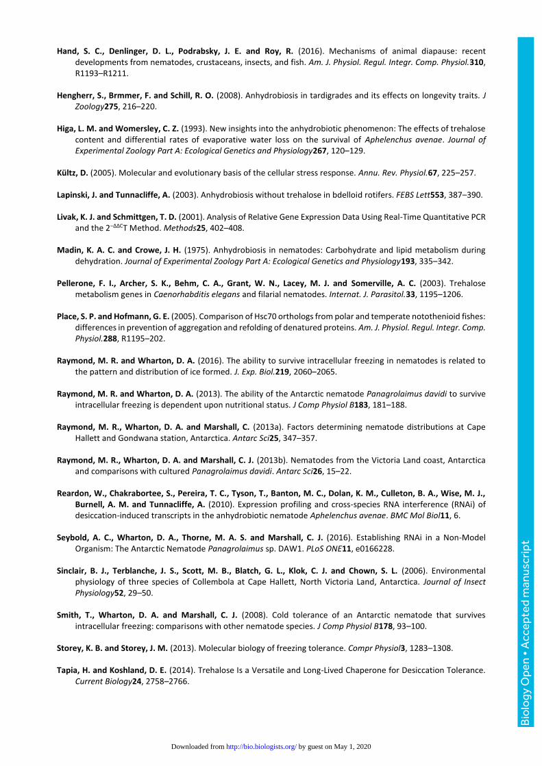
of one (Fig. 1). The final gene tested, Pd-gpx-1, did not vary significantly. Expression of both Pd-tps-2a
(relative expression = 3.9 ± 0.7, p = 0.017) and Pd-tps-2b (relative expression = 4.6 ± 0.6, p = 0.021) was
significantly increased consistent with an increase in trehalose content (Fig. 2A). Trehalose-6-phosphate
phosphatase (Pd-gob-1), an enzyme also involved in trehalose metabolism (relative expression = 2.3 ± 0.2,
p = 0.005) and Pd-lea-1 (relative expression = 3.4 ± 0.4, p = 0.008) were significantly up-regulated in the 5
°C samples. Pd-gpx-1 was slightly up-regulated in the 5 °C samples compared to the 20 °C samples but this
was not statistically significant (relative expression = 1.33 ± 0.14, p = 0.13). In contrast, Pd-hsp-70 was
slightly and significantly down-regulated (relative expression = 0.75 ± 0.05, p = 0.018).
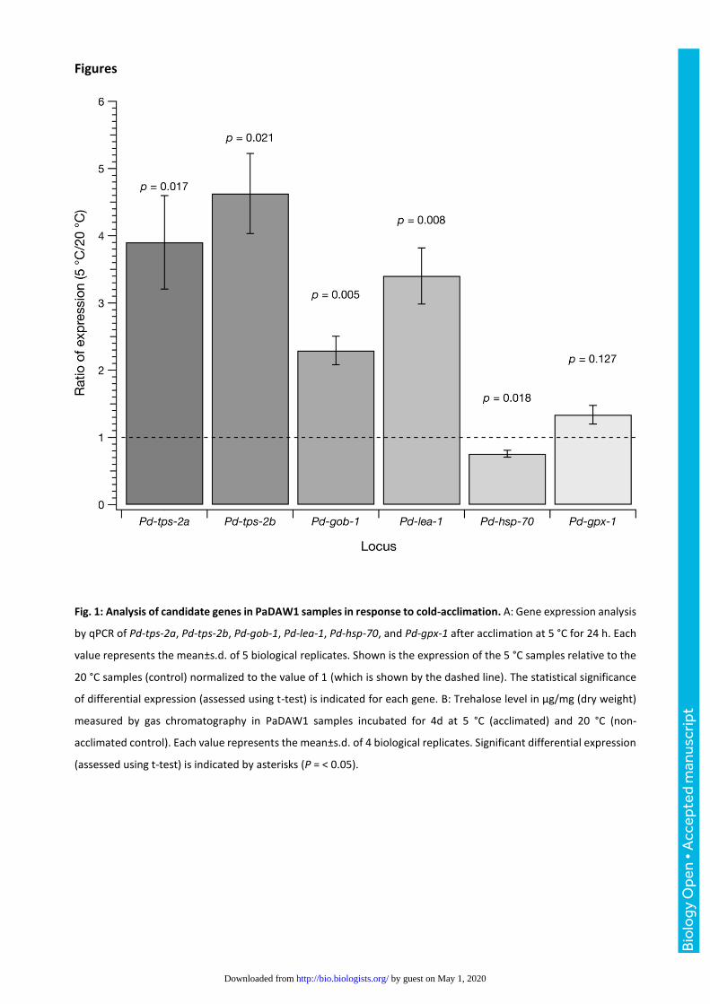
The up-regulation of Pd-tps-2 expression upon cold-acclimation in PaDAW1 was correlated with an
increase in total trehalose content. Our results showed that the trehalose level was significantly higher in
cold-acclimated samples (6.8 ± 0.5 µg/mg at 5 °C versus 1.3 ± 0.5 µg/mg at 20 °C, a 5.2-fold increase, p ≈2
× 10-6) (Fig. 2A). These data show a large and significant increase in trehalose content in response to
Bio
logy
Ope
n •
Acc
epte
d m
anus
crip
t
by guest on May 1, 2020http://bio.biologists.org/Downloaded from

acclimation at 5 °C, as noted previously (Wharton et al., 2000). This suggests that trehalose may be one of
the agents responsible for the correlation between cold-acclimation and freezing survival.
Can RNAi efficiently silence tps-2 expression and is there any phenotype?
Since gene expression analysis by qPCR showed a significant up-regulation of both Pd-tps-2a and Pd-tps-
2b in cold-acclimated samples (see Fig. 1A), these loci were chosen for further analysis by RNAi. Expression
of Pd-tps-2a assessed by qPCR after treatment with Pd-tps-2a(RNAi) showed a decrease relative to control
RNAi-treated nematodes of Pd-tps-2 (RNAi relative expression of 0.92 ± 0.25, p = 0.021 versus control
relative expression 1.26 ± 0.26, overall relative expression (RNAi/Control) = 0.73; Fig. 2B). It is not clear if
this difference is biologically significant even though it is statistically significant at p < 0.05. Such small
differences of expression may not be enough to generate a phenotype (Fraser et al., 2000).
RNAi of Pd-tps-2b (RNAi relative expression of 1.02 ± 0.09, p = 0.021 versus control relative expression 0.95
± 0.09, overall relative expression ≈1.07; Fig. 2C) showed no significant decrease in expression. To
determine whether a small change in TPS-2 synthesis was associated with any change in trehalose content,
we measured the amount of trehalose in nematodes after RNAi treatment. No significant difference in the
trehalose content between Pd-tps-2a(RNAi)-treated (9.1 ± 2.1 µg/mg) and non-treated control samples
(7.9 ± 1.7 µg/mg) was detected (Fig. 2D). We also measured freezing survival and found no difference
between RNAi-treated (57.7% ± 10.3%) and control samples (56.8% ± 12.1%) (Fig. 2E).
Discussion
Investigating candidate genes
Candidate genes were selected from those thought to be associated with cold tolerance in nematodes
(e.g. Adhikari et al., 2009) and using transcriptomic data from PaDAW1 (Thorne et al., 2014; Thorne et al.,
2017). Three of these are associated with trehalose synthesis and this is consistent with previous findings
that cold-acclimation of PaDAW1 under these conditions is correlated with a significant increase in
trehalose content (Wharton et al., 2000). Synthesis of trehalose in metazoans (which include nematodes)
occurs in two steps involving the formation of trehalose 6-phosphate from glucose 6-phosphate and UDP-
glucose, followed by the removal of phosphate to form trehalose (Fig. 3). The enzymes involved are
trehalose 6-phosphate synthase (TPS) and trehalose 6-phosphate phosphatase (GOB), and trehalase in the
breakdown of trehalose (Behm, 1997). Our data shows a significant up-regulation of both Pd-tps-2a, and
Pd-lea-1, whereas Pd-gpx-1 and Pd-hsp-70 showed no significant change in mRNA levels.
The observation that Pd-tps-2a and Pd-lea-1 are significantly up-regulated upon cold-acclimation, suggests
that these two genes are cold-inducible and therefore likely to be involved in cold-adaptation of PaDAW1.
Late embryogenesis proteins (lea) were originally described in plants (Tunnacliffe et al., 2010) and are now
known to have a role in preventing protein aggregation in a wide range of organisms including nematodes
Bio
logy
Ope
n •
Acc
epte
d m
anus
crip
t
by guest on May 1, 2020http://bio.biologists.org/Downloaded from
(Hand et al., 2016; Yaari et al., 2016). Two other genes, Pd-gpx-1 and Pd-hsp-70, involved in oxidative
metabolism and protein folding respectively, remained nearly unchanged after cold-acclimation, indicating
that they might be constitutively expressed in PaDAW1. Similar expression patterns in response to
desiccation have been demonstrated in other nematode species. For example, after cold-acclimation in P.
murrayi, Pm-tps and Pm-lea were highly up-regulated, while Pm-gpx was slightly up-regulated and Pm-
hsp-70 remained unchanged (Adhikari et al., 2009). Similarly, in Panagrolaimus superbus, Ps-lea was highly
up-regulated, while Ps-gpx was slightly up-regulated and Ps-hsp-70 remained unchanged (Tyson et al.,
2012).
The gene hsp-70 seems to be up-regulated (inducible) in only some nematode species (Choi et al., 2014)
and unchanged (constitutive) in others (Tyson et al., 2012). In P. murrayi, hsp-70 is constitutively
expressed, contributing to enhanced stress resistance overall. Generally, hsp are not constitutively
expressed but are expressed in response to stress due to incompatibility with normal metabolism but there
are exceptions such as in notothenioid fish (Buckley and Somero, 2008; Place and Hofmann, 2005).
However, P. murrayi may have evolved mechanisms to maintain HSP function during normal metabolism
in order to survive in an unpredictable environment like Antarctica, with sudden exposure to a variety of
stressors (Adhikari et al., 2009). Survival of PaDAW1 may also depend on maintaining constitutive
expression of this molecular chaperone.
Oxidative stress is experienced by organisms undergoing a wide range of abiotic stressors, resulting in
generation of reactive oxygen species (Reardon et al., 2010). An oxidative stress response has been defined
as part of the environmental stress response (Gasch et al., 2000), as well as part of the minimal stress
response (Kültz, 2005). In PaDAW1, the antioxidant gpx-1 was slightly but not significantly up-regulated,
indicating a minor role or a constitutive expression. The locus, gpx-1, has been shown to be up-regulated
in other nematode species in response to desiccation (Adhikari et al., 2009; Reardon et al., 2010; Tyson et
al., 2012). In Aphelenchus avenae, gpx-1 expression was 32 times greater in response to desiccation and
RNAi of gpx-1 reduced desiccation survival in P. superbus (Reardon et al., 2010).
In PaDAW1, trehalose accumulation has been shown to correlate with an increase in survival after
exposure to freezing. Trehalose may thus play a role in the freeze tolerance of PaDAW1 by protecting
membranes against the harmful effects of freeze-induced dehydration (Wharton et al., 2000). Our results
showed that the amount of trehalose was significantly higher in cold-acclimated samples than in non-
acclimated nematodes. This is consistent with cold-acclimation enhancing freezing survival in PaDAW1
(Raymond:2013ei; Wharton et al., 2000).
RNAi affects tps-2 expression but not trehalose synthesis
Gene expression analysis by qPCR showed a significant up-regulation of Pd-tps-2 in cold-acclimated
samples compared to non-acclimated samples. Furthermore, the level of trehalose, the product of TPS-2
Bio
logy
Ope
n •
Acc
epte
d m
anus
crip
t
by guest on May 1, 2020http://bio.biologists.org/Downloaded from
activity, is increased 4.4-fold in response to cold-acclimation. Therefore, Pd-tps-2 was chosen for further
analysis by RNAi. Gene expression analysis by qPCR showed a slight but significant down-regulation of Pd-
tps-2a(RNAi)-treated samples compared to the non-treated samples.
However, no significant difference in trehalose content between Pd-tps-2a(RNAi)-treated and non-treated
samples was found. The absence of any change in trehalose content could be caused by a number of things.
One possibility is that the pool of trehalose is large enough that changes in synthesis over 72 h are not
detectable. This seems unlikely since acclimation at 5 °C for 96 h increases trehalose content by about 4.4-
fold (Fig. 1B). Alternatively, tps-2 may not be efficiently silenced. This could be a reflection of a limited
reduction in mRNA content – only ≈25% in our data (Fig. 2B) – or because there are other genes for
trehalose synthesis. According to Pellerone et al. (2003), less than 100% knockdown of tps expression could
provide enough enzyme activity to allow residual trehalose metabolism. Therefore, a significant reduction
in mRNA may not alter the amount of the enzyme enough to detect a phenotype even though RNAi might
have reduced the amount of the target mRNA.
There is evidence that PaDAW1 has more than one tps gene which could compensate for the loss of
expression at one locus. In C. elegans, a reduction in trehalose level of > 90% (confirmed by qPCR) was
achieved after a double knockdown of both tps genes. However, no loss-of-function phenotypes were
observed (Pellerone et al., 2003). It is therefore questionable whether a phenotype, such as a decrease in
survival after exposure to freezing, would be observable in PaDAW1, particularly since the trehalose level
was not decreased after Pd-tps-2a knockdown. It is also possible, that the half-life of trehalose is
significantly longer than the RNAi effect, complicating detection of a decrease in trehalose levels.
PaDAW1 has multiple genes for trehalose synthesis
In contrast to Pd-tps-2a, gene expression analysis of Pd-tps-2b(RNAi)-treated samples showed no
significant down-regulation of the target mRNA compared to the non-treated samples. The fact that Pd-
tps-2a but not Pd-tps-2b is sensitive to RNAi is interesting and indicates that these genes probably act
differently. They have either evolved different features or are expressed in different tissues with different
accessibility to environmental RNAi. Expression in different tissues has been described for trehalase genes,
where membrane associated and soluble tre activities have been observed (reviewed in Behm, 1997).
Conant and Wagner (2004) found that mutational robustness is greatest for closely related gene
duplicates. Since duplicate genes often have similar functions, the loss of one duplicate can be tolerated
because other copies can buffer against this loss. They also found a positive correlation between the amino
acid distance and the number of duplicates with different knockdown effects (Conant and Wagner, 2004).
Thus, the more distant two duplicates are, the more likely it is that one has a stronger knock-down effect
than the other. This observation might explain our data on inhibition of tps-2a and tps-2b synthesis.
Symmetric divergence, which probably increases with amino acid distance and divergence time, could
Bio
logy
Ope
n •
Acc
epte
d m
anus
crip
t
by guest on May 1, 2020http://bio.biologists.org/Downloaded from

explain why distantly related duplicates often show different mutational effects (Conant and Wagner,
2004).
Freezing survival is not affected by tps-2 silencing
There was no statistical difference between the proportion of moving nematodes after freezing of non-
treated samples compared to that of Pd-tps-2a(RNAi)-treated samples (Fig. 2E). The lack of freezing
sensitivity in RNAi-treated samples is not surprising given that the trehalose content was not decreased
(Fig. 2D). It is also likely that genes other than those involved in trehalose synthesis are involved in freezing
survival.
Taken together, the characteristics of gene duplicates explain the different RNAi sensitivity between the
two tps-2 genes in PaDAW1 as well as the lack of effects on the trehalose content. The fact that not only
the two tps-2 genes, but also gob-1 is involved in trehalose synthesis, indicates a multiple backup system
in PaDAW1, underlining the importance of this sugar as a cryoprotectant against environmental stressors
common in Antarctica.
Acknowledgements
We thank Peter Dearden and Liz Duncan from the Department of Biochemistry (University of Otago) for
providing expertise, advice and reagents. We also thank Nicky McHugh from the Department of Zoology
(University of Otago) for providing technical support.
Competing interests
No competing interests declared
Author contributions
ACS performed the experiments. CJM, MAST, DAW and ACS conceived and designed the study and
experiments. ACS and MAST developed the probes and primers. ACS wrote the manuscript with input from
CJM, MAST and DAW. All authors gave final approval for publication.
Funding
This research was supported by a University of Otago Doctoral Scholarship and a Post Submission Doctoral
Publishing Bursary to ACS.
Bio
logy
Ope
n •
Acc
epte
d m
anus
crip
t
by guest on May 1, 2020http://bio.biologists.org/Downloaded from
References
Adhikari, B. N., Wall, D. H. and Adams, B. J. (2009). Desiccation survival in an Antarctic nematode: molecular analysis using expressed sequenced tags. BMC Genomics10, 69.
Argüelles, J.-C. (2014). Why Can’t Vertebrates Synthesize Trehalose? J Mol Evol79, 111–116.
Avonce, N., Mendoza-Vargas, A., Morett, E. and Iturriaga, G. (2006). Insights on the evolution of trehalose biosynthesis. BMC Evol. Biol.6, 109.
Barrett, J., Goodall, G., Marshall, C. J. and Ramløv, H. (2005). Ice-active proteins from the Antarctic nematode Panagrolaimus davidi. Cryobiology51, 198–207.
Barwick, V. J. (1999). Sources of uncertainty in gas chromatography and high-performance liquid chromatography. Journal of Chromatography A849, 13–33.
Behm, C. A. (1997). The role of trehalose in the physiology of nematodes. Internat. J. Parasitol.27, 215–229.
Brown, I. M., Wharton, D. A. and Millar, R. B. (2004). The influence of temperature on the life history of the Antarctic nematode Panagrolaimus davidi. Nematology6, 883–890.
Browne, J. A., Dolan, K. M., Tyson, T., Goyal, K., Tunnacliffe, A. and Burnell, A. M. (2004). Dehydration-specific induction of hydrophilic protein genes in the anhydrobiotic nematode Aphelenchus avenae. Eukaryotic Cell3, 966–975.
Buckley, B. A. and Somero, G. N. (2008). cDNA microarray analysis reveals the capacity of the cold-adapted Antarctic fish Trematomus bernacchii to alter gene expression in response to heat stress. Polar Biol32, 403–415.
Carpenter, J. F., Crowe, L. M. and Crowe, J. H. (1987). Stabilization of phosphofructokinase with sugars during freeze-drying: characterization of enhanced protection in the presence of divalent cations. Biochim. Biophys. Acta923, 109–115.
Choi, B.-G., Hepat, R. and Kim, Y. (2014). RNA interference of a heat shock protein, Hsp70, loses, its protection role in indirect chilling injury to the beet armyworm, Spodoptera exigua. Comp. Biochem. Physiol., A168, 90–95.
Conant, G. C. and Wagner, A. (2004). Duplicate genes and robustness to transient gene knock-downs in Caenorhabditis elegans. Proc. Biol. Sci.271, 89–96.
Crowe, J. H., Carpenter, J. F. and Crowe, L. M. (1998). The role of vitrification in anhydrobiosis. Annu. Rev. Physiol.60, 73–103.
Crowe, J. H., Crowe, L. M. and Chapman, D. (1984). Preservation of Membranes in Anhydrobiotic Organisms - the Role of Trehalose. Science223, 701–703.
Farelli, J. D., Galvin, B. D., Li, Z., Liu, C., Aono, M., Garland, M., Hallett, O. E., Causey, T. B., Ali-Reynolds, A., Saltzberg, D. J., et al. (2014). Structure of the Trehalose-6-phosphate Phosphatase from Brugia malayi Reveals Key Design Principles for Anthelmintic Drugs. PLoS Pathogens10, e1004245.
Feofilova, E. P., Usov, A. I., Mysyakina, I. S. and Kochkina, G. A. (2014). Trehalose: Chemical structure, biological functions, and practical application. Microbiology83, 184–194.
Flegg, J. F. and Hooper, D. J. (1970). Extraction of free-living stages from soil. In (ed. Southey, J. F., pp. 5–22. London: HMSO.
Fraser, A. G., Kamath, R. S., Zipperlen, P., Martinez-Campos, M., Sohrmann, M. and Ahringer, J. (2000). Functional genomic analysis of C. elegans chromosome I by systematic RNA interference. Nature408, 325–330.
Gasch, A. P., Spellman, P. T., Kao, C. M., Carmel-Harel, O., Eisen, M. B., Storz, G., Botstein, D. and Brown, P. O. (2000). Genomic expression programs in the response of yeast cells to environmental changes. Mol. Biol. Cell11, 4241–4257.
Bio
logy
Ope
n •
Acc
epte
d m
anus
crip
t
by guest on May 1, 2020http://bio.biologists.org/Downloaded from
Hand, S. C., Denlinger, D. L., Podrabsky, J. E. and Roy, R. (2016). Mechanisms of animal diapause: recent developments from nematodes, crustaceans, insects, and fish. Am. J. Physiol. Regul. Integr. Comp. Physiol.310, R1193–R1211.
Hengherr, S., Brmmer, F. and Schill, R. O. (2008). Anhydrobiosis in tardigrades and its effects on longevity traits. J Zoology275, 216–220.
Higa, L. M. and Womersley, C. Z. (1993). New insights into the anhydrobiotic phenomenon: The effects of trehalose content and differential rates of evaporative water loss on the survival of Aphelenchus avenae. Journal of Experimental Zoology Part A: Ecological Genetics and Physiology267, 120–129.
Kültz, D. (2005). Molecular and evolutionary basis of the cellular stress response. Annu. Rev. Physiol.67, 225–257.
Lapinski, J. and Tunnacliffe, A. (2003). Anhydrobiosis without trehalose in bdelloid rotifers. FEBS Lett553, 387–390.
Livak, K. J. and Schmittgen, T. D. (2001). Analysis of Relative Gene Expression Data Using Real-Time Quantitative PCR and the 2−ΔΔCT Method. Methods25, 402–408.
Madin, K. A. C. and Crowe, J. H. (1975). Anhydrobiosis in nematodes: Carbohydrate and lipid metabolism during dehydration. Journal of Experimental Zoology Part A: Ecological Genetics and Physiology193, 335–342.
Pellerone, F. I., Archer, S. K., Behm, C. A., Grant, W. N., Lacey, M. J. and Somerville, A. C. (2003). Trehalose metabolism genes in Caenorhabditis elegans and filarial nematodes. Internat. J. Parasitol.33, 1195–1206.
Place, S. P. and Hofmann, G. E. (2005). Comparison of Hsc70 orthologs from polar and temperate notothenioid fishes: differences in prevention of aggregation and refolding of denatured proteins. Am. J. Physiol. Regul. Integr. Comp. Physiol.288, R1195–202.
Raymond, M. R. and Wharton, D. A. (2016). The ability to survive intracellular freezing in nematodes is related to the pattern and distribution of ice formed. J. Exp. Biol.219, 2060–2065.
Raymond, M. R. and Wharton, D. A. (2013). The ability of the Antarctic nematode Panagrolaimus davidi to survive intracellular freezing is dependent upon nutritional status. J Comp Physiol B183, 181–188.
Raymond, M. R., Wharton, D. A. and Marshall, C. (2013a). Factors determining nematode distributions at Cape Hallett and Gondwana station, Antarctica. Antarc Sci25, 347–357.
Raymond, M. R., Wharton, D. A. and Marshall, C. J. (2013b). Nematodes from the Victoria Land coast, Antarctica and comparisons with cultured Panagrolaimus davidi. Antarc Sci26, 15–22.
Reardon, W., Chakrabortee, S., Pereira, T. C., Tyson, T., Banton, M. C., Dolan, K. M., Culleton, B. A., Wise, M. J., Burnell, A. M. and Tunnacliffe, A. (2010). Expression profiling and cross-species RNA interference (RNAi) of desiccation-induced transcripts in the anhydrobiotic nematode Aphelenchus avenae. BMC Mol Biol11, 6.
Seybold, A. C., Wharton, D. A., Thorne, M. A. S. and Marshall, C. J. (2016). Establishing RNAi in a Non-Model Organism: The Antarctic Nematode Panagrolaimus sp. DAW1. PLoS ONE11, e0166228.
Sinclair, B. J., Terblanche, J. S., Scott, M. B., Blatch, G. L., Klok, C. J. and Chown, S. L. (2006). Environmental physiology of three species of Collembola at Cape Hallett, North Victoria Land, Antarctica. Journal of Insect Physiology52, 29–50.
Smith, T., Wharton, D. A. and Marshall, C. J. (2008). Cold tolerance of an Antarctic nematode that survives intracellular freezing: comparisons with other nematode species. J Comp Physiol B178, 93–100.
Storey, K. B. and Storey, J. M. (2013). Molecular biology of freezing tolerance. Compr Physiol3, 1283–1308.
Tapia, H. and Koshland, D. E. (2014). Trehalose Is a Versatile and Long-Lived Chaperone for Desiccation Tolerance. Current Biology24, 2758–2766.
Bio
logy
Ope
n •
Acc
epte
d m
anus
crip
t
by guest on May 1, 2020http://bio.biologists.org/Downloaded from

Teets, N. M. and Denlinger, D. L. (2014). Surviving in a frozen desert: environmental stress physiology of terrestrial Antarctic arthropods. J. Exp. Biol.217, 84–93.
Thorne, M. A. S., Kagoshima, H., Clark, M. S., Marshall, C. J. and Wharton, D. A. (2014). Molecular analysis of the cold tolerant Antarctic nematode, Panagrolaimus davidi. PLoS ONE9, e104526–e104526.
Thorne, M. A. S., Seybold, A., Marshall, C. and Wharton, D. A. (2017). Molecular snapshot of an intracellular freezing event in an Antarctic nematode. Cryobiology75, 117–124.
Tunnacliffe, A., Hincha, D. K., Leprince, O. and Macherel, D. (2010). LEA Proteins: Versatility of Form and Function. In Topics in Current Genetics (eds. Lubzens, E., Cerda, J., and Clark, M., pp. 91–108. Berlin, Heidelberg.
Tyson, T., O'Mahony Zamora, G., Wong, S., Skelton, M., Daly, B., Jones, J. T., Mulvihill, E. D., Elsworth, B., Phillips, M., Blaxter, M., et al. (2012). A molecular analysis of desiccation tolerance mechanisms in the anhydrobiotic nematode Panagrolaimus superbus using expressed sequenced tags. BMC Res Notes5, 68.
Wharton, D. A. (2010). Osmoregulation in the Antarctic nematode Panagrolaimus davidi. J. Exp. Biol.213, 2025–2030.
Wharton, D. A. and Brown, I. M. (1989). A survey of terrestrial nematodes from the McMurdo Sound region, Antarctica. New Zealand J. of Zoology16, 467–470.
Wharton, D. A. and Brown, I. M. (1991). Cold-Tolerance Mechanisms of the Antarctic Nematode Panagrolaimus davidi. Journal of Experimental Biology155, 629–641.
Wharton, D. A. and Ferns, D. (1995). Survival of intracellular freezing by the Antarctic nematode Panagrolaimus davidi. J. Exp. Biol.198, 1381–1387.
Wharton, D. A., Downes, M. F., Goodall, G. and Marshall, C. J. (2004a). Freezing and cryoprotective dehydration in an Antarctic nematode (Panagrolaimus davidi) visualised using a freeze substitution technique. Cryobiology50, 21–28.
Wharton, D. A., Egeter, B. and Marshall, C. J. (2017). Comparisons between two Antarctic nematodes: cultured Panagrolaimus sp. DAW1 and field-sourced Panagrolaimus davidi. Nematology.
Wharton, D. A., Goodall, G. and Marshall, C. J. (2003). Freezing survival and cryoprotective dehydration as cold tolerance mechanisms in the Antarctic nematode Panagrolaimus davidi. Journal of Experimental Biology206, 215–221.
Wharton, D. A., Judge, K. F. and Worland, M. R. (2000). Cold acclimation and cryoprotectants in a freeze-tolerant Antarctic nematode, Panagrolaimus davidi. J Comp Physiol B170, 321–327.
Wharton, D. A., Mutch, J. S., Wilson, P. W., Marshall, C. and Lim, M. (2004b). A simple ice nucleation spectrometer. CryoLetters25, 335–340.
Womersley, C. and Smith, L. (1981). Anhydrobiosis in nematodes—I. The role of glycerol myo-inositol and trehalose during desiccation. Comp. Biochem. Physiol., B70, 579–586.
Yaari, M., Doron-Faigenboim, A., Koltai, H., Salame, L. and Glazer, I. (2016). Transcriptome analysis of stress tolerance in entomopathogenic nematodes of the genus Steinernema. Internat. J. Parasitol.46, 83–95.
Łopieńska-Biernat, E., Zaobidna, E. A. and Dmitryjuk, M. (2015). Expression of Genes Encoding the Enzymes for Glycogen and Trehalose Metabolism in L3 and L4 Larvae of Anisakis simplex. Journal of Parasitology Research2015, 1–8.
Bio
logy
Ope
n •
Acc
epte
d m
anus
crip
t
by guest on May 1, 2020http://bio.biologists.org/Downloaded from
Figures
Fig. 1: Analysis of candidate genes in PaDAW1 samples in response to cold-acclimation. A: Gene expression analysis
by qPCR of Pd-tps-2a, Pd-tps-2b, Pd-gob-1, Pd-lea-1, Pd-hsp-70, and Pd-gpx-1 after acclimation at 5 °C for 24 h. Each
value represents the mean±s.d. of 5 biological replicates. Shown is the expression of the 5 °C samples relative to the
20 °C samples (control) normalized to the value of 1 (which is shown by the dashed line). The statistical significance
of differential expression (assessed using t-test) is indicated for each gene. B: Trehalose level in µg/mg (dry weight)
measured by gas chromatography in PaDAW1 samples incubated for 4d at 5 °C (acclimated) and 20 °C (non-
acclimated control). Each value represents the mean±s.d. of 4 biological replicates. Significant differential expression
(assessed using t-test) is indicated by asterisks (P = < 0.05).
Bio
logy
Ope
n •
Acc
epte
d m
anus
crip
t
by guest on May 1, 2020http://bio.biologists.org/Downloaded from
Bio
logy
Ope
n •
Acc
epte
d m
anus
crip
t
by guest on May 1, 2020http://bio.biologists.org/Downloaded from

Fig. 2: Analysis of Pd-tps-2a(RNAi)-treated and non-treated (control) samples in response to cold-acclimation. A:
Trehalose content of nematodes acclimated at 5 °C and 20 °C for 72 h were determined by gas chromatography and
expressed as µg trehalose/mg of nematode dry weight. B and C: Gene expression of Pd-tps-2a and Pd-tps-2b after
RNAi. Relative expression of each locus is shown as for Figure 1. D: Trehalose content of control and RNAi-treated
nematodes. E: Freezing survival of control and RNAi-treated nematodes after freezing at –15 °C for 30 min as
described.
Bio
logy
Ope
n •
Acc
epte
d m
anus
crip
t
by guest on May 1, 2020http://bio.biologists.org/Downloaded from

Fig. 3: Schematic outline of trehalose synthesis and degradation in nematodes adapted from Behm (1997).
Bio
logy
Ope
n •
Acc
epte
d m
anus
crip
t
by guest on May 1, 2020http://bio.biologists.org/Downloaded from

PCR primers for amplification of genes for dsRNA production
Table S1: Genes and PCR primers used to amplify PaDAW1 genes for dsRNA production in E. coli. In addition to the gene names and primer sequences, primer melting points, clone location and size, cDNA
size, and the PaDAW1 contig name [1] is shown. The cDNA size refers to the size of the gene identified
at each Pd number.
Primername Sequence(5'to3') Tm(°C) Start
(bp)
End
(bp)
Clone
size(bp)
cDNA
size(bp)
Pdnumber
Pd-tps-2aF TGCTTGAGTTTGTGAATCTGGG 66.2788 1321 534 1564 PdU054198_
v1.1Pdtps-2aR ACGCTACAAGTTTATCATCCAG 59.7
Pd-tps-2bF AACTCCCAATGAACATGACCA 64.1849 1369 520 1432 PdU054346_
v1.1Pd-tps-2bR CGTTGTTGTTGAAGAAGGTGAT 62.8
Biology Open (2017): doi:10.1242/bio.023341: Supplementary information
Bio
logy
Ope
n •
Sup
plem
enta
ry in
form
atio
n
by guest on May 1, 2020http://bio.biologists.org/Downloaded from

qPCR primers
qPCR primers were designed and prepared as described for PCR primers, except the product
size range was set to 80-120 bp. Although the maximum temperature difference of primer
pairs was set to 1 °C using Primer3web, the final temperature difference of some primer pairs
exceeded this value. Sequences for all qPCR primers are shown in Table S2.
Primername Sequence(5'to3') Tm
(°C)
Use Efficiency
(%)
Pd-gob-1F CATTCCAACTCCATCAGCTTTATT 64.0 qPCRvalidation 99.4
Pd-gob-1R TGATCCATCGACAGAATTAGAAGT 62.6 qPCRvalidation 99.4
Pd-gpd-2F TGTGTTGCTGTTAATGATCCTT 61.0 Referencegene 98.4
Pd-gpd-2R CAAGATTTCCACCTTCTGCTTT 63.2 Referencegene 98.4
Pd-gpx-1F TGATCGTGGATTTGAAGTTGC 64.9 qPCRvalidation 97.8
Pd-gpx-1R TATCAATTTCACATGCAGGTTCC 64.6 qPCRvalidation 97.8
Pd-hsp-70F CTGAAACTCTTACTCGTGCCA 62.0 qPCRvalidation 99.4
Pd-hsp-70R CAGCATCTTCCATAACCTTTTGA 63.9 qPCRvalidation 99.4
Pd-lea-1F AAAGAAAGTGCCCAAAATGC 60.7 qPCRvalidation 108.8
Pd-lea-1R CTTTATCAGAAGCATGTTGAACT 62.8 qPCRvalidation 108.8
Pd-tba-1F CCGAGGGGATGTTGTACCTA 63.6 Referencegene 95.6
Pd-tba-1R TAAAGCCAGTTGGACACCAAT 63.1 Referencegene 95.6
Pd-tps-2aF AGTTTCAGAATCACCACAGACG 63.1 qPCRvalidation 109.3
Pd-tps-2aR TGATAAAGGGCAGGGCATTG 66.9 qPCRvalidation 109.3
Pd-tps-2bF ACCAAACCAGCAGAGGGATT 64.7 qPCRvalidation 103.7
Pd-tps-2bR ATCGGAGTTGTCACCCCAAA 66.5 qPCRvalidation 103.7
Table S2: Names, sequences, use and efficiency of qPCR primers. Primer names are formed according
to the following rules: Species name (e.g. Pd = PaDAW1, - gene (e.g. gob) - primer (F = forward, R =
reverse).
Biology Open (2017): doi:10.1242/bio.023341: Supplementary information
Bio
logy
Ope
n •
Sup
plem
enta
ry in
form
atio
n
by guest on May 1, 2020http://bio.biologists.org/Downloaded from
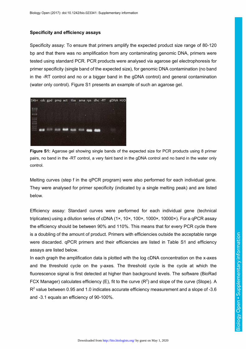
Specificity and efficiency assays
Specificity assay: To ensure that primers amplify the expected product size range of 80-120
bp and that there was no amplification from any contaminating genomic DNA, primers were
tested using standard PCR. PCR products were analysed via agarose gel electrophoresis for
primer specificity (single band of the expected size), for genomic DNA contamination (no band
in the -RT control and no or a bigger band in the gDNA control) and general contamination
(water only control). Figure S1 presents an example of such an agarose gel.
Figure S1: Agarose gel showing single bands of the expected size for PCR products using 8 primer pairs, no band in the -RT control, a very faint band in the gDNA control and no band in the water only
control.
Melting curves (step f in the qPCR program) were also performed for each individual gene.
They were analysed for primer specificity (indicated by a single melting peak) and are listed
below.
Efficiency assay: Standard curves were performed for each individual gene (technical
triplicates) using a dilution series of cDNA (1×, 10×, 100×, 1000×, 10000×). For a qPCR assay
the efficiency should be between 90% and 110%. This means that for every PCR cycle there
is a doubling of the amount of product. Primers with efficiencies outside the acceptable range
were discarded. qPCR primers and their efficiencies are listed in Table S1 and efficiency
assays are listed below.
In each graph the amplification data is plotted with the log cDNA concentration on the x-axes
and the threshold cycle on the y-axes. The threshold cycle is the cycle at which the
fluorescence signal is first detected at higher than background levels. The software (BioRad
FCX Manager) calculates efficiency (E), fit to the curve (R2) and slope of the curve (Slope). A
R2 value between 0.95 and 1.0 indicates accurate efficiency measurement and a slope of -3.6
and -3.1 equals an efficiency of 90-100%.
Biology Open (2017): doi:10.1242/bio.023341: Supplementary information
Bio
logy
Ope
n •
Sup
plem
enta
ry in
form
atio
n
by guest on May 1, 2020http://bio.biologists.org/Downloaded from
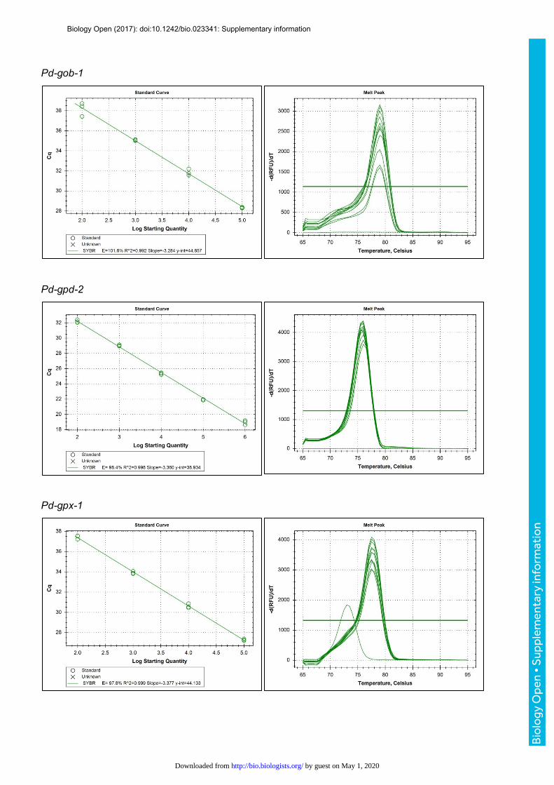
Pd-gob-1
Pd-gpd-2
Pd-gpx-1
Biology Open (2017): doi:10.1242/bio.023341: Supplementary information
Bio
logy
Ope
n •
Sup
plem
enta
ry in
form
atio
n
by guest on May 1, 2020http://bio.biologists.org/Downloaded from
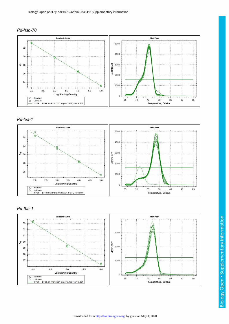
Pd-hsp-70
Pd-lea-1
Pd-tba-1
Biology Open (2017): doi:10.1242/bio.023341: Supplementary information
Bio
logy
Ope
n •
Sup
plem
enta
ry in
form
atio
n
by guest on May 1, 2020http://bio.biologists.org/Downloaded from

Pd-tps-2a
Pd-tps-2b
Biology Open (2017): doi:10.1242/bio.023341: Supplementary information
Bio
logy
Ope
n •
Sup
plem
enta
ry in
form
atio
n
by guest on May 1, 2020http://bio.biologists.org/Downloaded from
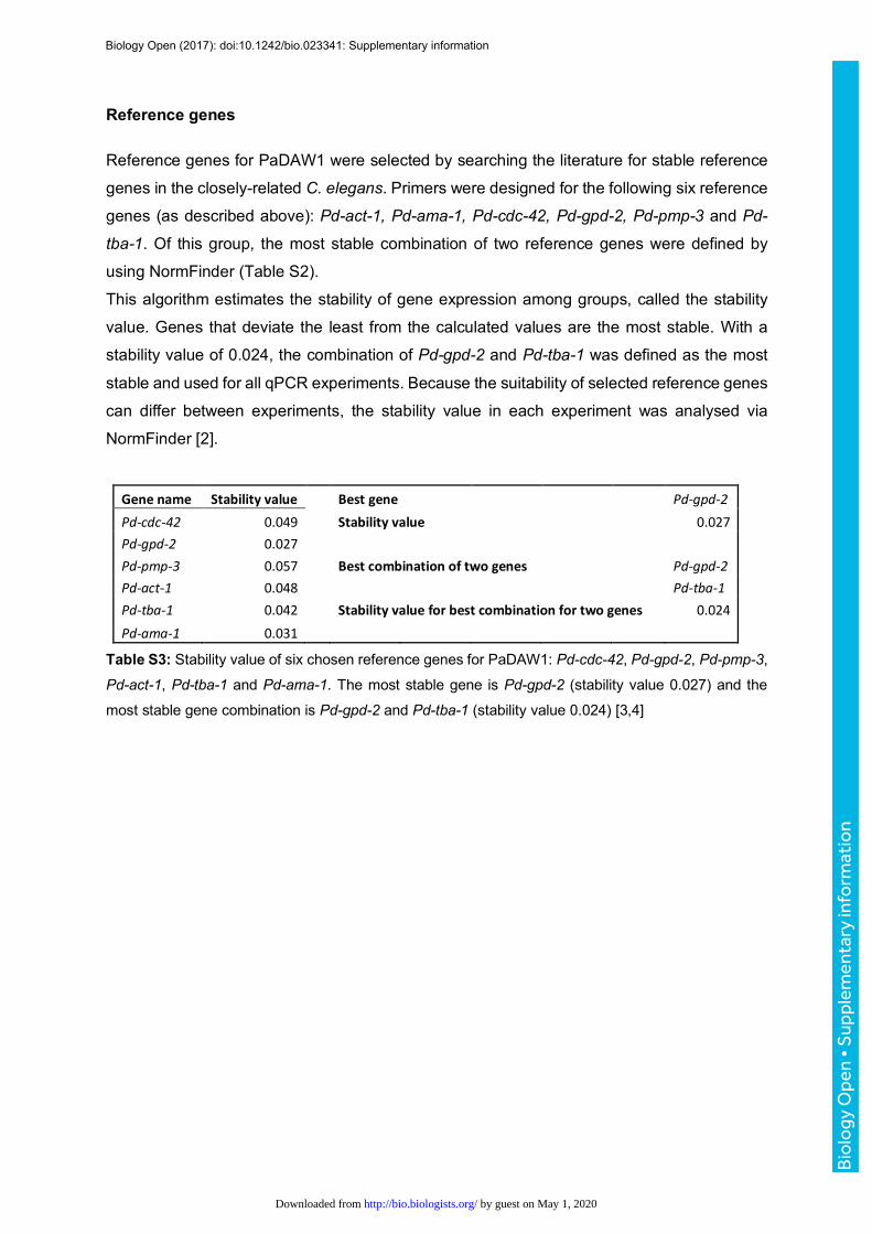
Reference genes
Reference genes for PaDAW1 were selected by searching the literature for stable reference
genes in the closely-related C. elegans. Primers were designed for the following six reference
genes (as described above): Pd-act-1, Pd-ama-1, Pd-cdc-42, Pd-gpd-2, Pd-pmp-3 and Pd-
tba-1. Of this group, the most stable combination of two reference genes were defined by
using NormFinder (Table S2).
This algorithm estimates the stability of gene expression among groups, called the stability
value. Genes that deviate the least from the calculated values are the most stable. With a
stability value of 0.024, the combination of Pd-gpd-2 and Pd-tba-1 was defined as the most
stable and used for all qPCR experiments. Because the suitability of selected reference genes
can differ between experiments, the stability value in each experiment was analysed via
NormFinder [2].
Genename Stabilityvalue Bestgene Pd-gpd-2Pd-cdc-42 0.049 Stabilityvalue 0.027Pd-gpd-2 0.027Pd-pmp-3 0.057 Bestcombinationoftwogenes Pd-gpd-2Pd-act-1 0.048 Pd-tba-1Pd-tba-1 0.042 Stabilityvalueforbestcombinationfortwogenes 0.024Pd-ama-1 0.031
Table S3: Stability value of six chosen reference genes for PaDAW1: Pd-cdc-42, Pd-gpd-2, Pd-pmp-3,
Pd-act-1, Pd-tba-1 and Pd-ama-1. The most stable gene is Pd-gpd-2 (stability value 0.027) and the
most stable gene combination is Pd-gpd-2 and Pd-tba-1 (stability value 0.024) [3,4]
Biology Open (2017): doi:10.1242/bio.023341: Supplementary information
Bio
logy
Ope
n •
Sup
plem
enta
ry in
form
atio
n
by guest on May 1, 2020http://bio.biologists.org/Downloaded from

Example chromatograms
Example chromatogram of a 20°C sample (elution times: 17 = Glycerol, 40 = Dulcitol (internal standard),
58 = Trehalose)
Example chromatogram of a 5°C sample
Biology Open (2017): doi:10.1242/bio.023341: Supplementary information
Bio
logy
Ope
n •
Sup
plem
enta
ry in
form
atio
n
by guest on May 1, 2020http://bio.biologists.org/Downloaded from

Example chromatogram of a non-treated acclimated (control) sample (elution times: 17 = Glycerol, 39 = Dulcitol (internal standard), 58 = Trehalose)
Example chromatogram of a Pd-tps-2a(RNAi) treated acclimated sample
References 1. Thorne MAS, Kagoshima H, Clark MS, Marshall CJ, Wharton DA. Molecular analysis of the cold
tolerant Antarctic nematode, Panagrolaimus davidi. PLoS ONE. 2014;9: e104526–e104526. doi:10.1371/journal.pone.0104526
2. Bustin SA, Benes V, Garson JA, Hellemans J, Huggett J, Kubista M, et al. The MIQE guidelines:minimum information for publication of quantitative real-time PCR experiments. Clin Chem.American Association for Clinical Chemistry; 2009;55: 611–622.doi:10.1373/clinchem.2008.112797
Biology Open (2017): doi:10.1242/bio.023341: Supplementary information
Bio
logy
Ope
n •
Sup
plem
enta
ry in
form
atio
n
by guest on May 1, 2020http://bio.biologists.org/Downloaded from

3. Seybold AC, Wharton DA, Thorne MAS, Marshall CJ. Establishing RNAi in a Non-ModelOrganism: The Antarctic Nematode Panagrolaimus sp. DAW1. PLoS ONE; 2016;11: e0166228.doi:10.1371/journal.pone.0166228
4. Seybold A. Molecular Adaptation Mechanisms in the Antarctic Nematode Panagrolaimus davidi.Phd Thesis, University of Otago. 2015.
Biology Open (2017): doi:10.1242/bio.023341: Supplementary information
Bio
logy
Ope
n •
Sup
plem
enta
ry in
form
atio
n
by guest on May 1, 2020http://bio.biologists.org/Downloaded from