investigation journal name date - museum of science and ......involves opening the chest and heart...
TRANSCRIPT

Live...from the He
art
Investigation Journ
al
Investigation Journal
Name Date

The Heart
Coronary Artery Disease
Heart Interior
Every muscle in the body needs a fresh supply of blood and nutrients in order to survive. The job of the heart is to pump nutrient-rich blood throughout the body.
1 2
Heart Exterior
When the arteries of the heart become blocked, blood flow to the heart muscle and the rest of the body is severely restricted. This means that the heart has to work much harder to maintain proper blood flow.
Plaque consists of deposits of fats, inflammatory cells, proteins and calcium material along the lining of arteries. In a person with coronary artery disease, the arteries can become blocked by this build-up of plaque, or the plaque may rupture causing the artery to become immediately blocked.
When a doctor suspects that a patient has heart disease, he or she may use a diagnostic tool call angiography, a special type of X-ray that allows a close look at the arteries of the heart. A catherter, or flexible tube, is inserted into a large artery in the patient's leg and is threaded up to the aorta. A special fluid called contrast dye is injected into the aorta and the X-ray machine captures pictures (angiograms) of the dye in the arteries. The doctor can then determine if there is blockage in any of the arteries that supply blood to the heart.
Heart & BlockageWe
lcome Welcome to Live… from the Heart!
The Museum of Science and Industry, Chicago is providing you with unique access to an operating room at Advocate Christ Medical Center to view live open-heart surgery. You’ll watch the surgical team in action and learn the role each person plays by talking with them while they work. You’ll also learn about the patient’s risk factors, diagnosis and treatment. Use this journal as your guide throughout the surgery to record questions, take notes, and diagram aspects of the procedure.
0% Blockage 50-60% Blockage 95% Blockage
Bypass surgery creates an alternate route for blood to flow to the heart muscle. This is similar to I-294 that provides an alternative route for cars to travel around Chicago.

INCHESMM
1
1 0 2 0 3 0 4 0 5 0 6 0 7 0 8 0 9 0 10 0 11 0 12 0 13 0 14 0 15 0 16 0 17 0 18 0 19 0 20 0 21 0 22 0 23 0 24 0 25 0
2 3 4 5 6 7 8 9 10
4
Patient ProfileBypass
Diagram
3
Bypass DiagramPatient Profile
RightCoronary Artery
Left AnteriorDescendingArtery
CircumflexBranch
LeftCoronaryArtery
Did you know? A saphenous vein is 4 to 6 mm wide, and an internal mammary artery is just 1 to 3 mm wide!
SEX: AGE:
RISK FACTORS:

Careers in the ORToday’s surgical team members are:
Surgeon: ________________________________________________________________________________________________
Physician Assistant: ______________________________________________________________________________________
Anesthesiologist: ________________________________________________________________________________________
Perfusionist: _____________________________________________________________________________________________
Scrub Nurse(s): __________________________________________________________________________________________
Circulating Nurse(s): ______________________________________________________________________________________
5
Scrub Nurse
Difibrillation Paddles
Pacemakerand Leads
Endoscopic Kit
Sternal Saw
Endoscopic Monitor
Patient Statistics Monitor
Electro-Cautery Unit
Scrub Nurse
Circulating Nurse
Scrub Nurse
Surgical Lights
Defibrillation Unit
CardiovascularSurgeon
PhysicianAssistant
Anesthesiologist
Perfusionist
Heart Lung Machine
Visit www.livefromtheheart.org to get more information about the personnel and equipment in the operating room. 6
Surgical Team Member I Will Shadow: ________________________________________________________________
Major Responsibilities: _____________________________________________________________________________
_________________________________________________________________________________________________
_________________________________________________________________________________________________
_________________________________________________________________________________________________
Education and Training: ____________________________________________________________________________
_________________________________________________________________________________________________
_________________________________________________________________________________________________
_________________________________________________________________________________________________
Careers in the ORCareers in
the OR
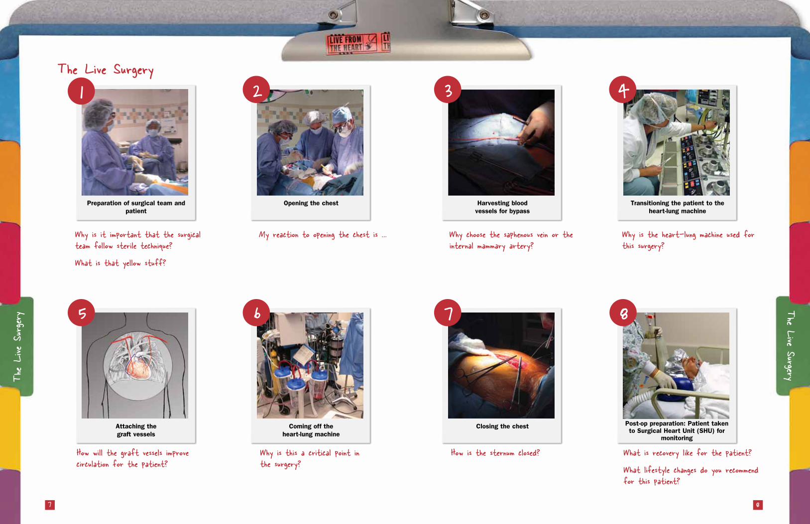
The Live Surgery
7 8
Preparation of surgical team and patient
Attaching the graft vessels
Harvesting blood vessels for bypass
Closing the chest
Opening the chest
Coming off the heart-lung machine
Transitioning the patient to the heart-lung machine
Post-op preparation: Patient taken to Surgical Heart Unit (SHU) for
monitoring
The Live SurgeryThe Live
Surgery
Why is it important that the surgical team follow sterile technique?
What is that yellow stuff?
How will the graft vessels improve circulation for the patient?
My reaction to opening the chest is …
Why is this a critical point in the surgery?
Why choose the saphenous vein or the internal mammary artery?
How is the sternum closed?
Why is the heart-lung machine used for this surgery?
What is recovery like for the patient?
What lifestyle changes do you recommend for this patient?
1 2 3 4
5 6 7 8

Anastomosis - A natural or surgical joining of parts or branches of tubular structures so as to make or become continuous.
Angioplasty – A procedure to open clogged arteries in which a tiny, hollow tube (catheter) with a balloon at its tip is inserted into an artery and inflated to compress plaque against the artery wall.
Artery - A blood vessel that carries blood away from the heart to the body.
Atherosclerosis - Commonly called “hardening of the arteries” - a variety of conditions caused by fatty or calcium deposits on the artery walls, causing them to narrow.
Blood pressure - Pressure of blood against the walls of a blood vessel or heart chamber.
Bypass - An alternative passage created surgically to divert the flow of blood around a blockage.
Capillaries - Tiny blood vessels between arteries and veins where the exchange of gases (oxygen and carbon dioxide) and nutrients takes place.
Cardiac - Pertaining to the heart.
Cardiac catheterization - A diagnostic procedure in which a tiny, hollow tube (catheter) is inserted into an artery or vein in order to evaluate the heart and blood vessels.
Cardiology - The clinical study and practice of treating the heart.
Cardiovascular - Pertaining to the heart and blood vessel (circulatory) system.
Catheter – A tiny, hollow tube inserted into an artery or vein for diagnostic and treatment purposes.
Cauterize – To cut soft tissue with heat; helps seal blood vessels to minimize bleeding.
Cholesterol - A substance normally made by the body but also found in foods from animal sources like beef, eggs and butter. Too much cholesterol in the body can lead to narrowing and blockage of the arteries, especially those that feed the heart and keep it healthy. Ideally, blood cholesterol levels should be less than 200mg/dL.
Coronary arteries - Two arteries that branch off the aorta and provide blood to the heart muscle.
Graft – A blood vessel surgically removed from the body and attached to the heart to create a new pathway around clogged arteries.
Heart attack (also called myocardial infarction) - Occurs when one or more regions of the heart muscle experience a severe or prolonged decrease in oxygen supply caused by blocked blood flow to the heart muscle.
Internal mammary artery – An artery in the chest wall frequently used to bypass blockage in the heart.
Ischemia - Decreased flow of oxygenated blood to an organ due to obstruction in an artery.
Left atrium - The upper left chamber of the heart. It receives oxygen-rich (red) blood from the lungs via the four pulmonary veins, and then sends this blood to the left ventricle. Blood is not actually red or blue. It is only depicted that way for diagrammatic purposes.
Left ventricle - The lower left-hand chamber of the heart. It receives oxygen-rich (red) blood from the left atrium and pumps it into the aorta, which delivers the blood to the body. The left ventricle must be strong and muscular in order to pump enough blood to the body to meet its requirements.
Lipid - A fatty substance in the blood.
Open-heart surgery - Surgery that involves opening the chest and heart while a heart-lung machine takes over for the heart and lungs.
Plaque - Deposits of fat or other substances attached to the artery wall.
Pulmonary artery - The blood vessel connecting the right ventricle to the lungs, allowing oxygen-poor (blue) blood to receive oxygen.
Pulmonary vein - The vessel that carries oxygenated blood from the lungs to the left side of the heart.
Right atrium - The upper right chamber of the heart, which receives oxygen-poor (blue) blood from the body and sends it to the right ventricle
Right ventricle - The lower right chamber of the heart, which receives oxygen-poor (blue) blood from the right atrium and sends it to the pulmonary artery.
Risk factor - A condition, element or activity that may adversely affect the heart.
Saphenous vein – A vein in the leg often removed and attached to the heart to bypass blockage in the heart.
Stent – A wire mesh tube inserted in a clogged artery to prevent the artery from narrowing again.
Suture - The process of joining two surfaces or edges together along a line by or as if by sewing; the material, such as thread, gut or wire, that is used in this procedure.
Vein - A blood vessel that carries blood from the body back to the heart.
Heart Valve DiseaseThe heart has four chambers. The upper two are the right and left atria. The lower two are the right and left ventricles. Blood is pumped through the chambers, aided by four heart valves. The valves open and close to let the blood flow in only one direction. Each valve has a set of flaps (also called leaflets or cusps). When working properly, the heart valves open and close fully.
•�The�tricuspid�valve�is�between�the�right�atrium�and right ventricle.
•��The�pulmonary�valve�is�between�the�right�ventricle and the pulmonary artery.
•��The�mitral�(bicuspid)�valve�is�between�the�left�atrium and left ventricle.
•��The�aortic�valve�is�between�the�left�ventricle�and�the aorta.
Heart valves don't always work as they should. A person can be born with an abnormal heart valve, a type of congenital heart defect. Also, a valve can become damaged by infections or conditions such as rheumatic fever.
A defective heart valve is one that fails to fully open or close. A stenotic heart valve can't open completely, so blood is pumped through a smaller-than-normal opening. A valve also may not be able to close completely. This leads to regurgitation (blood leaking back through the valve when it should be closed).
People with congenital heart valve defects may need treatment with drugs. Some valve defects may be repaired or replaced during surgery.
Robotic SurgeryWhere traditional open surgeries require large incisions and long recovery times, the da Vinci® Surgical system provides surgeons with an alternative to both traditional open surgery and conventional laparoscopy, putting surgeons hands at the controls of a state-of-the-art robotic platform. The da Vinci® System enables surgeons to perform even the most complex and delicate procedures through very small incisions with unmatched precision resulting in less pain, scarring and quicker return to normal activities. For years, Advocate Christ Medical Center has been pioneering the use of minimally invasive techniques.
Angioplasty and Stent In this procedure a thin, flexible tube called a catheter is inserted into the narrowed artery. A tiny balloon at the tip of the catheter is inflated, mashing the blockage against the vessel wall. A stent, a wire-mesh tube, is then inserted. The stent remains in the artery permanently and acts like a scaffold, preventing further closing or narrowing of the artery. Aortic AneurysmAn aortic aneurysm is a weakened and bulging area in the aorta, the major blood vessel that feeds blood to the body. The aorta, about the thickness of a garden hose, runs from your heart through the center of your chest and abdomen. Because the aorta is the body's main supplier of blood, a ruptured aortic aneurysm can cause life-threatening bleeding.
Surgery to repair an aortic aneurysm involves removing the damaged section of the aorta and replacing it with a synthetic tube (graft), which is sewn into place. This procedure requires open-abdominal or open-chest surgery.
Glossary of TermsOther Heart Conditions and Treatments
Definitions provided by Advocate Health Care 109
GlossaryOt
her Co
nditions
and
Treatm
ents

© 2010 Museum of Science and Industry, Chicago. This publication may be reproduced by teachers for education use only.
For more information, email us at [email protected]
3, 2, 1 Check for Understanding
3 things I learned today …
2 questions I still have …
1 thing I will tell someone else about …