investigation of an extracorporeal immobilized enzyme
TRANSCRIPT
Investigation of an Extracorporeal Immobilized EnzymeDevice; A Potential Treatment for Blood Deheparinization
by
Guillermo A. Ameer
B.S. Chemical EngineeringUniversity of Texas at Austin
(1993)
Submitted in partial fulfillment of the requirements for the Degree of
Doctor of Sciencein Chemical Engineering
at the
Massachusetts Institute of Technology
February, 1999
1999 Massachusetts itute of Technology
Al,r;~s reerved
Signature of AuthorDeartment of Chffmical EngineeringD6cember 14, 1998
Certified by
Thesis SupervisorK e Germeshausen Professor of Chemical
,/and Bio dical Engineeing
Accepted by Rbbert E. CohenSt. Laurent Professor of Chemical EngineeringChairman, Committee for Graduate Students
OCTs_
1=a "
This Doctoral Thesis has been examined by the following Thesis Committee:
Robert S. Langer, Sc.D.Thesis SupervisorGermeshausen Professor of Chemical and Biomedical EngineeringDepartment of Chemical EngineeringMassachusetts Institute of Technology
Charles Cooney, Ph.D.Professor of Chemical and Biochemical EngeeringDepartment of Chemical EngineeringMassachusetts Institute of Technology
William Harmon, M.D.Director of Pediatric NephrologyDepartment of NephrologyThe Children's HospitalHarvard Medical School
Paula T. Hammond, Ph.D.Development Assistant ProfessorDepartment of Chemical EngineeringMassachusetts Institute of Technology
Ram Sasisekharan, Ph.D.Assistant ProfessorDivision of Bioengineering and Environmental HealthMassachusetts Institute of Technology
2
Investigation of an Extracorporeal Immobilized EnzymeDevice; A Potential Treatment for Blood Deheparinization
by
Guillermo A. Ameer
Submitted to the Department of Chemical Engineering in partial fulfillment of the requirements forthe Degree of
Doctor of Sciencein Chemical Engineering
ABSTRACT
Heparin is an anticoagulant used in extracorporeal procedures such as hemodialysis andopen heart surgery. Unfortunately, heparin may induce potentially fatal complications in patients athigh risk of bleeding. A potential treatment to make heparin anticoagulation therapy safer is the useof an immobilized heparinase I reactor to achieve regional heparinization of a closed circuit. This isa method in which heparin is infused into the extracorporeal circuit pre dialyzer and neutralizedpost dialyzer. However, a significant problem has been the design of a safe and efficient reactor formedical use.
Taylor-Couette flow was evaluated for potential use in a reactor for blood deheparinization,taking into account clinical specifications and engineering principles. A Taylor-Couette flow devicewas designed with recirculation ports, strategically placed tangentially to the rotational flow, toallow recirculation of the agarose beads without the use of an external pump. This reactor removed90% of the heparin's anticoagulant activity from 450 cc of human citrated blood in vitro within 3minutes of operation at a flow rate of 100 ml/min. Even though this device demonstrated to beeffective, it was not practical due to the significant decrease in blood cell counts and the largedegree of hemolysis that was observed when whole blood was tested (plasma free hemoglobingreater than 800 mg/dL). Therefore, it was shown that the well known beneficial characteristics ofTaylor vortices on biological systems are lost when agarose beads are fluidized within wholeblood.
These findings motivated a new design which incorporated Taylor-Couette flow,simultaneous separation/reaction, and fluidization of the agarose immobilized heparinase.Fluidization was achieved via membrane induced circumferencial flow within the reactive chamberof the device which was separate from the blood cells and the Taylor vortices. Residence timedistribution studies were performed and a mathematical model was developed in order to predictand optimize heparin neutralization. The model predicted heparin conversions in saline within amean of 5% error relative to the experimental results. The new design, referred to as vortex flowplasmapheretic reactor (VFPR), was tested in vitro and ex vivo in sheep. The VFPR achievedheparin conversions of 44 ± 0.5% and 34 ± 2% in saline and human blood, respectively. Inaddition, there was no hemolysis or cell count decrease. Regional heparinization experiments insheep resulted in 39 ± 1.7% mean heparin conversion. The procedure did not effect any clinicallysignificant changes in complete blood counts and total protein concentrations for up to 2 hours.Furthermore, gross necropsy and histopathology reports did not show any negative effects on thebrain, kidney, liver, spleen, lungs, and heart.
Thesis Supervisor: Robert LangerTitle: Professor of Chemical and Biomedical Engineering
3
Acknowledgments
Growing up in Panama, I never imagined that I would contribute to the body of knowledge
in the Chemical and Biomedical engineering fields. Also beyond my imagination was the fact that I
would do so in the laboratory of Professor Robert Langer at M.I.T., for whom I have the greatest
respect. So, needless to say, there are numerous people I have to thank for their contribution
(through either direct help or emotional support) to the completion of this thesis. My thesis
advisor, Bob Langer, gave me the opportunity to work on this project which has been going on for
over two decades. Of course, the problem of safer anticoagulation had been around for much
longer and the need for safer heparin therapy among patients with acute renal failure remained
unmet. This problem has been the motivation for this research. Bob has been very enthusiastic,
supportive, available for the project, and has always given me the freedom to pursue different
paths to address the research problem and to develop myself as a scientist. I am also grateful for
the opportunity he gave me to go to Japan for two months to broaden my research skills and to
make international contacts for future collaborations.
I would like to thank William Harmon, M.D., Paula Hammond, Ph.D., Charlie Cooney,
Ph.D., and Ram Sasisekharan, Ph.D. for serving on my thesis committee. Dr. Harmon was a
valuable resource for the medical issues that I had to deal with during my research and also for
facilitating the contacts that I made at Children's Hospital, Boston MA. John Thompson and the
staff of the dialysis unit were very helpful in regards to equipment, the operation of an
extracorporeal circuit, and the practical use of heparin. To Paula Hammond and Charlie Cooney I
am thankful for the insightful discussions regarding chemistries and the vortex flow reactor,respectively. My thanks to Ram for involving me in the project when he was working with Bob
and for his expertise and friendship during my years as a graduate student. His emotional and
technical support will always be remembered.
The Langer lab has made my graduate years a fun experience thanks to the many friendly
people that have been part of it, both past years and present. Due to the large number of friends
that I have made over the years in the lab, it is not feasible for me to mention the highlights of each
of their contributions, but in general the discussions that we've had and the interest that they have
shown in my project are sincerely appreciated. I am, however, compelled to mention Eric
Grovender and Bojana Obradovic for their input on the reactor modeling studies. I also thank my
office mates (present and past), Rebecca, Bojana, Nenad, Samir, Mark and Lon, for their ability to
make sharing small quarters enjoyable. I could not conceive of a better office experience.
4

I would like to thank P. Dozois at the M.I.T. Central Machine Shop and Sylvester
Szczepanowski at the M.I.T. B.C.S. Machine Shop for machining the reactors. I also thank IBEX
Technologies (Montreal Canada) for providing the heparinase I. Recognition is also due to the
volunteer blood donors, Srivatsan Raghavan, Dave Ting, and the many other UROPs that I
supervised for helping with the experiments. The in vitro experiments with human blood were
possible thanks to the cooperation of the personnel at the Blood Donor Center of Children's
Hospital, Boston, Massachusetts.
To the many friends in Boston that I have met outside of M.I.T., I am also grateful.
Specifically, I'd like to thank Tirzah Spencer, who has been an excellent friend, Barbara Brothers
for her support and Mihaela Bazalakova for her support and for exposing me to the beauty of
Boston and its international crowd. I also appreciate the faith and encouragement that my friends
from Texas have shown towards me. In particular Raphaelle Johnson and Gargi Mukherji.
I am also thankful to my family for their support and encouragement. Without their
direction I would not have been the first of my family to achieve this degree. I thank my sisters
and brother - Sheila, Yvonne, Sharon, and Jorge - for being there when I needed them. Most
importantly, I dedicate this thesis to my Mother and Father, for they are the strongest personalities
I have ever met, have been available for me regardless of circumstances, and instilled into me the
foundation that makes me the person that I am today. Finally, I thank God for he has been with
me throughout my life.
Guillermo A. AmeerDecember 1998
This work was supported by the National Science Foundation and the National Institutes of Health
(GM 25810).
5
Table of Contents
Title Page .......................................................... 1
C om m ittee P age...................................................................... 2
Abstract............................................................ 3
Acknowledgments................................................... 4
Table of Contents ............................................................. 6
List of Figures ..................................................... 7
List of Tables......................................................8
1. Introduction.....................................................91.1 Motivation..................................................... 91.2 Specific Aims............................................... 111.3 Immobilized Proteins in Extracorporeal Systems .................................. 121.4 Heparin and its Role in Extracorporeal Blood Circulation ......................... 161.5 Heparinase I ............................................................... 181.6 Previous Immobilized Heparinase Devices ......................................... 191.7 Clinical M odel Selection................................................................. 221.8 References .................................................................... 26
2. The Vortex Flow Fluidized Bed Reactor.........................................302.1 Introduction ........................................................... ............. 302.2 M aterials and M ethods.................................................................. 312.3 Results and D iscussion................................................................ 352.4 References ................................................................ 44
3. The Vortex Flow Plasmapheretic Reactor........................................463.1 Introduction............................................................. .... 463.2 M aterials and M ethods.................................................................. 463.3 Results and Discussion................................................................ 493.4 R eferences ............................................................................... 58
4. Reactor4.14.24.34.44.5
5. Reactor5.15.25.35.4
Fluid Dynamics and Modeling...........................................59Introduction .............................................................................. 59M aterials and M ethods.................................................................. 59M athem atical A nalysis.................................................................. 60Results and Discussion................................................................ 63References ..................................................................... 70
Performance Ex Vivo in Sheep..........................................71Introduction............................................................... 71M aterials and M ethods.................................................................. 71Results and Discussion................................................................ 73R eferences .............................................................................. . 90
6. Conclusions and Recommendations for Future Work...........................91
Appendix...........................................................................95
6
List of Figures
Chapter 1.Figure 1.1.Figure 1.2.Figure 1.3.Figure 1.4.
Chapter 2.Figure 2.1.Figure 2.2.Figure 2.3.Figure 2.4.Figure 2.5.Figure 2.6.Figure 2.7.Figure 2.8.Figure 2.9.
Chapter 3.Figure 3.1.Figure 3.2.Figure 3.3.Figure 3.4.Figure 3.5.Figure 3.6.Figure 3.7.Figure 3.8.
Chapter 4.Figure 4.1.Figure 4.2.Figure 4.3.Figure 4.4.
Chapter 5.Figure 5.1.Figure 5.2.Figure 5.3.Figure 5.4.Figure 5.5.Figure 5.6.Figure 5.7.Figure 5.8.Figure 5.9.a.Figure 5.9.b.Figure 5.9.c.Figure 5.10.Figure 5.11.
IntroductionProposed method for regional heparinization of a patient ...........................Illustration of the components of a heparin polysaccharide chain ..................Diagram of the old heparinase I recirculating reactor ............................D iagram of the shaker reactor...........................................................
12172121
The Vortex Flow Fluidized Bed Reactor (VFFBR)Diagram of the vortex flow fluidized bed reactor.................................... 39In vitro batch heparin removal by the VFFBR (design I) ......................... 40Safety data for the VFFBR (design I) ............................................... 40Release of free hemoglobin into the plasma (design I) ............................ 41Effect of gel volume and rotation rate on hemolysis ............................... 41In vitro batch heparin removal by the VFFBR design II ............................ 42In vitro regional heparinization by the VFFBR design II.......................... 42Safety data for the VFFBR (design II) ................................................ 43Release of free hemoglobin into the plasma (design II) ........................... 43
The Vortex Flow Plasmapheretic Reactor (VFPR)Diagram of the vortex flow plasmapheretic reactor ..................................Regional heparinization using the packed bed ........................................Protein entrapment methods for a Taylor-Couette flow reactor.....................Flow dynamics within the VFPR ......................................................Performance of the immobilized heparinase I packed bed in saline ................Performance of the VFPR in saline ....................................................Operation of the VFPR with blood.....................................................Performance of the VFPR with human blood in vitro...............................
Reactor Fluid Dynamics and ModelingExperimental systems for residence time distribution studies.......................Diagram of the reactor sections to be modeled........................................Diagram of the compartment model for the VFPR ................................Exit age distribution for compartment 1 and the whole device......................
Reactor Performance Ex Vivo in SheepDouble lumen catheter and ex vivo experimental set up .............................Safety data for control sheep............................................................Safety data for regionally heparinized sheep 1........................................Safety data for regionally heparinized sheep 2........................................Safety data for regionally heparinized sheep 3........................................Safety data for regionally heparinized sheep 4........................................Grouped safety data for regionally heparinized sheep ...............................Environmental S.E.M. of the polyester membrane ..................................Regional heparinization of sheep.......................................................Regional heparinization of sheep.......................................................Regional heparinization of sheep.......................................................Environmental S.E.M. of plasma exposed bead .....................................Environmental S.E.M. of saline exposed bead.......................................
5354545556565757
67686869
79808182838485868787888989
Chapter 6. Conclusions and Recommendations for Future WorkFigure 6.1. Proposed modification to the VFPR ................................................... 94
7
List of Tables
Chapter 1.Table 1.1.Table 1.2.Table 1.3.
Chapter 2.Table 2.1.Table 2.2.
Chapter 3.Table 3.1.Table 3.2.Table 3.3.
Chapter 4.Table 4.1.Table 4.2.Table 4.3.Table 4.4.
IntroductionAmino acid composition of heparinase I ............................................ 19Efficacy of old recirculating reactor.................................................. 20Current alternatives to systemic heparinization ....................................... 23
The Vortex Flow Fluidized Bed Reactor (VFFBR)Dimensions for the VFFBR (designs I and II) ..................................... 35Minimum rotation rates required in the VFFBR.................................... 37
The Vortex Flow Plasmapheretic Reactor (VFPR)Dimensions of the vortex flow plasmapheretic reactor ............................ 47Complete blood cell and hemolysis data for the VFPR.............................. 51Safety data for 1 hour of operation of the VFPR..................................... 52
Reactor Fluid Dynamics and ModelingComparison of mathematical models for E and Eoal with experiment ............ 63Summary of the model output for four experimental conditions ................. 64Comparison of model predictions with experimental data........................... 65Sensitivity analysis on Elb .............................................................. 66
Chapter 5. Reactor Performance Ex Vivo in SheepTable 5.1. Total protein and fibrinogen values for sheep experiments ....................... 75
8
Chapter 1
Introduction
1.1 Motivation
The effective and safe detoxification of blood, toxified either due to high concentration of
metabolites or due to drug overdoses, remains a serious problem. Current methods to treat blood
toxicity include hemodialysis, hemofiltration or activated charcoal filters. These methods are
widely used in the clinic but they lack specificity in metabolite removal and therefore essential
metabolites have to be restored continuously (Takao, 1993). Several investigators have proposed
the use of immobilized enzymes in extracorporeal bioreactors as a means to specifically carry out a
desired reaction without disrupting other blood components (Ambrus et al., 1978; Dumler et al.,
1981; Horvath et al., 1973; Langer et al., 1982; Lavin et al., 1985; Olanoff et al., 1975). Even
though there is a good understanding of how enzymes can improve or correct pathological
conditions or eliminate undesirable metabolites from the blood, immobilized enzyme bioreactors
are not widely used in the clinical setting. The main reasons are: the high cost of production of
enzymes (specially from animal source), the need to obtain derivatives of the enzymes with
unaltered therapeutic activity, but with no toxicity and antigenicity (Klein and Langer, 1986) and
the lack of bioreactor designs that are safe and efficient.
Heparin is a very important and widely used drug in modem medicine and surgery.
Heparin is used to treat thrombotic disorders and as an anticoagulant to prevent clotting in
procedures requiring extracorporeal equipment such as hemodialysis, membrane oxygenation, and
cardiopulmonary bypass surgery. However, the use of heparin can lead to complications in
patients who are at high risk of bleeding such as those suffering from acute renal failure (Broyer et
al., 1986) and those who have undergone open heart surgery (Salzman et al., 1975). Heparin was
also found to be the prescribed drug responsible for the most deaths in patients that were otherwise
healthy (Porter and Fick, 1977). Currently, the anticoagulant effect of heparin is neutralized with
protamine sulfate, a highly basic protein that binds to heparin. However, the use of protamine can
cause severe side-effects including hypotension, vasodilation, pulmonary platelet accumulation and
bradycardia (Wakefield et al., 1986). This heparin reversal method may benefit patients that
require rapid removal of heparin after a surgical procedure but has very limited application in acute
renal failure because of the complexity of on line titration and the side effects mentioned earlier.
9
To address this problem, a novel application of enzymes was introduced by Langer and his
co workers in 1982 (Langer et al., 1982). The authors describe the enzymatic degradation of
heparin by heparinase I (an alpha eliminase extracted from Flavobacterium heparinum) in a dog
animal model. An immobilized heparinase blood filter could be used in a regional heparinization
system and it is a potential alternative to the current method of neutralizing heparin with protamine.
Recently, IBEX Technologies conducted clinical trials with soluble heparinase I to reverse
heparinization after cardiovascular surgery and the results are encouraging. In this application
heparinase is injected into the patient's bloodstream (Broughton, 1995). However, the repeated
use of soluble heparinase towards regional heparinization in acute kidney failure would face similar
problems of immunogenicity.
Throughout the last decade, several investigators studied the potential for an enzymatic
deheparinization system using an immobilized enzyme fluidized bed system in dog and sheep
animal models (Larsen et al., 1984; Freed et al., 1988; Bernstein et al., 1987). Hollow fiber
bundles were also explored as a possible heparinase reactor (Comfort et al., 1989). These studies
contributed a great deal of knowledge in the area of immobilized enzymes in extracorporeal devices
because mathematical models and first principles were developed to aid the design of the device.
However, in these reactors, blood flow rates allowed in the fluidized systems were limited to under
200 ml/min. (Freed et al., 1993). This limits the heparinase reactor to only neonates or small
children and can not be used on adult patients suffering from acute renal failure and therefore
jeopardizes its ability to compete with current heparin reversal methods. In addition to the flow
rate limitation, the clinical use of the heparinase reactors was not possible because of significant
blood damage (Larsen et al., 1984; Freed, 1988), scarcity and low purity of the native heparinase
I, and difficulty of use of the bioreactors in a clinical setting. In summary, the following problems
have to be addressed in order for a heparinase reactor to be clinically useful:
1. Bioreactor Design:
a) particle fluidization at flow rates of 150-400 ml/min.
b) reactor induced blood damage
c) efficiency
d) simplicity
2. Enzyme Immobilization:
a) high enzyme activity retention
b) stability in blood
c) enzyme purity
10
To address the bioreactor design issues, Taylor vortices are applied to the immobilized
heparinase system as will be discussed further. Taylor vortices have been shown to have excellent
mixing characteristics in reactors that processed biological systems such as cell cultures, blood
plasmapheresis and oxygenation (Gaylor and Smeby, 1976; Mottaghy and Hanse, 1985; Strong
and Carlucci, 1976; Jaffrin, 1989). In addition, Cooney and co workers investigated and modeled
reaction and mass transfer properties in a particle and membrane vortex flow reactor (Iosilevskii et
al., 1993; Moore, 1994). The enzyme immobilization issues are addressed by assessing
immobilization chemistries and supports using high purity heparinase I (95%+ by SDS-PAGE)
donated by IBEX Technologies. In this research, novel vortex flow reactor designs and their
clinical feasibility are investigated. The reactor performance is evaluated for regional heparinization
using a sheep animal model. By applying Taylor vortices to immobilized enzyme bioreactors in
extracorporeal systems, this research should enhance the knowledge and applications of Taylor
vortices in biological and enzymatic systems. The ultimate goal of this research is to allow a safer
extracorporeal treatment for the critically ill patient undergoing renal replacement therapy and facing
heparin related complications.
1.2 Specific Aims
The general objective of this thesis is to investigate the use of immobilized heparinase I in a
reactor that would allow regional heparinization of the extracorporeal circuit for potential use in the
treatment of acute renal failure (Figure 1.1). The specific objectives of this thesis are:
1. To design, construct, and characterize prototype reactors that utilize porous beads as an
enzymatic support
2. To evaluate reactor safety and efficacy through in vitro and ex vivo experiments in a sheep
animal model; specifically,
2.1 To investigate the effect of the device on the blood components
2.2 To assess whether high conversions can be achieved at clinically relevant flow rates
for adults (150-400 ml/min.) At the beginning of this research, a high conversion
was defined to be 80% per pass through the reactor. After consulting with various
nephrologists and clinicians, it was determined that such a high goal for conversion
would be necessary only if the circuit were overanticoagulated with heparin. At
adequate heparin concentrations, sufficient to prevent clotting in the circuit, 80%
conversions could induce clotting at the vascular access of the patient. Therefore,
the earlier goal of 80% conversion was reconsidered and replaced with the range of
11
45-50% conversion. The criteria for this new value are discussed later in the
Chapter.
3. To develop a mathematical model that would adequately describe the
deheparinization process in the reactor for generalization of the results.
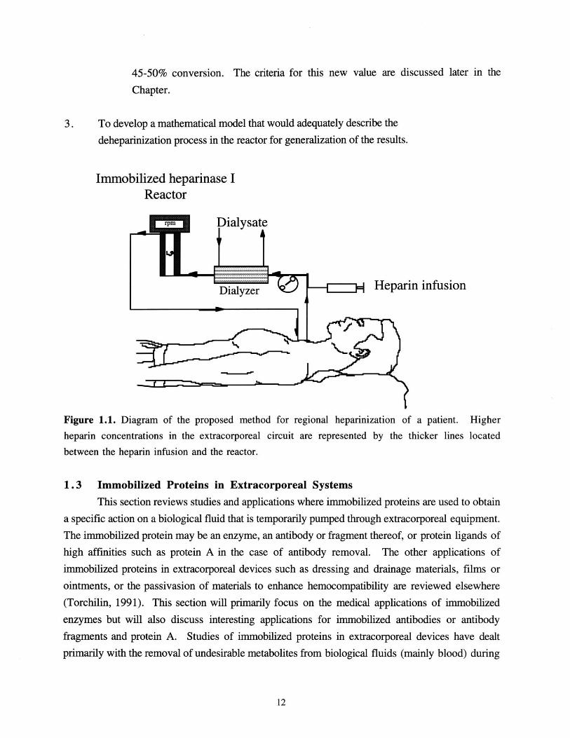
Immobilized heparinase IReactor
r~m Dialysate
alyz E Heparin infusion
Figure 1.1. Diagram of the proposed method for regional heparinization of a patient. Higher
heparin concentrations in the extracorporeal circuit are represented by the thicker lines located
between the heparin infusion and the reactor.
1.3 Immobilized Proteins in Extracorporeal Systems
This section reviews studies and applications where immobilized proteins are used to obtain
a specific action on a biological fluid that is temporarily pumped through extracorporeal equipment.
The immobilized protein may be an enzyme, an antibody or fragment thereof, or protein ligands of
high affinities such as protein A in the case of antibody removal. The other applications of
immobilized proteins in extracorporeal devices such as dressing and drainage materials, films or
ointments, or the passivasion of materials to enhance hemocompatibility are reviewed elsewhere
(Torchilin, 1991). This section will primarily focus on the medical applications of immobilized
enzymes but will also discuss interesting applications for immobilized antibodies or antibody
fragments and protein A. Studies of immobilized proteins in extracorporeal devices have dealt
primarily with the removal of undesirable metabolites from biological fluids (mainly blood) during
12
perfusion. The emphasis on detoxification using immobilized enzymes can be attributed to the
high specificity and catalytic activity of enzymes. The main areas of interest for immobilized
enzymes in extracorporeal devices are: treatment of liver and/or kidney failure, cancer, blood
diseases, and more recently, drug overdoses.
Kidney insufficiency can be treated by the removal of excessive urea from the blood in a
reactor with immobilized urease. This approach was proven by using a hollow fiber reactor with
immobilized urease (Salmona et al., 1974). An important issue that had to be addressed was the
elimination of ammonia formed due to the enzymatic hydrolysis of urea. A detoxifying reactor for
the removal of urea has also been described (Makarov and Kibardin, 1980). In their arrangement,
columns with activated charcoal, immobilized urease, and an inorganic cation exchanger were
sequentially arranged. The activated charcoal column achieved complete sorption of creatinine, and
partial sorption of urea, the urease column removed most of the urea, and the cation exchanger
eliminated the ammonia formed in the urease column.
The accumulation of toxic metabolites like ammonia, mercaptanes, phenols, phenylalanine
and free fatty acids in the blood can cause liver insufficiency and coma. In order to treat these
conditions a variety of immobilized enzyme reactors have been suggested (Kalghatgi et al., 1980;
Pedersen et al., 1979). The current method of blood purification, by passing it through activated
charcoal, shows improvement but is usually accompanied with different side reactions because of
the non-specific adsorption of many blood proteins and relatively extensive blood damage. Sung
and co workers (1986) described the removal of excess bilirubin from the blood for the treatment
of endogenous jaundice using an extracorporeal device with immobilized bilirubin oxidase from
Myrothecium verrucaria. The enzyme was immobilized on CNBr-activated Sepharose. Its
thermostability was 5 times higher than that of the soluble enzyme. The column reactor contained
15 ml of the immobilized enzyme gel and oxidized 60% of the elevated bilirubin.
Immobilized enzyme devices have also been investigated as a potential treatment of amino
acid-dependent tumors. L-asparaginase was immobilized by incorporation into cellulose triacetate
fibers or dacron (Salmona et al., 1974) and covalently bound on the inner surface of nylon tubing
(Horvath et al., 1973) or collagen films (Olanoff et al., 1975). L-asparaginase can reduce the
plasma L-asparagine concentration significantly, thereby depriving the tumor cells of this amino
acid. Normal cells synthesize asparagine while certain lymphomas depend on circulating
asparagine to grow. Systems have been developed that resulted in almost quantitative removal of
asparagine from the blood in dogs, monkeys (Olanoff et al., 1975) and man (Sampson et al.,
1974). The first results from clinical trials with immobilized asparaginase were described by
13
Dumler and co workers in 1981. L-asparaginase was immobilized in a regenerated cellulose
hollow fiber reactor via trichloro-s-triazine. Blood asparagine levels decreased by 30% at flow
rates of 200 ml/min. in a hollow fiber reactor with immobilized L-asparaginase when compared to
traditional dialysis. The use of the L-asparaginase reactor led to noticeable temporary improvement
in two out of three patients with tumors. The L-asparaginase reactor did not provoke any
immunological reactions even though some of the patients demonstrated anaphylaxis after
parenteral administration of the native enzyme. Arginine-dependent tumors can be treated with
extracorporeal reactors with immobilized arginase. Arginase, immobilized in a hollow fiber
reactor, was used to treat familial hyperargininemia in model experiments performed with rabbit
blood (Rossi, 1981; Kanalas et al., 1982). Within the reactor, arginine was separated from blood
by means of a semipermeable membrane to low molecular weight substances.
The absence of the enzyme phenylalanine hydroxylase, which degrades the amino acid
phenylalanine, results in the genetic disorder Phenylketonuria. Patients suffering from this disease
have a very elevated plasma level of phenylalanine which results in neuropsychiatric disorders. A
promising treatment of this disorder may be the enzyme phenylalanine ammonia lyase which
converts phenylalanine into non-toxic transcinnanamic acid. Ambrus and co workers (1978)
proved the possibility of using devices with immobilized phenylalanine ammonia lyase when they
quantitatively removed phenylalanine rapidly from a sample of citrated blood. Incorporating a
multi-tubular device containing phenylalanine ammonia lyase immobilized on the inner surface of
nylon tubings lead to a 2-fold decrease in blood phenylalanine. Low levels of phenylalanine could
be maintained for at least two days (Pedersen et al., 1979). In similar experiments, the use of a
hollow fiber enzyme reactor shunted between the femoral artery and femoral vein of a dog for two
hours decreased blood phenylalanine levels from 15 to 2 mg/100 ml. The number of red blood
cells, leukocytes and platelets remained constant throughout the treatment (Ambrus et al., 1983).
Another application of immobilized enzymes to cancer therapy involves carboxypeptidase,
which cleaves folate and methotrexate. Folate is required for cancerous cells and the effect of the
enzyme is a severe folate deficiency. Methotrexate is a folate antagonist which is administered to
cancer patients during chemotherapy. By using an immobilized carboxypeptidase reactor, higher
drug doses of methotrexate can be administered for shorter time periods and the excess drug can be
eliminated on demand. This approach was investigated by Bertino and co workers (1979), who
constructed an immobilized carboxypeptidase reactor to remove methotrexate in canine blood in
vivo. The enzyme was linked via glutaraldehyde to nylon tubing and anisotropic hollow fibers.
The in vivo experiments were conducted at blood flow rates of 500 ml/min. for a 2 hour period.
Neither reactor was able to lower methotrexate blood levels significantly.
14
Plotz and co workers (1974) described the use of agarose immobilized albumin, packed
into a column, for the removal of protein-bound metabolites and toxins. Thyroxine,
taurolithocholate, chenodeoxycholate, and digitoxin bound tightly. Digoxin, as expected, was not
removed. The authors reported the in vitro removal of bilirubin from 10 ml of anticoagulated
whole blood with slight decreases in calcium and magnesium. There were no significant changes
in blood cells, platelets, electrolytes or clotting factors. However, the apparent hemocompatibility
of the column was demonstrated for very low flow rates (0.5-2.5 ml/min.). The beads could be
repeatedly reused without loss of efficiency after ethanol elution.
The use of immunoadsorption methods may take place in the clinical setting when an
enzyme is not feasible or readily available to remove an undesirable substance from blood.
Immunoadsorption relies on the specificity and high affinity of antibodies to deplete antigen plasma
concentrations. A potential application for immunoadsorption is in the treatment of dialysis related
amyloidosis (DRA), a debilitating and potentially fatal disease that could benefit from the specific
and significant removal of the protein beta-2-microglobulin ([ 2m) from blood. This protein is
implicated in the amyloid fibrils that deposit in the joints and create cystic bone lesions, destructive
arthropathy, and soft tissue damage (Schaeffer et al., 1995). DRA is a consequence of the longer
survival times that are now associated with patients with end-stage renal disease (ESRD).
Currently, the incidence of ESRD in the United States exceeds 250,000 patients and is rising by
approximately 8% per year. Hemodialysis is the principal therapy for these patients, all of which
are expected to develop DRA after long-term treatment. Immunoadsorbent approaches to
extracorporeal removal of P2m have previously been investigated. These studies consisted of
immobilizing murine derived, anti-human P 2m, monoclonal antibodies onto either regenerated
cellulose hollow fibers or Sepharose beads (Vallar et al., 1994; Vallar et al., 1995). The studies
with the hollow fiber device determined that the membrane-antibody coupling capacity was
insufficient to remove the necessary quantities of P2m to effectively treat or prevent P2 m. The
Sepharose based device was used as an immunoadsorbent column to remove P2m from plasma in
vitro. The drawbacks of this system were the required separation of the blood cells from plasma
and the low P 2m binding capacity of the device.
Another use of immobilized proteins for the specific removal of substances from blood is
the use of the protein A column (Arbiser et al., 1995). Protein A immunoadsorption is a therapy
for the treatment of diseases that are caused by pathogenic autoantibodies. The method works
because protein A binds to the Fc portion of IgG. Therefore, this antibody class can be removed
from the blood of the patient. However, the procedure also relies on plasma separation prior to
15
circulation, significantly increasing the cost of the therapy. Currently, the protein A column is
predominantly used in the treatment of idiopathic thrombocytopenic purpura but other applications
that rely on the removal of autoantibodies are under investigation.
An important problem in extracorporeal blood circulation therapy is the fast and specific
removal of heparin from the blood after completion of the treatment or as a means to control
heparin levels. A desirable treatment modality for patients who are at high risk of bleeding such as
in acute hemodialysis is regional heparinization to minimize the patient's exposure to heparin. To
solve this problem, an extracorporeal reactor with immobilized heparinase I was proposed (Langer
et al., 1982). Heparinase I, an enzyme that specifically degrades heparin, was immobilized on
Sepharose 4B and fluidized in a blood filter modified with an external recirculation line and a
peristaltic pump. The extracorporeal shunt containing the immobilized enzyme was tested in vitro
in human blood and incorporated into the circulation of a dog. Human blood was anticoagulated
with 90 U of heparin /ml and passed through the reactor at a flow rate of 50 ml/min. After one
pass (2 min.), 60 % of the heparin's anticoagulant activity was destroyed and almost all the activity
was gone after 6 minutes according to activated partial thromboplastin time (apTT) measurements.
For the in vivo experiments in dogs, nearly all of the heparin's anticoagulant activity measured by
apTT, whole blood recalcification time (WBRT), and azure assays was gone within 2 minutes. In
the experiments, blood continued to flow unrestricted through the filter even after six passes.
Products of the enzymatically degraded heparin were tested for cytotoxic and mutagenic effects and
none of these effects were observed. Blood taken from the dogs showed no decrease in
hematocrit, a 30% decrease in white blood cells, and a 70% decrease in platelet count. These
values were in accordance to those obtained for tests of extracorporeal circuits in dogs. Further
studies aiming to improve the safety and efficacy of the heparinase reactor introduced an oscillating
mechanism to help suspend the particles and eliminate the recirculation line and peristaltic pump
that was used in the original reactor (Freed, 1988). In these studies, the reactor effected an 83%
deheparinization over 2 hours in human blood and 79% deheparinization over 40 minutes in
systematically heparinized lambs at flow rates of 100 ml/min.
1.4 Heparin and its Role in Extracorporeal Blood Circulation
Heparin is a polysaccharide composed of alternating units (and variously sulfated residues)
of alpha-D-glucosamine monosaccharides and uronic acids joined together by 1-4 glycosidic
linkages. The uronic acid residues are either L-iduronic acid or D-glucuronic acid. The D-
glucosamine residues are either N-sulfated or N-acetylated. A more complete description of
heparin is found in Figure 1.2. Commercial heparin is extracted predominantly from bovine
lung or from porcine intestinal mucosa. Heparin has been widely used by the medical community
16
throughout the world as the anticoagulant of choice to prevent blood from clotting within an
extracorporeal circuit. It is also used to treat and prevent thrombosis. It is now believed that the
anticoagulant activity of heparin comes mainly from its ability to bind antithrombin III and
accelerate the inhibition of thrombin and inhibit the activation of prothrombin. The biological
activity of heparin is expressed in U.S. Pharmacopoeial (USP) units. The USP unit of heparin is
the quantity that will prevent 1.0 ml of citrated sheep plasma from clotting for 1 hour after the
addition of 1:100 CaCl2 solution.
00 s0S00 0S03 COO' OSO 3 0-0 osoi
OH OH COO OH H O0
OS0 3, S OH NHAc H NHSO3' OSO 3- NHS03'
A B 1 %C D E F G H
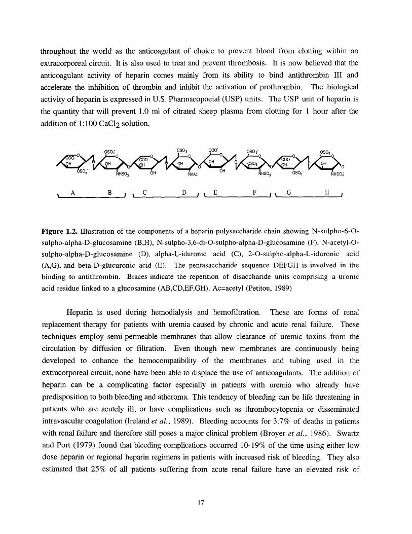
Figure 1.2. Illustration of the components of a heparin polysaccharide chain showing N-sulpho-6-O-
sulpho-alpha-D-glucosamine (B,H), N-sulpho-3,6-di-O-sulpho-alpha-D-glucosamine (F), N-acetyl-O-
sulpho-alpha-D-glucosamine (D), alpha-L-iduronic acid (C), 2-0-sulpho-alpha-L-iduronic acid
(A,G), and beta-D-glucuronic acid (E). The pentasaccharide sequence DEFGH is involved in the
binding to antithrombin. Braces indicate the repetition of disaccharide units comprising a uronic
acid residue linked to a glucosamine (AB,CD,EF,GH). Ac=acetyl (Petitou, 1989)
Heparin is used during hemodialysis and hemofiltration. These are forms of renal
replacement therapy for patients with uremia caused by chronic and acute renal failure. These
techniques employ semi-permeable membranes that allow clearance of uremic toxins from the
circulation by diffusion or filtration. Even though new membranes are continuously being
developed to enhance the hemocompatibility of the membranes and tubing used in the
extracorporeal circuit, none have been able to displace the use of anticoagulants. The addition of
heparin can be a complicating factor especially in patients with uremia who already have
predisposition to both bleeding and atheroma. This tendency of bleeding can be life threatening in
patients who are acutely ill, or have complications such as thrombocytopenia or disseminated
intravascular coagulation (Ireland et al., 1989). Bleeding accounts for 3.7% of deaths in patients
with renal failure and therefore still poses a major clinical problem (Broyer et al., 1986). Swartz
and Port (1979) found that bleeding complications occurred 10-19% of the time using either low
dose heparin or regional heparin regimens in patients with increased risk of bleeding. They also
estimated that 25% of all patients suffering from acute renal failure have an elevated risk of
17
bleeding. Heparin free dialysis has been used occasionally but it is generally unsuccessful due to
the high frequency of systemic clotting (Deary et al., 1991). Typical heparin level in hemodialysis
treatment is approximately 0.5 USP units/ml blood.
It is well recognized that major activation of the coagulation cascade takes place during
cardiopulmonary bypass surgery (CPB) and higher doses of heparin than those used for
hemodialysis are necessitated (Jobes et al., 1981). This type of surgery is an every day occurrence
in many hospitals. CPB equipment has a much greater surface area and employs much higher flow
rates (5 1/min. compared to 300 ml/min.). A heparin level of 3.6 USP units/ml was recommended
as the minimum safe level for avoidance of micro-coagulation and its sequelae (Senning, 1959). A
clear correlation was observed between excess circulating heparin and blood loss (Wright et al.,
1964). High risk patients include those with recent myocardial infarction, post-operative patients,
patients with ulcers and diabetics (Basu et al., 1984).
1.5 Heparinase I
Heparinase I is an eliminase originally extracted from Flavobacterium heparinum that
cleaves heparin at alpha-glycosidic linkages in heparin's repeating unit. The elimination reaction
results in a site of unsaturation in the terminal uronic acid, but the glucosamine reducing end
remains an unaltered residue (Linker and Hovingh, 1972). The molecular weight of the enzyme is
approximately 43,000 Daltons and its Km for heparin is around 4 pM (0.11 mg/cc) (Yang et al.,
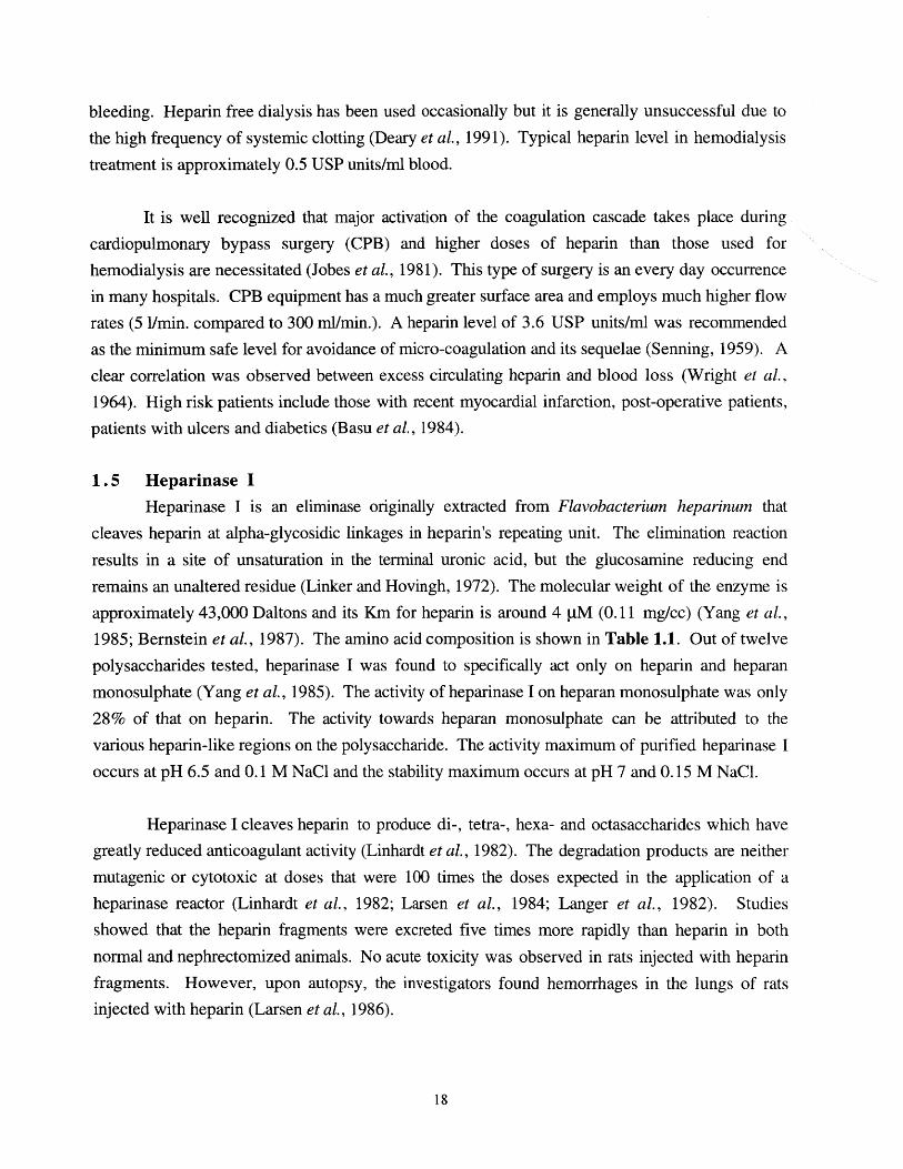
1985; Bernstein et al., 1987). The amino acid composition is shown in Table 1.1. Out of twelve
polysaccharides tested, heparinase I was found to specifically act only on heparin and heparan
monosulphate (Yang et al., 1985). The activity of heparinase I on heparan monosulphate was only
28% of that on heparin. The activity towards heparan monosulphate can be attributed to the
various heparin-like regions on the polysaccharide. The activity maximum of purified heparinase I
occurs at pH 6.5 and 0.1 M NaCl and the stability maximum occurs at pH 7 and 0.15 M NaCl.
Heparinase I cleaves heparin to produce di-, tetra-, hexa- and octasaccharides which have
greatly reduced anticoagulant activity (Linhardt et al., 1982). The degradation products are neither
mutagenic or cytotoxic at doses that were 100 times the doses expected in the application of a
heparinase reactor (Linhardt et al., 1982; Larsen et al., 1984; Langer et al., 1982). Studies
showed that the heparin fragments were excreted five times more rapidly than heparin in both
normal and nephrectomized animals. No acute toxicity was observed in rats injected with heparin
fragments. However, upon autopsy, the investigators found hemorrhages in the lungs of rats
injected with heparin (Larsen et al., 1986).
18
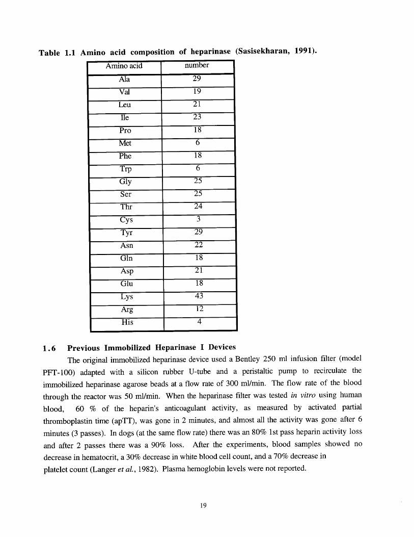
Table 1.1 Amino acid composition of heparinase (Sasisekharan, 1991).
Amino acid number
Ala 29
Val 19
Leu 21
Be 23
Pro 18
Met 6
Phe 18
Trp 6
Gly 25
Ser 25
Thr 24
Cys 3
Tyr 29
Asn 22
Gln 18
Asp 21
Glu 18
Lys 43
Arg 12
His 4
1.6 Previous Immobilized Heparinase I Devices
The original immobilized heparinase device used a Bentley 250 ml infusion filter (model
PFT- 100) adapted with a silicon rubber U-tube and a peristaltic pump to recirculate the
immobilized heparinase agarose beads at a flow rate of 300 ml/min. The flow rate of the blood
through the reactor was 50 mI/min. When the heparinase filter was tested in vitro using human
blood, 60 % of the heparin's anticoagulant activity, as measured by activated partial
thromboplastin time (apTT), was gone in 2 minutes, and almost all the activity was gone after 6
minutes (3 passes). In dogs (at the same flow rate) there was an 80% 1st pass heparin activity loss
and after 2 passes there was a 90% loss. After the experiments, blood samples showed no
decrease in hematocrit, a 30% decrease in white blood cell count, and a 70% decrease in
platelet count (Langer et al., 1982). Plasma hemoglobin levels were not reported.
19
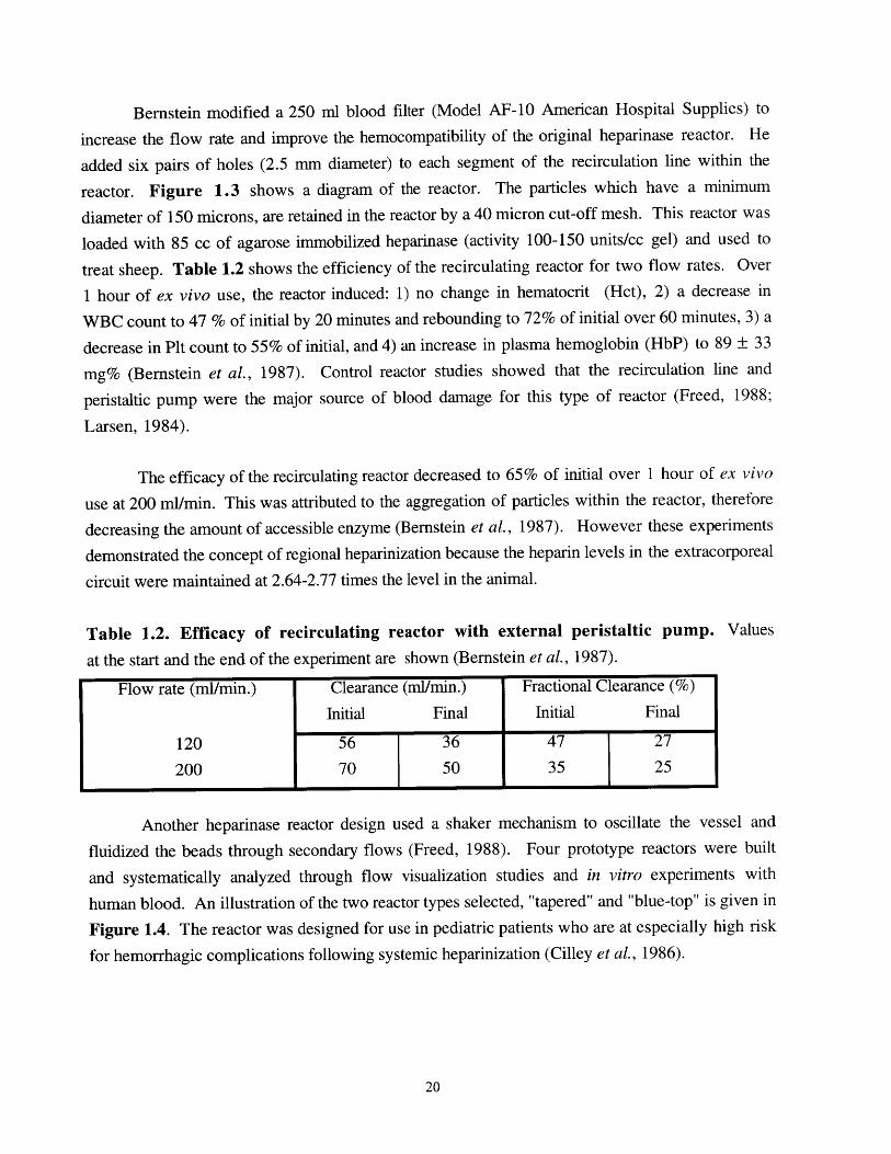
Bernstein modified a 250 ml blood filter (Model AF-10 American Hospital Supplies) to
increase the flow rate and improve the hemocompatibility of the original heparinase reactor. He
added six pairs of holes (2.5 mm diameter) to each segment of the recirculation line within the
reactor. Figure 1.3 shows a diagram of the reactor. The particles which have a minimum
diameter of 150 microns, are retained in the reactor by a 40 micron cut-off mesh. This reactor was
loaded with 85 cc of agarose immobilized heparinase (activity 100-150 units/cc gel) and used to
treat sheep. Table 1.2 shows the efficiency of the recirculating reactor for two flow rates. Over
1 hour of ex vivo use, the reactor induced: 1) no change in hematocrit (Hct), 2) a decrease in
WBC count to 47 % of initial by 20 minutes and rebounding to 72% of initial over 60 minutes, 3) a
decrease in Plt count to 55% of initial, and 4) an increase in plasma hemoglobin (HbP) to 89 ± 33
mg% (Bernstein et al., 1987). Control reactor studies showed that the recirculation line and
peristaltic pump were the major source of blood damage for this type of reactor (Freed, 1988;
Larsen, 1984).
The efficacy of the recirculating reactor decreased to 65% of initial over 1 hour of ex vivo
use at 200 m/min. This was attributed to the aggregation of particles within the reactor, therefore
decreasing the amount of accessible enzyme (Bernstein et al., 1987). However these experiments
demonstrated the concept of regional heparinization because the heparin levels in the extracorporeal
circuit were maintained at 2.64-2.77 times the level in the animal.
Table 1.2. Efficacy of recirculating reactor with external peristaltic pump. Values
at the start and the end of the experiment are shown (Bernstein et al., 1987).
Flow rate (ml/min.) Clearance (ml/min.) Fractional Clearance (%)
Initial Final Initial Final
120 56 36 47 27
200 70 50 35 25
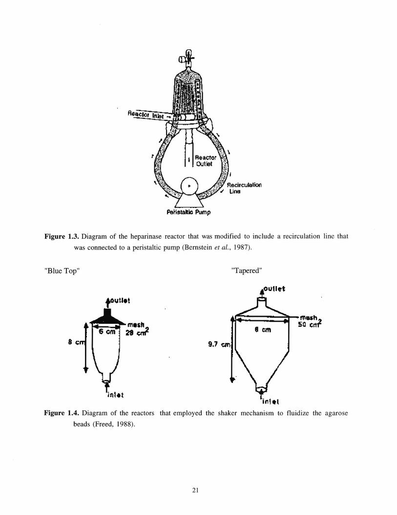
Another heparinase reactor design used a shaker mechanism to oscillate the vessel and
fluidized the beads through secondary flows (Freed, 1988). Four prototype reactors were built
and systematically analyzed through flow visualization studies and in vitro experiments with
human blood. An illustration of the two reactor types selected, "tapered" and "blue-top" is given in
Figure 1.4. The reactor was designed for use in pediatric patients who are at especially high risk
for hemorrhagic complications following systemic heparinization (Cilley et al., 1986).
20
Reactor ~t Outl lt.
Recirculalin
Pestawtho Pump
Figure 1.3. Diagram of the heparinase reactor that was modified to include a recirculation line that
was connected to a peristaltic pump (Bernstein et al., 1987).
"Blue Top" "Tapered"
rutlt
- mesti
6 cm 29 i
20le
9.7
OUl let
!50 C;Wcm v
Figure 1.4. Diagram of the reactors that employed the shaker mechanism to fluidize the agarose
beads (Freed, 1988).
21
i
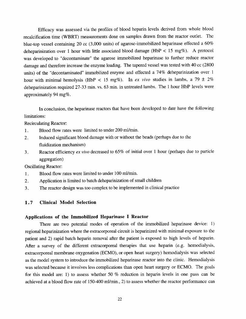
Efficacy was assessed via the profiles of blood heparin levels derived from whole blood
recalcification time (WBRT) measurements done on samples drawn from the reactor outlet. The
blue-top vessel containing 20 cc (3,000 units) of agarose-immobilized heparinase effected a 60%
deheparinization over 1 hour with little associated blood damage (HbP < 15 mg%). A protocol
was developed to "decontaminate" the agarose immobilized heparinase to further reduce reactor
damage and therefore increase the enzyme loading. The tapered vessel was tested with 40 cc (2800
units) of the "decontaminated" immobilized enzyme and effected a 74% deheparinization over 1
hour with minimal hemolysis (HbP < 15 mg%). In ex vivo studies in lambs, a 79 ± 2%
deheparinization required 27-33 min. vs. 63 min. in untreated lambs. The 1 hour HbP levels were
approximately 94 mg%.
In conclusion, the heparinase reactors that have been developed to date have the following
limitations:
Recirculating Reactor:
1. Blood flow rates were limited to under 200 ml/min.
2. Induced significant blood damage with or without the beads (perhaps due to the
fluidization mechanism)
3. Reactor efficiency ex vivo decreased to 65% of initial over 1 hour (perhaps due to particle
aggregation)
Oscillating Reactor:
1. Blood flow rates were limited to under 100 ml/min.
2. Application is limited to batch deheparinization of small children
3. The reactor design was too complex to be implemented in clinical practice
1.7 Clinical Model Selection
Applications of the Immobilized Heparinase I Reactor
There are two potential modes of operation of the immobilized heparinase device: 1)
regional heparinization where the extracorporeal circuit is heparinized with minimal exposure to the
patient and 2) rapid batch heparin removal after the patient is exposed to high levels of heparin.
After a survey of the different extracorporeal therapies that use heparin (e.g. hemodialysis,
extracorporeal membrane oxygenation (ECMO), or open heart surgery) hemodialysis was selected
as the model system to introduce the immobilized heparinase reactor into the clinic. Hemodialysis
was selected because it involves less complications than open heart surgery or ECMO. The goals
for this model are: 1) to assess whether 50 % reduction in heparin levels in one pass can be
achieved at a blood flow rate of 150-400 ml/min., 2) to assess whether the reactor performance can
22
be maintained for 4 hours at 37'C, and 3) to minimize blood damage to an amount below or
comparable to that of the hemodialyzer.
An immobilized heparinase reactor would have its greatest impact in the treatment of acute
renal insufficiency. Anticoagulation is one of the most important components of renal replacement
therapy and heparin is the oldest and most frequently used anticoagulant. In patients suffering
from acute renal failure, the risk of bleeding is greatly increased and excessive anticoagulation may
result in bleeding complications reported to occur in 5-26% of treatments (Zusman et al., 1981).
Current alternatives to systemic heparinization include variable heparin dosing, low molecular
weight heparin, regional heparinization and neutralization with protamine, regional citrate
anticoagulation with trisodium citrate, nafamostat mesilate, and prostaglandin analogue infusion.
Despite these alternatives, acute kidney failure is a serious problem with an unmet need for
effective deheparinization. The relative advantages and disadvantages of the alternate techniques
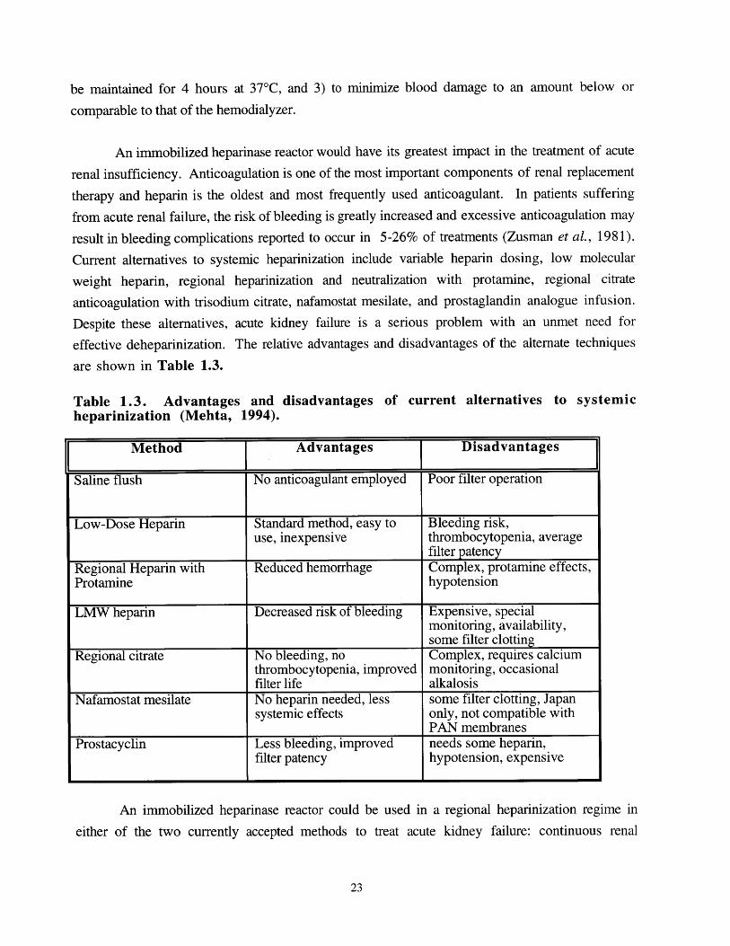
are shown in Table 1.3.
Table 1.3. Advantages and disadvantages of current alternatives to systemicheparinization (Mehta, 1994).
Method Advantages Disadvantages
Saline flush No anticoagulant employed Poor filter operation
Low-Dose Heparin Standard method, easy to Bleeding risk,use, inexpensive thrombocytopenia, average
filter patencyRegional Heparin with Reduced hemorrhage Complex, protamine effects,Protamine hypotension
LMW heparin Decreased risk of bleeding Expensive, specialmonitoring, availability,some filter clotting
Regional citrate No bleeding, no Complex, requires calciumthrombocytopenia, improved monitoring, occasionalfilter life alkalosis
Nafamostat mesilate No heparin needed, less some filter clotting, Japansystemic effects only, not compatible with
PAN membranesProstacyclin Less bleeding, improved needs some heparin,
filter patency hypotension, expensive
An immobilized heparinase reactor could be used in a regional heparinization regime in
either of the two currently accepted methods to treat acute kidney failure: continuous renal
23
replacement therapy (CRRT) and intermittent hemodialysis (IHD). Continuous arteriovenous
hemofiltration (CAVH) and continuous venovenous hemofiltration (CVVH) are forms of CRRT
which differ in vascular access and the treatment may last for several days. A stated advantage of
CRRT is that it is particularly suitable for patients in intensive care units who have multiple organ
failure and are generally hemodynamically unstable. It is also easier to administer the large daily
volumes of parenteral nutrition than in the case of IHD (Mehta, 1994). CAVH relies on the natural
pressure difference between the vein and the artery for circulation through the extracorporeal
circuit; therefore, flow rates range from 50-100 ml/min. in adults. CVVH uses a pump to circulate
the blood at a flow rate of 200 ml/min. Acute renal failure treatment with IHD typically may last
from 2-4 hours and flow rates range from 200-400 ml/min. in adults. The immobilized heparinase
reactor issues that need to be addressed for CRRT are:
1) prolongation of the stability of the immobilized heparinase in blood, and
2) a replacement method of the enzymatic support that would not disrupt the operation of the
dialyzer or filter.
Criteria for Reactor Design
The Taylor vortex flow reactor (VFR), was selected to evaluate the application of
immobilized heparinase I for regional heparinization. A vortex flow reactor consist of two
concentric cylinder in which the inner cylinder is able to rotate and the outer is stationary. At a
critical rotation rate, secondary flow instabilities termed Taylor vortices appear in the annulus and
their morphology can vary depending on the magnitude of the axial flow rate and the rotation rate.
Therefore, the VFR induced secondary flows without the physical hazards of external momentum.
The design criteria for a clinically feasible immobilized heparinase reactor for use in regional
heparinization involves the following:
1) Efficacy: reactor inlet blood activated clotting time (ACT) of 220-240 sec and reactor outlet
ACT of 170-180 sec (45-50% average steady state single pass conversion),
2) Safety: no significant effect on whole blood cells such as hemolysis (less than 150 mg plasma
free hemoglobin /dL), rapid cell and platelet count reduction, or excessive white cell or platelet
activation,
3) Stability: Stable operation at flow rates of up to 300 ml/min., and
4) Simplicity: Simple operation and low cost.
There are two conventional forms of operation for the VFR:
1) a fluidized bed VFR in which the immobilized heparinase beads are suspended within
the whole blood, annular path, and
24
2) a membrane VFR in which heparinase is immobilized onto a membrane placed on the
inner and outer cylinders.
During this research a third form of operation for the VFR was developed which combines
a microporous membrane and fluidization of agarose beads. A modular design was implemented
so that the reactor would be separate from the motor and control panel. This would allow for a
disposable unit and ease of use.
The Fluidized Bed VFR. The fluidized bed configuration would allow the highest
contact surface area for reaction with minimal external mass transfer limitations as demonstrated by
Freed and co workers (1993). But clogging of the agarose beads against a retaining mesh was still
a problem for flow rates above 100 ml/min. Therefore a recirculating loop that did not require an
external pump was designed to shunt the top and bottom of the device and allow recirculation of
the agarose via the natural rotational flow dynamics.
The Membrane VFR. The conventional method of immobilizing heparinase onto a
membrane and placing the membrane on the inner and outer cylinders of the VFR was not pursued
because the retention of heparinase activity after immobilization to several porous membranes was
minimal (< 5%) and the surface to volume ratio required was not feasible (Appendix). Therefore,
a modified membrane design was adopted using agarose beads as the support for the immobilized
heparinase and a porous membrane for cell separation. In this novel configuration, the membrane
was placed in the inner surface of the outer cylinder and the immobilized heparinase was injected
into the compartments created by the membrane and the cylinder (reactive compartment).
Criteria for Enzyme Immobilization
Several important issues must be taken into account when designing an immobilized
enzyme device for blood contact. First, the chemistry used for the immobilization process should
result in a stable covalent bond between the enzyme and the support and should not introduce toxic
substances in the support that could be difficult to eliminate. Secondly, the support for the
immobilization should be mechanically and chemically stable, non toxic (before and after
immobilization), and readily available. Supports in the shape of beads and membranes are
examined. The former allows a high reactive surface area but at the same time has reduced
hemocompatibility and potential limitations for high flow rate applications. In addition, the beads
require a mechanism to prevent their escape from the reactor. If a retaining mesh is used, the
possibility of clogging at the outlet becomes an important issue. Membranes have better
mechanical advantages but typically have the disadvantage of reduced reactive surface area if the
process is mass transfer limited. In either case, immobilization typically reduces the activity of the
enzyme and may even destroy it.
25

References
Ambrus, C., Ambrus, J.L., Horvath, C., Pedersen, H., Sharma, S., Kant, C., Mirand, E.,Guthrie, R., and Paul, T. 1978. Phenylalanine depletion for the management of phenylketonuria:Use of enzyme reactor with immobilized enzymes. Science 201: 837-839.
Ambrus, J. J., Codey, C., and Virand, E.A. 1983. In vivo safety of hollow-fiber enzyme-reactorswith immobilized phenylalanine ammonia lyase in a large animal model for phenylketonurea. J.Pharmacol. Exp. Ther. 224: 598.
Arbiser, J.L., Dzieczkowski, J.S., Harmon, J.V., and Duncan, L.M. 1995. Leukocytoclasticvasculitis following staphylococcal protein A column immunoadsorption therapy. Arch Dermatol.131:707-709.
Basu, D., Gallus, A. J., Hirsh, and Cade, J. 1984. New Eng. J. Med. 100: 358.
Bernstein, H., Yang, V., Lund, D., Randhawa, M., Harmon, W., and Langer, R. 1987.Extracorporeal enzymatic heparin removal: Use in a sheep dialysis model. Kidney Int. 32: 452-463.
Bertino, J., Condos, S., Horvath, C., Khaltagi, K., and Pedersen, H. 1979. Cancer Research 38:1936,1978.
Broughton, R. 1995. Personal Communication. IBEX Technologies, Montreal, Canada.
Broyer, M., Brunner, F.P., Brynger, H., Fassbinder, W., Guillou, P.J., and Oules, R. 1986.Demography of dialysis and transplantation in Europe. Nephrology, Dialysis and Transplantation1: 1.
Cilley, R. E., Zwischenberger, J.B., Andrews, A.F., Bowerman, R.A., Rodolff, D.W., andBarlett, R.H. 1986. Intracranial hemorrhage during extracorporeal membrane oxygenation inneonates. Pediatrics 78: 69-74.
Comfort, A. R., Albert, E.C., and Langer, R. 1989. Immobilized enzyme cellulose hollow fibers:I. Immobilization of Heparinase. Biotech. Bioeng. 34: 1366-1373.
Deary, D.F., Gajaria, M., Fryer-Keene, S., and Willumsen, J. 1991. Pediatric Nephrology 5:220-224.
Dumler, F., Singh, P., Jackson, C.E., Kini, K.R., Samhouri, A.M., Halvorson, H.R., andShore, J.D. 1981. Extracorporeal enzyme therapy. Use of an L-asparaginase reactor-dialyser in aclinical setting. ASAIO J. 4: 70.
Freed, L.E. 1988. An enzymatic fluidized bed reactor for blood deheparinization; Development andtesting in lambs on extracorporeal circulation. Ph.D. dissertation, M.I.T.
Freed, L.E., Vuniak, G.V., Drinker, P.A., and Langer, R. 1988. A novel bioreactor based onsuspended particles of agarose-immobilized species. Trans. Am. Intern. Organs 34: 732-738.
Freed, L.E., Vunjak-Novakovic, G.V., Bernstein, H., Cooney, C. L., and Langer, R. 1993.Kinetics of immobilized heparinase in human blood. Ann. Biomed. Engng. 21: 67-76.
26
Freed, L.E., Vunjak-Novakovic, G.V., Drinker, P.A., and Langer, R. 1993. Bioreactor based onsuspended particles of immobilized enzyme. Ann. Biomed. Engng. 21: 57-65.
Gaylor, J.D.S., and Smeby, L.C. In: Physiological and Clinical Aspects of Oxygenator Design;Dawids, S.G., and Engell, H.C. 1976. Eds., Elsevier: North-Holland, Amsterdam; pp. 65-79.
Horvath, C., Sardi, A., and Woods, F.S. 1973. L-asparaginase tubes: Kinetic behavior andapplication in physiological studies. J. Appl. Physiol. 34: 181.
Iosilevskii, G., Brenner, H., Moore, C.M.V., and Cooney, C.L. 1993. Mass transport andchemical reaction in Taylor-vortex flows with entrained catalytic particles: applications to a novelclass of immobilized enzyme biochemical reactors. Phil. Trans. R. Soc. Lond. 345: 259-294.
Ireland, H., Rylance, P.B. and Kesteven, P. 1989. In: Heparin: Chemical and biologicalproperties, clinical applications; Lane, D.A., and U. Lindahl, Eds., CRC Press: Boca Raton, pp.549.
Jaffrin, M.Y., 1989. Innovative processes for membrane plasma separation. J. Membrane Sci. 44:115-129.
Jobes, D.R., Schwartz, A.J., Ellison, N., Andrews, R., Ruffini, R.A., and Ruffini, J.J. 1981.Monitoring heparin anticoagulation and its neutralization. Annals of Thoracic Surgery 31: 161.
Kalghatgi, K.K., Horvath, C. and Ambrus, C.M. 1980. Multi-tubular reactors with immobilized-phenylalanine ammonia lyase for use in extracorporeal shunts. Res. Commun. Chem. Pathol.Pharmacol. 27: 551.
Kanalas, J.J., Spector, E.B. and Cederbaum, S.D. 1982. Hollow-fiber reactors containingmammalian arginase: An approach to enzyme replacement therapy. Biochem. Med. 27: 46.
Klein, M.D., and Langer, R. 1986. Immobilized enzymes in clinical medicine: An emergingapproach to new drug therapies. Trends in Biotech. 14: 179-186.
Langer, R., Linhardt, R.J. Hoffberg, S., Larsen, A.K., Cooney, C.L., Tapper, D. and Klein, M.1982. An enzymatic system for removing heparin in extracorporeal therapy. Science 217: 261-263.
Larsen, A.K., Newberne, P.M., and Langer, R. 1986. Comparative studies of heparin andheparin fragments: Distribution and toxicity in the rat. Fundamental and Applied Toxicology, 7:86-93.
Larsen, A.K., Linhardt, R.J., Tapper, D., Klein, M. and Langer, R. 1984. Effect ofextracorporeal deheparinization on formed blood components. Artif. Organs. 8: 198-203.
Lavin, A., Sung, C., Klibanov, A.M. and Langer, R. 1985. Enzymatic removal of bilirubin fromblood; A potential treatment for neonatal jaundice. Science, 230: 543-545.
Linhardt, R.J., Grant, A., Cooney, C.L. and Langer, R. 1982. Differential anticoagulant activityof heparin fragments prepared using microbial heparinase. J. Biol. Chem. 257: 7310-7313.
Linker, A., and Hovingh, P. 1972. Heparinase and heparitinase from Flavobacteria. Meth.Enzymol. 28: 902-911.
Makarov, K.A., and Kibardin, S.A. 1980 'Immobilized biopreparations in medicine' , Moscow.
27
Mehta, R.L., 1994. Anticoagulation during continuous renal replacement. ASAIO J. 40: 931-935.
Moore, C.M.V., 1994. Characterization of a Taylor-Couette vortex flow reactor. Ph.D.dissertation, M.I.T.
Mottaghy, K., and Hanse, H.J. 1985. Effect of combined shear, secondary and axial flow ofblood on oxygen uptake. Chem. Engng. Commun. 36: 269-279.
Olanoff, L.S., Bernath, F.R. and Venkatasubramanian, K. 1975. Perfusion trials with collagenimmobilized enzyme in an extracorporeal reactor. Amer. Chem. Soc. Polym. Prepr. 16: 203.
Pedersen, H., Horvath, C., and Ambrus, C.M. 1979. Preparation of immobilized L-phenylalanineammonia lyase in tubular forms for depletion of L-phenylalanine. Res. Commun. Chem. Pathol.Pharmacol. 20: 559.
Petitou, M. 1989. In: Heparin: Chemical and biological properties, clinical applications; Lane,D.A., and U. Lindahl, Eds., CRC Press Inc.: Boca Raton, pp. 65-79.
Plottz, P.H., Berk, P.D., Scharschmidt, B.F., Gordon, J.K. and Vergalla, J. 1974. Removingsubstances from blood by affinity chromatograpy. I. Removing bilirubin and other albumin-boundsubstances from plasma and blood with albumin conjugated agarose-beads. J. Clin. invest. 5:778-785.
Porter, J., and Fick, H. 1977. Drug related deaths among medical inpatients. J. Amer. Med.Assoc. 237: 879-881.
Rossi, V., 1981. Immobilization of arginase on hollow-fiber hemodialyzer. Int. J. Artif. Organs4: 102.
Salmona, M., Saronio, C.I., Bartosek, J., Vecchi, A. and Mussini, E. 1974. In: Insolubilizedenzymes; M. Salmona, C.S., Garattini, S. Eds., Raven Press: New York, pp. 189.
Salzman, E.W., Deykin, D. and Shapiro, R.M. 1975. Management of heparin therapy, controlledprospective trial. New Eng. J. Med., 292: 1046.
Sampson, D., Han, T., Hersh, L.S. and Murphy, G.P. 1974. Extracorporeal chemotherapy withL-asparaginase in man. J Surg Oncol., 6: 39.
Sasisekharan, R., 1991. Cloning and biochemical characterization of heparinase fromFlavobacterium heparinum. Ph.D. dissertation, Harvard University.
Senning, A., 1959. Plasma heparin concentrations in extracorporal circulation. Acta. Chirurgica.Scandinavica 117: 55.
Schaeffer, J., Floege, J., Ehlerding, G. and Koch., K.M. 1995. Pathogenetic and diagnosticaspects of dialysis-related amyloidosis. Nephrol Dial Transplant. 10(3): 4-8.
Strong, A. B., and Carlucci, L. 1976. An experimental study of mass transfer in rotating Couetteflow with low axial Reynolds number. Can. J. Chem. Engng. 54: 295-298.
Sung, C., Lavin, A., Klibanov, A.M. and Langer, R. 1986. An immobilized enzyme reactor forthe detoxification of bilirubin. Biotech. Bioeng. 28: 1531.
Swartz, R.D., and Port, F.K. 1979. Preventing hemorrhage in high risk hemodialysis. Regionalversus low dose heparin. Kidney Int. 16: 513-518.
28
Takao, N. 1993. In: Biomedical applications of polymeric materials; Tsuruta, T., Hayashi, T.,Kataoka, K., Ishihara, K., Kimura, Y. Eds., CRC Press: Boca Raton; pp. 192-208.
Torchilin, V. P. 1991. In: Immobilized enzymes in medicine; Springer-Verlag: Berlin; pp. 135-145.
Vallar, L., and Rivat, C. 1994. Regenerated cellulose-based hemodialyzers with immobilizedproteins as potential devices for extracorporeal immunoadsorption procedures: An assessment ofprotein coupling capacity and in vitro dialysis performances. Artif. Organs 20:8-16.
Vallar, L., Costa, P.M.P., Teixeira, A., Pfister, M., Barrois, R., Costa, P.P. and Rivat, C. 1995.Immunoadsorption procedure as a potential Method for the specific p2m removal from plasma ofpatients with chronic renal failure. J. of Chromatography B: Biomed. App. 664: 97-106.
Wakefield, T.W., Lindblad, B., Whitehouse, W., Hantler, C., and Stanley, J.C. 1986.Attenuation of hemodynamic and hematologic effects of heparin protamine sulfate interaction afteraortic reconstruction in a canine model. Surgery 100: 45-51.
Wright, J. S., Osborn, J.J., Perkins, H.A. and Gerbode, F. 1964. Heparin levels during andafter hypothermic perfusion. Journal of Cardiovascular Surgery 5: 244.
Yang, V. C., Linhardt, R.J. Bernstein, H. Cooney, C.L. and Langer, R. 1985. Purification andcharacterization of heparinase from flavobacterium heparinum. J. Biol. Chem. 260: 1849-1857.
Zusman, R. M., Rubin, R.H., Cato, A.E., Cocchetto, B.S., Crow, J.W., Tolkoff-Rubin, N.1981. Hemodialysis using prostacyclin instead of heparin as the sole antithrombotic agent. NewEng. J. Med. 304: 934-939.
29
Chapter 2
The Vortex Flow Fluidized Bed Reactor
2.1 Introduction
The optimum management of heparin therapy in the critically ill patient is an important
problem that continues to affect the medical community. The use of heparin and its antagonist
protamine has been associated with potentially fatal complications in high risk patients such as
those suffering from acute renal failure and those who have undergone open heart surgery (Broyer
et al., 1986; Salzman et al., 1975). The immobilized heparinase I approach could be an alternative
therapy to protamine reversal and also a way to achieve regional heparinization in extracorporeal
circuits for hemodialysis and hemoperfusion. However, an important issue that needs to be
addressed for the successful application of extracorporeal therapies that involve immobilized
enzymes or antibodies is the development of bioreactors that are safe, efficient, easy to use, and
cost effective.
Current bioreactor designs can be divided into two categories: membrane based devices
such as hollow fiber cartridges, and porous particle devices in the form of packed columns or
fluidized beds. Several investigators have bound the protein of interest to the walls of a hollow
fiber dialyzer to enhance the whole blood compatibility of the system but in many cases the surface
area requirements for the efficacy of such devices remain impractical (Comfort, 1988a; Vallar, et
al., 1996). Others have suggested the use of porous particles, such as agarose or cellulose beads,
in either the packed bed or fluidized bed mode as a way to optimize the protein or adsorbent
loading per volume of device (Gejyo et al., 1993; Langer et al., 1982). Vunjak-Novakovic and co
workers (1988) have demonstrated the importance of secondary flows in an oscillating bioreactor
for the even fluidization of agarose. Nevertheless, despite the presence of secondary flows, the
oscillating bioreactor was limited to flow rates of up to 100 ml blood/min due to the packing of the
beads at the reactor outlet (Freed et al., 1988).
In this chapter, secondary flows (Taylor vortices) and "flow-induced" agarose recirculation
was combined to facilitate the use of agarose immobilized heparinase I in the clinical setting.
Taylor vortices are flow instabilities that occur within the annular gap of concentric cylinders when
the inner cylinder is rotated above a critical rotation rate. Taylor vortices have been shown to have
excellent mixing characteristics in reactors that processed biological systems such as cell cultures,
blood plasmapheresis, and oxygenation (Beaudoin et al., 1987). However, no one has
30
investigated the use of Taylor vortices with immobilized enzyme fluidized systems for medical
applications.
The studies in this chapter examined whether the advantages of Taylor vortices could be
incorporated into an immobilized heparinase I fluidized bed reactor that would meet the following
criteria: 1) high whole blood flow rates of 150 - 400 ml/min, 2) efficient conversions, and 3)
minimal blood damage as determined by blood cell counts, platelet counts, and hemolysis.
2.2 Materials and Methods
The reactor vessel is made from concentric polycarbonate cylinders (6.38 cm OD and 5.10
cm OD with 0.32 cm wall thickness) and sheets purchased from Commercial Plastics (Somerville,
MA) with polycarbonate inlet and outlet ports (0.96 cm ID) purchased from Avecor Cardiovascular
Inc. (Plymouth, MN). The inner cylinder rotates on 0.32 cm OD stainless steel pins from Small
Parts Inc. (Miami Lakes, FL). Silicone 0-rings were purchased from Marco Rubber Inc.
(Andover, MA). The reactor components were machined according to specifications at the MIT
Central Machine Shop.
The inner cylinder rotated via a custom made magnetic coupling drive system which
consisted of eight neodymium iron boron disc magnets (Dia = 1.28 cm Length = 0.64 cm Cat#
27DNE3216) from Magnet Sales & Manufacturing Inc. (Culver City, CA) and an external Bodine
PM electric motor and controller (Bodine Type FPM) from Bodine Electric Company (Chicago,
IL). Four neodymium iron boron disc magnets were placed in the top end of the inner cylinder
with the north/south magnet pair perpendicular and in opposite polarity to the east/west pair. A
similar magnet set up was placed at the end of the motor. Magnetic coupling was achieved when
the magnets on the motor and the inner cylinder aligned themselves according to magnetic polarity.
The magnetic force between the inner cylinder and the motor driver was strong enough to rotate the
inner cylinder, eliminating direct contact of the blood path with the motor shaft. Rotation rate was
detected through an optical sensor and displayed on a digital panel meter from Cole Parmer
Instruments Co. (Niles, IL). Temperature measurements were made by T-type thermocouples
connected to a digital panel meter from Cole Parmer Instruments Co. (Niles, IL). Medical grade
photoreactive adhesive ELC type 4M12 was from Electro-Lite Corporation (Danbury, CT).
Drip chambers (12 ml, polyvinylchloride) were from Lifemed (Compton, CA) and Qosina
Corporation (Edgewood, NY). Tygon medical grade tubing (0.64 cm ID and 0.96 cm ID) was
from VWR Scientific (Boston, MA). The peristaltic blood pump (model Draker Willock 7401)
was donated by Children's Hospital (Boston, MA). Ethylene oxide was from Andersen (Oyster
Bay, NY). Medical grade heparin solution for blood studies (porcine, 1,000 units/ml) was from
31
Elkin-Sinn (Cherry Hill, NJ). Powdered heparin for enzyme activity determination (porcine, 166
units/mg) was from Hepar Industries (Franklin, OH). Normal saline (0.9% NaCl) was from
Abbott (Chicago, IL) and PBS (0.154 M NaCl and 0.01M sodium phosphate, pH = 7.4) was
from Life Technologies (Grand Island, NY). Heparin levels in blood were monitored with model
801 Hemochron coagulation timers and P214 glass activated test tubes from Cardio Medical
Products (Rockaway, NJ).
Agarose particles (8%, 50-100 mesh, 200 jim average diameter) were from BioRad
(Rockville, NY); Cyanogen Bromide and Lysine Hydrochloride were from Sigma Chemical
Company (St. Louis, MO); Heparinase I, produced from Flavobacterium heparinum, was
generously donated by IBEX Technologies (Quebec, Canada). The enzyme preparation was 95%
pure heparinase I as determined by RP-HPLC (company documentation) with a specific activity of
242 IU/ml and protein concentration of 2.6 mg/ml. One IU (international unit) is defined as the
amount of enzyme required to produce 1 [tmol of product per minute. Human blood was obtained
from healthy male and female volunteers. The blood was collected by a professional staff at the
Blood Donor Center of Children's Hospital (Boston, MA). Plasma hemoglobin levels were
determined with an assay kit from Sigma Chemical Company (St. Louis, MO).
Protein concentration in saline
The coomasie blue dye method of Bradford was used to measure the concentration of
heparinase in buffered saline. A volume of 800 [1 of sample was mixed with 200 pl of dye
solution and incubated at room temperature for 5 minutes. The absorbance at 595 nm was
measured and compared to a standard curve prepared with known bovine albumin concentrations
ranging from 1 to 20 pg/ml. The standard curve was linear in the above range.
Activity of soluble heparinase I
The activity of heparinase I was defined using the international unit (IU). One IU is the
amount of heparinase that would produce 1 jimol of double bonds/min. The appearance of heparin
degradation products was monitored at 232 nm in a quartz cuvette. Briefly, 20 p1 of heparinase
solution was added to 3 ml of heparin solution (2 mg/ml) in 100 mM MOPS 5 mM calcium acetate
pH 7.4 at 30 *C. The initial rate was calculated from the slope of the curve and converted to IU
using 3800 M1 molar absorptivity.
Cyanogen bromide activation of agarose
1 volume of agarose gel (200 jm average bead diameter) was washed with 10 volumes of
deionized (DI) water to remove any preservatives. After resuspending the gel in 1 volume of DI
32
water and 2 volumes of 2 M sodium carbonate the solution was chilled in an ice bath. Next, 0.2
volume of a solution of cyanogen bromide in acetonitrile (Ig/ml) was added to the gel solution and
the mixture was stirred vigorously in the fume hood for 5 minutes. Using a sintered glass funnel,
the activated gel was isolated and washed with the following: 10 volumes of DI water, 5 volumes
of 1 mM hydrochloric acid, and 5 volumes of 0.1 M sodium bicarbonate 0.5 M NaCl pH 8.3
buffer.
Immobilization of heparinase I
After extensive washing of the activated gel, the heparinase solution (18 mg in 100 ml 0.1
M sodium bicarbonate 0.5M NaCl pH 8.3) was incubated with the beads (100 ml) at 4'C for 5 hrs
to allow binding. Following heparinase binding, the immobilized heparinase was incubated
overnight at 4'C in a lysine solution (0.2 M lysine hydrochloride 0. 1M sodium bicarbonate 0.5M
NaCl) to cap any unreacted sites. The immobilized heparinase was then washed with 5 volumes of
cold pH 8.3 buffer and 30 volumes of phosphate buffered saline (PBS) pH 7.4, and 10 volumes
of distilled water. The amount of bound protein was determined by a mass balance between the
original enzyme solution and the washes. The immobilized heparinase I was resuspended in an
equal volume of PBS, pH 7.4, and stored at 4 'C.
Activity of the immobilized heparinase I
The activity of the heparinase bound to the beads was measured using a modified UV 232
activity assay. A known volume of beads, approximately 0.1 ml, was added to 4 ml of a heparin
solution (25 mg/ml). Under vigorous swirling at 37 'C, 0.1 ml samples were taken at 1 minute
intervals for 4 minutes and quenched in 0.9 ml of 30 mM HCl. The samples were centrifuged and
the absorbance of the supernatant was measured at 232 nm. The activity was then calculated from
the slope between the data points using 5000 MI as the molar absorptivity.
Measurement of heparin activity in blood
Whole blood recalcification times (WBRT) were used to indirectly determine the amount of
heparin present in blood. Briefly, 200 pl of citrated blood (3.8% 1:10 dilution) was added to
Hemochron p214 test tubes containing glass particles. At t=0, 200 Rl of CaCl 2 was added to the
test tube and the Hemochron 801 clot timer machine was immediately started. The test tube was
gently mixed for 10 seconds and inserted into the test well of the Hemochron 801. The time
required for a clot to form was recorded. The unknown samples were compared to a standard
curve which was linear in the range of 0 - 4 USP units heparin/ml blood.
33
Measurement of free hemoglobin in plasma
A hemoglobin detection kit from Sigma was used to monitor the release of hemoglobin into
the plasma. The kit is based on the catalytic effect of hemoglobin on the oxidation of 3,3',5,5'-
tetramethylbenzidine (TMB) by hydrogen peroxide. Briefly, 10kl of sample is added to 4 ml of
reagent and measured at 600 nm at 25'C. The color formation in the reaction is proportional to the
hemoglobin concentration in the sample. Any measurements above 2.6 mg hemoglobin/dL-pass
were considered to be unsafe. The criteria for significant hemolysis was derived from interviews
with physicians and hemolysis data from currently used medical devices.
Measurement of complete blood counts
Blood samples were taken to the M.I.T. Medical Laboratory for automated analysis of the
red cell, white cell, and platelet populations.
In vitro batch removal of heparin from blood
The assembled reactor was incorporated into a closed circuit primed with normal saline
which consisted of a blood warmer, blood pump, and a drip chamber and pressure monitor. The
circuit was then connected to a 450 cc reservoir of citrated human blood which was spiked with
heparin to achieve a final concentration of approximately 2 USP units Heparin /cc blood. The
blood flow and rotation rate of the reactor were adjusted to either 100 ml/min or 200 ml/min and
500 or 200 rpm, respectively. The temperature of the system was monitored directly with a T type
thermocouple probe. At time = 0, a 50% slurry of agarose immobilized heparinase was injected
via the drip chamber into the blood circuit and allowed to enter the annular space of the reactor
where Taylor vortices were present. Samples were constantly taken from the outlet port during the
experiment and analyzed for efficacy and safety of the device.
Design and construction of the vortex flow fluidized bed reactor (VFFBR)
The reactor was machined from polycarbonate tubing and had an outside diameter of 6.38
cm. Two versions were tested: a narrow gap (r outer - rinner = 0.32 cm), 24.00 cm length device
(design I) and a wide gap (r outer - rinner = 0.64 cm), 14.00 cm length device (design II). In
addition to the inlet and outlet ports, the reactor was fitted with two tangential ports (0.96 cm Dia.)
to accommodate a recirculation line. The dimensions and the diagram for the device are shown in
Table 2.1 and Figure 2.1, respectively. A 1 inch length retaining mesh with a mesh opening of
44 gm was placed along the circumference of the outer cylinder proximal to the outlet port to
prevent the beads from escaping the device. The maximum volume of the device was limited to
150 ml. This volume was a clinical specification to minimize hemodilution of the patient.
34
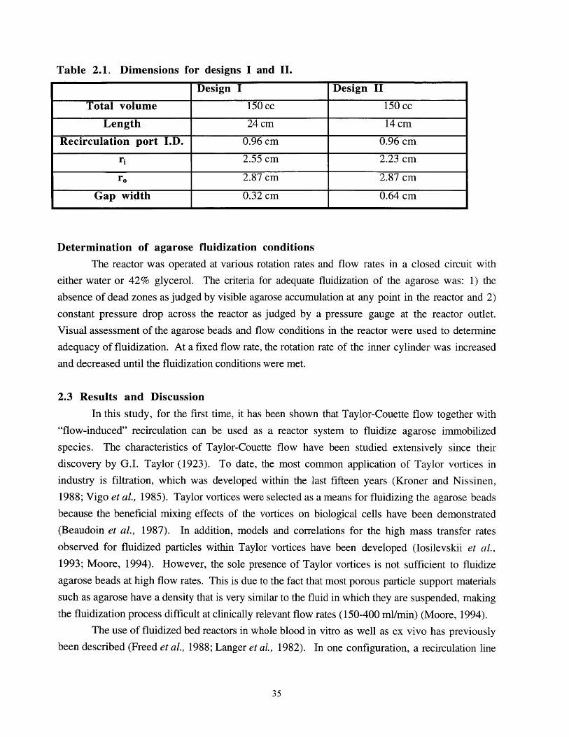
Table 2.1. Dimensions for designs I and II.
Design I Design II
Total volume 150 cc 150 cc
Length 24 cm 14 cm
Recirculation port I.D. 0.96 cm 0.96 cm
ri 2.55 cm 2.23 cm
ro 2.87 cm 2.87 cm
Gap width 0.32 cm 0.64 cm
Determination of agarose fluidization conditions
The reactor was operated at various rotation rates and flow rates in a closed circuit with
either water or 42% glycerol. The criteria for adequate fluidization of the agarose was: 1) the
absence of dead zones as judged by visible agarose accumulation at any point in the reactor and 2)
constant pressure drop across the reactor as judged by a pressure gauge at the reactor outlet.
Visual assessment of the agarose beads and flow conditions in the reactor were used to determine
adequacy of fluidization. At a fixed flow rate, the rotation rate of the inner cylinder was increased
and decreased until the fluidization conditions were met.
2.3 Results and Discussion
In this study, for the first time, it has been shown that Taylor-Couette flow together with
"flow-induced" recirculation can be used as a reactor system to fluidize agarose immobilized
species. The characteristics of Taylor-Couette flow have been studied extensively since their
discovery by G.I. Taylor (1923). To date, the most common application of Taylor vortices in
industry is filtration, which was developed within the last fifteen years (Kroner and Nissinen,
1988; Vigo et al., 1985). Taylor vortices were selected as a means for fluidizing the agarose beads
because the beneficial mixing effects of the vortices on biological cells have been demonstrated
(Beaudoin et al., 1987). In addition, models and correlations for the high mass transfer rates
observed for fluidized particles within Taylor vortices have been developed (Iosilevskii et al.,
1993; Moore, 1994). However, the sole presence of Taylor vortices is not sufficient to fluidize
agarose beads at high flow rates. This is due to the fact that most porous particle support materials
such as agarose have a density that is very similar to the fluid in which they are suspended, making
the fluidization process difficult at clinically relevant flow rates (150-400 ml/min) (Moore, 1994).The use of fluidized bed reactors in whole blood in vitro as well as ex vivo has previously
been described (Freed et al., 1988; Langer et al., 1982). In one configuration, a recirculation line
35
and a peristaltic pump were used to help fluidize the beads to prevent bead packing at the reactor
outlet. Further studies determined that using a peristaltic pump to recirculate an agarose-blood
slurry significantly contributed to the observed blood damage (Larsen et al., 1984). Therefore,elimination of a recirculating pump was a design objective.
The immobilization procedure used for the present study bound 95% of the soluble enzyme
with an activity retention of 30-40%. The average activity of the agarose immobilized heparinase I
was 12-16 IU/ml packed wet gel. Another important issue for the use of immobilized enzymes is
detachment or leaching of the enzyme from the support. Over a period of 4 days, no detectable
leaching of heparinase I occurred from the agarose beads in buffer as judged by protein
concentration and soluble enzyme activity assays (the UV 232 nm activity assay is sensitive to 0.5
gg/ml of pure heparinase). A previous analysis of the immobilization of heparinase on solid
supports also showed no significant leaching using cyanogen bromide activation chemistry vs.
other covalent methods, as long as thorough post immobilization washes with high salt buffer were
performed (Comfort et al., 1988b; Leckband and Langer, 1991). In regards to toxicity issues,
animal experiments demonstrated that the heparin degradation products were non toxic for doses of
up to 100 times the expected clinical exposure concentration (Larsen et al., 1986). Furthermore,healthy volunteers from a phase 1 clinical trial did not elicit any acute reaction to soluble heparinase
I (Broughton, 1995).
In the present study, a Taylor-Couette flow device was constructed and modified with a
recirculation line in order to allow even fluidization of agarose beads at high flow rates (Figure
2.1). The selected tangential location of the recirculation ports allowed continuous flow and
fluidization of the beads without the aid of an external pump. Design I (narrow gap) was able to
remove 90% of the heparin's anticoagulant activity from 450 cc of human citrated blood in vitro
within 3 minutes of operation at a flow rate of 100 ml/min (Figure 2.2). However, for the
operating conditions used, fluidization of the agarose beads within the Taylor vortices caused
extensive damage to the red cells, white cells, and platelets (Figure 2.3). Figure 2.4 shows
hemolysis during the experiment, an effect of the fluidization of agarose beads within the Taylor
vortices on the red cell membrane. The red cells were ruptured and hemoglobin was released into
the plasma (HbP). The hemolysis that resulted was above the safety criteria of 2.6 mg
hemoglobin/dL-pass (1 pass = 4.5 min). Further experiments demonstrated that the degree of
hemolysis was directly proportional to the volume fraction of agarose and to the rotation rate of the
inner cylinder (Figure 2.5).
In order for the fluidization to occur at relatively high flow rates, the rotation rate had to bemaintained above a minimum predetermined value in order to continuously recirculate the agarose
36
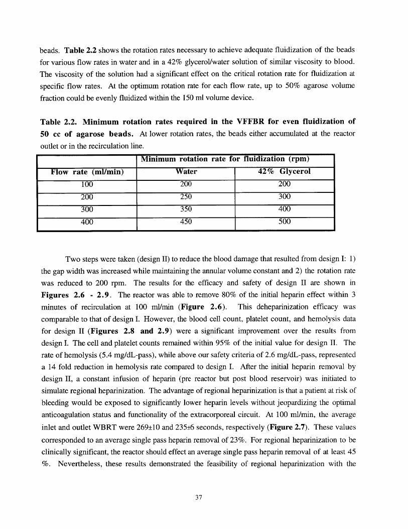
beads. Table 2.2 shows the rotation rates necessary to achieve adequate fluidization of the beads
for various flow rates in water and in a 42% glycerol/water solution of similar viscosity to blood.
The viscosity of the solution had a significant effect on the critical rotation rate for fluidization at
specific flow rates. At the optimum rotation rate for each flow rate, up to 50% agarose volume
fraction could be evenly fluidized within the 150 ml volume device.
Table 2.2. Minimum rotation rates required in the VFFBR for even fluidization of
50 cc of agarose beads. At lower rotation rates, the beads either accumulated at the reactor
outlet or in the recirculation line.
Minimum rotation rate for fluidization (rpm)
Flow rate (ml/min) Water 42% Glycerol
100 200 200
200 250 300
300 350 400
400 450 500
Two steps were taken (design II) to reduce the blood damage that resulted from design I: 1)
the gap width was increased while maintaining the annular volume constant and 2) the rotation rate
was reduced to 200 rpm. The results for the efficacy and safety of design II are shown in
Figures 2.6 - 2.9. The reactor was able to remove 80% of the initial heparin effect within 3
minutes of recirculation at 100 ml/min (Figure 2.6). This deheparinization efficacy was
comparable to that of design I. However, the blood cell count, platelet count, and hemolysis data
for design II (Figures 2.8 and 2.9) were a significant improvement over the results from
design I. The cell and platelet counts remained within 95% of the initial value for design II. The
rate of hemolysis (5.4 mg/dL-pass), while above our safety criteria of 2.6 mg/dL-pass, represented
a 14 fold reduction in hemolysis rate compared to design I. After the initial heparin removal by
design II, a constant infusion of heparin (pre reactor but post blood reservoir) was initiated to
simulate regional heparinization. The advantage of regional heparinization is that a patient at risk of
bleeding would be exposed to significantly lower heparin levels without jeopardizing the optimal
anticoagulation status and functionality of the extracorporeal circuit. At 100 ml/min, the average
inlet and outlet WBRT were 269±10 and 235±6 seconds, respectively (Figure 2.7). These values
corresponded to an average single pass heparin removal of 23%. For regional heparinization to be
clinically significant, the reactor should effect an average single pass heparin removal of at least 45
%. Nevertheless, these results demonstrated the feasibility of regional heparinization with the
37
VFFBR. An increase in conversion could be achieved by increasing the specific activity of the
immobilized enzyme. Even though heparin was continuously removed, the potential formation ofclots around the agarose beads can not be determined in the current system because the blood was
also citrated.
As mentioned above, the dimensions and rotation rate (200 rpm) used for design II reduced
the red cell hemolysis rate by 14 fold for the same agarose volume fraction used in design I.However, the rotation rate required to maintain good fluidization at blood flow rates higher than
150 ml/min was higher than 200 rpm. For 50 cc of gel, 200 rpm was the maximum rotation rate
that could be used without significant hemolysis (data not shown). Because it was found that the
rate of hemolysis is proportional to the gel volume used, it should be possible to increase the flow
rate capacity by reducing the gel volume and increasing the rotation rate accordingly.
In summary, the concept of using Taylor-Couette flow as a potential way to fluidize
agarose immobilized heparinase in whole blood was introduced. A modified Taylor-Couette flow
device evenly fluidized high gel volume fractions at high flow rates (100 - 400 ml/min) when tested
in water and in a blood substitute. However, when the device was tested in blood, some damage
to blood cells and platelets was noticed. Even though the mixing properties of Taylor vortices have
been shown to be gentle on blood cells, the effects of suspending solid particles within the vortices
and in whole blood have not been examined. The results in this chapter suggest that when Taylor-
Couette flow is combined with macroscopic solid particles such as agarose beads, lysis of the redcells becomes an important issue. Chapter 4 investigates a novel method, based on Taylor-Couetteflow and the principle of simultaneous separation/reaction, to eliminate the hemolysis problemreported in this chapter. Nevertheless, design and operational criteria that can be used to minimizehemolysis in a whole blood fluidized particle system were established, especially if blood flowrates higher than 150 ml/min are desired.
38
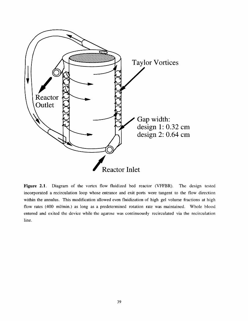
ReactorOutlet
Taylor Vortices
Gap width:design 1: 0.32 cmdesign 2: 0.64 cm
Reactor Inlet
Figure 2.1. Diagram of the vortex flow fluidized bed reactor (VFFBR). The design tested
incorporated a recirculation loop whose entrance and exit ports were tangent to the flow direction
within the annulus. This modification allowed even fluidization of high gel volume fractions at high
flow rates (400 ml/min.) as long as a predetermined rotation rate was maintained. Whole blood
entered and exited the device while the agarose was continuously recirculated via the recirculation
line.
39
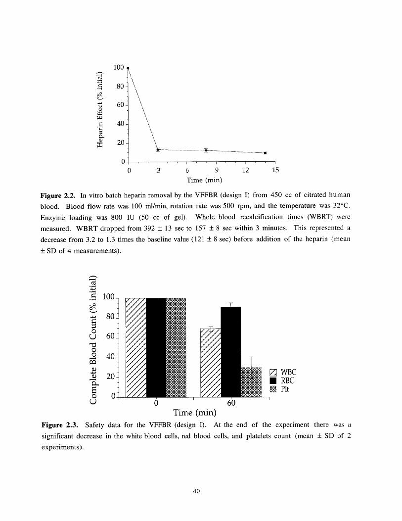
100-0
r- 80-
tU 60
-4 40-
20-
0-i0 3 6 9 12 15
Time (min)
Figure 2.2. In vitro batch heparin removal by the VFFBR (design I) from 450 cc of citrated human
blood. Blood flow rate was 100 ml/min, rotation rate was 500 rpm, and the temperature was 32'C.
Enzyme loading was 800 IU (50 cc of gel). Whole blood recalcification times (WBRT) were
measured. WBRT dropped from 392 ± 13 sec to 157 8 sec within 3 minutes. This represented a
decrease from 3.2 to 1.3 times the baseline value (121 8 sec) before addition of the heparin (mean
SD of 4 measurements).
100-
0
-o 4 // // -u 606
"C 40-
~20-
U 06
Time (min)
Figure 2.3. Safety data for the VFFBR (design I). At the end of the experiment there was a
significant decrease in the white blood cells, red blood cells, and platelets count (mean ± SD of 2
experiments).
40
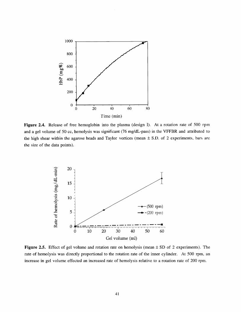
1000
800
# 600
400
200
0 I I
0 20 40 60 80
Time (min)
Figure 2.4. Release of free hemoglobin into the plasma (design I). At a rotation rate of 500 rpm
and a gel volume of 50 cc, hemolysis was significant (76 mg/dL-pass) in the VFFBR and attributed to
the high shear within the agarose beads and Taylor vortices (mean S.D. of 2 experiments, bars are
the size of the data points).
~' 20
15
104
-a-(500 rpm)5 --- -(200 rpm)
0
P4 00 10 20 30 40 50 60
Gel volume (ml)
Figure 2.5. Effect of gel volume and rotation rate on hemolysis (mean SD of 2 experiments). The
rate of hemolysis was directly proportional to the rotation rate of the inner cylinder. At 500 rpm, an
increase in gel volume effected an increased rate of hemolysis relative to a rotation rate of 200 rpm.
41
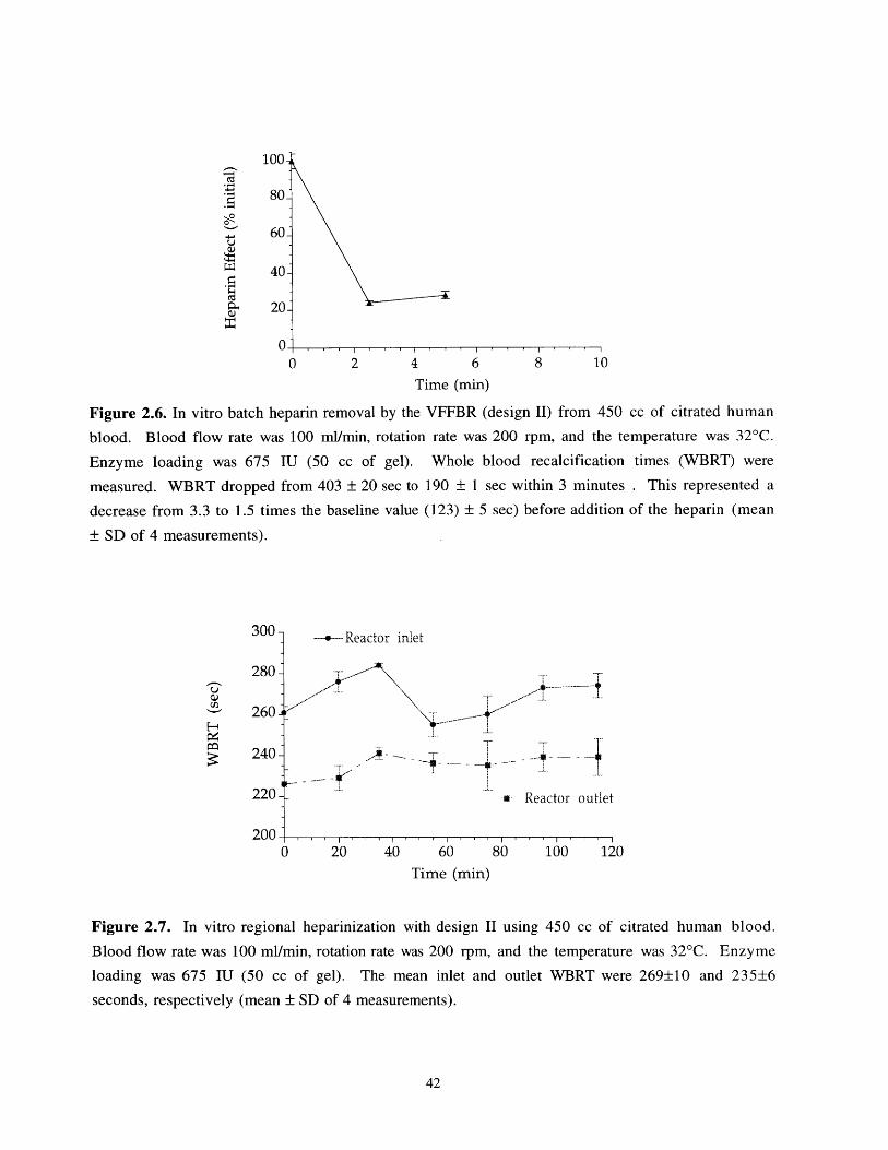
100-
03 880
Z' 60
40-
4 20
0 ,0 2 4 6 8 10
Time (min)
Figure 2.6. In vitro batch heparin removal by the VFFBR (design II) from 450 cc of citrated human
blood. Blood flow rate was 100 ml/min, rotation rate was 200 rpm, and the temperature was 32'C.
Enzyme loading was 675 IU (50 cc of gel). Whole blood recalcification times (WBRT) were
measured. WBRT dropped from 403 ± 20 sec to 190 ± 1 sec within 3 minutes . This represented a
decrease from 3.3 to 1.5 times the baseline value (123) ± 5 sec) before addition of the heparin (mean
SD of 4 measurements).
300- -+-Reactor inlet
280 -39\
' 260A
240_
220 - -Reactor outlet
200 .0 20 40 60 80 100 120
Time (min)
Figure 2.7. In vitro regional heparinization with design II using 450 cc of citrated human blood.
Blood flow rate was 100 ml/min, rotation rate was 200 rpm, and the temperature was 32'C. Enzyme
loading was 675 IU (50 cc of gel). The mean inlet and outlet WBRT were 269±10 and 235±6
seconds, respectively (mean ± SD of 4 measurements).
42
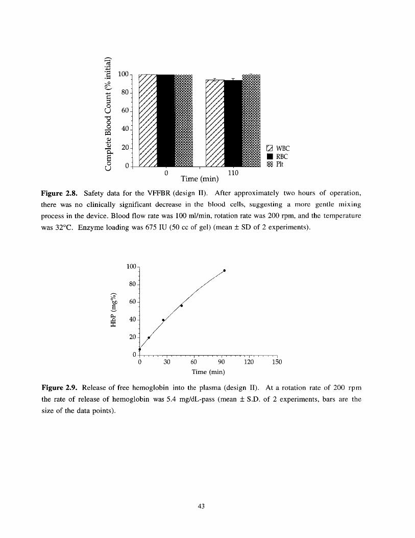
100-
4' 8 0
U 60
40
~4-
20 - WBCU RBC
01 PltU 0 110
Time (min)
Figure 2.8. Safety data for the VFFBR (design II). After approximately two hours of operation,
there was no clinically significant decrease in the blood cells, suggesting a more gentle mixing
process in the device. Blood flow rate was 100 ml/min, rotation rate was 200 rpm, and the temperature
was 32'C. Enzyme loading was 675 IU (50 cc of gel) (mean SD of 2 experiments).
100-
807
S60-
40
20-
0 30 60 90 120 150
Time (min)
Figure 2.9. Release of free hemoglobin into the plasma (design 1I). At a rotation rate of 200 rpm
the rate of release of hemoglobin was 5.4 mg/dL-pass (mean ± S.D. of 2 experiments, bars are the
size of the data points).
43
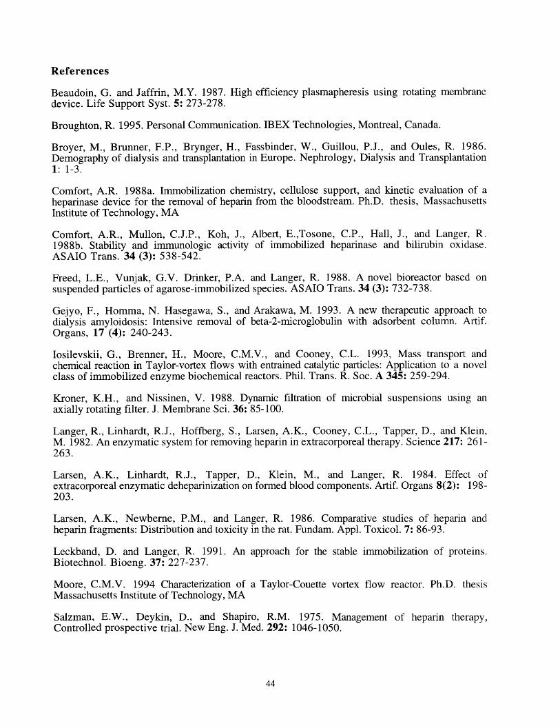
References
Beaudoin, G. and Jaffrin, M.Y. 1987. High efficiency plasmapheresis using rotating membranedevice. Life Support Syst. 5: 273-278.
Broughton, R. 1995. Personal Communication. IBEX Technologies, Montreal, Canada.
Broyer, M., Brunner, F.P., Brynger, H., Fassbinder, W., Guillou, P.J., and Oules, R. 1986.Demography of dialysis and transplantation in Europe. Nephrology, Dialysis and Transplantation1: 1-3.
Comfort, A.R. 1988a. Immobilization chemistry, cellulose support, and kinetic evaluation of aheparinase device for the removal of heparin from the bloodstream. Ph.D. thesis, MassachusettsInstitute of Technology, MA
Comfort, A.R., Mullon, C.J.P., Koh, J., Albert, E.,Tosone, C.P., Hall, J., and Langer, R.1988b. Stability and immunologic activity of immobilized heparinase and bilirubin oxidase.ASAIO Trans. 34 (3): 538-542.
Freed, L.E., Vunjak, G.V. Drinker, P.A. and Langer, R. 1988. A novel bioreactor based onsuspended particles of agarose-immobilized species. ASAIO Trans. 34 (3): 732-738.
Gejyo, F., Homma, N. Hasegawa, S., and Arakawa, M. 1993. A new therapeutic approach todialysis amyloidosis: Intensive removal of beta-2-microglobulin with adsorbent column. Artif.Organs, 17 (4): 240-243.
Iosilevskii, G., Brenner, H., Moore, C.M.V., and Cooney, C.L. 1993, Mass transport andchemical reaction in Taylor-vortex flows with entrained catalytic particles: Application to a novelclass of immobilized enzyme biochemical reactors. Phil. Trans. R. Soc. A 345: 259-294.
Kroner, K.H., and Nissinen, V. 1988. Dynamic filtration of microbial suspensions using anaxially rotating filter. J. Membrane Sci. 36: 85-100.
Langer, R., Linhardt, R.J., Hoffberg, S., Larsen, A.K., Cooney, C.L., Tapper, D., and Klein,M. 1982. An enzymatic system for removing heparin in extracorporeal therapy. Science 217: 261-263.
Larsen, A.K., Linhardt, R.J., Tapper, D., Klein, M., and Langer, R. 1984. Effect ofextracorporeal enzymatic deheparinization on formed blood components. Artif. Organs 8(2): 198-203.
Larsen, A.K., Newberne, P.M., and Langer, R. 1986. Comparative studies of heparin andheparin fragments: Distribution and toxicity in the rat. Fundam. Appl. Toxicol. 7: 86-93.
Leckband, D. and Langer, R. 1991. An approach for the stable immobilization of proteins.Biotechnol. Bioeng. 37: 227-237.
Moore, C.M.V. 1994 Characterization of a Taylor-Couette vortex flow reactor. Ph.D. thesisMassachusetts Institute of Technology, MA
Salzman, E.W., Deykin, D., and Shapiro, R.M. 1975. Management of heparin therapy,Controlled prospective trial. New Eng. J. Med. 292: 1046-1050.
44

Taylor, G.I. 1923. Stability of a viscous liquid contained between two rotating cylinder. Phil.Trans. R. Soc. A 223: 289-343.
Vallar L, and Rivat, C. 1996. Regenerated cellulose-based hemodialyzers with immobilizedproteins as potential devices for extracorporeal immunoadsorption procedures: Assessment ofprotein coupling capacity and in vitro dialysis performances. Artif. Organs 20 (1): 8-11.
Vigo, F., Uliana, C., and Lupino, P. 1985. The performance of a rotating module in oilyemulsions ultrafiltration. Sep. Sci. Technol. 20: 213-230.
Vunjak-Novakovic, G., Shiva, V.A., Freed, L.E., Cooney, C.L. and Langer, R. 1988.Computer-aided visualization and analysis of fluid and particle motion in multiphase systems.Biochem. Eng. Quarterly 2: 249-252.
45
Chapter 3
The Vortex Flow Plasmapheretic Reactor (VFPR)
3.1 Introduction
The results from Chapter 2 showed that agarose immobilized heparinase I fluidized within
high blood flow rates and turbulent vortices resulted in blood damage and hemolysis (Ameer et al.,
1999). To minimize blood damage, a device that simultaneously effected plasma separation and the
enzymatic reaction with the capacity to treat high whole blood flow rates was designed and
constructed. The new reactor design uses Taylor-Couette flow (also known as Taylor vortex flow)
as a means to achieve good mixing and to minimize cell concentration polarization on the
membrane (Beaudoin and Jaffrin, 1987). Taylor-Couette flow is established when the inner
cylinder of an annulus is rotated beyond a critical rotation rate determined by the critical Taylor
number (Taylor, 1923). The flow instabilities generated within the annulus create a secondary
flow patem in the form of periodic vortices termed Taylor vortices. Taylor-Couette flow devices
have primarily been used in filtration applications (Holeschovsky and Cooney, 1991; Kroner and
Nissinen, 1988). During filtration, the Taylor vortices effectively reduce concentration polarization
at the membrane surface. Reported filtrate fluxes are significantly higher than those observed in
conventional hollow fiber devices (Ohashi et al., 1988). Membrane induced circumferencial flow
was observed in the immobilized enzyme compartment and was able to fluidize the agarose beads.
The objective of this study was to evaluate the feasibility and efficacy of this novel device for
regional heparinization in vitro using buffered saline and human blood.
3.2 Materials and Methods
The materials used to build and operate the VFPR were the same as those described in
Chapter 2. The agarose particles (6%, 100-200 mesh, 100 gm average diameter) were from
BioRad (Rockville, NY). Blue dextran (2,000 kD) was from Sigma Chemical Company (St.
Louis, MO). Polyester membranes (1km pore size) were from Whatman Inc. (Clifton, NJ).
Heparin Concentration in Saline
An assay using Azure II dye was used to monitor heparin concentrations during the
experiments. (Lam et al., 1976). A 4.5 ml volume of the Azure II dye solution (0.01 mg/ml) was
added to 0.5 ml of the heparin solution to be tested. The sample was mixed and incubated at room
temperature for 1 minute before measuring the absorbance at 500 nm. A standard curve for this
assay was prepared by using solutions of known heparin concentrations ranging from 0 to 3 USP
units/ml heparin (- 0 - 19 gg/ml). The standard curve was linear in the above range.
46
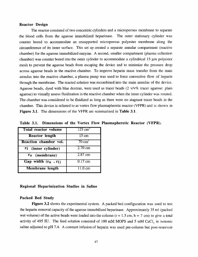
Reactor Design
The reactor consisted of two concentric cylinders and a microporous membrane to separate
the blood cells from the agarose immobilized heparinase. The outer stationary cylinder was
counter bored to accommodate an unsupported microporous polyester membrane along the
circumference of its inner surface. This set up created a separate annular compartment (reactive
chamber) for the agarose immobilized enzyme. A second, smaller compartment (plasma collection
chamber) was counter bored into the outer cylinder to accommodate a cylindrical 15 jim polyester
mesh to prevent the agarose beads from escaping the device and to minimize the pressure drop
across agarose beads in the reactive chamber. To improve heparin mass transfer from the main
annulus into the reactive chamber, a plasma pump was used to force convective flow of heparin
through the membrane. The reacted solution was recombined into the main annulus of the device.
Agarose beads, dyed with blue dextran, were used as tracer beads (2 v/v% tracer agarose: plain
agarose) to visually assess fluidization in the reactive chamber when the inner cylinder was rotated.
The chamber was considered to be fluidized as long as there were no stagnant tracer beads in the
chamber. This device is referred to as vortex flow plasmapheretic reactor (VFPR) and is shown in
Figure 3.1. The dimensions of the VFPR are summarized in Table 3.1.
Table 3.1. Dimensions of the Vortex Flow Plasmapheretic Reactor (VFPR).
Total reactor volume 125 cm'
Reactor length 15 cm
Reaction chamber vol. 70 cm'
ri (inner cylinder) 2.70 cm
ro (membrane) 2.87 cm
Gap width (ro - ri) 0.17 cm
Membrane length 11.0 cm
Regional Heparinization Studies in Saline
Packed Bed Study
Figure 3.2 shows the experimental system. A packed bed configuration was used to test
the heparin removal, capacity of the agarose immobilized heparinase. Approximately 35 ml (packed
wet volume) of the active beads were loaded into the column (r = 1.3 cm, h = 7 cm) to give a total
activity of 495 IU. The feed solution consisted of 100 mM MOPS and 5 mM CaCl2 in isotonic
saline adjusted to pH 7.4. A constant infusion of heparin was used pre-column but post-reservoir
47
to simulate regional heparinization. The heparin concentrations entering and exiting the packed bed
were measured using the Azure II assay described above. A peristaltic pump was used to control
the flowrate of solution through the packed bed which was maintained at 60 ml/min. The system
was maintained at 37 'C with a water bath.
VFPR Study
Regional heparinization studies, similar to the packed bed experiments described above,
were performed with the VFPR. A 500 ml volume of the feed solution, heated to 33 'C, was
recirculated through the circuit at 120 ml/min with the plasma pump adjusted to 60 ml/min. The
rotation rate of the inner cylinder was set to 1,200 rpm, which corresponds to a shear rate of 9,200
s-1. This shear rate is below the hemolysis limit of 20,000 s-1 reported in the literature (Fischel et
al. 1988; Heuser and Opitz, 1980). Approximately 20 ml of immobilized heparinase (Specific
activity 10-15 IU/ml wet gel) was injected into the reactive chamber of the VFPR and was fluidized
by the flow dynamics in that chamber. The infusion of heparin pre-reactor was adjusted to achieve
clinically relevant heparin plasma concentrations which are in the range of 5 - 12 gg heparin/mi
plasma. Heparin concentrations were measured at the inlet and outlet of the reactor using the Azure
II assay.
In Vitro Regional Heparinization Studies with Human Blood
The assembled reactor was incorporated into a closed circuit primed with normal saline.
The circuit consisted of a blood warmer, blood pump, drip chamber and pressure monitor. The
experimental set up was similar to the VFPR saline studies. A 450 cc volume of human donor
blood was collected in heparin at the Blood Donor Center of Children's Hospital of Boston.
Approval was obtained from the M.I.T. Committee for the use of Humans as Experimental
Subjects and the required consent forms and protocols were implemented. The hematocrit (v/v%
of blood cells) of the bag was adjusted to 30-35 % by dilution with saline to match the values
normally encountered in the clinical setting. The blood was pumped at a flow rate of 160 ml/min.
The VFPR rotation rate was set at 1,200 rpm and the plasma pump was set at 60 ml/min. Cell
leakage into the reactive chamber was prevented by placing a strip of medical grade adhesive tape
(type 7759 from Adhesive Research, Inc., Glen Rock, PA) within the reactor's outer cylinder and
adhering the polyester membrane onto the tape with photoreactive adhesive. The temperature of
the blood circuit was constant at 33 'C. A pressure gauge was placed between the reactive chamber
outlet and the plasma pump to monitor clogging of the microporous membrane. Blood samples
were taken at the inlet and outlet of the device and heparin levels were indirectly assayed by
WBRT's, as described in the Materials and Methods section of Chapter 2. Blood samples also
were drawn at the reactor outlet for further hemocompatibility tests.
48
Reactor Hemocompatibility
Human blood samples were taken to the M.I.T. Medical Laboratory for automated analysis
of the red cell, white cell, and platelet populations. In addition, fibrinogen, antithrombin III, and
total protein levels were assayed at Quest Laboratories. Hemolysis was measured through the
release of free hemoglobin into the plasma. A hemoglobin detection kit from Sigma was used.
The kit is based on the catalytic effect of hemoglobin on the oxidation of 3,3',5,5'-
tetramethylbenzidine (TMB) by hydrogen peroxide. In this assay, a 10 R1 sample is added to 4 ml
of reagent and measured at 600 nm at 25'C. The color formation in the reaction is proportional to
the hemoglobin concentration in the sample.
3.3 Results and Discussion
Figure 3.3 shows the schematic of a Taylor-Couette flow device and three possible
approaches, including the proposed design, to include a surface immobilized species. Suspension
of the agarose immobilized species within Taylor vortices in whole blood, as shown in Figure
3.3-A, resulted in significant lysis of the red cells (Ameer et al., 1999). Figure 3.3-B
illustrates the use of a membrane as an immobilization support. A concern with a membrane
Taylor-Couette flow device was the lower surface area to volume ratio that could be attained for a
reasonable size device. In addition, the membrane mass transfer coefficient has been reported to be
orders of magnitude smaller than the mass transfer coefficient to a fluidized bead (Moore, 1994).
The reactor configuration in Figure 3.3-C was designed so that the blood cells and platelets
would not come into contact with the agarose beads and therefore minimize hemolysis. The
unsupported polyester microporous membrane used for cell separation undulated as a result of the
rotation rate of the inner cylinder. These undulations generated a secondary circumferencial flow
in the reactive chamber. The author hypothesizes that the mechanism for the generation of the flow
in the reactive chamber is similar to that of peristalsis. The flow was determined to be
circumferencial after monitoring the trajectory of tracer beads and small bubbles that were formed
in the reactive chamber. This flow fluidized the agarose beads throughout the reactive chamber as
judged visually by the distribution of tracer beads. The fluidization of the beads within the plasma
is illustrated in Figure 3.4. An additional advantage to this design is the fact that the immobilized
species is only exposed to blood plasma and the device can handle blood flow rates of up to 400
ml/min. Such high flow rates are possible because there are no "bead retaining" meshes in the
main blood path that could clog or clot.
49
In vitro Regional Heparinization Studies in Saline
Packed Bed Study
The results for the regional heparinization experiments, using the packed bed, are shown in
Figure 3.5. The steady state heparin conversions (diamonds) were between 80 and 99 % at 37 0 C
and a flow rate of 60 ml/min. The activity of the beads was checked at the end of the experiment
and they lost only 10% of initial. Therefore, the observed decline in conversion towards the end of
the run may have been due to fluid channeling through the bed. Nevertheless, these results,
though obtained in saline, demonstrate the feasibility and efficacy of regional heparinization using
immobilized heparinase. However, the use of an immobilized heparinase packed bed with blood is
not feasible due to hemocompatibility issues. In addition, the high pressure drop generated across
the column is not desirable when working with a patient's blood. Therefore, alternative
approaches had to be pursued to achieve similar conversions in a "blood compatible" reactor
system.
VFPR Study
As discussed earlier, the VFPR design was intended to overcome the "blood compatibility"
and throughput issues associated with agarose immobilized heparinase. The results of the VFPR
experiments in saline are shown in Figure 3.6. The reactor maintained mean inlet and outlet
heparin concentrations of 11 ± 0.5 and 6.0 ± 0.2 gg/ml ( S.E.M.), respectively. These values
corresponded to a mean heparin conversion of 44 ±0.5% ( S.E.M.) for an inlet flow rate into the
reactor of 120 ml/min and a plasma pump flow rate of 60 ml/min. This heparin conversion, if
achieved in a patient, would meet the minimum efficacy design criteria for an immobilized
heparinase reactor as stated in the Introduction. It is expected that the overall heparin conversion
through the device would depend on the amount of immobilized enzyme, the fluidization
conditions, and the flow split through the microporous membrane.
VFPR Regional Heparinization Studies In Vitro with Human Blood
Reactor Efficacy
Even though good heparin neutralization results were obtained in the above saline
experiments, the important measure for an immobilized heparinase reactor is its performance in
blood. Figure 3.7 shows a picture of the reactor during operation with blood. The results for
regional blood heparinization using 10 ml of agarose immobilized heparinase (specific activity 16
IU/ml gel) are shown in Figure 3.8. The reactor maintained a mean WBRT difference of 67
seconds between the inlet and outlet. This difference corresponded to a mean heparin conversion
50
of 34 ± 2% (± S.E.M.) at a blood flow rate of 160 ml/min and a plasma pump flow rate of 60
ml/min. This mean conversion, obtained with half the volume of immobilized enzyme that was
used in the saline studies, does not fall within the target range of 45-50%, however, the data
demonstrates the feasibility of using the VFPR design to achieve regional heparinization with
negligible blood damage. Bead volumes of up to 30 ml (42% of the reactive chamber volume)
have been fluidized within the reactive chamber. Therefore, an increase in the volume of the
immobilized enzyme injected into the reactive chamber (up to 30 ml) should improve the observed
heparin conversion as long as adequate agarose fluidization is present. Further use of the enzyme
with the VFPR was reserved for the reactor modeling studies in saline and the ex vivo evaluation
of the reactor in sheep, as discussed in Chapters 4 and 5, respectively.
Reactor Safety
The results for the hemocompatibility of the VFPR are summarized in Tables 3.2 and
3.3. The white and red cell counts changed by less than 3% of their initial values. The platelet
count had an 8% reduction relative to the concentrations at the beginning of the run. The
concentrations of free hemoglobin released into the plasma were well below the safety criteria of
150 mg/dL. Therefore, eliminating contact between the cells and the agarose beads was an
efficient way to significantly reduce hemolysis. These results are favorable when compared to
those of previous immobilized heparinase reactors in which fluidized agarose beads within whole
blood negatively affected the cell counts (Larsen et al., 1984). In the in vitro studies with human
blood performed by Larsen, white cell and platelet counts decreased to 10% of their initial values
when tested in a recirculating reactor. This blood damage was attributed to mixing conditions in
the reactor. As discussed in Chapter 2, the fluidization of agarose beads within Taylor vortices in
whole blood resulted in white cell and platelet counts of 70% and 30% of their initial values,
respectively.
Table 3.2. Complete blood cell and hemolysis data for the VFPR.
Time (min) WBC (% RBC (% Plt (% initial) HbP (mg/dL)
initial) initial)
0 100 100 100 11
30 97 98 103 18
60 100 99 92 15
51
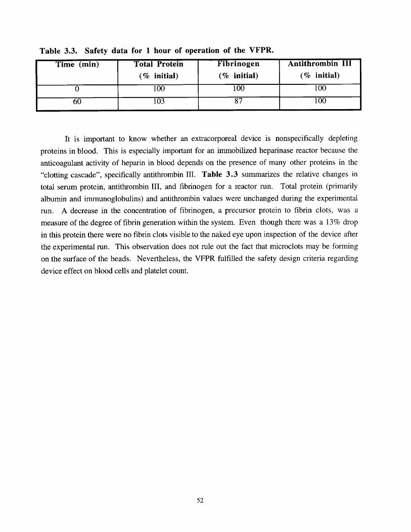
Table 3.3. Safety data for 1 hour of operation of the VFPR.
Time (min) Total Protein Fibrinogen Antithrombin III
(% initial) (% initial) (% initial)
0 100 100 100
60 103 87 100
It is important to know whether an extracorporeal device is nonspecifically depleting
proteins in blood. This is especially important for an immobilized heparinase reactor because the
anticoagulant activity of heparin in blood depends on the presence of many other proteins in the
"clotting cascade", specifically antithrombin III. Table 3.3 summarizes the relative changes in
total serum protein, antithrombin III, and fibrinogen for a reactor run. Total protein (primarily
albumin and immunoglobulins) and antithrombin values were unchanged during the experimental
run. A decrease in the concentration of fibrinogen, a precursor protein to fibrin clots, was a
measure of the degree of fibrin generation within the system. Even though there was a 13% drop
in this protein there were no fibrin clots visible to the naked eye upon inspection of the device after
the experimental run. This observation does not rule out the fact that microclots may be forming
on the surface of the beads. Nevertheless, the VFPR fulfilled the safety design criteria regarding
device effect on blood cells and platelet count.
52
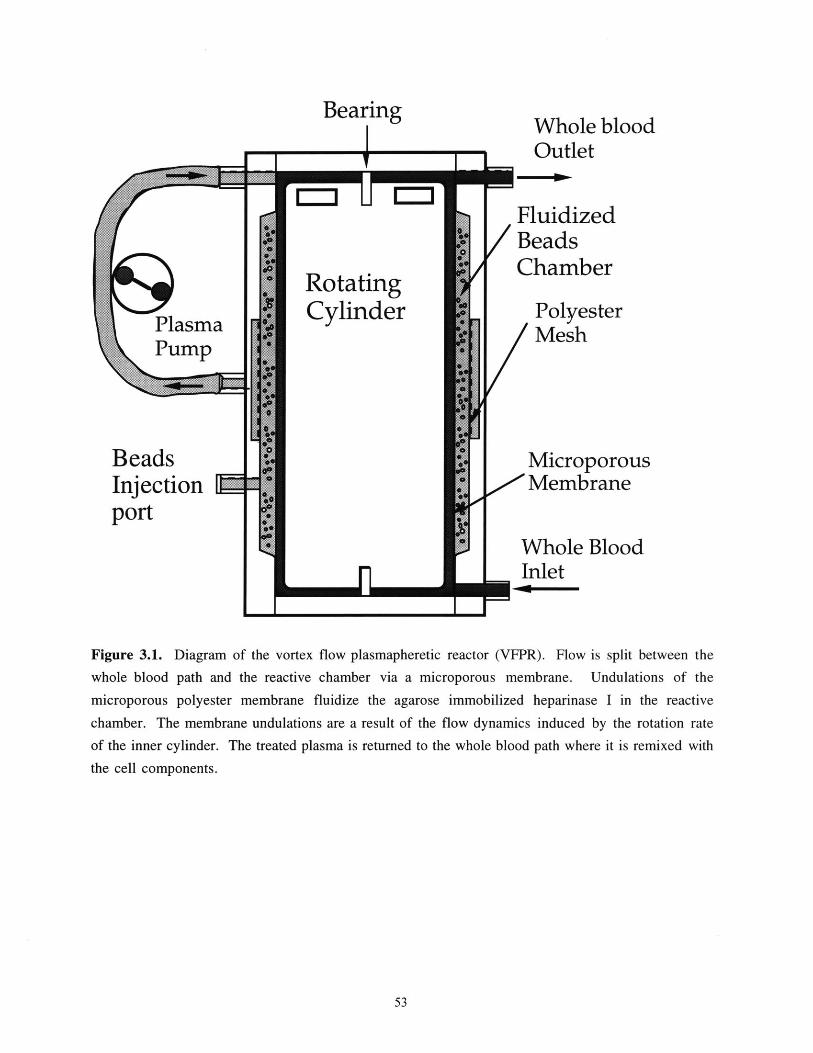
Bearing Whole bloodOutlet
FluidizedBeads
Rotatin ChamberSRotating
Cylinder PolyesterMesh
Pump
Beads MicroporousInjection Membraneport
Whole BloodInlet
Figure 3.1. Diagram of the vortex flow plasmapheretic reactor (VFPR). Flow is split between the
whole blood path and the reactive chamber via a microporous membrane. Undulations of the
microporous polyester membrane fluidize the agarose immobilized heparinase I in the reactive
chamber. The membrane undulations are a result of the flow dynamics induced by the rotation rate
of the inner cylinder. The treated plasma is returned to the whole blood path where it is remixed with
the cell components.
53
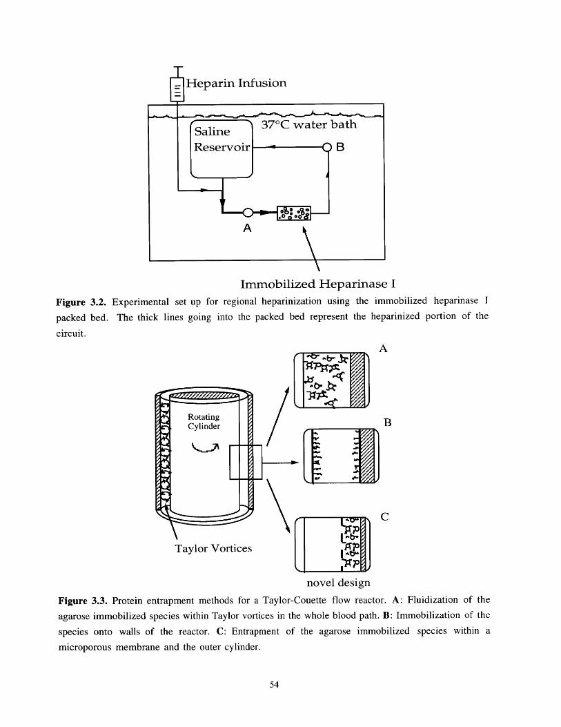
Heparin Infusion
.n 37'C water bath
Reservoir B
A
Immobilized Heparinase I
Figure 3.2. Experimental set up for regional heparinization using the immobilized heparinase I
packed bed. The thick lines going into the packed bed represent the heparinized portion of the
circuit.
A
31
B
\tI~i----
Taylor Vortices
novel design
Figure 3.3. Protein entrapment methods for a Taylor-Couette flow reactor. A: Fluidization of the
agarose immobilized species within Taylor vortices in the whole blood path. B: Immobilization of the
species onto walls of the reactor. C: Entrapment of the agarose immobilized species within a
microporous membrane and the outer cylinder.
54
RotatingCylinder
---- ' -
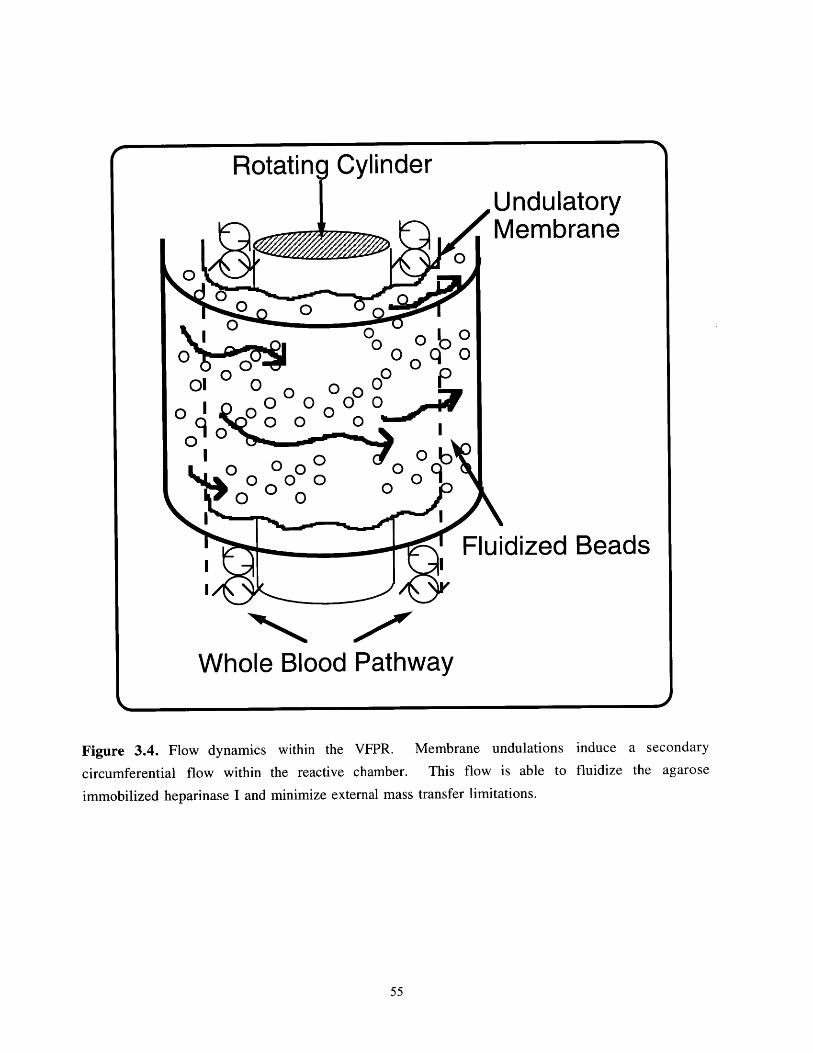
Rotating CylinderUndulatoryMembrane
0 0
Fluidized Beads
Whole Blood Pathway
Figure 3.4. Flow dynamics within the VFPR. Membrane undulations induce a secondary
circumferential flow within the reactive chamber. This flow is able to fluidize the agarose
immobilized heparinase I and minimize external mass transfer limitations.
55
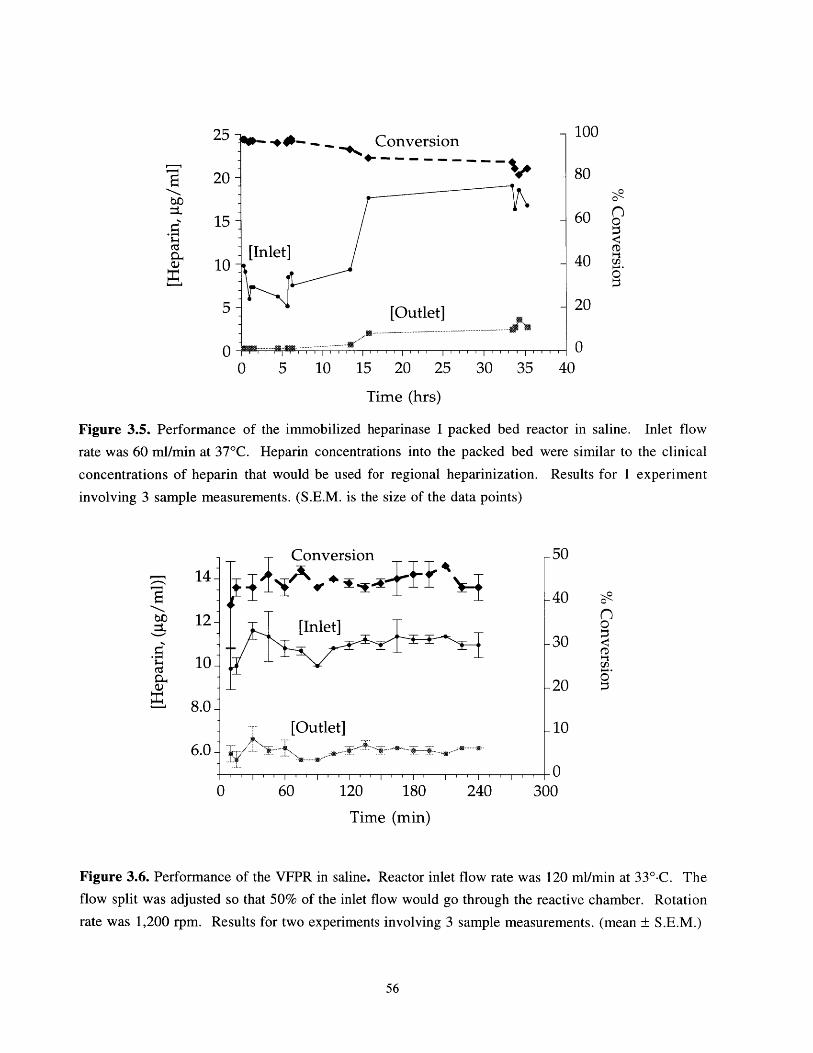
25 Conversion - 100ap-~~ -+-.... Convers-i-on
20 - 80
15 - 60 o
CIO [Inlet] -4
10 - 40 .
20
5 [Outlet] 20
0 5 10 15 20 25 30 35 40
Time (hrs)
Figure 3.5. Performance of the immobilized heparinase I packed bed reactor in saline. Inlet flow
rate was 60 ml/min at 37 0C. Heparin concentrations into the packed bed were similar to the clinical
concentrations of heparin that would be used for regional heparinization. Results for 1 experiment
involving 3 sample measurements. (S.E.M. is the size of the data points)
Conversion 50
14 -40 /
12- [Inlet]30
100
20 e8.0
[Outlet] -10
6.0 - .1 ..... .,..,.
0 60 120 180 240 300
Time (min)
Figure 3.6. Performance of the VFPR in saline. Reactor inlet flow rate was 120 ml/min at 33'-C. The
flow split was adjusted so that 50% of the inlet flow would go through the reactive chamber. Rotation
rate was 1,200 rpm. Results for two experiments involving 3 sample measurements. (mean ± S.E.M.)
56
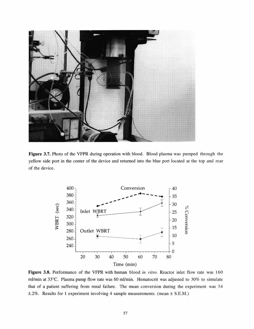
Figure 3.7. Photo of the VFPR during operation with blood. Blood plasma was pumped through the
yellow side port in the center of the device and returned into the blue port located at the top and rear
of the device.
400- Conversion -40
380 . - 35
360 ,,... '-3
S 340 Inlet WBRT -25 )320- 0
H 320-204 300-
- 15 4.280 Outlet WBRT 0
-10260-
240- 5. . . . . . . 0
20 30 40 50 60 70 80
Time (min)
Figure 3.8. Performance of the VFPR with human blood in vitro. Reactor inlet flow rate was 160
ml/min at 33*C. Plasma pump flow rate was 60 ml/min. Hematocrit was adjusted to 30% to simulate
that of a patient suffering from renal failure. The mean conversion during the experiment was 34
±.2%. Results for 1 experiment involving 4 sample measurements. (mean ± S.E.M.)
57
References
Ameer, G.A., Harmon, W., Sasisekharan, R., and Langer, R. 1999. Investigation of a wholeblood fluidized bed Taylor-Couette flow device for enzymatic heparin neutralization. Biotechnol.Bioeng. In Press.
Beaudoin, G. and Jaffrin, M.Y. 1987. High efficiency plasmapheresis using rotating membranedevice. Life Support Syst. 5: 273-278.
Fischel, R.J., Fischer, H., Shatzel, A., Langer, W.P., Cahill, D., Gervail, D., and Ascher, N.L.1988. Couette membrane filtration with constant shear stress. Trans. Am. Soc. Artif. Intern.Organs 34: 375-385.
Heuser, G. and Opitz, R., 1980. A couette viscometer for short time shearing of blood. Rheology17: 17-24.
Holeschovsky, U.B. and Cooney, C.L., 1991. Quantitative description of ultrafiltration in arotating filtration device, AIChE J., 37(8):1219-1226.
Kroner, K.H., and Nissinen, V., 1988. Dynamic filtration of microbial suspensions using anaxially rotating filter. J. Membrane Sci. 36: 85-100.
Lam, L.H., Silbert, J.E., and Rosenberg, R.D., 1976. The separation of active and inactive formsof heparin. Biochem. Biophys. Res. Commun. 69: 570-577.
Larsen, A.K., Linhardt, R.J., Tapper, D., Klein, M., and Langer, R. 1984. Effect ofextracorporeal enzymatic deheparinization on formed blood components. Artif. Organs 8(2): 198-203.
Moore, C.V.L. 1994 Characterization of a Taylor-Couette vortex flow reactor. Ph.D. thesisMassachusetts Institute of Technology, Cambridge MA
Ohashi, K., Tashiro, K., Kushiya, F., et. al 1988. Rotation-induced Taylor vortex enhancesfiltrate flux in plasma separation. Trans. Am. Soc. Artif. Intern. Organs 34: 300-307.
Taylor, G.I., 1923. Stability of a viscous liquid contained between two rotating cylinders. Phil.Trans. Roy. Soc. A223: 289-343.
58
Chapter 4
Residence Time Distribution Analysis and Modeling of
the Vortex Flow Plasmapheretic Reactor
4.1 Introduction
The VFPR separates the blood cell components via a microporous membrane and
enzymatically degrades plasma heparin. The degradation takes place in a separate, fluidized,
agarose immobilized heparinase I, compartment. The reactor also can accommodate high inlet flow
rates (150-400 ml/min). This novel design eliminates contact between the blood cells and the
agarose beads. Also, it has shown to be effective and "biocompatible" in vitro (Chapter 3) and ex
vivo in sheep (Chapter 5).
The goal of the study presented herein was to characterize the flow dynamics within the
novel bioreactor on a macroscopic scale and to predict heparin conversion or neutralization.
Understanding the flow dynamics also would allow optimization of the design. The residence time
distribution of a conventional Taylor-Couette flow device has been reported (Pudjiono et al.,
1992). However, the proposed bioreactor design, which relies on simultaneous separation and
reaction, is a novel configuration that has not been previously characterized. In the present study,
residence time distribution experiments were performed at various operating conditions using the
inert tracer/pulse injection method. A mathematical compartment model was developed to describe
the residence time distributions and was tested against experimental results for kinetics of heparin
conversions.
4.2 Materials and Methods
Residence Time Distribution Studies
Residence time distribution (RTD) studies were performed through the pulse injection of an
inert dye (1 ml, 50 mg/ml Blue Dextran MW 2,000,000 from Sigma Chemical Company, St.
Louis, MO) into the reactor inlet and the measurement of the exit dye concentration over time.
RTD studies were performed on the whole device, as shown in Figure 4.1. a, to obtain the total
exit age distribution function, E,. Additional RTD studies were performed with the plasma line
draining into a separate container, as shown in Figure 4.1. b. These studies measured the exit
age distribution function of the annular compartment, E,. The total device inlet flow rate was set at
either 120 or 150 mi water/min with the plasma pump set at 55, 60, and 75 ml/min. These flow
59
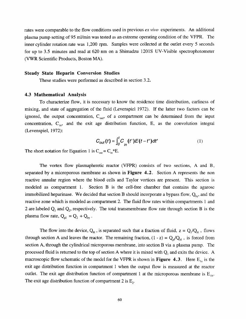
rates were comparable to the flow conditions used in previous ex vivo experiments. An additional
plasma pump setting of 95 ml/min was tested as an extreme operating condition of the VFPR. The
inner cylinder rotation rate was 1,200 rpm. Samples were collected at the outlet every 5 seconds
for up to 3.5 minutes and read at 620 nm on a Shimadzu 1201S UV-Visible spectrophotometer
(VWR Scientific Products, Boston MA).
Steady State Heparin Conversion Studies
These studies were performed as described in section 3.2.
4.3 Mathematical Analysis
To characterize flow, it is necessary to know the residence time distribution, earliness of
mixing, and state of aggregation of the fluid (Levenspiel 1972). If the latter two factors can be
ignored, the output concentration, CO,, of a compartment can be determined from the input
concentration, Cin, and the exit age distribution function, E, as the convolution integral
(Levenspiel, 1972):
Cout(O)= C. (t')E(t-t')dt' 1
The short notation for Equation 1 is C.,1 = Ci*E.
The vortex flow plasmapheretic reactor (VFPR) consists of two sections, A and B,
separated by a microporous membrane as shown in Figure 4.2. Section A represents the non
reactive annular region where the blood cells and Taylor vortices are present. This section is
modeled as compartment 1. Section B is the cell-free chamber that contains the agarose
immobilized heparinase. We decided that section B should incorporate a bypass flow, Qby, and the
reactive zone which is modeled as compartment 2. The fluid flow rates within compartments 1 and
2 are labeled Q, and Q2, respectively. The total transmembrane flow rate through section B is the
plasma flow rate, Qj = Q2 + Qby .
The flow into the device, Qbl , is separated such that a fraction of fluid, z = QI/Qbl I flows
through section A and leaves the reactor. The remaining fraction, (1 - z) = Q1/Qb1 , is forced from
section A, through the cylindrical microporous membrane, into section B via a plasma pump. The
processed fluid is returned to the top of section A where it is mixed with Q, and exits the device. A
macroscopic flow schematic of the model for the VFPR is shown in Figure 4.3. Here Ela is the
exit age distribution function in compartment 1 when the output flow is measured at the reactor
outlet. The exit age distribution function of compartment 1 at the microporous membrane is Ei.The exit age distribution function of compartment 2 is E2.
60
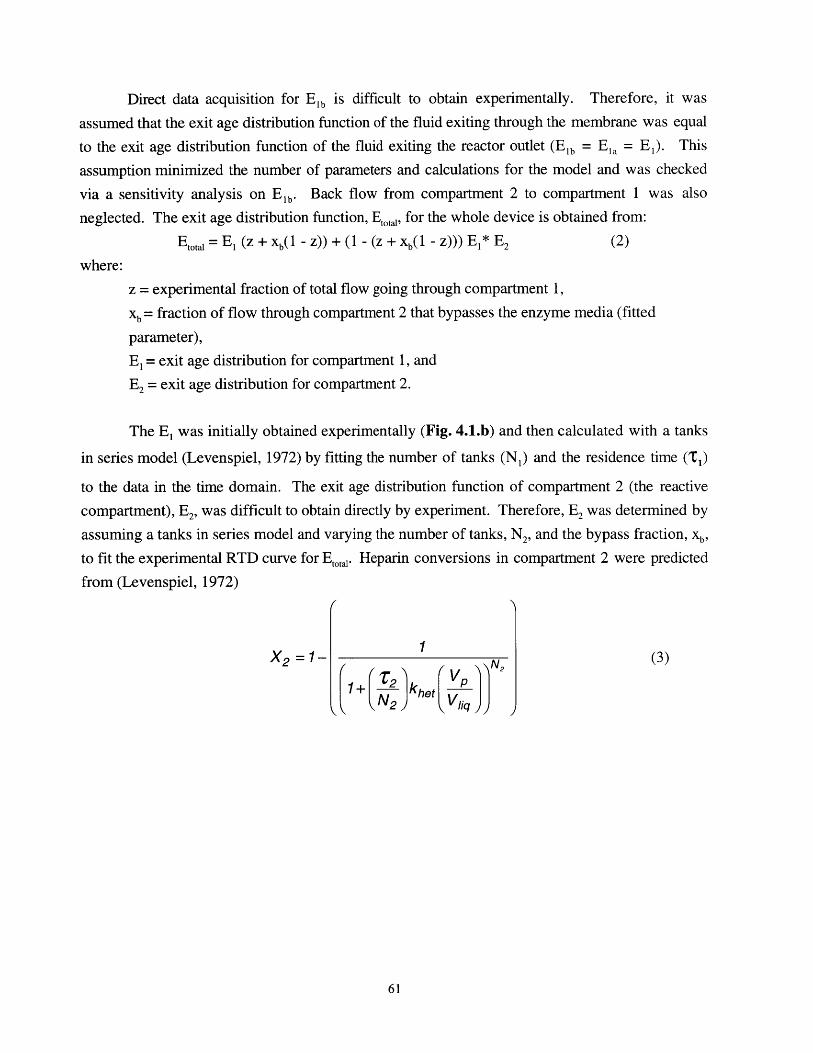
Direct data acquisition for Eib is difficult to obtain experimentally. Therefore, it was
assumed that the exit age distribution function of the fluid exiting through the membrane was equal
to the exit age distribution function of the fluid exiting the reactor outlet (Eib = Eia = E1). This
assumption minimized the number of parameters and calculations for the model and was checked
via a sensitivity analysis on Eib. Back flow from compartment 2 to compartment 1 was also
neglected. The exit age distribution function, Ea, for the whole device is obtained from:
Eta = EI (Z + x - Z)) + (1 - (Z+ x(l - z))) E* E2 (2)
where:
z = experimental fraction of total flow going through compartment 1,
xb= fraction of flow through compartment 2 that bypasses the enzyme media (fitted
parameter),
E1 = exit age distribution for compartment 1, and
E2= exit age distribution for compartment 2.
The E was initially obtained experimentally (Fig. 4.1.b) and then calculated with a tanks
in series model (Levenspiel, 1972) by fitting the number of tanks (N1) and the residence time (T,)
to the data in the time domain. The exit age distribution function of compartment 2 (the reactive
compartment), E2, was difficult to obtain directly by experiment. Therefore, E2 was determined by
assuming a tanks in series model and varying the number of tanks, N2, and the bypass fraction, xb,
to fit the experimental RTD curve for Etotal. Heparin conversions in compartment 2 were predicted
from (Levenspiel, 1972)
1
X2 =1- )(3)
r1+ 2 khet&,,l jVjN
61
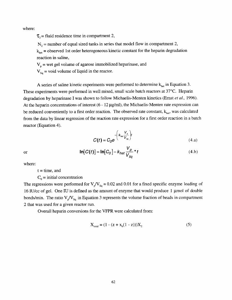
where:
T2 = fluid residence time in compartment 2,
N2 = number of equal sized tanks in series that model flow in compartment 2,
khet = observed 1st order heterogeneous kinetic constant for the heparin degradation
reaction in saline,
VP = wet gel volume of agarose immobilized heparinase, and
Vq = void volume of liquid in the reactor.
A series of saline kinetic experiments were performed to determine khet in Equation 3.
These experiments were performed in well mixed, small scale batch reactors at 37'C. Heparin
degradation by heparinase I was shown to follow Michaelis-Menten kinetics (Ernst et al., 1996).
At the heparin concentrations of interest (6 - 12 gg/ml), the Michaelis-Menten rate expression can
be reduced conveniently to a first order reaction. The observed rate constant, khet, was calculated
from the data by linear regression of the reaction rate expression for a first order reaction in a batch
reactor (Equation 4).
**khet VPh (4aC(t)=Coe " (4.a)
Vor In[C(t)] = In[Co]-khet P*t (4.b)
Vliq
where:
t = time, and
Co = initial concentration
The regressions were performed for V,/V iq= 0.02 and 0.01 for a fixed specific enzyme loading of
16 IU/cc of gel. One IU is defined as the amount of enzyme that would produce 1 gmol of double
bonds/min. The ratio Vp/Vq in Equation 3 represents the volume fraction of beads in compartment
2 that was used for a given reactor run.
Overall heparin conversions for the VFPR were calculated from:
Xtotai = (1 - (z + xb(1 - z)))X 2 (5)
62
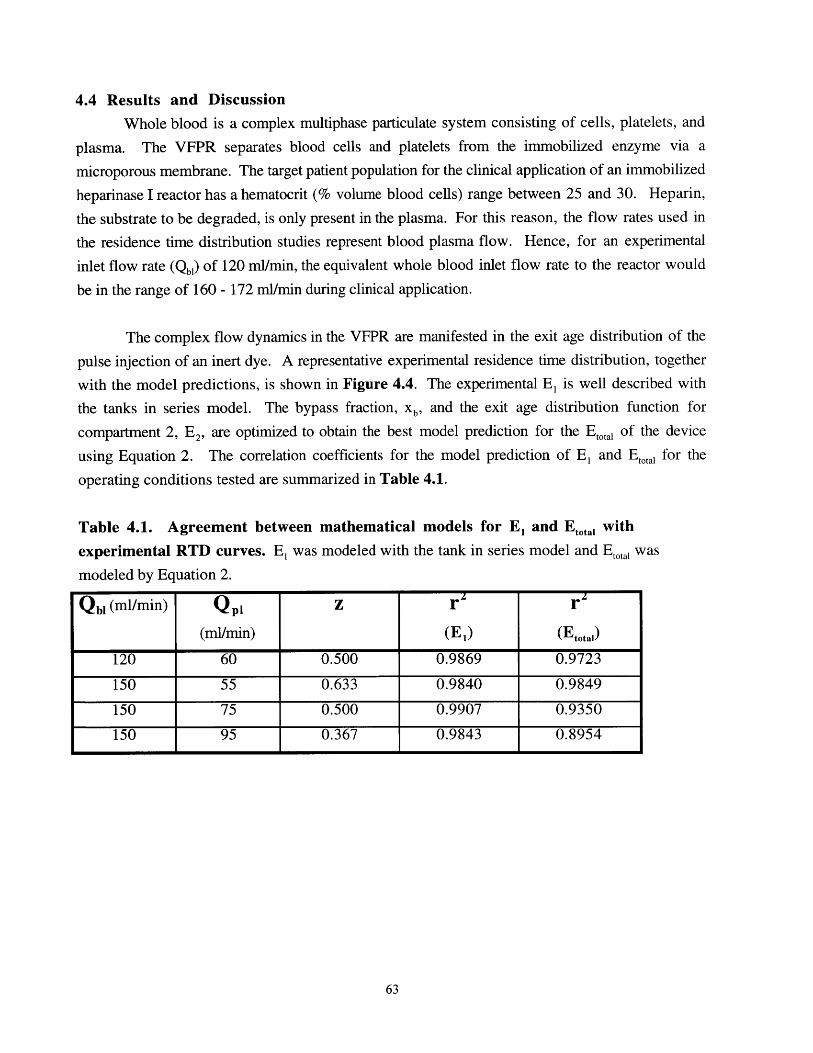
4.4 Results and Discussion
Whole blood is a complex multiphase particulate system consisting of cells, platelets, and
plasma. The VFPR separates blood cells and platelets from the immobilized enzyme via a
microporous membrane. The target patient population for the clinical application of an immobilized
heparinase I reactor has a hematocrit (% volume blood cells) range between 25 and 30. Heparin,
the substrate to be degraded, is only present in the plasma. For this reason, the flow rates used in
the residence time distribution studies represent blood plasma flow. Hence, for an experimental
inlet flow rate (Qbl) of 120 ml/min, the equivalent whole blood inlet flow rate to the reactor would
be in the range of 160 - 172 ml/min during clinical application.
The complex flow dynamics in the VFPR are manifested in the exit age distribution of the
pulse injection of an inert dye. A representative experimental residence time distribution, together
with the model predictions, is shown in Figure 4.4. The experimental E, is well described with
the tanks in series model. The bypass fraction, xb, and the exit age distribution function for
compartment 2, E2, are optimized to obtain the best model prediction for the E,, of the device
using Equation 2. The correlation coefficients for the model prediction of E, and Eal for the
operating conditions tested are summarized in Table 4.1.
Table 4.1. Agreement between mathematical models for El and Etotal with
experimental RTD curves. El was modeled with the tank in series model and Etwa was
modeled by Equation 2.
QbI (ml/min) QP1 Z(mil/min) (E1) (Etota)
120 60 0.500 0.9869 0.9723
150 55 0.633 0.9840 0.9849
150 75 0.500 0.9907 0.9350
150 95 0.367 0.9843 0.8954
63
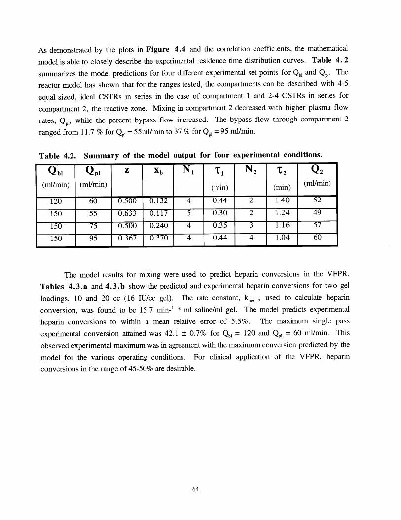
As demonstrated by the plots in Figure 4.4 and the correlation coefficients, the mathematical
model is able to closely describe the experimental residence time distribution curves. Table 4.2
summarizes the model predictions for four different experimental set points for Qbl and QP1. The
reactor model has shown that for the ranges tested, the compartments can be described with 4-5
equal sized, ideal CSTRs in series in the case of compartment 1 and 2-4 CSTRs in series for
compartment 2, the reactive zone. Mixing in compartment 2 decreased with higher plasma flow
rates, QP1, while the percent bypass flow increased. The bypass flow through compartment 2
ranged from 11.7 % for QP1= 55ml/min to 37 % for QP1= 95 ml/min.
Table 4.2. Summary of the model output for four experimental conditions.
Qbl P Z Xb N1 T, N 2 T2 2
(ml/min) (ml/min) (min) (min) (ml/min)
120 60 0.500 0.132 4 0.44 2 1.40 52
150 55 0.633 0.117 5 0.30 2 1.24 49
150 75 0.500 0.240 4 0.35 3 1.16 57
150 95 0.367 0.370 4 0.44 4 1.04 60
The model results for mixing were used to predict heparin conversions in the VFPR.
Tables 4.3.a and 4.3.b show the predicted and experimental heparin conversions for two gel
loadings, 10 and 20 cc (16 IU/cc gel). The rate constant, khet , used to calculate heparin
conversion, was found to be 15.7 min-' * ml saline/ml gel. The model predicts experimental
heparin conversions to within a mean relative error of 5.5%. The maximum single pass
experimental conversion attained was 42.1 ± 0.7% for QbI = 120 and QP1 = 60 ml/min. This
observed experimental maximum was in agreement with the maximum conversion predicted by the
model for the various operating conditions. For clinical application of the VFPR, heparin
conversions in the range of 45-50% are desirable.
64
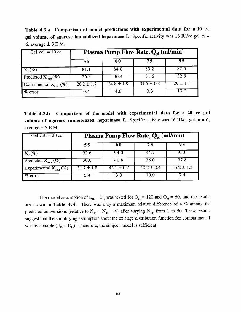
Table 4.3.a Comparison of model predictions with experimental data for a 10 cc
gel volume of agarose immobilized heparinase I. Specific activity was 16 IU/cc gel. n =
6, average S.E.M.
Gel vol.= 10 cc Plasma Pump Flow Rate, Qj (ml/min)
55 60 75 95
X2(%)81.1 84.0 83.2 82.5
Predicted Xtota (%) 26.3 36.4 31.6 32.8
Experimental Xs (%) 26.2 ± 1.7 34.8± 1.9 31.5 ± 0.3 29 ± 1.1
% error 0.4 4.6 0.3 13.0
Table 4.3.b Comparison of the model with experimental data
volume of agarose immobilized heparinase I. Specific activity was
average ± S.E.M.
for a 20 cc gel
16 IU/cc gel. n = 6,
Gel vol. = 20 cc Plasma Pump Flow Rate, Qj (ml/min)
55 60 75 95
X2(%)92.6 94.0 94.7 95.0
Predicted Xtota (%) 30.0 40.8 36.0 37.8
Experimental Xtota (%) 31.7 ± 1.8 42.1 ± 0.7 40.2 ± 0.4 35.2 ± 1.3
% error 5.4 3.0 10.0 7.4
The model assumption of E = Ei was tested for QbI = 120 and Qj = 60, and the results
are shown in Table 4.4. There was only a maximum relative difference of 4 % among the
predicted conversions (relative to Nla = Nib = 4) after varying Nib from 1 to 50. These results
suggest that the simplifying assumption about the exit age distribution function for compartment 1
was reasonable (Eib = Eia). Therefore, the simpler model is sufficient.
65
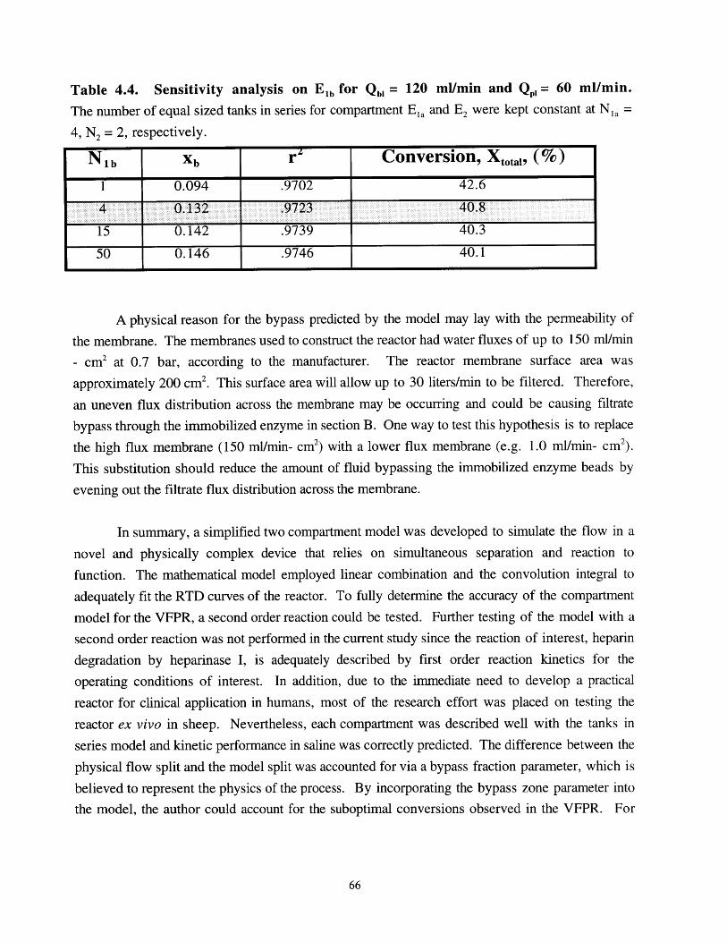
Table 4.4. Sensitivity analysis on Elb for Qb, = 120 ml/min and Q,= 60 ml/min.
The number of equal sized tanks in series for compartment Eia and E2 were kept constant at Na =
4, N2 = 2, respectively.
Nib xb r' Conversion, Xtoi (%)
1 0.094 .9702 42.6
4 0.132 .9723 40.8
15 0.142 .9739 40.3
50 0.146 .9746 40.1
A physical reason for the bypass predicted by the model may lay with the permeability of
the membrane. The membranes used to construct the reactor had water fluxes of up to 150 ml/min
- cm2 at 0.7 bar, according to the manufacturer. The reactor membrane surface area was
approximately 200 cm 2. This surface area will allow up to 30 liters/min to be filtered. Therefore,
an uneven flux distribution across the membrane may be occurring and could be causing filtrate
bypass through the immobilized enzyme in section B. One way to test this hypothesis is to replace
the high flux membrane (150 ml/min- cm 2) with a lower flux membrane (e.g. 1.0 mI/min- cm 2).
This substitution should reduce the amount of fluid bypassing the immobilized enzyme beads by
evening out the filtrate flux distribution across the membrane.
In summary, a simplified two compartment model was developed to simulate the flow in a
novel and physically complex device that relies on simultaneous separation and reaction to
function. The mathematical model employed linear combination and the convolution integral to
adequately fit the RTD curves of the reactor. To fully determine the accuracy of the compartment
model for the VFPR, a second order reaction could be tested. Further testing of the model with a
second order reaction was not performed in the current study since the reaction of interest, heparin
degradation by heparinase I, is adequately described by first order reaction kinetics for the
operating conditions of interest. In addition, due to the immediate need to develop a practical
reactor for clinical application in humans, most of the research effort was placed on testing the
reactor ex vivo in sheep. Nevertheless, each compartment was described well with the tanks in
series model and kinetic performance in saline was correctly predicted. The difference between the
physical flow split and the model split was accounted for via a bypass fraction parameter, which is
believed to represent the physics of the process. By incorporating the bypass zone parameter into
the model, the author could account for the suboptimal conversions observed in the VFPR. For
66
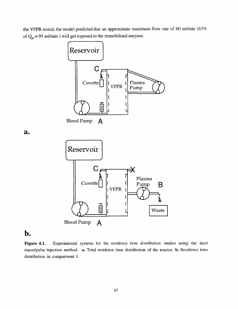
the VFPR tested, the model predicted that an approximate maximum flow rate of 60 ml/min (63%
of QI= 95 ml/min ) will get exposed to the immobilized enzyme.
Reservoir
C F-
Cuvette
C
Cuvette
rI IVFPR
I I
1 rI IjVFPR I
I I
ILI
PlasmaPump
PlasmaPump B
Waste
Blood Pump A
b.Experimental systems for the residence time distribution studies using the inert
tracer/pulse injection method. a. Total residence time distribution of the reactor. b. Residence time
distribution in comparment 1.
67
Blood Pump A
a.
Reservoir
Figure 4.1.
M 0
all
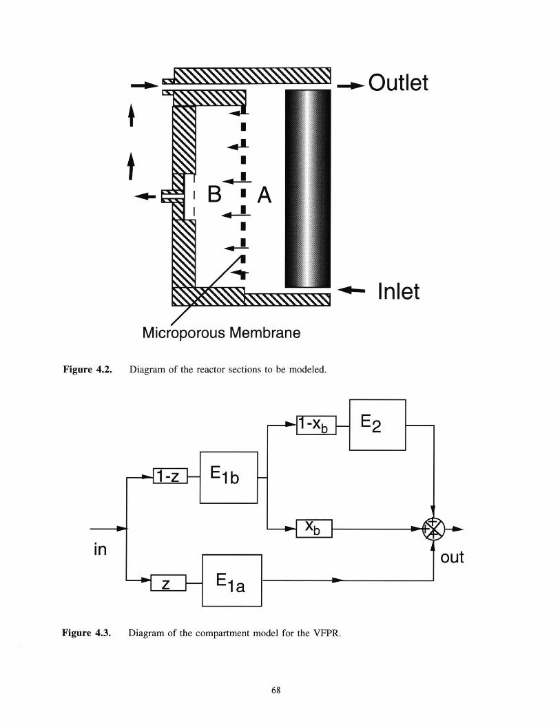
Outlet
IInlet
Microporous Membrane
Figure 4.2. Diagram of the reactor sections to be modeled.
1-xb - E
in out
Figure 4.3. Diagram of the compartment model for the VFPR.
68
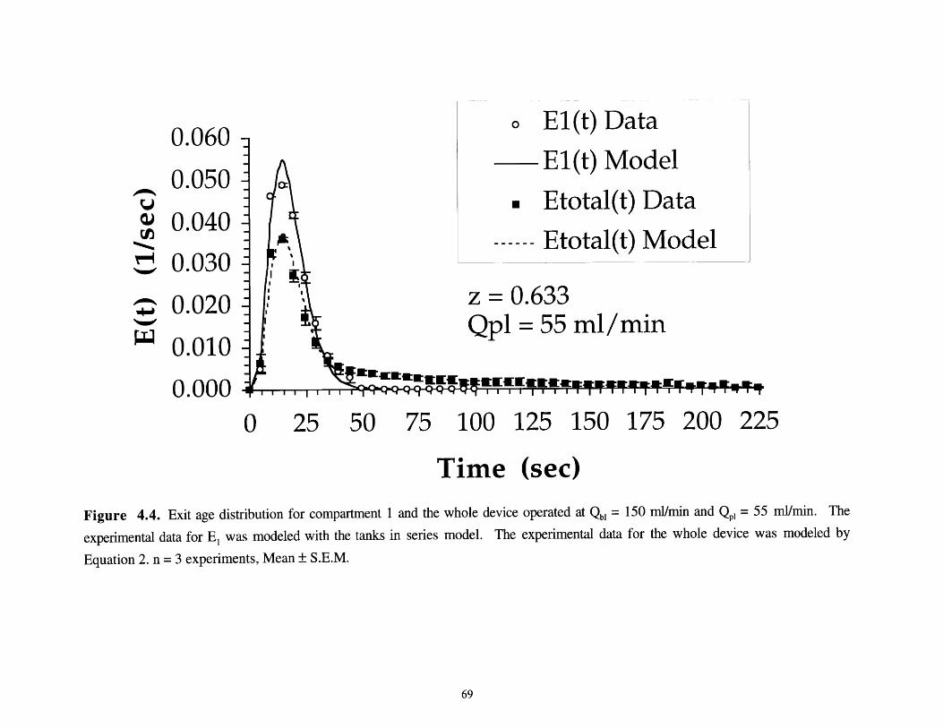
0.060
0.050
0.040
0.030
0.020
0.010
0.0000
o E1(t) Data
E1(t) Model
. Etotal(t) Data------ Etotal(t) Model
z = 0.633Qpl = 55 ml/min
REEK"" EEKEEE"a: K
25 50 75 100 125 150 175 200 225
Time (sec)
Figure 4.4. Exit age distribution for compartment 1 and the whole device operated at QbI = 150 ml/min and QP, = 55 ml/min. The
experimental data for El was modeled with the tanks in series model. The experimental data for the whole device was modeled by
Equation 2. n = 3 experiments, Mean ± S.E.M.
69
References
Ernst, S., Venkataraman, G., Winkler, S., Godavarti, R., Langer, R., Cooney, C.L., andSasisekharan, R. 1996. Expression in Escherichia coli, purification and characterization ofheparinase I from Flavobacterium heparinum. Biochem. J. 315: 589-597.
Levenspiel, 0., 1972. Chemical Reaction Engineering, John Wiley & Sons, New York.
Pudjiono, P.I., Tavare, N.S., Garside, J., and Nigam, D.P., 1992. Residence time distributionfrom a continuous Couette flow device. Chem. Eng. J. 48: 101-110.
70
Chapter 5
Reactor Performance Ex Vivo in Sheep
5.1 Introduction
The previous studies using the VFPR demonstrated that regional heparinization could be
effected in saline as well as in human blood in vitro. The positive safety results from in vitro
experiments with human blood supported further testing ex vivo. In addition, data from animal
studies are a prerequisite to clinical trials in humans. The objective of the studies in this chapter
was to investigate the efficacy and safety of the novel reactor when used for regional heparinization
in a sheep animal model. The sheep was the animal model of choice because of its comparable size
to humans (no need for scale up), its similar hemodynamic characteristics to that of humans
(Burhop et al., 1991), and feasibility to compare the current data with that of previous research in
this model (Bernstein et al., 1987). Clotting times were monitored pre and post reactor to
indirectly measure heparin neutralization. Reactor safety was assessed by white blood cell and
platelet counts, degree of hemolysis, and specific protein concentrations.
5.2 Materials and Methods
Animal Preparation and Regional Heparinization of the Circuit
The experimental protocol for the use of animals was approved by the M.I.T. Committee
on Animal Care and followed throughout the experiments. A total of 5 animals were used for the
feasibility studies of the reactor. One animal was exposed to the reactor without agarose beads to
assess whether the reactor materials and the complex fluid flow within the device would negatively
affect the blood cells or the animal. The other four sheep were exposed to the agarose immobilized
heparinase I to simulate regional heparinization. A double lumen catheter (Medcomp Inc.,Harleysville, PA) was used to gain vascular access to the blood circulation of healthy male sheep
(35-45 Kg). The catheter was inserted through the left internal jugular vein and guided into the
right atrium of the heart (Figure 5.1.a). Blood flows of up to 200 ml/min were attained with no
visible adverse effects to the animal. An extracorporeal procedure was performed up to 4 times on
each animal depending on the patency of the catheter.
A diagram of the experimental set up is shown in Figure 5.1. b. The extracorporeal
circuit consisted of a closed loop which included: the animal, a blood pump, a plasma pump, a
bubble trap, a thrombus trap, and the reactor. The bubble and thrombus traps help prevent bubbles
and emboli, which may form during operation of the reactor, from entering into animal. To
71
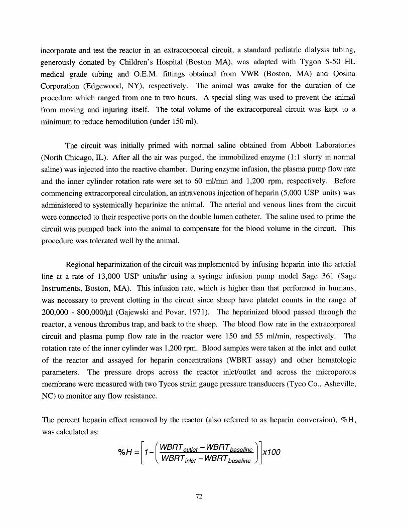
incorporate and test the reactor in an extracorporeal circuit, a standard pediatric dialysis tubing,
generously donated by Children's Hospital (Boston MA), was adapted with Tygon S-50 HL
medical grade tubing and O.E.M. fittings obtained from VWR (Boston, MA) and Qosina
Corporation (Edgewood, NY), respectively. The animal was awake for the duration of the
procedure which ranged from one to two hours. A special sling was used to prevent the animal
from moving and injuring itself. The total volume of the extracorporeal circuit was kept to a
minimum to reduce hemodilution (under 150 ml).
The circuit was initially primed with normal saline obtained from Abbott Laboratories
(North Chicago, IL). After all the air was purged, the immobilized enzyme (1:1 slurry in normal
saline) was injected into the reactive chamber. During enzyme infusion, the plasma pump flow rate
and the inner cylinder rotation rate were set to 60 ml/min and 1,200 rpm, respectively. Before
commencing extracorporeal circulation, an intravenous injection of heparin (5,000 USP units) was
administered to systemically heparinize the animal. The arterial and venous lines from the circuit
were connected to their respective ports on the double lumen catheter. The saline used to prime the
circuit was pumped back into the animal to compensate for the blood volume in the circuit. This
procedure was tolerated well by the animal.
Regional heparinization of the circuit was implemented by infusing heparin into the arterial
line at a rate of 13,000 USP units/hr using a syringe infusion pump model Sage 361 (Sage
Instruments, Boston, MA). This infusion rate, which is higher than that performed in humans,
was necessary to prevent clotting in the circuit since sheep have platelet counts in the range of
200,000 - 800,000/pl (Gajewski and Povar, 1971). The heparinized blood passed through the
reactor, a venous thrombus trap, and back to the sheep. The blood flow rate in the extracorporeal
circuit and plasma pump flow rate in the reactor were 150 and 55 ml/min, respectively. The
rotation rate of the inner cylinder was 1,200 rpm. Blood samples were taken at the inlet and outlet
of the reactor and assayed for heparin concentrations (WBRT assay) and other hematologic
parameters. The pressure drops across the reactor inlet/outlet and across the microporous
membrane were measured with two Tycos strain gauge pressure transducers (Tyco Co., Asheville,
NC) to monitor any flow resistance.
The percent heparin effect removed by the reactor (also referred to as heparin conversion), %H,
was calculated as:
%H = 1- WBRToutlet - WBRTbaseline x100WBRTinet - WBRTbaseline 00
72
The priming volume (whole blood path) and cell free volume (reaction chamber) of the reactor
were 45 ml and 70 ml, respectively. The reactor was loaded with approximately 27 ml of agarose
immobilized heparinase with a specific activity of 18 IU/cc of gel.
Hematology
Blood samples were collected from the reactor outlet into the appropriate vacutainer tubes
(Becton Dickinson, Rockville, MA) at various time points. The following parameters were
measured: white cell and platelet counts, hematocrit, fibrinogen, total protein (albumin + IgG) and
plasma free hemoglobin. The samples were analyzed manually for white cell counts, platelet
counts, and hematocrit at the diagnostic laboratory of the M.I.T Division of Comparative Medicine.
Fibrinogen and total protein were sent out for analysis at Quest Laboratories (Cambridge, MA).
Cell counts and fibrinogen concentrations were normalized by the animal's baseline values and
reported as percents. Plasma free hemoglobin, which is an indicator of hemolysis, was measured
in the laboratory using a hemoglobin detection kit from Sigma Chemical Co.(St. Louis, MO).
Necropsy
The animals were euthanized using an overdose of sodium pentobarbitol, which was
injected directly into the catheter. A complete necropsy with a careful gross examination of all
organs and tissues was performed. Tissue samples from the brain, kidney, spleen, liver, heart,
and lung were taken for histological evaluation at the diagnostic laboratory of the M.I.T. Division
of Comparative Medicine.
5.3 Results and Discussion
Reactor Safety
The results of the reactor safety control study, which exposed the sheep to the VFPR
without the agarose beads, are shown in Figure 5.2a and 5.2b. The values reported are for the
mean of 4 experiments. There was no observed transient initial decrease in white cell counts (F i g.
5.2a) and platelet counts were within 10 % of the initial values. Initial decreases in the white cell
population are usually correlated with complement activation which may promote sequestration of
the white cells by the lungs (Holmes, 1995). The concentrations of C3a and C5a (indicators of
complement activation) were not measured for the ex vivo experiments due to lack of an available
standardized ovine complement assay. However, the absence of the white cell count drop within
the first 30 minutes of operation suggests minimal complement activation. Nevertheless, there is
no substantial evidence of a negative clinical outcome in the form of increased morbidity or
mortality among patients (Holmes, 1995).
73
Figure 5.2b shows the mean hematocrit (a measure of the volume percent of cells in
plasma), which was also maintained within physiological levels (22-35 v/v %). The maintenance
of the hematocrit during the extracorporeal circulation is a positive sign since it is known that sheep
can alter their hematocrit significantly when they are under stress (Hecker, 1993). A significant
decrease in hematocrit could also suggest internal bleeding within the animal or excessive red cell
lysis (hemolysis). To further confirm the integrity of the red cells, plasma free hemoglobin (HbP)
concentrations were measured before and after the extracorporeal procedure. The beginning and
final (2 hour) plasma free hemoglobin concentrations were found to be 6 ± 0.6 and 7 ± 1.5 mg/dL,
respectively. These low plasma free hemoglobin concentrations reflect a significant improvement
in reactor design when compared to past hemolysis observations reported in the literature
(Bernstein et al., 1987; Freed, 1988). In these reports, previous immobilized heparinase devices
based on fluidized agarose beads, were tested in sheep and plasma free hemoglobin concentrations
of up to 90 mg/dL were found after one hour of operation. The data on the mean hematocrit
together with the low levels of plasma free hemoglobin generated during the experiment with the
control sheep suggest a very small degree of hemolysis. It should be mentioned that no decrease in
heparin anticoagulant activity was observed during the control experiments.
The reactor safety data for 11 regional heparinization studies performed on each of four
animals are presented in Figures 5.3 - 5.6. The safety data for the four animals were pooled
and the results are shown in Figure 5.7. The white cell counts seemed to follow the reported
trends that are associated with complement activation. However, in most cases the white cell count
rebounded to approximately 80% of the initial values within one hour and throughout the end of
extracorporeal circulation. In addition, the white cell counts were within normal values prior to the
beginning of subsequent experiments. Platelet counts were normal throughout the experiments.
The maintenance of a normal platelet count during the experiments is another significant
improvement over a previous reactor which depleted platelets down to 50% of the initial value after
one hour of operation without the dialyzer (Bernstein et al. 1987). By the end of the regional
heparinization procedure with the VFPR, plasma free hemoglobin concentrations were below 15
mg/dL, on average. Therefore, separation of the blood cells from the agarose immobilized
heparinase accomplished the objective of significantly minimizing cell destruction.
The results for the total protein and fibrinogen measurements are shown in Table 5.1.
After one hour of extracorporeal circulation, there was no significant change in the total protein for
the one control or the four treated sheep. The mean of the normalized fibrinogen concentrations for
3 regionally heparinized animals and 1 control animal decreased to 94 ± 2% and 92 ± 3% of initial
baseline concentrations, respectively, after one hour of extracorporeal circulation through the
74
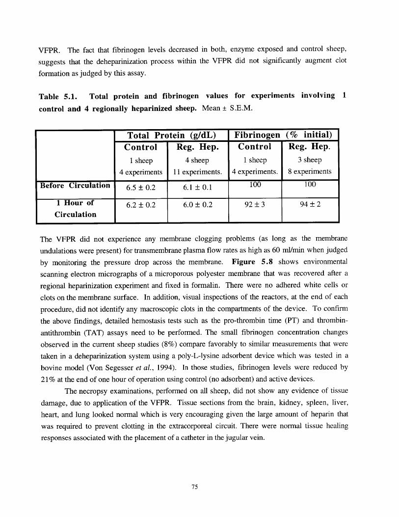
VFPR. The fact that fibrinogen levels decreased in both, enzyme exposed and control sheep,
suggests that the deheparinization process within the VFPR did not significantly augment clot
formation as judged by this assay.
Table 5.1. Total protein and fibrinogen values for experiments involving 1
control and 4 regionally heparinized sheep. Mean S.E.M.
Total Protein (g/dL) Fibrinogen (% initial)Control Reg. Hep. Control Reg. Hep.
1 sheep 4 sheep 1 sheep 3 sheep
4 experiments 11 experiments. 4 experiments. 8 experiments
Before Circulation 6.5 ± 0.2 6.1 ± 0.1 100 100
1 Hour of 6.2± 0.2 6.0± 0.2 92 ±3 94 ± 2Circulation
The VFPR did not experience any membrane clogging problems (as long as the membrane
undulations were present) for transmembrane plasma flow rates as high as 60 ml/min when judged
by monitoring the pressure drop across the membrane. Figure 5.8 shows environmental
scanning electron micrographs of a microporous polyester membrane that was recovered after a
regional heparinization experiment and fixed in formalin. There were no adhered white cells or
clots on the membrane surface. In addition, visual inspections of the reactors, at the end of each
procedure, did not identify any macroscopic clots in the compartments of the device. To confirm
the above findings, detailed hemostasis tests such as the pro-thrombin time (PT) and thrombin-
antithrombin (TAT) assays need to be performed. The small fibrinogen concentration changes
observed in the current sheep studies (8%) compare favorably to similar measurements that were
taken in a deheparinization system using a poly-L-lysine adsorbent device which was tested in a
bovine model (Von Segesser et al., 1994). In those studies, fibrinogen levels were reduced by
21% at the end of one hour of operation using control (no adsorbent) and active devices.
The necropsy examinations, performed on all sheep, did not show any evidence of tissue
damage, due to application of the VFPR. Tissue sections from the brain, kidney, spleen, liver,
heart, and lung looked normal which is very encouraging given the large amount of heparin that
was required to prevent clotting in the extracorporeal circuit. There were normal tissue healing
responses associated with the placement of a catheter in the jugular vein.
75
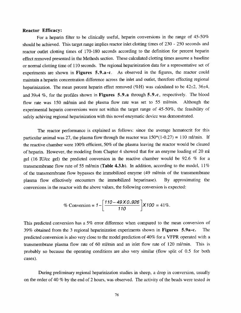
Reactor Efficacy:
For a heparin filter to be clinically useful, heparin conversions in the range of 45-50%
should be achieved. This target range implies reactor inlet clotting times of 230 - 250 seconds and
reactor outlet clotting times of 170-180 seconds according to the definition for percent heparin
effect removed presented in the Methods section. These calculated clotting times assume a baseline
or normal clotting time of 110 seconds. The regional heparinization data for a representative set of
experiments are shown in Figures 5.9.a-c. As observed in the figures, the reactor could
maintain a heparin concentration difference across the inlet and outlet, therefore effecting regional
heparinization. The mean percent heparin effect removed (%H) was calculated to be 42+2, 36+4,
and 39±4 %, for the profiles shown in Figures 5.9.a through 5.9.c, respectively. The blood
flow rate was 150 ml/min and the plasma flow rate was set to 55 ml/min. Although the
experimental heparin conversions were not within the target range of 45-50%, the feasibility of
safely achiving regional heparinization with this novel enzymatic device was demonstrated.
The reactor performance is explained as follows: since the average hematocrit for this
particular animal was 27, the plasma flow through the reactor was 150*(1-0.27) = 110 ml/min. If
the reactive chamber were 100% efficient, 50% of the plasma leaving the reactor would be cleared
of heparin. However, the modeling from Chapter 4 showed that for an enzyme loading of 20 ml
gel (16 IU/cc gel) the predicted conversion in the reactive chamber would be 92.6 % for a
transmembrane flow rate of 55 ml/min (Table 4.3.b). In addition, according to the model, 11%
of the transmembrane flow bypasses the immobilized enzyme (49 ml/min of the transmembrane
plasma flow effectively encounters the immobilized heparinase). By approximating the
conversions in the reactor with the above values, the following conversion is expected:
% Conversion = 1 - 110 49X . 92 6 X100 = 41%._ 110 _
This predicted conversion has a 5% error difference when compared to the mean conversion of
39% obtained from the 3 regional heparinization experiments shown in Figures 5.9a-c. The
predicted conversion is also very close to the model prediction of 40% for a VFPR operated with a
transmembrane plasma flow rate of 60 ml/min and an inlet flow rate of 120 ml/min. This is
probably so because the operating conditions are also very similar (flow split of 0.5 for both
cases).
During preliminary regional heparinization studies in sheep, a drop in conversion, usually
on the order of 40 % by the end of 2 hours, was observed. The activity of the beads were tested in
76
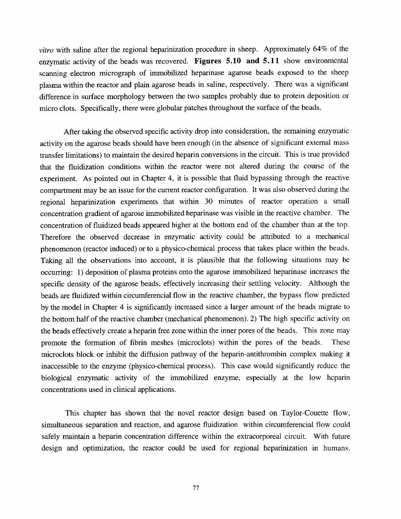
vitro with saline after the regional heparinization procedure in sheep. Approximately 64% of the
enzymatic activity of the beads was recovered. Figures 5.10 and 5.11 show environmental
scanning electron micrograph of immobilized heparinase agarose beads exposed to the sheep
plasma within the reactor and plain agarose beads in saline, respectively. There was a significant
difference in surface morphology between the two samples probably due to protein deposition or
micro clots. Specifically, there were globular patches throughout the surface of the beads.
After taking the observed specific activity drop into consideration, the remaining enzymatic
activity on the agarose beads should have been enough (in the absence of significant external mass
transfer limitations) to maintain the desired heparin conversions in the circuit. This is true provided
that the fluidization conditions within the reactor were not altered during the course of the
experiment. As pointed out in Chapter 4, it is possible that fluid bypassing through the reactive
compartment may be an issue for the current reactor configuration. It was also observed during the
regional heparinization experiments that within 30 minutes of reactor operation a small
concentration gradient of agarose immobilized heparinase was visible in the reactive chamber. The
concentration of fluidized beads appeared higher at the bottom end of the chamber than at the top.
Therefore the observed decrease in enzymatic activity could be attributed to a mechanical
phenomenon (reactor induced) or to a physico-chemical process that takes place within the beads.
Taking all the observations into account, it is plausible that the following situations may be
occurring: 1) deposition of plasma proteins onto the agarose immobilized heparinase increases the
specific density of the agarose beads, effectively increasing their settling velocity. Although the
beads are fluidized within circumferencial flow in the reactive chamber, the bypass flow predicted
by the model in Chapter 4 is significantly increased since a larger amount of the beads migrate to
the bottom half of the reactive chamber (mechanical phenomenon). 2) The high specific activity on
the beads effectively create a heparin free zone within the inner pores of the beads. This zone may
promote the formation of fibrin meshes (microclots) within the pores of the beads. These
microclots block or inhibit the diffusion pathway of the heparin-antithrombin complex making it
inaccessible to the enzyme (physico-chemical process). This case would significantly reduce the
biological enzymatic activity of the immobilized enzyme, especially at the low heparin
concentrations used in clinical applications.
This chapter has shown that the novel reactor design based on Taylor-Couette flow,
simultaneous separation and reaction, and agarose fluidization within circumferencial flow could
safely maintain a heparin concentration difference within the extracorporeal circuit. With future
design and optimization, the reactor could be used for regional heparinization in humans.
77

Recommendations and optimization strategies for future studies to improve reactor performance
and safety are outlined in Chapter 6.
78
a.
b.Figure 5.1 a. A double lumen catheter was inserted into the left jugular vein of the sheep to gain
vascular access. b. Experimental set up for the ex vivo studies in sheep. The animal was conscious
during the procedure but restrained within a specially designed sling.
79
Control Sheep150
100
50 Baseline-o-WBC-*-plt
-20 0 20 40 60 80 100 120 140Time (min)
a .
3530
25
6- 2015 Baseline
10
5
-20 0 20 40 60 80 100 120 140
Time (min)
b.Figure 5.2. Safety data for a control sheep. a. White cell and platelet count profiles were normalized
with baseline values. b. Hematocrit. Mean ± S.E.M 4 experiments.
80
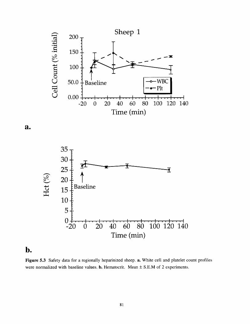
Sheep 1
aseline o WBC-- Plt
. . . . . . .40 60 80 100 120 140
Time (min)
a.
35
30
25
20
15-10
51
-
Baseline
I I I I I I I
20 40 60 80 100 120 1400 0...!-20 0
Time (min)
b.Figure 5.3 Safety data for a regionally heparinized sheep. a. White cell and platelet count profiles
were normalized with baseline values. b. Hematocrit. Mean ± S.E.M of 2 experiments.
81
200-
150-
100-
--
0.00
50.0-]B
,... 0.-20 0 20
40
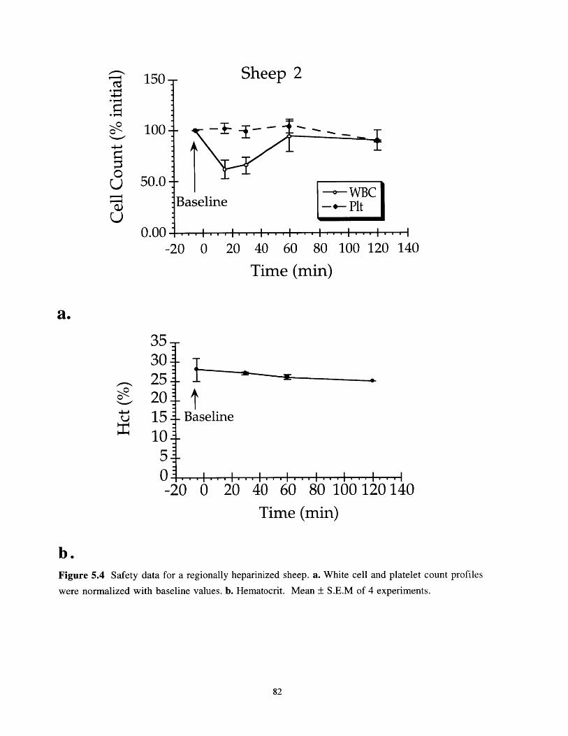
150-
10-
50.0-
0.00
Sheep 201N4'-
Ur---
U Baseline
-20 0 20 40 60 80 100 120 140
Time (min)
a.35-30~2520151050 I.-2U
4
Baseline
I I I I I I I . . I
I. . .. . ..i. . ..4o0 20 40 60 80 100 120 14
Time (min)
b.Figure 5.4 Safety data for a regionally heparinized sheep. a. White cell and platelet count profiles
were normalized with baseline values. b. Hematocrit. Mean ± S.E.M of 4 experiments.
82
I-- PltI
0
IWBCD1-
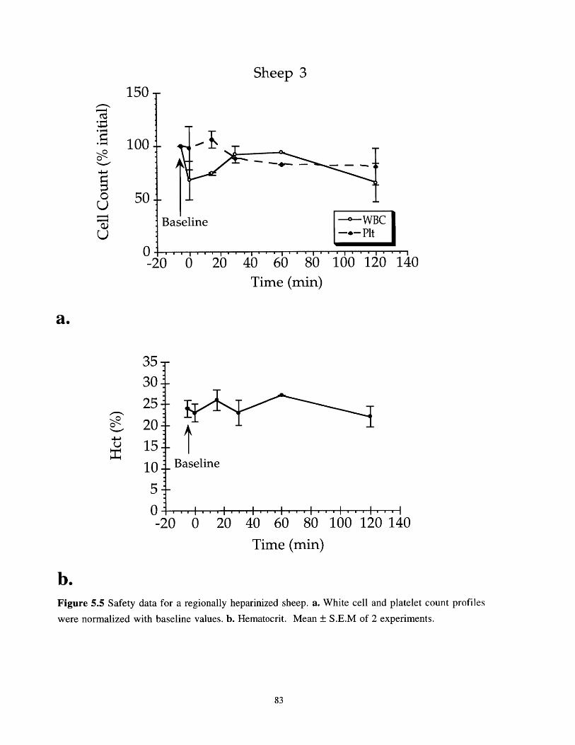
Sheep 3
-I
U
U
20 40 60 80 100Time (min)
120 140
40
35.30
25-:
20-
15-
107
5-04--2C
Baseline
.-
0 20 40 60 80 160 120 140Time (min)
b.Figure 5.5 Safety data for a regionally heparinized sheep. a. White cell and platelet count profiles
were normalized with baseline values. b. Hematocrit. Mean ± S.E.M of 2 experiments.
83
150-
-o WBC-. 6- PltI
100.
50.
Baseline
.-20
a.
i0
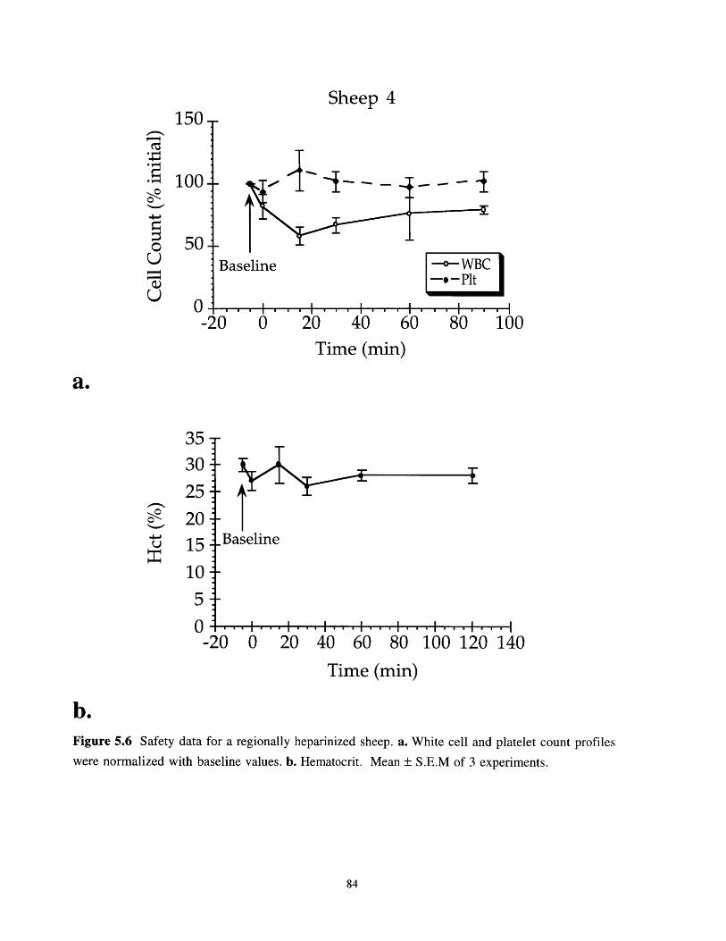
Sheep 4150
100_
500
8 50-U Baseline [ WBC
-20 0 20 40 60 80 100
Time (min)
a.
35--30.
25 A
20
415 -Baseline
10-
5
0-20 0 20 40 60 80 100 120 140
Time (min)
b.Figure 5.6 Safety data for a regionally heparinized sheep. a. White cell and platelet count profiles
were normalized with baseline values. b. Hematocrit. Mean ± S.E.M of 3 experiments.
84
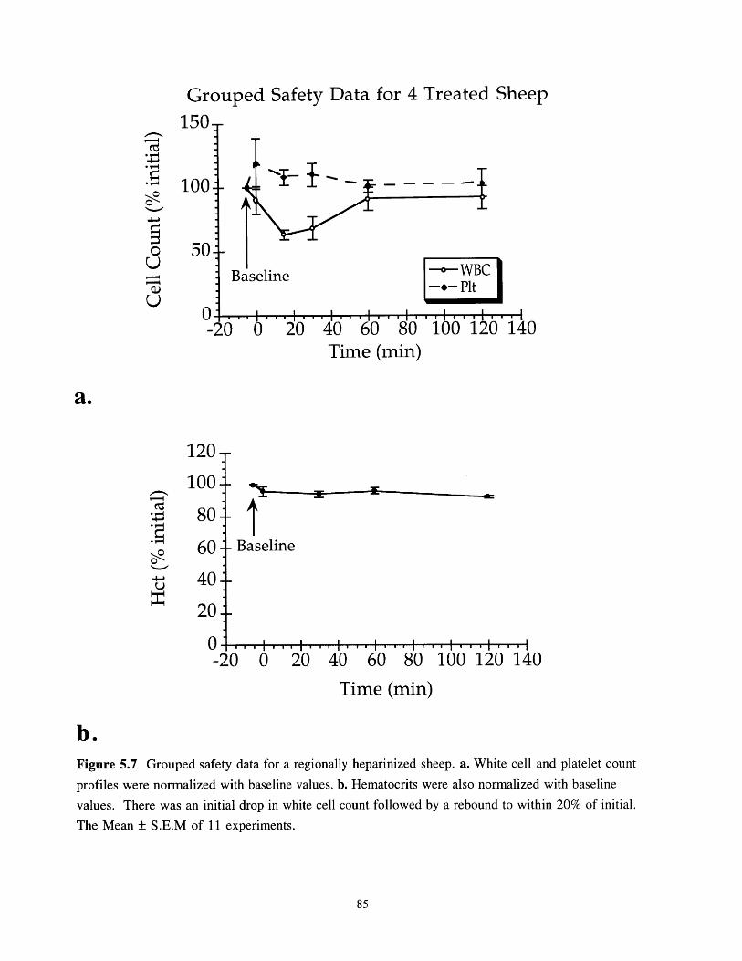
Grouped Safety Data for 4 Treated Sheep1 5 0 -T
4-j
0-
40
U
CU
100'
50-
I- -I
Baseline -o-WBC- Plt
10 1i20 40 60 (8) 120 140
Time (min)
a.
120,
100:
80
+0j 60 + Baseline
40:
20
-20 0 20 40 60 80 100 120 140
Time (min)
b.Figure 5.7 Grouped safety data for a regionally heparinized sheep. a. White cell and platelet count
profiles were normalized with baseline values. b. Hematocrits were also normalized with baseline
values. There was an initial drop in white cell count followed by a rebound to within 20% of initial.
The Mean ± S.E.M of 11 experiments.
85
A-2n
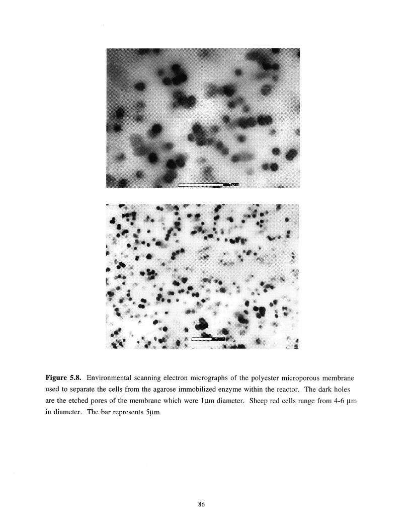
Figure 5.8. Environmental scanning electron micrographs of the polyester microporous membrane
used to separate the cells from the agarose immobilized enzyme within the reactor. The dark holes
are the etched pores of the membrane which were Igm diameter. Sheep red cells range from 4-6 Rm
in diameter. The bar represents 5gm.
86
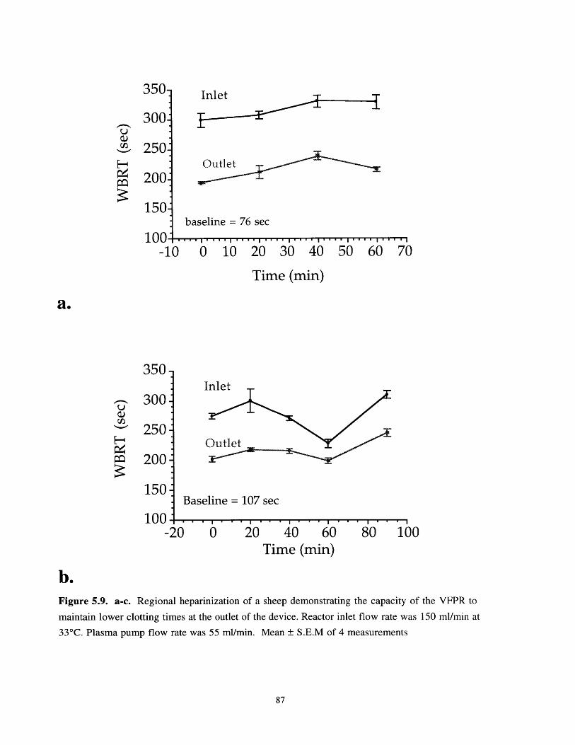
350-
300
250
200
150:
1001.-10
Inlet
Outlet
baseline = 76 sec
0 1 26- 30 40 '1 60 70
Time (min)
a.
Baseline = 107 sec
.1-20 0 20 40 60 80 100
Time (min)
b.Figure 5.9. a-c. Regional heparinization of a sheep demonstrating the capacity of the VFPR to
maintain lower clotting times at the outlet of the device. Reactor inlet flow rate was 150 ml/min at
33'C. Plasma pump flow rate was 55 ml/min. Mean ± S.E.M of 4 measurements
87
UCU~
U
H
350-
300:
250
200
150-
100
Inlet
.I I .I . .
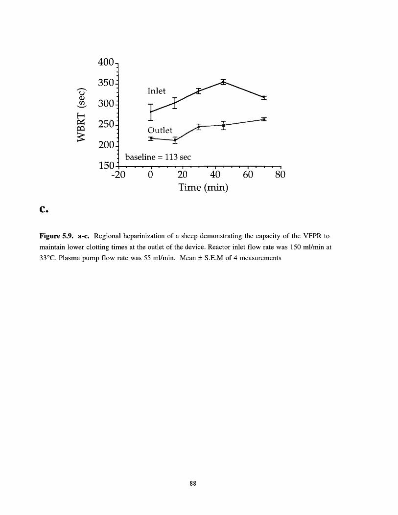
400-
350' N Inlet
300
250
200-baseline = 113 sec
150.-20 0 20 40 60 80
Time (min)
C.
Figure 5.9. a-c. Regional heparinization of a sheep demonstrating the capacity of the VFPR to
maintain lower clotting times at the outlet of the device. Reactor inlet flow rate was 150 ml/min at
33*C. Plasma pump flow rate was 55 ml/min. Mean ± S.E.M of 4 measurements
88
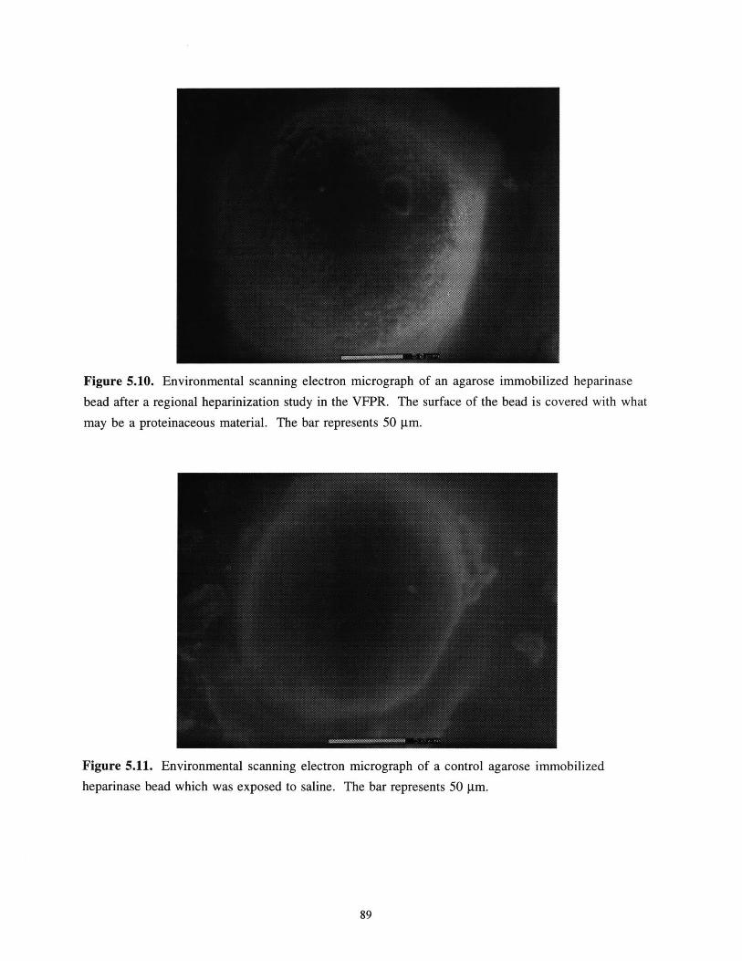
Figure 5.10. Environmental scanning electron micrograph of an agarose immobilized heparinase
bead after a regional heparinization study in the VFPR. The surface of the bead is covered with what
may be a proteinaceous material. The bar represents 50 gm.
Figure 5.11. Environmental scanning electron micrograph of a control agarose immobilized
heparinase bead which was exposed to saline. The bar represents 50 gm.
89
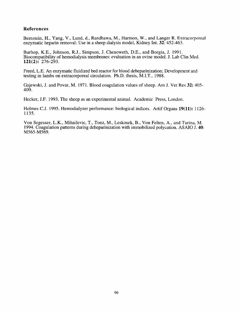
References
Bernstein, H., Yang, V., Lund, d., Randhawa, M., Harmon, W., and Langer R. Extracorporealenzymatic heparin removal: Use in a sheep dialysis model, Kidney Int. 32: 452-463.
Burhop, K.E., Johnson, R.J., Simpson, J. Chenoweth, D.E., and Borgia, J. 1991.Biocompatibility of hemodialysis membranes: evaluation in an ovine model. J. Lab Clin Med.121(2): 276-293.
Freed, L.E. An enzymatic fluidized bed reactor for blood deheparinization; Development andtesting in lambs on extracorporeal circulation. Ph.D. thesis, M.I.T., 1988.
Gajewski, J. and Povar, M. 1971. Blood coagulation values of sheep. Am J. Vet Res 32: 405-409.
Hecker, J.F. 1993. The sheep as an experimental animal. Academic Press, London.
Holmes C.J. 1995. Hemodialyzer performance: biological indices. Artif Organs 19(11): 1126-1135.
Von Segesser, L.K., Mihailevic, T., Tonz, M., Leskosek, B., Von Felten, A., and Turina, M.1994. Coagulation patterns during deheparinization with immobilized polycation. ASAIO J. 40:M565-M569.
90
Chapter 6
Conclusions and Recommendations for Future Work
Taylor-Couette flow was evaluated for potential use in an immobilized heparinase I medical
reactor. Specifically, this thesis evaluated the use of immobilized heparinase I to achieve regional
heparinization of a closed circuit. From the studies in Chapter 2, it was concluded that the
fluidization of small porous agarose particles (under 200 grm diameter) in whole blood resulted in
significant blood damage in the form of hemolysis and cell count depletion. Therefore, fluidization
of small particles would require a totally new approach to maintain the flow rate flexibility, safety,and the maximum use of the immobilized heparinase demanded by clinicians. The lesson learned
from the studies with agarose immobilized heparinase, fluidized in whole blood, paved the way to
design a reactor that would separate the blood cells from the agarose beads. Nevertheless, these
are the first studies to evaluate the use of agarose immobilized species fluidized within Taylor
vortices for a medical application and the results have been accepted for publication in a peer
reviewed journal.
The concept of the vortex flow plasmapheretic reactor (VFPR) was a direct consequence of
the conclusions formulated in Chapter 2. The basis for the VFPR design assumed that if the
agarose beads were kept separate from the blood cells while maintaining adequate fluidization, an
effective and safe reactor may result. The challenge was to obtain high plasma clearances within
the device without causing premature clotting or membrane clogging. The studies in Chapter 3confirmed that separation of the cells from the agarose resulted in good hemocompatibility and
efficacy. Moreover, fluidization of the agarose beads via circumferencial flow was observed
within the reactive chamber. The observed fluidization was independent of the axial flow ratethrough the reactor which is an important operating parameter in hospitals. The new type offluidization, observed and reported for the first time during the course of this research, was theresult of undulatory motions of the microporous membrane. The periodic membrane undulationsare attributed to both, fluid dynamics caused by rotation of the inner cylinder and the membranematerial.
In Chapter 4, residence time distribution studies were performed with the VFPR tocharacterize the type of mixing within the device in order to predict heparin conversion. Thecomplex nature of the device necessitated a multi-compartment model to adequately describe theexperimental residence time distribution curve. The convolution integral was a useful mathematicaltool that enabled integration of the model compartments and the prediction of heparin conversions.
91
The model revealed the existence of a bypass zone within the reactive chamber of the VFPR which
could account for the lower observed conversions.
Finally, in Chapter 5, the device was tested ex vivo in sheep to determine the safety and
efficacy of regional heparinization, a potential treatment modality to make heparin anticoagulation
safer for the patient suffering from acute renal failure. Steady state conversions in the range of 36-
42% were obtained for up to one hour of operation at a blood flow rate of 150 ml/min. These
conversions are close to the initial objective which is in the range of 45-50%. These results,
although not within the specified range of 45-50%, demonstrated the feasibility of regional
heparinization of a closed circuit with a reactor using Taylor-Couette flow. The safety profile of
the device was also evaluated by monitoring changes in cell counts, total protein, fibrinogen, and
the release of free hemoglobin into the plasma. The white cell count profiles suggest some degree
of complement activation but this was shown to be due to the presence of agarose beads and not
the reactor materials. In addition, the white cell count rebounded to within 25% of the initial
values. In general, by all of the latter criteria, the device demonstrated good "biocompatibility".
These studies lay the foundation for a small scale animal preclinical trial in order to test the device
in humans.
Recommendations for Future Work:
The following issues should be addressed before the VFPR can be used on humans:
Efficacy:
The mechanism for decrease in conversion has to be addressed as the device should
function for up to 5 hours if necessary. The modeling studies from Chapter 4 predicted a bypass
that is a function of the transmembrane flow and the permeability of the membrane. Therefore, a
membrane with the proper mechanical strength and permeability (smaller pore size) should be
identified in collaboration with the manufacturer (Whatman Inc.). In addition, according to the
model, another way to enhance reaction efficacy and reduce flow bypass would be the
concentration of the beads within the reactive chamber (smaller reactive chamber volume). This
line of research should also address the bead concentration gradient that was observed in some of
the experiments.
Safety:
A very important consideration when designing a medical device is the safety of the patient.
Based on the experiments, the immediate danger to the patients would be accidental rupture of the
microporous membrane which would release the beads into the extracorporeal circuit. To prevent
92
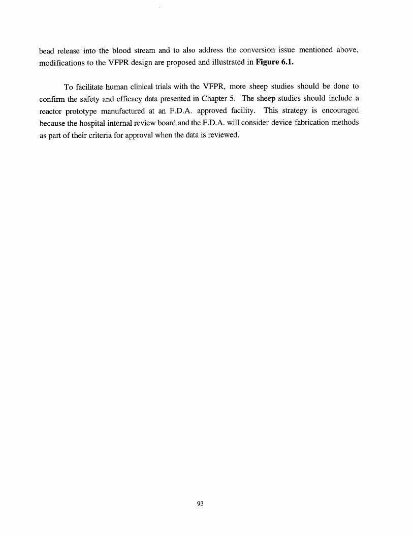
bead release into the blood stream and to also address the conversion issue mentioned above,
modifications to the VFPR design are proposed and illustrated in Figure 6.1.
To facilitate human clinical trials with the VFPR, more sheep studies should be done to
confirm the safety and efficacy data presented in Chapter 5. The sheep studies should include a
reactor prototype manufactured at an F.D.A. approved facility. This strategy is encouraged
because the hospital internal review board and the F.D.A. will consider device fabrication methods
as part of their criteria for approval when the data is reviewed.
93
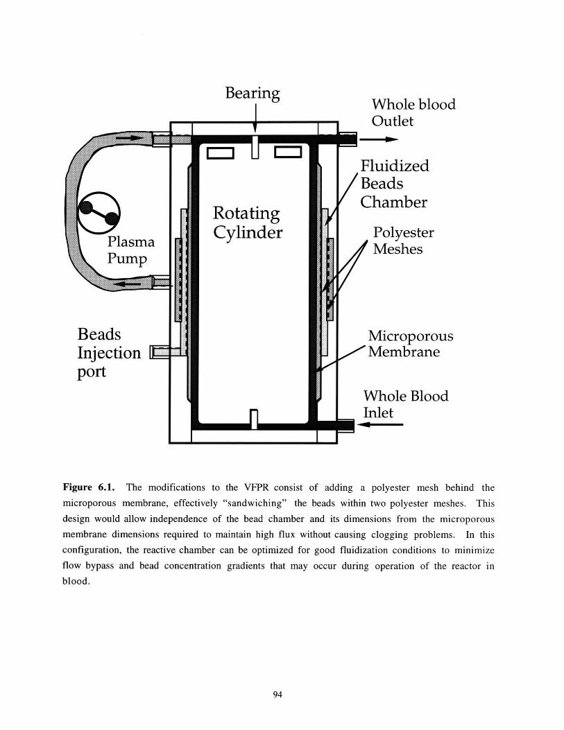
Bearing
I
PlasmaPump
BeadsInjection [port
-
Figure 6.1. The modifications to the VFPR consist of adding a polyester mesh behind the
microporous membrane, effectively "sandwiching" the beads within two polyester meshes. This
design would allow independence of the bead chamber and its dimensions from the microporous
membrane dimensions required to maintain high flux without causing clogging problems. In this
configuration, the reactive chamber can be optimized for good fluidization conditions to minimize
flow bypass and bead concentration gradients that may occur during operation of the reactor in
blood.
94
2
Y
RotatingCylinder
Whole bloodOutlet
FluidizedBeadsChamber
PolyesterMeshes
Microporous,XMembrane
Whole BloodInlet-0
- U
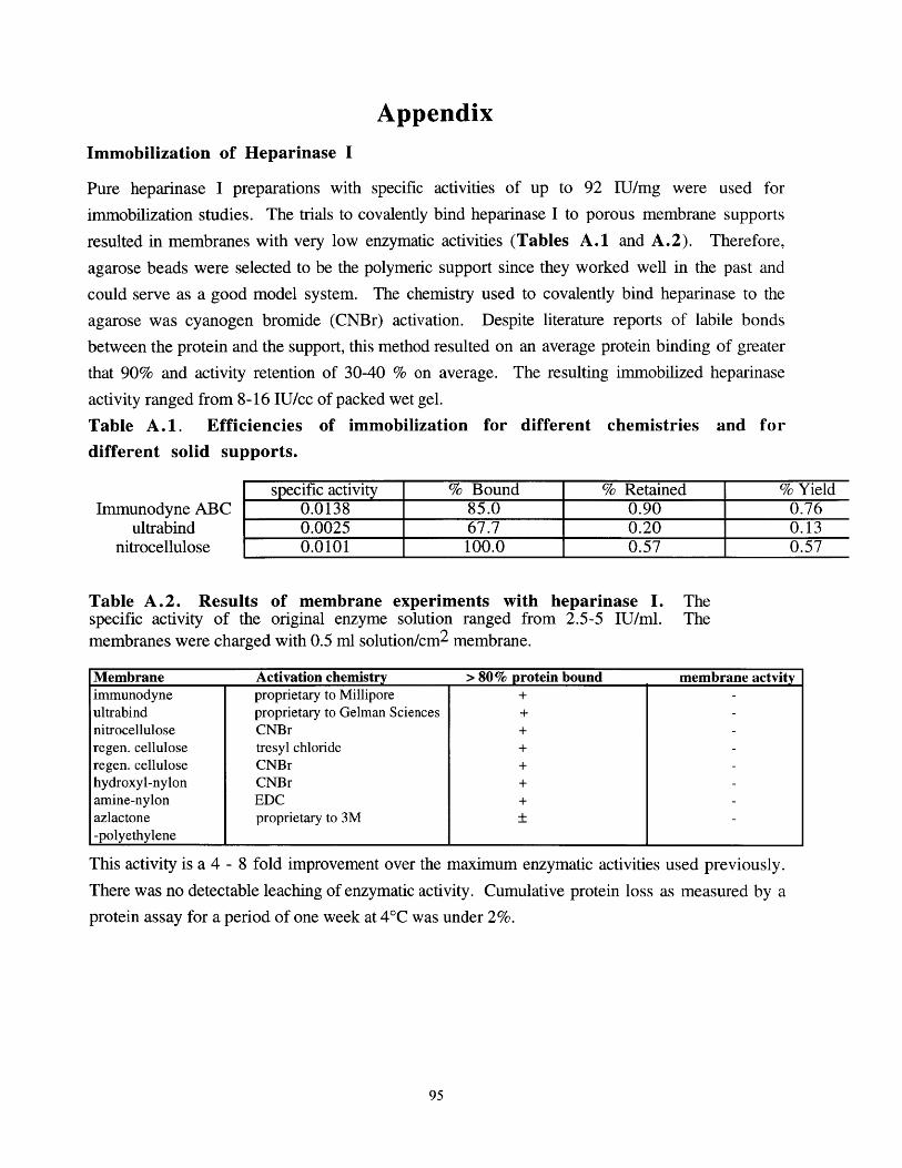
Appendix
Immobilization of Heparinase I
Pure heparinase I preparations with specific activities of up to 92 IU/mg were used for
immobilization studies. The trials to covalently bind heparinase I to porous membrane supports
resulted in membranes with very low enzymatic activities (Tables A. 1 and A.2). Therefore,
agarose beads were selected to be the polymeric support since they worked well in the past and
could serve as a good model system. The chemistry used to covalently bind heparinase to the
agarose was cyanogen bromide (CNBr) activation. Despite literature reports of labile bonds
between the protein and the support, this method resulted on an average protein binding of greater
that 90% and activity retention of 30-40 % on average. The resulting immobilized heparinase
activity ranged from 8-16 IU/cc of packed wet gel.
Table A. 1. Efficiencies of immobilization for different chemistries and for
different solid supports.
Immunodyne ABCultrabind
nitrocellulose
specific activity % Bound % Retained % Yield0.0138 85.0 0.90 0.760.0025 67.7 0.20 0.130.0101 100.0 0.57 0.57
Table A.2. Results of membrane experiments with heparinase I. Thespecific activity of the original enzyme solution ranged from 2.5-5 IU/ml. Themembranes were charged with 0.5 ml solution/cm 2 membrane.
Membrane Activation chemistry >80% protein bound membrane actvityimmunodyne proprietary to Millipore + -ultrabind proprietary to Gelman Sciences +nitrocellulose CNBr +regen. cellulose tresyl chloride +-regen. cellulose CNBr +-hydroxyl-nylon CNBr +-amine-nylon EDC +-azlactone proprietary to 3M +--polyethylene
This activity is a 4 - 8 fold improvement over the maximum enzymatic activities used previously.
There was no detectable leaching of enzymatic activity. Cumulative protein loss as measured by a
protein assay for a period of one week at 4'C was under 2%.
95
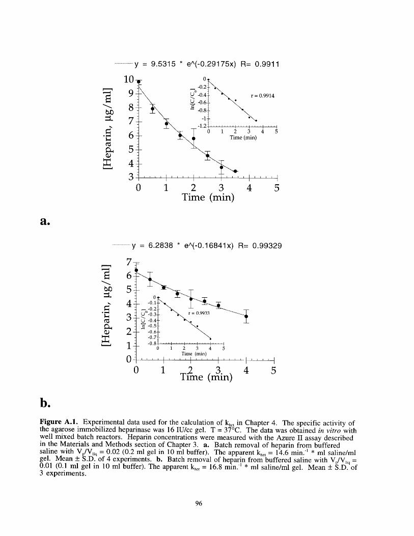
y = 9.5315 * e^(-0.29175x) R= 0.9911
10 02o+- -0.2
9 U-0.4-L 0.9914
-14-
7 -7-- 1.21 ...
0 1 2 3 4 56 Time (mn)
S5
30 1 2 3 4 5
Time (min)
a.
- y = 6.2838 * eA(-0.16841x) R= 0.99329
7-$ 6
00.1. r 0.9933
1 -0.52~-0.6:
-0.7:-
Time (min)-0.0 1 2 3 . 4 5Time (mm)
b.Figure A.1. Experimental data used for the calculation of khet in Chapter 4. The specific activity ofthe agarose immobilized heparinase was 16 IU/cc gel. T = 37 0 C. The data was obtained in vitro withwell mixed batch reactors. Heparin concentrations were measured with the Azure II assay describedin the Materials and Methods section of Chapter 3. a. Batch removal of heparin from bufferedsaline with V,/V,,q= 0.02 (0.2 ml gel in 10 ml buffer). The apparent khet = 14.6 min.- * ml saline/mlgel. Mean ± S.D. of 4 experiments. b. Batch removal of heparin from buffered saline with VP/Vs =0.01 (0.1 ml gel in 10 ml buffer). The apparent khet = 16.8 min.- * ml saline/ml gel. Mean ± S.D. of3 experiments.
96