investigation of early cell–surface interactions of human...
TRANSCRIPT

on September 26, 2018http://rsfs.royalsocietypublishing.org/Downloaded from
rsfs.royalsocietypublishing.org
ResearchCite this article: Medda R et al. 2014
Investigation of early cell – surface interactions
of human mesenchymal stem cells on
nanopatterned b-type titanium – niobium
alloy surfaces. Interface Focus 4: 20130046.
http://dx.doi.org/10.1098/rsfs.2013.0046
One contribution of 10 to a Theme Issue
‘Nano-engineered bioactive interfaces’.
Subject Areas:biomaterials, nanotechnology
Keywords:block copolymer micelle nanolithography, cell
adhesion, human mesenchymal stem cells,
nanopattern, titanium alloys
Author for correspondence:Elisabetta A. Cavalcanti-Adam
e-mail: [email protected]
heidelberg.de
& 2013 The Author(s) Published by the Royal Society. All rights reserved.
Electronic supplementary material is available
at http://dx.doi.org/10.1098/rsfs.2013.0046 or
via http://rsfs.royalsocietypublishing.org.
Investigation of early cell – surfaceinteractions of human mesenchymal stemcells on nanopatterned b-typetitanium – niobium alloy surfaces
Rebecca Medda1,2, Arne Helth3, Patrick Herre4, Darius Pohl4,Bernd Rellinghaus4, Nadine Perschmann1,2, Stefanie Neubauer5,Horst Kessler5, Steffen Oswald3, Jurgen Eckert3, Joachim P. Spatz1,2,Annett Gebert3 and Elisabetta A. Cavalcanti-Adam1,2
1Institute for Physical Chemistry, Ruprecht-Karl-University of Heidelberg, Heidelberg 69120, Germany2Max Planck Institute for Intelligent Systems, Stuttgart 70569, Germany3Institute for Complex Materials, and 4Institute for Metallic Materials, IFW Dresden, PO Box 270116,Dresden 01171, Germany5Institute for Advanced Study (IAS) and Center of Integrated Protein Science (CIPSM), Technische UniversitatMunchen, Lichtenbergstrasse 4, Garching 85747, Germany
Multi-potent adult mesenchymal stem cells (MSCs) derived from bone
marrow have therapeutic potential for bone diseases and regenerative medi-
cine. However, an intrinsic heterogeneity in their phenotype, which in turn
results in various differentiation potentials, makes it difficult to predict
the response of these cells. The aim of this study is to investigate initial
cell–surface interactions of human MSCs on modified titanium alloys. Gold
nanoparticles deposited on b-type Ti–40Nb alloys by block copolymer micelle
nanolithography served as nanotopographical cues as well as specific bind-
ing sites for the immobilization of thiolated peptides present in several
extracellular matrix proteins. MSC heterogeneity persists on polished and
nanopatterned Ti–40Nb samples. However, cell heterogeneity and donor
variability decreased upon functionalization of the gold nanoparticles with
cyclic RGD peptides. In particular, the number of large cells significantly
decreased after 24 h owing to the arrangement of cell anchorage sites, rather
than peptide specificity. However, the size and number of integrin-mediated
adhesion clusters increased in the presence of the integrin-binding peptide
(cRGDfK) compared with the control peptide (cRADfK). These results suggest
that the use of integrin ligands in defined patterns could improve MSC-
material interactions, not only by regulating cell adhesion locally, but also
by reducing population heterogeneity.
1. IntroductionOwing to the steadily increasing number of elderly people and the resulting
rise in age-related diseases, such as osteoporosis and osteoarthritis, there is a
high demand for long-lasting and biocompatible orthopaedic implant materials
[1]. Nowadays, titanium (Ti) alloys are widely used for orthopaedic and dental
implants owing to their mechanical characteristics, biocorrosion resistance
and adequate biocompatibility. However, a significant discrepancy between the
high elastic moduli (more than 100 GPa) of materials currently used for orthopae-
dic implants, such as cp-Ti and Ti–6Al–4V, and the low elastic modulus (less than
30 GPa) of bone, impairs bone turnover at the implant site. This eventually leads
to implant failure, also called the stress-shielding effect [2]. Moreover, depending
on the alloying element, biocompatibility issues need to be carefully considered,
especially for in vivo situations. Ti–6Al–4V (a þ b type), the most commonly

rsfs.royalsocietypublishing.orgInterface
Focus4:20130046
2
on September 26, 2018http://rsfs.royalsocietypublishing.org/Downloaded from
used Ti alloy, was reported to trigger immunologic reactions in
hip replacements [1,3]. Dissolved vanadium and aluminium
were shown to induce severe reactions within the tissue and
to affect growth rates in fibroblasts and osteoblasts [2,4,5].
Therefore, more biocompatible b-type Ti alloys with
minimal side effects and satisfactory mechanical features
have been developed. These alloys contain b-stabilizing
elements, such as niobium (Nb), tantalum and zirconium,
and exhibit superior mechanical properties, e.g. much lower
elastic moduli compared with cp-Ti (a-type) and Ti–6Al–4V,
as well as low metal release rates. Furthermore, these b-type
alloys show excellent performance regarding the inflammatory
response and osteoconductivity [6]. As for Ti–Nb alloys, the
use of a high Nb content lowers the elastic moduli further,
thereby rendering those alloys preferred materials for medical
applications [7]. With 40–45 wt% Nb, it is possible to obtain an
elastic modulus of 60–62 GPa, which can be even further low-
ered to 40–50 GPa by thermo-mechanical processing and
microalloying [8,9].
Lately, there is an ever-growing interest in using human
adult mesenchymal stem cells (MSCs) for regenerative medi-
cine approaches. Derived from bone marrow, these cells can
differentiate into a variety of lineages, including osteoblasts,
chondrocytes and adipocytes [4,5,10]. Stem cell fate is also
determined by their interaction with the microenvironment,
namely the extracellular matrix (ECM). Stem cells are respon-
sive to physical features of the extracellular environment,
such as topography and stiffness, as well as to chemical fea-
tures, such as molecular composition of the ECM and ligand
density [6,11–13]. A population of MSCs from the same
individual comprise a heterogeneous mixture of cells with
differing differentiation and proliferation potentials [14,15].
This heterogeneity is further increased upon isolation and
during in vitro selection, resulting in cells exhibiting various
degrees of maturation [16,17]. In vitro senescence of MSCs
is accompanied by an increase in cell size. These large-sized
cells ultimately stop proliferation but can be maintained in
this state for several months in culture [16].
The presence of non-proliferative senescent cells is a pro-
blematic issue in regenerative medicine. On the one hand,
they may prevent adhesion and settling of desired proliferat-
ing cells, simply by covering large fractions of the implant’s
surface. On the other hand, senescent cells can modify their
microenvironment by inducing senescence in neighbouring
and remote cells extrinsically through their altered secretome
[18]. Therefore, it is of upmost significance to carefully control
initial cell settling on implant materials. A general approach
is the modification of the material’s surface. Thus, enhanc-
ing or preventing adhesion of these cells can be achieved
by immobilization of specific ligands, such as proteins or
bioactive peptides derived from the ECM [19,20].
A number of studies describe different approaches for
immobilizing peptides on surfaces using self-assembled poly-
mers. As was shown by Zorn et al. [21,22], a self-assembled
monolayer was grafted to electropolished and oxidized
Ti–45Nb surfaces and bioactive peptides immobilized onto
this monolayer. Frith et al. [23] investigated the effect of lat-
eral peptide spacing on the ability of MSCs to establish
mature focal adhesion (FA). As already extensively shown
in the past, cell adhesion to different substrates via integrins
can be improved by presentation of short integrin-binding
motifs found in a variety of ECM proteins. Among them,
the commonly used RGD (arginine–glycine–aspartic acid)
motif present in e.g. fibronectin, vitronectin and osteopontin
is widely used to study MSC proliferation and differentiation
[24–26]. Furthermore, RGD and the laminin-derived binding
motif IKVAV (isoleucine–lysine–valine–alanine–valine),
were also shown to influence, respectively, long-term viabi-
lity and osteoblastic differentiation of MSCs [27]. RGD in
combination with growth factors was immobilized on micro-
spheres to construct scaffolds for MSC adhesion, proliferation
and differentiation [28]. Early adhesion and spreading of
primary osteoblasts on trimmed and sandblasted Ti alloys
(Ti-6Al-4V) was positively affected by cyclic RGD coating,
as was shown by Mas-Moruno et al. [29]. An elegant
method to control the spatial density of bioactive peptides
and the orientation of immobilized ligands is a dip-coating
technique based on self-assembly of diblock copolymer
micelles (block copolymer micelle nanolithography, BCMN)
[30]. Using gold as the micelle core, a hexagonally ordered
pattern of gold nanoparticles can be created on various
materials, including orthopedically relevant surfaces, for
example Ti, and used to direct cell adhesion [31,32].
In this study, we present the patterning of Ti–40Nb alloy
discs and showed the effects of nanotopography and biofunc-
tionalization with integrin ligands on human MSCs early
adhesion and phenotype selection. Here, we use BCMN
to create gold nanopatterns on Ti–40Nb alloys. Specific func-
tionalization of the gold nanoparticles was achieved by using
the thiolated ligands cRGDfK and cRADfK. Based on the
characterization of early cell–surface interactions on the modi-
fied and functionalized alloys, we demonstrate that the
heterogeneity of human MSC is reduced and their adhesion,
in terms of cell size and FA formation, is optimized by using
the cRGDfK peptide.
2. Material and methods2.1. Preparation of titanium alloysHigh-purity Ti and Nb were arc-melted in argon atmosphere
to obtain Ti–40Nb (wt%) ingots. These pre-alloys were sub-
sequently melted by induction heating and cold crucible
cast with a pressure of 500 mbar in a water-cooled copper
mould with a diameter of 10 mm and a length of 100 mm. The
samples were sealed in quartz tubes and homogenized in puri-
fied argon atmosphere at 10008C for 24 h, and subsequently
water quenched. Discs of 2–3 mm thickness were cut from the
rods. Finally, the alloy composition was determined by induc-
tively coupled plasma optical emission spectroscopy (iCAP
6500 Duo, Thermo Fisher Scientific GmbH, Germany). The struc-
tural state of the cast and homogenized samples was checked
with X-ray diffraction (STOE Stadi P instrument, STOE &
CIE GmbH, Germany) and scanning electron microscopy
(SEM, LEO 1530 Gemini, Carl Zeiss, Germany) and verified a
polycrystalline single b-phase state with a mean grain size of
approximately 70 mm [9].
In a standard procedure, the aforementioned 10 mm Ti–
40Nb discs were plane ground with SiC emery paper up to
grid P400, followed by lapping with diamond suspension (9, 6
and 3 mm) using a flat grinding plate (Struers, Germany). The
final polishing was carried out with a mixture of colloidal
silica and hydrogen peroxide. After the fine polishing procedure
ending with a mixture of colloidal silica and hydrogen peroxide,
the alloy surface was extremely smooth (mirror-like). The surface
roughness was analysed with optical profilometry using a Micro-
Prof system (FRT, Germany), whereby a surface area of 2 � 2 mm
was mapped. Typical roughness data are Ra ¼ 0.02+0.01 mm

rsfs.royalsocietypublishing.orgInterface
Focus4:20130046
3
on September 26, 2018http://rsfs.royalsocietypublishing.org/Downloaded from
(Ra, mean roughness) and Rz ¼ 0.12+0.02 mm (Rz, averaged
roughness depth). These values are extremely low compared
with those resulting from other polishing grades or etching treat-
ments applied to Ti–Nb alloy samples [1]. However, they are
comparable with those obtained for commercial silicate glass
samples measured with the same system, i.e. Ra ¼ 0.079+0.012 mm and Rz ¼ 0.670+ 0.328 mm, thus indicating a similar
surface roughness.
2.2. Preparation of nanopatterns on Ti – 40Nb discsThe technique of gold deposition on surfaces by BCMN is based
on Glass et al. [30]. Here, we achieved efficient surface pattern-
ing by adjusting plasma treatment and surface preparation
prior to the dip-coating process. Polished and ground Ti–40Nb
discs were activated in oxygen plasma (0.4 mbar, 150 W,
10 min) prior to BCMN. The micelle solution was prepared in
toluene with either polymer ‘1056’ (consisting of 1056 poly-
styrene units and 671 vinyl pyridine units, 7 mg ml21) or with
polymer ‘2074’ (consisting of 2074 polystyrene and 571 vinyl pyr-
idine units, 2 mg ml21) and HAuCl4 to obtain a gold loading of
0.5 (HAuCl4 from Sigma, Germany). After dip-coating the discs,
the organic compounds on the surface were removed by oxygen
plasma (0.4 mbar, 150 W, 60 min), resulting in coalescence of the
gold nanoparticles.
Passivation and biofunctionalization of nanopatterned (NP)
Ti–40Nb discs: following plasma treatment, Ti–40Nb discs were
passivated by incubation with 0.1 mg ml21 PLL-g-PEG in
HEPES (Surface SolutionS, Switzerland) for 30 min at room temp-
erature. Following several rinsing with distilled water, the discs
were incubated with 25 mM cyclic RGDfK or cyclic RADfK at
room temperature for at least 4 h. Prior to cell plating, the discs
were washed with sterile phosphate buffered saline (PBS).
2.3. Characterization of the quasi-hexagonal patternof the gold nanoparticles by scanning electronmicroscopy
To characterize the quasi-hexagonal gold nanoparticle pattern,
the surface of Ti–40Nb discs was examined with SEM (LEO
1530 Gemini, Carl Zeiss), as shown in figure 2. Prior to SEM ima-
ging, carbon was sputtered (MED 020 Coating System Sputter,
Bal-Tec, Liechtenstein) on the Ti–40Nb discs to enhance electron
scattering, thus enhancing contrast. The interparticle distances as
well as the order of the hexagonal patterns were derived by ana-
lysing the SEM micrographs (50 000� magnification) with the
IMAGEJ plugin ‘Dot analyzer’ (kindly provided by Philippe
Girard, University of Heidelberg) according to the underlying
theory of Kansal et al. [33].
2.4. Auger electron spectroscopySample surfaces were characterized with Auger electron spec-
troscopy (AES) using a JAMP-9500 F Field Emission Auger
Microprobe (Jeol, Japan) with primary electrons of 10 keV at an
electron current of 10 nA. For depth profiling, the sample sur-
faces were sputtered by a scanned beam of 1 keV Arþ ions and
spectra were recorded in probe areas of 10 � 10 mm after sputter
intervals of 0.3 min.
2.5. Transmission electron microscopyCross-sectional transmission electron microscopy (TEM) samples
were prepared by focused ion beam (FIB) cutting of lamellae
from the near-surface region of the processed Ti–40Nb samples
decorated with gold nanoparticles using an FEI Helios nanolab
600i dual beam system (FEI EUROPE, The Netherlands). Prior to
the FIB cut, layers of amorphous carbon and platinum were
subsequently deposited onto the surface in order to protect the
area to be investigated from damages through the 30 kV Gaþ
ions. Structural and local chemical characterization was conducted
by conventional and aberration-corrected high-resolution-TEM
(HR-TEM, shown in figure 3) using an FEI Tecnai G2 20 microscope
(with LaB6 emitter, scanning unit (STEM), energy dispersive X-ray
spectrometer (EDXS), FEI company) and an FEI Titan3 80–300
microscope operated at 300 kV (equipped with a field emission
gun, CEOS CetCor CS-image corrector, high angle annular dark
field detector, Gatan Tridiem 863ER for electron energy loss spec-
troscopy (EELS) and EDXS, FEI company), respectively (figure 4).
2.6. Cell culture and indirect immunofluorescencestaining
Human MSCs (hMSCs) derived from bone marrow (Promocell,
Heidelberg, Germany) were cultured and plated in proliferation
medium including the supplements provided by the manufac-
turer (Promocell), without antibiotics. Cells from two different
donors were investigated (donor1: male, Caucasian, 65 years
and donor2: male, Caucasian, 64 years). hMSCs (less than
10 000 cm– 2) were plated on biofunctionalized Ti–40Nb discs for
24 h and fixed with 4% (w/v) paraformaldehyde (Sigma-Aldrich,
St Louis, USA) in PBS (pH 7.4) for 20 min followed by permeabi-
lization with 0.1% (v/v) Triton X-100 (Sigma-Aldrich) in PBS for
5 min. After blocking in 1% (w/v) BSA in PBS, the discs were incu-
bated with anti-vinculin mouse IgG (Sigma-Aldrich) detected by
Alexa488 goat anti-mouse IgG (Life Technologies, Carlsbad,
USA) to stain FAs and phalloidin-TRITC (Sigma-Aldrich) to
label F-actin. The antibodies as well as the phalloidin conjugate
were diluted according to the manufacturer’s recommendations.
The fluorescently labelled samples were embedded in Mowiol
supplemented with 1 mg ml21 DAPI (Sigma-Aldrich) for nuclear
staining and mounted onto a coverslip (standard thickness 1,
Carl Roth, Karlsruhe, Germany).
2.7. Fluorescence microscopy and data analysisFluorescence imaging was carried out with an Olympus IX
inverted microscope (Olympus, Hamburg, Germany) using a
Delta Vision RT system (Applied Precision Inc., Issaquah,
USA). Here, cells were examined using either a 10� (Neofluor
10�/0.3 phase contrast, Carl Zeiss) air or 60� (PlanApo 60�/
1.4, Olympus) oil immersion lens with the resulting pixel sizes
of 0.589 and 0.158 mm, respectively. Images were acquired
using a cooled CCD camera (Photometrics, Kew, Australia) and
processing was controlled by RESOLVE3D (Applied Precision
Inc.). All images were analysed with IMAGEJ v. 1.43 (https://
rsb.info.nih.gov/ij/). Cell area was determined from actin micro-
graphs such that binary images were created by adjusting the
threshold individually for each image. If required, neighbouring
cells (on control and polished samples) were separated manually.
Cells were counted and analysed using the analyse particle tool
from IMAGEJ with a threshold set as 10 mm2—infinity and exclud-
ing edges from the analysis to ensure only taking completely
displayed cells into account. The area of the cell nuclei was deter-
mined similarly by creating binary images. Cell number was
determined by counting the nuclei, whereas nucleus area and cir-
cularity were determined additionally by using the analyse
particle tool with the threshold set to 50–2000 mm2. To rule out
the effects owing to sample or cell seeding inhomogeneity,
8–10 random images throughout the whole sample surfaces
were acquired from at least three different experiments. Focal
adhesion analysis was performed on images acquired with a
60� oil lens as described above. Clusters were outlined and ana-
lysed with IMAGEJ using the ROI manager and ROI color coder
(by Tiago Ferreira) tools. Only clusters larger than 0.5 mm2
were considered for analysis.

control Ti–40Nb non-patterned
100 mm
dono
r 2
dono
r 1
* *
(a) (b)
(c) (d)
**
Figure 1. Morphology of hMSCs derived from two different donors on flatglass (control, a,c) and polished, non-patterned Ti – 40Nb (b,d ) surfacesafter 24 h. Fluorescence microscopy reveals cell heterogeneity (F-actin: red,DAPI: green). Small spindle-shaped cells, ‘fried-egg-shaped’ spread cells aswell as large irregular-shaped or flat cells (asterisks) are found in bothdonors and on both substrates.
rsfs.royalsocietypublishing.orgInterface
Focus4:20130046
4
on September 26, 2018http://rsfs.royalsocietypublishing.org/Downloaded from
Cell size distribution was plotted with Excel and box plots
were created in Origin displaying the box between 25% and
75% and the whiskers between 10% and 90% of the data range.
Significance was assessed by the Mann–Whitney U-test (PRISM
6.0d, GraphPad Software, Inc.)
3. Results and discussion3.1. Human mesenchymal stem cells on polished
Ti – 40NbTi–40Nb discs were manufactured by using a melting procedure
as described in the experimental section. To demonstrate the
heterogeneity of human bone-marrow-derived MSCs (hMSCs)
in independent experiments, we seeded cells derived from two
different donors of comparable age (donor1, D1: 65 years and
donor2, D2: 64 years) on polished non-patterned Ti–40Nb
discs. Cells were stained for actin (red) and nuclei (green) and
their morphology on the discs was investigated in comparison
to control condition (glass coverslips). As depicted in figure 1,
hMSCs seeded for 24 h in proliferative conditions attach and
spread similarly on glass (figure 1a,b) and Ti–40Nb surfaces
(figure 1b,d). Note the typical heterogeneous mixture of cells,
showing different phenotypes. This inherent heterogeneity
becomes particularly apparent for cell size, ranging from small
spindle-shaped cells with a typical area less than 5000 mm2
to up to 30 000 mm2 for large spread cells. The spindle-shaped
cells have a fibroblastic-like morphology, whereas the
large cells are spread, and/or irregular shaped and very flat
with the exception of the peri-nuclear region. The former type
resembles fibroblast morphology; it should be noted however
that these cells cannot be considered as fibroblasts, thus differing
in their function of secreting and assembling ECM. The latter
type is especially undesired for further use in regenerative medi-
cine because it accounts for senescent and/or committed cells
[34–36]. A cell size below 1000 mm2 mostly indicates poorly
attached or dividing cells.
Besides the inherent heterogeneity of the hMSC pheno-
type, an additional challenge is the diversity between
different donors. We chose hMSCs from two donors as men-
tioned above and evaluated their cell size distribution. As can
be clearly seen in figure 1a,b, donor 1 comprises many large
cells and only very few small cells (less than 5000 mm2),
whereas several small cells are observed in the case of
donor 2 (figure 1c,d ). These results were also confirmed for
cells derived from other frozen stocks of the same donors
(data not shown).
3.2. Characterization of gold nanoparticleson Ti – 40Nb discs
Kilian et al. [37] reported that cell shape and contractility influ-
ence lineage commitment of bone-marrow-derived hMSCs.
Therefore, a defined cell microenvironment, in particular for
its physical and chemical features, might represent a successful
approach to control hMSC anchorage to the implant material as
well as to direct cell responses. The defined deposition of gold
nanoparticles on surfaces is an elegant way to spatially control
and direct cell adhesion through topography or by functionaliz-
ing the gold nanoparticles with adhesion-promoting ligands.
Here, we used polished Ti–40Nb alloy surfaces for nano-
patterning and subsequent biofunctionalization. To achieve a
controlled surface topography, a hexagonally ordered pattern
of gold nanoparticles was created on the surface by a dip-
coating technique based on self-assembly of diblock copolymer
micelles (BCMN). The scheme in figure 2a depicts the surface
treatment and BCMN process. Here, we chose the copolymers
such that with a constant retraction velocity of 25 mm min21
average interparticle distance of 68+13 nm (order 0.54) for
smaller spacing or 96+17 nm (order 0.56) for larger spacing
can be obtained. The order of the hexagonal gold nanoparticle
pattern as well as the interparticle distances was determined by
evaluation of SEM images (figure 2b,c) as described in the
experimental section. For cell experiments, a distance of
approximately 68 nm was chosen, being the threshold for integ-
rin lateral clustering and assembly of stable FAs in cells of
mesenchymal origin [38,39].
AES (see electronic supplementary material, figure S1) was
used to chemically characterize the surface state. Survey spec-
tra of the outermost surface reveal the main presence of Ti- and
Nb-oxide species. Local AES analyses at selected points (cover-
ing probe areas of about 50 nm), which are visible in the SEM
images in figure 2 as white contrast, clearly confirm that these
are indeed gold particles. Upon depth profiling, Au was
detected up to a sputter depth of approximately 5 nm. How-
ever, the main characteristics of the depth profile indicate the
presence of a Ti- and Nb-oxide layer with a thickness of
approximately 18 nm. This oxide layer on Ti–40Nb generated
by the above-described process is much thicker than the natu-
ral one with only less than 5 nm [9]. Altogether, the AES results
demonstrate the presence of nanoscale Au species above a
(Ti,Nb)-oxide layer. However, being the characterization of
the Au species at the lower detection limit of the method, it is
necessary to apply analyses at a higher resolution level.
TEM investigations of cross-sectional areas of Ti–40Nb
samples confirm the presence of an oxide layer at the sample
surface (figure 3). This layer has a thickness of at least 10 nm
and is evidenced by the darker zone at the sample surface in

Ti–40Nb disc nanopatterning passivation functionalization cell seeding
interparticle distance: 96 ± 17nmorder: 0.56
interparticle distance: 68 ± 13 nmorder: 0.54
200 nm 200 nm
(a)
(b) (c)
Figure 2. BCMN on Ti – 40Nb surfaces. (a) Scheme depicting the manufacturing process of BCMN on Ti – 40Nb discs shows the deposition of gold nanoparticles via adip-coating technique followed by passivation of the interparticle space and functionalization of the gold particles. SE micrographs shown in (b,c) displaynanoparticle-decorated surfaces with two different interparticle distances, which can be controlled by using different polymers for the generation of micelles.Note the quasi-hexagonal pattern for both types of spacing. (Online version in colour.)
Au
10 nm
1 nm 5 nm5 nm
10 nm
1 nm
1-1-300-4
-11-3
-22-2
-220
-222-113004
1-13
2-22
2-20
2-2-2
1-1-1 00-2
-11-1
-111002
1-11
1-1-3
00-21-1-1
1-11 -11-1
-111002
-113
oxide
TiNb
(a)
(c) (d )
(b)
(e) ( f )
Figure 3. Structural characterization of the surface of a Ti – 40Nb substrate covered with gold nanoparticles. (a) TEM micrograph surveying a cross section of the surface.The interface between the near-surface oxide layer and the metallic Ti – 40Nb underneath is marked with a dashed line. (b) STEM image of the same surface. (c,e) TypicalHR-TEM images of (c) a large and (e) a small gold particle. (d,f ) The diffractograms as obtained from FFT of the original images are consistent with fcc Au crystals seenalong k110l zone axes. The Bragg reflections are labelled with their respective Miller indices. (Online version in colour.)
rsfs.royalsocietypublishing.orgInterface
Focus4:20130046
5
on September 26, 2018http://rsfs.royalsocietypublishing.org/Downloaded from
the scanning TEM (STEM) images displayed in figures 3b and
4b. Here, the reduction of the average atomic number owing to
the incorporation of light O atoms effectively reduces the STEM
contrast. Evidence for this surface oxidation arises from EELS
measurements shown in figure 4. EEL spectra were collected
in STEM mode at positions 1–10 along the yellow line across
the oxide–metal interface as indicated in the STEM image in
figure 4b. Hereto, the dispersion and energy range of the EEL
spectrometer were adjusted to allow for a simultaneous
acquisition of losses at the Nb–M5,4, Ti–L3,2 and O–K absorp-
tion edges, respectively. The relative amounts of Nb, Ti and O
were determined from an evaluation of the near edge absorp-
tion intensities after appropriate background subtraction
(see dashed line in figure 4c). The resulting concentration pro-
files across the interface are displayed in figure 4a. The
measurements clearly evidence the enrichment of oxygen
within the surface layer. Detailed analyses of the absorption
edges further reveal a small chemical shift of the Ti–L3,2

70(a)
(c)
1 2 3 4 5 6 7 8 9 10
Ti
Nb
NbM5.4
TiL3.2
signals (×3)
O
60
50
40
30
elem
enta
l fra
ctio
n (%
)in
tens
ity (
arb.
uni
ts)
20
10
00
200 300 400energy loss (eV)
500 600 700
position (nm)5
CK
OK
10 151234
56789
5 nm 10
spectrumbackground
(b)
Figure 4. EEL spectra as collected from the subsurface positions 1 and 2indicated in the STEM image in (b). The displayed range of loss energiescovers the Ti – L3,2 and O – K absorption edges. A small chemical shiftbetween the two Ti – L3,2 edges is highlighted by a dashed vertical line.
rsfs.royalsocietypublishing.orgInterface
Focus4:20130046
6
on September 26, 2018http://rsfs.royalsocietypublishing.org/Downloaded from
edges upon crossing the oxide–metal interface which is indica-
tive of an oxidation of Ti within the oxide layer (not shown
here). HR-TEM images further demonstrate that the surface
oxide is amorphous, as no indications of crystalline structures
were ever observed (also not shown).
Both the TEM and STEM images in figure 3a,b display
nanoparticles whose strong contrast is already indicative of
the high atomic number of the material they are comprised.
There are large particles with sizes of 6–7 nm and a number
of smaller particles with diameters in the range of 2–3 nm. In
similar micrographs (as the one displayed in the TEM image
in figure 3a) from other cross-sectional areas of the sample,
we could detect a comparable number of Au particles which
is indicative of a homogeneous (areal) particle density across
the whole sample. HR-TEM images of the particles confirm
that the particles are indeed gold. The examples presented in
figure 3c for a large particle and figure 3e for a small particle,
and their diffractograms as obtained by fast Fourier transform-
ation (FFT) clearly show the crystal structure of face-centred
cubic (fcc) Au as seen along k110l zone axes.
In the past, several groups investigated the topographical
influence on a variety of cellular functions. It has been shown
that fibroblasts sense changes in the surface roughness of
approximately 10 nm, which affects their proliferation and
spreading [40]. Sjostrom et al. [32] described an increase in
MSC osteogenic potential on 15 nm compared with 8 nm
sized gold nanoparticles. Here, we use gold nanoparticles of
approximately 5 nm in size to investigate their influence on
hMSC heterogeneity, in particular on cell size. In contrast to
the aforementioned works dealing with topographical
investigations for several days, we studied early cell–surface
interactions after 24 h.
3.3. Human mesenchymal stem cells on nanopatternedTi – 40Nb alloys
To determine the effects of surface topography on cell pheno-
type, surfaces were modified by deposition of gold
nanoparticles on Ti–40Nb via BCMN. Here, hMSCs were
seeded on polished as well as on NP discs for 24 h and het-
erogeneity of the population was analysed, in particular
with respect to the number and fraction of large-sized cells.
Cells plated on patterned Ti–40Nb adhere and spread simi-
larly to cells grown on non-patterned Ti–40Nb alloys and
control (figure 5a–c). However, on patterned surfaces the
overall cell number (figure 5g) as well as cell confluence
(data shown in the electronic supplementary material) is
reduced. The absolute numbers of large-sized (more than
10 000 mm2) cells are comparable in all three conditions: con-
trol surfaces, non-patterned and NP Ti–40Nb alloys (data not
shown). Hence, modifying the surface topography by the
deposition of nanoparticles of approximately 5 nm could
not improve the ratio of fibroblast-like cells nor resulted in
a decreased number of large cells. In this study, we analysed
short-term effects induced by nanoscale surfaces to reduce
heterogeneity within a cell population and among donors.
Dalby and co-workers [41,42], for example, investigated the
influence of nanoscale topographies on long-term maintenance
of the stem cell phenotype or the induction of osteogenesis.
Also, migration of osteoblasts was found to be influenced by
topographical modifications on the nanoscale [43]. Further
studies will be necessary to determine the influence of nano-
scale topographies on long-term cell behaviour, and, in turn,
the surface modifications after cell matrix deposition.
Besides their effects on surface topography, nanoparticles
arranged in patterns are an elegant tool for controlling cell
adhesion via immobilization of specific thiolated biomolecules
and for confining cell–surface interaction to defined sites on
the surface. Passivation of the surface between the particles is
crucial to assign the observed effects to the immobilized
ligands only. To obtain a passivation of the space between
the nanoparticles, a polyethylene glycol (PEG) layer was phy-
sisorbed on the surface, thereby restricting cell–substrate
interactions only at the particles. As expected, adhesion of
hMSCs after 24 h on these passivated surfaces is very poor
and spread cells are absent (figure 5d).
To specifically design the cell–surface interface, directly after
PEG-passivation the gold nanoparticles were functionalized
with adhesion-promoting peptides. Here, we immobilized
either the thiolated, cyclic pentapeptide cRGDfK (cyclo-Arg-
Gly-Asp-D-Phe-Lys) or the control peptide cRADfK (cyclo-
Arg-Ala-Asp-D-Phe-Lys) on the gold nanoparticles. Thus, cells
seeded on these surfaces are only able to adhere to the surface
through the presented immobilized ligands. Figure 5e,f presents
overview images of hMSCs on both ligands after 24 h. This time
point was chosen to allow cells to adhere and spread properly,
on the one hand. On the other hand, this short-term incubation
ensured that the observed effects only stem from the functiona-
lized surfaces and no significant cell-induced alterations of the
0
20
40
60
80
100
control NP cRADfK cRGDfK
1001–5000 µm2 5001–10 000 µm2 >10 000 µm2
100 µm
control Ti–40Nb non-patterned Ti–40Nb + NP
Ti–40Nb + NP + PEG Ti–40Nb + NP + PEG + cRGDfK Ti–40Nb + NP + PEG + cRADfK
(g) (h)
control
0
10
20
30
40
50
60
non-patterned NP cRADfK cRGDfK
cell
num
ber
per
fram
e
rela
tive
cell
num
ber
(%)
*
*
(a) (b) (c)
(d) (e) ( f )
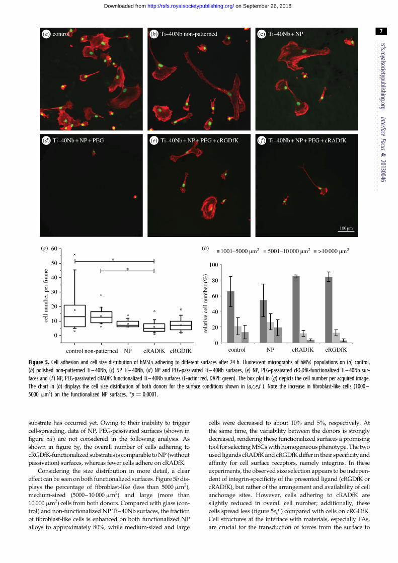
Figure 5. Cell adhesion and cell size distribution of hMSCs adhering to different surfaces after 24 h. Fluorescent micrographs of hMSC populations on (a) control,(b) polished non-patterned Ti – 40Nb, (c) NP Ti – 40Nb, (d ) NP and PEG-passivated Ti – 40Nb surfaces, (e) NP, PEG-passivated cRGDfK-functionalized Ti – 40Nb sur-faces and ( f ) NP, PEG-passivated cRADfK functionalized Ti – 40Nb surfaces (F-actin: red, DAPI: green). The box plot in (g) depicts the cell number per acquired image.The chart in (h) displays the cell size distribution of both donors for the surface conditions shown in (a,c,e,f ). Note the increase in fibroblast-like cells (1000 –5000 mm2) on the functionalized NP surfaces. *p ¼ 0.0001.
rsfs.royalsocietypublishing.orgInterface
Focus4:20130046
7
on September 26, 2018http://rsfs.royalsocietypublishing.org/Downloaded from
substrate has occurred yet. Owing to their inability to trigger
cell-spreading, data of NP, PEG-passivated surfaces (shown in
figure 5d) are not considered in the following analysis. As
shown in figure 5g, the overall number of cells adhering to
cRGDfK-functionalized substrates is comparable to NP (without
passivation) surfaces, whereas fewer cells adhere on cRADfK.
Considering the size distribution in more detail, a clear
effect can be seen on both functionalized surfaces. Figure 5h dis-
plays the percentage of fibroblast-like (less than 5000 mm2),
medium-sized (5000–10 000 mm2) and large (more than
10 000 mm2) cells from both donors. Compared with glass (con-
trol) and non-functionalized NP Ti–40Nb surfaces, the fraction
of fibroblast-like cells is enhanced on both functionalized NP
alloys to approximately 80%, while medium-sized and large
cells were decreased to about 10% and 5%, respectively. At
the same time, the variability between the donors is strongly
decreased, rendering these functionalized surfaces a promising
tool for selecting MSCs with homogeneous phenotype. The two
used ligands cRADfK and cRGDfK differ in their specificity and
affinity for cell surface receptors, namely integrins. In these
experiments, the observed size selection appears to be indepen-
dent of integrin-specificity of the presented ligand (cRGDfK or
cRADfK), but rather of the arrangement and availability of cell
anchorage sites. However, cells adhering to cRADfK are
slightly reduced in overall cell number; additionally, these
cells spread less (figure 5e,f ) compared with cells on cRGDfK.
Cell structures at the interface with materials, especially FAs,
are crucial for the transduction of forces from the surface to

20 µm
10 mm
0 255
(a) Ti–40Nb non-patterned (b) Ti–40Nb + NP
(c) Ti–40Nb + NP + PEG + cRADfK (d) Ti–40Nb + NP + PEG + cRGDfK
non-patterned NP NP + cRADfK NP + cRGDfK0
2
4
area
(mm
2 )
(e) focal adhesions per cell ( f ) focal adhesion size
0
20
40
60
80
100
120
140
160
180
200
non-patterned NP NP + cRADfK NP + cRGDfK
no. F
A p
er c
ell
**
*
Figure 6. Focal adhesions of hMSCs grown on different surfaces. Immunofluorescent labelling of vinculin on (a) polished, non-patterned Ti – 40Nb, (b) NP, notpassivated Ti – 40Nb, (c) cRADfK functionalized NP and passivated Ti – 40Nb and (d ) cRGDfK-functionalized NP and passivated Ti – 40Nb surfaces. The red box in thegrey scale overview image is magnified on the right, showing FAs in more detail. The lookup table shown in (a) represents fluorescence intensity. In the chart shownin (e), the number of FAs per cells is depicted, whereas the box plot in ( f ) highlights the FA cluster sizes measured on the four different surfaces. *p , 0.0001.
rsfs.royalsocietypublishing.orgInterface
Focus4:20130046
8
on September 26, 2018http://rsfs.royalsocietypublishing.org/Downloaded from
the actin cytoskeleton and vice versa. Kilian et al. [37] used
shape confinement at the microscale to study cell tension and
found that increased acto-myosin contractility promotes osteo-
genesis in MSCs. Here, we confined not the cell shape but rather
their anchorage by patterns consisting of single nanoparticles.
Thus, the adhering cells can adopt any shape, as long as they
adapt to the substrate by establishing new FAs. Furthermore,
adhesion signalling might be involved in the regulation of cell
senescence, as it has been shown for example, for paxillin and
c-Src as well as cytoskeletal proteins [44]. It remains however
unclear, if the reduced expression in key adhesion molecules
during cell senescence could modulate cell ability to adhere
and adapt to different materials because of altered protein
turnover in FAs.
3.4. Focal adhesions on nanopatterned alloyshMSCs were seeded on Ti–40Nb NP with either cRGDfK or
cRADfK ligands as described above. Twenty-four hours after
cell seeding, FAs were identified in fixed cells by indirect immu-
nofluorescence staining for vinculin and visualized by
fluorescence microscopy. Both on polished and on NP non-
passivated Ti-40Nb discs, hMSCs show robust peripheral FAs
(figure 6a,b). Cells grown on Ti–40Nb discs passivated and pat-
terned with cRADfK-decorated nanoparticles, display smaller
and undefined vinculin clusters which are not localized at the
cell periphery, whereas cells adhering to cRGDfK exhibit
elongated and defined FAs at the cell margin (figure 6c,d).
Note that the number of FAs per cell is reduced on all NP
samples in comparison with the non-patterned surface as

rsfs.royalsocietypublishing.orgInterface
Focus4:20130046
9
on September 26, 2018http://rsfs.royalsocietypublishing.org/Downloaded from
shown in figure 6e. However, offering cRGDfK increases the
number of FAs per cell on these substrates dramatically. These
results indicate that the reduced cell size observed on samples
functionalized with the cRADfK ligand is owing to poor
adhesion and lack of forming a sufficient number of mature FAs.
For the use of biological material in regenerative medi-
cine, it is important to guarantee a high reproducibility and
homogeneity between different batches on the one hand,
and predictable and/or uniform samples, on the other
hand. To gain information about surface uniformity, cell
coverage (surface area covered by cells) on the different coat-
ings was analysed (data displayed as box plots in the
electronic supplementary material, figure S2). On control and
polished, untreated Ti–40Nb surfaces, the cell coverage is
extremely variable for different areas analysed, evidencing
inhomogeneous cell settling throughout the samples. This
variability is reduced on the nanostructured surfaces, even
on the non-functionalized nanoparticles.
Our observations that hMSCs form large vinculin clusters
on untreated and on non-functionalized Ti–40Nb discs is in
agreement with the recent findings of Sjostrom et al. [32].
The presence of nanotopographies owing to nanoparticles
of 5–10 nm size might therefore promote on the long-term
osteogenic differentiation of hMSC via the local enhancement
of FA maturation. Regarding the use of RGD motifs cova-
lently attached to biometals, Jager et al. [45] showed that
hMSC osteogenic stimulation is not significantly improved
in the presence of these peptides. This report further supports
our findings that RGD motifs promote adhesion of hMSC
and the formation of mature FA, while the cells still exhibit
their typical non-committed stem cell phenotype.
4. ConclusionUsing Ti–40Nb alloys, which present the advantage of closely
matching the elastic modulus of bone, we probed the response
of hMSCs to surface nanopatterning and further functiona-
lization with cRGDfK peptides. We determined the effects of
these surface modifications on the settling of hMSC having
heterogeneous phenotype and on adhesion. This shows a
proof-of-principle for the use of such materials for the selection
of hMSC subpopulations and decreasing donor variability. The
results shown here suggest that by combining spatial cues with
specific adhesive cues at the nanoscale, hMSC adhesion is opti-
mized while maintaining the typical phenotype of non-
committed stem cells. Studies with such materials could test
in the future whether these cells would show the same dif-
ferentiation potential and whether the expression of specific
senescence markers is abolished.
Acknowledgements. We thank Dr Katharina Klein and Dr SeraphineWegner for carefully reading the manuscript.
Funding statement. The work was financially supported by grants from theGerman Research Foundation (DFG SFB-TR79 projects M1 and M6). Wegratefully acknowledge the support from the Max Planck Society.
References
1. Hoesel LM, Pausch M, Schnettler R, Heiss C. 2008 Theimpact of osteoporosis on the classification of hip andwrist fractures. Med. Sci. Monit. 14, HY1 – HY8.
2. Geetha M, Singh AK, Asokamani R, Gogia AK. 2009Ti based biomaterials, the ultimate choice fororthopaedic implants: a review. Prog. Mater. Sci. 54,397 – 425. (doi:10.1016/j.pmatsci.2008.06.004)
3. Witt JD, Swann M. 1991 Metal wear and tissueresponse in failed titanium alloy total hipreplacements. J. Bone Joint Surg. Br. 73, 559 – 563.
4. Laing PG, Ferguson AB, Hodge ES. 1967 Tissuereaction in rabbit muscle exposed to metallicimplants. J. Biomed. Mater. Res. 1, 135 – 149.(doi:10.1002/jbm.820010113)
5. Rao S, Ushida T, Tateishi T, Okazaki Y, Asao S. 1996Effect of Ti, Al, and V ions on the relative growthrate of fibroblasts (L929) and osteoblasts (MC3T3-E1) cells. Biomed. Mater. Eng. (IOS Press). 6,79 – 86.
6. Matsuno HH, Yokoyama AA, Watari FF, Uo MM,Kawasaki TT. 2001 Biocompatibility andosteogenesis of refractory metal implants, titanium,hafnium, niobium, tantalum and rhenium.Biomaterials 22, 1253 – 1262. (doi:10.1016/S0142-9612(00)00275-1)
7. Hon Y-H, Wang J-Y, Pan Y-N. 2003 Composition/Phase structure and properties of titanium –niobium alloys. Mater. Trans. 44, 2384 – 2390.(doi:10.2320/matertrans.44.2384)
8. Matsumoto H, Watanabe S, Hanada S. 2005 BetaTiNbSn alloys with low Young’s modulus and highstrength. Mater. Trans. 46, 1070 – 1078. (doi:10.2320/matertrans.46.1070)
9. Helth A et al. Chemical nanoroughening of Ti40Nbsurfaces and its effect on human mesenchymalstromal cell response. J. Biomed. Mater. Res. B.(doi:10.1002/jbm.b.32976)
10. Wang XX et al. 2006 Characterization ofmesenchymal stem cells isolated from mouse fetalbone marrow. Stem Cells 24, 482 – 493. (doi:10.1634/stemcells.2005-0219)
11. Rowlands ASA, George PAP, Cooper-White JJJ. 2008Directing osteogenic and myogenic differentiation ofMSCs: interplay of stiffness and adhesive ligandpresentation. Am. J. Physiol. Cell Physiol. 295,C1037 – C1044. (doi:10.1152/ajpcell.67.2008)
12. Sun Y, Chen CS, Fu J. 2012 Forcing stem cells tobehave: a biophysical perspective of the cellularmicroenvironment. Annu. Rev. Biophys. 41, 519 –542. (doi:10.1146/annurev-biophys-042910-155306)
13. Kulangara K, Yang Y, Yang J, Leong KW. 2012Nanotopography as modulator of humanmesenchymal stem cell function. Biomaterials33, 4998 – 5003. (doi:10.1016/j.biomaterials.2012.03.053)
14. Vogel W, Grunebach F, Messam CA, Kanz L, BruggerW, Buhring H-J. 2003 Heterogeneity among humanbone marrow-derived mesenchymal stem cells and
neural progenitor cells. Haematologica 88,126 – 133.
15. Kolf CM, Cho E, Tuan RS. 2007 Biology of adultmesenchymal stem cells: regulation of niche, self-renewal and differentiation. Arthritis Res. Ther. 9,204 – 214. (doi:10.1186/ar2116)
16. Wagner W, Ho AD, Zenke M. 2010 Different facets ofaging in human mesenchymal stem cells. TissueEng. B Rev. 16, 445 – 453. (doi:10.1089/ten.teb.2009.0825)
17. Pevsner-Fischer M, Levin S, Zipori D. 2011 Theorigins of mesenchymal stromal cell heterogeneity.Stem Cell Rev. Rep. 7, 560 – 568. (doi:10.1007/s12015-011-9229-7)
18. Beltrami AP, Cesselli D, Beltrami CA. 2011 At thestem of youth and health. Pharmacol. Ther. 129,3 – 20. (doi:10.1016/j.pharmthera.2010.10.005)
19. Marko K, Kohidi T, Hadinger N, Jelitai M, Mezo G,Madarasz E. 2011 Isolation of radial glia-like neuralstem cells from fetal and adult mouse forebrain viaselective adhesion to a novel adhesive peptide-conjugate (Deli MA, editor). PLoS ONE 6, e28538.(doi:10.1371/journal.pone.0028538)
20. Gronthos S, Zannettino ACW. 2008 A method toisolate and purify human bone marrow stromalstem cells. Methods Mol. Biol. 449, 45 – 57.
21. Zorn G, Gotman I, Gutmanas EY, Adadi R, Salitra G,Sukenik CN. 2005 Surface modification of Ti45Nballoy with an alkylphosphonic acid self-assembled

rsfs.royalsocietypublishing.orgInterface
Focus4:20130046
10
on September 26, 2018http://rsfs.royalsocietypublishing.org/Downloaded from
monolayer. Chem. Mater. 17, 4218 – 4226. (doi:10.1021/cm050477f )
22. Zorn G, Gotman I, Gutmanas EY, Adadi R, SukenikCN. 2007 Surface modification of Ti45Nb alloy byimmobilization of RGD peptide via self assembledmonolayer. J. Mater. Sci. Mater. Med. 18,1309 – 1315. (doi:10.1007/s10856-006-0117-7)
23. Frith JE, Mills RJ, Cooper-White JJ. 2012 Lateralspacing of adhesion peptides influences humanmesenchymal stem cell behaviour. J. Cell. Sci. 125,317 – 327. (doi:10.1242/jcs.087916)
24. Alvarez-Barreto JF, Landy B, VanGordon S, Place L,DeAngelis PL, Sikavitsas VI. 2010 Enhancedosteoblastic differentiation of mesenchymal stem cellsseeded in RGD-functionalized PLLA scaffolds andcultured in a flow perfusion bioreactor. J. Tissue Eng.Regen. Med. 5, 464 – 475. (doi:10.1002/term.338)
25. Hosseinkhani H, Hosseinkhani M, Tian F, KobayashiH, Tabata Y. 2006 Osteogenic differentiationof mesenchymal stem cells in self-assembledpeptide-amphiphile nanofibers. Biomaterials27, 4079 – 4086. (doi:10.1016/j.biomaterials.2006.03.030)
26. Zhang H-F, Li Z-J, Fu X, Ma J-X, Ma X-L. 2013Interactions of bone marrow stromal cells withnative and RGD surface modified acellular bonematrix: a biocompatibility study. Arch. Med. Res. 44,69 – 74. (doi:10.1016/j.arcmed.2012.11.006)
27. Frith JE, Mills RJ, Hudson JE, Cooper-White JJ.2012 Tailored integrin – extracellular matrixinteractions to direct human mesenchymal stem celldifferentiation. Stem Cells Dev. 21, 2442 – 2456.(doi:10.1089/scd.2011.0615)
28. Park JS, Yang HN, Jeon SY, Woo DG, Na K, Park K-H.2010 Osteogenic differentiation of humanmesenchymal stem cells using RGD-modified BMP-2coated microspheres. Biomaterials 31, 6239 – 6248.(doi:10.1016/j.biomaterials.2010.05.002)
29. Mas-Moruno CC, Dorfner PMP, Manzenrieder FF,Neubauer SS, Reuning UU, Burgkart RR, Kessler H.
2012 Behavior of primary human osteoblasts ontrimmed and sandblasted Ti6Al4V surfacesfunctionalized with integrin avb3-selectivecyclic RGD peptides. J. Biomed. Mater. Res. A 101,87 – 97.
30. Glass R, ller MM, Spatz JP. 2003 Block copolymermicelle nanolithography. Nanotechnology 14,1153 – 1160. (doi:10.1088/0957-4484/14/10/314)
31. Bagno A et al. 2007 Human osteoblast-like celladhesion on titanium substrates covalentlyfunctionalized with synthetic peptides. Bone 40, 7.(doi:10.1016/j.bone.2006.10.007)
32. Sjostrom T, McNamara LE, Meek RMD, Dalby MJ, SuB. 2013 2D and 3D Nanopatterning of titanium forenhancing osteoinduction of stem cells at implantsurfaces. Adv. Healthc. Mater. 2, 1285 – 1293.(doi:10.1002/adhm.201200353)
33. Kansal AR, Truskett TM, Torquato S. 2000Nonequilibrium hard-disk packings with controlledorientational order. J. Chem. Phys. 113,4844 – 4851. (doi:10.1063/1.1289238)
34. Muraglia AA, Cancedda RR, Quarto RR. 2000 Clonalmesenchymal progenitors from human bonemarrow differentiate in vitro according to ahierarchical model. J. Cell. Sci. 113, 1161 – 1166.
35. Lange C, Schroeder J, Lioznov MV, Zander AR. 2005High-potential human mesenchymal stem cells.Stem Cells Dev. 14, 236. (Mary Ann Liebert, Inc. 2Madison Avenue Larchmont, NY 10538 USA; 14,70 – 80). (doi:10.1089/scd.2005.14.70)
36. Ho M, Yu D, Davidsion MC, Silva GA. 2006Comparison of standard surface chemistries forculturing mesenchymal stem cells prior to neuraldifferentiation. Biomaterials 27, 4333 – 4339.(doi:10.1016/j.biomaterials.2006.03.037)
37. Kilian KA, Bugarija B, Lahn BT, Mrksich M.2010 Geometric cues for directing the differentiationof mesenchymal stem cells. Proc. Natl Acad. Sci.USA 107, 4872 – 4877. (doi:10.1073/pnas.0903269107)
38. Arnold M, Cavalcanti-Adam EA, Glass R, Blummel J,Eck W, Kantlehner M, Kessler H, Spatz JP. 2004Activation of integrin function by nanopatternedadhesive interfaces. ChemPhysChem 5, 383 – 388.(doi:10.1002/cphc.200301014)
39. Cavalcanti-Adam EA, Volberg T, Micoulet A, KesslerH, Geiger B, Spatz JP. 2007 Cell spreading and focaladhesion dynamics are regulated by spacing ofintegrin ligands. Biophys. J. 92, 2964 – 2974.(doi:10.1529/biophysj.106.089730)
40. Gentile FF, Medda RR, Cheng LL, Battista EE,Scopelliti PEP, Milani PP, Cavalcanti-Adam EA,Decuzzi P. 2013 Selective modulation of cellresponse on engineered fractal silicon substrates.Sci. Rep. 3, 1461. (doi:10.1038/srep01461)
41. Dalby MJ, Gadegaard N, Tare R, Andar A, RiehleMO, Herzyk P, Wilkinson CD, Oreffo RO. 2007 Thecontrol of human mesenchymal cell differentiationusing nanoscale symmetry and disorder. Nat. Mater.6, 997 – 1003. (doi:10.1038/nmat2013)
42. McMurray RJR et al. 2011 Nanoscale surfaces forthe long-term maintenance of mesenchymal stemcell phenotype and multipotency. Nat. Mater. 10,637 – 644. (doi:10.1038/nmat3058)
43. Lamers E, van Horssen R, Riet te J, van Delft FC,Luttge R, Walboomers XF, Jansen JA. 2010 Theinfluence of nanoscale topographical cues on initialosteoblast morphology and migration. Eur. CellMater. 20, 329 – 343.
44. Nishio K, Inoue A. 2005 Senescence-associatedalterations of cytoskeleton: extraordinary productionof vimentin that anchors cytoplasmic p53 insenescent human fibroblasts. Histochem. Cell Biol.123, 263 – 273. (doi:10.1007/s00418-005-0766-5)
45. Jager M, Boge C, Janissen R, Rohrbeck D, Hulsen T,Lensing-Hohn S, Krauspe R, Herten M. 2013Osteoblastic potency of bone marrow cells cultivatedon functionalized biometals with cyclic RGD-peptide. J. Biomed. Mater. Res. A 101, 2905 – 2914.(doi:10.1002/jbm.a.34590)