invited paper innovations leading capsule endoscopy … · capsule endoscopy a very patient...
TRANSCRIPT

526IEICE TRANS. COMMUN., VOL.E98–B, NO.4 APRIL 2015
INVITED PAPER Special Section on Innovation of Medical Information and Communication Technology for Dependable Society
Innovations Leading Capsule Endoscopy into the New Frontier:Screening and Therapy at Home
Hidetoshi OHTA†a), Member
SUMMARY This paper proposed patient friendly capsule endoscopy(CE) for not only screening but also treatment. Two different types of CEswith an Internet utility were investigated. The first type used magneticnavigation in the stomach and colon for screening. Magnetic navigationenabled the capsule to explore the whole of the gastrointestinal tract withless risk of missing lesions and complete the screening within the batterylife. The system’s design was patiently friendly as it allowed the subjects toleave the hospital after the capsule had been navigated in the stomach. Thesecond investigated two different therapeutic robotic endoscopes. Both pro-totypes were driven by DC motors and controlled remotely via the internet.In addition, they were equipped with therapeutic tools and each prototype’sability with the tools was assessed. The investigation showed it was possi-ble to remotely control both prototypes and operate therapeutic tools via theInternet. The investigation identified areas for improvement, such as size,connection speed, security of data, and the holding the capsule’s positionduring treatment, In conclusion, both methods have the potential to makecapsule endoscopy a very patient friendly procedure that can be carried outanywhere.key words: capsule endoscopy, magnetic navigation, robotic endoscope,remote control, Internet
1. Introduction
Capsule endoscopy (CE) was clinically introduced 14 yearsago by Given Imaging and has been improved to extend theapplication from the small intestine, where CE has estab-lished the position as the gold standard modality for screen-ing, to other parts of gastrointestinal tract. As CE is a lessinvasive endoscopic modality than conventional wired (ca-ble) endoscopy many endoscopists were looking forward toan endoscopic evolution when CE was introduced into clin-ical practice. However, to our disappointment, CE has sev-eral cumbersome drawbacks, which need to be overcomebefore useful applications can be added. Firstly the cap-sule’s movement is dependent upon peristalsis and gravity,secondly the short battery life, thirdly there is no methodfor cleansing on demand, and fourthly there is no therapeu-tic methodology. These drawbacks have fixed the paradigmthat CE is available only for diagnosis in the long narrowtubes of the gastrointestinal tract.
To shift this stubborn paradigm, some researchers haveendeavoured to overcome the drawbacks with recent ad-vances in technology. Two major approaches to control thecapsule endoscope have been proposed to date. The first is
Manuscript received December 15, 2014.Manuscript revised January 13, 2015.†The author is with Sapporo Orthopaedics and Cardiovascular
Hospital, Sapporo-shi, 004-0861 Japan.a) E-mail: [email protected]
DOI: 10.1587/transcom.E98.B.526
extracorporeal magnetic control [1]–[7] and the second iswireless (radio) control [8]–[10]. The former utilizes theinteractive forces between a small magnet in or over thecapsule endoscope and the magnetic field generated extra-corporeally. The latter is propelled by a motor or actuatorand fed electric power from the batteries. Over the past fewyears, we have presented our versions of the two systems atmedical and engineering conferences [11]–[15].
In relation to the battery life, newer more powerful bat-teries with a longer battery life are continually being devel-oped and at the same time the amount of power that is con-sumed by electric circuits and ultra-tiny motors is decreas-ing rapidly owing to meeting the specifications for portabledevices and robotic engineering. Improvements in powerconsumption and battery life will continue to extend the cap-sule’s running time. However, having enough energy in thebatteries to power other functions as well as imaging willbe a big challenge. External sources of power for CE couldbe the answer. For example, there has been preliminary re-search into wireless magnetic induction [16]. However forthe time being, the necessary electrical energy which is fun-damentally used in the capsule can only be adequately sup-plied from batteries.
The clinical approach to the preparation of the colonhas been focused on recently because many articles in med-ical journals validated that the cleanliness of the observationarea are closely related to the detection rate of colorectal le-sions [17], [18]. The American Gastroenterological Asso-ciation (AGA) runs a big colorectal cancer awareness cam-paign every March, which is aimed at elderly people whohave a higher risk of colon cancer. Two reasons for peoplenot getting a colonoscopy are, (1) the procedure is painfuland (2) they have to drink a large volume of unpleasanttasting preparation. CE is a less painful and more accept-able modality for screening than colonoscopy, but the doseof the preparation (6 liters) for colon capsule endoscopy(CCE) is too much for a typical Asian person to drink. Jennaet al. proposed an interesting preparation using mechanicalcleansing by gas generated from a chemical reaction [19].Several pharmaceutical companies have provided some newpreparation agents such as Suprep R©, Prepopick R© which ac-cording to their literature are supposed to reduce the prepa-ration dose for conventional colonoscopy. Pharmaceuticalcompanies and many doctors (including me) are investigat-ing acceptable preparations for CCE and a number of possi-ble protocols have already been presented at medical confer-ences. As this area is getting a lot of attention, we estimated
Copyright c© 2015 The Institute of Electronics, Information and Communication Engineers
OHTA: INNOVATIONS LEADING CAPSULE ENDOSCOPY INTO THE NEW FRONTIER: SCREENING AND THERAPY AT HOME527
that this drawback should be solved in a few years. Intro-ducing treatment by CE into medicine is one of the dreamsof gastroenterologists, but it is still a long and tough way toour destination, even though innovation has realized variousdreams. Only a few researchers have reported on their pro-totypes for therapeutic CE to date [20], [21]. Furthermore,to the best of our knowledge, there has been no other re-port of CE utilizing network technology which is being usedto develop portable and wearable devices and is becomingcommon in the healthcare system. Gradually, step by step,technical innovations are about to lead to a paradigm shiftin CE. In this report, I present our newest magnetic naviga-tion system for CE which at the time of writing this paperwas undergoing a clinical trial, and our preliminary studyon therapeutic robotic endoscopes controlled via the Inter-net remotely, which we hope will be patient friendly. Inaddition, I will evaluate the advantages of this original ideaof introducing network technology into endoscopy by com-paring it with recent developments in CE.
2. Materials and Methods
2.1 Magnetic Navigation (MNCE)
2.1.1 System Architecture
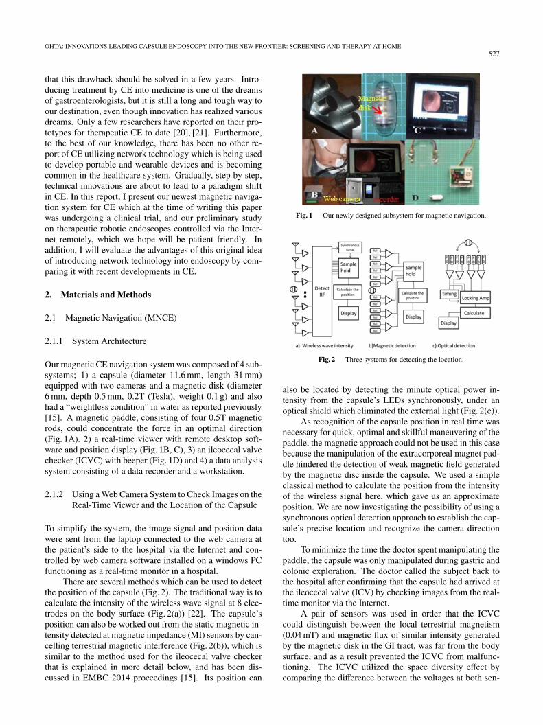
Our magnetic CE navigation system was composed of 4 sub-systems; 1) a capsule (diameter 11.6 mm, length 31 mm)equipped with two cameras and a magnetic disk (diameter6 mm, depth 0.5 mm, 0.2T (Tesla), weight 0.1 g) and alsohad a “weightless condition” in water as reported previously[15]. A magnetic paddle, consisting of four 0.5T magneticrods, could concentrate the force in an optimal direction(Fig. 1A). 2) a real-time viewer with remote desktop soft-ware and position display (Fig. 1B, C), 3) an ileocecal valvechecker (ICVC) with beeper (Fig. 1D) and 4) a data analysissystem consisting of a data recorder and a workstation.
2.1.2 Using a Web Camera System to Check Images on theReal-Time Viewer and the Location of the Capsule
To simplify the system, the image signal and position datawere sent from the laptop connected to the web camera atthe patient’s side to the hospital via the Internet and con-trolled by web camera software installed on a windows PCfunctioning as a real-time monitor in a hospital.
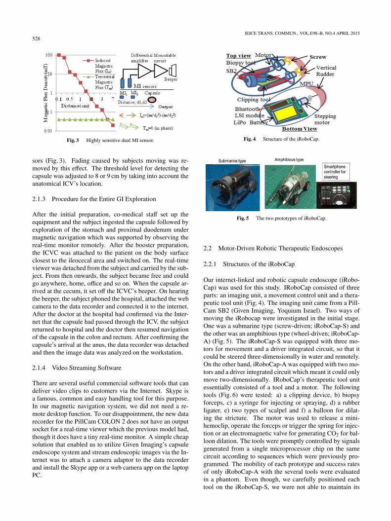
There are several methods which can be used to detectthe position of the capsule (Fig. 2). The traditional way is tocalculate the intensity of the wireless wave signal at 8 elec-trodes on the body surface (Fig. 2(a)) [22]. The capsule’sposition can also be worked out from the static magnetic in-tensity detected at magnetic impedance (MI) sensors by can-celling terrestrial magnetic interference (Fig. 2(b)), which issimilar to the method used for the ileocecal valve checkerthat is explained in more detail below, and has been dis-cussed in EMBC 2014 proceedings [15]. Its position can
Fig. 1 Our newly designed subsystem for magnetic navigation.
Fig. 2 Three systems for detecting the location.
also be located by detecting the minute optical power in-tensity from the capsule’s LEDs synchronously, under anoptical shield which eliminated the external light (Fig. 2(c)).
As recognition of the capsule position in real time wasnecessary for quick, optimal and skillful maneuvering of thepaddle, the magnetic approach could not be used in this casebecause the manipulation of the extracorporeal magnet pad-dle hindered the detection of weak magnetic field generatedby the magnetic disc inside the capsule. We used a simpleclassical method to calculate the position from the intensityof the wireless signal here, which gave us an approximateposition. We are now investigating the possibility of using asynchronous optical detection approach to establish the cap-sule’s precise location and recognize the camera directiontoo.
To minimize the time the doctor spent manipulating thepaddle, the capsule was only manipulated during gastric andcolonic exploration. The doctor called the subject back tothe hospital after confirming that the capsule had arrived atthe ileocecal valve (ICV) by checking images from the real-time monitor via the Internet.
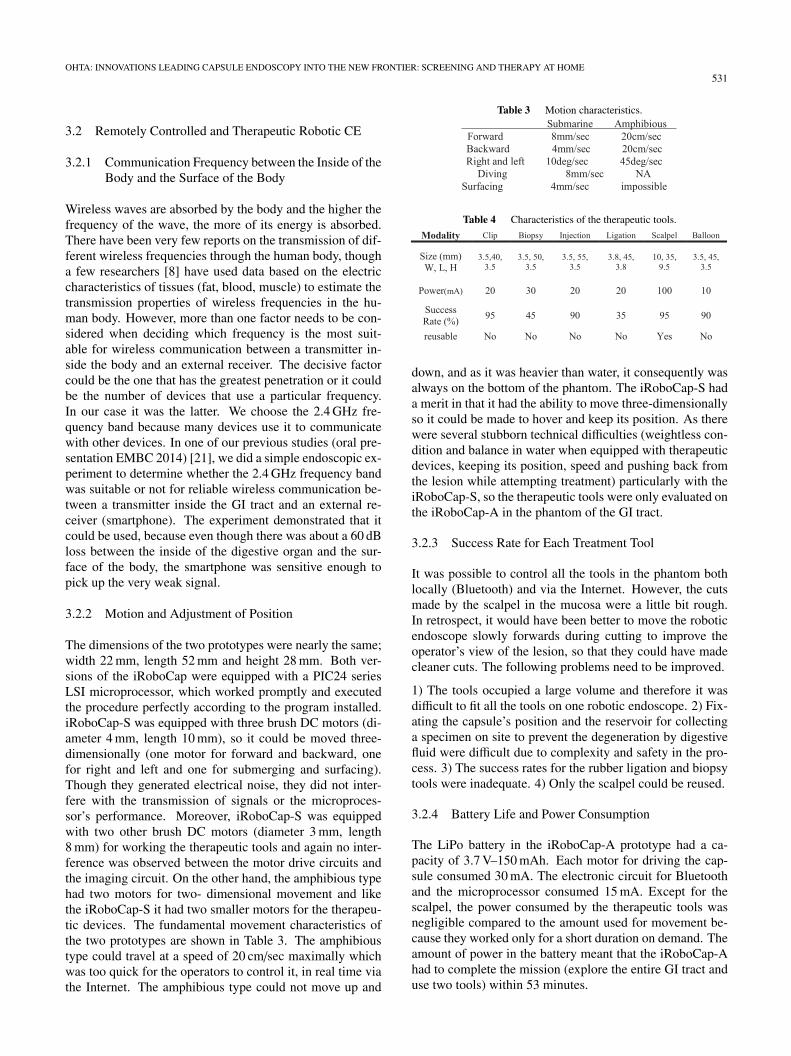
A pair of sensors was used in order that the ICVCcould distinguish between the local terrestrial magnetism(0.04 mT) and magnetic flux of similar intensity generatedby the magnetic disk in the GI tract, was far from the bodysurface, and as a result prevented the ICVC from malfunc-tioning. The ICVC utilized the space diversity effect bycomparing the difference between the voltages at both sen-
528IEICE TRANS. COMMUN., VOL.E98–B, NO.4 APRIL 2015
Fig. 3 Highly sensitive dual MI sensor.
sors (Fig. 3). Fading caused by subjects moving was re-moved by this effect. The threshold level for detecting thecapsule was adjusted to 8 or 9 cm by taking into account theanatomical ICV’s location.
2.1.3 Procedure for the Entire GI Exploration
After the initial preparation, co-medical staff set up theequipment and the subject ingested the capsule followed byexploration of the stomach and proximal duodenum undermagnetic navigation which was supported by observing thereal-time monitor remotely. After the booster preparation,the ICVC was attached to the patient on the body surfaceclosest to the ileocecal area and switched on. The real-timeviewer was detached from the subject and carried by the sub-ject. From then onwards, the subject became free and couldgo anywhere, home, office and so on. When the capsule ar-rived at the cecum, it set off the ICVC’s beeper. On hearingthe beeper, the subject phoned the hospital, attached the webcamera to the data recorder and connected it to the internet.After the doctor at the hospital had confirmed via the Inter-net that the capsule had passed through the ICV, the subjectreturned to hospital and the doctor then resumed navigationof the capsule in the colon and rectum. After confirming thecapsule’s arrival at the anus, the data recorder was detachedand then the image data was analyzed on the workstation.
2.1.4 Video Streaming Software
There are several useful commercial software tools that candeliver video clips to customers via the Internet. Skype isa famous, common and easy handling tool for this purpose.In our magnetic navigation system, we did not need a re-mote desktop function. To our disappointment, the new datarecorder for the PillCam COLON 2 does not have an outputsocket for a real-time viewer which the previous model had,though it does have a tiny real-time monitor. A simple cheapsolution that enabled us to utilize Given Imaging’s capsuleendoscope system and stream endoscopic images via the In-ternet was to attach a camera adaptor to the data recorderand install the Skype app or a web camera app on the laptopPC.
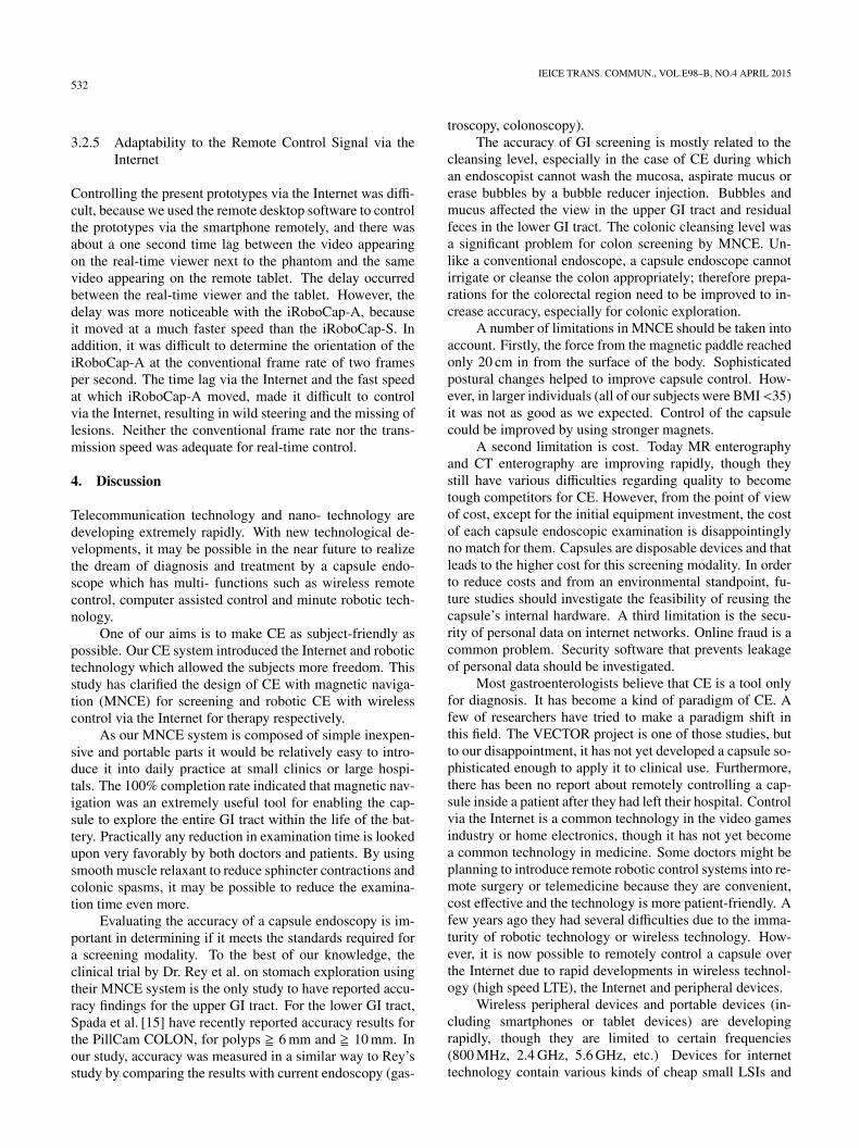
Fig. 4 Structure of the iRoboCap.
Fig. 5 The two prototypes of iRoboCap.
2.2 Motor-Driven Robotic Therapeutic Endoscopes
2.2.1 Structures of the iRoboCap
Our internet-linked and robotic capsule endoscope (iRobo-Cap) was used for this study. IRoboCap consisted of threeparts: an imaging unit, a movement control unit and a thera-peutic tool unit (Fig. 4). The imaging unit came from a Pill-Cam SB2 (Given Imaging, Yoquium Israel). Two ways ofmoving the iRobocap were investigated in the initial stage.One was a submarine type (screw-driven; iRoboCap-S) andthe other was an amphibious type (wheel-driven; iRoboCap-A) (Fig. 5). The iRoboCap-S was equipped with three mo-tors for movement and a driver integrated circuit, so that itcould be steered three-dimensionally in water and remotely.On the other hand, iRoboCap-A was equipped with two mo-tors and a driver integrated circuit which meant it could onlymove two-dimensionally. IRoboCap’s therapeutic tool unitessentially consisted of a tool and a motor. The followingtools (Fig. 6) were tested: a) a clipping device, b) biopsyforceps, c) a syringe for injecting or spraying, d) a rubberligater, e) two types of scalpel and f) a balloon for dilat-ing the stricture. The motor was used to release a mini-hemoclip, operate the forceps or trigger the spring for injec-tion or an electromagnetic valve for generating CO2 for bal-loon dilation. The tools were promptly controlled by signalsgenerated from a single microprocessor chip on the samecircuit according to sequences which were previously pro-grammed. The mobility of each prototype and success ratesof only iRoboCap-A with the several tools were evaluatedin a phantom. Even though, we carefully positioned eachtool on the iRoboCap-S, we were not able to maintain its

OHTA: INNOVATIONS LEADING CAPSULE ENDOSCOPY INTO THE NEW FRONTIER: SCREENING AND THERAPY AT HOME529
Fig. 6 Six types of therapeutic tool units.
“weightless condition” or the weight balance in water.
2.2.2 Architecture of the iRoboCap System
Figure 7 shows the architecture of the iRoboCap system.Both the S and A versions of iRoboCap used two differentfrequency bands and transceiver LSIs. 430 MHz was usedfor images and 2.4 GHz (Bluetooth or Wi-Fi specification)for control signal transmission. For security of the wirelesssignal, WEP or WPA encryption was used to pair each de-vice. On the other hand, image signals from the capsulewere unilaterally received by antennas on the surface of thephantom and stored on the conventional data recorder. Inaddition, they were sometimes simultaneously transmittedwirelessly back to the hospital via a real-time viewer linkedto the Internet. A tablet device in our hospital was used tocontrol the prototypes remotely by using application soft-ware installed on a smartphone (Galaxy; Samsung Co. or
Fig. 7 System architecture of the iRoboCap.
iPhone, Apple Co.) which was next to the phantom. Thesmartphone was used as a repeater for the control signals be-tween the tablet and each prototype. When each prototypewas controlled wirelessly via the Internet, it was done in adifferent room in our hospital which was beyond the rangeof communication by Bluetooth or Wi- Fi. Each prototype’smicroprocessor was equipped with a Bluetooth or Wi-Fitransceiver which allowed signals to be securely transferredbetween each prototype and the smartphone. TeamViewer(Germany), a remote desktop application was used to con-firm basic transmission, control the capsule with the smart-phone and to view images between the smartphone near theiRoboCap and the remote portable device. We are currentlybuilding our own bespoken app using the Xcode5 platformand it is nearly ready for testing on an iPhone 5S.
3. Results
3.1 Magnetic Navigation
3.1.1 Visual Improvements (Two Cameras and an Adap-tive Frame Rate)
In CCE (PillCam COLON 2; Given Imaging) there are twomajor improvements which have helped to reduce the num-ber of lesions it misses compared to conventional CE (Pill-Cam SB2). The first is the capsule has two cameras, one ateach end of the capsule, and the other improvement is theadaptive frame rate, which adjusts the timing of the flashingof the LEDs and imaging, in relation to the velocity of thecapsule’s movement which is measured by the accelerationsensor. The two cameras give a wider angle of view, almost360 degrees of visual coverage. The adaptive frame rate islinked to its low rate for missing lesions when moving fastand to the capsule’s low power consumption. Another of thecapsule’s low-power features is that it does not capture thesame image twice. The fixed frame rate of two frames persecond (fps) of the PillCam SB2 could not prevent it frommissing the lesions when the capsule was navigated quickly.Sixteen fps is the minimal rate for the video not to be jerky.Even though the frame rate of 6 fps (the maximum availablefor CCE) made the video a bit jerky, it was quick enough forthe researchers to be able to control the capsule smoothly.

530IEICE TRANS. COMMUN., VOL.E98–B, NO.4 APRIL 2015
Fig. 8 The depth of penetration for each modality.
Table 1 Precision of 3 localization of capsule endoscopy.
Therefore, we decided that the Pillcam COLON 2 with itstwo cameras and a frame rate of 6 fps was the best combina-tion for our system.
3.1.2 Localization of the Capsule Endoscope
The location system required precision to some extent andsimplicity. Under the same conditions without any ad-ditional items added to the capsule or changes in powerconsumption we investigated three modalities, the electro-magnetic wave (430 MHz) intensity [21], the static mag-netic intensity [15] and the intensity of light (630 nm) [23]for capturing endoscopic images. Figure 8 shows the signalloss in extracted porcine livers by each detection method un-der the sensitivity of our present detector. The conventionalmethod by means of wireless image signal (430 MHz) inten-sity was the only available method. Table 1 shows a roughprecision for each method, though the precision dependedon the number of detector sensors and the wavelength of themodality. Using a calibration technique to revise the posi-tion, we improved the precision level. The results indicatethat neither static magnetic detection nor optical detectioncould detect the signals well enough to calculate the cap-sule location to our satisfaction. Even if the optical detec-tion system failed in our tests due to the optical scatteringcharacteristics, we still think it could in principle be the bestmethod for not only detecting the capsule’s location accu-rately, but also the direction of the cameras. We hope toimprove the optical permittivity in tissues by using the pi-cosecond pulse of near infrared laser diodes. However, ifthat does not improve the optical permittivity, we will try toimprove the signal-to-noise ratio (S/N) by using the feedfor-ward compensation technique or the synchronous detection.
Table 2 Transit times (min.).
3.1.3 Transit Times and Completion Rates
Table 2 shows the average transit times, gastric transit times(GTT), small bowel transit times (SBTT), and colon-rectumtransit times (CRTT) for CCE, with and without magneticnavigation. The examination time for navigation was muchshorter than that for conventional CCE. With navigation theGTT was reduced by half, though the SBTT was similarto the time for CCE. Navigation resulted in completion oftotal GI screening within the battery life (completion rate(CR): 100%), mainly because the CRTT was only 48 min-utes compared with 475 minutes for conventional CCE. Torealize this high CR, postural changes were also employedduring colon navigation. To our disappointment, the mag-netic force was not strong enough for the capsule to over-come peristalsis or sphincter contractions.
3.1.4 Preparation and Cleanliness
For the typical Asian, 6 Liters of PEG preparation as recom-mended by the European Society of Gastroenterological En-doscopy [17] is too much and not acceptable. In our clinicaltrials, we used a total of 4.2 Liters of PEG intake, which isthe same as the Japanese Capsule Endoscopic Association’sprotcol and was administrated in their first clinical trial forCCE [18]. However, in our trials, only 45% of the subjectsdrank all of the preparation due to the volume of liquid andits monotone taste. 4.2 L was especially a problem for sub-jects older than 65 years old, because only 10% of themmanaged to drink all of the preparation. Furthermore, clean-liness (in the colon and rectum), which was evaluated by a4-point scale (excellent, good, fair, poor), scored better thanfair, in only 70% of the subjects. Clinically, visibility wasnot good enough to satisfy most endoscopists.
3.1.5 Evaluation of the Streaming Software
In the previous model we used TeamViewer (remote desk-top software) to confirm the arrival of the capsule at the ileo-cecal valve from the hospital. However, we found that theTeamViewer software was not suitable software for stream-ing video images while navigating the capsule with the mag-netic paddle, because some of those images were lost and allof the images were also delayed for a few seconds. In thisclinical study, we did not need a bilateral communicationsystem between the patient and the hospital, therefore weused Skype (Microsoft) and LiveCapture3 (Daddy corp.).The former had a one second delay and the latter 0.5 sec-onds due to their respective coding methods, but there wereno lost frames.
OHTA: INNOVATIONS LEADING CAPSULE ENDOSCOPY INTO THE NEW FRONTIER: SCREENING AND THERAPY AT HOME531
3.2 Remotely Controlled and Therapeutic Robotic CE
3.2.1 Communication Frequency between the Inside of theBody and the Surface of the Body
Wireless waves are absorbed by the body and the higher thefrequency of the wave, the more of its energy is absorbed.There have been very few reports on the transmission of dif-ferent wireless frequencies through the human body, thougha few researchers [8] have used data based on the electriccharacteristics of tissues (fat, blood, muscle) to estimate thetransmission properties of wireless frequencies in the hu-man body. However, more than one factor needs to be con-sidered when deciding which frequency is the most suit-able for wireless communication between a transmitter in-side the body and an external receiver. The decisive factorcould be the one that has the greatest penetration or it couldbe the number of devices that use a particular frequency.In our case it was the latter. We choose the 2.4 GHz fre-quency band because many devices use it to communicatewith other devices. In one of our previous studies (oral pre-sentation EMBC 2014) [21], we did a simple endoscopic ex-periment to determine whether the 2.4 GHz frequency bandwas suitable or not for reliable wireless communication be-tween a transmitter inside the GI tract and an external re-ceiver (smartphone). The experiment demonstrated that itcould be used, because even though there was about a 60 dBloss between the inside of the digestive organ and the sur-face of the body, the smartphone was sensitive enough topick up the very weak signal.
3.2.2 Motion and Adjustment of Position
The dimensions of the two prototypes were nearly the same;width 22 mm, length 52 mm and height 28 mm. Both ver-sions of the iRoboCap were equipped with a PIC24 seriesLSI microprocessor, which worked promptly and executedthe procedure perfectly according to the program installed.iRoboCap-S was equipped with three brush DC motors (di-ameter 4 mm, length 10 mm), so it could be moved three-dimensionally (one motor for forward and backward, onefor right and left and one for submerging and surfacing).Though they generated electrical noise, they did not inter-fere with the transmission of signals or the microproces-sor’s performance. Moreover, iRoboCap-S was equippedwith two other brush DC motors (diameter 3 mm, length8 mm) for working the therapeutic tools and again no inter-ference was observed between the motor drive circuits andthe imaging circuit. On the other hand, the amphibious typehad two motors for two- dimensional movement and likethe iRoboCap-S it had two smaller motors for the therapeu-tic devices. The fundamental movement characteristics ofthe two prototypes are shown in Table 3. The amphibioustype could travel at a speed of 20 cm/sec maximally whichwas too quick for the operators to control it, in real time viathe Internet. The amphibious type could not move up and
Table 3 Motion characteristics.
Table 4 Characteristics of the therapeutic tools.
down, and as it was heavier than water, it consequently wasalways on the bottom of the phantom. The iRoboCap-S hada merit in that it had the ability to move three-dimensionallyso it could be made to hover and keep its position. As therewere several stubborn technical difficulties (weightless con-dition and balance in water when equipped with therapeuticdevices, keeping its position, speed and pushing back fromthe lesion while attempting treatment) particularly with theiRoboCap-S, so the therapeutic tools were only evaluated onthe iRoboCap-A in the phantom of the GI tract.
3.2.3 Success Rate for Each Treatment Tool
It was possible to control all the tools in the phantom bothlocally (Bluetooth) and via the Internet. However, the cutsmade by the scalpel in the mucosa were a little bit rough.In retrospect, it would have been better to move the roboticendoscope slowly forwards during cutting to improve theoperator’s view of the lesion, so that they could have madecleaner cuts. The following problems need to be improved.
1) The tools occupied a large volume and therefore it wasdifficult to fit all the tools on one robotic endoscope. 2) Fix-ating the capsule’s position and the reservoir for collectinga specimen on site to prevent the degeneration by digestivefluid were difficult due to complexity and safety in the pro-cess. 3) The success rates for the rubber ligation and biopsytools were inadequate. 4) Only the scalpel could be reused.
3.2.4 Battery Life and Power Consumption
The LiPo battery in the iRoboCap-A prototype had a ca-pacity of 3.7 V–150 mAh. Each motor for driving the cap-sule consumed 30 mA. The electronic circuit for Bluetoothand the microprocessor consumed 15 mA. Except for thescalpel, the power consumed by the therapeutic tools wasnegligible compared to the amount used for movement be-cause they worked only for a short duration on demand. Theamount of power in the battery meant that the iRoboCap-Ahad to complete the mission (explore the entire GI tract anduse two tools) within 53 minutes.
532IEICE TRANS. COMMUN., VOL.E98–B, NO.4 APRIL 2015
3.2.5 Adaptability to the Remote Control Signal via theInternet
Controlling the present prototypes via the Internet was diffi-cult, because we used the remote desktop software to controlthe prototypes via the smartphone remotely, and there wasabout a one second time lag between the video appearingon the real-time viewer next to the phantom and the samevideo appearing on the remote tablet. The delay occurredbetween the real-time viewer and the tablet. However, thedelay was more noticeable with the iRoboCap-A, becauseit moved at a much faster speed than the iRoboCap-S. Inaddition, it was difficult to determine the orientation of theiRoboCap-A at the conventional frame rate of two framesper second. The time lag via the Internet and the fast speedat which iRoboCap-A moved, made it difficult to controlvia the Internet, resulting in wild steering and the missing oflesions. Neither the conventional frame rate nor the trans-mission speed was adequate for real-time control.
4. Discussion
Telecommunication technology and nano- technology aredeveloping extremely rapidly. With new technological de-velopments, it may be possible in the near future to realizethe dream of diagnosis and treatment by a capsule endo-scope which has multi- functions such as wireless remotecontrol, computer assisted control and minute robotic tech-nology.
One of our aims is to make CE as subject-friendly aspossible. Our CE system introduced the Internet and robotictechnology which allowed the subjects more freedom. Thisstudy has clarified the design of CE with magnetic naviga-tion (MNCE) for screening and robotic CE with wirelesscontrol via the Internet for therapy respectively.
As our MNCE system is composed of simple inexpen-sive and portable parts it would be relatively easy to intro-duce it into daily practice at small clinics or large hospi-tals. The 100% completion rate indicated that magnetic nav-igation was an extremely useful tool for enabling the cap-sule to explore the entire GI tract within the life of the bat-tery. Practically any reduction in examination time is lookedupon very favorably by both doctors and patients. By usingsmooth muscle relaxant to reduce sphincter contractions andcolonic spasms, it may be possible to reduce the examina-tion time even more.
Evaluating the accuracy of a capsule endoscopy is im-portant in determining if it meets the standards required fora screening modality. To the best of our knowledge, theclinical trial by Dr. Rey et al. on stomach exploration usingtheir MNCE system is the only study to have reported accu-racy findings for the upper GI tract. For the lower GI tract,Spada et al. [15] have recently reported accuracy results forthe PillCam COLON, for polyps � 6 mm and � 10 mm. Inour study, accuracy was measured in a similar way to Rey’sstudy by comparing the results with current endoscopy (gas-
troscopy, colonoscopy).The accuracy of GI screening is mostly related to the
cleansing level, especially in the case of CE during whichan endoscopist cannot wash the mucosa, aspirate mucus orerase bubbles by a bubble reducer injection. Bubbles andmucus affected the view in the upper GI tract and residualfeces in the lower GI tract. The colonic cleansing level wasa significant problem for colon screening by MNCE. Un-like a conventional endoscope, a capsule endoscope cannotirrigate or cleanse the colon appropriately; therefore prepa-rations for the colorectal region need to be improved to in-crease accuracy, especially for colonic exploration.
A number of limitations in MNCE should be taken intoaccount. Firstly, the force from the magnetic paddle reachedonly 20 cm in from the surface of the body. Sophisticatedpostural changes helped to improve capsule control. How-ever, in larger individuals (all of our subjects were BMI<35)it was not as good as we expected. Control of the capsulecould be improved by using stronger magnets.
A second limitation is cost. Today MR enterographyand CT enterography are improving rapidly, though theystill have various difficulties regarding quality to becometough competitors for CE. However, from the point of viewof cost, except for the initial equipment investment, the costof each capsule endoscopic examination is disappointinglyno match for them. Capsules are disposable devices and thatleads to the higher cost for this screening modality. In orderto reduce costs and from an environmental standpoint, fu-ture studies should investigate the feasibility of reusing thecapsule’s internal hardware. A third limitation is the secu-rity of personal data on internet networks. Online fraud is acommon problem. Security software that prevents leakageof personal data should be investigated.
Most gastroenterologists believe that CE is a tool onlyfor diagnosis. It has become a kind of paradigm of CE. Afew of researchers have tried to make a paradigm shift inthis field. The VECTOR project is one of those studies, butto our disappointment, it has not yet developed a capsule so-phisticated enough to apply it to clinical use. Furthermore,there has been no report about remotely controlling a cap-sule inside a patient after they had left their hospital. Controlvia the Internet is a common technology in the video gamesindustry or home electronics, though it has not yet becomea common technology in medicine. Some doctors might beplanning to introduce remote robotic control systems into re-mote surgery or telemedicine because they are convenient,cost effective and the technology is more patient-friendly. Afew years ago they had several difficulties due to the imma-turity of robotic technology or wireless technology. How-ever, it is now possible to remotely control a capsule overthe Internet due to rapid developments in wireless technol-ogy (high speed LTE), the Internet and peripheral devices.
Wireless peripheral devices and portable devices (in-cluding smartphones or tablet devices) are developingrapidly, though they are limited to certain frequencies(800 MHz, 2.4 GHz, 5.6 GHz, etc.) Devices for internettechnology contain various kinds of cheap small LSIs and

OHTA: INNOVATIONS LEADING CAPSULE ENDOSCOPY INTO THE NEW FRONTIER: SCREENING AND THERAPY AT HOME533
antennas which are suitable for miniaturizing equipmentand establishing various functions on one circuit. Unfor-tunately, those frequencies mentioned earlier have large di-electric losses in the human body (so called electronic ovenfrequency) and therefore it was believed that transmissionin the human body was impossible. But the quality of thosefrequency bands have been improving rapidly and their sen-sitivity levels have come into the target range for realizingcommunication between intra and extracorporeal transmit-ters and receivers. Valdastri [24] and Iris De Falco [8] indi-cated the possibility of 0.9 GHz communication in vivo. Ourprevious study provided the direct evidence that the 2.4 GHzfrequency band could be used in vivo. The 2.4 GHz fre-quency band is very useful for connecting to the Internet viawireless HUBs. Fortunately, most smartphones or tabletssupport 2.4 GHz Wi-Fi connectivity and/or Bluetooth con-nectivity. Moreover, smartphones and tablets have becomevery common electronic devices. If such portable devicescan detect a 2.4 GHz high baud rate signal such as imagesignals from a capsule in the GI tract, we only need to in-stall an application on them that is capable of storing data lo-cally or transmitting it via the Internet, and this would meanthe conventional data recorder would no longer be needed.Hopefully our quite simple system will pave the way for fu-turistic, impeccable and advanced remote medicine that isnot only for capsule endoscopy, but also for interventionalmedicine including gastroenterology and surgery.
By building and trying two robotic prototypes, we haveidentified several areas that need to be improved in orderto realize a practical version. They include the followingfour points, miniaturization, battery life, internet security,frame rate and high-speed transfer of video data for real-time control [25]–[27].
To miniaturize an iRoboCap to half of the size of thepresent prototypes, we have to look at every part. We arefocusing our attention on 1) integrating the transceiver LSIwith the conventional image transmitter or motor driver IC,2) acquiring a smaller battery with a longer battery life and3) acquiring smaller motors with low power consumption.We hope that the first problem will be overcome by intro-duction of a Bluetooth or ZigBee IC working at higher clockrates in the very near future. Our results indicate that thecurrent LiPo battery would have enough power for naviga-tion in the stomach and colon and simple therapy, howeverit would not have enough power for therapy that required along operation time, such as an endoscopic mucosal resec-tion (EMR) or an endoscopic submucosal dissection (ED).This type of battery is rapidly developing and has been in-troduced into various fields such as electric cars, personalcomputers and portable device. Unfortunately, at the mo-ment they have safety issues and bio-batteries are still intheir infancy and it will almost certainly take quite a fewyears before they come on to the market. If we did not needto worry about the price of the iRoboCap or think about re-ducing costs by reusing it, the third problem could be solvedquite easily by buying expensive small low power brushlessmotors (ϕ1.5 mm) which should help to prolong the battery
life as well as aiding miniaturization.The most crucial problem might be internet security
including secure access to cloud services. Various codingtechnology might be one solution as well as various new en-cryption techniques that are currently under development.Another approach is a fail-safe mechanism that may be alsoone of the better options to surmount any difficulties due tojamming of wireless communication.
The last problem concerns real-time control which wasdifficult via the Internet due to a delay in the video reach-ing the remote tablet, the slow frame rate of two frame persecond and in the case of the iRoboCap-A, it was acerbatedby the high speed at which iRoboCap-A traveled. Generallyspeaking, the length of the time lag depended on the speedof at which images were compressed and decompressed, andthe transmission rate over the internet network. Using a4G/LTE smartphone connected to a 4G network should rec-tify the time lag problem, allow for faster frame rates (adap-tive frame rate) and it may be fast enough to do without spe-cial software for compressing and decompressing the videoimages. We intend to test a 4G network connection in thevery near future.
5. Conclusion
Our single capsule endoscopic system with magnetic navi-gation for screening the entire GI tract has the possibility tobecome a more subject friendly and more accurate modalityfor screening than conventional endoscopy, though it will benecessary to reduce costs and improve security.
Our preliminary results for robotic endoscopes showthat it is possible to remotely control a robotic capsuleand operate therapeutic tools via the Internet. Though ourrobotic system is a long way from the finished article, weare sure that rapid innovations in robotic and wireless tech-nology will make it possible to realize many types of patientfriendly systems, not only in gastroenterology but also inother healthcare areas.
Acknowledgements
I would like to especially thank Professor MakotoKawashima of Chubu University for discussing various as-pects of the circuit design and Shinichi Katsuki, a visitingprofessor of the Asian Institute of TeleSurgery for his help-ful advice and support during our clinical trial.
References
[1] P. Swain, A. Toor, F. Volke, J. Keller, J. Gerber, E. Rabinovitz, andR.I. Rothstein, “Remote magnetic manipulation of a wireless cap-sule endoscope in the esophagus and stomach of humans,” Gastroin-test Endosc, vol.71, pp.1290–1293, 2010.
[2] J. Keller, C. Fibble, F. Volke, J. Gerber, A.C. Mosse, M.R. Zawadzki,E. Rabinovitz, P. Layer, and P. Swain, “Remote magnetic control ofa wireless capsule endoscope in the esophagus is safe and feasible:Results of a randomized, clinical trial in healthy volunteers,” Gas-trointest Endosc, vol.72, pp.941–946, 2010.
[3] J.F. Rey, H. Ogata, N. Hosoe, K. Ohtsuka, N. Ogata, K. Ikeda, H.
534IEICE TRANS. COMMUN., VOL.E98–B, NO.4 APRIL 2015
Aihara, I. Pangtay, T. Hibi, S. Kudo, and H. Tajiri, “Feasibility ofstomach exploration with a guided capsule endoscope,” Endoscopy,vol.42, pp.541–545, 2010.
[4] E. Morita, N. Ohtsuka, Y. Shindo, S. Nouda, T. Kuramoto, T. Inoue,M. Murano, E. Umegaki, and K. Higuchi, “In vivo trial a driving sys-tem for a self-propelling capsule endoscope using a magnetic fiel,”Gastrointest Endosc, vol.72, no.4, pp.836–840, 2010.
[5] J. Keller, C. Fibble, F. Volke, J. Gerber, A.C. Mosse, M.R. Zawadzki,E. Rabinovitz, P. Layer, D. Schmitt, V. Andresen, U. Rossiren, andP. Swain, “Inspection of the human stomach using remote-controlledcapsule endoscopy: A feasibility study in healthy volunteers,” Gas-trointest Endosc, vol.73, pp.22–28, 2011.
[6] J.F. Rey, H. Ogata, N. Hosoe, K. Ohtsuka, N. Ogata, K. Ikeda, H.Aihara, I. Pangtay, T. Hibi, S. Kudo, and H. Tajiri, “Blinded nonrandomized comparative study of gastric examination with a mag-netically guided capsule endoscope and standard videoendoscope,”Gastrointest Endosc, vol.75, pp.373–381, 2012.
[7] U.W. Denzer, T. Rosch, B. Hotayt, N. Ogata, N. Hosoe, H. Ogata, K.Ohtsuka, X. Hebuterne, G. Vanbiervliet, J. Filippi, M. Greff, and J.F.Rey, “Prospective evaluation of magnetic gastric capsule examina-tion (MGCE) in patients with indications for upper GI endoscopy,”Endoscopy supplement No1, 20th United European Gastroenterol-ogy Week, Amsterdam, The Netherland, pp.60–61, Oct. 2012.
[8] I. Iris De Falco, G. Tortora, P. Dario, and A. Menciassi, “A inte-grated system for Wireless capsule Endoscopy in a liquid-distendedStomach,” IEEE Trans. Biomed. Eng., vol.61, pp.794–804, March2014.
[9] R. Carta, G. Tortora, B. Lenaerts, P. Valdastri, A. Menciassi, P.Dario, and R. Puers, “Wireless powering for a self-propelled andsteerable endoscopic capsule for stomach inspection,” Biosensorsand Bioelectronics, vol.25, pp.845–851, 2009.
[10] M. Quirini, A. Menciassi, S. Scapellato, P. Dario, F. Rieber, C. Ho,S. Schostek, and M.O. Schurr, “Feasibility proof of a legged locomo-tion capsule for the GI tract,” Gastrointest Endosc, vol.67, pp.1153–1158, 2008.
[11] H. Ohta, Y. Sato, S. Tanaka, T. Okuda, T. Doi, A. Fujimi, and Y.Kanisawa, “A new extracorporeal magnetic navigation tool for cap-sule endoscopy,” Gastrointest Endosc supplement, vol.69, DigestiveDisease Week (DDW), pp.98–99, Chicago, USA, May 2009.
[12] H. Ohta, Y. Sato, S. Tanaka, F. Tamura, and T. Doi, “Capsule en-doscopy for the whole of the Gastrointestinal tract using extracorpo-real magnetic navigation,” Gastrointest Endosc supplement, vol.71,pp.238–239, DDW, New Orleans, USA, May 2010.
[13] H. Ohta, S. Katsuki, K. Tanaka, and T. Ebata, “A real-time dualviewer (3-D position and endoscopic image) for magnetic naviga-tion of capsule endoscopes: Realizing simple, economical quick andaccurate gastrointestinal endoscopy,” Gastrointest Eondosc supple-ment, vol.73, pp.370–371, DDW, Chicago, USA, May 2011.
[14] H. Ohta, S. Katsuki, T. Fjita, Y. Sato, and T. Sagawa, “Total Gas-trointestinal Tract Screening by using only one capsule endoscopewith navigation,” Gastrointest Endosc supplement, vol.75, pp.127–128, DDW, San Diego, USA, May 2012.
[15] H. Ohta and S. Katsuki, “Subject-friendly entire gastrointestinalscreening with a single capsule endoscope by magnetic navigationand the Internet,” Proc. 36th annual Int. Conf. of IEEE on EMB,pp.6997–7000, Chicago, USA, Aug. 2014.
[16] Y. Mao, F. Lian, and C. Yuhua, “An efficient wireless power trans-mission system for the capsule endoscopy application,” 2011 IEEE,Biomedical cuircuits and System Conf., pp.221–224, 2011.
[17] C. Spada, C. Hassan, M. Munoz-Navas, et al., “Second generationcolon capsule endoscopycompared with colonoscopy,” GastrointestEndosc, vol.74, pp.581–589, 2011.
[18] S. Oka, S. Tanaka, Y. Saito, S. Saito, Y. Kakugawa, M. Matsumoto,H. Aikawa, I. Watari, T. Aoyama, S. Nouda, T. Kuramoto, K.Watanabe, N. Ohmiya, K. Higuchi, H. Goto, T. Arakawa, andH. Tajiri, “Evaluation of the clinical efficacy of colon capsule en-doscopy in the detection of lesion of the colon-prospective multicen-
ter study in Japan,” Gastrointest Endosc supplement, vol.79, DDWpp.170–171, Chicago, USA, May 2014.
[19] J.L. Toennie, G. Cuiti, B.F. Smith, A. Menciassi, P. Valdastri, andR.J. Webster, “Toward Thetherless Insufflation of the GI Tract,”32nd Annual Int. Conf. IEEE on EMB, pp.1946–1949, Boston USA,Aug. 2011
[20] P. Valdastri, C. Quaglla, E. Susilo, A. Menciassi, P. Dario, C.N. Ho,G. Anhoeck, and M.O. Schurr, “Wireless therapeutic endoscopiccapsule: In vivo experiment,” Endoscopy, vol.40, pp.979–982, 2008.
[21] H. Ohta and M. Kawashima, “Technical feasibility of patient-friendly screening and treatment of digestive disease by remote con-trol robotic capsule endoscopes via the Internet,” 36th annual Int.Conf. of IEEE on EMB, pp.7001–7004, Chicago, USA, Aug. 2014.
[22] T. Ito, D. Anzai, and J. Wang, “Novel joint TOA/RSSI-based WCElocation tracking method without prior knowledge of biological hu-man body tissue,” 36th annual Int. Conf. of IEEE on EMB, pp.6993–6996, Chicago, USA, Aug. 2014.
[23] T. Nakamura, Y. Nishiwaki, S. Suzuki, S. Sakaguchi, K. Oota, and Y.Yamashita, “Optical transparency of the liver tissue in various liverdisease,” J. Japan Laser Medicine, vol.8, no.3, pp.121–122, Dec.1987. (in Japanese)
[24] P. Valdastri, S. Rossi, A. Menciassi, V. Lionetti, F. Bernini, F.A.Recchia, and P. Dario, “An implantable ZigBee ready telemetricplatform for in vivo monitoring of physiological parameters,” Sen-sors and Actuators, vol.142, pp.368–378, 2008.
[25] H. Ohta, “The dawn of internet linked robotic capsule endoscopy;A radio-controlled and motor-driven capsule endoscope that can becontrolled via the Internet,” Gastrointest Endosc supplement, vol.77,pp.465–466, DDW Orland USA, May 2013.
[26] H. Ohta, S. Katsuki, T. Fujita, Y. Sato, and T. Sagawa, “First trial of awireless robotic capsule endoscope equipped with therapeutic tools,”Endoscopy supplement no.1 21st United European GastroenterologyWeek, Berlin Germany, pp.83–84, Oct. 2013.
[27] H. Ohta and S. Katsuki, “A therapeutic wireless robotic endoscopecontrolled via the Internet remotely,” 22nd United European Gas-troenterology Week, Vienna Austria, pp.789–790, Oct. 2014.
Hidetoshi Ohta received his B.S. andM.S. degrees in Electronic Engineering fromHokkaido University in 1971 and 1973, andhis B.S. and Ph.D. in Medicine from SapporoMedical University in 1988 and 1994 respec-tively. From 1973-82, he worked at YokosukaTelecommunication Laboratory, NTT to studycoaxial cable and optical fiber transmission sys-tems. From 1988-2014, he has worked at severalclinical hospitals, Sapporo Medical UniversityHospital, Hokkaido Prefectural Sapporo Kitano
Hospital, Oji General Hospital, and during that time he studied medicalengineering related to gastroenterology. From 2004-11 he mentored andtrained resident doctors as a clinical associate professor of Sapporo Medi-cal University.