ionizing radiation enhances - erc.bioscientifica.com · 3dipartimento di diagnostica per immagini e...
TRANSCRIPT
En
do
crin
e-R
ela
ted
Can
cer
ResearchC Passaro, A Abagnale et al. dl922–947 and ionizing
radiation20 :5 633–647
Ionizing radiation enhancesdl922–947-mediated cell death ofanaplastic thyroid carcinoma cells
Carmela Passaro*, Antonella Abagnale*, Silvana Libertini1, Massimiliano Volpe,
Ginevra Botta†, Laura Cella2, Roberto Pacelli3, Gunnel Hallden4, David Gillespie1
and Giuseppe Portella
Dipartimento di Biologia e Patologia Cellulare e Molecolare, Facolta di Medicina e Chirurgia, Universita di Napoli
Federico II, via S. Pansini 5, 80131 Napoli, Italy1The Beatson Institute for Cancer Research, Switchback Road, Bearsden, Glasgow G61 1BD, UK2Istituto di Biostrutture e Bioimmagini, Consiglio Nazionale delle Ricerche, Napoli, Italy3Dipartimento di Diagnostica per Immagini e Radioterapia, Universita Federico II, Napoli, Italy4Barts Cancer Institute, Centre for Molecular Oncology, Queen Mary University of London, London, UK
*(C Passaro and A Abagnale contributed equally to this work)†G Botta is now at Department of Medical Oncology, Dana-Farber Cancer Institute, 450 Brookline Avenue, Boston,
Massachusetts 02215, USA
http://erc.endocrinology-journals.org q 2013 Society for EndocrinologyDOI: 10.1530/ERC-13-0001 Printed in Great Britain
Published by Bioscientifica Ltd.
Downloa
Correspondence
should be addressed
to G Portella
Abstract
dl922–947 is an oncolytic adenovirus potentially suitable for the treatment of aggressive
localized tumors, such as anaplastic thyroid carcinoma (ATC). In this study, we have analyzed
the effects of dl922–947 in combination with ionizing radiations, testing different schedules
of administration and observing synergistic effects only when ATC cells were irradiated 24 h
prior to viral infection. Cells undergoing combined treatment exhibited a marked increase in
cell death and viral replication, suggesting that irradiation blocks cells in a more permissive
state for viral life cycle. We also show that dl922–947 triggers a DNA damage response,
characterized by mobilization of the MRN complex (composed by Mre11-Rad50-Nbs1),
accumulation of gH2AX, and activation of the checkpoint kinases ataxia telangiectasia
mutated (ATM) and Chk1. Based on these observations, we speculate that the DNA damage
response acts as a cellular protective mechanism to hinder viral infection and replication. To
confirm this hypothesis, we demonstrate that the ATM inhibitor KU55933 increased the
oncolytic activity of dl922–947 and its replication. Finally, we validate the potential
therapeutic use of this approach by showing in vivo that the combined treatment slows
tumor xenograft growth more potently than either irradiation or infection alone.
Key Words
" anaplastic thyroid carcinoma
" oncolytic virus
" irradiation
" DNA damage
ded
Endocrine-Related Cancer
(2013) 20, 633–647
Introduction
Anaplastic thyroid carcinoma (ATC) represents one of
the most aggressive human malignancies. It arises from
thyroid follicular cells showing morphological features
of a malignant neoplasm, high proliferation rate, and
marked aneuploidy. The median survival of patients with
ATC is w6 months from diagnosis. Multimodality
therapy, which includes surgical debulking, external
radiation therapy, and chemotherapy, has failed to show
any improvements in survival and ATC still remains a
formidable medical challenge (Smallridge et al. 2009).
Therefore, novel therapies with different mechanisms of
action are required.
from Bioscientifica.com at 02/16/2019 03:49:21PMvia free access
En
do
crin
e-R
ela
ted
Can
cer
Research C Passaro, A Abagnale et al. dl922–947 and ionizingradiation
20 :5 634
Oncolytic viruses (OVs) are replication competent
viral mutants able to complete their life cycle exclusively
in tumor cells. With respect to classical anti-cancer drugs,
OVs present several advantages: tumor-selective replica-
tion with amplification of the input dose, stimulation of
anti-tumoral immune responses, and relatively few mild
side effects when administered to human patients. Also,
the development of cross-resistance is unlikely because
their mechanism of action is independent of conventional
anti-cancer therapeutics (Eager & Nemunaitis 2011,
Hallden & Portella 2012). A number of OVs, such as the
vaccinia virus GLV-1h68 (Lin et al. 2008b) and the
oncolytic attenuated herpes virus G207 (Lin et al.
2008a), have shown promising effects against ATC cells
in preclinical studies.
We have previously demonstrated that the oncolytic
adenoviruses dl1520 and dl922–947 could be useful for the
therapy of ATC (Portella et al. 2002, 2003, Libertini et al.
2007, 2008, 2011). dl1520, bearing a E1B 55K deletion, is
the first mutant engineered for tumor-specific cytolysis
(Heise et al. 1997). As its replication is severely attenuated
not only in normal cells but also in the majority of cancers
(O’Shea et al. 2004), dl1520 has shown limited utility as
single agent. Hence, novel and more effective OVs have
been engineered. dl922–947 is a second-generation ade-
noviral mutant bearing a 24 bp deletion in E1A-conserved
region 2 (CR2; Heise et al. 2000a). This region binds to and
inactivates the pRb tumor suppressor protein, dissociating
the pRb-E2F complex and driving S-phase entry and viral
replication. Because the mutant E1A protein encoded by
dl922–947 cannot bind pRb, the virus is unable to trigger
S-phase entry in normal cells and can therefore only
replicate in cells with an aberrant G1-S checkpoint, a
defect observed in over 90% human cancers (Sherr &
McCormick 2002). Superior cell killing potency of
dl922–947 with respect to dl1520 has been observed
in vitro and in vivo in human cancer cells of different origin
such as pancreas (Bhattacharyya et al. 2011), prostate
(Radhakrishnan et al. 2010), ovarian (Lockley et al. 2006),
thyroid (Libertini et al. 2008), and brain (Yong et al. 2009,
Botta et al. 2010, 2012). In addition, other mutants
with E1ACR2 region deletion have proved effective in
experimental models (Jiang et al. 2009, Oberg et al. 2010).
Emerging evidences indicate that combining OVs with
standard chemotherapy and other types of anti-cancer
treatments has the potential to increase the antitumor
activity (Heise et al. 2000b, Cheong et al. 2008). Preclinical
and clinical studies (phase I and II/III) have clearly
shown that OVs and radiation therapy show additional
or synergistic antitumor effects (Immonen et al. 2004,
http://erc.endocrinology-journals.org q 2013 Society for EndocrinologyDOI: 10.1530/ERC-13-0001 Printed in Great Britain
Touchefeu et al. 2011). Despite these encouraging results,
relatively little is known about the mechanisms that result
in enhanced tumor cell death. Understanding these
mechanisms could enable further optimization of this
strategy for future clinical development.
Ionizing radiation (IR) is known to induce DNA
damage and a subsequent block of the cell cycle
progression with accumulation of cells either in G1 or
G2, depending on the integrity of cell cycle checkpoints.
After DNA repair, these cell cycle checkpoint arrests are
released. It has been reported that drugs that block cells in
G2/M or inhibit cytokinesis can enhance OV-mediated
cell killing (Seidman et al. 2001). For example, we recently
demonstrated that the cytopathic effects of dl922–947 are
enhanced by the Aurora B inhibitor AZD1152, which
induces mitotic arrest, accumulation in G2/M phase, and
polyploidy (Libertini et al. 2011).
Here, we have evaluated the effect of dl922–947 in
association with IR, showing that the combination
enhances the effect of dl922–947 against ATC cells
in vitro. Comparing different schedules of treatment
(virus administered before or after irradiation), we
observed synergistic cell killing only when irradiation is
administered prior to viral infection. The efficacy of the
optimal combined treatment has also been confirmed in
ATC tumor xenografts in vivo.
Materials and methods
Cells, adenoviruses, and drugs
Human thyroid anaplastic carcinoma cell lines BHT101-5,
FRO, and Cal62 have been described and authenticated as
shown elsewhere (Schweppe et al. 2008). For growth curve
experiments, FRO and BHT101-5 cells were seeded in
12-well culture plates at a density of 104 cells/well and,
24 h later, treated as indicated. Cell count was performed
using an automatic cell counter (TC-10 Bio-Rad).
dl922–947 and AdGFP viral stocks were expanded in the
human embryonic kidney cell line HEK-293, purified, stored,
and quantified as previously reported (Botta et al. 2010).
zVAD-fmk and KU55933 (both from Tocris Bioscience,
Bristol, UK) were dissolved in DMSO to a final concen-
tration of 10 and 5 mM respectively and stored at K20 8C.
zVAD-fmk was added 2 h prior irradiation.
Cytotoxicity assay
Cytotoxicity was evaluated using the sulforhodamine B
assay as described previously (Vichai & Kirtikara 2006).
Published by Bioscientifica Ltd.
Downloaded from Bioscientifica.com at 02/16/2019 03:49:21PMvia free access
En
do
crin
e-R
ela
ted
Can
cer
Research C Passaro, A Abagnale et al. dl922–947 and ionizingradiation
20 :5 635
Dose–response curves were generated to calculate the
concentration of each agent required to kill 50% of cells
(median lethal dose, LD50). Untreated cells or cells treated
with single agents were used as a control.
Viral replication
FRO and BHT101-5 cells were infected with dl922–947,
media collected at 48 hours post infection (hpi), and viral
DNA was extracted using the High pure viral nucleic acid
kit (Roche Diagnostics). Specific primers for the viral
hexon gene (from 99 to 242 bp) were used to measure viral
replication by real-time PCR: 5 0-GCC ACC GAG ACG TAC
TTC AGC CTG-3 0 (upstream primer) and 5 0-TTG TAC GAG
TAC GCG GTA TCC TCC GCG GTC-3 0 (downstream
primer). A standard curve was constructed by assaying
serial dilutions of dl922–947.
For tissue culture infectious dose 50% (TCID50) assays,
105 cells were irradiated with 5 Gy and, after 24 h, infected
with five multiplicity of infection (MOIs) (FRO cells) or
one MOI (BHT101-5) of dl922–947. At 24 hpi, cells were
harvested and subjected to three rounds of freezing/
thawing and then centrifuged. The supernatant was
titrated on HEK-293 cells by serial dilution (Connell et al.
2011). Viral replication in tumor xenografts was evaluated
as described previously (Libertini et al. 2008).
FACS analysis
Cell cycle FRO and BHT101-5 cells treated as indicated
were collected and fixed in 70% (v/v) ethanol for at least
1 h at K20 8C. Washed pellets were resuspended in PBS
containing RNaseA (Roche) (0.4 U) and propidium iodide
(PI; Sigma) (0.015 mol/l), incubated for 20 min at room
temperature, and analyzed for emission in the FL3
channel. To remove artifacts – doublets and aggregates –
from the analysis, an electronic doublet discrimination
was performed using the area and width of the fluor-
escence (FL3) pulse.
AdGFP infection FRO and BHT101-5 cells were
infected with AdGFP (25 MOIs) and harvested 48 h later.
Pellet was resuspended in PBS and emission in the FL1
channel was analyzed.
Annexin V/PI staining Attached and detached FRO
and BHT101-5 cells were collected and washed with
Annexin V binding buffer (Biolegend, San Diego, CA,
USA). Pellet was resuspended in 50 ml of Annexin V
http://erc.endocrinology-journals.org q 2013 Society for EndocrinologyDOI: 10.1530/ERC-13-0001 Printed in Great Britain
binding buffer containing 2 ml FITC-conjugated Annexin
V (Biolegend, #640906). After 15 min of incubation at
room temperature, 250 ml PI solution 0.0015 mol/l in PBS
was added to each sample just before analysis.
gH2AX/PI and pH3/PI stainings Cells were fixed in
70% (v/v) ethanol for 2 h at K20 8C. Pellet was washed
with PBS/Tween buffer (PBT) (0.5% w/v BSA and 0.1% v/v
Tween 20 in PBS) and resuspended in PBT containing
gH2AX antibody (Millipore, #05-636, 1:100, Billerica, MA,
USA) or p-histone H3 (ser10) antibody (Millipore, #06-570,
1:100). After 1-h incubation at room temperature, samples
were washed in PBT and then resuspended in PBT
containing Alexa488 anti-mouse (Invitrogen, #A11001,
1:100) or Alexa488 anti-rabbit (Invitrogen, #A11008,
1:100) at darkness. After further 30 min, cells were washed
with PBT and resuspended in PI 0.015 mol/l for 20 min
and analyzed for the emission in FL1 and FL3 channels. All
samples were acquired with a BD LSRFortessa (BD
Biosciences, San Jose, CA, USA) and analyzed using BD
FACSDiva Software.
Protein extraction and western blot analysis
After the indicated treatments, attached and detached
cells were harvested and lysates were prepared as already
described (Botta et al. 2010). Fifty micrograms of protein
lysates were probed with the following antibodies: caspase
3 (Abcam, #Ab13585, 1:500, Cambridge, UK), p-ATM
s1981 (Cell Signaling, #4526, 1:1000, Boston, MA, USA),
p-chk1 s345 (Cell Signaling, #2348, 1:1000), MRE11
(SantaCruz, #sc-5859, 1:1000, Santa Cruz, CA, USA), cyclin
B1 (SantaCruz, #sc-245, 1:1000), and a-tubulin (Sigma,
#T9026, 1:5000).
Tumorigenicity assay
FRO cells (1!106) were injected into the right flank of 80
athymic mice. After 30 days, tumor volume was evaluated
and the animals were divided into four groups (20
animals/group) with similar average tumor size. At day 0,
a single radiation dose of 10 Gy was administered on the
tumor volume at a distance of 80 cm with a bolus
interposition to avoid lower doses at the tumor external
edge. dl922–947 (2!106 MOIs) was injected in the
peritumoral area administered three times per week by
intratumoral injection to avoid first pass effect in two
groups. The control group was injected with saline
solution. Tumor diameters were measured with caliber
and tumor volumes (V) were calculated by the formula of
Published by Bioscientifica Ltd.
Downloaded from Bioscientifica.com at 02/16/2019 03:49:21PMvia free access
En
do
crin
e-R
ela
ted
Can
cer
Research C Passaro, A Abagnale et al. dl922–947 and ionizingradiation
20 :5 636
rotational ellipsoid: VZA!B2/2 (A, axial diameter; B,
rotational diameter).
Mice were maintained at the Dipartimento di Biologia
e Patologia Cellulare e Molecolare Animal Facility. All
animal experiments were conducted in accordance with
accepted standards of animal care and in accordance with
the Italian regulations for the welfare of animals used in
the studies of experimental neoplasia. Study was approved
by our institutional committee on animal care.
Statistical analysis
The analysis of the cytotoxic effect in vitro was made by
isobolograms generated to calculate the combination
index (CI), as described previously (Tallarida 2001). The
isobologram method is a graphical representation of a
two-drug pharmacological interaction and is obtained by
selecting a desired fractional cell kill (LD50) and plotting
the individual drug doses required to generate that LD50
on their respective x- and y-axes. A straight line is then
drawn to connect the points. The observed dose com-
bination of the two agents that achieved that particular
LD50 is then plotted on the isobologram. A CI of 1
indicates an additive effect between two agents, whereas
a CI!1 or CIO1 indicates synergism or antagonism
respectively. Drug synergy was determined by the iso-
bologram and CI methods as described previously
(Tallarida 2001).
Comparisons among different treatment groups in
the experiments in vivo were made by the ANOVA method
and the Bonferroni post hoc test using commercial software
(GraphPad Prism 4, San Diego, CA, USA). Assessment of
differences among the rate of tumor growth in mice was
made for each time point of the observation period.
Results
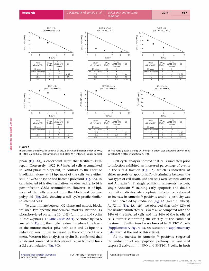
Effects of IR on dl922–947
To determine whether IR could enhance the oncolytic
activity of the mutant adenovirus dl922–947, ATC cell lines
FRO, BHT101-5, and Cal62 were infected and irradiated. To
identify most appropriate treatment sequence, cells were
infected and then irradiated after 24 h or vice versa. Cell
survival was evaluated 7 days after infection.
When cells were infected 24 h after irradiation, CI
showed a potent, statistically significant synergy of cell
killing in Cal62 and BHT101-5 cells at all combinations.
In FRO cells, using the same treatment schedule, a
slight synergic effect was observed in two combinations
http://erc.endocrinology-journals.org q 2013 Society for EndocrinologyDOI: 10.1530/ERC-13-0001 Printed in Great Britain
(CIZ0.95 for 2 Gy plus 7.5 MOIs and CIZ0.9 for 4 Gy plus
5 MOIs) whereas at 8 Gy plus 2 MOIs an additive effect
(CIZ1) was observed (Fig. 1, upper panels).
In contrast, an antagonist effect was observed in FRO
and Cal62 cells when infection was followed by irradi-
ation, whereas in BHT101-5 cells only 4 Gy combined with
2.5 MOIs showed synergy with this schedule (Fig. 1, lower
panels). These data demonstrate that the cytotoxicity of
dl922–947 is significantly enhanced when cells are
irradiated 24 h prior to infection.
Viral entry and viral replication analysis
It has been proposed that radiations could enhance
oncolytic activity by increasing viral entry in target cells
(Anders et al. 2003). To monitor this step, FRO and
BHT101-5 cells were irradiated and after 24 h infected
with a non-replicating reporter adenovirus transducing
GFP (AdGFP). After an additional 48 h, GFP emission was
evaluated by cytofluorimetric analysis. As shown in
Fig. 2A, irradiation neither increases the percentage of
GFP-positive cells nor the average green emission of
individual cells, except for the 2 Gy sample, where a
slight, albeit significant positive shift in the GFP fluor-
escence was observed in both cell lines.
Next, a real-time PCR assay was performed to evaluate
viral replication. FRO and BHT101-5 cells were irradiated
(2–4–5 Gy) and after 24 h infected with different MOIs
(1–5–10) of dl922–947. After an additional 48 h, viral
genome copies were evaluated by real-time PCR. This
analysis revealed that IR induced a significant (*P!0.05)
or highly significant (**P!0.005) dose-dependent increase
in viral replication (Fig. 2B and C, left panels). A TCID50
assay confirmed an increased viral production in
irradiated cells (Fig. 2B and C, right panels).
Analysis of cell cycle profile and cell death
It has been reported that drugs able to block cells in G2/M
or inhibit cytokinesis could enhance the effects of OVs
(Seidman et al. 2001). Irradiation is known to induce a
transitory block in G2 phase (Lisby et al. 2004); therefore,
we analyzed cell cycle profiles to evaluate differences in
cell cycle phases and timing of cell death. FRO and
BHT101-5 cells were irradiated (8 Gy) and, 24 h later,
infected with five or one MOIs of dl922–947 respectively.
Starting from 6 hpi, cells were collected, stained with PI,
and cell cycle analyzed by FACS.
At the time of the infection, that is 24 h after
irradiation (24hpIR), most of the cells were in G2/M
Published by Bioscientifica Ltd.
Downloaded from Bioscientifica.com at 02/16/2019 03:49:21PMvia free access
10
8
6
4
2dl92
2–94
7(M
OIs
)
dl92
2–94
7(M
OIs
)
0
10
8
6
4
2dl92
2–94
7(M
OIs
)
dl92
2–94
7(M
OIs
)
0 0
1
2
3
4
5
0 2 4
100248
0101098
1.21.31.6
1:2.51:1.64:1 8
42010
22.55
1.20.91.2
1:1.5 42108
689100
1.11.051.02
1:41:9
50
6 8 10IR(Gy)
10
8
6
4
2dl92
2–94
7(M
OIs
)
0
0 2 4 6 8IR(Gy)
0 2 4 6 8 10IR(Gy)
0 2
Ratio(Gy)/(MOIs)
EC50IR(Gy)
EC50dl922–947
(MOIs)CI
Ratio(Gy)/(MOIs)
EC50IR(Gy)
EC50dl922–947
(MOIs)CI
100
0107.552
0.950.91
248
10 80124
010642
0.730.650.7
051
0.50.1
0.40.50.82
0248
IR alonedl922–947 alone
1:3.75
1:51:1.25
1:1
Ratio(Gy)/(MOIs)
EC50IR(Gy)
EC50dl922–947
(MOIs)CI
Ratio(Gy)/(MOIs)
EC50IR(Gy)
EC50dl922–947
(MOIs)CI
Ratio(Gy)/(MOIs)
EC50IR(Gy)
EC50dl922–947
(MOIs)CI
Ratio(Gy)/(MOIs)
EC50IR(Gy)
EC50dl922–947
(MOIs)CI
IR alonedl922–947 alone
IR alonedl922–947 alone
IR alonedl922–947 alone
IR alonedl922–947 alone
IR alonedl922–947 alone
2:18:180:1
1:61:22:1
1:1.254:1
4 6 8 10IR(Gy)
10
8
6
4
2dl92
2–94
7(M
OIs
)
00 2 4 6 8
IR(Gy)
0
1
2
3
4
5
0 2 4 6 8 10IR(Gy)
FRO cells(IR dl922–947)
FRO cells(dl922–947 IR)
BHT101-5 cells(dl922–947 IR)
Ca162 cells(dl922–947 IR)
BHT101-5 cells(IR dl922–947)
Ca162 cells(IR dl922–947)
Figure 1
IR enhances the cytopathic effects of dl922–947. Combination index of FRO,
BHT101-5, and Cal62 cells irradiated and after 24 h infected (upper panels)
or vice versa (lower panels). A synergistic effect was observed only in cells
infected 24 h after irradiation (CI!1).
En
do
crin
e-R
ela
ted
Can
cer
Research C Passaro, A Abagnale et al. dl922–947 and ionizingradiation
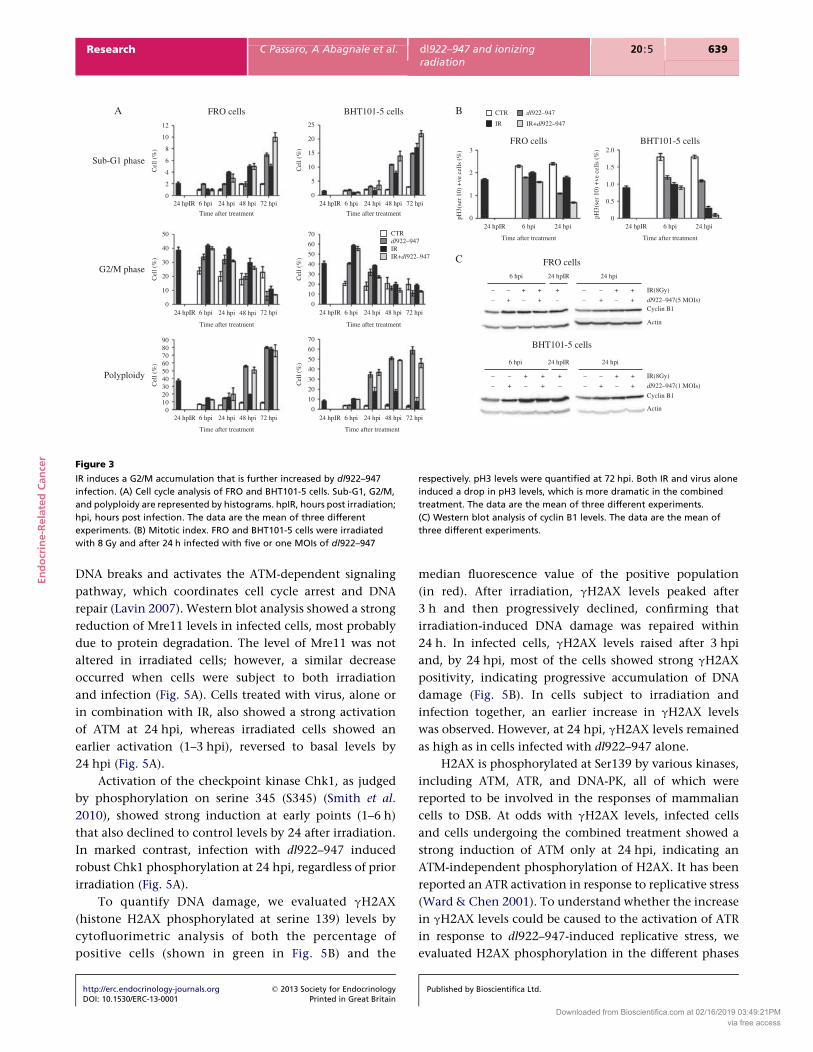
20 :5 637
phase (Fig. 3A), a checkpoint arrest that facilitates DNA
repair. Conversely, dl922–947-infected cells accumulated
in G2/M phase at 6 hpi but, in contrast to the effect of
irradiation alone, at 48 hpi most of the cells were either
still in G2/M phase or had become polyploid (Fig. 3A). In
cells infected 24 h after irradiation, we observed up to 24 h
post-infection G2/M accumulation. However, at 48 hpi,
most of the cells escaped from the block and become
polyploid (Fig. 3A), showing a cell cycle profile similar
to infected cells.
To discriminate between G2 phase and mitotic block,
we used two specific biochemical markers: histone H3
phosphorylated on serine 10 (pH3) for mitosis and cyclin
B1 for G2 phase (Lao-Sirieix et al. 2004). As shown by FACS
analysis in Fig. 3B, the single treatments reduced the levels
of the mitotic marker pH3 both at 6 and 24 hpi; this
reduction was further increased in the combined treat-
ment. Western blot analysis of cyclin B1 confirmed that
single and combined treatments induced in both cell lines
a G2 accumulation (Fig. 3C).
http://erc.endocrinology-journals.org q 2013 Society for EndocrinologyDOI: 10.1530/ERC-13-0001 Printed in Great Britain
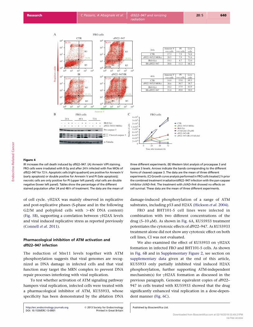
Cell cycle analysis showed that cells irradiated prior
to infection exhibited an increased percentage of events
in the subG1 fraction (Fig. 3A), which is indicative of
either necrosis or apoptosis. To discriminate between the
two types of cell death, unfixed cells were stained with PI
and Annexin V. PI single positivity represents necrosis,
single Annexin V staining early apoptosis and double
positivity indicates late apoptosis. Infected cells showed
an increase in Annexin V positivity and this positivity was
further increased by irradiation (Fig. 4A, green numbers).
At 72 hpi (Fig. 4A, left), we observed that only 12% of
the irradiated/infected cells were alive compared with the
24% of the infected cells and the 54% of the irradiated
cells, further confirming the efficacy of the combined
treatment. Similar trend was observed in BHT101-5 cells
(Supplementary Figure 1A, see section on supplementary
data given at the end of this article).
As the increase in Annexin V positivity suggested
the induction of an apoptotic pathway, we analyzed
caspase 3 activation in FRO and BHT101-5 cells. In both
Published by Bioscientifica Ltd.
Downloaded from Bioscientifica.com at 02/16/2019 03:49:21PMvia free access
C
Vir
al D
NA
cop
ies
(×10
5 )
Vir
al ti
ter
(pfu
/ml)
4.03.53.02.52.01.51.00.50.0 0
5.0 ×10⁶
1.0 ×10⁷
1.0 ×10⁷
2.0 ×10⁷
dl922–947 dl922–947/IR
IR(Gy)0
2
4
5
**
** **
**
**
**
**
**
*
BHT101-5 cells BHT101-5 cells
1 5 10
dl922–947(MOIs)
B
Vir
al D
NA
cop
ies
(×10
5 )
Vir
al ti
ter
(pfu
/ml)
10
8
6
4
2
01
**
FRO cells
**
**
FRO cells
3.0 ×10⁷
2.0 ×10⁷
1.0 ×10⁷
0dl922–947 dl922–947/IR
**
*
5 10
dl922–947(MOIs)
IR(Gy)0
2
4
5
100
A
75
50
25
GFP
pos
itive
cel
ls (
%)
00 2 4 8
1.5*
1.0
0.5
0.0
IR(Gy)
0 2 4 8
IR(Gy)
FRO cells
FACS analysis
BHT101-5 cells
GFP
em
issi
on(f
old
over
bas
al)
Figure 2
IR does not affect viral entry but enhances viral replication of dl922–947.
(A) FACS analysis of AdGFP-infected cells. FRO and BHTH101-5 were
irradiated (2–4–8 Gy) and after 24 h infected with AdGFP (25 MOIs).
The histogram on the left side shows the percentage of GFP-positive
cells whereas the mean GFP emission is reported on the right side. Both
parameters, with the exception of 2 Gy, were not increased in irradiated
cells. The data are the mean of three different experiments (*P!0.01).
(B and C) Replication of dl922–947 was measured by real-time PCR genome
equivalent analysis (left panels) and by TCID50 assay (right panels).
For real-time PCR analysis, FRO and BHTH101-5 cells were irradiated
(2–4–5 Gy) and 24 h later infected with dl922–947 (1–5–10 MOIs).
For TCID50 assay, FRO and BHT101-5 were irradiated (5 Gy) and after
24 h infected with five and one MOIs respectively. Irradiated cells showed
significant or highly significant differences in viral replication levels with
respect to non-irradiated cells. The data are the mean of three different
experiments (*P!0.05; **P!0.005).
En
do
crin
e-R
ela
ted
Can
cer
Research C Passaro, A Abagnale et al. dl922–947 and ionizingradiation
20 :5 638
cell lines, a decrease in procaspase 3 and an increase in
cleaved caspase 3 were observed in the combined
treatment with respect to single treatments (Fig. 4B and
Supplementary Figure 1B). To better understand the role of
caspases in the cell death mechanisms elicited by the
combined treatments, we performed a growth curve in
the presence of the pan-caspase inhibitor zVAD-fmk.
Addition of the drug 2 h prior IR/dl922–947 did not
modify cell proliferation, suggesting that other cell death
pathways are activated by the combined treatment
(Fig. 4C and Supplementary Figure 1C).
http://erc.endocrinology-journals.org q 2013 Society for EndocrinologyDOI: 10.1530/ERC-13-0001 Printed in Great Britain
DNA damage repair system and dl922–947 infection
IR induces DNA damage and, after its detection, cellular
pathways are activated to halt cell cycle progression
and repair the damage. Adenoviral replication also
induces DNA damage (Touchefeu et al. 2011); therefore,
we analyzed in FRO cells the effects of viral infection,
alone or in combination with radiation, on DNA damage
repair system.
The MRN complex (composed by Mre11-Rad50-Nbs1)
acts as a double-strand breaks (DSBs) sensor. It localizes to
Published by Bioscientifica Ltd.
Downloaded from Bioscientifica.com at 02/16/2019 03:49:21PMvia free access
12
10
8
6
4
2
0
50
40
30
20
10
0
9080706050403020100
70
60
50
40
30
20
10
0
70
60
50
40
30
20
10
0
25
20
15
10
5
0
Cel
l (%
)
Cel
l (%
)
Cel
l (%
)
Cel
l (%
)
Cel
l (%
)
Cel
l (%
)
24 hpIR 6 hpi 24 hpi 48 hpi 72 hpi
FRO cellsA B
C
FRO cells
FRO cells
–
– – –+ +
– + + –
– –+ +
– + +
–
– –+ +
– + +
+
–
– – –+ +
– + + +
BHT101-5 cells
BHT101-5 cells
BHT101-5 cells
Sub-G1 phase
G2/M phase
Polyploidy
Time after treatment
24 hpIR
24 hpIR
24 hpIR
6 hpi
6 hpi
6 hpi
24 hpi
24 hpi
24 hpi
24 hpIR6 hpi 24 hpi
48 hpi 72 hpi
Time after treatment
Time after treatment
24 hpIR 6 hpi 24 hpi
Time after treatment
24 hpIR 6 hpi 24 hpi 48 hpi 72 hpi
Time after treatment
24 hpIR 6 hpi 24 hpi 48 hpi 72 hpi
Time after treatment
24 hpIR 6 hpi 24 hpi 48 hpi 72 hpi
Time after treatment
24 hpIR 6 hpi 24 hpi 48 hpi 72 hpi
Time after treatment
3
2
1
0pH3(
ser
10)
+ve
cel
ls (
%)
pH3(
ser
10)
+ve
cel
ls (
%) 2.0
1.5
1.0
0.5
0
IR(8Gy)
dl922–947(1 MOIs)
Actin
Actin
Cyclin B1
IR(8Gy)
dl922–947(5 MOIs)Cyclin B1
CTR
IRIR+dl922–947
dl922–947
CTR
IR IR+dl922–947
dl922–947
Figure 3
IR induces a G2/M accumulation that is further increased by dl922–947
infection. (A) Cell cycle analysis of FRO and BHT101-5 cells. Sub-G1, G2/M,
and polyploidy are represented by histograms. hpIR, hours post irradiation;
hpi, hours post infection. The data are the mean of three different
experiments. (B) Mitotic index. FRO and BHT101-5 cells were irradiated
with 8 Gy and after 24 h infected with five or one MOIs of dl922–947
respectively. pH3 levels were quantified at 72 hpi. Both IR and virus alone
induced a drop in pH3 levels, which is more dramatic in the combined
treatment. The data are the mean of three different experiments.
(C) Western blot analysis of cyclin B1 levels. The data are the mean of
three different experiments.
En
do
crin
e-R
ela
ted
Can
cer
Research C Passaro, A Abagnale et al. dl922–947 and ionizingradiation
20 :5 639
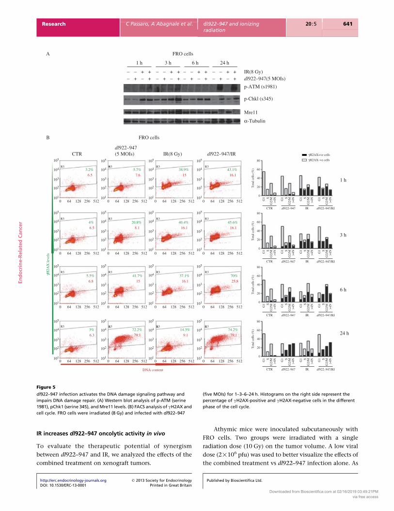
DNA breaks and activates the ATM-dependent signaling
pathway, which coordinates cell cycle arrest and DNA
repair (Lavin 2007). Western blot analysis showed a strong
reduction of Mre11 levels in infected cells, most probably
due to protein degradation. The level of Mre11 was not
altered in irradiated cells; however, a similar decrease
occurred when cells were subject to both irradiation
and infection (Fig. 5A). Cells treated with virus, alone or
in combination with IR, also showed a strong activation
of ATM at 24 hpi, whereas irradiated cells showed an
earlier activation (1–3 hpi), reversed to basal levels by
24 hpi (Fig. 5A).
Activation of the checkpoint kinase Chk1, as judged
by phosphorylation on serine 345 (S345) (Smith et al.
2010), showed strong induction at early points (1–6 h)
that also declined to control levels by 24 after irradiation.
In marked contrast, infection with dl922–947 induced
robust Chk1 phosphorylation at 24 hpi, regardless of prior
irradiation (Fig. 5A).
To quantify DNA damage, we evaluated gH2AX
(histone H2AX phosphorylated at serine 139) levels by
cytofluorimetric analysis of both the percentage of
positive cells (shown in green in Fig. 5B) and the
http://erc.endocrinology-journals.org q 2013 Society for EndocrinologyDOI: 10.1530/ERC-13-0001 Printed in Great Britain
median fluorescence value of the positive population
(in red). After irradiation, gH2AX levels peaked after
3 h and then progressively declined, confirming that
irradiation-induced DNA damage was repaired within
24 h. In infected cells, gH2AX levels raised after 3 hpi
and, by 24 hpi, most of the cells showed strong gH2AX
positivity, indicating progressive accumulation of DNA
damage (Fig. 5B). In cells subject to irradiation and
infection together, an earlier increase in gH2AX levels
was observed. However, at 24 hpi, gH2AX levels remained
as high as in cells infected with dl922–947 alone.
H2AX is phosphorylated at Ser139 by various kinases,
including ATM, ATR, and DNA-PK, all of which were
reported to be involved in the responses of mammalian
cells to DSB. At odds with gH2AX levels, infected cells
and cells undergoing the combined treatment showed a
strong induction of ATM only at 24 hpi, indicating an
ATM-independent phosphorylation of H2AX. It has been
reported an ATR activation in response to replicative stress
(Ward & Chen 2001). To understand whether the increase
in gH2AX levels could be caused to the activation of ATR
in response to dl922–947-induced replicative stress, we
evaluated H2AX phosphorylation in the different phases
Published by Bioscientifica Ltd.
Downloaded from Bioscientifica.com at 02/16/2019 03:49:21PMvia free access
104
103
10.3% 46.4%
9.2% 29.5%
41.0%12.1%
Annexin V
32.5%
Prop
idiu
m io
dide
47.2%
102
101
100
100 101 102 103 104 100 101 102 103 104
100 101 102 103 104 100 101 102 103 104
104
103
102
101
R324 h
Annexin V+ve cells
17.3 7.9 74.871.6
72.068.2
7.9
8.711.2
20.5
19.320.6
14.9
30.6
18.2
21.0
0 24
Num
ber
of c
ells
4 × 10⁵
3 × 10⁵
8 × 10⁴
6 × 10⁴
4 ×10⁴
2 × 10⁴
0
2 × 10⁵
1 × 1051.2×105
48 72
Time after treatment (h)
CTR
IR(8 Gy)zVAD-fmk (20 µM)
dl922–947(5 MOIs)
dl922–947+IRdl922–947+IR+zVAD-fmk
15.8
30.7
17.9
49.0
69.3
38.7
63.9
30.0
PI+ve cells
Livecells
Annexin V+ve cells
PI+ve cells
Livecells
CTRdl922–947(5 MOIs)
IR(8 Gy)IR+dl922–947
48 h
CTRdl922–947(5 MOIs)
dl922–947(5 MOIs)
IR(8 Gy)
IR(8 Gy)
24 h
––
– ++ – + – + – + – + – +
+ – – + + – – + +
48 h 72 h
Pro caspase 3
Cleaved caspase 3
α-Tubulin
IR+dl922–947
R3
R5
R5R5
R3
R455.4%
R3
R411.8%
R5 R4R480.5% 24.1%
100
104
103
102
101
100
104
103
102
101
100
CTR
IR
dl922–947
dl922–947/IR
FRO cellsA
B C FRO cellsFRO cells
Figure 4
IR increases the cell death induced by dl922–947. (A) Annexin V/PI staining.
FRO cells were irradiated with 8 Gy and after 24 h infected with five MOIs of
dl922–947 for 72 h. Apoptotic cells (right quadrant) are positive for Annexin V
(early apoptosis) or double positive for Annexin V and PI (late apoptosis);
necrotic cells are only positive for PI (upper left panel); vital cells are double
negative (lower left panel). Tables show the percentage of the different
stained population after 24 and 48 h of treatment. The data are the mean of
three different experiments. (B) Western blot analysis of procaspase 3 and
caspase 3 levels. Arrows indicate the bands corresponding to the different
forms of cleaved caspase 3. The data are the mean of three different
experiments. (C) Growth curve analysis performed in FRO cells treated 2 h prior
the combined treatment irradiation/dl922–947 infection with the pan-caspase
inhibitor zVAD-fmk. The treatment with zVAD-fmk showed no effects on
cell survival. These data are the mean of three different experiments.
En
do
crin
e-R
ela
ted
Can
cer
Research C Passaro, A Abagnale et al. dl922–947 and ionizingradiation
20 :5 640
of cell cycle. gH2AX was mainly observed in replicative
and post-replicative phases (S-phase and in the following
G2/M and polyploid cells with O4N DNA content)
(Fig. 5B), supporting a correlation between gH2AX levels
and viral induced replicative stress as reported previously
(Connell et al. 2011).
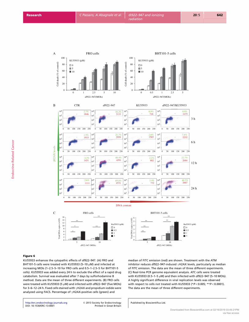
Pharmacological inhibition of ATM activation and
dl922–947 infection
The reduction of Mre11 levels together with ATM
phosphorylation suggests that viral genomes are recog-
nized as DNA damage in infected cells and that viral
function may target the MRN complex to prevent DNA
repair processes interfering with viral replication.
To test whether activation of ATM signaling pathway
hampers viral replication, infected cells were treated with
a pharmacological inhibitor of ATM, KU55933, whose
specificity has been demonstrated by the ablation DNA
http://erc.endocrinology-journals.org q 2013 Society for EndocrinologyDOI: 10.1530/ERC-13-0001 Printed in Great Britain
damage-induced phosphorylation of a range of ATM
substrates, including p53 and H2AX (Hickson et al. 2004).
FRO and BHT101-5 cell lines were infected in
combination with two different concentrations of the
drug (5–10 mM). As shown in Fig. 6A, KU55933 treatment
potentiates the cytotoxic effects of dl922–947. As KU55933
treatment alone did not show any cytotoxic effect on both
cell lines, CI was not evaluated.
We also examined the effect of KU55933 on gH2AX
formation in infected FRO and BHT101-5 cells. As shown
in Fig. 6B and in Supplementary Figure 2, see section on
supplementary data given at the end of this article,
KU55933 only partially inhibited viral induced H2AX
phosphorylation, further supporting ATM-independent
mechanism(s) for gH2AX formation as discussed in the
previous paragraph. Genome equivalent copies of dl922–
947 in cells treated with KU55933 showed that the drug
significantly enhanced viral replication in a dose-depen-
dent manner (Fig. 6C).
Published by Bioscientifica Ltd.
Downloaded from Bioscientifica.com at 02/16/2019 03:49:21PMvia free access
CTR
R3 R3 R3 R3
R3 R3 R3 R3
R3 R3 R3 R3
R3 R3 R3 R3
B
105
104
103
102
1010 64 128 256 512
105
104
103
102
1010 64 128 256 512
105
104
103
102
1010 64 128 256 512
105
104
103
102
1010 64 128 256 512
105
104
103
102
101
0 64 128 256 512
105
104
103
102
101
0 64 128 256 512
105
104
103
102
101
0 64 128 256 512
105
104
103
102
101
0 64 128 256 512
105
γH2A
X le
vels
3.2%
6.5
5.7%
7.6
38.9%
15
43.1%
16.1
45.6%
16.1
40.4%
16.1
20.8%
8.1
4%
6.5
5.5%
6.8
41.7%
15
37.1%
16.1
70%
25.8
74.2%
79.1
14.3%
9.1
72.2%
79.1
DNA content
3%
6.3
104
103
102
101
0 64 128 256 512
105
104
103
102
101
0 64 128 256 512
105
104
103
102
101
0 64 128 256 512
105
104
103
102
101
0 64 128 256 512
105
104
103
102
1010 64 128 256 512
105
104
103
102
1010 64 128 256 512
105
104
103
102
1010 64 128 256 512
105
104
103
102
1010 64 128 256 512
dl922–947(5 MOIs) dl922–947/IR
80
γH2AX+ve cells
γH2AX–ve cells
60
40 1 h
3 h
6 h
24 h
20Tot
al c
ells
(%
)
0
G1
CTR IRdl922–947 dl922–947/RI
G2/
M>
4N
S
G1
G2/
M>
4N
S
G1
G2/
M>
4N
S
G1
G2/
M>
4N
S
80
60
40
20Tot
al c
ells
(%
)
0
G1
CTR IRdl922–947 dl922–947/RI
G2/
M>
4N
S
G1
G2/
M>
4N
S
G1
G2/
M>
4N
S
G1
G2/
M>
4N
S
80
60
40
20Tot
al c
ells
(%
)
0
G1
CTR IRdl922–947 dl922–947/RI
G2/
M>
4N
S
G1
G2/
M>
4N
S
G1
G2/
M>
4N
S
G1
G2/
M>
4N
S80
60
40
20Tot
al c
ells
(%
)
0
G1
CTR IRdl922–947 dl922–947/RI
G2/
M>
4N
S
G1
G2/
M>
4N
S
G1
G2/
M>
4N
S
G1
G2/
M>
4N
S
IR(8 Gy)
FRO cells
1 h
– – – – – – – ––––––––– + + + + + + + +
+ IR(8 Gy)dl922–947(5 MOIs)
p-ATM (s1981)
p-Chkl (s345)
Mre11
A
α-Tubulin
+++++++
3 h
FRO cells
6 h 24 h
Figure 5
dl922–947 infection activates the DNA damage signaling pathway and
impairs DNA damage repair. (A) Western blot analysis of p-ATM (serine
1981), pChk1 (serine 345), and Mre11 levels. (B) FACS analysis of gH2AX and
cell cycle. FRO cells were irradiated (8 Gy) and infected with dl922–947
(five MOIs) for 1–3–6–24 h. Histograms on the right side represent the
percentage of gH2AX-positive and gH2AX-negative cells in the different
phase of the cell cycle.
En
do
crin
e-R
ela
ted
Can
cer
Research C Passaro, A Abagnale et al. dl922–947 and ionizingradiation
20 :5 641
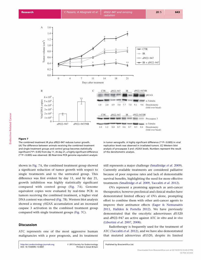
IR increases dl922–947 oncolytic activity in vivo
To evaluate the therapeutic potential of synergism
between dl922–947 and IR, we analyzed the effects of the
combined treatment on xenograft tumors.
http://erc.endocrinology-journals.org q 2013 Society for EndocrinologyDOI: 10.1530/ERC-13-0001 Printed in Great Britain
Athymic mice were inoculated subcutaneously with
FRO cells. Two groups were irradiated with a single
radiation dose (10 Gy) on the tumor volume. A low viral
dose (2!106 pfu) was used to better visualize the effects of
the combined treatment vs dl922–947 infection alone. As
Published by Bioscientifica Ltd.
Downloaded from Bioscientifica.com at 02/16/2019 03:49:21PMvia free access
100
80
60
40
20
0
100
80
60
40
20
0
Cel
l dea
th (
% o
f co
ntro
l)
Cel
l dea
th (
% o
f co
ntro
l)
KU55933 (µM)
B
C
CTR
FRO cells BHT101-5 cells
2.7% 16.2% 0.9% 0.9%
7.6%2.6%21.7%1.9%
3.2% 48.4% 2.2% 22.7%
40.2%3.4%69.0%2.6%
dl922–947 KU55933
3 h
6 h
12 h
24 h
DNA content
1846 2133 718 1475
2232156529591933
1431 4391 882 2684
266773245311111
dl922–947/KU55933
0510
0
104
103
102
101
0 50 100 150 200 250 0 50 100 150 200 250 0 50 100 150 200 250 0 50 100 150 200 250
0 50 100 150 200 250 0 50 100 150 200 250 0 50 100 150 200 250 0 50 100 150 200 250
0 50 100 150 200 250 0 50 100 150 200 250 0 50 100 150 200 250 0 50 100 150 200 250
0
16
14
12
10
8
6
4
2
05
*
*
**
**
**
**
**
**
**
**
10
Ku55933 (µM)
0
6
5
4
3
2
1
0
0.515
dl922–947 (MOIs)
5 10
dl922–947 (MOIs)
Vir
al D
NA
cop
ics
(×10
5)
Vir
al D
NA
cop
ics
(×10
5)
50 100 150 200 250 0 50 100 150 200 250 0 50 100 150 200 250 0 50 100 150 200 250
104
103
102
101
104
103
102
101
104
103
102
101
104
103
102
101
104
103
102
101
104
103
102
101
104
103
102
101
104
103
102
101
104
103
102
101
104
103
102
101
104
103
102
101
104
103
102
101
104
103
102
101
104
103
102
101
104
103
102
101
1 2.5
dl922–947(MOIs) dl922–947(MOIs)
5 10 0 0.5 1 2.5 5
γH2A
X le
vels
Ku55933 (µM)
00.515
KU55933 (µM)
0510
FRO cellsA BHT101-5 cells
Figure 6
KU55933 enhances the cytopathic effects of dl922–947. (A) FRO and
BHT101-5 cells were treated with KU55933 (5–10 mM) and infected at
increasing MOIs (1–2.5–5–10 for FRO cells and 0.5–1–2.5–5 for BHT101-5
cells). KU55933 was added every 24 h to exclude the effect of a rapid drug
catabolism. Survival was evaluated after 7 days by sulforhodamine B
method. Data are the mean of three different experiments. (B) FRO cells
were treated with KU55933 (5 mM) and infected with dl922–947 (five MOIs)
for 3–6–12–24 h. Fixed cells stained with gH2AX and propidium iodide were
analyzed using FACS. Percentage of gH2AX-positive cells (green) and
median of FITC emission (red) are shown. Treatment with the ATM
inhibitor reduces dl922–947-induced gH2AX levels, particularly as median
of FITC emission. The data are the mean of three different experiments.
(C) Real-time PCR genome equivalent analysis. ATC cells were treated
with KU55933 (0.5–1–5 mM) and then infected with dl922–947 (5–10 MOIs).
A highly significant difference in viral replication levels was observed
with respect to cells not treated with KU55933 (*P!0.005; **P!0.0001).
The data are the mean of three different experiments.
En
do
crin
e-R
ela
ted
Can
cer
Research C Passaro, A Abagnale et al. dl922–947 and ionizingradiation
20 :5 642
http://erc.endocrinology-journals.org q 2013 Society for EndocrinologyDOI: 10.1530/ERC-13-0001 Printed in Great Britain
Published by Bioscientifica Ltd.
Downloaded from Bioscientifica.com at 02/16/2019 03:49:21PMvia free access
1.6A
B C
1.4
1.2
1.0
0.8
0.6
0.4
0.2
Tum
or v
olum
e (c
m3 )
0.0
6 × 10⁴
5 × 10⁴
4 × 10⁴
3 × 10⁴
2 × 10⁴
1 ×10⁴
0
Vir
al D
NA
cop
ies
0 4 7 11 14 18 21
CTR
IR
****
* IR + dl922–947
dl922–947
dl922–947
dl922–947 dl922–947/NR
**
CTR
1.0
1.0 1.2 0.8 0.7 0.6 0.7 0.3 0.4
2.0 4.9 5.8 7.7 7.0 9.1 9.6
γH2AX
α-Tubulin
Procaspase 3
Densitometry(fold over basal)
α-Tubulin
Densitometry(fold over basal)
IR
dl922–947CTR IR
dl922–947/IR
dl922–947/IR
Days after treatment
Figure 7
The combined treatment IR plus dl922–947 reduces tumor growth.
(A) The difference between animals receiving the combined treatment
and single treatment groups and control group becomes statistically
significant (*P!0.05) from day 11. At day 21, a highly significant difference
(**P!0.005) was observed. (B) Real-time PCR genome equivalent analysis
in tumor xenografts. A highly significant difference (**P!0.005) in viral
replication levels was observed in irradiated tumors. (C) Western blot
analysis of procaspase 3 and gH2AX levels. Numbers represent the result
of the densitometric analysis.
En
do
crin
e-R
ela
ted
Can
cer
Research C Passaro, A Abagnale et al. dl922–947 and ionizingradiation
20 :5 643
shown in Fig. 7A, the combined treatment group showed
a significant reduction of tumor growth with respect to
single treatments and to the untreated group. This
difference was first evident by day 11, and by day 21,
growth inhibition was highly statistically significant
compared with control group (Fig. 7A). Genome
equivalent copies were evaluated by real-time PCR: in
tumors receiving the combined treatment, a higher viral
DNA content was observed (Fig. 7B). Western blot analysis
showed a strong gH2AX accumulation and an increased
caspase 3 activation in the combined treatment group
compared with single treatment groups (Fig. 7C).
Discussion
ATC represents one of the most aggressive human
malignancies with a poor prognosis, and its treatment
http://erc.endocrinology-journals.org q 2013 Society for EndocrinologyDOI: 10.1530/ERC-13-0001 Printed in Great Britain
still represents a major challenge (Smallridge et al. 2009).
Currently available treatments are considered palliative
because of poor response rates and lack of demonstrable
survival benefits, highlighting the need for more effective
treatments (Smallridge et al. 2009, Taccaliti et al. 2012).
OVs represent a promising approach as anti-cancer
therapeutics; however preclinical and clinical studies have
demonstrated limited efficacy of OVs alone, prompting
effort to combine them with other anti-cancer agents to
improve their antitumor effects (Eager & Nemunaitis
2011, Hallden & Portella 2012). We have previously
demonstrated that the oncolytic adenoviruses dl1520
and dl922–947 are active against ATC in vitro and in vivo
(Libertini et al. 2007, 2008).
Radiotherapy is frequently used for the treatment of
ATC (Taccaliti et al. 2012), and we have also demonstrated
that mutated adenovirus dl1520, despite its limited
Published by Bioscientifica Ltd.
Downloaded from Bioscientifica.com at 02/16/2019 03:49:21PMvia free access
En
do
crin
e-R
ela
ted
Can
cer
Research C Passaro, A Abagnale et al. dl922–947 and ionizingradiation
20 :5 644
efficacy, enhances the effects of IR against ATC cells and
xenograft tumors (Portella et al. 2003). Here, we have
analyzed the effects of a more potent second-generation
OV, dl922–947, in combination with IR. We show that
both the cytopathic activity and replication of dl922–947
are significantly enhanced by IR.
The potentiating effect of irradiation on the cyto-
pathic potential of OVs, other than dl922–947, has already
been demonstrated. However, optimal strategies for
combining these therapeutic modalities still remain to
be established as evidence suggests that they interact in
complex ways that vary in a cell line- or virus-dependent
fashion (Touchefeu et al. 2011). Here, we have identified
the most effective treatment sequence for ATC cells
showing that irradiation prior to dl922–947 infection
results in enhanced viral replication and synergistic cell
killing. Irradiation after viral infection did not show
synergistic or even additive effects.
It has been proposed that irradiation can increase viral
uptake (Zhang et al. 2003) either by enhancing coxsackie
and adenovirus receptor (CAR) and integrin expression
on cell membranes or by modulating the expression of
Dynamin 2, an intracellular protein involved in adeno-
viral internalization (Qian et al. 2005). In a study using a
telomerase-specific replication selective adenovirus (OBP-
301), viral infection was shown to result in radiosensitivity
in A459 small-cell lung cancer cells (Kuroda et al. 2010). In
the same study, a dose-dependent increase in adenoviral
uptake and coxsackie adenovirus receptor expression was
also observed.
By infecting the cells with an AdGFP virus, we
evaluated the effects of IR on viral uptake on ATC cells.
In contrast to previous studies, we observed only very
modest effects of IR on viral uptake. At 2 Gy, a small
increase was observed, whereas higher radiation doses do
not significantly modify viral uptake. Moreover, CAR
receptor levels were not modified by IR (data not
shown). Differences in treatment sequence, virus, and
cell lines may explain the discrepancies with other studies.
Despite this, we did observe a significant increase in
viral replication in irradiated cells. Existing data regarding
the effect of radiation on viral replication are conflicting.
Enhanced viral replication following IR has been reported
in some studies (Liu et al. 2007, Bieler et al. 2008), but not
in others (Lamfers et al. 2002, Geoerger et al. 2003),
suggesting that increased viral replication is not an
absolute prerequisite for enhanced cell killing.
Subversion of the host cell cycle is an important
feature of adenoviral infection. The expression of E1A
gene products drives an unscheduled DNA synthesis
http://erc.endocrinology-journals.org q 2013 Society for EndocrinologyDOI: 10.1530/ERC-13-0001 Printed in Great Britain
followed by an abortive cytokinesis, inducing G2 accumu-
lation and O4N DNA content (Davy & Doorbar 2007). The
potential benefit of the G2 accumulation is represented
by the pseudo S phase that maintains host cells in a
replicative state (Davy & Doorbar 2007, Nichols et al.
2009). It has been proposed that drugs blocking cells in G2
phase could enhance the effects of OVs. Indeed, we have
demonstrated that the inhibitor of the mitotic kinase
Aurora B, AZD1152, enhances the effects of dl922–947 by
inducing a G2 arrest (Libertini et al. 2011). Accordingly,
the accumulation in G2 phase induced by radiation likely
enhances the cytopathic effects of dl922–947. This
hypothesis is further supported by the observation that
infection followed by irradiation did not potentiate cell
killing or viral replication (data not shown).
We have analyzed the effects of the combined
treatment, showing an increase in number of cells in
sub-G1 phase and a sharp reduction in the number of vital
cells with respect to single treatments. Infection alone also
triggered similar effects, although with lesser efficiency. In
the present work, we observed that dl922–947, alone or
in combination with IR, induced phosphatidylserine
exposure and caspase 3 cleavage, suggesting the activation
of apoptosis. However, the treatment with the caspase
inhibitor zVAD-fmk was not able to restore cell prolifer-
ation, supporting the involvement of other cell death
pathways rather than the classical apoptosis. Our results
are in agreement with the previous reports suggesting
alternative cell death mechanisms induced by dl922–947
(Abou El Hassan et al. 2004, Baird et al. 2008, Libertini
et al. 2011).
It is known that the cells respond to adenovirus
infection by activating a DNA damage response (Lilley
et al. 2007, Nichols et al. 2009, Touchefeu et al. 2011);
indeed, it has been proposed that the prolonged S-phase
induced by the virus could resemble the cellular environ-
ment occurring in response to replication-associated DNA
damage (Connell et al. 2011). Also, the linear adenovirus
genome has double-stranded DNA termini that could
represent targets for cellular DSB repair pathways. IR itself
induces DNA damage; therefore, we analyzed the effects of
the combined treatment on key components of DSB
pathway. In ATC-irradiated cells, DNA damage is correctly
repaired, as demonstrated by the progressive reduction
toward basal levels of gH2AX at 24 h after irradiation.
Conversely, upon infection with dl922–947, an inefficient
DNA damage response was observed. Indeed, despite the
expected ATM and Chk1 activation, gH2AX accumulation
was still revealed at 24 hpi, indicating the presence of an
unrepaired DNA. The strong reduction of Mre11 levels
Published by Bioscientifica Ltd.
Downloaded from Bioscientifica.com at 02/16/2019 03:49:21PMvia free access
En
do
crin
e-R
ela
ted
Can
cer
Research C Passaro, A Abagnale et al. dl922–947 and ionizingradiation
20 :5 645
observed 24 h after the infection, likely due to degradation
induced by viral proteins (Nichols et al. 2009), and
the viral induced replicative stress could explain the
accumulation of an unrepaired DNA. A similar inadequate
DNA damage response was reported in cells subject to
irradiation prior to infection. However, a more rapid
increase in gH2AX-positive cells was seen, probably due to
IR-induced DNA damage combined with that induced by
viral replication.
A strong activation of ATM was observed only at
24 hpi, whereas gH2AX levels rose already at 3 hpi,
suggesting a ATM-independent phosphorylation of
H2AX as previously reported (Ward & Chen 2001, Nichols
et al. 2009). H2AX is phosphorylated by various kinases
other than ATM, including ATR that activates Chk1. Chk1
is phosphorylated as early as 3 hpi, indicating a temporal
correlation between gH2AX accumulation and ATR–Chk1
activation. This observation is in agreement with previous
studies showing a robust induction of ATR–Chk1 pathway
upon infection with wild-type and mutant adenoviruses,
including dl922–947 (Connell et al. 2011). Moreover, our
data suggest a correlation with the replicative stress
induced by the virus, as gH2AX is mainly observed in
replicative and post-replicative phases.
During the infection with the telomerase-driven
oncolytic adenovirus OBP301, the degradation of MRN
complex was accompanied by greatly reduced levels of
ATM activation (Kuroda et al. 2010). It is possible that
dl922–947 infection is not able to fully suppress the early
events in DNA damage signaling, as MRN complex
degradation may not in itself be sufficient to preclude/
reduce ATM and ATR kinase activation, as previously
reported (Nichols et al. 2009). Several reports indicate that
ATM activation counterbalances viral infection (Nichols
et al. 2009); therefore, we postulated that ATM activation
could be detrimental for viral infection/replication. The
increased cell death observed in cells infected in the
presence of the specific ATM inhibitor KU55933
confirmed this hypothesis.
It has been shown that the block of ATR–Chk1
pathways augments dl922–947 cytotoxicity (Connell
et al. 2011). In this study, we demonstrated that also
ATM inhibition could represent an approach to increase
dl922–947 cytotoxicity. Our data, together with previous
studies, indicate that the DNA damage response pathway
acts as an intrinsic cellular defense against the virus
and could be targeted to potentiate the effects of oncolytic
adenovirus.
Finally, we confirmed the therapeutic potential of
combining irradiation with dl922–947 infection using
http://erc.endocrinology-journals.org q 2013 Society for EndocrinologyDOI: 10.1530/ERC-13-0001 Printed in Great Britain
xenografts formed from ATC cells. Although the s.c.
implant of neoplastic cells do not fully reproduce the
stromal response and the pattern of spread of the neoplastic
cells to lymph nodes and other metastatic sites as in
orthotopic models, the data obtained clearly demonstrate
the efficacy of the combination therapy in vivo.
A majority of patients with ATC die from aggressive
local regional disease, primarily from upper airway
respiratory obstruction (Taccaliti et al. 2012). For this
reason, radiotherapy has been used to control local
growth, evolving from palliation on to preoperative
or/and postoperative therapy to prolong survival.
Given the importance of radiotherapy in the control
of ATC, our results suggest that dl922–947 could be
combined with this modality in order to improve local
control and offer the possibilities of new clinical trials for
this intractable disease.
Supplementary data
This is linked to the online version of the paper at http://dx.doi.org/10.1530/
ERC-13-0001.
Declaration of interest
The authors declare that there is no conflict of interest that could be
perceived as prejudicing the impartiality of the research reported.
Funding
This study was supported by the Associazione Italiana per la Ricerca sul
Cancro (AIRC). S Libertini is a Marie Curie fellow.
Acknowledgements
The authors thank Salvatore Sequino for his excellent technical assistance.
References
Abou El Hassan MA, van der Meulen-Muileman I, Abbas S & Kruyt FA 2004
Conditionally replicating adenoviruses kill tumor cells via a basic
apoptotic machinery-independent mechanism that resembles necrosis-
like programmed cell death. Journal of Virology 78 12243–12251.
(doi:10.1128/JVI.78.22.12243-12251.2004)
Anders M, Christian C, McMahon M, McCormick F & Korn WM 2003
Inhibition of the Raf/MEK/ERK pathway up-regulates expression of the
coxsackievirus and adenovirus receptor in cancer cells. Cancer Research
63 2088–2095.
Baird SK, Aerts JL, Eddaoudi A, Lockley M, Lemoine NR & McNeish IA 2008
Oncolytic adenoviral mutants induce a novel mode of programmed cell
death in ovarian cancer. Oncogene 27 3081–3090. (doi:10.1038/sj.onc.
1210977)
Bhattacharyya M, Francis J, Eddouadi A, Lemoine NR & Hallden G 2011 An
oncolytic adenovirus defective in pRb-binding (dl922–947) can
efficiently eliminate pancreatic cancer cells and tumors in vivo in
Published by Bioscientifica Ltd.
Downloaded from Bioscientifica.com at 02/16/2019 03:49:21PMvia free access
En
do
crin
e-R
ela
ted
Can
cer
Research C Passaro, A Abagnale et al. dl922–947 and ionizingradiation
20 :5 646
combination with 5-FU or gemcitabine. Cancer Gene Therapy 18
734–743. (doi:10.1038/cgt.2011.45)
Bieler A, Mantwill K, Holzmuller R, Jurchott K, Kaszubiak A, Stark S,
Glockzin G, Lage H, Grosu AL, Gansbacher B et al. 2008 Impact of
radiation therapy on the oncolytic adenovirus dl520: implications on
the treatment of glioblastoma. Radiotherapy and Oncology 86 419–427.
(doi:10.1016/j.radonc.2007.10.009)
Botta G, Perruolo G, Libertini S, Cassese A, Abagnale A, Beguinot F,
Formisano P & Portella G 2010 PED/PEA-15 modulates coxsackievirus-
adenovirus receptor expression and adenoviral infectivity via
ERK-mediated signals in glioma cells. Human Gene Therapy 21
1067–1076. (doi:10.1089/hum.2009.181)
Botta G, Passaro C, Libertini S, Abagnale A, Barbato S, Maione AS,
Hallden G, Beguinot F, Formisano P & Portella G 2012 Inhibition of
autophagy enhances the effects of E1A-defective oncolytic adenovirus
dl922–947 against glioma cells in vitro and in vivo. Human Gene Therapy
23 623–634. (doi:10.1089/hum.2011.120)
Cheong SC, Wang Y, Meng JH, Hill R, Sweeney K, Kirn D, Lemoine NR &
Hallden G 2008 E1A-expressing adenoviral E3B mutants act synergis-
tically with chemotherapeutics in immunocompetent tumor models.
Cancer Gene Therapy 15 40–50. (doi:10.1038/sj.cgt.7701099)
Connell CM, Shibata A, Tookman LA, Archibald KM, Flak MB, Pirlo KJ,
Lockley M, Wheatley SP & McNeish IA 2011 Genomic DNA damage and
ATR–Chk1 signaling determine oncolytic adenoviral efficacy in human
ovarian cancer cells. Journal of Clinical Investigation 121 1283–1297.
(doi:10.1172/JCI43976)
Davy C & Doorbar J 2007 G2/M cell cycle arrest in the life cycle of viruses.
Virology 368 219–226. (doi:10.1016/j.virol.2007.05.043)
Eager RM & Nemunaitis J 2011 Clinical development directions in
oncolytic viral therapy. Cancer Gene Therapy 18 305–317. (doi:10.1038/
cgt.2011.7)
Geoerger B, Grill J, Opolon P, Morizet J, Aubert G, Lecluse Y,
van Beusechem VW, Gerritsen WR, Kirn DH & Vassal G 2003
Potentiation of radiation therapy by the oncolytic adenovirus dl1520
(ONYX-015) in human malignant glioma xenografts. British Journal of
Cancer 89 577–584. (doi:10.1038/sj.bjc.6601102)
Hallden G & Portella G 2012 Oncolytic virotherapy with modified
adenoviruses and novel therapeutic targets. Expert Opinion on
Therapeutic Targets 16 945–958. (doi:10.1517/14728222.2012.712962)
Heise C, Sampson-Johannes A, Williams A, McCormick F, Von Hoff DD &
Kirn DH 1997 ONYX-015, an E1B gene-attenuated adenovirus, causes
tumor-specific cytolysis and antitumoral efficacy that can be augmen-
ted by standard chemotherapeutic agents. Nature Medicine 3 639–645.
(doi:10.1038/nm0697-639)
Heise C, Hermiston T, Johnson L, Brooks G, Sampson-Johannes A,
Williams A, Hawkins L & Kirn D 2000a An adenovirus E1A mutant that
demonstrates potent and selective systemic anti-tumoral efficacy.
Nature Medicine 6 1134–1139. (doi:10.1038/80474)
Heise C, Lemmon M & Kirn D 2000b Efficacy with a replication-selective
adenovirus plus cisplatin-based chemotherapy: dependence on
sequencing but not p53 functional status or route of administration.
Clinical Cancer Research 6 4908–4914.
Hickson I, Zhao Y, Richardson CJ, Green SJ, Martin NM, Orr AI, Reaper PM,
Jackson SP, Curtin NJ & Smith GC 2004 Identification and character-
ization of a novel and specific inhibitor of the ataxia-telangiectasia
mutated kinase ATM. Cancer Research 64 9152–9159. (doi:10.1158/
0008-5472.CAN-04-2727)
Immonen A, Vapalahti M, Tyynela K, Hurskainen H, Sandmair A,
Vanninen R, Langford G, Murray N & Yla-Herttuala S 2004 AdvHSV-tk
gene therapy with intravenous ganciclovir improves survival in human
malignant glioma: a randomised, controlled study. Molecular Therapy
10 967–972. (doi:10.1016/j.ymthe.2004.08.002)
Jiang H, Gomez-Manzano C, Lang FF, Alemany R & Fueyo J 2009 Oncolytic
adenovirus: preclinical and clinical studies in patients with human
malignant gliomas. Current Gene Therapy 9 422–427. (doi:10.2174/
156652309789753356)
http://erc.endocrinology-journals.org q 2013 Society for EndocrinologyDOI: 10.1530/ERC-13-0001 Printed in Great Britain
Kuroda S, Fujiwara T, Shirakawa Y, Yamasaki Y, Yano S, Uno F, Tazawa H,
Hashimoto Y, Watanabe Y, Noma K et al. 2010 Telomerase-dependent
oncolytic adenovirus sensitizes human cancer cells to ionizing
radiation via inhibition of DNA repair machinery. Cancer Research 70
9339–9348. (doi:10.1158/0008-5472.CAN-10-2333)
Lamfers ML, Grill J, Dirven CM, Van Beusechem VW, Geoerger B, Van Den
Berg J, Alemany R, Fueyo J, Curiel DT, Vassal G et al. 2002 Potential of
the conditionally replicative adenovirus Ad5-Delta24RGD in the
treatment of malignant gliomas and its enhanced effect with
radiotherapy. Cancer Research 62 5736–5742.
Lao-Sirieix P, Brais R, Lovat L, Coleman N & Fitzgerald RC 2004 Cell cycle
phase abnormalities do not account for disordered proliferation in
Barrett’s carcinogenesis. Neoplasia 6 751–760. (doi:10.1593/neo.04280)
Lavin MF 2007 ATM and the Mre11 complex combine to recognize
and signal DNA double-strand breaks. Oncogene 26 7749–7758.
(doi:10.1038/sj.onc.1210880)
Libertini S, Iacuzzo I, Ferraro A, Vitale M, Bifulco M, Fusco A & Portella G
2007 Lovastatin enhances the replication of the oncolytic adenovirus
dl1520 and its antineoplastic activity against anaplastic thyroid
carcinoma cells. Endocrinology 148 5186–5194. (doi:10.1210/
en.2007-0752)
Libertini S, Iacuzzo I, Perruolo G, Scala S, Ierano C, Franco R, Hallden G &
Portella G 2008 Bevacizumab increases viral distribution in human
anaplastic thyroid carcinoma xenografts and enhances the effects of
E1A-defective adenovirus dl922–947. Clinical Cancer Research 14
6505–6514. (doi:10.1158/1078-0432.CCR-08-0200)
Libertini S, Abagnale A, Passaro C, Botta G, Barbato S, Chieffi P & Portella G
2011 AZD1152 negatively affects the growth of anaplastic thyroid
carcinoma cells and enhances the effects of oncolytic virus dl922–947.
Endocrine-Related Cancer 18 129–141. (doi:10.1677/ERC-10-0234)
Lilley CE, Schwartz RA & Weitzman MD 2007 Using or abusing: viruses and
the cellular DNA damage response. Trends in Microbiology 15 119–126.
(doi:10.1016/j.tim.2007.01.003)
Lin SF, Gao SP, Price DL, Li S, Chou TC, Singh P, Huang YY, Fong Y &
Wong RJ 2008a Synergy of a herpes oncolytic virus and paclitaxel for
anaplastic thyroid cancer. Clinical Cancer Research 14 1519–1528.
(doi:10.1158/1078-0432.CCR-07-4628)
Lin SF, Price DL, Chen CH, Brader P, Li S, Gonzalez L, Zhang Q, Yu YA,
Chen N, Szalay AA et al. 2008b Oncolytic vaccinia virotherapy of
anaplastic thyroid cancer in vivo. Journal of Clinical Endocrinology and
Metabolism 93 4403–4407. (doi:10.1210/jc.2008-0316)
Lisby M, Barlow JH, Burgess RC & Rothstein R 2004 Choreography of the
DNA damage response: spatiotemporal relationships among check-
point and repair proteins. Cell 118 699–713. (doi:10.1016/j.cell.2004.
08.015)
Liu C, Sarkaria JN, Petell CA, Paraskevakou G, Zollman PJ, Schroeder M,
Carlson B, Decker PA, Wu W, James CD et al. 2007 Combination of
measles virus virotherapy and radiation therapy has synergistic activity
in the treatment of glioblastoma multiforme. Clinical Cancer Research
13 7155–7165. (doi:10.1158/1078-0432.CCR-07-1306)
Lockley M, Fernandez M, Wang Y, Li NF, Conroy S, Lemoine N & McNeish I
2006 Activity of the adenoviral E1A deletion mutant dl922–947 in
ovarian cancer: comparison with E1A wild-type viruses, biolumines-
cence monitoring, and intraperitoneal delivery in icodextrin. Cancer
Research 66 989–998. (doi:10.1158/0008-5472.CAN-05-2691)
Nichols GJ, Schaack J & Ornelles DA 2009 Widespread phosphorylation
of histone H2AX by species C adenovirus infection requires viral DNA
replication. Journal of Virology 83 5987–5998. (doi:10.1128/
JVI.00091-09)
Oberg D, Yanover E, Adam V, Sweeney K, Costas C, Lemoine NR &
Hallden G 2010 Improved potency and selectivity of an oncolytic
E1ACR2 and E1B19K deleted adenoviral mutant in prostate and
pancreatic cancers. Clinical Cancer Research 16 541–553. (doi:10.1158/
1078-0432.CCR-09-1960)
O’Shea CC, Johnson L, Bagus B, Choi S, Nicholas C, Shen A, Boyle L,
Pandey K, Soria C, Kunich J et al. 2004 Late viral RNA export, rather
Published by Bioscientifica Ltd.
Downloaded from Bioscientifica.com at 02/16/2019 03:49:21PMvia free access
En
do
crin
e-R
ela
ted
Can
cer
Research C Passaro, A Abagnale et al. dl922–947 and ionizingradiation
20 :5 647
than p53 inactivation, determines ONYX-015 tumor selectivity. Cancer
Cell 6 611–623. (doi:10.1016/j.ccr.2004.11.012)
Portella G, Scala S, Vitagliano D, Vecchio G & Fusco A 2002 ONYX-015, an
E1B gene-defective adenovirus, induces cell death in human anaplastic
thyroid carcinoma cell lines. Journal of Clinical Endocrinology and
Metabolism 87 2525–2531. (doi:10.1210/jc.87.6.2525)
Portella G, Pacelli R, Libertini S, Cella L, Vecchio G, Salvatore M & Fusco A
2003 ONYX-015 enhances radiation-induced death of human
anaplastic thyroid carcinoma cells. Journal of Clinical Endocrinology
and Metabolism 88 5027–5032. (doi:10.1210/jc.2003-030385)
Qian J, Yang J, Dragovic AF, Abu-Isa E, Lawrence TS & Zhang M 2005
Ionizing radiation-induced adenovirus infection is mediated by
Dynamin 2. Cancer Research 65 5493–5497. (doi:10.1158/0008-5472.
CAN-04-4526)
Radhakrishnan S, Miranda E, Ekblad M, Holford A, Pizarro MT, Lemoine NR
& Hallden G 2010 Efficacy of oncolytic mutants targeting pRb and p53
pathways is synergistically enhanced when combined with cytotoxic
drugs in prostate cancer cells and tumor xenografts. Human Gene
Therapy 21 1311–1325. (doi:10.1089/hum.2010.019)
Schweppe RE, Klopper JP, Korch C, Pugazhenthi U, Benezra M, Knauf JA,
Fagin JA, Marlow LA, Copland JA, Smallridge RC et al. 2008
Deoxyribonucleic acid profiling analysis of 40 human thyroid cancer
cell lines reveals cross-contamination resulting in cell line redundancy
and misidentification. Journal of Clinical Endocrinology and Metabolism
93 4331–4341. (doi:10.1210/jc.2008-1102)
Seidman MA, Hogan SM, Wendland RL, Worgall S, Crystal RG &
Leopold PL 2001 Variation in adenovirus receptor expression and
adenovirus vector-mediated transgene expression at defined stages of
the cell cycle. Molecular Therapy 4 13–21. (doi:10.1006/mthe.2001.
0414)
http://erc.endocrinology-journals.org q 2013 Society for EndocrinologyDOI: 10.1530/ERC-13-0001 Printed in Great Britain
Sherr CJ & McCormick F 2002 The RB and p53 pathways in cancer.
Cancer Cell 2 103–112. (doi:10.1016/S1535-6108(02)00102-2)
Smallridge RC, Marlow LA & Copland JA 2009 Anaplastic thyroid cancer:
molecular pathogenesis and emerging therapies. Endocrine-Related
Cancer 16 17–44. (doi:10.1677/ERC-08-0154)
Smith J, Tho LM, Xu N & Gillespie DA 2010 The ATM–Chk2 and ATR–Chk1
pathways in DNA damage signaling and cancer. Advances in Cancer
Research 108 73–112. (doi:10.1016/B978-0-12-380888-2.00003-0)
Taccaliti A, Silvetti F, Palmonella G & Boscaro M 2012 Anaplastic thyroid
carcinoma.Frontiers inEndocrinology384. (doi:10.3389/fendo.2012.00084)
Tallarida RJ 2001 Drug synergism: its detection and applications. Journal of
Pharmacological and Experimental Therapeutics 298 865–872.
Touchefeu Y, Vassaux G & Harrington KJ 2011 Oncolytic viruses
in radiation oncology. Radiotherapy and Oncology 99 262–270.
(doi:10.1016/j.radonc.2011.05.078)
Vichai V & Kirtikara K 2006 Sulforhodamine B colorimetric assay for
cytotoxicity screening. Nature Protocols 1 1112–1116. (doi:10.1038/
nprot.2006.179)
Ward IM & Chen J 2001 Histone H2AX is phosphorylated in an
ATR-dependent manner in response to replicational stress.
Journal of Biological Chemistry 276 47759–47762. (doi:10.1074/jbc.
M009785200)
Yong RL, Shinojima N, Fueyo J, Gumin J, Vecil GG, Marini FC, Bogler O,
Andreeff M & Lang FF 2009 Human bone marrow-derived mesenchy-
mal stem cells for intravascular delivery of oncolytic adenovirus
Delta24-RGD to human gliomas. Cancer Research 69 8932–8940.
(doi:10.1158/0008-5472.CAN-08-3873)
Zhang M, Li S, Li J, Ensminger WD & Lawrence TS 2003 Ionizing radiation
increases adenovirus uptake and improves transgene expression in
intrahepatic colon cancer xenografts. Molecular Therapy 8 21–28.
(doi:10.1016/S1525-0016(03)00143-6)
Received in final form 17 June 2013Accepted 8 July 2013Made available online as an Accepted Preprint9 July 2013
Published by Bioscientifica Ltd.
Downloaded from Bioscientifica.com at 02/16/2019 03:49:21PMvia free access