ions released from metal-on-metal hip implantsdigitool.library.mcgill.ca/thesisfile86703.pdf ·...
TRANSCRIPT

2
Ions Released from Metal-on-Metal Hip
Implants:
In Vitro and in Vivo Investigations
Cathy Mélanie Tkaczyk, M.Sc.
Biomedical Engineering Department
McGill University,
Montréal, Canada
A thesis submitted to McGill University, Faculty of Graduate and
Postdoctoral Studies, in partial fulfillment of the requirements of the
degree of doctor of philosophy.
Copyright © Cathy Tkaczyk 2009

3
A mon conjoint Lilian et à ma fille Athénais Clara Constance J’ignorais que je pouvais tant aimer
4
ABSTRACT
Studies have shown that Co and Cr particles and ions can enter the
bloodstream and accumulate in tissues and organs of patients after metal-on-metal
(MM) total hip arthroplasty (THA). These ions can generate reactive oxygen
species (ROS) that can be deleterious for cells. We first assessed the biological
effects of Cr(VI), Co(II), and Cr(III) by testing their effect on antioxidant
enzymes (SODs, CAT, GPx, HO-1) that represent a primary defense system
against ROS. We demonstrated that Cr(VI) induced the protein expression
(translation) of antioxidant enzymes, whereas it had no effect on the mRNA
expression (transcription). Co(II) induced the expression of both protein and
mRNA of HO-1 only. Cr(III) had no effect on the activity of these enzymes. We
then suggested that a difference in molecular structure may be at the origin of
their differential effects and showed that Cr(III) can form precipitable complexes,
whereas Co(II) and Cr(VI) cannot form complexes in the same experimental
conditions. These Cr(III) complexes, formed in simulated-physiological fluids,
were constituted by an organic phase (amino acids, phosphate) tangled with an
inorganic phase (Cr, Ca, Na). Interestingly, these Cr(III) complexes interacted
only with albumin in presence of fetal bovine serum, whereas they interacted with
8 different human serum proteins in presence of human serum. The interaction of
Cr(III) complexes with serum proteins affect their internalization by
macrophages, complexes formed with human serum being more easily
internalized than those bound to bovine proteins. Lastly, results suggested that the
5
levels of Co and Cr ions in patients with MM THA are not sufficient to induce
significant oxidative stress in the blood of these patients, bringing optimism over
concern for the long term biological effects of Co and Cr ions released from
metal-metal bearings. In conclusion, this thesis gives very valuable information
on the biological effects of Cr and Co ions and gives insight into Cr metabolism.

6
RESUME
De nombreuses études ont montré que les particules et ions métalliques
(Cr(III), Co(II) et Cr(VI)) générés par l’usure de prothèses de hanche métalliques
(PHM) se retrouvent dans le flux sanguin des patients et s’accumulent également
dans leurs tissus et leurs organes. Ces ions peuvent génèrer des radicaux libres qui
peuvent à leur tour être nocifs pour la cellule. Nous avons déterminé les effets de
ces ions sur les enzymes antioxidantes (SODs, CAT, GPx, HO-1) qui représentent
la première ligne de défense cellulaire contre les radicaux libres. Nos travaux ont
révélé que le Cr(VI) induit l’expression de ces protéines (traduction) mais non
l’expression de leur ARNm (transcription). Le Co(II) induit l’expression de la
proteine et de l’ARNm de HO-1 seulement alors que le Cr(III) n’a aucun effet sur
l’expression de ces enzymes. Nous avons par la suite suggéré, que la structure
moléculaire de ces ions pouvait avoir une influence sur leurs différents effets. Les
résultats ont montré que le Cr(III) formait des complexes constitués d’une partie
organique (acides aminés et phosphates) et d’une partie inorganique (Cr, calcium
et sodium), alors que ni le Co(II), ni le Cr(VI) ne formaient de complexes dans les
mêmes conditions expérimentales. En présence de sérum bovin fétal, les
complexes de Cr(III) pouvent se lier seulement à l’albumine, alors qu’en présence
de sérum humain, ces mêmes complexes intéragissent avec 8 protéines de nature
differente. Cette intéraction avec les protéines humaines semble augmenter
l’internalisation des complexes par les macrophages. Finalement, les résultats
démontrent que la concentration d’ions Co et Cr présents dans le sang de patients

7
portant une PHM est insuffisante pour induire un stress oxydant et apportent un
souffle optimiste sur les effets à long terme des ions métalliques chez les patients.
Dans son ensemble ce projet apporte de nouveaux éléments de connaissances sur
les effets biologiques des ions Co et Cr et sur le métabolisme du Cr.

8
REMERCIEMENTS
Tout d’abord, je tiens à remercier le Dr Maryam Tabrizian du
Département de Génie Biomédical de l’Université McGill et le Dr Olga Huk du
Département de Chirurgie Orthopédique de l’Hôpital Général Juif, de m’avoir
acceptée au sein de leur laboratoire, de m’avoir fourni un excellent environnement
pour ma recherche doctorale et de m’avoir permis de présenter mes travaux lors
de conférences internationales.
Je suis extrêmement reconnaissante envers le Dr Maryam Tabrizian de
m’avoir donné l’opportunité d’élaborer mon projet, guidée par son sens
scientifique, ses conseils avertis et ses encouragements afin de travailler sur les
thèmes que j’ai jugés importants. J’ai pu mettre à l’épreuve mon sens de
l’initiative, perfectionner mon autonomie et acquérir un grand sens de l’adaptation
inhérents à ce parcours. C’est grâce au Dr Tabrizian que j’ai pu ainsi travailler sur
le projet multi-disciplinaire qui m’importait tant. Je la remercie également
profondément pour sa grande gentillesse et sa compréhension. Merci pour tout
Maryam, tu es mon modèle de réussite.
Je suis extrêmement reconnaissante envers Le Dr Olga Huk d’avoir été ma
co-superviseur, autant disponible qu’elle le pouvait et d’avoir été un merveilleux
guide pour mon projet clinique. Son analyse clinique, sa grande gentillesse et son
sens de l’humanité m’ont énormement apporté. De plus, elle représente à mes
yeux le médecin que tout patient devrait avoir la chance d’avoir.

9
Maryam et Olga, vous êtes des modèles de réussite à tous points de vue et je vous
respecte et je vous admire profondément.
Je ne remercierais jamais assez le Dr Alain Petit de m’avoir supportée
dans le laboratoire pendant ces années. Toujours disponible lorsque je ressentais
le besoin d’avoir un conseil scientifique ou même non scientifique, il m’a apporté
une aide précieuse dans ma thèse et m’a enseigné des choses fondementales. Il
m’a, de ce fait, apporté ses lumières dans de nombreux domaines et ce fut ainsi
très enrichissant de passer du temps avec lui. J’ose espérer qu’il lui restera encore
quelques cheveux lorsque j’aurai terminé. Longue vie à toi Alain, tu es une perle.
Je remercie très chaleureusement mes superviseurs et le Dr Zukor pour
leur soutien financiers sans lequel je n’aurais pu mener à bout ce travail. Je leur en
suis et en serai toujours reconnaissante.
Je remercie toutes les personnes, qui de près ou de loin, ont apporté une
pierre à l’édifice de ce projet: Dr Fackson Mwale pour ses discussions, les bons
moments et pour m’avoir permis de travailler sur son bureau lors de ses absences,
le Dr Ismail Ashraf pour m’avoir permis de venir travailler de nombreuses heures
dans son laboratoire avec le spectromètre Infrarouge, Marco Di Falco de la
plateforme de Protéomique de Masse de McGill pour avoir confirmer les analyses
que j’avais faites sur les centaines de spectres, Suzie Poulain pour son assistance
concernant les analyses en XPS, je tiens à remercier également le Dr Vali, Line
Mongeon, Kelly Sears, Lucie Marcotte et tous mes collègues du Département de
Génie Biomédical et de l’Institut Lady Davis que je ne nommerai pas par soucis
d’espace, mais qui ont été des compagnons tres agréables et sympathiques et qui

10
me permettent d’associer ces années d’études à de très bons souvenirs. Je
remercie Pina, Lina, Nadia et Andrew pour leur gentillesse, leur sourire et leur
efficacité et diligence concernant tout le côté administratif de la thèse, Caroline
Demers pour une phrase anodine qu’elle m’a dite apres une présentation et qui
m’a cependant beaucoup encouragée dans mes démarches « ton projet est celui
que j’aurais rêvé de faire si j’avais continué sur un PhD » et enfin je remercie
toutes les personnes qui m’ont aiguillée vers des personnes ou des lieux sources.
Pour terminer, je souhaiterais exprimer ma reconnaissance à mes parents,
mes frères adorés d’avoir toujours été présents pour moi, mes amis restés au pays
du tournesol et des vignes, qui font partie de ma vie depuis plus de 20ans et qui
sont plus que jamais présents, mes amis plus récents rencontrés au Canada,
notamment au sein de l’Institut. Et enfin je remercie de tout cœur mon conjoint de
m’avoir encouragée, de croire en moi et d’être constamment fier de moi. Je le
remercie pour ses opinions scientifiques toujours excellentissimes ainsi que son
amour et son soutien indéfectibles.

11
CONTRIBUTION OF AUTHORS
This thesis is presented as a collection of manuscripts which have been
published or submitted. The manuscripts are based on experimental data
generated from experiments designed and performed by the candidate, who was
also responsible for data collection and analysis.
Dr Maryam Tabrizian (McGill University Montreal, QC) and Dr. Olga
Huk (Jewish General Hospital, Montreal, QC) were involved as supervisors
during the execution of the work and in the manuscript preparation. Dr Alain Petit
was the collaborator the most involved at any scientific and technical standpoints.
John Antoniou and Fackson Mwale had a lesser contribution in the research, but
provided me with the equipment. Dr Maryam Tabrizian, Dr Olga Huk, and Dr
David Zukor were involved financially in the achievement of this thesis.

12
TABLE OF CONTENTS
ABSTRACT………..........……………………………………………………………….3
RESUME………...........……………………………………………………………....….5
REMERCIEMENTS………………………..….….….….….….….…………….……...7
CONTRIBUTIONS OF THE AUTHORS……………………….….….….….….....10
CHAPTER 1: THE PHILOSOPHY OF ORTHOPEDIC RESEARCH…......26
CHAPTER 2: INTRODUCTION…………………………………………........….27
2.1. Rationale…………………………………………………………………….............27
2.2. Motivation Behind the Study of Chromium and Cobalt Ions In Vitro……….…......28
2.3. Hypotheses of the Thesis……………………………………………………............29
2.4. Objectives of the Thesis………………………………………………………..........30
CHAPTER 3 REVIEW OF LITERATURE…………………………......….……31
3.1. Metal-on-Metal Hip Prostheses and Ion Release………………..….….……..….….31
3.1.1. Total hip arthroplasty……………………………………………......…....31
3.1.2. Metal-on-polyethylene prostheses…………………………….….….…...32
3.1.3. Metal-on-metal prostheses……………………………………………......33
3.2. Ion Properties, Characterization, and Metabolism………………………...………...34
3.2.1. Physiochemical properties…………………………………….....…...…..34
3.2.1.1. Chromium element……………………………………….......……........34
3.2.1.2. Cobalt element…………………………………………….…........…....36
3.2.2. Characterization techniques for ions and ion complexes………………....38
3.2.2.1. Fourier Transform Infra Red Focal Plan Array (FT-IR FPA)............38
3.2.2.2. Carbon/Hydrogen/Nitrogen elemental analysis......................................39
13
3.2.2.3. X-ray Photoelectron Spectroscopy (XPS)................................................40
3.2.2.4. Liquid Chromatography Quadrupole Time of Flight Mass Spectrometry
(LC-Q-TOf)........................................................................................................................41
3.2.2.5. Inductively-coupled Mass spectrometry (ICP-MS)..................................42
3.2.3. Metabolism of ions..............................................................................43
3.2.3.1. Chromium metabolism.............................................................................43
3.2.3.2. Cobalt metabolism...................................................................................45
3.3. Wear, Corrosion, and Ion Release..............................................................................46
3.3.1. Tribocorrosion............................................................................................46
3.3.2. Corrosion....................................................................................................46
3.3.3. Generation of ions......................................................................................48
3.4. Biological Effects.......................................................................................................48
3.5. Ion Toxicity ...............................................................................................................49
3.5.1. Oxidative stress..........................................................................................50
3.5.1.1. Reactive oxygen species..........................................................................50
3.5.1.2. Reactive oxygen species generated by metal ions...................................51
3.5.2. Effect of reactive oxygen species on biomacromolecules..........................53
3.5.2.1. Lipid peroxidation...................................................................................54
3.5.2.2. Oxidation and nitration of proteins.........................................................55
3.5.2.3. DNA damage...........................................................................................57
3.5.3. Antioxidant systems...................................................................................59
3.5.3.1. Non-enzymatic antioxidant molecules.....................................................59
3.5.3.2. Antioxidant enzymes................................................................................60
3.6. References..................................................................................................................64
CHAPTER 4: EFFECT OF Cr(III), Cr(VI), AND Co(II) ON
MACROPHAGES IN VITRO....................................................................................76
4.1. Abstract.......................................................................................................................78
4.2. Introduction.................................................................................................................78
4.3. Materials and Methods................................................................................................80
4.3.1. Cell culture..................................................................................................80
4.3.2. Protein expression……………………………………………………..….81 4.3.3. RNA extraction……………………………………………………….…..82

14
4.3.4. Reverse transcriptase (RT) reaction……………………………………...82
4.3.5. Polymerase Chain reaction.........................................................................82
4.4. Results.........................................................................................................................84
4.4.1. Effects of Cr(VI).........................................................................................84
4.4.2. Effects of Cr(III).........................................................................................86
4.4.3. Effects of Co(II)..........................................................................................88
4.5. Discussion...................................................................................................................90
4.6. Conclusion..................................................................................................................93
4.7. References..................................................................................................................94
CHAPTER 5: COMPLEXES FORMED BY CHROMIUIM IONS................99
5.1. Abstract.....................................................................................................................101
5.2. Introduction...............................................................................................................102
5.3. Materials and Methods..............................................................................................104
5.3.1. Material.....................................................................................................104
5.3.2. Isolation of Co(II), Cr(III) and Cr(VI)......................................................106
5.3.3. Electron Microscopy Analyses.................................................................106
5.3.4. Energy Dispersive X-ray Analysis...........................................................106
5.3.5. Fourier Transform- Infrared Spectrometry...............................................107
5.3.6. Elemental Analysis...................................................................................108
5.3.7. X-ray photoelectric spectroscopy.............................................................108
5.4. Results.......................................................................................................................108
5.5. Discussion.................................................................................................................120
5.6. Conclusion ...............................................................................................................124
5.7. References.................................................................................................................125
CHAPTER 6: INTERACTIONS OF Cr(III)-COMPLEXES WITH SERUM
PROTEINS...................................................................................................................132
6.1. Abstract.....................................................................................................................134
6.2. Introduction...............................................................................................................135
6.3. Materials and Methods..............................................................................................138
6.3.1. Isolation of Cr(III) complexes..................................................................138
15
6.3.2. Detection of proteins by Mass Spectrometry............................................139
6.3.3. TEM Microscopy......................................................................................140
6.4. Results.......................................................................................................................141
6.5. Discussion.................................................................................................................149
6.6. Conclusion................................................................................................................154
6.7. References.................................................................................................................154
CHAPTER 7: EVALUATION OF OXIDATIVE STRESS IN VIVO............163
7.1. Abstract.....................................................................................................................165
7.2. Introduction...............................................................................................................166
7.3. Materials and Methods..............................................................................................168
7.3.1. Study groups.............................................................................................168
7.3.2. Blood samples and ion concentration.......................................................169
7.3.3. Plasma preparation....................................................................................170
7.3.4. Oxidative stress markers...........................................................................170
7.3.5. Antioxidant Enzymes................................................................................171
7.3.6. Statistical analysis.....................................................................................172
7.4. Results.......................................................................................................................172
7.5. Discussion.................................................................................................................179
7.6. Conclusion................................................................................................................184
7.7. References.................................................................................................................184
CHAPTER 8: MEETING THE OBJECTIVES AND FUTURE
PERSPECTIVES.........................................................................................................191
8.1. Achievements of the Thesis Objectives....................................................................191
8.1.1 Cytotoxicity of Cr(III) complexes.............................................................192
8.1.2 Long term fate of Cr(III) complexes.........................................................192
8.1.3. Interaction of bovine and human serum proteins with Cr(III)
complexes...........................................................................................................193
8.1.4. Clinical relevance of increased levels of Co and Cr ions in patients with
MM hip implants .................................................................................193
8.2. A Model Integrating the Different Results...............................................................194
16
8.3. Future Work..............................................................................................................199
8.3.1. In vitro ......................................................................................................199
8.3.1.1. Determination of the tridimensional structure of Cr(III) complexes.....199
8.3.1.2. Cr(III) complexes formed in RPMI and in DMEM................................199
8.3.1.3. Fate of Cr(III) complexes in cells and in medium.................................200
8.3.1.4. Fate of Co(II) and Cr(VI) ions in cells..................................................200
8.3.1.5. Internalization of the Cr(III) complexes................................................201
8.3.2. In vivo.......................................................................................................201
8.3.2.1. Effects of ions on blood cells.................................................................201
8.3.2.2. Effects of ions on young patients...........................................................202
8.4. References................................................................................................................203
CHAPTER 9: GENERAL CONCLUSION..........................................................205
APPENDIX...................................................................................................................206

17
LIST OF FIGURES
Figure 3.1: Insertion of hip prosthesis in the acetabulum (cup) and the femur
(stem)
Figure 3.2: Hypothetic scheme used to illustrate the Cr ions internalization
pathways
Figure 3.3: Standard reduction for chromium
Figure 3.4: Standard reduction potentials for cobalt
Figure 3.5: Biological reduction of Cr6+ and the different associated reactions
producing ROS
Figure 3.6: Peroxidation of lipids by ROS
Figure 3.7: Oxidation of proteins by ROS
Figure 3.8: Formation of nitrotyrosine by ROS
Figure 3.9: Modification of Guanine by ROS
Figure 3.10: Effect of SOD on ROS
Figure 3.11: Effect of catalase on ROS
Figure 3.12: Effect of GPx on ROS
Figure 3.13: Degradation of heme by HO-1
Figure 4.1: Effect of Cr(VI) on antioxidant enzyme expression
Figure 4.2: Effect of Cr(VI) on the expression of antioxidant enzyme proteins
Figure 4.3: Effect of Cr(III) on the expression of antioxidant enzymes
Figure 4.4: Effect of Cr(III) on the expression of antioxidant enzyme proteins
Figure 4.5: Effect of Cr(III) on the gene expression of antioxidant enzyme

18
Figure 4.6: Effect of Co(II) on the expression of antioxidant enzymes
Figure 4.7: Effect of Co(II) on the expression of HO-1
Figure 5.1: Formation of ion complexes in cell culture media
Figure 5.2: FEG-SEM pictures of Cr(III) complexes
Figure 5.3: Heavy atoms present in Cr(III) complexes
Figure 5.4: Analysis of Cr(III) complexes by transmittance FT-IR
Figure 5.5: Analysis of Cr(III) complexes by ATR FT-IR
Figure 5.6: Effect of the concentration of Cr(III) on the weight of Cr(III)
complexes pellets
Figure 5.7: X-ray photoelectron spectroscopy analysis of Cr(III) complexes
Figure 6.1: Electrophoresis gels of Cr(III) complexes formed in the presence of
serum
Figure 6.2: MS/MS spectrum of the peptide CCTESLVNR
Figure 6.3: TEM pictures of Cr(III) complexes with U937 macrophage-like cells
in serum supplemented media
Figure 6.4: EDXA spectra of Cr(III) complexes internalized by U937
macrophage-like cells in RPMI 1640 with FBS (a) and in DMEM
high glucose supplemented with FBS (b)
Figure 6.5: Internalization of Cr(III) complexes by U937 macrophage-like cells
Figure 7.1: Co and Cr ion concentrations in the blood of patients with MM
THAs.
Figure 7.2: Total antioxidant status (TAS) in the plasma of patients with MM
THAs

19
Figure 7.3: Peroxide concentrations in the plasma of patients with MM THAs
Figure 7.4: Nitrotyrosine concentrations in the plasma of patients with MM
THAs
Figure 7.5: Superoxide dismutases activity in the plasma of patients with MM
THAs
Figure 7.6: Catalase activity in the plasma of patients with MM THAs
Figure 7.7: Glutathione peroxidase activity in the plasma of patients with MM
THAs.
Figure 7.8: Heme oxygenase-1 concentrations in the plasma of patients with
MM THAs.
Figure 8.1: A proposed model for the nanoscale Cr(III) complexes formed in
RPMI 1640 (A) and DMEM high glucose (B) media.
Figure 8.2: Internalization of Cr(III), Co(II), and Cr(VI) by cell.
Figure 8.3: Fusion of pre-endosome and phagocytosis vesicle to form an
endosome containing Cr(III) complexes.
Figure 8.4: Effects of Cr(III), Co(II), and Cr(VI) in cells: A model.

20
LIST OF TABLES
Table 1: Relationship between antioxidant enzymes and diseases in human
Table 2: Sequences of primers used for PCR
Table 3: Formulations of RPMI 1640 and high glucose DMEM media
Table 4: Elemental analysis of nitrogen, carbon, hydrogen, and sulphur in Cr(III)
complexes
Table 5: Atoms present in Cr(III) complexes
Table 6: Nature of the atomic bounds in the Cr(III) complexes
Table 7: Comparison of the most abundant proteins present in human and fetal
bovine serum
Table 8: Mascot analysis of the fragmentation of the RNVLSETCC peptide
Table 9: Most probable protein candidates in bovine and human sera interacting
with Cr(III) complexes
Table 10: Demographic and outcome measures of patients in the different study
groups

21
GLOSSARY OF TERMS
•NO: nitric oxide
•O2-: superoxide radical
•OH: hydroxyl radical
8-oxo-dG: 8-oxo-7,8-dihydro-20-deoxyguanosine
A: Adenosine
ABTS: 2-2’-Azino-di-[3-ethylbenzthiazoline sulphonite
Arachidonic acid: C20H32O2
ARNm: Acide ribonucléique messager
BHK cells: Baby hamster kidney
C : Carbon
C3 : Complement C3
Ca: Calcium
CAT: Catalase
CHO cells: Chinese ovarian cell
Cl: Chlorine
Co: Cobalt
CO: Carbon monoxide
Cr: Chromium
CrPO4: Chromium phosphate
Cu/Zn-SOD: Copper-Zinc superoxide dismutase

22
CV: Inter-assay coefficient of variation
Cys: Cysteine
DMEM: Eagle’s minimal essential medium
DNA: Deoxyribonucleic acid
EC-SOD: Extracellular superoxide dismutase
EDTA: Ethylenediaminetetraacetic acid
EDXA: Energy dispersive X-ray analysis
EPA: U.S. Environmental Protection Agency
EtOH: Ethanol
eV: Electron volt (binding energy)
FAPy-G: 2,6-diamino-5-formamido-4-hydroxypyrimidine
FBS: Fetal bovine serum
Fe(II): Ferrous ion
Fe(III): Ferric ion
Feg-SEM: Field Emission Gun Scanning Electron microscopy

23
FT-IR FPA: Fourier transform infra-red focal plan array
G: Guanine
GPx: Glutathione peroxidase
GSH: Glutathione
GSSR: Glutathione disulfide
H: Hydrogen
H+: Proton
H2O: Water
H2O2: Hydrogen peroxide
HCl: Hydrochloric acid
HHS: Harris hip score
HNE: 4-hydrxy-2-nonenal
HO-1: Heme-oxygenase-1
HS: Human serum
IARC: International Agency for Research on Cancer
ICP-MS: Inductively coupled plasma mass spectroscopy
Ig: Immunoglobulins
LC-Q-Tof: Liquid chromatography–quadrupole–time of flight
Linoleic acid: C18H32O2
24
Lipid-OOH: Lipid peroxide
Lys: Lysine
M: Metallic atom
m/z: Mass-to-charge ratio
MDA: Malondialdehyde
Met: Methionine
MM: Metal-on-metal
Mn+: Ion+
Mn2+: Ion2+
Mn-SOD: Manganese-superoxide dismutase
Mo: Molybdenum
mRNA: Messenger ribonucleic acid
MS: Mass spectrometry
N: Nitrogen
N2: Nitrogen gas (chemical formula)
Na: Sodium
NAD(P)+: Oxidized form of Nicotinamide adenine
dinucleotide phosphate
NAD(P)H: Reduced form of Nicotinamide adenine
dinucleotide phosphate
NaOH: Sodium hydroxide
Ni: Nickel

25
O2-: Superoxide anion
OH-: Hydroxyl anion
ONOO-: Peroxinitrite
P: Phosphorus
PCR: Polymerase chain reaction
PHM: Prothèses de hanche métalliques
PO4: Phosphate
Ppb: Parts per billion
Ppm: Parts per million
Pre-OP: Preoperative
Pro: Proline
R: Gas constant (8.314)
ROS: Reactive oxygen species
RPMI: Roswell Park Memorial Institute medium
RSH: Structure of thiol functional group
RT reaction: Reverse transcriptase reaction
S: Sulfur
SH: Sulfudryl group
SO2: Sulfur dioxide
SOD: Superoxide dismutase
T: Temperature

26
TAS: Total antioxidant status
TEM: Transmission electron microscopy
THA: Total hip arthroplasty
Thr: Threonine
Ti: Titanium
Tryp: Tryptophan
Tyr: Tyrosine
UHMWPE: Ultra High molecular weight polyethylene
XPS: X-ray photoelectron spectroscopy
ΔGo: Free energy of reaction in a define standard state
ΔGred: Free energy for reduction reaction

27
CHAPTER 1
The Philosophy of Orthopedic Research
The fastest growing segment of the population is constituted by people
over the age of 65 in Canada. Pain in older adults is often wrongly assumed to be
a direct consequence of aging but in fact, it often results from specific injuries or
diseases. Although the prevalence of some causes of pain, such as osteoarthritis
and hip fractures increase with age, there is no reason to expect or accept pain as
a consequence of aging. There is no reason as well, to accept pain in young
persons who suffer from hip disorders. However, the psychological consequences
of pain associated with hip troubles have not been well established but subjects
with hip fracture experience confirm a significant deterioration in general health,
psychological wellbeing and body image, in addition to impaired physical and
social functioning. These troubles can also result in serious repercussions on their
family circle.
Hip prostheses have revolutionized the treatment of osteoarthritis and are
still the most effective therapy to treat the pain and disability of these patients and
improve their quality-of-life. Indeed, hip implants have emerged as one of the
success stories of modern orthopaedic surgery.

28
CHAPTER 2
Introduction
2.1. Rationale
Due to the increase of life expectancy and aging population, changes have
been brought in managing degenerative conditions of the musculosketal system.
Durable treatments strategies, that safety and effectively restore joint function,
have thus emerged over the last 30 years. One of these treatments is the
replacement of the hip joint by a biocompatible prosthesis.
In the 1960s, the concept of low-friction arthroplasty with a uniquely small ball in
a cemented ultra-high molecular weight polyethylene cup appeared. Nevertheless,
a high rate of osteolysis and implant loosening was observed with this metal-on-
polyethylene prosthesis. Aseptic loosening was the major complication and
caused a very high rate of revisions hip surgery in North America. Important
efforts were made to reduce the revision rates and alternative bearings such as
cobalt-chromium alloys are usually used in Total Hip Arthroplasty (THA).
Metal-on-metal (MM) prostheses, made of cobalt-chromium (Co-Cr)
alloys, were first introduced in the 1960s’ but have been progressively abandoned
because of the high failure rate resulting from unacceptably high loosening rates.
After investigations, it was determined that poor manufacturing and finishing
techniques were the cause of the observed complications. Therefore, the second
generation of MM bearings emerged with an improved design that gave excellent
29
outcome results. The implantation of MM prostheses rose an estimated of 70%
from 2000 to 2002. Co-Cr-Mo alloys have the hardest, strongest and most fatigue
resistant of all materials used for hip implants.
2.2. Motivation Behind the Study of Chromium and Cobalt Ions in Vitro
Even though Co-Cr alloys are resistant, they are subject, like all metals, to
wear, corrosion, and both phenomena lead to ion release. Co and Cr ions can be
generated from Co-Cr alloys by different ways; They can come directly from the
corrosion of the passive layer or from the corrosion of wear particles released
from the implant. Ions generated during these processes are Co2+ or Co(II) and
hexavalent Cr6+or Cr(VI), being rapidly reduced in a physiological environment
to trivalent Cr3+ or Cr(III).
These ions are potentially toxic, since the reduction of Cr6+ to Cr3+ is
accompanied by the generation of unstable and toxic intermediates such as Cr4+,
Cr5+, and reactive oxygen species (ROS). These radicals are extremely unstable
and react with molecules or other radicals to reach a stable structure. In a
balanced cell state, ROS are generated as products of metabolism and are
annihilated by antioxidants and the potential damages are repaired. When the
level of ROS overwhelms the antioxidant level, an oxidative stress occurs,
inducing deleterious effects on cells, such as lipid peroxidation, nitration and
oxidation of proteins, and DNA damage.
Once Cr is ingested or released from an implant, its excretion is not fully
effective and Cr ions accumulate in cells and tissues. Furthermore, Co and Cr

30
particles and ions released from MM hip implant can enter the bloodstream and
accumulate both in surrounding tissues and organs of patients after MM total hip
arthroplasty. Indeed, the main concern associated with such bearings is the
presence of circulating and accumulating ions in the organism.
2.3. Hypotheses of the Thesis
This thesis was articulated in 4 parts dealing with in vitro and in vivo
investigations of Co and Cr ions released from metallic hip implants.
1- Co and Cr ions are known to induce protein damage in human
macrophages. Our first hypothesis was that these ions could modify the
expression of antioxidant enzymes in human macrophages, which
represent the first defense system against oxidative stress.
2- The second hypothesis was that a difference in molecular structure of
these ions in simulated physiological fluids is the origin of their difference
in toxicity.
3- Since Co and Cr are released in blood which contains numerous proteins,
we hypothesized that proteins could have a predominant role in the
behavior and properties in vitro of Cr and Co ions.

31
4- Lastly, we hypothesized that Co and Cr ions released from MM bearings
could generate oxidative stress in patients with MM THA
2.4. Objectives of the Thesis
The overall objective of this thesis is to study the fate of Cr and Co ions released
from metallic hip implants and to assess and explain their toxicity.
The detailed objectives are:
1- Compare the toxicity of Cr(VI), Cr(III), and Co(II) in human U937 cell-
like macrophages
2- Determine the molecular structures of Cr(VI), Cr(III), and Co(II), that are
believed to be the origin of their toxicities.
3- Attempt to understand why Cr excretion is not fully effective by
determining the binding of Cr(III) complexes with serum proteins.
4- Assess the clinical relevance of increased levels of Cr and Co ions in
patients with metal-on-metal implants.

32
CHAPTER 3
Review of Literature
3.1. Metal-on-Metal Hip Prostheses
3.1.1. Total hip arthroplasty
Millions of people in the world are affected by joint diseases, especially
those of the hip articulation. One of the most predominant hip pathologies is
fracture due to osteoporosis, which affects primarily elderly people. In fact, the
fastest growing segment of the population is constituted by the people over age of
65 in Canada (1). Durable treatment strategies that safely and effectively restore
joint function have emerged over the past 30 years. One of those is the
replacement of the hip joint by a biocompatible prosthesis (2-4). Several
properties are required to obtain an optimal hip implant. The material needs to be
biocompatible, it must have good fatigue resistance, and its elastic modulus
should be ideally the same of the bone’s modulus. Total hip arthroplasty (THA) is
an orthopedic procedure that involves the insertion of a prosthesis, composed of a
femoral component (stem) and an acetabular component (cup), in a hip in which
the head and the proximal neck of the femur have been surgically excised and the
cartilage and the subchondral bone of the acetabulum removed (5). (Fig 3.1).

33
Figure 3.1. Insertion of hip prosthesis in the acetabulum (cup) and the femur
(stem).
3.1.2. Metal-on-polyethylene prostheses
In the 1960s, Sir John Charnley introduced the concept of low-friction
arthroplasty with a uniquely small ball (22.25 mm) in a (UHMWPE) cup (6,7).
The design and bearings of this metal-on-polyethylene prosthesis led for the first
time to good performance of hip arthroplasty. Nevertheless, a high rate of wear-
mediated osteolysis and implant loosening was observed close to both the femoral
stem and the acetabular component (8,9). Aseptic loosening, which is bone
resorption around the prosthesis, was the major complication and caused a very
high rate of revision hip surgery in North America. Furthermore, revision surgery
is dangerous for the patient and not as effective as the primary arthroplasty (10).
These problems were first thought to be generated by the use of acrylic cement in
the fixation, but the most common explanation accepted was that these
phenomena were generated by the presence of wear debris released from
→
34
polyethylene bearings. In fact, presence of polyethylene wear particles activate
macrophages, which activate osteoclasts or become osteoclasts themselves and
initiate bone resorption (11). Actually, this is the major factor limiting longevity
of modern hip replacement, leading to revision surgery associated with high
morbidity. Consequently, important efforts were made to reduce the revision rates
and alternative bearings such as cobalt-chromium alloys are now used in THA.
3.1.3. Metal-on-metal prostheses
Metal-on-metal (MM) prostheses, made of cobalt-chromium (Co-Cr)
alloys, have been first introduced in the 1960s’ but have been progressively
abandoned because of the high failure rate resulting from the premature design
and unacceptably high loosening rates. Time has showed that despite the early
failures, some excellent long-term survival could be achieved with these MM
bearings. After investigation, it was demonstrated that manufacturing design
limitations, were the cause of observed complications (12). Therefore, the second
generation of MM bearings emerged after significant improvements in design,
finishing techniques and manufacturing that gave excellent outcome results. In
addition, they have shown to have less volumetric wear rate than the first
generation. As a result, they were launched in the market in the late 1980’s.
Indeed, American Association of Orthopaedic Surgeons reported that MM hip
prostheses were implanted in more than 750,000 patients in United States between
1998 and 2000 (13).

35
Among the available Co-Cr alloy bearings, only two of them are used as
joint implants: cobalt-chromium-molybdenum (Co-Cr-Mo) and cobalt-chromium-
nickel (Co-Cr-Ni). Co-Cr alloys consist of a primary Co alloy matrix phase and a
secondary metal carbide phase. The metal elements are represented as followed:
62-67% Co, 27-30% Cr, 5-7% Mo, but less than 1% of nickel and manganese
(14,15). In the Co phase, the presence of Cr enhances the mechanical properties of
the alloy and promotes the formation of a passive oxide layer, while Mo is
included to enhance corrosion resistance. Moreover, the size and distribution of
the carbide phase contribute to the hardness and mechanical behavior of the alloy.
Co-Cr-Mo alloys have the hardest, strongest and most fatigue resistant of all
materials used for hip implants (16).
The first generation of MM prostheses was made of cast Co-Cr alloys,
also designed as ASTM-F75. They were subsequently modified to make them
forgeable, leading to new specifications for surgical implants: ASTM-799 for
forging Co-Cr alloy and ASTM-F1537 for bar-stock (17,18). Different
combinations of Co-Cr-Mo exist: cast alloys with high carbon content or those
with low carbon, for instance.
3.2. Ion Properties, Characterization and Metabolism
3.2.1. Physicochemical properties
3.2.1.1. Chromium element
Chromium is the element with atomic number 24 and an atomic mass of
51.996 g/mol. Its density is 7.19 g/cm3 at 20°C. It is unstable in oxygen and

36
immediately produces a thin oxide layer that is impermeable to oxygen and
protects the underlying metal layers. Cr can exist under different valence states
(Fig 3.2), the most common are trivalent Cr(III) (or Cr3+, green in solution) and
hexavalent Cr(VI) (or Cr6+, orange in solution), but the most thermodynamically
stable specie is Cr(III), explaining the rapid reduction of Cr(VI) to Cr(III) in
solution. Cr can form different compounds such as chromium fluoride (CrF2), Cr
chloride (CrCl3), Cr bromide (CrBr2), Cr iodides (CrI2), Cr oxides (CrO2), Cr
sulfides (CrS), Cr selenides (CrSe), and Cr tellurides (Cr2Te3). Cr is also known
to form complexes such as hexaaquachromium nitrate (Cr(NO3)3* 9H2O), for
instance (19,20).
Figure 3.2: Standard reduction for chromium.

37
Chelation of chromium
Due to its different valences, Cr is able to bind many molecules (20). The
affinity of a chelating agent for a given metal is a function of its ionic charge,
hydrated ionic radius, and ligand bonding with exposed electron pairs on nitrogen
and oxygen. The chelation process is also dependent on the entropy, meaning that
the change of Gibbs free energy is negative. It has been demonstrated that Cr can
bind to other metals, such as vanadium, iron or zinc: this binding can modify its
absorption. Chelating substances such as EDTA and phytates (phytic acid or
inositol hexaphosphates which are present in legumes) are known to interact with
Cr (21,22). Binding to EDTA has no effect on its absorption but the interaction
with phytates decreases it in the intestine of rats. Oxalates such as ethanedioate
(present in spinach and rhubarb) generate an increase of the absorption (23).
Cr can also bind to some amino acids such as histidine, which chelates Cr
in the small intestine (24). These amino acids prevent the precipitation of Cr at the
basic pH of the intestine and thereby increase its absorption. Furthermore, Cr has
been demonstrated to bind phosphate (PO4) groups, generating CrPO4 (chromium
phosphate) in phosphate buffers (25). The chelating capacity of Cr(III) is higher
than that of Cr(VI).
3.2.1.2. Cobalt element
Cobalt is the element with atomic number 27 and an atomic mass of
58.933 g/mol. Its density is 8.86 g/cm3 at 20°C. Cobalt is a silver-white, hard,
lustrous, brittle element. It is a member of group VIII of the periodic table. Like

38
iron, it can be magnetized; it is similar to iron and nickel in its physical properties.
Co is active chemically, forming many compounds. Contrary to Cr, Co is stable in
air and unaffected by water, but is slowly attacked by dilute acids. Cobalt exists
under 2 different valence states (Fig 3.3): Co(II) (or Co2+) and Co(III) or Co3+ the
most stable of these being Co(II). Its ionic radius is 0.078 nm for Co(II) and 0.063
nm for Co(III). Co can form different compounds such as cobalt fluoride (CoF2),
Cr chloride (CoCl2), Co bromide (CoBr2), Cr iodides (CoI2), Co oxides (CoO), Co
sulfides (CoS), Co selenides (CoSe), and Co tellurides (CoTe). It can also form
complexes such as hexaaquocobalt dichloride (CoCl2.6H2O) (26).
Figure 3.3. Standard reduction potentials for cobalt.
Chelation of cobalt
Co is known to have strong affinity for sulfudryl (SH) groups. It also binds
EDTA, deferoxamine, a chelating agent uses to remove excess iron from the
organism, and (27,28), a compound used as a precursor of pharmaceuticals or a
precursor of chelating agents. However, the chelating capacity of Co is less
important than that of Cr.

39
3.2.2. Characterization techniques for ions and ion complexes
Various techniques can be used for the characterization of ions and ions
compounds. The following sections briefly describe the main techniques used in
the present thesis for the characterization of Cr and Co complexes
3.2.2.1. Fourier-Transform Infra-Red Focal Plan Array (FT-IR FPA)
FT-IR is a non-destructive technique used for the characterization of
chemical structures of various compounds in powder or in solution (28,29). The
principal of this technique is based on the Infra-red radiation passing through a
sample; some of the infra-red radiation is absorbed by the sample and some of it
is transmitted. The resulting spectrum represents the molecular absorption and
transmission, creating a unique molecular fingerprint of the sample. This makes
infra-red spectroscopy useful for several types of analysis. For instance, it can
identify unknown materials; it can determine the quality or consistency of a
sample, or the amount of components in a mixture. FT-IR provides a precise
measurement method which requires no external calibration.
While the two-dimensional format of an FPA provides the ability to
spatially analyze a sample, a spectroscopic imaging instrument must still
incorporate a method of wavelengths discrimination. FT-IR FPA functions in a
similar fashion as a traditional mapping FT-IR, but in addition, it is possible to
visualize the sample with a camera and to register the spectra at different
specified points of the sample (29).

40
In the in chapter 5, FT-IR was useful to characterize complexes formed by
Cr(III) ions in simulated-physiological fluids. This technique allowed us to reveal
the presence of amino acids by the presence of specific bounds (such as N-H and
C=O), and allowed us to determine as well the presence of phosphate groups
(P=O bounds). Additionally, FT-IR FPA was useful to conclude that the chemical
composition is representative of the overall Cr(III) complexes.
3.2.2.2. Carbon/ Hydrogen/Nitrogen (C/H/N) Elemental Analysis
The sample under test is weighed in using a tin capsule. The tin capsule
enclosing the sample falls into the reactor chamber where excess oxygen is
introduced before. At about 990°C the material is "mineralized". Formation of
carbon monoxide is probable at this temperature even under these conditions of
excess oxygen. The complete oxidation is reached and the resulting mixture thus
consists of CO2, H2O und NOx. The product gas mixture flows through a silica
tube packed with copper granules. In this zone – held at about 500°C – the
remaining oxygen is bound and nitric/nitrous oxides are reduced. The exiting gas
stream includes the analytically important species CO2, H2O und N2. Eventually,
included SO2 or hydrohalogenides are absorbed at the appropriate traps. High
purity helium is used as a carrier gas. Finally the gas mixture is brought to a
defined pressure/volume state and is passed to a gas chromatographic system.
Separation of the species is done by so called zone chromatography. In this
technique a staircase type signal is registered (30).

41
Complementary to FT-IR, elemental analysis confirmed the implication of
amino acids in the formation of the Cr(III) complexes but allowed as well to
determine the percentage of nitrogen (N), carbon (C), hydrogen (H), and sulphur
(S) in this nanoscale structure. Importantly, C/N ratio made thus possible to
determine only three combinations of amino acids present in Cr(III) complexes
(Chapter 5).
3.2.2.3. X-ray Photoelectron Spectroscopy (XPS)
Like FT-IR, XPS provides information about the chemical binding of the
chemical but only at a few nanometers of the surface. The principle of XPS is
based on the photoelectric effect: photons of an adequate energy from an X-ray
beam ionize atoms in a sample, resulting in the emission of core-level electrons.
Photo-ionization comprises: photo absorption, photo emission, displacement of
the electron within the solid and the escape of the electron from the solid into the
vacuum of the spectrophotometer. According to the principle of conservation of
energy, the sum of the energy of the initial state, plus the proton energy is equal to
the sum of the final state plus the kinetic energy of the emitted photoelectron. As
each element has characteristic core-electron binding energies, it therefore emits
photoelectrons with a characteristic kinetic energy for a given photon energy.
Element identification can thus be accomplished by recording the photoelectron
energy distribution (spectrum), which shows intensity peaks corresponding to
different element (31).

42
In our project, XPS analysis confirmed the presence of Cr, Ca, N, and
phosphorus (P) in the Cr(III) complexes, as well as the presence of sodium (Na)
and chlorine (Cl). XPS reported the presence of C-N, N-H, C-C, and P-O bond
displacements, confirming once more, the presence of amino acids and phosphate
groups in Cr(III) complexes (Chapter 5).
3.2.2.4. Liquid Chromatography- Quadrupole–Time of Flight Spectrometry
(LC-Q-Tof)
With LC-Q-Tof, the peptides are first separated chromatographically,
often by ion exchange prior to nanoscale reversed-phase HPLC. Eluting peptides
are fed to a nanoflow electrospray source for ionization. During electrospray
ionization the sample is dissolved in a volatile polar solvent by applying a high
voltage, this creates a strong electric field that causes the solvent and sample to
elute from the tip of the capillary as an aerosol of highly charged droplets. The
charged droplets decrease in size as the solvent is evaporated by the flow of
heated nitrogen gas that blows across the front of the ionization source. The
charged ions eventually become freed from the solvent and it is then directed
through a small aperture into the analyzer section where molecules are separated
according to molecular mass. The basis principle of LC-Q-Tof is to generate ions
from either inorganic or organic compounds to separate these ions by their mass-
to-charge ratio (m/z) and to detect them qualitatively and quantitatively by their
respective m/z and abundance. The Tof analyzer uses an electric field to
accelerate the ions through the same potential, and then measures the time they

43
take to reach the detector. If the particles all have the same charge, the kinetic
energies will be identical, and their velocities will depend only on their masses.
Lighter ions will reach the detector first (31-33).
In chapter 6, LC-Q-Tof allowed us to confirm the presence of proteins that
could bind to Cr(III) complexes and revealed their exact nature.
3.2.2.5. Inductively Coupled Plasma Mass Spectrometry (ICP-MS)
The plasma is formed in a stream of argon gas flowing through an
assembly of three concentric quartz tubes known as the plasma torch. The torch is
encircled at the top by an induction coil, connected to a free-running
radiofrequency generator. The induction coil is cooled by the argon gas. The
magnetic field generated by the generator current through the load coil induces a
current in the argon gas stream. A plasma is formed almost instantaneously when
the argon gas is seeded with energetic electrons. The ICP is capable of
exciting/ionizing a wide range of elements, particularly metals, and it therefore
allows simultaneous multi-element determination. This technique is very sensitive
and allows determining ions concentration in a ppb range (31,34,35).
ICP-MS was used in chapter 7 to determine the concentration of trace
metals, namely Cr and Co, in whole blood of patients wearing a metallic hip
implant.

44
3.2.3. Metabolism of ions
3.2.3.1. Chromium metabolism
Cr is an essential nutrient for glucose metabolism and Cr is suspected to
play a role in type II diabetes (36). This retention is a function of the chemical
form, the age of the individual, and the presence of diabetes (37). The metabolism
of Cr is dependant on its valence, its ligands, and the way in which it enters the
organism. Cr is present in human diets as organic and inorganic forms (38). The
hexavalent form (Cr(VI)) is present in organism diets and can also be inhaled
from industrial contamination . However, Cr(VI) is unstable and is rapidly
reduced via Cr(V) to Cr(III) by ascorbic acid and thiols, including reduced
glutathione and cysteine (39). Not all forms of Cr(III) are biologically active. The
biological active form of Cr(III) is present in the diet; it is combined to nicotinic
acid or glutathione. It can also be synthesized directly by the organism (40).
Even though Cr metabolism is not fully elucidated, it is known that it is
absorbed in the intestinal mucosa. In humans, the sites of absorption include the
small intestine, the ileum, the duodenum, and the jejunum (41). The absorption of
organic Cr salts is variable: about 0.5% for CrCl3 and Cr-acetate
[(CH3CO2)2Cr·H2O]2 and approximately 40% for 51Cr3+ (42). The absorption of
inorganic Cr (Chromium chloride (ClCH3) is low, from 0.4 to 3%. but fast, (less
than 15 min) and dose-dependent. Cr is preferentially excreted in the urine, hair,
sweat, and bile (43). Its urinary excretion is high during the first 2 hours post-
absorption. However, studies dealing with the intestinal absorption of Cr are not
in total agreement.

45
Cr accumulates in cells and tissues but its excretion is not fully effective.
Due to this partial excretion, it is believed that Cr can bind plasma proteins. Few
studies have elucidated that Cr ions can bind proteins, such as transferrin or
ferritin (44). Cr can be released from their carriers in the endosome or in the
cytosol after binding to the DMT1 protein transporter. They can then be oxidized
and generate the formation of ROS.
Some studies have shown that Cr(VI) can cross the cell membrane using
anion channels while other studies suggested the use of carriers (transport
proteins) (Fig 11). What is known is that Cr(VI) easily enters into the cells (45).
Cr can also cross the nuclear membrane and cause damages to DNA (46).
However, little is known about the internalization of Cr(III), some studies
suggesting that Cr(III) could be phagocytosed, but nothing has been demonstrated
yet.

46
Figure 3.4: Hypothetic scheme used to illustrate the Cr ions internalization
pathways.
3.2.3.2. Cobalt metabolism
Co is an essential element for the formation of vitamin B12
(hydroxocobalamin) in humans, and the main source of Co is from the diet (47).
Its concentration in drinking water is about 5 mg/L, while it is present in the air at
small concentrations (1–2 ng/m3) in urban areas and somewhat less in remote
areas. Once ingested, the availability of Co via the gastrointestinal tract depends
on the solubility of the compound and the amount of ingested material (48). Inter-
individual variations in availability also exist. Substantial uptake of Co can also
occur through the lungs, while its highest concentrations are in the liver and

47
kidneys. The elimination of Co occurs rapidly and most of it (60-80% of the
absorbed dose) appears in urine. Co usually has a biological half-life of several
days, but a small amount has a half-life of several years (48).
3.3. Wear, Corrosion and Ion Release
Even though Co-Cr alloys are resistant, they are subject, like all metals, to
wear and corrosion, both phenomena leading to ion release.
3.3.1. Tribocorrosion
The simultaneous mechanical (wear) and degradation (corrosion)
processes is named tribocorrosion (49). Tribological design is then critical for the
long term clinical performance of prostheses. When a MM hip prosthesis is
implanted, its wear rate is highly dependent on the lubrication regime which is
governed by tribological properties of the starting composition (low or high
carbon content), the manufacturing conditions (casting, forging or powder
metallurgy), and thermal treatments. More importantly, a correlation exists
between the wear rate and the carbide volume fraction: the lowest wear will occur
where the carbide volume fraction is largest (50).
3.3.2. Corrosion
In regard to the phenomenon of corrosion, the advantage of Co-Cr alloys
resides in the fact that they form a passive oxide layer (chromium oxide), making
them much more resistant to corrosion than the other metals (except titanium).

48
Two essential parameters determine how and why a metal corrodes. The first one
concerns thermodynamic forces, which cause oxidation and reduction. During
corrosion, the valence state of metal atoms increases to form ions. The
thermodynamic force can be calculated using the following equation:
ΔGred = ΔGo + RT ln [M]/ [Mn+] [e-]n
where ΔGred is the free energy for the reduction reaction, ΔGo is the free energy of
the reaction in a define standard state, R is the gas constant, T is the temperature,
and the bracketed values are the species (M = metallic atom, Mn+ = ion, e- =
electron). If ΔG is greater than zero, oxidation processes will occur
spontaneously.
The second parameter is determined by kinetic barriers, such as an
oxidative passive layer, that limit these reactions. This is not an energetic process
but a mechanical one that protects metals against oxidation and reduction
processes. Passive oxide films are the best example of kinetic barriers. The
passive layers of Co-Cr-Mo alloys have specific characteristics that make them
really efficient. For instance, these passive layers are formed spontaneously and
cover the entire surface of the material. Moreover, they are porous, allowing for
better cell adhesion. Finally, they are able to remain on the surface despite stress
and mechanical abrasion. However, corrosion in hip implants can occur when the
passive layer is disrupted because of fretting, stress, or fatigue corrosion (51,52).

49
3.3.3. Generation of ions
Co and Cr ions can be generated from Co-Cr alloys in different ways. As
mentioned above, they can be produced as a result of the corrosion of the passive
layer or from the corrosion of wear particles. Ions generated during these
processes are Co2+ and hexavalent Cr6+, the latter being rapidly reduced in
physiological environments to trivalent Cr3+. This reduction is accompanied by
the generation of unstable and toxic intermediates such as Cr4+, Cr5+, and oxygen
radicals (ROS) (53,54).
3.4. Biological effects
Chromium is an element with two faces, as far as health effects are
concerned. Small amounts of chromium are essential for the health of plants and
animals. In humans, a chromium deficiency leads to diabetes-like symptoms, for
instance. In larger amounts, chromium is harmful, particularly Cr(VI). Cr(VI) are
dangerous, causing a rash or sores if spilled on the skin. They can also cause sores
in the mouth and throat if inhaled. If swallowed, some chromium compounds can
seriously damage the throat, stomach, intestines, kidneys, and circulatory (blood)
system. Finally, they are believed to cause cancer. As a result, the U.S.
Environmental Protection Agency (EPA) has established rules about the amount
of chromium to which workers can be exposed (54,55).
Cobalt is widely dispersed in the environment (56). Humans may be
exposed to it by breathing air, drinking water and eating food that contains cobalt.
Skin contact with soil or water that contains cobalt may also enhance exposure.

50
Cobalt is not often freely available in the environment, but when cobalt particles
are not bound to soil or sediment particles the uptake by plants and animals is
higher and accumulation in plants and animals may occur.
However, high concentrations of cobalt may damage human health (55).
When we breathe in very high concentrations of cobalt through air we may
experience lung effects, such as asthma and pneumonia. Health effects that are a
result of the uptake of high concentrations of cobalt may include: vomiting and
nausea, vision problems, heart problems and thyroid damage. Cobalt dust may
cause an asthma-like disease with symptoms ranging from cough, shortness of
breath and dyspnea to decreased pulmonary function, nodular fibrosis, permanent
disability, and death. Exposure to cobalt may also cause weight loss, dermatitis,
and respiratory hypersensitivity (57). Finally, cobalt is believed to be
carcinogenic. As a result, the International Agency for Research on Cancer
(IARC) has listed cobalt compounds within group 2B (agents which are possibly
carcinogenic to humans) (58).
3.5. Ion Toxicity
The mechanism of toxicity of these chromium and cobalt ions is not fully
elucidated but there is good evidence that their cytotoxicity is a consequence of
the reactive oxygen species (ROS) and/or an oxidative stress they generate
(59,60). These notions are explained in the following points.

51
3.5.1. Oxidative stress
In a balanced cell state, ROS are generated as products of metabolism and
are annihilated by antioxidants and the potential damages are then repaired. When
the level of ROS overwhelms the antioxidant level, an oxidative stress occurs
(61).
3.5.1.1. Reactive oxygen species
Oxygen radicals, also known as ROS, are formed by a cluster of atoms
containing unpaired electron. These radicals are extremely unstable and react with
molecules or other radicals to reach a stable structure. The family of ROS is
composed of superoxide anion (O2-), superoxide radical (•O2
-), nitric oxide (•NO),
hydroxyl radical (•OH), hydroxyl anion (OH-), and hydrogen peroxide (H2O2).
The most reactive is •OH whereas H2O2 is not a radical but is a very powerful
oxidizer and can be converted to •OH. H2O2 is stable in absence of transitional
metal ions. It readily diffuses across biological membranes and forms •OH in
presence of transitional metal ions, especially iron and copper by the reaction of
Fenton (62):
Fe(III) + H2O2 → Fe(II) + OH- + •OH
H2O2 is also involved in the Haber-Weiss reaction, leading to the formation of
•OH as well (63):
•O2- + H2O2 → •OH + OH- + O2

52
ROS can be produced from both exogenous and endogenous substances. Potential
endogenous sources include mitochondria, inflammatory cell activation, and
peroxisomes. Under physiological conditions, mitochondria produce a
considerable quantity of H2O2 estimated to account for about 2% of the total
oxygen uptake by an organism. Furthermore, mitochondria generate 2-3 nmol of
O2-/min/mg of protein. In mitochondria producing a high amount of ROS,
antioxidant molecules and antioxidant proteins are present in the organelle to
avoid oxidative stress. O2- is formed on both sides of mitochondria inner and is
detoxified initially to H2O2 and then to water by antioxidant enzymes.
Macrophages and neutrophiles are additional endogenous sources of ROS. They
are also present in other cells involved in cholesterol metabolism and synthesis of
steroids (ovary, interstitium of the testis). As a matter of fact, activated
macrophages lead to an increase in consummation of oxygen, generating a variety
of ROS, including O2-, H2O2, and •NO. Additionally, peroxisomes can produce
H2O2 under physiological conditions. Although livers are the organs where the
peroxisomal contribution for H2O2 is the most important, other organs containing
peroxisomes, are also exposed to the production of H2O2. ROS can also be
generated by exogenous sources, such as radiations, barbiturates, and metal ions.
3.5.1.2. Reactive oxygen species generated by metal ions
In vivo, Cr6+ can interact with glutathione (GSH) to generate the radical
GS• and Cr5+ (Fig 3.5, reactions 1 and 2). Once formed, Cr5+ can react with

53
hydrogen peroxide (H2O2) via Fenton reactions to generate Cr6+ and hydroxyl
radical (•OH) (Fig 3.5, reaction 3) as described in the following formula:
Mn+ + H2O2 = M(n+1)+ + •OH + OH-
where Mn+ is a transition metal such as Co or Cr (64). This hydroxyl radical is
highly reactive with a half-life in aqueous solution of less than 1 nanosecond. On
the other hand, GS• radical is able to induce cellular damages and can further
react with other thiol molecules (-SH) in oxygenated tissues to generate
superoxide radical (•O2-) (Fig 3.5, reactions 4 and 5). Superoxide radicals can also
reduce Cr6+ in Cr5+ (Fig 3.5, reaction 6) that in turn can produce hydroxyl radical
through a Fenton-like reaction (Fig 3.5, reaction 7) (65). Finally, cellular
reductants, such as ascorbate and glutathione can also reduce Cr5+ to Cr4+ (Fig
3.5, reaction 8), which can in turn generate hydroxyl radical and Cr6+ through
another Fenton-like reaction (Fig 3.5, reaction 9) (53).

54
Figure 3.5: Biological reduction of Cr6+ and the different associated reactions
producing ROS.
Co is also involved in many biological reactions and is able to generate
hydroxyl radicals from hydrogen peroxide under physiological conditions.
However, not all Co ions reacting with hydrogen peroxide can generate ROS.
Studies have demonstrated that superoxide anions can be produced from the
interaction of Co2+ and hydrogen peroxide. In addition, Co can be involved in a
Fenton-like reaction as follows (66):
Co+ + H2O2 = Co2+ + •OH + OH-
2Co+ + H2O2 = Co3+ + •OH + OH-
3.5.2. Effect of reactive oxygen species on macromolecules
Metal ions – in this case, Co and Cr – can induce deleterious effects on
cells through the formation of ROS. The effects of these ROS on macromolecules
are summarized in following sections.

55
3.5.2.1. Lipid peroxidation
Lipids can interact with hydroxyl radical in a lipid-peroxidation process.
This process occurs in three phases, the initiation, the propagation, and the
termination, that can be schematized as presented in figure 3.6 (Fig3.6).
Figure 3.6: Peroxidation of lipids by ROS.
The main concern associated with the lipid oxidation is the fact they are
the major constituents of cell membranes. In fact, phospholipids represent about
50% of the macromolecules of the cellular membrane (67). Because of their
highly unsaturated level, they are very susceptible to oxidization. In a pure
system, it has been demonstrated that 60 molecules of linoleic acids and 200 of
arachidonic acids are consumed per initiation event. However, the length of lipid
radicals depends on many factors in cells. Peroxidation of lipids can disturb the
assembly of the membrane, causing changes in fluidity and permeability,
alterations of ion transport, and inhibition of metabolic processes (68). In
addition, damage generated in mitochondrial membranes can lead to additional
ROS formation.

56
A variety of lipid byproducts can be produced as a consequence of lipid
peroxidation. Malondialdehyde (MDA) and 4-hydroxy-2-nonenal (HNE) are two
of the basic byproducts of lipid oxidation which have been shown to be mutagenic
in bacteria and mammalian cells and frequently used as oxidative stress markers
(69,70). MDA can react with DNA to form adducts to deoxyguanosine and
deoxyadenosine. HNE, which can electrophilically attack many molecules such as
proteins and nucleotides, causes dysfunction of these target molecules. HNE can
also affect transduction signals in vivo, namely the extracellular signal-regulated
kinase phosphorylation that is a signaling pathway associated with cellular
proliferation, survival, and homeostasis.
3.5.2.2. Oxidation and nitration of proteins
Protein oxidation is defined as the covalent modification of a protein
induced either directly by ROS or indirectly by reaction with secondary
byproducts of oxidative stress. Depending on the ROS present, the resulting
damage to the protein may take the form of oxidation or nitration of various
amino acid residues. Consequently, there are numerous types of protein
modifications through oxidative process.
Damages to proteins by •OH can be generated at two different sites in the
polypeptide backbone: the α-carbon of amino acids and the aliphatic side chains
of hydrophobic amino acids residues. These reactions lead to the formation of an
alkylperoxyl radical, which generates alkylperoxide. In the presence of dioxygen
radical (•OH2), this alkylperoxide is converted to a radical intermediate and finally

57
to a hydroxyl protein derivative (Fig 3.7). It is notable that during the oxidation of
proteins, the generated amino acid radicals can be propagated to secondary sites,
potentially causing disproportionately modified amino acids (71). Tyr and Tryp
residues are electron-rich sinks ideal for such free-radical chains. Indeed, if these
residues are functionally critical, their oxidation reactions at the protein surface
might disrupt or even occlude some active-site residues.
Figure 3.7: Oxidation of proteins by ROS.
The most common products of protein oxidation are the carbonyl
derivatives of proline (Pro), arginine (Arg), lysine (Lys), and threonine (Thr).
These protein derivatives or peptide fragments possess highly reactive carbonyl
groups such as aldehydes and ketones. These derivatives may serve as markers of
oxidative stress for most types of ROS.
Another byproduct of protein oxidation by ROS is nitrotyrosine (72).
Nitric oxide (•NO) can rapidly react with superoxide ion (•O2-) to form
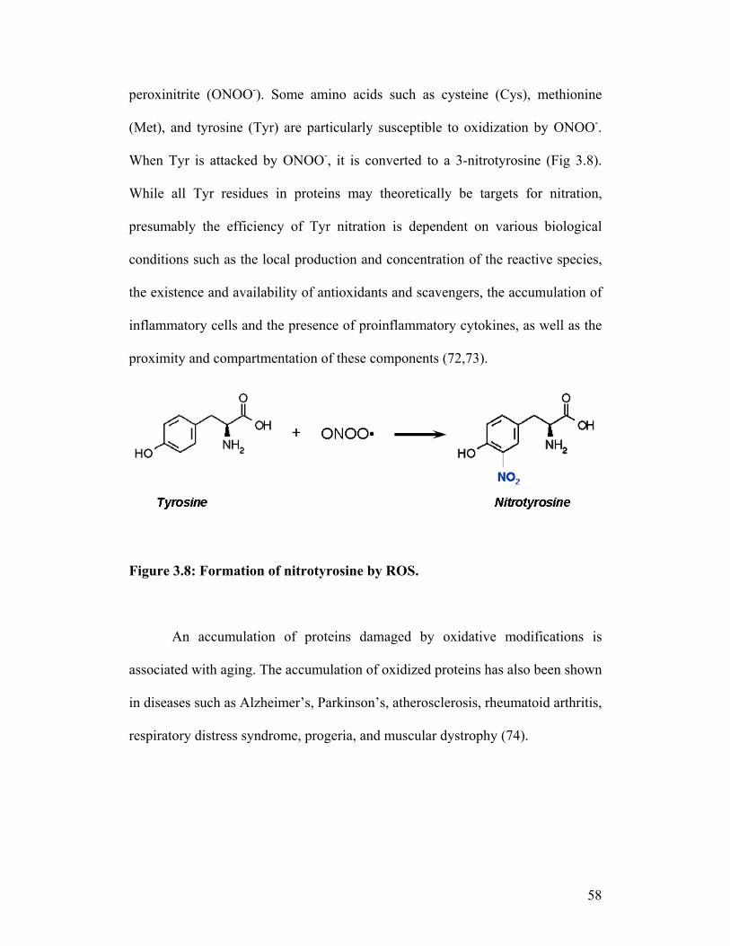
58
peroxinitrite (ONOO-). Some amino acids such as cysteine (Cys), methionine
(Met), and tyrosine (Tyr) are particularly susceptible to oxidization by ONOO-.
When Tyr is attacked by ONOO-, it is converted to a 3-nitrotyrosine (Fig 3.8).
While all Tyr residues in proteins may theoretically be targets for nitration,
presumably the efficiency of Tyr nitration is dependent on various biological
conditions such as the local production and concentration of the reactive species,
the existence and availability of antioxidants and scavengers, the accumulation of
inflammatory cells and the presence of proinflammatory cytokines, as well as the
proximity and compartmentation of these components (72,73).
Figure 3.8: Formation of nitrotyrosine by ROS.
An accumulation of proteins damaged by oxidative modifications is
associated with aging. The accumulation of oxidized proteins has also been shown
in diseases such as Alzheimer’s, Parkinson’s, atherosclerosis, rheumatoid arthritis,
respiratory distress syndrome, progeria, and muscular dystrophy (74).

59
3.5.2.3. DNA damage
Hydroxyl radicals can interact with DNA bases leading to a variety of
oxidative products. In fact, the interaction of hydroxyl radical (•OH) with
Guanine (G) generates the formation of 8-oxo-7,8-dihydro-20-deoxyguanosine (8-
oxo-dG) and of 2,6-diamino-5-formamido-4-hydroxypyrimidine (FAPy-G) (Fig
3.9). 8-oxoG is a highly mutagenic miscoding lesion that can lead to G:C to T:A
transversion mutations. Hydroxyl radical can also interact with Adenine (A) but
these lesions are less frequent than those associated with G (75).
Figure 3.9: Modification of Guanine by ROS.
Oxidative lesions to DNA can lead to single-strand breaks. These lesions
present mutagenic and/or cytotoxic (blocking) challenges to the cell when
encountered during chromosome replication or gene transcription. Indeed, the
most significant consequences in DNA lesions result in genomic instability and

60
various pathologies such as cancer. Oxidation of DNA is also implicated in
normal aging. Due to their major physiological effects in several aspects of
intracellular signaling, ROS can also indirectly interfere with the expression of
several genes and signal transduction pathways (76).
3.5.3. Antioxidant systems
Biological antioxidants are natural molecules that can prevent the
uncontrolled formation of free radicals and ROS or inhibit their relation with
biological structures. Antioxidants can be divided in three general families. The
first group is composed of small molecules such as vitamin E and C, traps radicals
and prevents chain reactions. The second one made of antioxidant enzymes and
metal-binding proteins, prevents the formation of new ROS. Lastly, those
composed of DNA repair enzymes, repair biomolecules damaged by ROS. The
focus of our research was mostly on the two first families of molecules which are
further described below.
3.5.3.1. Non-enzymatic antioxidant molecules
Non-enzymatic antioxidants are scavengers that either physically
quenches excited species, such as singlet oxygen, or chemically traps oxidizing
free radicals and ROS. The kinetics of their scavenging reactions is very fast,
making them very efficient. Importantly, free radicals reacting with scavengers
decay through dismutation, recombination, or reduction by secondary scavengers.
Therefore, these free radicals do not serve to initiate uncontrolled chain reactions.

61
Non-enzymatic antioxidant molecules exist as water soluble (free glutathione
(GSH) and vitamin C (ascorbic acid), and lipid soluble (vitamins A and E)
antioxidant. Bilirubin and ubiquinol, the latter being the two-electron product of
ubiquinone, have the power to inhibit lipid peroxidation. Uric acid and flavonoids,
which are ubiquitous in plants and contain phenolic hydroxyl groups, also have
antioxidant effects (77,78).
3.5.3.2. Antioxidant enzymes
Even though non-enzymatic scavengers are essential to avoid damages
induced by most ROS, their protection is not sufficient against radicals such as
peroxides and hydroperoxides that are continuously produced by aerobic cells.
Specific antioxidant enzymes play this essential role. The advantages of such
antioxidant enzymes reside in the fact that the steady-state concentration of
peroxides can be adapted to the cell requirements. Indeed, these enzymes can be
induced, inhibited, or even inactivated. They also play an important role in the
regulation of metabolic pathways and specific functions of aerobic cells. For this
reason, their expression is modified in several diseases (Table 1) (79). The
activities of the enzymes analyzed in the present research project are described
below.

62
DISEASE CELL TISSUS ANTIOXIDANT
Female infertility Luteinizing hormone
(brain)
Cu/Zn-SOD
Diabetes Beta cells (mice) Ec-SOD
HIV Plasma SOD
Parkinson Neurons CAT, GPx, SOS
Heart diseases Arteries GPx, SOD
Crohn’s disease Gastro-intestinal tract CAT, GPx, SOD
Allergy to pollen cochlea Cu/Zn-SOD
Table 1: Relationship between antioxidant enzymes and diseases in human.
Superoxide dismutase.
Superoxide dismutase (SOD) converts superoxide radical (•O2-) into
hydrogen peroxide (Fig 3.10). There are three forms of human SOD: cytosolic
copper/zinc-SOD (Cu/Zn-SOD), mitochondrial manganese-SOD (Mn-SOD), and
extracellular-SOD (EC-SOD). Cu/Zn-SOD contains 2 subunits of 32 kDa and
constitutes the first line of antioxidant defense. The Mn-SOD is a tetramer of 96
kDa that is depressed or induced by different factors such as cytokines. EC-SOD
is a tetrameric enzyme of 135 kDa containing copper and zinc and is found in
interstitial spaces of tissues and in extracellular fluids (synovial fluid, plasma, and

63
lymph). A low level of SODs is constantly generated by the cells in reaction to the
formation of ROS by the respiratory chain. Interestingly, since they are induced
by oxidants, they can be used as markers of oxidative stress (80).
Figure 3.10: Effect of SOD on ROS.
Catalase
Catalase (CAT) is a tetramer of 240 kDa containing one ferriprotorphyrin
group per subunit. It plays a key role in the acquisition of tolerance to oxidative
stress in adaptative response of cells. CAT converts hydrogen peroxide to water
and oxygen (Fig 3.11) (81).
Figure 3.11: Effect of catalase on ROS.
Glutathione peroxidase
Glutathione peroxidase (GPx) is a tetramer of 80 kDa that converts
hydroperoxides to water and oxygen (Fig 3.12). It is also able to catalyze the
reduction of hydroperoxides into water and oxygen. Although it acts similarly as

64
CAT, GPx is the only enzyme that can react efficiently with other organic
hydroperoxides and is crucial for cell homeostasis (82).
Figure 3.12: Effect of GPx on ROS.
Heme Oxygenase
Heme oxygenase (HO) is a nontraditional antioxidant enzyme exhibiting
antioxidant capacities. Three isoforms (HO-1, HO-2, and HO-3) of this enzyme
exist. All these isoforms are located in the membrane of the endoplasmatic
reticulum and catalyze the degradation of heme into carbon monoxide, iron, and
biliverdin, which is subsequently reduced to bilirubin, a potent antioxidant (Fig
3.13) (83). HO-1 is inducible by ROS and is found throughout the organs, but
especially in tissues rich in reticuloendothelial cells, such as the spleen and the
bone marrow. HO-1 is involved in human diseases such as Alzheimer and
inflammatory diseases and its expression is down-regulated in hepatitis C.
HO-2 and HO-3 are constitutively expressed and are not inducible. HO-2 and
HO-3 are widely distributed throughout the body but are especially localized in
endothelial cell, adventitial nerves of blood vessels, and in neurons in autonomic
ganglia (83,84).

65
Figure 3.13: Degradation of heme by HO-1.
3.6. References.
1. Yancik R. Cancer burden in the aged: an epidemiologic and demographic
overview. Cancer 1997 Oct 1;80(7):1273-1283.
2. Saikko V, Ahlroos T, Calonius O, Keranen J. Wear simulation of total hip
prostheses with polyethylene against CoCr, alumina and diamond-like carbon.
Biomaterials 2001 Jun;22(12):1507-1514.
3. Skinner HB. Ceramic bearing surfaces. Clin Orthop Relat Res 1999
Dec(369):83-91.
4. Tipper JL, Ingham E, Hailey JL, Besong AA, Fisher J, Wroblewski BM, et al.
Quantitative analysis of polyethylene wear debris, wear rate and head damage
in retrieved Charnley hip prostheses. J Mater Sci Mater Med 2000
Feb;11(2):117-124.
5. Fessy MH, Seutin B, Bejui J. Anatomical basis for the choice of the femoral
implant in the total hip arthroplasty. Surg Radiol Anat 1997;19(5):283-286.
6. Donald SM. Sir John Charnley (1911-1982): inspiration to future generations
of orthopaedic surgeons. Scott Med J 2007 May;52(2):43-46.
7. Wroblewski M, Siney PD, Nagai H, Fleming PA. Wear of ultra-high-
molecular-weight polyethylene cup articulating with 22.225 mm zirconia

66
diameter head in cemented total hip arthroplasty. J Orthop Sci 2004;9(3):253-
255.
8. Wroblewski BM. Osteolysis due to particle wear debris following total hip
arthroplasty: the role of high-density polyethylene. Instr Course Lect
1994;43:289-294.
9. Zhu YH, Chiu KY, Tang WM. Review Article: Polyethylene wear and
osteolysis in total hip arthroplasty. J Orthop Surg (Hong Kong) 2001
Jun;9(1):91-99.
10. Synder M, Kozlowski P, Marciniak M, Dorman T. Revision hip arthroplasty
in aseptic loosening of total hip replacement. Ortop Traumatol Rehabil
2001;3(1):34-37.
11. Ingham E, Fisher J. The role of macrophages in osteolysis of total joint
replacement. Biomaterials 2005 Apr;26(11):1271-1286.
12. Swanson SA. Engineering considerations in the design of double-cup hip
replacement prostheses. Clin Orthop Relat Res 1978 Jul-Aug(134):12-18.
13. Rieker CB, Schon R, Kottig P. Development and validation of a second-
generation metal-on-metal bearing: laboratory studies and analysis of
retrievals. J Arthroplasty 2004 Dec;19(8 Suppl 3):5-11.
14. Higuchi F, Inoue A, Semlitsch M. Metal-on-metal CoCrMo McKee-Farrar
total hip arthroplasty: characteristics from a long-term follow-up study. Arch
Orthop Trauma Surg 1997;116(3):121-124.

67
15. Hughes AN, Lane RA. The development of high tensile properties in a
wrought Co-Cr-Ni-W alloy of potential orthopaedic surgery use. Eng Med
1976 Apr;5(2):38-40.
16. Wang SC, Browne M, Ubhi HS, Starink MJ. Determination of the fatigue
fracture planes of Co-Cr-Mo biomedical alloys using electron backscatter
diffraction. J Microsc 2005 Feb;217(Pt 2):118-121.
17. Saldivar-Garcia AJ, Lopez HF. Microstructural effects on the wear resistance
of wrought and as-cast Co-Cr-Mo-C implant alloys. J Biomed Mater Res A
2005 Aug 1;74(2):269-274.
18. Salinas-Rodriguez A, Rodriguez-Galicia JL. Deformation behavior of low-
carbon Co-Cr-Mo alloys for low-friction implant applications. J Biomed
Mater Res 1996 Jul;31(3):409-419.
19. Shupack SI. The chemistry of chromium and some resulting analytical
problems. Environ Health Perspect 1991 May;92:7-11.
20. Kartal SN. Removal of copper, chromium, and arsenic from CCA-C treated
wood by EDTA extraction. Waste Manag 2003;23(6):537-546.
21. Hazell T. Minerals in foods: dietary sources, chemical forms, interactions,
bioavailability. World Rev Nutr Diet 1985;46:1-123.
22. Yu XZ, Gu JD. The role of EDTA in phytoextraction of hexavalent and
trivalent chromium by two willow trees. Ecotoxicology 2008 Apr;17(3):143-
152.
23. Chen NS, Tsai A, Dyer IA. Effect of chelating agents on chromium absorption
in rats. J Nutr 1973 Aug;103(8):1182-1186.

68
24. Gammelgaard B, Jensen K, Steffansen B. In vitro metabolism and permeation
studies in rat jejunum: organic chromium compared to inorganic chromium. J
Trace Elem Med Biol 1999 Jul;13(1-2):82-88.
25. Zhitkovich A, Voitkun V, Costa M. Formation of the amino acid-DNA
complexes by hexavalent and trivalent chromium in vitro: importance of
trivalent chromium and the phosphate group. Biochemistry 1996 Jun
4;35(22):7275-7282.
26. Blackburn AC, Gallucci JC, Gerkin RE. Structure of hexaaquacobalt(II)
bromate. Acta Crystallogr B 1990 Dec 1;46 ( Pt 6):712-716.
27. Nagababu P, Latha JN, Pallavi P, Harish S, Satyanarayana S. Studies on
antimicrobial activity of cobalt(III) ethylenediamine complexes. Can J
Microbiol 2006 Dec;52(12):1247-1254.
28. Hutson TB, Mitchell ML, Keller JT, Long DJ, Chang MJ. A technique for
monitoring mammalian cell growth and inhibition in situ via Fourier
transform infrared spectroscopy. Anal Biochem 1988 Nov 1;174(2):415-422.
29. Stenlund H, Gorzsas A, Persson P, Sundberg B, Trygg J. Orthogonal
projections to latent structures discriminant analysis modeling on in situ FT-
IR spectral imaging of liver tissue for identifying sources of variability. Anal
Chem 2008 Sep 15;80(18):6898-6906.
30. Keszthelyi L, Varga L, Demeter I, Hollos-Nagy K, Szokefalvi-Nagy Z.
Elemental analysis of samples of biological origin relative to their protein
content by means of charged particle bombardment. Anal Biochem 1984
Jun;139(2):418-426.
69
31. Pradier CM, Rubio C, Poleunis C, Bertrand P, Marcus P, Compere C. Surface
characterization of three marine bacterial strains by Fourier transform IR, X-
ray photoelectron spectroscopy, and time-of-flight secondary-ion mass
spectrometry, correlation with adhesion on stainless steel surfaces. J Phys
Chem B 2005 May 19;109(19):9540-9549.
32. Cordewener JH, Luykx DM, Frankhuizen R, Bremer MG, Hooijerink H,
America AH. Untargeted LC-Q-TOF mass spectrometry method for the
detection of adulterations in skimmed-milk powder. J Sep Sci 2009
Apr;32(8):1216-1223.
33. Chassaigne H, Norgaard JV, Hengel AJ. Proteomics-based approach to detect
and identify major allergens in processed peanuts by capillary LC-Q-TOF
(MS/MS). J Agric Food Chem 2007 May 30;55(11):4461-4473.
34. Deutsch AS, Cohen BI, Musikant BL. Inductively coupled plasma-emission
spectroscopy and atomic absorption for the use of elemental analysis of a root
canal after lasing with a holmium:YAG laser. J Endod 2003 Jun;29(6):404-
406.
35. Bayon MM, Rodriguez Garcia A, Garcia Alonso JI, Sanz-Medel A. Indirect
determination of trace amounts of fluoride in natural waters by ion
chromatography: a comparison of on-line post-column fluorimetry and ICP-
MS detectors. Analyst 1999 Jan;124(1):27-31.
36. Althuis MD, Jordan NE, Ludington EA, Wittes JT. Glucose and insulin
responses to dietary chromium supplements: a meta-analysis. Am J Clin Nutr
2002 Jul;76(1):148-155.

70
37. Anderson RA, Kozlovsky AS. Chromium intake, absorption and excretion of
subjects consuming self-selected diets. Am J Clin Nutr 1985 Jun;41(6):1177-
1183.
38. Anderson RA, Bryden NA, Polansky MM, Reiser S. Urinary chromium
excretion and insulinogenic properties of carbohydrates. Am J Clin Nutr 1990
May;51(5):864-868.
39. Avudainayagam S, Megharaj M, Owens G, Kookana RS, Chittleborough D,
Naidu R. Chemistry of chromium in soils with emphasis on tannery waste
sites. Rev Environ Contam Toxicol 2003;178:53-91.
40. McCarty MF. Chromium(III) picolinate. FASEB J 1996 Feb;10(2):365-369.
41. Dowling HJ, Offenbacher EG, Pi-Sunyer FX. Absorption of inorganic,
trivalent chromium from the vascularly perfused rat small intestine. J Nutr
1989 Aug;119(8):1138-1145.
42. Borguet F, Wallaeys B, Cornelis R, Lameire N. Transperitoneal absorption
and kinetics of chromium in the continuous ambulatory peritoneal dialysis
patient--an experimental and mathematical analysis. Nephron 1996;72(2):163-
170.
43. Donaldson DL, Smith CC, Yunice AA. Renal excretion of chromium-51
chloride in the dog. Am J Physiol 1984 Jun;246(6 Pt 2):F870-878.
44. Sun Y, Ramirez J, Woski SA, Vincent JB. The binding of trivalent chromium
to low-molecular-weight chromium-binding substance (LMWCr) and the
transfer of chromium from transferrin and chromium picolinate to LMWCr. J
Biol Inorg Chem 2000 Feb;5(1):129-136.

71
45. Suwalsky M, Castro R, Villena F, Sotomayor CP. Cr(III) exerts stronger
structural effects than Cr(VI) on the human erythrocyte membrane and
molecular models. J Inorg Biochem 2008 Apr;102(4):842-849.
46. Quievryn G, Goulart M, Messer J, Zhitkovich A. Reduction of Cr (VI) by
cysteine: significance in human lymphocytes and formation of DNA damage
in reactions with variable reduction rates. Mol Cell Biochem 2001 Jun;222(1-
2):107-118.
47. Tiffany ME, Spears JW, Xi L, Horton J. Influence of dietary cobalt source and
concentration on performance, vitamin B12 status, and ruminal and plasma
metabolites in growing and finishing steers. J Anim Sci 2003
Dec;81(12):3151-3159.
48. Barceloux DG. Cobalt. J Toxicol Clin Toxicol 1999;37(2):201-206.
49. Azzi M, Szpunar JA. Tribo-electrochemical technique for studying
tribocorrosion behavior of biomaterials. Biomol Eng 2007 Nov;24(5):443-
446.
50. Vandamme NS, Wayman BH, Topoleski LD. Wear behavior of carbide
coated Co-Cr-Mo implant alloy. J Mater Sci Mater Med 2003 Jan;14(1):47-
53.
51. Jacobs JJ, Gilbert JL, Urban RM. Corrosion of metal orthopaedic implants. J
Bone Joint Surg Am 1998 Feb;80(2):268-282.
52. Cook SD, Gianoli GJ, Clemow AJ, Haddad RJ, Jr. Fretting corrosion in
orthopaedic alloys. Biomater Med Devices Artif Organs 1983;11(4):281-292.

72
53. Tsou TC, Yang JL. Formation of reactive oxygen species and DNA strand
breakage during interaction of chromium (III) and hydrogen peroxide in vitro:
evidence for a chromium (III)-mediated Fenton-like reaction. Chem Biol
Interact 1996 Dec 20;102(3):133-153.
54. Davidson T, Kluz T, Burns F, Rossman T, Zhang Q, Uddin A, et al. Exposure
to chromium (VI) in the drinking water increases susceptibility to UV-induced
skin tumors in hairless mice. Toxicol Appl Pharmacol 2004 May
1;196(3):431-437.
55. Day GA, Virji MA, Stefaniak AB. Characterization of exposures among
cemented tungsten carbide workers. Part II: Assessment of surface
contamination and skin exposures to cobalt, chromium and nickel. J Expo Sci
Environ Epidemiol 2009 May;19(4):423-434.
56. Domingo JL. Cobalt in the environment and its toxicological implications.
Rev Environ Contam Toxicol 1989;108:105-132.
57. Saglam AM, Baysal V, Ceylan AM. Nickel and cobalt hypersensitivity
reaction before and after orthodontic therapy in children. J Contemp Dent
Pract 2004 Nov 15;5(4):79-90.
58. Steinhoff D, Mohr U. On the question of a carcinogenic action of cobalt-
containing compounds. Exp Pathol 1991;41(4):169-174.
59. Lison D, Carbonnelle P, Mollo L, Lauwerys R, Fubini B. Physicochemical
mechanism of the interaction between cobalt metal and carbide particles to
generate toxic activated oxygen species. Chem Res Toxicol 1995
Jun;8(4):600-606.

73
60. Harrop TC, Mascharak PK. Fe(III) and Co(III) centers with carboxamido
nitrogen and modified sulfur coordination: lessons learned from nitrile
hydratase. Acc Chem Res 2004 Apr;37(4):253-260.
61. Turi S, Nemeth I, Torkos A, Saghy L, Varga I, Matkovics B, et al. Oxidative
stress and antioxidant defense mechanism in glomerular diseases. Free Radic
Biol Med 1997;22(1-2):161-168.
62. Lloyd RV, Hanna PM, Mason RP. The origin of the hydroxyl radical oxygen
in the Fenton reaction. Free Radic Biol Med 1997;22(5):885-888.
63. Shi XL, Dalal NS. The role of superoxide radical in chromium (VI)-generated
hydroxyl radical: the Cr(VI) Haber-Weiss cycle. Arch Biochem Biophys 1992
Jan;292(1):323-327.
64. Morehouse KM, Mason RP. The transition metal-mediated formation of the
hydroxyl free radical during the reduction of molecular oxygen by ferredoxin-
ferredoxin:NADP+ oxidoreductase. J Biol Chem 1988 Jan 25;263(3):1204-
1211.
65. Luzzatto E, Cohen H, Stockheim C, Wieghardt K, Meyerstein D. Reactions of
low valent transition metal complexes with hydrogen peroxide. Are they
"Fenton-like" or not? 4. The case of Fe(II)L, L = edta; hedta and tcma. Free
Radic Res 1995 Nov;23(5):453-463.
66. Leonard S, Gannett PM, Rojanasakul Y, Schwegler-Berry D, Castranova V,
Vallyathan V, et al. Cobalt-mediated generation of reactive oxygen species
and its possible mechanism. J Inorg Biochem 1998 Jul;70(3-4):239-244.

74
67. Parthiban A, Vijayalingam S, Shanmugasundaram KR, Mohan R. Oxidative
stress and the development of diabetic complications--antioxidants and lipid
peroxidation in erythrocytes and cell membrane. Cell Biol Int 1995
Dec;19(12):987-993.
68. Arduini A, Stern A, Storto S, Belfiglio M, Mancinelli G, Scurti R, et al. Effect
of oxidative stress on membrane phospholipid and protein organization in
human erythrocytes. Arch Biochem Biophys 1989 Aug 15;273(1):112-120.
69. Nielsen F, Mikkelsen BB, Nielsen JB, Andersen HR, Grandjean P. Plasma
malondialdehyde as biomarker for oxidative stress: reference interval and
effects of life-style factors. Clin Chem 1997 Jul;43(7):1209-1214.
70. Shiraishi K, Naito K. Effects of 4-hydroxy-2-nonenal, a marker of oxidative
stress, on spermatogenesis and expression of p53 protein in male infertility. J
Urol 2007 Sep;178(3 Pt 1):1012-1017; discussion 1017.
71. Hawkins CL, Davies MJ. Generation and propagation of radical reactions on
proteins. Biochim Biophys Acta 2001 Apr 2;1504(2-3):196-219.
72. Mattson MP, Goodman Y, Luo H, Fu W, Furukawa K. Activation of NF-
kappaB protects hippocampal neurons against oxidative stress-induced
apoptosis: evidence for induction of manganese superoxide dismutase and
suppression of peroxynitrite production and protein tyrosine nitration. J
Neurosci Res 1997 Sep 15;49(6):681-697.
73. Petit A, Mwale F, Tkaczyk C, Antoniou J, Zukor DJ, Huk OL. Cobalt and
chromium ions induce nitration of proteins in human U937 macrophages in
vitro. J Biomed Mater Res A 2006 Dec 1;79(3):599-605.

75
74. Schuessler H, Schilling K. Oxygen effect in the radiolysis of proteins. Part 2.
Bovine serum albumin. Int J Radiat Biol Relat Stud Phys Chem Med 1984
Mar;45(3):267-281.
75. Ober M, Linne U, Gierlich J, Carell T. The two main DNA lesions 8-Oxo-7,8-
dihydroguanine and 2,6-diamino-5-formamido-4-hydroxypyrimidine exhibit
strongly different pairing properties. Angew Chem Int Ed Engl 2003 Oct
20;42(40):4947-4951.
76. Thannickal VJ, Fanburg BL. Reactive oxygen species in cell signaling. Am J
Physiol Lung Cell Mol Physiol 2000 Dec;279(6):L1005-1028.
77. Franci O, Ranfi F, Scaccini C, Amici A, Merendino N, Tommasi G, et al.
Differential effect of alpha-tocopherol and ascorbate on oxidative injury
induced in immune cells by thermal stress. J Biol Regul Homeost Agents
1996 Apr-Sep;10(2-3):54-59.
78. Schettler V, Wieland E, Methe H, Schuff-Werner P, Muller GA. Oxidative
stress during dialysis: effect on free radical scavenging enzyme (FRSE)
activities and glutathione (GSH) concentration in granulocytes. Nephrol Dial
Transplant 1998 Oct;13(10):2588-2593.
79. Nepomniashchikh VA, Lomivorotov VV, Deryagin MN, Lomivorotov VN,
Kniazkova LG. Oxidative stress and monooxygenase liver function in patients
with coronary heart disease and multiple organ dysfunction syndrome. Eur J
Anaesthesiol 2009 Feb;26(2):140-146.
80. Wlaschek M, Scharffetter-Kochanek K. Oxidative stress in chronic venous leg
ulcers. Wound Repair Regen 2005 Sep-Oct;13(5):452-461.

76
81. Cutler RG. Oxidative stress and aging: catalase is a longevity determinant
enzyme. Rejuvenation Res 2005 Fall;8(3):138-140.
82. Doroshow JH. Glutathione peroxidase and oxidative stress. Toxicol Lett 1995
Dec;82-83:395-398.
83. Baranano DE, Rao M, Ferris CD, Snyder SH. Biliverdin reductase: a major
physiologic cytoprotectant. Proc Natl Acad Sci U S A 2002 Dec
10;99(25):16093-16098.
84. Le WD, Xie WJ, Appel SH. Protective role of heme oxygenase-1 in oxidative
stress-induced neuronal injury. J Neurosci Res 1999 Jun 15;56(6):652-658.

77
CHAPTER 4
Effects of Chromium and Cobalt Ions on the Expression
of Antioxidant Enzymes in Human U937 Macrophage-
like Cells
The first article presented in this chapter and accepted for publication in
Journal of Biomedical Materials Research Part A, fulfills the first objective of
this thesis which was to compare the toxicity of Cr(VI), Cr(III), and Co(II) in
human U937 macrophages-like cells.
Previous studies performed in our laboratory showed that Cr(III) and
Co(II) ions could induce damage to lipids and proteins in human macrophages in
vitro. Moreover, this damage was suggested to be the result of reactive oxygen
species (ROS) generated by Cr and Co ions. Consequently, one of the aims of this
thesis was to understand the fate of Cr(III) and Co(II) ions in generating oxidative
damage by studying in vitro a defense system against oxidative stress. It is known
that Cr(VI) ions are the first ions generated from metallic implants and that they
are rapidly reduced to Cr(III) in the body. As a result, we further investigated the
effects of Cr(III), Cr(VI) and Co(II) on macrophages. Our findings showed that
Cr(III) has no effect on the expression of Mn-SOD, Cu/Zn-SOD, CAT, GPx, and
HO-1. Cr(VI) induced the protein expression of all the antioxidant enzymes
studied but had no effect on the expression of their mRNA. Co(II) induced the
expression of both proteins and mRNA of HO-1 only.

78
EFFECT OF CHROMIUM AND COBALT IONS ON THE EXPRESSION
OF ANTIOXIDANT ENZYMES IN HUMAN U937 MACROPHAGE-LIKE
CELLS
Cathy Tkaczyk1,2,3, Olga L. Huk2,3, Fackson Mwale2, John Antoniou2,3, David. J
Zukor2,
Alain Petit2, Maryam Tabrizian1,3,4,5
1Department of Biomedical Engineering, Duff Medical Building, 3775 University
Street, Montreal, QC, H3A 2B4, Canada.
2Division of Orthopaedic Surgery, McGill University,
Lady Davis Institute for Medical Research,
The Sir Mortimer B. Davis-Jewish General Hospital,
3755 Côte-Sainte-Catherine Road, Montreal, QC, H3T 1E2, Canada.
3Center of Biorecognition and Biosensors (CBB)
4 McGill Institute for Advanced Materials, 5 Faculty of Dentistry

79
4.1. Abstract
The main concern associated with metal-on-metal (MM) hip prosthesis is
the presence of metal ions, mainly chromium (Cr) and cobalt (Co), which are
found both systemically and locally in the organism of patients. Previous studies
revealed that Cr(III) and Co(II) ions could induce damages to proteins in
macrophage-like cells in vitro, probably through the formation of reactive oxygen
species (ROS). We then hypothesized that these ions can modify the expression of
antioxidant enzymes in these cells. Results showed that Cr(VI) induced the
protein expression of Mn-superoxide dismutase, Cu/Zn-superoxide dismutase,
catalase, glutathione peroxidase, and heme oxygenase-1 (HO-1) but had no effect
of the expression of their mRNA. Cr(III) have no effect on the expression of all
these antioxidant enzymes. Co(II) induced the expression of both protein and
mRNA of HO-1 only. In conclusion, results showed that Cr(VI), Cr(III), and
Co(II) had differential effects on the expression of antioxidant enzymes in
macrophage-like cells in vitro.
4.2. Introduction
Metal-on-metal (MM) hip prostheses, made of cobalt-chromium (Co-Cr)
alloys, represent an excellent alternative to metal-polyethylene bearings in the
treatment of osteoarthritis of the hip because of their substantially lower wear
rates (1). However, the main concern associated with such bearings is the
presence of circulating metal ions in the organism (2). These ions are potentially
toxic (3,4), may cause metal hypersensitivity (5,6), chromosomal aberrations (7),

80
and may induce changes in the proportions of peripheral blood lymphocytes (8-
10). On the other hand, epidemiological studies performed in patients with MM
hip implants were unable to demonstrate any causal effect of the presence of
metallic ions on the risk of cancer (11). Moreover, the mechanism of metal ion
toxicity is not completely understood: it is however known that they can generate
reactive oxygen species (ROS), such as superoxide ions (O2-), hydrogen peroxide
(H2O2), hydroxyl radical (OH ), and nitrogen oxide (NO ) through Fenton/ Haber-
Weiss chemistry (12). These ROS are involved in numerous human diseases,
including degenerative lung and heart conditions, Alzheimer, rheumatoid arthritis,
and aging (13). ROS are implicated in the induction of oxidative stress generating
cell and tissue damage. In previous studies, we have shown that Co(II) and Cr(III)
ions can induce the formation of protein carbonyls (14) and nitrotyrosines (15),
two markers of ROS-induced protein damage linked to oxidative stress in human
U937 macrophage-like cells and in human MG-63 osteoblast-like cells (16).
However, oxidative stress may result from an increased production of oxidants
(ROS) and/or a decrease in antioxidant defense. In fact, the organisms possess an
efficient enzymatic defense system against these ROS, avoiding the oxidative
stress status which occurs when the level of ROS overwhelms the antioxidant
level.
Superoxide dismutases (SODs) are antioxidant enzymes found in three
isoforms, manganese-SOD (Mn-SOD), copper/zinc-SOD (Cu/Zn-SOD), and
extracellular-SOD. They catalyze the conversion of superoxide radical (•O2-) into
hydrogen peroxide (H2O2). Catalase (CAT) is another antioxidant enzyme that

81
assumes the next step by converting 2 molecules of hydrogen peroxide in water
and oxygen. Glutathione peroxidase (GPx) supports CAT by converting one
molecule of hydrogen peroxide in water and oxygen. Heme-oxygenase-1 (HO-1)
is not considered as a conventional antioxidant enzyme, but it catalyses the
degradation of heme in biliverdin and CO2: these by-products have high
antioxidant properties. In addition, this enzyme can be induced in an oxidative
stress environment. In the present study, we aim to further understand the toxicity
of Cr and Co at the cellular level by studying the effect of these ions on the
expression of antioxidant enzymes in human U937 macrophage-like cells in vitro.
4.3. Material and Methods
4.3.1. Cell culture
Human U937 macrophage-like cells have been chosen as the cell culture
model because macrophages are the main cells present in periprosthetic tissues.
Moreover, macrophages are the cells that engulf foreign bodies by phagocytosis.
U937 cells were cultured in RPMI 1640 medium supplemented with 5% fetal
bovine serum (HyClone, Logan, UT), 100 U/mL penicillin, and 100 µg/mL
streptomycin in a humidified 5% CO2 atmosphere at 37ºC. Cells were incubated
for up to 72h in the absence (control) or the presence of 0-50 ppm Cr(VI)
(Na2CrO4, Sigma-Aldrich, Oakville, ON), 0-250 ppm Cr(III) (CrCl3, Sigma-
Aldrich), and 0-10 ppm Co(II) (CoCl2, Fisher Scientific, Montreal, QC). The
choice of the ions is due to the fact that Co and Cr are the main corrosion products

82
of MM hip implants. Moreover, Cr(VI) are the first Cr ions released from
metallic implants and are rapidly reduced to a more thermodynamically stable
specie Cr(III) (17). Concentrations were chosen in function of the non-
prestimulated nature of U937 cells in which higher levels of ions are necessary for
stimulation. They are also based on our previous studies on the effect of Cr and
Co ions on the stimulation of the oxidation of proteins in macrophages (14,15)
and osteoblasts (16).
4.3.2. Protein expression
After incubation, cells were lyzed in a caspase assay lysis buffer
(BioSource, Nivelles, Belgium) as previously described. Proteins were then
separated on 4–20% acrylamide gels (SDS-PAGE) and transferred to
nitrocellulose membranes. Anti-oxidant enzymes were detected by Western blot
using anti-Mn-SOD (1:500 - Upstate, Billerica, MA), anti-Cu/Zn-SOD (1:1000 -
Upstate, Billerica, MA), anti-CAT (1:1000- Abcam, Cambridge, MA), anti-GPx
(1:1500 - Abcam), and anti-HO-1 (1:1000 - Abcam) antibodies. All antibodies
were diluted in ChemiBlocker™:PBS-tween (0.05%) (1:1) (Chemicon
International Norcross, GA). Actin (NeoMarkers, Fremont, CA) expression
served as a house keeping gene for gel loading and served to normalize the
results. Proteins were detected using NEN Renaissance chemiluminescence
reagents (Perkin-Elmer, Woodbridge, ON) and analyzed using a VersaDoc image
analysis system equipped with a cooled charge coupled device (CCD) 12-bit
camera (Bio-Rad Laboratories, Mississauga, ON).
83
4.3.3. RNA Extraction
Total RNA was extracted from U937 cells by a modified method of
Chomcynski and Sacchi (18) using TRIzol® reagent (Invitrogen, Burlington,
ON). Purity of the RNA was measured through the A260/A280 ratio.
4.3.4. Reverse Transcriptase (RT) Reaction
The RT reaction was performed for 50 min at 42ºC using 1.0 µg total
RNA. 20 μL of reaction volume contained 50 mM Tris-HCl (pH 8.3), 75 mM
KCL, 3 mM MgCl2, 10 mM DTT, 50 μM dNTP mixture, and 200 units of
Superscript II RNAse H reverse transcriptase (Invitrogen). The inactivation
reaction was achieved at 70ºC for 15 min.
4.3.5. Polymerase Chain Reaction (PCR)
PCR was performed in a total volume of 25 μL containing 10 mM Tris-
HCl (pH 8.3), 1.5 mM MgCl2, 0.2 mM dNTP mixture, 0.8 μM of forward and
reverse primers, 1μl of RT mixture, and 1.25 units of Taq DNA polymerase
(Invitrogen). The PCR procedure was done in accordance with the standard
method (19). Thirty five cycles were used for all genes. Every cycle consists of 1
min denaturation at 95°C, 1 min annealing at 51°C (GPx, HO-1) or 56°C (CAT,
SODs), 1 min polymerization at 72°C, followed by a final 10 min extension at
72°C. PCR products were separated on 2% agarose gel, visualized by ethidium
bromide staining, and analyzed using a Bio-Rad VersaDoc image analysis system.
GAPDH was used as a house keeping gene for gel loading and served to

84
normalize the results. To avoid contamination with genomic DNA, DNase I
treatment was performed using 1 unit of deoxyribonuclease I, (Invitrogen) at 25ºC
for 1 h. DNase I was inactivated by adding 1 μL of 25 mM EDTA to the reaction
mixture and by heating for 10 min at 65ºC. To confirm the lack of genomic DNA
contamination of RNA samples, PCR was also performed with RNA aliquots. The
primers, specific for human genes, were obtained from Invitrogen. The sequences
of the primers used for PCR are described in Table 2.
Gene Primers Product size
Forward 5’- ACATCTATGTGGCCCTGGAG HO-1 Reverse 5’- TGTTGGGGAAGGTGAAGAAG 348
Forward 5’- CTGCAGCTCCGCAATCCTAC -CAT Reverse 5’- TCCTTCCACTGCTTCATCTGG 229
Forward 5’- CCTCAAGTACGTCCGACCTG -GPX Reverse 5’- CAATGTCGTTGCGGCACACC 197
Forward 5’- AAGGCCGTGTGCGTGCTGAA- Cu-Zn-
SOD Reverse 5’- CAGGTCTCCAACATGCCTCT- 246
Forward 5’- GCACATTAACGCGCAGATCA Mn-SOD Reverse 5’- AGCCTCCAGCAACTCTCCTT - 241
Forward 5’- CCATCACCATCTTCCAGGAG -GAPDH Reverse 5’- CCTGCTTCACCACCTTCTTG - 576
Table 2: Sequences of primers used for PCR.

85
4.4. Results
4.4.1. Effect of Cr(VI)
Figures 1 and 2 show the effect of Cr(VI) on the expression of antioxidant
enzymes. Indeed, Cr(VI) induced a sustained dose-dependent expression of all
studied antioxidant enzyme proteins at 24h, 48h and 72h (Fig. 4.1A and Fig. 4.2).
The induction reached 2.5, 2.8, 2.9, 2.8, and 4.5 times the control with 50 ppm
Cr(VI) after 72h for Mn-SOD, Cu/Zn-SOD, CAT, GPx, and HO-1 respectively.
Cr(VI) had no effect on their mRNA expression in the same experimental
conditions (Fig. 4.1B).
Figure 4.1: Effect of Cr(VI) on antioxidant enzyme expression. Human U937
cells were incubated for 48 h in the absence (Control) or the presence of
increasing concentrations of Cr(VI). Protein expression (A) was measured by

86
Western blot while mRNA expression (B) was measured by PCR. Results are
representative of 3-6 experiments.
Figure 4.2: Effect of Cr(VI) on the expression of antioxidant enzyme
proteins. U937 cells were incubated for up to 72h in the presence of 0-50 ppm
Cr(VI). Protein expression was measured by Western blot. Results are the mean ±
standard deviation of 3-4 experiments.

87
4.4.2. Effect of Cr(III)
Figures 4.3 and 4.4 show the effect of Cr(III) on the expression of
antioxidant enzymes. After 48h, Cr(III) had no effect on the expression of
antioxidant enzyme proteins (Fig. 4.3A and 4.4) and mRNA (Fig. 4.3B and 4.5) in
the cell culture conditions and time of incubations used in this study. Results were
similar at 24h and 72h.
Figure 4.3: Effect of Cr(III) on the expression of antioxidant enzymes. Human
U937 cells were incubated for 48 h in the absence (Control) or the presence of
increasing concentrations of Cr(III). Protein expression (A) was measured by
Western blot while mRNA expression (B) was measured by PCR. Results are
representative of 3-6 experiments.

88
Figure 4.4: Effect of Cr(III) on the expression of antioxidant enzyme
proteins. U937 cells were incubated up to 72h in the presence of 0-250 ppm
Cr(III). Protein expression was measured by Western blot. Results are the mean ±
standard deviation of 4-6 experiments.

89
Figure 4.5: Effect of Cr(III) on the gene expression of antioxidant enzyme.
U937 cells were incubated up to 72h in the presence of 0-250 ppm Cr(III). Gene
expressions were measured by RT-PCR. Results are the mean ± standard
deviation of 3-4 experiments.
4.4.3. Effect of Co(II)
Figures 4.6 and 4.7 show the effect of Co(II) on the expression of
antioxidant enzymes. Figure 4.6A shows a representative example of the
expression of proteins, while Figure 4.6B is a representative example of mRNA
expression as illustrated by ethidium bromide staining of amplified sequences.

90
Figure 4.7 shows the semi-quantitative results of the effect of Co(II) on HO-1
expression. Results show that Co(II) had no effect on the expression of SODs,
CAT, and GPx proteins (Fig. 4.6A) and mRNA (Fig. 4.6B). However, Co(II)
induced a time- and dose-dependent expression of both protein and mRNA. The
protein expression of HO-1 reached 0.65, 11.86, and 13.4 times the control with
10 ppm Co(II) after 24h, 48h, and 72h, respectively (Fig. 4.7A). The expression
of HO-1 mRNA paralleled the protein expression with stimulation reaching 3.3,
5.9, 7.5 times the control with 10 ppm Co(II) after 72h.
Figure 4.6: Effect of Co(II) on the expression of antioxidant enzymes. Human
U937 cells were incubated for 48 h in the absence (Control) or the presence of
increasing concentrations of Co(II). Protein expression (A) was measured by
Western blot while mRNA expression (B) was measured by PCR. Results are
representative of 3-6 experiments.

91
Figure 4.7: Effect of Co(II) on the expression of HO-1. U937 cells were
incubated for up to 72h in the presence of 0-10 ppm Co(II). Protein expression
(A) was measured by Western blot while mRNA expression (B) was measured by
PCR. Results are the mean ± standard deviation of 4-6 experiments.
4.5. Discussion
Many stresses, including exposure to heavy metals, can induce damage to
cells either directly or indirectly through a burst of reactive oxygen species (ROS)
(20). However, mammalian cells are able to respond to elevated levels of ROS by
activating their antioxidant defense systems. Results of the present study showed
that Cr(VI), Cr(III), and Co(II) had differential effects on the expression of
antioxidant enzymes in human U937 macrophage-like cells. More specifically,
Cr(VI) induced the protein expression, but not the mRNA expression, of all the
antioxidant enzymes, suggesting that several defense mechanisms are necessary to
protect the U937 cells against this ion. In fact, three antioxidant groups of

92
molecules constitute the antioxidant defense system of the organism: the primary
system prevents the formation of ROS, the secondary system eliminates radicals
to prevent chain reactions, and the tertiary system works to repair damaged
molecules (21). The secondary system, analyzed in the present study, is composed
by Mn-SOD, Cu/Zn-SOD, and extracellular-SOD that converts superoxide radical
(•O2-) into hydrogen peroxide, CAT, that converts hydrogen peroxide to water and
oxygen, and GPx, that converts hydroperoxide to water and oxygen (22). All
these enzymes were induced by Cr(VI). Our results are in agreement with a
previous study showing that long-term exposition (up to 4 weeks) to low doses of
Cr(VI) induced the protein expression of SOD, CAT, and GPx in U937 cells (23).
It was demonstrated that pre-stimulated macrophages, as in this study, respond to
a larger extent than non-pre-stimulated cells, as in the present study (24). This
implies that the concentrations necessary to induce cell response in vitro are more
important than in vivo and justifies the concentrations of ions used in our study.
Nevertheless, both the use of higher dose in non pre-stimulated cells or the use of
repeated low doses of ions mimicking pre-stimulated cells suggests that Cr(VI)
can induce the expression of antioxidant enzymes in vitro. Results also showed
that Cr(VI) had no effect on HO-1 mRNA expression, in agreement with the lack
of effect of Cr(VI) on the HO-1 mRNA expression in human airway epithelial
cells in vitro (25).
Results also showed that Cr(III) has no effect on the expression of
antioxidant enzymes (both protein and gene), suggesting that Cr(III) is less toxic
than Cr(VI). In fact, most studies showed that Cr(VI) is more toxic than Cr(III)

93
(26). The reason for this difference of toxicity is not well known. It was however
demonstrated that Cr(III) formed precipitable complexes in simulated
physiological fluids, whereas Cr(VI) did not (27). Contrary to Cr(VI) which is
known to enter the cells easily through anion channels (28), the Cr(III) complexes
seemed to be internalized via a phagocytosis-like pathways (29) that may protect
the cells from damages and may explain the difference in toxicity between the
two forms of Cr.
Finally, Co(II) induced the expression of both the protein and the mRNA
of HO-1, suggesting that the regulation of the expression is at the transcription
level. Our results are in agreement with another study showing that CoCl2 induced
the expression of HO-1 mRNA in Chinese hamster ovarian cells (CHO cells)
(30). Results are also in agreement with the induction of HO-1 transcription by
hypochlorous acid, which is known to induce oxidative stress (31). HO-1 is a
microsomal enzyme that catalyses the rate limiting step in heme degradation into
carbon monoxide (CO), biliverdin, and iron with effects against apoptosis,
inflammation, and oxidative stress (32). HO-1 can be induced by a variety of
oxidative stress-associated agents including inflammatory cytokine, and heavy
metals (33). Indeed, results suggest that the induction of HO-1 transcription may
exert a cytoprotective in defense of Co(II) in human U937 macrophage-like cells.
However, Co(II) had no effect on the expression of Mn-SOD, Cu/Zn-SOD, CAT,
and GPx suggesting that the effects of Co(II) and Cr(VI) on ROS in vitro are
different. The generation of these ROS by Co(II) and Cr(VI) in U937 cells
remains however to be investigated. Nevertheless, our results are in agreement

94
with study by Kamiya et al, showing that Co(II) had no effect on the expression
of Cu/Zn-SOD, Mn-SOD, CAT, and GPx in COS7 cells (34).
4.6. Conclusion
Using the U937 human cell lines, we have demonstrated that Cr(VI), Cr(III),
and Co(II) might be involved in oxidative stress. However, their mechanism of
action varies from one ion to another: Cr(VI) induced the translation of all
antioxidant enzymes studied, but had no effect on their transcription. Cr(III)
revealed neither effect on the transcription, nor on the translation of the same
antioxidant enzymes, and finally, Co(II) had a role only on HO-1 by inducing
both its transcription and translation. This might be related to a different
mechanism of cellular uptake of each ion. Since Cr(III) form complexes, whereas
Cr(VI) and Co(II) do not form any structure in the same experimental conditions,
the mechanism would be mostly endocytosis or phagocytosis for Cr(III), as
opposed to Cr(VI) and Co(II), which could mostly enter into the cell using an
anion channel, generating more deleterious consequences at different cellular
levels. However, because of the in vitro set-up (static medium without any flow)
and the use of high concentrations in a ppm range, these might limit the
extrapolation of in vitro results to in vivo. Ongoing research with the blood
samples of patients bearing MM implants is aiming at determining the relevance
of our findings in real clinical situations.

95
4.7. References
1. Jacobs JJ, Urban RM, Hallab NJ, Skipor AK, Fischer A, Wimmer MA.
Metal-on-metal bearing surfaces. J Am Acad Orthop Surg 2009;17(2): 69-76.
2. MacDonald SJ. Can a safe level for metal ions in patients with metal-on-
metal total hip arthroplasties be determined? J Arthroplasty 2004; 8:71-7.
3. Rae T. A study on the effects of particulate metals of orthopaedic interest on
murine macrophages in vitro. J Bone Joint Surg [Br] 1975;57-B: 444-50.
4. Haynes DR, Rogers D, Pearcy MJ, Howie DW. The differences in toxicity
and release of bone-resorbing mediators induced by titanium and cobalt-
chromium-alloy wear particles. J Bone Joint Surg [Am] 1993;75-A: 825-34.
5. Hallab NJ, Merritt K, Jacobs JJ. Metal sensitivity in patients with orthopaedic
implants. J Bone Joint Surg [Am] 2001;83-A: 428-36.
6. Gawkrodger DJ. Metal sensitivities and orthopaedic implants revisited: the
potential for metal allergy with the new metal-on-metal joint prostheses. Br J
Dermatol 2003;148: 1089-93.
7. Case CP. Chromosomal changes after surgery for joint replacement
(Editorial). J Bone Joint Surg [Br] 2001;83-B: 1093-5.
8. Savarino L, Granchi D, Ciapetti, G, et al. Effects of metal ions on white
blood cells of patients with failed total joint arthroplasties. J Biomed Mater
Res 1999;47: 543-50.
9. Case CP, Langkamer VG, Lock RJ, Perry MJ, Palmer MR, Kamp AJ.
Changes in the proportions of peripheral blood lymphocytes in patients with
worn implants. J Bone Joint Sur [Br] 2000;82-B: 748-54.

96
10. Granchi D, Savarino L, Ciapetti G, Cenni E, Rotini R, Mieti M, Baldini N,
Giunti A. Immunological changes in patients with primary osteoarthritis of
the hip after total joint replacement. J Bone Joint Surg [Br] 2003;85-B: 758-
64.
11. Visuri T, Pukkala E, Paavolainen P, Pulkkinen P, Riska EB. Cancer risk after
metal on metal and polyethylene on metal total hip arthroplasty. Clin Orthop
Relat Res 1996;329: S280-9.
12. Stohs SJ, Bagchi D. Oxidative mechanisms in the toxicity of metal ions. Free
Radic Biol Med 1995;18: 321-36.
13. Spector A. Oxidative stress and disease. J Ocul Pharmacol Ther 2000;16: 193-
201.
14. Petit A, Mwale F, Tkaczyk C, Antoniou J, Zukor DJ, Huk OL. Cobalt and
chromium ions induce nitration of proteins in human U937 macrophages in
vitro. J Biomed Mater Res A 2006;79(3): 599-605.
15. Petit A, Mwale F, Tkaczyk C, Antoniou J, Zukor DJ, Huk OL. Induction of
protein oxidation by cobalt and chromium ions in human U937 macrophages.
Biomaterials 2005;26(21): 4416-22.
16. Fleury C, Petit A, Mwale F, Antoniou J, Zukor DJ, Tabrizian M, Huk OL.
Effect of cobalt and chromium ions on human MG-63 osteoblasts in vitro:
morphology, cytotoxicity, and oxidative stress. Biomaterials 2006; 27(18):
3351-60.
17. Merritt K, Brown SA. Distribution of cobalt chromium wear and corrosion
products and biologic reactions. Clin Orthop Relat Res 1996;329 (Suppl):
S233-43.

97
18. Chomczynski P, Sacchi N. Single-step method of RNA isolation by acid
guanidinium thiocyanate-phenol-chloroform extraction. Anal Biochem
1987;162: 156-9.
19. Nelea V, Luo L, Demers CN, Antoniou J, Petit A, Lerouge S, R Wertheimer
M. Selective inhibition of type X collagen expression in human mesenchymal
stem cell differentiation on polymer substrates surface-modified by glow
discharge plasma. J Biomed Mater Res Part A. 2005;75-A(1): 216–23.
20. Stosh SJ, Bagchi D. Oxidative mechanisms in the toxicity of metal ions. Free
Radical Biol Med 1995;18: 321-36.
21. Chaudière J, Ferrari-Iliou R. Intracellular antioxidants: from chemical to
biochemical mechanisms. Food Chem Toxicol 1999;37(9-10): 949-62.
22. Matés JM, Sánchez-Jiménez F. Antioxidant enzymes and their implications
in pathophysiologic processes. Front Biosci 1999;15(4): D339-45.
23. Raghunathan VK, Ellis EM, Grant MH. Response to chronic exposure to
hexavalent chromium in human monocytes. Toxicol In Vitro 2009;23(4):
647-52.
24. S.H. Lee, F.R. Brennan, J.J. Jacobs, R.M. Urban, D.R. Ragasa and T.T.
Glant, Human monocyte/macrophage response to cobalt–chromium corrosion
products and titanium particles in patients with total joint replacements. J
Orthop Res 1997;15: 40–9.
25. O’hara KA, Nemec AA, Alam J, Klei LR, Mossman BT, Barchowski A.
Chromium (VI) inhibits heme oxygenase-1 expression in vivo and in arsenic-
exposed human airway epithelial cells. J Cell Physiol 2006;209: 113-21.

98
26. Stasinakis AS, Thomaidis NS, Mamais D, Karivali M, Lekkas TD.
Chromium species behaviour in activated sludge process. Chemosphere
2003;52: 1059–67.
27. Tkaczyk C, Huk OL, Mwale F, Antoniou J, Zukor DJ, Petit A, Tabrizian M.
The molecular structure of complexes formed by chromium or cobalt ions in
simulated physiological fluids. Biomaterials 2009;30: 460-7.
28. Wiegand HJ, Ottenwälder H, Bolt HM. Fast uptake kinetics in vitro of 51Cr
(VI) by red blood cells of man and rat. Arch. Toxicol 1985;57: 31–4.
29. Tkaczyk C, Huk OL, Mwale F, Antoniou J, Zukor DJ, Petit A, Tabrizian M.
Investigation of the binding of Cr(III) complexes to bovine and human serum
proteins: a proteomic approach. J Biomed Mater Res. In press, 2009.
30. Gong P, Hu B, Stewart D, Ellerbe M, Figueroa YG, Blank V, Beckman BS,
Alam J. Cobalt induces heme oxygenase-1 expression by a hypoxia-inducible
factor-independent mechanism in Chinese hamster ovary cells: regulation by
Nrf2 and MafG transcription factors. J Biol Chem 2001;276(29): 27018-25.
31. Wei Y, Liu XM, Peyton KJ, Wang H, Johnson FK, Johnson RA, Durante W.
Hypochlorous acid-induced heme oxygenase-1gene expression promotes
human endothelial cell survival. Am J Physiol Cell Physiol July 22 2009,
[Epub ahead of print].
32. Takahashi T, Morita K, Akagi R, Sassa S. Heme oxygenase-1: a novel
therapeutic target in oxidative tissue injuries. Curr Med Chem 2004;11: 1545-
61.

99
33. Alcaraz MJ, Fernández P, Guillén MI. Anti-inflammatory actions of the heme
oxygenase-1 pathway. Curr Pharm Des 2003;9(30): 2541-51.
34. Kamiya T, Hara H, Yamada H, Imai H, Inagaki N, Adachi T . Cobalt chloride
decreases EC-SOD expression through intracellular ROS generation. Free
Radic Res 2008;42(11-12): 949-56.

100
CHAPTER 5 The Molecular Structure of Complexes Formed by
Chromium or Cobalt Ions in Simulated Physiological
Fluids
The second paper presented in this chapter, and published in Biomaterials,
fulfills the second objective of this thesis which was to determine the molecular
structures of Cr(III), Cr(VI), and Co(II) that are believed to be the origin of their
toxicities.
In the first chapter, we assessed the effect of Cr(III), Cr(VI), and Co(II) on
the expression of antioxidant enzymes in human U937 macrophages in vitro.
Results suggested that Co(II) and Cr(VI) were more toxic than Cr(III). The
present chapter aims at understanding this difference in toxicity. As mentioned in
the Introduction (3.2.1. Metabolism of chromium), Co(II) and Cr(VI) seem to
enter cells more easily than Cr(III).
We determined the molecular structure of Cr(III) nanoscale complexes.
The following results present the analysis of the molecular structure of these
Cr(III) complexes formed in both RPMI 1640 and DMEM high glucose media,
which are commonly used in cell culture experiments and that Cr(III) can form
different structures in simulated-physiological fluids.

101
MOLECULAR STRUCTURE OF COMPLEXES FORMED BY Cr(III)
AND Co(II) IONS IN SIMULATED PHYSIOLOGICAL FLUIDS
Cathy Tkaczyk1,3, Olga L. Huk3,4, Fackson Mwale3, John Antoniou3,4, David. J
Zukor3,
Alain Petit3, Maryam Tabrizian1,2,4
1Department of Biomedical Engineering, 2Faculty of Dentistry,4Centre for
Biorecognition and Bionsensors (CBB)
Duff Medical Building, 3775 University St, Montreal, QC H3A 2B4, Canada.
3Division of Orthopaedic Surgery, McGill University,
Lady Davis Institute for Medical Research,
The Sir Mortimer B. Davis-Jewish General Hospital,
3775 Chemin de la Côte Sainte Catherine, Montreal, QC, H3T 1E2, Canada.

102
5.1. Abstract
Cobalt (Co) and chromium (Cr) and their various alloys are widely used as
implantable devices. Numerous studies point out elevated levels of Co and Cr
ions in the serum and urine of patients with Co-Cr alloy-based articular prostheses
and dental implants. It is also well recognized that workers in mines and
manufacturers of hard-metals have increased risk of cancer. However, very few
studies have carried out the characterization of the various chemical forms of
these ions in living organisms and on their interaction with biological
environments. Here, we sought to elucidate the molecular structures formed by
Co and Cr ions in simulated biological fluids via transmission electron
microscopy (TEM), energy dispersive X-ray analysis (EDXA), Fourier transform-
infrared (FT-IR) spectroscopy, and X-ray photoelectron spectroscopy (XPS).
TEM observation revealed that Cr3+ (or Cr(III)) formed nanoscale complexes
which precipitate in both RPMI 1640 and DMEM high glucose. However, no
complexes were observed with Co(II) and Cr(VI). EDXA, elementary analysis
and FT-IR spectroscopy investigations showed that the Cr(III)-complexes contain
phosphate ions and amino acids. In addition, FT-IR spectrum indicated that
Cr(III) ions are not a form of “amino acid trap” but a real chelating entity between
PO4 groups, amino acids, and Ca. Although the exact nature of the bonds remains
to be investigated, the presence of PO4 may favour the formation of low energy
hydrogen bonds. Interestingly, the nature of amino acids varied in Cr(III)-
complexes formed in RPMI 1640 compared to those formed in DMEM high
glucose. The absence of sulphur in the elemental analysis spectrum suggested that

103
amino acids containing sulphur such as methionine or cystine, are not involved in
the formation of Cr(III)-complexes. Since it is known that Cr(III) is less toxic than
Co(II) or Cr(VI), and that only Cr(III) ions can form complexes with media, the
lower Cr(III) ion toxicity may be related to the formation of chemical complexes.
These results may bring some insight in understanding the relationship between
toxicity and the chelating capabilities of various metal ions in vitro and in vivo.
5.2. Introduction
Chromium and Cr alloys has been in widespread commercial use for over
100 years and several million workers worldwide are exposed to airborne fumes,
mists, and dust and other components containing Cr and Co (1). Cobalt is used to
make corrosion and wear-resistant alloys for aircraft engine, magnets, and in
high-strength steels (2). Good mechanical and physicochemical properties, and
particularly the high corrosion resistance of Co-Cr alloys, also make them very
suitable for surgical devices and implants (3). Among the most popular medical
applications of Co-Cr alloys is their use in total hip arthroplasty (THA)
armamentarium.
Several studies have shown the presence of cobalt and chromium ions in
blood, urine, and in the organs of patients after THA using Co-Cr alloy-based
implants (4-8). It is also well recognized that individuals working in hard-metal
production facilities have increased risk of cancer (9). This is due to the fact that
ions with variable degrees of oxidation such as Cr can undergo slow oxidation,
resulting in the release of higher oxidative forms, such as Cr(VI) (10, 11), which

104
is a human carcinogen, primarily affecting the respiratory tract (12). A generally
accepted mechanism of Cr(VI)-induced genotoxicity includes the active transport
of Cr(VI) into cells through ion channels for water soluble chromates or by
phagocytosis of insoluble chromates (13). Inside cells, Cr(VI) undergoes
reduction via a process associated with the production of unstable Cr(V),
Cr(IV),Cr (III) ions, and organic radicals (14-16). Cr(V) and Cr(VI) are able to
link to amino acids during their reduction step by the thiol group of glutathione
(17). However, phosphate groups are necessary for the interaction of Cr(III) with
DNA (18). Nevertheless, the biological significance of these complexes is
difficult to assess because of their unknown composition.
Literature has also reported on the cytotoxic effects of Co ions on/in
macrophages (19,20) and osteoblasts (21). In fact, the toxicity of Co(II) is much
higher than the toxicity of Cr(III), 25 times less Co(II) being necessary to generate
the same level of toxicity within cells. Moreover, Co(II) induces the oxidation
(22) and the nitration (23) of proteins, which both contribute to cell damage
(24,25), with a higher potency than Cr(III).
Although many reports are available on the biological and toxic effects of
Co and Cr in vitro, there has been little or no attempt to determine the chemical
form of implant-derived metal ions in adjacent tissues or in biofluids. We
hypothesized that Cr(III) interacts in a different way with cells as a consequence
of its interaction with cell membranes compared to Co(II) and Cr(VI), and thereby
affects the cell viability in a different manner. We used two different types of cell
culture media namely RPMI 1640 and DMEM high glucose media, to determine

105
the nature of the media interaction with Co and Cr ions as a mean to simulate
biological fluids. Transmission electron microscopy (TEM) was used to visualize
any Co or Cr complexes formed with the cell culture media Fourier transform-
infrared (FT-IR) spectroscopy and high resolution X-ray photoelectron
spectroscopy (XPS) were employed to identify the chemical structure of the
complexes.
5.3. Materials and Methods
5.3.1. Materials.
Cr (III) and Cr (VI) (CrCl3 and Na2CrO4 respectively) were purchased
form Sigma-Aldrich (Oakville, ON, Canada) and Co(II) (CoCl2), CrPO4• xH2O
and amino acids were supplied by Fisher Scientific (Ville St-Laurent, QC,
Canada). RPMI 1640 and DMEM high glucose media were obtained from
Hyclone (Logan, UT). The media composition and content are reported in Table
3. Both media were chosen in function of their composition similarity with
physiological fluids. They are commonly used to harvest prostheses surrounding
cells and to assess the different metallic ions toxicity.

106
DESCRIPTION
RPMI (mg/L)
DMEM (mg/L)
INORGANIC SALTS
Ca(NO3)2·4H2O CaCl2 (anhydrous) Fe(NO3)·9H2O KCl MgSO4 NaCl NaH2PO4 (anhydrous) NaH2PO4·H2O
100.00 0 0 400.00 48.84 6000.00 800.00 0
0 200.00 0.10 400.00 97.67 6400.00 0 125.00
AMINO ACIDS
L-Arginine L-Asparagine L-Aspartic Acid L-Cystine 2HCl L-Glutamic Acid L-Glutamine Glycine L-Histidine FB L-Hydroxyproline L-Isoleucine L-Leucine L-Lysine HCl L-Methionine L-Phenylalanine L-Proline L-Serine L-Threonine L-Tryptophan L-Tryosine 2Na·2H2O L-Valine
200.00 50.00 20.00 65.15 20.00 300.00 10.00 15.00 20.00 50.00 50.00 40.00 15.00 15.00 20.00 30.00 20.00 5.00 28.83 20.00
84.00 0 0 62.57 0 584.00 30.00 42.00 0 104.80 104.80 146.20 30.00 66.00 0 42.00 95.20 16.00 103.79 93.60
VITAMINS
d-Biotin D-Ca Pantothenate Choline Chloride Folic Acid Myo-Inositol Niacinamide Pyridoxine HCl Riboflavin Thiamine HCl Vitamin B-12
0.20 0.25 3.00 1.00 35.00 1.00 1.00 0.20 1.00 0.0050
0 4.00 4.00 4.00 7.00 4.00 4.00 0.40 4.00 0
OTHER
D-Glucose Para-Aminobenzoic Acid Glutathione(Reduced) Phenol Red (Sodium) NaHCO3
2000.00 1.00 1.00 5.30 0
4500.00 0 0 15.90 3700.00
Table 3: Formulations of RPMI 1640 and high glucose DMEM media.

107
5.3.2. Isolation of Co(II), Cr(III), and Cr(VI) complexes
Fifty (50) ppm of Cr (III), Cr (VI) and Co(II) (CoCl2) were incubated
separately for 1h at 37°C in 1 mL of RPMI 1640 or DMEM high glucose. After
incubation, structures were isolated by centrifugation at 5000 x g for 20 min,
washed 3 times with water and once with ethanol (EtOH). Pellets were dried for
4h at 18°C and resuspended in different volumes of EtOH (10 to 100 μl).
5.3.3. Electron Microscopy Analyses
One drop of isolated precipitates was deposited on a copper grid, dried,
and then analyzed by transmission electron microscopy (TEM) using a JEOL
2000FX TEM (Akishima, Tokyo, Japan) at an accelerating voltage of 80 kV.
Isolated precipitates were processed for field-emission-gun scanning electron
microscopy (Feg-SEM, S-3000N Variable Pressure, Hitachi, Japan) at an
accelerating voltage of 20 kV. Samples were critical-point dried, mounted on
aluminum pin stubs, and coated with a 5-nm layer of platinum-palladium.
Platinum-palladium coating was assessed using an Agar Scientific high-resolution
sputter fitted with an Agar Scientific thickness monitor.
5.3.4. Energy Dispersive X-Ray Analysis (EDXA)
Qualitative analysis of the elemental composition of isolated precipitates
was performed using an energy-dispersive spectrometer (Princeton Gamma Tech,
Princeton, NJ, USA) equipped with an ultrathin window for the detection of low

108
atomic number elements. Data acquisition time was set at 100 sec and 20 spectra
were collected with a take-off angle of 70°.
5.3.5. Fourier Transform-Infra-red Spectroscopy
Fourier transform-infrared (FT-IR) spectroscopy, in both transmittance
and reflectance (ATR) modes, was performed to obtain the IR spectra of isolated
precipitates along with the spectrum of each amino acid present in the cell culture
media used as a control (Table 3). One drop of the precipitate in ethanol was
deposited on a zinc-selenium (ZnSe) slide (75mm×25mm×1mm) and examined
with a UMA-600 infrared microscope (Digilab, Randolph, MA, USA) coupled to
a charge-coupled device CDD camera. FT-IR analyses was performed using a
Digilab Excalibur imaging spectrometer equipped with a Digilab UMA-600
infrared microscope with a 32×32 mercury-cadmium-telluride (MCT) focal plane
array detector with a cut-off at 950 cm-1 and operating under Win-IR Pro 3.3
software. All spectra were collected by co-adding 256 scans at a resolution of 8
cm-1 from 4000 to 950 cm-1 and were normalized against a background spectrum
recorded from a bare section of the microscope slide. A constant flow of dry air
was used to purge the spectrometer and the microscope in order to limit spectral
contributions from carbon dioxide and atmospheric water vapor. A focal plan area
(FPA)-FT-IR spectrometer equipped with an n×n array detector (n2 pixels)
collects data simultaneously from n2 spatially adjacent points within the
microscope’s field of view. A total of 1024 spectra were recorded simultaneously

109
from an area of 180 µm×180 µm, corresponding to a spatial resolution of about
5.6 μm, in a total acquisition time of about 2 min.
5.3.6. Elemental Analysis
Elemental analysis was performed by carbon, hydrogen, nitrogen, sulphur
(CHNS) dynamic flash combustion on a Fison EA 1108 (Fison Instruments,
Rodano, Italy).
5.3.7. X-ray Photoelectron Spectroscopy (XPS)
The X-ray photoelectron spectroscopy (XPS) experiments were performed
using a VG Scientific ESCA LAB MK-II spectrometer equipped with a
magnesium-aluminium (Mg–Al) X-ray spectroscope (Thermo Electron
Corporation, Waltham, MA, USA). Only an Al excitation source was used,
selected for its ultrahigh-energy resolution (300W, 15 kV, 1486,6 eV).
The basal pressure in the sample chamber was controlled in the range of
108 to 109 Torr. Emitted photoelectrons were collected from an approximately 3
mm diameter circle of the sample. High-resolution spectra were recorded at a
take-off angle of 0° by a concentric hemispherical analyzer operating in the
constant-pass-energy mode.
5.4. Results
Figure 5.1 shows TEM micrographs of ion complexes formed in cell
culture media. The presence of vesicle-like complexes in the nanoscale range

110
were observed with Cr(III) in both RPMI 1640 and DMEM high glucose (Fig.
5.1A and B, respectively), whereas no such structures were identified with Cr
(VI) and Co(II).
Figure 5.1: Formation of ion complexes in cell culture media. Complexes
obtained with Cr(III) in RPMI 1640 (A) or in DMEM high glucose (B) and with
Cr(VI) (C) and Co (D) in RPMI 1640 were analyzed by TEM. Magnification: x
85000.
We cannot however exclude the formation of Co(II) and Cr(VI)
complexes smaller than 0.4 nm, which is the sensitivity limit of TEM. The Cr (III)
complexes in RPMI 1640 were smaller (Fig. 1A) than those in DMEM high
C D
B A

111
glucose (Fig. 5.1 B) (~60 nm vs. 100 nm). FEG-SEM studies confirmed the
formation of the Cr(III) complexes in RPMI 1640 (Fig. 5.2A) and in DMEM high
glucose (Fig. 5.2B) with similar aspects.
Figure 5.2: FEG-SEM pictures of Cr(III) complexes. Nanoscale complexes
were formed in both RPMI 1640 (left) and DMEM high glucose (right). Pictures
show agglomeration of complexes at 10k magnification. Inlet shows a higher
magnification (200k) of complexes. However, closer examination of complex
morphology revealed a slight variation between the two types of complex. Cr (III)
complexes formed in DMEM show increased surface roughness.
EDXA spectra of Cr(III)-complexes isolated from RPMI 1640 (Fig. 5.3)
and from DMEM high glucose (Fig. 5.3B) media showed similar structural
composition. The major peaks in these spectra corresponded to Cr, phosphate
(PO4), and calcium (Ca), indicating an interaction between Cr(III), Ca, and PO4
present in the media allowing complex formation. In addition, the EDXA spectra

112
taken from numerous points on the complexes showed identical patterns,
suggesting that the complex composition is homogenous.
Figure 5.3: Heavy atoms present in Cr(III) complexes. Cr(III) complexes
prepared in RPMI 1640 (A) and in DMEM high glucose (B) were analyzed by
EDXA. Copper peaks visible on the right of the spectra (8-9 keV) are generated
by the grids

113
Transmittance FT-IR spectroscopy was then used to further characterize
the composition and compare the Cr(III) complexes formed in RPMI 1640 and
DMEM high glucose (Fig. 5.4).
Figure 5.4: Analysis of Cr(III)-complexes by transmittance FT-IR. Spectra
were obtained for structures prepared in RPMI 1640 (dashed line) and in DMEM
high glucose (full line).
Spectra showed the presence of P=O groups at 1040 cm-1 and the presence
of three peaks characteristic of amino acids at 1395 cm-1, 1500 cm-1, and 1602 cm-
1, corresponding respectively to N-H, C=O, and N-H bonds. The presence of the
O-H stretch (2770 cm-1 to 3650 cm-1) corresponded to moisture from the air. The
results indicated the presence of both amino acids (organic phase) and PO4 groups

114
(inorganic phase) in the Cr(III)-complexes formed within the two media. The
spectra also showed that the intensity ratio of P=O/amino acids was different for
complexes obtained in RPMI 1640 and in DMEM high glucose. This may be
related to the higher concentration of PO4 found in RMPI 1640 (800 mg/L)
compared to DMEM high glucose (125 mg/L). Finally, the smoothness of the
curves suggested that amino acids were chemically bound to Cr(III), but not free
in the complexes. The polynomial baseline correction of the FT-IR results
confirmed the presence of amino acids in Cr(III)-complexes with characteristic
peaks of C-H bonds at 1640 cm-1, 2840 cm-1, 2940 cm-1and 2900 cm-1 in RPMI,
and at 2935 cm-1, 2920 cm-1 and 2860 cm-1 in DMEM high glucose (results not
shown).
In order to further determine the contribution of PO4 in forming Cr(III)-
complexes, FT-IR was used to compare Cr(III)-complexes and commercial
chromium phosphate. Results showed that the peak corresponding to P=O in
Cr(III)-complexes (Fig. 5A and 5B) had the same aspect and wavelength than the
peak of P=O in CrPO4 (Fig. 5C), suggesting that Cr(III) is covalently bound to
PO4.
Complementary to FT-IR, elemental analysis not only confirmed the
implication of amino acids in the formation of the Cr(III)-complexes but also
allowed determination of the percentage of nitrogen (N), carbon (C), hydrogen
(H), and sulphur (S) in this nanoscale structure (Table 4).
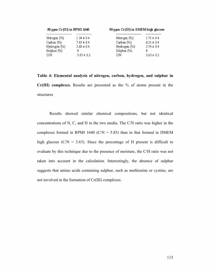
115
Table 4: Elemental analysis of nitrogen, carbon, hydrogen, and sulphur in
Cr(III) complexes. Results are presented as the % of atoms present in the
structures
Results showed similar chemical compositions, but not identical
concentrations of N, C, and H in the two media. The C/N ratio was higher in the
complexes formed in RPMI 1640 (C/N = 5.85) than in that formed in DMEM
high glucose (C/N = 3.63). Since the percentage of H present is difficult to
evaluate by this technique due to the presence of moisture, the C/H ratio was not
taken into account in the calculation. Interestingly, the absence of sulphur
suggests that amino acids containing sulphur, such as methionine or cystine, are
not involved in the formation of Cr(III) complexes.

116
Figure 5.5: Analysis of Cr(III)-complexes by ATR FT-IR. Cr(III) complexes
obtained in RPMI 1640 (A) and DMEM high glucose (B) were compared to
CrPO4 (C). AA: amino acid characteristic pics.
To show the chelating role of Cr(III) governing the formation of
complexes in cell culture media, nanoscale structures containing various
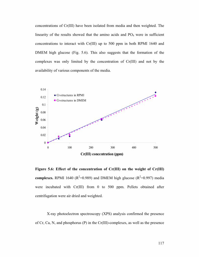
117
concentrations of Cr(III) have been isolated from media and then weighted. The
linearity of the results showed that the amino acids and PO4 were in sufficient
concentrations to interact with Cr(III) up to 500 ppm in both RPMI 1640 and
DMEM high glucose (Fig. 5.6). This also suggests that the formation of the
complexes was only limited by the concentration of Cr(III) and not by the
availability of various components of the media.
Figure 5.6: Effect of the concentration of Cr(III) on the weight of Cr(III)
complexes. RPMI 1640 (R2=0.989) and DMEM high glucose (R2=0.997) media
were incubated with Cr(III) from 0 to 500 ppm. Pellets obtained after
centrifugation were air dried and weighted.
X-ray photoelectron spectroscopy (XPS) analysis confirmed the presence
of Cr, Ca, N, and phosphorus (P) in the Cr(III)-complexes, as well as the presence
0
0.02
0.04
0.06
0.08
0.1
0.12
0.14
0 100 200 300 400 500
Cr(III) concentration (ppm)
Wei
ght (
g)
Cr-structures in RPMICr-structures in DMEM

118
of sodium (Na) and chlorine (Cl), but in different ratios (Fig. 5.7, Table 5). The
presence of oxygen in the survey spectrum was attributed to the presence of
moisture and therefore has not been taken into account in the calculation. Cr and
Cl were predominant, followed by N and P. Table IV reports on the presence of
C-N, N-H, C-C, and P-O bond displacements and their relative ratios as identified
by high resolution XPS (spectra not shown) confirming once more the role of
Cr(III) in chelating amino acids and PO4 and to form nanoscale structures.

119
Figure 5.7: X-ray photoelectron spectroscopy analysis of Cr(III) complexes.
Spectra were obtained from complexes formed in both RPMI 1640 (A) and
DMEM high glucose (B). Spectra show the presence of Cr, Na, P, C and O.
Table 5: Atoms present in Cr(III) complexes. Table summarizes the results
obtained by XPS (Figure 5.7) and shows the different elements present in the
DMEM and RPMI nanostructures (noticed DMEM Cr(III) complexes and RPMI-
Cr(III) complexes in the table 5 and their relative amount. Sensitivity factor are
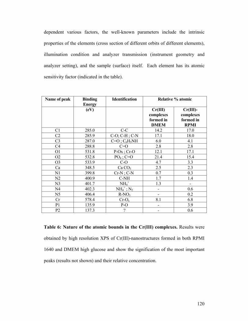
120
dependent various factors, the well-known parameters include the intrinsic
properties of the elements (cross section of different orbits of different elements),
illumination condition and analyzer transmission (instrument geometry and
analyzer setting), and the sample (surface) itself. Each element has its atomic
sensitivity factor (indicated in the table).
Name of peak Binding Energy
Identification Relative % atomic
(eV) Cr(III) complexes formed in DMEM
Cr(III)-complexes formed in
RPMI C1 285.0 C-C 14.2 17.0 C2 285.9 C-O; C-H ; C-N 17.1 18.0 C3 287.0 C=O ; C6H5NH 6.0 4.1 C4 288.8 C=O 2.8 2.8 O1 531.8 P-Ox ; Cr-O 12.1 17.1 O2 532.8 PO4 ; C=O 21.4 15.4 O3 533.9 C-O 4.7 3.3 Ca 348.5 Ca CO3 2.5 2.3 N1 399.8 Cr-N ; C-N 0.7 0.3 N2 400.9 C-NH 1.7 1.4 N3 401.7 NH4
+ 1.3 - N4 402.3 NH4
+ ; N2 - 0.6 N5 406.4 R-NO3 - 0.2 Cr 578.4 Cr-Ox 8.1 6.8 P1 135.9 P-O - 3.9 P2 137.3 ? - 0.6
Table 6: Nature of the atomic bounds in the Cr(III) complexes. Results were
obtained by high resolution XPS of Cr(III)-nanostructures formed in both RPMI
1640 and DMEM high glucose and show the signification of the most important
peaks (results not shown) and their relative concentration.

121
5.5. Discussion
Few publications to date have focused on the characterization of the
chemical nature of implant-derived metal ions in biofluids, relating the chemical
structure to their relative toxicity. The present work is an attempt to characterize
the molecular structure of Co(II), Cr(III) and Cr(VI) in simulated biological
fluids.
It is known that cobalt-chromium metallic implants release cobalt and
chromium ions in organism, by the natural corrosion process. However, a
complexation phenomenon is instantaneous, making impossible to extract
implant-derived metal ions in vivo, before their involvement in any chemical
process. Although commercial ions are used in this study, they are similar to
those generated from metallic surfaces.
We showed that of the group only Cr(III) could form insoluble nanoscale
complexes by interacting with the media components after centrifugation (5000 x
g, 20 min). The absence of precipitate with Co(II) and Cr(VI) was persistent even
though harsher experimental conditions were used (i.e. 16000 x g for 40 min
centrifugation). The Cr(III) complexes could not be dissolved in either acidic or
basic solutions. Solution pH has been modified in order to obtain a large range of
pH from basic to acidic by adding respectively NaOH or HCl. For further
investigations, isolated complexes were immerged in pure NaOH or HCl. No
solubilisation was observed. Polar and non-polar organic solvents also failed to
dissolve the precipitates. 27 solvents with different polarities have been used from
0.043 solvent polarity parameter (triethylamine) to 1.00 (water), including

122
common solvents such as toluene (0.099), chloroform (0.259), acetone (0.355),
methanol (0.762) and more specifically solvents such as dioxane (0.164)
hexamethylphosphoramide (0.315) or methoxyether (0.667). The low solubility of
the complexes caused some limitations when using liquid phase analytical
methods for characterization of the complex (NMR for example).
Our results, which are mostly based on solid phase characterization of the
complexes, indicated that Cr(III) binds PO4, Ca, and amino acids in cell culture
media . Complexes with a size ranging from 70 nm to 100 nm are formed in
RPMI 1640 and DMEM high glucose, respectively. This finding is in agreement
with the work of Zhikovitch et al who showed the formation of chemically
inactive CrPO4 in phosphate buffer containing Cr(III) (26).
Some studies showed that CrPO4 compounds have been found in tissues
surrounding orthopaedic implants (27). Other studies have demonstrated that
Cr(III) interacts with DNA and that the PO4 groups were necessary for that
interaction (28). Cr-DNA could be formed as a ternary complex involving amino
acids or glutathione bridges (29). These findings support our hypothesis
concerning the interaction of Cr, PO4, and amino acids for the formation of
Cr(III)-complexes in both RPMI 1640 and DMEM high glucose. Since the
concentration of amino acids was 100 times higher than the concentration of
vitamins in the media, it is highly unlikely the vitamins are significantly involved
in the formation of these complexes, if at all.
In order to define the exact molecular structure of the Cr(III)-complexes,
crystallization assays with various solvents were performed. X-ray diffraction

123
analysis showed that at the most only 33% crystallization of the complexes can be
achieved in cell culture media. Our attempt to match the crystal structure with one
of those available in the latest X-ray database failed. Elemental analysis however
clearly indicated that the amino acids composition of the Cr(III)-complexes
formed in RPMI 1640 is significantly different than the one formed in DMEM
high glucose. Ratio of 5.81 for C/N is obtained in RPMI 1640 media, while a ratio
of 3.63 was observed in DMEM high glucose (Table 4).
Considering the types of amino acids and their respective concentrations,
only three combinations of amino acids could correspond to the C/N ratio in the
RPMI 1640 Cr(III)-complexes:, 1) phenylalanine, proline, serine, and tryptophane
for a C/N=5.85; 2) tyrosine, proline, serine, or tryptophane for a C/N=5.85; and 3)
phenylalanine, tyrosine, serine, and tryptophane for a C/N=5.88. Furthermore, the
C/N ratio in DMEM high glucose could be represented by the following two
combinations of amino acids: 1) aspartic acid, threonine, valine, serine, and
isoleucine/leucine for a C/N=3.75; 2) aspartic acid, threonine, valine, serine, and
glutamic acid for a C/N=3.60. The presence of C6H5 groups in high resolution
XPS spectrum (Table 5) suggests that the likely involvement of phenylalanine or
tyrosine in the formation of complexes.
Since the 256 scans of FPA-FT-IR at a single point, on 30 different spots
of the samples having a diameter of 1 cm (vs. ~70 nm for the Cr(III)-complexes)
had the same profile, we can conclude that the chemical composition as defined
by the C/N ratios are representative of the overall Cr(III)-structures. In addition,
the aspect of the FT-IR spectrum indicated that Cr(III) ions are not a type of

124
“amino acids trap” but a chelating entity between PO4 groups, amino acids, and
Ca. Although the exact nature of the bonds remains to be investigated, the
presence of PO4 may favour the formation of low energy hydrogen bounds. The
efficacy of a given metal as a chelating agent in a biological system depends on
the binding constants of the chelator, the concentration of the metals, and the
presence and concentrations of other ligands competing for binding with the
chelating metal. In our case, it seems that Cr(III) ions are better chelating agents
than Co(II) or Cr(VI) ions in biological media under our experimental conditions.
Our results are in agreement with some studies reporting on the higher chelating
ability of Cr(III) compared to that of Co(II) and Cr(VI) (30,31). This is extremely
interesting because chelation is a very important process in biology. Chelating
therapy, for example, could be used as a medical treatment of thalassaemia, which
is a genetic disease that interferes with a person's ability to make hemoglobin
(32).
The presence of Ca, coming from inorganic salts available in high
concentration in RPMI 1640 (100 mg/L of Ca(NO3)2) and in DMEM high glucose
(200 mg/L of CaCl2), is another interesting finding. Indeed, this is in agreement
with the study of Harris et al (33) that showed a strong co-localization of Cr with
Ca in small areas of cytoplasm in A549 human lung adenocarcinoma epithelial
cells.
pH also seems to play a role in the formation of Cr-complexes. The pH of
the two chosen media is slightly basic, which precipitates both Cr(III) complexes
and commercial CrPO4. In fact, amino acids are zwitterions (PKa of COOH is

125
between 1.8 and 2.2 and PKa of NH2 is between 8.8 and 10.6) resulting in a major
concentration of COO-. Since Cr(III) has a strong affinity towards O-, it interacts
either with PO4 groups or with the carboxylic groups of amino acids. Also, both
glutamic acid and aspartic acid have their side chain negatively charged (PKa of
COOH side chain = 4.7) at the pH of the media. This would allow a strong
interaction between the Cr ions and these amino acids via the COO- group in
DMEM high glucose media. This may also explain the difference in the
morphology of complexes formed in the two different media, as observed by
TEM (Fig. 5.1).
These findings strongly suggest that there is a relationship between the
formation of Cr(III) nanoscale complexes and the lower toxicity of Cr(III)
compared to Cr(VI) (34-39). We believe that due to the size and nature of the
Cr(III) nanoscale-complexes, their cellular internalization follow different
mechanisms which in turn might lead to their lower toxicity. As demonstrated by
some in vivo studies, Cr(III) ions are primarily taken up by cells and internalized
via the cell membrane (40,41) while Cr(VI) ions have the ability to enter cells
through ion channels and accumulate inside cells at concentrations of up to a
hundred-fold greater than the extracellular concentration (42,43). Since we found
that neither Co(II) or Cr(VI) form complexes in the tested media, it should affect
their overall toxicity.

126
5.6. Conclusion
Our results show that among three ions, namely Co (II), Cr (III) and Cr
(VI), only Cr(III) formed nanoscale complexes in simulated physiological fluids,
namely in RPMI 1640 and DMEM high glucose. Although the use of many
analytical techniques allowed us to identify the involvement of phosphate,
calcium and amino acids in the chemical composition of the Cr(III)-complexes
formed in RPMI 1640 and in DMEM high glucose, they could not be matched to
any organic or inorganic crystal structure available to date in X-ray diffraction
databases. Formation of metal ion complexes in biological fluids is of great
importance for the understanding of ion cytotoxicity in vivo and our findings
could explain the difference between chromium and cobalt ions toxicity.
5.7. References
1. Brockett C, Williams S, Jin Z, Isaac G, Fisher J. Friction of total hip
replacements with different bearings and loading conditions. J Biomed Mater
Res B Appl Biomater 2007; 81 :508-15.
2- Sargeant A, Goswami T. Pathophysiological aspects of hip implants. J Surg
Orthop Adv 2006; 15:111-2.
3- Ren WP, Markel DC, Zhang R, Peng X,Wu B, Monica H, Wooley PH.
Association between UHMWPE particle-induced inflammatory
osteoclastogenesis and expression of RANKL, VEGF, and Flt-1 in vivo.
Biomaterials 2006; 30:5161-9.

127
4- Geller JA, Malchau H, Bragdon C, Greene M, Harris WH, Freiberg AA.
Large diameter femoral heads on highly cross-linked polyethylene: minimum
3-year results. Clin Orthop Rel Res 2006; 447:53-9.
5- Sharkey PF, Hozack WJ, Dorr LD, Maloney WJ, Berry D. The bearing
surface in total hip arthroplasty: evolution or revolution. Instr Course Lect
2000; 49:41-56.
6- Williams S, Tipper JL, Ingham E, Stone MH, Fisher J. In vitro analysis of the
wear, wear debris and biological activity of surface-engineered coatings for
use in metal-on-metal total hip replacements. Proc Inst Mech Eng [H] 2003;
217:155-63.
7- Jacobs JJ, Skipor AK, Patterson LM, Hallab NJ, Paprosky WG, Black J,
Galante JO. Metal release in patients who have had a primary total hip
arthroplasty. A prospective, controlled, longitudinal study. J Bone Joint Surg
[Am] 1998; 80:1447-58.
8- Jacobs JJ, Skipor AK, Doorn PF, Campbell P, Schmalzried TP, Black J,
Amstutz HC. Cobalt and chromium concentrations in patients with metal on
metal total hip replacements. Clin Orthop Ret Res 1996; (329 Suppl):S256-63.
9- Langkamer VG, Case CP, Heap P, Taylor A, Collins C, Pearse M, Solomon L.
Systemic distribution of wear debris after hip replacement. A cause for
concern? J Bone Joint Surg [Br] 1992; 74:831-9.
10- Merritt K, Brown SA. Distribution of cobalt chromium wear and corrosion
products and biologic reactions. Clin Orthop Rel Res 1996; (329 Suppl):S233-
43.
128
11- Zhitkovich A, Peterson-Roth E, Reynolds M. Killing of chromium-damaged
cells by mismatch repair and its relevance to carcinogenesis. Cell Cycle 2005;
8:1050-2.
12- Levina A, Harris HH, Lay PA. Binding of chromium(VI) to histones:
implications for chromium(VI)-induced genotoxicity. J Biol Inorg Chem
2006; 11:225-34.
13- Moghaddas S, Gelerinter E, Bose RN. Mechanisms of formation and
decomposition of hypervalent chromium metabolites in the glutathione-
chromium (VI) reaction. J Inorg Biochem 1995; 57:135-46.
14- Stearns DM, Kennedy LJ, Courtney KD, Giangrande PH, Phieffer LS,
Wetterhahn KE. Reduction of chromium(VI) by ascorbate leads to chromium-
DNA binding and DNA strand breaks in vitro. Biochemistry 1995; 34:910-9.
15- Gez S, Luxenhofer R, Levina A, Codd R, Lay PA. Chromium(V) complexes
of hydroxamic acids: formation, structures, and reactivities. Inorg Chem
2005; 44:2934-43.
16- Moghaddas S, Gelerinter E, Bose RN. Mechanisms of formation and
decomposition of hypervalent chromium metabolites in the glutathione-
chromium (VI) reaction. J Inorg Biochem 1995; 57:135-46.
17- Voitkun V, Zhitkovich A, Costa M. Cr(III)-mediated crosslinks of glutathione
or amino acids to the DNA phosphate backbone are mutagenic in human cells.
Nucleic Acids Res 1998; 6:2024-30.

129
18- Cobb AG, Schmalzreid TP. The clinical significance of metal ion release from
cobalt-chromium metal-on-metal hip joint arthroplasty. Proc Inst Mech Eng
[H] 2006; 220:385-98.
19- Catelas I, Petit A, Vali H, Fragiskatos C, Meilleur R, Zukor DJ, Antoniou J,
Huk OL. Quantitative analysis of macrophage apoptosis vs. necrosis induced
by cobalt and chromium ions in vitro. Biomaterials 2005; 26:2441-53.
20- Fleury C, Petit A, Mwale F, Antoniou J, Zukor DJ, Tabrizian M, Huk OL.
Effect of cobalt and chromium ions on human MG-63 osteoblasts in vitro:
morphology, cytotoxicity, and oxidative stress. Biomaterials 2006; 27:3351-
60.
21- Petit A, Mwale F, Tkaczyk C, Antoniou J, Zukor DJ, Huk OL. Induction of
protein oxidation by cobalt and chromium ions in human U937 macrophages.
Biomaterials 2005; 26:4416-22.
22- Petit A, Mwale F, Tkaczyk C, Antoniou J, Zukor DJ, Huk OL. Cobalt and
chromium ions induce nitration of proteins in human U937 macrophages in
vitro. J Biomed Mater Res A 2006; 79A:599-605.
23- Salnikow K, Su W, Blagosklonny MV, Costa M. Carcinogenic metals induce
hypoxia-inducible factor-stimulated transcription by reactive oxygen species-
independent mechanism. Cancer Res 2000; 60:3375-8.
24- Kitahara J, Yamanaka K, Kato K, Lee YW, Klein CB, Costa M. Mutagenicity
of cobalt and reactive oxygen producers. Mutat Res 1996; 370:133-40.
25- Zhitkovich A, Voitkun V, Costa M. Formation of the amino acid-DNA
complexes by hexavalent and trivalent chromium in vitro: importance of

130
trivalent chromium and the phosphate group. Biochemistry 1996; 35:7275-
82.
26- Case CP, Langkamer VG, James C, Palmer MR, Kemp AJ, Heap PF,
Solomon L. Widespread dissemination of metal debris from implants. J Bone
Joint Surg [Br] 1994; 76:701-12.
27- Chaudhary S, Pinkston J, Rabile MM, Van Horn JD. Unusual reactivity in a
commercial chromium supplement compared to baseline DNA cleavage with
synthetic chromium complexes. J Inorg Biochem 2005; 99:787-94.
28- Messer J, Reynolds M, Stoddard L, Zhitkovich A. Causes of DNA single-
strand breaks during reduction of chromate by glutathione in vitro and in cells.
Free Radic Biol Med 2006; 40:1981-92.
29- Harris HH, Levina A, Dillon CT, Mulyani I, Lai B, Cai Z, Lay PA. Time-
dependent uptake, distribution and biotransformation of chromium(VI) in
individual and bulk human lung cells: application of synchrotron radiation
techniques. J Biol Inorg Chem 2005; 10:105-18.
30- Walter PB, Macklin EA, Porter J, Evans P, Kwiatkowski JL, Neufeld EJ,
Coates T, Giardina PJ, Vichinsky E, Olivieri N, Alberti D, Holland J, Harmatz
P. Inflammation and oxidant-stress in beta-thalassemia patients treated with
iron chelators deferasirox (ICL670) or deferoxamine: an ancillary study of the
Novartis CICL670A0107 trial. Haematologica 2008; 93:817-23.
31- Cai L, Li XK, Song Y, Cherian MG. Essentiality, toxicology and chelation
therapy of zinc and copper. Curr Med Chem 2005; 12:2753-63.

131
32- Abdel-Latif S, El-Roudi O, Mohamed M. Chelation behaviour of nitroso-
pyrazolones with Mn(II), Co(II), Ni(II), Cu(II) AND Zn(II). Journal of
Thermal Analysis and Calorimetry 2003; 73: 939-50.
33- Bagchi D, Stohs SJ, Downs BW, Bagchi M, Preuss HG. Cytotoxicity and
oxidative mechanisms of different forms of chromium. Toxicology 2002;
180:5-22.
34- Bagchi D, Bagchi M, Stohs SJ. Chromium (VI)-induced oxidative stress,
apoptotic cell death and modulation of p53 tumor suppressor gene. Mol Cell
Biochem 2001; 222:149-58.
35- Blasiak J, Kowalik J. A comparison of the in vitro genotoxicity of tri- and
hexavalent chromium. Mutat Res 2000; 469:135-45.
36- Shelnutt SR, Goad P, Belsito DV. Dermatological toxicity of hexavalent
chromium. Crit Rev Toxicol 2007; 37:375-87.
37- Nethercott J, Paustenbach D, Adams R, Fowler J, Marks J, Morton C, Taylor
J, Horowitz S, Finley B. A study of chromium induced allergic contact
dermatitis with 54 volunteers: implications for environmental risk assessment.
Occup Environ Med 1994; 51:371-80.
38- Goulart M, Batoréu MC, Rodrigues AS, Laires A, Rueff J. Lipoperoxidation
products and thiol antioxidants in chromium exposed workers. Mutagenesis
2005; 20:311-5.
39- Sipowicz MA, Anderson LM, Utermahlen WE Jr, Issaq HJ, Kasprzak KS.
Uptake and tissue distribution of chromium(III) in mice after a single
intraperitoneal or subcutaneous administration. Toxicol Lett 1997; 93:9-14.

132
40- Suzuki T, Miyata N, Horitsu H, Kawai K, Takamizawa K, Tai Y, Okazaki M.
NAD(P)H-dependent chromium (VI) reductase of Pseudomonas ambigua G-
1: a Cr(V) intermediate is formed during the reduction of Cr(VI) to Cr(III). J
Bacteriol 1992; 174:5340-5.
41- Buttner B, Beyersmann D. Modification of the erythrocyte anion carrier by
chromate. Xenobiotica 1985; 15:735-41.
42- Biedermann KA, Landolph JR. Role of valence state and solubility of
chromium compounds on induction of cytotoxicity, mutagenesis, and
anchorage independence in diploid human fibroblasts. Cancer Res 1990;
50:7835-42.
43- Sathwara NG, Patel KG, Vyas JB, Patel S, Trivedi MR, Dave LM, Madia
MM, Kulkarni PK, Parikh DJ, Saiyed HN. Chromium exposure study in
chemical based industry 2007; 28:405-8.

133
CHAPTER 6
Interactions of Cr(III) Complexes with Serum Proteins
The 3rd paper presented here, and accepted for publication in Journal of
Biomedical Materials Research Part (Ref.: JBMR-A-09-0371.R2), fulfills the
objective of this thesis which aimed at understanding why Cr excretion is not
fully effective by determining the binding of Cr(III) complexes with serum
proteins.
Our previous in vitro studies suggested that Cr(III) are less toxic than
Cr(VI) and Co(II) and that this difference in toxicity may be explained by the
formation of Cr(III) complexes in physiological fluids. It is known that the
retention of organic Cr in the organism is much higher than the retention of
inorganic Cr compounds. This suggests that Cr ions circulating in blood can
interact with serum proteins, generating complexes with a high organic density.
In the present chapter, we determined through a proteomic approach the
nature of proteins bound to Cr(III) complexes in both bovine and human serum
and investigated their effect on the internalization of these complexes by
macrophages. Our main finding showed that Cr(III) complexes could bind only to
bovine albumin in bovine serum but to 8 proteins in human serum. We also
demonstrated that Cr(III) complexes formed in both RPMI 1640 and DMEM high
glucose and in the presence of human serum were more easily internalized by
macrophages than the complexes formed in bovine serum, suggesting that human
proteins playing a role of opsonins.

134
INVESTIGATION OF THE BINDING OF Cr(III) COMPLEXES TO
HUMAN AND BOVINE SERUM PROTEINS: A PROTEOMIC
APPROACH
Cathy Tkaczyk1,2,3, Olga L. Huk1,3, Fackson Mwale1, John Antoniou1,3, David.
J Zukor1,
Alain Petit1, Maryam Tabrizian2,3,4,5
1Division of Orthopaedic Surgery, McGill University,
Lady Davis Institute for Medical Research,
The Sir Mortimer B. Davis-Jewish General Hospital,
3755 Côte-Sainte-Catherine Road, Montreal, QC, H3T 1E2, Canada.
Fax: (514) 340-7502
2Department of Biomedical Engineering, 3Center of Biorecognition and
Biosensors (CBB),
4 McGill Institute for Advanced Materials, 5 Faculty of Dentistry,
Duff Medical Building, 3775 University Street, Montreal, QC, H3A 2B4, Canada.
Fax: (514) 398-7461

135
6.1. Abstract
One of the main concerns in the use of cobalt-chromium (Co-Cr) alloys-
based implants is the long-term fate of Co and Cr ions present in the blood,
organs, and urine of patients. Our previous studies have shown that Cr(III) can
form complexes in different cell culture media alone, whereas Co(II) and Cr(VI)
do not form any detectable structure under the same conditions. Since Cr(VI) and
Co(II) are known to be more toxic than Cr(III), we hypothesized that the
difference in their molecular structure could be the origin of the difference in their
toxicity. Herein, we investigated the interaction of the Cr(III) complexes formed
in RPMI 1640 and DMEM high glucose media with serum proteins and their
internalization by macrophages. Electrophoresis gels stained by Coomassie Blue
showed the presence of proteins for complexes formed in both media. Using a
liquid chromatography–quadrupole–time of flight (LC-Q-Tof) mass spectrometer,
we showed that in the presence of fetal bovine serum, Cr(III) complexes
interacted only with albumin, while they interact mainly with albumin, transferrin
and immunoglobulins (Ig) in the presence of human serum. Other proteins, such
as complement C3, alpha-2-macroglobulin, ceruloplasmin, plasminogen, and
apolipoprotein B may also interact with Cr(III) complexes but with lower
probability. The cellular uptake of Cr(III) complexes, visualized by transmission
electron microscopy, was also higher in RPMI 1640. Furthermore, Cr(III)
complexes were more easily engulfed by macrophages when they were formed
with human serum. To the best of our knowledge, this is the first report on Cr ions

136
and their interaction with macrophages in the presence of bovine and human
serum proteins.
6.2. Introduction
Numerous studies reported on the chromium (Cr) compounds toxicity to
organisms (1). Workers exposed to Cr often suffer from allergies, dermatitis,
asthma, and cancer. While Cr(VI) has been considered the main culprit for
“heavy” responses, Cr(III) has been shown to react more as an irritant (2). As far
as the orthopedic community is concerned, several studies have shown the
presence of Cr ions in the blood, urine, and organs of patients after metal-on-
metal (MM) total hip arthroplasty (THA) (3). The most significant concern on the
use of these implants, made of cobalt-chromium alloys, is the production of metal
ions, which could lead to cellular toxicity (4,5), metal hypersensitivity (6,7), and
chromosomal changes (8,9). In addition, these ions have the potential to generate
an oxidative stress that can impair cells via DNA damage, lipid peroxidation or
protein oxidation (10). Some investigations have also shown that Cr ions may be
responsible for protein nitration in human macrophages (11) and morphological
changes in human osteoblasts (12). Cr(VI) is known to be more toxic than Cr(III),
both in vitro and in vivo (13). So far, one study has been directed towards
understanding the molecular forms of these ions in biological fluids after their
release from metallic implants. Cr-phosphate corrosion products have been found
around retrieved hip implants and surrounding tissues (14). However, no further

137
investigation was performed to determine either their composition or to relate this
chemical composition to their level of toxicity.
Some studies attempted to elucidate the kinetic and the mechanism of
urinary exchange of Cr (15), and found that the urinary excretion of Cr is not fully
effective and Cr ions tend to accumulate in tissues and red blood cells (16,17). As
an explanation for such behavior, the literature points out possible interactions of
Cr with proteins (18). Organic Cr complexes have then been described to have a
higher retention coefficient in organs than inorganic Cr complexes or other forms
of Cr (19). However, the exact nature and mechanism of Cr interactions with
proteins and Cr complex retention still need to be understood.
Conversely, some investigations relate the lower toxicity of Cr (III) in
comparison to Cr(VI) to its mechanism of uptake through a phagocytosis-like
process (20). In a previous in vitro study, we have shown that Cr(III) formed
complexes in RPMI 1640 and DMEM high glucose cell culture media, whereas
Cr(VI) did not produce any detectable complexes under the same conditions (21).
Furthermore, the Cr(III) complexes were composed of organic and inorganic
components, tangled with Cr(III) ions. The organic compounds included amino
acids and phosphates, while calcium, sodium, and chromium were the main
compounds present in the inorganic phase. This study enabled us to relate, to
some extent, the chemical form of Cr to their mechanism of uptake and therefore
to their potential toxicity.
In light of these findings, the aim of this work was to provide new insight
on the predominant role of organic compounds, particularly of proteins, in the

138
formation of Cr(III) complex and their behavior and properties in vitro. To
elucidate the cellular uptake of Cr(III) ions, CrCl3 salt generating Cr(III) ions was
used. It was demonstrated that only Cr(III) is present in the crystal salt of CrCl3
(22) and is first released in the solution. However, Cr(II) and Cr(VI) may
theoretically also be formed from Cr(III). However, Cr(III) is the most
thermodynamically stable species and does not tend to reduce to Cr(II) or oxidize
to Cr(VI) that are more unstable. The valence of the ion is then similar to what is
observed in metal implants in which Cr(III) is released by the natural corrosion
process (23). Since the main cells present around orthopedic implants are
macrophages (24), the U937 cells, a monoblastoid cell line of human histiocytic
lymphoma (macrophage-like cells) (25) was used as cell culture model. They are
well known to engulf foreign bodies via different mechanisms. The “standard”
one is phagocytosis and involves immunoglobulins (Ig) and/or complement C3
playing the role of opsonins (26). In fact, opsonins are serum components acting
as attachment sites on the foreign bodies, which aid the internalization process.
Most of in vitro studies are performed with media supplemented with fetal bovine
serum (FBS). Therefore, the RPMI 1640 and DMEM high glucose media were
supplemented with FBS to determine biological differences with human serum
(HS) and point out the limitation of the use of FBS for in vitro studies.
This will help to improve our understanding of the fate and transit of metal
ions as well as the possible biological consequences of their interactions with
proteins both in vitro and in vivo. More than 600 proteins are present in sera,
albumin, transferrin, and fibrinogen being amongst the most abundant. There are

139
however, important differences in terms of protein composition and concentration
between FBS and HS as outlined in Table 7.
HUMAN SERUM (HS) FETAL BOVINE SERUM (FBS)
Albumin Albumin
Transferrin Alpha-1-Antiproteinase
Fibrinogen Plasminogen
IgA total Lactoperoxidase
Alpha-2-macroglobulin Kininogen
IgM total NADH-Ubiquinine-oxidoreductase
Alpha-1-Antitrypsin IgA total
C3 complement Alpha-2-Glycoprotein
Haptoglobin Hemiferrin
Table 7: Comparison of the most abundant proteins present in human and fetal
bovine serum.
6.3. Materials and Methods
6.3.1. Isolation of Cr(III) Complexes
The concentration of Cr(III) used in the present study is based on a
previous in vitro work showing that Cr(III) concentrations in the ppm range are
necessary to elicit a U937 cell response (11,27,28) and on our previous work on

140
the formation of Cr(III) complexes in cell culture media. (21). Fifty (50) ppm of
Cr ions (CrCl3, Sigma-Aldrich, Oakville, ON) were added to RPMI 1640 and
DMEM high glucose (HyClone, Logan, UT) supplemented with 5% FBS
(Hyclone) and 5% HS Sigma-Aldrich, Oakville, ON) as described below:
1) 50 ppm Cr(III) + RPMI 1640 + 5% FBS
2) 50 ppm Cr(III) + RPMI 1640 + 5% HSS
3) 50 ppm Cr(III) + DMEM high glucose + 5% FBS
4) 50 ppm Cr(III) + DMEM high glucose + 5% HS
After incubation for 1 h at 37°C, precipitates were isolated by
centrifugation at 5000 x g for 20 min, washed 3 times with water as previously
described (21) and loaded on 4-20% acrylamide gel. Proteins were then separated
by SDS-PAGE and gels stained with Coomassie Blue.
6.3.2. Detection of Proteins by Mass Spectrometry
Bands of proteins visible on acrylamide gels stained with Coomassie Blue
were cut out of the gels and taken for in-gel digestion on a robotic MassPrep
Workstation (Micromass; Waters Corp., Milford, MA). The gel pieces were
destained, reduced with 10 mM dithiothreitol, alkylated with 55 mM
iodoacetamide, and incubated with 6 ng/µl trypsin for 5 h at 37°C. Peptides were
then extracted with 1% formic acid/2% acetonitrile. Identification of the digested
proteins was completed using a Liquid Chromatography–Quadrupole–Time of
Flight (LC-Q-Tof) Mass Spectrometer (Micro Mass, Milford, MA). The digests
were loaded into a 10 cm capillary PicoFrit column filled with C18 stationary

141
phase and eluted by linear gradient of 5–70% acetonitrile in 0.1% formic acid at
the flow rate 200 µl/min. The eluted peptides were electrosprayed into Q-Tof and
the precursor ions were subjected to fragmentation by collision with argon
(MS/MS). The MS/MS data were submitted to Mascot (Matrix Science, London,
UK) for a search against the NCBI nonredundant database.
6.3.3. TEM Microscopy
U937 human macrophages (ATCC, Rockville, MD) were cultured in
RPMI 1640 and DMEM high glucose (Hyclone, Logan, UT) supplemented with
5% FBS or 5% HS, 100 U/ml penicillin, and 100 μg/ml streptomycin at 37°C
under 5% CO2 humidified atmosphere at a density of 5 × 10⁵ cells/ml. Cells were
incubated with 50 ppm of Cr(III) for 30 min at 37°C. After incubation, cells were
centrifuged at 500 × g for 6 min, supernatants were removed, and pellets were
fixed overnight in 0.5 ml of 2.5% glutaraldehyde. After centrifugation at
16,000 × g for 20 min, supernatants were removed and 0.5 ml of 1% osmium
tetroxide was added for 2 h. Cells were then dehydrated by increasing
concentrations of acetone (from 30% to 100%) to wash the pellets. After
centrifugation at 16,000 × g for 20 min, supernatants were removed and
epon:acetone with a ratio of 1:1 was added for 1 h, followed by epon:acetone with
a ratio of 2:1 for 1 h, and epon:acetone with a ratio of 3:1 overnight. At least
0.3 ml of pure epon resin was added and samples were incubated for 3 h under
vacuum to avoid any air bubble. After polymerization at 60°C for 48 h, the plastic
tubes were removed to obtain solid pieces of resin with cells pellets at the bottom.

142
These pellets were then sectioned in thin diamond knife cuts (about 90 nm). Cells
were observed using a JEOL 2000FX transmission electronic microscope (TEM)
(Akishima, Tokyo, Japan) at an accelerating voltage of 80 kV. To determine the
percentage of internalization, 500 cells from randomly selected fields were
counted and analyzed for the presence of nano-sized complexes (dark spots on the
TEM microphotographs). Finally, energy dispersive X-ray analysis (EDXA) was
used to confirm the nature of the internalized structures.
6.4 Results
Figure 6.1 shows that Cr(III) complexes obtained in different media
interacted with serum proteins. No proteins were found when Cr(III) was
incubated with media without serum, which was used as negative control. No
pelleted proteins could either be obtained after centrifugation of media and serum
without Cr(III) (results not shown), or after centrifugation of Cr(III) and serum
without medium dilution (21). The patterns of electrophoresis were similar for
complexes formed in the presence of FBS in RPMI 1640 (Fig. 6.1a) to those
formed in DMEM high glucose (Fig. 6.1b) with bands visible at 69 and 200 kDa.
In HS (Fig. 6.1c), major protein bands were detected at 10, 66, 68, 77, and 90 kDa
with similar electrophoresis patterns in both media.

143
Figure 6.1: Electrophoresis gels of Cr(III) complexes formed in the presence
of serum. Fifty (50) ppm of CrCl3 was incubated with the different media as
indicated in Materials and Methods. Proteins were separated on 4-20%
acrylamide gels and stained with Coomassie Blue.
a) Lane 1: molecular weight marker; Lane 2: RPMI + 50 ppm Cr3+; Lane 3: RPMI
+ 5% FBS + 50 ppm Cr3+.
b) Lane 1: molecular weight marker; Lane 2: DMEM + 50 ppm Cr3+; Lane 3:
DMEM + 5% FBS + 50 ppm Cr3+.
c) Lane 1: molecular weight marker; Lane 2: RPMI + 5% HS + 50 ppm Cr3+;
Lane 3: DMEM + 5% HS + 50 ppm Cr3+.
Figure 6.2 shows an illustration of MS/MS spectrum corresponding to the
fragmentation of the peptide CCTESLVNR by the MS. The generated data was
submitted to Mascot, a mass spectrometry search engine, in order to retrieve

144
proteins from primary sequence databases. The experimental mass value was
compared with calculated peptide mass or fragment ion mass values, obtained by
applying cleavage rules to the entries in a comprehensive primary sequence
database.
Figure 6.2: MS/MS spectrum of the peptide CCTESLVNR (serum albumin,
Homo sapiens) showing different ion fragments and their relative mass
(mass/charge (M/Z)). Electrophoresis gel bands of Cr(III) complexes in the
presence of FBS or HS were cut and digested with trypsin. The resulting peptide
mixture was analyzed by mass spectrometry and MS/MS.
By using an appropriate scoring algorithm, the closest match or matches
were identified. Results for the RNVLSETCC peptide are presented in Table 8. In
this example, 8 ion fragments on 70 matched with albumin for a peptide score of
60.1, demonstrating the presence of a human albumin. Ions score is 10*Log (P),
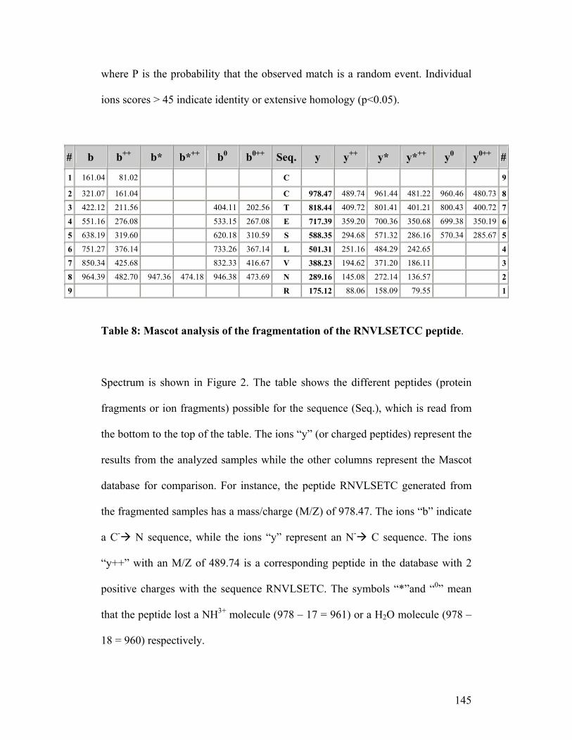
145
where P is the probability that the observed match is a random event. Individual
ions scores > 45 indicate identity or extensive homology (p<0.05).
# b b++ b* b*++ b0 b0++ Seq. y y++ y* y*++ y0 y0++ #
1 161.04 81.02 C 9
2 321.07 161.04 C 978.47 489.74 961.44 481.22 960.46 480.73 8
3 422.12 211.56 404.11 202.56 T 818.44 409.72 801.41 401.21 800.43 400.72 7
4 551.16 276.08 533.15 267.08 E 717.39 359.20 700.36 350.68 699.38 350.19 6
5 638.19 319.60 620.18 310.59 S 588.35 294.68 571.32 286.16 570.34 285.67 5
6 751.27 376.14 733.26 367.14 L 501.31 251.16 484.29 242.65 4
7 850.34 425.68 832.33 416.67 V 388.23 194.62 371.20 186.11 3
8 964.39 482.70 947.36 474.18 946.38 473.69 N 289.16 145.08 272.14 136.57 2
9 R 175.12 88.06 158.09 79.55 1
Table 8: Mascot analysis of the fragmentation of the RNVLSETCC peptide.
Spectrum is shown in Figure 2. The table shows the different peptides (protein
fragments or ion fragments) possible for the sequence (Seq.), which is read from
the bottom to the top of the table. The ions “y” (or charged peptides) represent the
results from the analyzed samples while the other columns represent the Mascot
database for comparison. For instance, the peptide RNVLSETC generated from
the fragmented samples has a mass/charge (M/Z) of 978.47. The ions “b” indicate
a C- N sequence, while the ions “y” represent an N- C sequence. The ions
“y++” with an M/Z of 489.74 is a corresponding peptide in the database with 2
positive charges with the sequence RNVLSETC. The symbols “*”and “0” mean
that the peptide lost a NH3+ molecule (978 – 17 = 961) or a H2O molecule (978 –
18 = 960) respectively.

146
All bands from Figure 6.1 were analyzed in the same way. It is important
to note that our choice was limited to proteins having at least 8 corresponding
peptides with ion scores over 45 in order to ascertain their presence in the Cr(III)
complexes. Results showed that with FBS, only albumin (123 peptides with
scores from 45 to 90) interacts with the Cr(III) complexes. With HS, albumin
(132 peptides with scores from 45 to 102), transferrin (98 peptides with scores
from 45 to 89), immunoglobulins (99 peptides with scores from 45 to 98),
apolipoprotein B (12 peptides with scores from 45 to 97), complement C3 (11
peptides with scores from 45 to 87), ceruloplasmin (11 peptides with scores from
45 to 79), alpha-2-macroglobulin (9 peptides with scores from 45 to 81), and
plasminogen (8 peptides with scores from 45 to 69) are all possibly interacting
with the Cr(III) complexes. The results obtained for albumin and transferrin are
summarized in Table 9.

147
Protein
Name
Gel
position
Accession no. MW
BOVINE Albumin 1 & 2 gi| 162648 71244
Albumin 1 & 2 gi| 229552 68083
HUMAN Albumin 1, 2, 4,
5 & 6
gi| 23307793 71344
Albumin 1, 2, 4,
5 & 6
gi| 28592 71316
Transferrin 3 & 4 gi| 553788 55207
Transferrin 3 & 4 gi| 31415705 79280
Transferrin 3 & 4 gi| 37747855 79329
Table 9: Most probable protein candidates in bovine and human sera
interacting with Cr(III) complexes. All bands from electrophoresis gels were
analyzed by MS/MS. The generated data was submitted to Mascot and the best
matches were identified as shown in Table 8.

148
Figure 6.3: TEM pictures of Cr(III) complexes with U937 macrophage-like
cells in serum supplemented media. Black arrows show Cr(III) complexes
inside the cells while white arrowheads show Cr(III) complexes outside the cells.
Note the presence of internalized vesicles (black arrow head). The black
arrowhead in the insert (RPMI with FBS) shows the formation of pseudopodia.
TEM images (Figure 6.3) along with EDXA analysis (Figure 6.4) clearly
indicated the presence of Cr(III) complexes in U937 macrophage-like cells or
interacting with them. The size of these complexes varied from 60 nm when

149
formed in RPMI 1640 (in both FBS and HS) to 100 nm when formed in DMEM
high glucose (in both FBS and HS).
Figure 6.4: EDXA spectra of Cr(III) complexes internalized by U937
macrophage-like cells in RPMI 1640 with FBS (a) and in DMEM high
glucose supplemented with FBS (b). EDXA spectra obtained in HS are the same
(data not shown). Copper peaks visible on the right of the spectra (8-9 keV) are
generated by the grids. Note the presence of chromium (Cr), phosphate (P) and
calcium (Ca) in both Cr(III) complexes.
Figure 6.5 shows the percentage of U937 cells that internalized Cr(III)
complexes (integrated into the cytoplasm, black arrow), as obtained by evaluating
500 cells under TEM for each medium/serum condition. A more important
cellular uptake of Cr(III) complexes was observed in RPMI 1640 than in DMEM
high glucose. The internalization of the Cr(III) complexes was also facilitated by
HS compared to FBS. Notably, 21% and 48% of U937 cells contained Cr(III)
complexes or interacting with Cr(III) complexes in RPMI 1640 in presence of

150
FBS and HS, respectively. Conversely, only 6% and 21% of U937 cells contained
Cr(III) complexes in DMEM high glucose in the presence of FBS and HS,
respectively.
Figure 6.5: Internalization of Cr(III) complexes by U937 macrophage-like
cells. Results are presented as the percentage (± standard deviation) of cells that
internalized the Cr(III) complexes over the total number of cells.
6.5. Discussion
Proteomic approach was used to study the interaction of Cr(III) complexes
formed in cell culture media (21) with serum proteins. Although proteomic is an
established technology for protein detection and discovery, it is not a widely used
methodology for determining the interactions between metal and proteins. We
found that Cr(III) complexes interacted only with albumin in the presence of FBS,

151
while they interacted with albumin, transferrin, and immunoglobulins in the
presence of HS. Alpha-2-macroglobulin, ceruloplasmin, plasminogen,
immunoglobulin, complement C3, and apolipoprotein B also seem to interact with
the Cr(III) complexes in HS. It is also interesting to point out the fact that
although albumin, transferrin, complement C3, alpha-2-macroglobulin,
ceruloplasmin, plasminogen, and apolipoprotein B are parts of the “standard
proteins” of plasma, they are not necessarily the most abundant ones (29),
suggesting that the concentration of proteins is not the prime factor to promote
their interaction with Cr(III) complexes and their internalization by U937
macrophage-like cells.
Our findings support a previous study demonstrating that 51Cr can bind to
transferrin and albumin in patients on continuous ambulatory peritoneal dialysis 30
and also enforce other studies showing the transfer of Cr(III) ions from blood to
urine in the presence of insulin (31-32). In fact, human albumin is known to bind
and transport a multitude of ligands such as bilirubin, fatty acids, retinoids,
steroids, heavy metals, and various drugs (33-35). The primary, secondary, and
tertiary structure of albumin provides the unique capability to bind, covalently or
reversibly, a great number of various endogenous and exogenous compounds (36-
39) with hydrophobic properties. However, heavy metals are also known to bind
to this protein (40-43) as it is the major transport protein in blood for Zn2+, which
is necessary for various physiological processes such as protein and DNA
synthesis, cell growth and division (44). It can also interact with Cd2+ and Cl- ions
(45-49). The presence of Cl- ions in Cr(III) complexes, as shown in our previous

152
study (21), suggests that Cl- might promote the interaction of Cr(III) complexes
with albumin.
Bovine and human albumin presents a 76% identical sequence (50). The
main difference between the two proteins is the presence of two tryptophan
residues (W131 and W214) in bovine albumin, whereas human albumin has only
one (W214) (51). This difference can play a role in their interactions with other
components. For instance, in a study on the interactions of both human and
bovine albumin with different detergents, spectroscopic results suggested distinct
interactions of detergent molecules with W131 and W214 (52). Moreover, it seems
that the binding of surfactants to HS is more complex than for FBS (53). This
complexity of binding of albumin is in agreement with the fact that more different
human proteins interact with Cr(III) complexes in human serum than in bovine
serum. This might also be the reason behind having a greater internalization by
U937 macrophage-like cells. Since it was demonstrated that cell lines of the
monocytic lineage had lower uptake of titanium oxide (TiO2) particles than
macrophages (54), it is possible that our results underestimated the capacity of
macrophages to internalize Cr(III) particles.
Like albumin, transferrin is involved in ion transport and is known as an
iron transporter protein. Ti(IV) can bind to transferrin, be recognized by the
transferrin receptor, and be up taken by the cells (55). Transferrin belongs to the
family of monomeric glycoproteins of~80 kDa which comprises two lobes (N and
C) (56), with each lobe containing a single metal-binding center. However,
despite substantial structural similarity between the two lobes, the existence of

153
significant differences in the lobes’ metal-binding properties has been
demonstrated (57). Indeed, TEM study showed that the Cr(III) complexes formed
with human serum are more easily internalized than those formed with bovine
serum. This difference can be explained by the absence of transferrin as a
mediator of cellular uptake of Cr(III) complexes when the medium is
supplemented with FBS (58).
Interestingly, no immunoglobin was involved in the formation of Cr(III)
complexes in the presence of FBS while they were involved in the formation of
Cr(III) complexes in HS, even though both human and bovine sera are composed
of 8-26% (5-18 mg/ml) of immunoglobins (59). While the reason for this
difference still needs to be investigated, we found that Cr(III) complexes formed
in HS interacted with immunoglobins and complement C3 and that these
complexes were more easily engulfed by human U937 cells than Cr(III)
complexes formed in FBS. Macrophages and monocytes can internalize
molecules and complexes mainly through 3 different pathways. When the
material is small (i.e. nutriments), it can be internalized by pinocytosis or
endocytosis. Larger substances are internalized by phagocytosis, in which
opsonins are involved. Indeed, our results suggest that the internalization of
Cr(III) complexes formed in HS occurs through the classical phagocytosis
pathway where immunoglobins and complement C3 are the opsonins that are
facilitating the recognition of foreign bodies by macrophages (26). However, the
absence of opsonin in the Cr(III) complexes formed in FBS suggests that
pinocytosis and/or endocytosis pathways may also be involved. This remains to

154
be investigated. Most importantly, it seems that Cr(III) ions enter cells by
different way than Cr(VI) that enter via anions channels (20). Those differences
might explain the difference of toxicity of these ions. Thus, the formation of
Cr(III) complexes along with the presence of proteins may be the origin in the
difference of toxicity between Cr(VI) and Cr(III). Moreover, our previous study
demonstrated that Cr(III) complexes cannot be dissolved at low pH (21),
suggesting that the Cr(III) complexes are not destroyed in the intracellular
endosomes following endocytosis/pinocytosis or in the phagosomes following
phagocytosis. This may also explain the high cellular and tissue retention of Cr
comparatively to Co that is easily eliminated in urine (16).
Results also suggest the presence of alpha-2-macroglobulin, known to be
involved in controlling inflammatory responses to different pathogens, in the
Cr(III) complexes formed in HS. Some studies have demonstrated that alpha-2-
macroglobulin has a very similar histidine and cysteine-rich region as albumin
(60). Additionally, alpha-2-macroglobulin is able to bind zinc (Zn2+) and
manganese (Mn2+) ions (61). Therefore, the ability of alpha-2-macroglobulin and
its similarity with albumin may explain the capacity of this protein to bind Cr(III)
complexes. In addition, alpha-2-macroglobulin shares the copper pool of plasma
with albumin and ceruloplasmin, an α2-glycoprotein that binds 90–95% of blood
plasma copper (62).

155
6.6. Conclusion
This work is the very first that uses a proteomic approach to study the
interaction of Cr(III) ions with bovine and human proteins present in sera. Our
results showed that Cr(III) complexes were able to bind albumin, transferrin, and
immunoglobulins in HS but only to albumin in FBS even though the other protein
were present in FBS. They also suggest a mechanism, the formation of organic Cr
complexes, for their retention in organ and cells. This study provides valuable
information on the specificity of interactions between Cr(III) complexes and
proteins in simulated physiological fluids.
6.7. References
1. Langard S. One hundred years of chromium and cancer: a review of
epidemiological evidence and selected case reports. Am J Ind Med
1990;17:189-215.
2. Iyer VJ, Banerjee G, Govindram CB, Kamath V, Shinde S, Gaikwad A,
Jerajani HR, Raman G, Cherian KM. Role of different valence states of
chromium in the elicitation of allergic contact dermatitis. Contact Dermatitis
2002;47:357-360.
3. MacDonald SJ, Brodner W, Jacobs JJ. A consensus paper on metal ions in
metal-on-metal hip arthroplasties. J Arthroplasty 2004;19(Suppl 3):12-16.
4. Haynes DR, Rogers SD, Hay S, Pearcy MJ, Howie DW. The differences in
toxicity and release of bone-resorbing mediators induced by titanium and

156
cobalt-chromium-alloy wear particles. J Bone Joint Surg [Am] 1993;75-
A:825-834.
5. Rae T. A study on the effects of particulate metals of orthopaedic interest on
murine macrophages in vitro. J Bone Joint Surg [Br] 1975;57:444-450.
6. Gawkrodger DJ. Metal sensitivities and orthopaedic implants revisited: the
potential for metal allergy with the new metal-on-metal joint prostheses. Br J
Dermatol 2003;148:1089-1093.
7. Hallab NJ, Mikecz K, Jacobs JJ. A triple assay technique for the evaluation
of metal-induced, delayed-type hypersensitivity responses in patients with or
receiving total joint arthroplasty. J Biomed Mater Res 2000;53:480-489.
8. Case CP. Chromosomal changes after surgery for joint replacement. J Bone
Joint Surg [Br] 2001;83:1093-1095.
9. Moriwaki H, Osborne MR, Phillips DH. Effects of mixing metal ions on
oxidative DNA damage mediated by a Fenton-type reduction. Toxicol In
Vitro 2008;22:36-44.
10. Stohs SJ, Bagchi D, Hassoun E, Bagchi M. Oxidative mechanisms in the
toxicity of chromium and cadmium ions. J Environ Pathol Toxicol Oncol
2001;20:77-88.
11. Petit A, Mwale F, Tkaczyk C, Antoniou J, Zukor DJ, Huk OL. Cobalt and
chromium ions induce nitration of proteins in human U937 macrophages in
vitro. J Biomed Mater Res Part A 2006;79:599-605.
12. Fleury C, Petit A, Mwale F, Antoniou J, Zukor DJ, Tabrizian M, Huk OL.
Effect of cobalt and chromium ions on human MG-63 osteoblasts in vitro:

157
morphology, cytotoxicity, and oxidative stress. Biomaterials 2006;27:3351-
3360.
13. McGregor DB, Baan RA, Partensky C, Rice JM, Wilbourn JD. Evaluation of
the carcinogenic risks to humans associated with surgical implants and other
foreign bodies - a report of an IARC Monographs Programme Meeting. Eur J
Cancer 2000;36:307-13.
14. Jacobs JJ, Urban RM, Gilbert JL, Skipor AK, Black J, Jasty M, Galante JO.
Local and distant products from modularity. Clin Orthop Relat Res
1995;319:94-105.
15. O'Flaherty EJ, Kerger BD, Hays SM, Paustenbach DJ. A physiologically
based model for the ingestion of chromium(III) and chromium(VI) by
humans. Toxicol Sci 2001;60:196-213.
16. Bitsch RG, Zamorano M, Loidolt T, Heisel C, Jacobs JJ, Schmalzried TP.
Ion production and excretion in a patient with a metal-on-metal bearing hip
prosthesis. A case report. J Bone Joint Surg [Am] 2007;89-A:2758-2763.
17. Granadillo VA, Parra de Machado L, Romero RA. Determination of total
chromium in whole blood, blood components, bone, and urine by fast
furnace program electrothermal atomization AAS and using neither analyte
isoformation nor background correction. Anal Chem 1994;66:3624-3631.
18. Kottwitz K, Laschinsky N, Fischer R, Nielsen P. Absorption, excretion and
retention of 51Cr from labelled Cr-(III)-picolinate in rats. Biometals
2009;22:289-295.

158
19. Kerger BD, Paustenbach DJ, Corbett GE, Finley BL. Absorption and
elimination of trivalent and hexavalent chromium in humans following
ingestion of a bolus dose in drinking water. Toxicol Appl Pharmacol
1996;141:145-158.
20. Wiegand HJ, Ottenwalder H, Bolt HM. Recent advances in biological
monitoring of hexavalent chromium compounds. Sci Total Environ
1988;71:309-315.
21. Tkaczyk C, Huk OL, Mwale F, Antoniou J, Zukor DJ, Petit A, Tabrizian M.
The molecular structure of complexes formed by chromium or cobalt ions in
simulated physiological fluids. Biomaterials 2009;30:460-467.
22. Kuhlov B. Magnetic ordering in CrCl3 at the phase transition. Phys.
Stat.sol(a) 1982; 72:161-168.
23. Merritt K, Brown SA. Distribution of cobalt chromium wear and corrosion
products and biologic reactions. Clin Orthop Relat Res 1996;329
(Suppl):S233-S243.
24. Ingham E, Fisher J. The role of macrophages in osteolysis of total joint
replacement. Biomaterials 2005;26:1271-1286.
25. Baek YS, Haas S, Hackstein H, Bein G, Hernandez-Santana M, Lehrach H,
Sauer S, Seitz H. Identification of novel transcriptional regulators involved
in macrophage differentiation and activation in U937 cells. BMC Immunol
2009;10:18 (15 pages).
26. Stuart LM, Ezekowitz RA. Phagocytosis: elegant complexity. Immunity
2005;22:539-550.

159
27. Catelas I, Petit A, Zukor DJ, Antoniou J, Huk OL. TNF-α secretion and
macrophage mortality induced by cobalt and chromium ions in vitro -
Quantitative analysis of apoptosis. Biomaterials 2003;24:383-391.
28. Petit A, Mwale F, Tkaczyk C, Antoniou J, Zukor DJ, Huk OL. Induction of
protein oxidation by cobalt and chromium ions in human U937
macrophages. Biomaterials. 26: 4416-4422, 2005.
29. Issaq HJ, Xiao Z, Veenstra TD. Serum and plasma proteomics. Chem Rev
2007;107:3601-3620.
30. Borguet F, Cornelis R, Delanghe J, Lambert MC, Lameire N. Study of the
chromium binding in plasma of patients on continuous ambulatory peritoneal
dialysis. Clin Chim Acta 1995;238:71-84.
31. Clodfelder BJ, Emamaullee J, Hepburn DD, Chakov NE, Nettles HS,
Vincent JB. The trail of chromium(III) in vivo from the blood to the urine:
the roles of transferrin and chromodulin. J Biol Inorg Chem 2001;6:608-617.
32. Clodfelder BJ, Vincent JB. The time-dependent transport of chromium in
adult rats from the bloodstream to the urine. J Biol Inorg Chem 2005;10:383-
393.
33. Chen J, Fitos I, Hage DS. Chromatographic analysis of allosteric effects
between ibuprofen and benzodiazepines on human serum albumin. Chirality
2006;18:24-36.
34. Fasano M, Curry S, Terreno E, Galliano M, Fanali G, Narciso P, Notari S,
Ascenzi P. The extraordinary ligand binding properties of human serum
albumin. IUBMB Life 2005;57:787-796.
160
35. Kandagal PB, Ashoka S, Seetharamappa J, Shaikh SM, Jadegoud Y, Ijare
OB. Study of the interaction of an anticancer drug with human and bovine
serum albumin: spectroscopic approach. J Pharm Biomed Anal 2006;41:393-
399.
36. Colmenarejo G. In silico prediction of drug-binding strengths to human
serum albumin. Med Res Rev 2003;23:275-301.
37. Curry S, Mandelkow H, Brick P, Franks N. Crystal structure of human serum
albumin complexed with fatty acid reveals an asymmetric distribution of
binding sites. Nat Struct Biol 1998;5:827-835.
38. Sudlow G, Birkett DJ, Wade DN. The characterization of two specific drug
binding sites on human serum albumin. Mol Pharmacol 1975;11:824-832.
39. Sugio S, Kashima A, Mochizuki S, Noda M, Kobayashi K. Crystal structure
of human serum albumin at 2.5 A resolution. Protein Eng 1999;12:439-446.
40. Filipe P, Morliere P, Patterson LK, Hug GL, Maziere JC, Freitas JP,
Fernandes A, Santus R. Oxygen-copper (II) interplay in the repair of semi-
oxidized urate by quercetin bound to human serum albumin. Free Radic Res
2004;38:295-301.
41. Liang H, Huang J, Tu CQ, Zhang M, Zhou YQ, Shen PW. The subsequent
effect of interaction between Co(2+) and human serum albumin or bovine
serum albumin. J Inorg Biochem 2001;85:167-171.
42. Stewart AJ, Blindauer CA, Berezenko S, Sleep D, Sadler PJ. Interdomain
zinc site on human albumin. Proc Natl Acad Sci (USA) 2003;100:3701-
3706.

161
43. Wang J, Zhang H, Yang K, Niu C, Ni J. Computer simulation of Zn(II)
speciation and effect of Gd(III) on Zn(II) speciation in human blood plasma.
Biol Trace Elem Res 2003;96:125-131.
44. Ahel I, Ahel D, Matsusaka T, Clark AJ, Pines J, Boulton SJ, West SC.
Poly(ADP-ribose)-binding zinc finger motifs in DNA repair/checkpoint
proteins. Nature 2008;451:81-85.
45. Bal W, Christodoulou J, Sadler PJ, Tucker A. Multi-metal binding site of
serum albumin. J Inorg Biochem 1998;70:33-39.
46. Goumakos W, Laussac JP, Sarkar B. Binding of cadmium(II) and zinc(II) to
human and dog serum albumins. An equilibrium dialysis and 113Cd-NMR
study. Biochem Cell Biol 1991;69:809-820.
47. Sadler PJ, Tucker A, Viles JH. Involvement of a lysine residue in the N-
terminal Ni2+ and Cu2+ binding site of serum albumins. Comparison with
Co2+, Cd2+ and Al3+. Eur J Biochem 1994;220:193-200.
48. Tinoco AD, Valentine AM. Ti(IV) binds to human serum transferrin more
tightly than does Fe(III). J Am Chem Soc 2005;127:11218-11219.
49. Zhou Y, Hu X, Dou C, Liu H, Wang S, Shen P. Structural studies on metal-
serum albumin. IV. The interaction of Zn(II), Cd(II) and Hg(II) with HSA
and BSA. Biophys Chem 1992;42:201-211.
50. Sen P, Fatima S, Khan JM, Khan RH. How methyl cyanide induces
aggregation in all-alpha proteins: a case study in four albumins. Int J Biol
Macromol 2009;44:163-169.
51. Peters T, Jr. 1985 Serum albumin. Adv Protein Chem 37:161-245.

162
52. Gelamo EL, Tabak M. Spectroscopic studies on the interaction of bovine
(BSA) and human (HSA) serum albumins with ionic surfactants.
Spectrochim Acta A Mol Biomol Spectrosc 2000;56-A:2255-2271.
53. Gelamo EL, Silva CH, Imasato H, Tabak M. Interaction of bovine (BSA)
and human (HSA) serum albumins with ionic surfactants: spectroscopy and
modelling. Biochim Biophys Acta 2002;1594:84-99.
54. Stringer B, Imrich A, Kobzik L. Flow cytometric assay of lung macrophage
uptake of environmental particulates. Cytometry 1995;20:23–32.
55. Tinoco AD, Eames EV, Valentine AM. Reconsideration of serum Ti(IV)
transport: albumin and transferrin trafficking of Ti(IV) and its complexes. J
Am Chem Soc 2008;130:2262-2270.
56. Sun H, Li H, Mason AB, Woodworth RC, Sadler PJ. N-lobe versus C-lobe
complexation of bismuth by human transferrin. Biochem J 1999;337 ( Pt
1):105-111.
57. Guo M, Sun H, McArdle HJ, Gambling L, Sadler PJ. Ti(IV) uptake and
release by human serum transferrin and recognition of Ti(IV)-transferrin by
cancer cells: understanding the mechanism of action of the anticancer drug
titanocene dichloride. Biochemistry 2000;39:10023-10033.
58. Aisen P. Transferrin, the transferrin receptor, and the uptake of iron by cells.
Met Ions Biol Syst 1998;35:585-631.
59. Stoica GH, Samborschi C, Michiu V. Influence of sex and age on serum
immunoglobulin concentrations in healthy subjects. Med Interne 1978;16:23-
31.

163
60. Moriya M, Ho YH, Grana A, Nguyen L, Alvarez A, Jamil R, Ackland ML,
Michalczyk A, Hamer P, Ramos D, Kim S, Mercer JF, Linder MC. Copper is
taken up efficiently from albumin and alpha2-macroglobulin by cultured
human cells by more than one mechanism. Am J Physiol Cell Physiol
2008;295:C708-C721.
61. Gettins P, Cunningham LW. A unique pair of zinc binding sites in the human
alpha 2-macroglobulin tetramer. A 35Cl and 37Cl NMR study. Biochemistry
1986;25:5004-5010.
62. Fehily D, Kiswani AS, Jenkins DM, Cremin FM. Redistribution of plasma
zinc between albumin and alpha 2-macroglobulin in pregnancy. J Trace
Elem Electrolytes Health Dis 1987;1:63-68.
164
CHAPTER 7
EVALUATION OF OXIDATIVE STRESS IN VIVO
The 4th and last paper presented in this chapter, submitted to Journal of
Arthroplasty (Ref 09-0432U), fulfills the last objective of the present thesis,
which was to assess the clinical relevance of increased levels of Co and Cr ions in
patients with MM implants.
The previous chapters suggested that the formation of Cr(III) complexes in
physiological fluids may explain the lowest toxicity of Cr(III) in vitro in
comparison to Cr(VI) and Co(II). The last aim of the present project was to assess
the relationship between the in vitro findings and the in vivo clinical situations.
In this chapter, we determined the concentrations of Cr and Co ions in five
groups of patients having a 28 mm-head metal-on-metal (MM) primary total hip
replacement for up to 10 years. We also measured the level of oxidative stress
markers (total antioxidants, peroxides, and nitrotyrosines) and the expression of
antioxidant enzymes (SODs, CAT, GPx, and HO-1). Results showed an increase
of both Cr and Co ion levels in the blood of these patients. However, these
increases had no effect on oxidative stress markers and very slight effect on the
expression of SOD and HO-1 expression, suggesting that the increased levels of
Co and Cr ions are not associated with significant oxidative stress damage in vivo.

165
SIGNIFICANCE OF ELEVATED BLOOD METAL ION LEVELS IN
PATIENTS WITH METAL-ON-METAL PROSTHESES: AN
EVALUATION OF OXIDATIVE STRESS MARKERS
Cathy Tkaczyk1,3,4, Olga L. Huk3,4, Fackson Mwale3, John Antoniou3,4, David. J
Zukor3,
Alain Petit3, Maryam Tabrizian1,2,4
1Department of Biomedical Engineering, 2Faculty of Dentistry,4Centre for
Biorecognition and Biosensors (CBB)
Duff Medical Building, 3775 University St, Montreal, QC H3A 2B4, Canada.
3Division of Orthopaedic Surgery, McGill University,
Lady Davis Institute for Medical Research,
The Sir Mortimer B. Davis-Jewish General Hospital,
3755 Chemin de la Côte Sainte Catherine, Montréal, QC, H3T 1E2, Canada.

166
7.1. Abstract
The main concern associated with Metal-on-Metal hip prostheses made of
chromium (Cr)-cobalt (Co) alloys, is the release of chromium and cobalt ions in
the blood and their accumulation in organs of patients. Moreover, it is widely
known that Cr and Co ions can enhance the production of reactive oxygen species
(ROS), known to be damageable for cells. In a matter of fact, these ROS have the
capacity to disturb the redox status of cells, leading to the oxidative stress which
generates damage at different levels in cells, from the extracellular environment to
the nucleus.
Different antioxidants are present in cells in order to prevent cell damage,
to stop the chain reactions generated by ROS and finally to repair the damage
caused by the oxidative stress. Antioxidant enzymes, such as superoxide
dismutases (SOD), catalase (CAT) and glutathione peroxidase (GPx) and a non-
classical one, heme-oxygenase-1 (HO-1) represent the major antioxidant defense
system in organism and have been widely studied recently because of their
involvement in many human diseases.
The aim of this work was to establish a relation between Cr and Co ions
released in serum of patients with a metal-metal hip implant and their capacity to
generate an oxidative stress. To answer this question, we assessed the antioxidant
enzymes, namely SOD, CAT, GPx and HO-1 at different time points. In addition,
we studied the presence of lipid and protein oxidation, which is a sign of
oxidative stress. Our findings suggested that SOD activity is related to ions
concentrations, suggesting that it could be used as oxidative stress biomarker in

167
vivo. Taken together, results showed that Cr and Co ions did no generate any
oxidative stress in patients, suggesting the efficacy of the complex antioxidant
system present in serum. This work is the very first to study the effects of metallic
ions released from metallic prostheses in the serum of patients, in term of
oxidative stress and to propose a biomarker to study oxidative stress in vivo.
7.2. Introduction
Metal-on-metal (MM) hip prostheses, made of cobalt-chromium (Co-Cr)
alloys, represent an excellent alternative to metal-polyethylene bearings in the
treatment of osteoarthritis of the hip because of their substantially lower wear
rates (1). However, the main concern associated with such bearings is the
presence of circulating ions in the organism (2). These metal alloys undergo
corrosion, either electrochemically or mechanically, and release metallic particles
and ions that disperse systemically (3). Indeed, several studies have shown that
Co and Cr particles and ions can enter the bloodstream and accumulate both in
surrounding tissues and organs of patients after MM total hip arthroplasty (THA)
(4-6). These ions are potentially toxic (7,8), may cause metal hypersensitivity
(9,10), and chromosomal aberrations (11), and may induce changes in the
proportions of peripheral blood lymphocytes (12-14).
The mechanism of metal ion toxicity is not completely understood but it is
known that they can generate reactive oxygen species (ROS), such as superoxide
ions (O2-), hydrogen peroxide (H2O2), hydroxyl radical (OH ), and nitrogen oxide
(NO ) through Fenton/ Haber-Weiss chemistry (15). These ROS are known to be

168
involved in numerous human diseases, including degenerative lung and heart
conditions, Alzheimer disease, rheumatoid arthritis, and aging (16). ROS are
implicated in the induction of oxidative stress generating cell and tissue damage.
Oxidative stress may result from an increased production of oxidants and/or a
decrease in antioxidant defense. Since the damage that a ROS produces depends
on its origin and type, the most accurate and clinically relevant measurement of
oxidative damage is to measure multiple products of this damage (17).
In the present study, we hypothesized that metal ions released from MM
bearings, namely Co and Cr, could generate oxidative stress in patients with MM
THA. We first measured the concentration of both Co and Cr ions in blood of
patients with MM 28 mm-head prostheses. We then evaluated the concentrations
of total antioxidants (TAS), peroxides, and nitrated protein as markers of
oxidative stress in the plasma of these patients. TAS measures the overall
antioxidant capacity of serum samples while peroxides were used as a marker of
lipid peroxidation and nitrated proteins (nitrotyrosines) as a marker of damage to
proteins. Since the induction of oxidative stress is also related to the activity of
specific antioxidant enzymes, in a second step we measured the activity of some
of these enzymes in the plasma of these patients. We specifically looked at
classical antioxidant enzymes (18) such as catalase (CAT), glutathione peroxidase
(GPx), and superoxide dismutase (SOD), as well as the non-classical antioxidant
enzyme heme oxygenase-1 (HO-1) (19). These enzymes were chosen because
their expression (CAT, GPX, and SOD) is modulated by Co and Cr ions in human
MG-63 pre-osteoblasts in vitro (20), and are implicated in multiple human disease

169
processes (18) and because the induction of HO-1 is a general response to
oxidative stress in mammalian cells (21).
In a recent study (22), it has been showed that the levels of oxidative stress
markers are not elevated in the plasma of patients 1 year after the implantation of
MM bearings. To the best of our knowledge, this is the very first study of its kind
attempting to further understand the clinical significance of elevated metal ion
levels by evaluating their long-term effect at the cellular oxidant/antioxidant level.
7.3. Materials and Methods
7.3.1. Study groups
A total of 127 patients having a 28 mm-head MM articulating interface
(Metasul, Zimmer, Warsaw, IN) and who gave a total of 156 blood samples were
included in the study. The serum of 46 patients scheduled for THA served as
control. The patients were separated into 5 groups: 1) the control group includes
46 patients; 2) 40 patients were included in the “less than 6 months” group; 3) 23
patients were included in the “1-2 years” group; 4) 28 patients were included in
the “3-4 years” group; 5) 19 patients were included in the “more than 4 years”
group. The age of patients at the time of blood sampling was similar for the 5
groups with a percentage of smokers varying from 14% of the patients (less than 6
months group) to 7% in the control group (Table 10). The UCLA activity and the
Harris Hip scores were also similar, except for the control and the less than 6

170
months groups that had lower results. Prior ethics committee approval and
informed written consent were obtained.
Table 10: Demographic and outcome measures of patients in the different study
groups.
7.3.2. Blood samples and ion concentrations
Whole blood samples were collected in Sarstedt Monovette tubes
equipped with needles for trace metal analysis (Sarstedt, Montreal, QC) and kept
at -80C until analysis. Co and Cr ions levels were measured as previously
described by inductively coupled plasma mass spectrometry (ICP-MS,
PerkinElmer SCIEX Elan 6100 DRC ICP-MS system; PerkinElmer Instruments,
Norwalk, CT) at the Geochemical Laboratories of McGill University (22). The
biological reference standard SeroNorm Trace Elements Whole Blood, Level 2
(Sero AS, Billingstad, Norway) was analyzed as a quality-control sample.
Age (range) UCLA HHS Smoker
Pre-OP 55 ± 9 (39-76 y) 4.8 ± 2.3 47 ± 14 7%
≤ 6 months 60 ± 10 (42-71 y) 4.7 ± 1.9 75 ± 16 14%
1-2 years 56 ± 11 (31-72 y) 6.0 ± 1.3 85 ± 14 11%
3-4 years 51 ± 8 (32-64 y) 6.6 ± 1.6 87 ± 13 8%
>4 years 52 ± 8 (33-65 y) 7.1 ± 1.6 90 ± 11 8%

171
7.3.3. Plasma preparation
Whole blood of patients (1.5 ml) was also collected into Sarstedt Li-
Heparin tubes and centrifuged at 500 x g for 10 min. Supernatant plasma was
stored at -80ºC for later analysis. Plasma was chosen for this part of the study
instead of whole blood because the assays for oxidative stress are not
recommended for whole blood and can lead to erroneous data.
7.3.4. Oxidative stress markers
Total antioxidant status (TAS) was measured in plasma using the Oxford
Biomedical total antioxidant power kit (Oxford, MI). This assay is based on the
capacity to prevent the ABTS (2-2’-Azino-di-[3-ethylbenzthiazoline sulphonite])
oxidation by metmyoglobin and has an inter-assay coefficient of variation (CV) of
3%.
Lipid peroxidation was measured as the total peroxide concentration in
plasma using the Biomedica OxyStat assay (Medicorp, Montreal, QC), which
assesses the concentration of an orange complex formed by the oxidation of Fe2+
by peroxides contained in samples. The assay has a 5.1% CV inter-assay
precision.
Plasma nitrotyrosine levels (nitrated proteins) were quantified using the
BIOXYTECH® Nitrotyrosine-EIA assay (OxisResearch™, Portland, OR) that has
an 11.2%CV inter-assay precision.
172
7.3.5.Antioxidant enzymes
The three types of superoxide dismutases (Cu/Zn-, Mn-, and Fe-SOD)
present in plasma were assessed by a colorimetric assay (Superoxide Dismutase
Assay kit, Cayman, Ann Arbor, MI). Cayman’s superoxide dismutase assay kit
utilizes a tetrazolium salt for detection of superoxide radicals generated by
xanthine oxidase and hypoxanthine. One unit of SOD is defined as the amount of
enzyme needed to exhibit 50% dismutation of the superoxide. It has a 3.2%CV
inter-assay precision.
Catalase (CAT) activity was measured in plasma of patients using the
Catalase Assay kit of Cayman, a colorimetric test based on the capacity of CAT to
produce formaldehydes after hydrolysing H2O2. The inter-assay CV is 3.8%.
The Glutathione Peroxidase Assay kit of Cayman was used to determine
the activity of all the glutathione peroxidases (GPX) present in patient plasma.
This assay measures GPX activity indirectly by a coupled reaction with
glutathione reductase and has a 5.7%CV inter-assay precision.
The presence of heme oxygenase-1 (HO-1) was assessed using the Human
HO-1 ELISA kit from Assay Designs (Ann Arbor, MI) in which a mouse
monoclonal antibody specific to human HO-1 is immobilized on wells and
captured HO-1 present in samples. The sensitivity of the assay is 0.78 ng/mL.
173
7.3.6. Statistical analysis
TAS, peroxides, CAT, and GPX data were symmetrically distributed.
Therefore, ANOVA followed by Scheffe’s test was used to compare the different
study groups. Metal ion, nitrotyrosine, SOD, and HO-1 data distributions were
asymmetric and variability between groups was significant. The Kruskall-Wallis
test that is a nonparametric equivalent of a one-way analysis of variance was used.
It is calculated as a regular ANOVA, but uses the ranks of the data and is
therefore resistant to outliers represented by the dots (•) in the box-plot
visualization of results, in which the box by itself represent the middle 50% (25%
to 75% percentiles) of the data. To facilitate calculations, all null values were
defined as the half of lowest sensitivity range of the assay. A p value < 0.05 was
considered as significant.
7.4. Results
Figure 7.1 shows the levels of Co and Cr ions in blood of patients with 28
mm-head MM prostheses. Results showed a significant increase of both Co (Fig.
7.1A) and Cr (Fig. 7.1B) in the blood of these patients within 6 month after their
hip replacement surgery. There was no difference in the levels of both Co and Cr
ions after the 1st year.
Figures 7.2 to 7.4 show the concentration of oxidative stress markers in
plasma of patients with 28 mm-head MM prostheses. Results showed no
differences between the various time groups for TAS (Fig. 7.2), peroxides (Fig.
7.3), and nitrotyrosines (Fig. 7.4), suggesting that the increased levels of Co and

174
Cr ions did not induce oxidative stress damage in plasma of these patients.
Figures 7.5 to 7.8 shows the activity of antioxidant enzymes in patient plasma.
Results showed that SOD significantly decreased by 65.8% in the plasma of
patients up to 2 years after surgery (p=0.005) (Fig. 7.5).
Figure 7.1: Co and Cr ion concentrations in the blood of patients with MM
THAs. Co and Cr were measured in the blood of patients with 28 mm-head MM
prosthesis up to 10 years post-operatively. Due to non-parametric distribution,
results are presented as box plot in which outliers are represented by the dots (•)

175
and the box by itself represents the middle 50% (25% to 75% percentiles) of the
data. Δ p< 0.05 versus control
Figure 7.2: Total antioxidant status (TAS) in the plasma of patients with MM
THAs. Total antioxidants were measured in the plasma of patients with 28 mm-
head MM prosthesis up to 10 years post-operatively. Results are expressed as the
mean ± standard deviation.

176
Figure 7.3: Peroxide concentrations in the plasma of patients with MM
THAs. Lipid peroxides were measured in plasma of patients with 28 mm-head
MM prosthesis up to 10 years post-operatively. Results are expressed as the mean
± standard deviation.
Figure 7.4: Nitrotyrosine concentrations in the plasma of patients with MM
THAs. Nitrotyrosines were measured in plasma of patients with 28 mm-head MM

177
prosthesis up to 10 years post-operatively. Due to non-parametric distribution,
results are presented as box plot in which outliers are represented by the dots (•)
and the box by itself represents the middle 50% (25% to 75% percentiles) of the
data.
This decrease was transient with a progressive return to control value after
3 years. The activity of SOD in the more than 4 year group was even higher than
in the less than 6 month group (2.4 times; p= 0.04), the 1-2 year (7.8 times; p=
0.006), and the 3-4 year (1.3 times; p=0.02) groups (Fig. 7.5). These results
suggest that Co and Cr ions may modify SOD activity in plasma of patients with
28 mm-head MM prostheses. Results also showed slow, not significant changes in
CAT activity for all time intervals (Fig. 7.6). There were no statistical differences
in the activity of GPX (Fig. 7.7) between the five groups.
Finally, the level of HO-1 decreased after implantation of the prostheses
with statistical difference observed after 1 year (p= 0.03), at 3-4 years (p= 0.005),
and after 4 years (p= 0.009) comparatively to the control group (Fig 7.8). In the
more than 4 year group, the decrease reached 29.8% of the control group. The
HO-1 level was also lower in the 1-2 year (p= 0.01) and the 3-4 year (p=0.006)
groups compared to the less than 6 month group. These decreases in HO-1 levels
suggest a role for Co and Cr ions in the modulation of circulating HO-1 in
patients with 28 mm-head MM prostheses.

178
Figure 7.5: Superoxide dismutases (SODs) activity in the plasma of patients
with MM THAs. SOD activity was measured in the plasma of patients with 28
mm-head MM prosthesis up to 10 years post-operatively. Due to non-parametric
distribution, results are presented as box plot in which outliers are represented by
the dots (•) and the box by itself represents the middle 50% (25% to 75%
percentiles) of the data. Δ p<0.05 versus control; * p< 0.05 versus ≤ 6
months.
NB: The p value for the > 4 year group compared to ≤ 6 month group is 0.0486,
which is at the limit of significance. The p value for the > 4 year group compared
to control group is 0.038.

179
Figure 7.6: Catalase (CAT) activity in the plasma of patients with MM
THAs. CAT activity was measured in the plasma of patients with 28 mm-head
MM prosthesis up to 10 years post-operatively. Results are expressed as the mean
± standard deviation.
Figure 7.7: Glutathione peroxidase (GPX) activity in the plasma of patients with
MM THAs. GPX activity was measured in the plasma of patients with 28 mm-

180
head MM prosthesis up to 10 years post-operatively. Results are expressed as the
mean ± standard deviation.
Figure 7.8: Heme oxygenase-1 (HO-1) concentrations in the plasma of
patients with MM THAs. HO-1 was measured in the plasma of patients with 28
mm-head MM prosthesis up to 10 years post-operatively. Due to non-parametric
distribution, results are presented as box plot in which outliers are represented by
the dots (•) and the box by itself represents the middle 50% (25% to 75%
percentiles) of the data. Δ p< 0.05 versus control; * p< 0.05 versus ≤ 6
months.
7.5. Discussion
The present study showed that levels of Co and Cr ions increased rapidly
after surgery in blood of patients with 28 mm-head MM THA. The levels of both
Co and Cr ions in our study were in the same range as those reported in previous

181
studies (23-30) using different prostheses, with mean (or median) Co ion levels
varying from 0.7 to 2.2 ppb (12 to 38 nmol/L) and mean (or median) Cr ion levels
varying from 1.3 to 2.8 ppb (24 to 53 nmol/L). However, due to differences in the
choice of samples (erythrocyte vs. serum vs. whole blood) and in the
methodology (digested samples vs. diluted samples; ICP-MS vs. graphite
furnace), it is impossible to identify a prosthesis that releases the least amount of
ions into patient circulation. Interestingly, the concentration of Cr and Co ions
was much lower than those observed in periprosthetic fibrous tissue adjacent to
the implants (31) but are comparable to those found in synovial fluid (32).
The rapid increase in blood ions found in the first year is also comparable
to what was observed by other groups (28-30). This increase was followed by a
plateau by 4 years. This suggests that, in vivo, the run-in phase of accelerated
wear is indeed limited to the first 2 years postoperatively. A steady-state phase is
reached from 6 months after surgery. In the present study, the number of patients
per group and the fact that the study is not longitudinal (as the groups are formed
of different patients) did not allow confirmation of this plateau. This suggests that
after initial run-in-phase production, excretion of ions remains relatively constant
to maintain constant level in patients. The similarity between previous
longitudinal studies (28-30), and the present study supports the validity of our
results even if their non-longitudinal aspect may represent a bias in the analysis of
metal ions. Results from the present study have in counterpart the advantage of
having been obtained with the same method of measurement (needles, tubes,
solutions, standard, and technology (ICP-MS)) up to 10 years post-operatively.

182
Indeed, when we subdivided the more than 4 years group, there was no statistical
differences in the levels of Co and Cr ions between the 5-6 years group (12
patients) and the ≥ 7 years group (7 patients) (p > 0.8) (results not shown). This
remains to be confirmed with larger groups of patients. However, the role of renal
excretion in the maintenance of metal ion levels has been shown by the dramatic
increases in serum Co and Cr in patients with renal failure (29). Indeed, the
observed constant levels of Co and Cr ions may also represent a change in the
excretion of ions via the kidney that balances the generation of ions in patients
with MM implants (3).
The single most significant obstacle preventing a broader application of
MM hip arthroplasties and resurfacings continues to be the concerns regarding
elevated metal ion levels in the blood, urine, and tissues of patients with these
bearing surfaces, especially in young patients (2). An elevation of chromosomal
aberrations was described in peripheral blood lymphocytes within 2 years
postoperatively in patients with MM implants. However, as mentioned by the
authors themselves, chromosomal aberrations are not necessarily associated with
increased cancer incidence (33). Moreover, the same group also showed
chromosome aberrations in patients with metal-polyethylene implants requiring
revision (34). Visuri et al. initially reported a relative higher incidence of
hematopoietic cancers in patients with MM prostheses (35). In the later study,
however, the patient population was relatively small and no adjustments were
made for co-morbidities that increase the patient risk of developing neoplasm
(such as rheumatoid arthritis, Paget’s disease, or bone infarcts) (35). More

183
recently, in a thorough evaluation of a larger cohort of patients, Visuri et al. were
unable to demonstrate a relative increased risk of cancer in patients with MM
prostheses (36). Finally, in a meta-analysis of 8 epidemiologic studies including
over 130,000 patients with total hip and knee arthroplasties, no causal link could
be established between MM hip bearings and cancer (37). Nevertheless, continued
surveillance of patients with MM bearings is warranted to understand the clinical
relevance of raised metal ions in these patients.
In an ongoing need to assess the potential consequences of elevated ion
levels, we chose to evaluate oxidative stress markers in our patient population
with MM THAs. Metal ions have the potential to induce the production of
reactive oxygen species (ROS), making them prime suspects for disturbing the
balance of oxidants/antioxidants in circulating cells (15,38,39). This
oxidant/antioxidant unbalance has been identified in numerous degenerative
processes including Alzheimer’s disease, macular degeneration, rheumatoid
arthritis, neoplasm, and aging (16). Three antioxidant groups of molecules
constitute the antioxidant defense system of the organism: the primary system
prevents the formation of ROS, the secondary system eliminates radicals to
prevent chain reactions, and the tertiary system works to repair damaged
molecules (39). TAS assesses these three antioxidant groups. In the present study,
we showed that there were no changes in the levels of three oxidative stress
markers (TAS, nitrotyrosine, and peroxides) and few changes in the activity of
four antioxidant enzymes in patients with 28 mm-head MM prostheses compared
to the control group, suggesting that the increased levels of metal ions in patients

184
with MM THA had no or little effect on oxidative stress in the blood of these
patients. The results also stayed the same if we subdivided the more than 4 years
group into a 5-6 years group and a more than 7 years group as observed for metal
ions. Our results suggest that the increased levels of metal ions were not sufficient
to disturb the overall defense antioxidant system (as measured by TAS) of
patients with 28 mm-head MM prostheses. Furthermore, when looking at more
specific markers of oxidative stress, namely lipid damage and nitrotyrosines, we
were unable to show any macromolecule damage in our patient population.
Further studies, with patients having prostheses with different designs, bigger
head-size, as well as with hip resurfacing, are however necessary to conclusively
determine the effects of elevated circulating ions on oxidative stress in the blood
of patients with MM bearings.
Oxidative stress occurs when the balance of formation of oxidants exceeds
the ability of antioxidant systems to remove ROS. In this regards, it was shown
that Co and Cr could modify the expression of CAT and GPX in human MG-63
pre-osteoblasts (20) and that the lack of SOD in knockout mice have dramatic
repercussions on phenotypes with perinatal or early postnatal mortality (40).
Moreover, these enzymes are involved in many pathophysiological processes
including neurodegeneration, cancer, and aging (18,41). Results of the present
study show that the overall capacity of the antioxidant enzymes to remove ROS
was not affected by metal ions in patients with 28 mm-head MM prostheses. We
observed a transient decrease of SOD up to 2 years postoperatively group with a
return to control values thereafter. The SOD activity we measured in the plasma

185
corresponded to SOD3 (extracellular SOD) that is present in low concentration
compared to cellular SODs. Furthermore, SOD3 present in extracellular spaces of
tissues accounts for 90-99% of the SOD3 present in the body. This low
concentration of SOD3 circulating in plasma may make it more sensitive to minor
changes of oxidative status (42). This may explain the variation of SOD activity
observed in plasma of patients with 28 mm-head MM prostheses whereas the
other antioxidant enzymes were not affected in the same conditions. Our results
are in agreement with a previous study in COS7 cells where cobalt chloride led to
a decrease in the resistance to oxidative damage by down-regulating the
expression of SOD3 (43).
HO-1 is also known to be implicated in many human diseases such as
Alzheimer and inflammatory diseases (21). Previous findings showed that Cr(VI)
induced the expression of HO-1 in human dermal fibroblasts (44). This suggests
that Cr ions were not implicated in the progressive decrease of HO-1 we observed
in patients with MM THA up to 4 years post-operatively. However, HO-1
increased at longer term (> 4 years), suggesting that the effect might be reversible.
A down-regulation of HO-1 was shown in vivo in patients with hepatitis C, while
other antioxidant enzymes, such as CAT and SOD, were not affected (45). This
suggests that parameters other than the presence of metal ions might be
responsible for the decreased levels we observed in patients with MM prostheses.

186
7.6. Conclusion
This work is the first to determine the biological effects of metal ions
released from MM hip implants with regards to mid-term systemic oxidative
stress and showed that the increased levels of Co and Cr ions are not associated
with significant oxidative stress damage.
7.7. References
1. Singh R, Dahotre NB. Corrosion degradation and prevention by surface
modification of biometallic materials. J Mater Sci - Mater Med 2007,
18:725.
2. MacDonald SJ. Can a safe level for metal ions in patients with metal-on-
metal total hip arthroplasties be determined? J Arthroplasty 2004; 8:71.
3. Cobb AG, Schmalzreid TP. The clinical significance of metal ion release
from cobalt-chromium metal-on-metal hip joint arthroplasty. Proc Inst Mech
Eng [H] 2006; 220:385.
4. Case CP, VG Langkamer, C James, et al. Wide spread dissemination of
metal debris implants. J Bone Joint Surg [Br] 1994; 76-B:701.
5. Merritt K, SA Brown. Distribution of cobalt and chromium wear corrosion
products and biological reactions. Clin Orthop Relat Res 1996; 329 (Suppl):
S233.
6. Urban RM, JJ Jacobs, MJ Tomlinson, et al. Dissemination of wear particles
to the liver, spleen, and abdominal lymph nodes of patients with hip or knee
replacement. J Bone Joint Surg [Am] 2000; 82-A:457.

187
7. Rae T. A study on the effects of particulate metals of orthopaedic interest on
murine macrophages in vitro. J Bone Joint Surg [Br] 1975; 57-B:444.
8. Haynes DR, Rogers D, Pearcy MJ, et al. The differences in toxicity and
release of bone-resorbing mediators induced by titanium and cobalt-
chromium-alloy wear particles. J Bone Joint Surg [Am] 1993; 75-A:825.
9. Hallab NJ, Merritt K, Jacobs JJ. Metal sensitivity in patients with
orthopaedic implants. J Bone Joint Surg [Am] 2001; 83-A:428.
10. Gawkrodger DJ. Metal sensitivities and orthopaedic implants revisited: the
potential for metal allergy with the new metal-on-metal joint prostheses. Br
J Dermatol 2003; 148:1089.
11. Case CP. Chromosomal changes after surgery for joint replacement
(Editorial). J Bone Joint Surg [Br] 2001; 83-B:1093.
12. Savarino L, Granchi D, Ciapetti, G, et al. Effects of metal ions on white
blood cells of patients with failed total joint arthroplasties. J Biomed Mater
Res 1999; 47:543.
13. Case CP, Langkamer VG, Lock RJ, et al. Changes in the proportions of
peripheral blood lymphocytes in patients with worn implants. J Bone Joint
Sur [Br] 2000; 82-B:748.
14. Granchi D, Savarino L, Ciapetti G, et al. Immunological changes in patients
with primary osteoarthritis of the hip after total joint replacement. J Bone
Joint Surg [Br] 2003; 85-B:758.
15. Stohs SJ, Bagchi D. Oxidative mechanisms in the toxicity of metal ions.
Free Radic Biol Med 1995; 18:321.

188
16. Spector A. Oxidative stress and disease. J Ocul Pharmacol Ther 2000;
16:193.
17. Argüelles S, García S, Maldonado M, et al. Do the serum oxidative stress
biomarkers provide a reasonable index of the general oxidative stress status?
Biochim Biophys Acta 2004; 1674:251.
18. Matés JM, Pérez-Gómez, Núnez de Castro T. Antioxidant enzymes and
human diseases. Clin Biochem 1999; 32:595.
19. Foresti R, Motterlini R. The heme oxygenase pathway and its interaction
with nitric oxide in the control of cellular homeostasis. Free Radic Res
1999; 31:459.
20. Fleury C, Petit A, Mwale F, et al. Effect of cobalt and chromium ions on
human MG-63 osteoblasts in vitro: morphology, cytotoxicity, and oxidative
stress. Biomaterials 2006; 27:3351.
21. Applegate LA, Luscher P, Tyrrell RM. Induction of heme oxygenase: a
general response to oxidant stress in cultured mammalian cells. Cancer Res
1991; 51:974.
22. Antoniou J, Zukor DJ, Mwale F, et al. Metal ion levels in the blood of
patients after hip resurfacing: a comparison between twenty-eight and thirty-
six-millimeter-head metal-on-metal prostheses. J Bone Joint Surg [Am]
2008; 90-A (Suppl) 3:142.
23. MacDonald SJ, McCalden RW, Chess DG, Bourne RB, Rorabeck CH,
Cleland D, Leung F. Metal-on-metal versus polyethylene in hip arthroplasty:
a randomized clinical trial. Clin Orthop Relat Res 2003; 406:282.

189
24. Clarke MT, Lee PTH, Arora A, et al. Levels of metal ions after small- and
large-diameter metal-on-metal hip arthroplasty. J Bone Joint Surg [Br]
2003; 85-B:913.
25. Savarino L, Granchi D, Ciapetti G. et al. Ion release in patients with metal-
on-metal bearings in total joint replacement: a comparison with metal-on-
polyethylene bearings. J Biomed Mater Res – Applied Biomater 2002;
63:467.
26. Damie F, Favard L. [Metal serum levels in 48 patients bearing a chromium-
cobalt total hip arthroplasty with a metal-on-polyethylene combination]
[French]. Rev Chir Orthop Rep App Moteur 2004; 90:241.
27. Daniels J, Ziaee H, Salama A, et al. The effect of the diameter of metal-on-
metal bearings on systemic exposure to cobalt and chromium. J Bone Joint
Surg [Br] 2006; 88-B:443.
28. Savarino L, Granchi D, Ciapetti G, et al. Ion release in stable hip
arthroplasties using metal-on-metal articulating surfaces: a comparison
between short- and medium-term results. J Biomed Mater Res Part A 2003;
66-A:450.
29. Brodner W, Bitzan P, Meisinger V, et al. Serum cobalt levels after metal-on-
metal total hip arthroplasty. J Bone Joint Surg [Am] 2003; 85-A:2168.
30. Lhotka C, Szekeres T, Steffan I, et al. Four-year study of cobalt and
chromium blood levels in patients managed with two different metal-on-
metal total hip replacements. J Orthop Res 2003; 21:189.

190
31. Dorr LD, Bloebaum R, Emmanual J, et al. Histologic, biochemical, and ion
analysis of tissue and fluids retrieved during total hip arthroplasty. Clin
Orthop Relat Res 1990; 261:82.
32. Brien WW, Salvati EA, Betts F, et al. Metal levels in cemented total hip
arthroplasty. Clin Orthop Relat Res 1992; 276:66.
33 Bhamra MS , Case CP. Biological effects of metal-on-metal hip
replacements. Proc Inst Mech Eng [H] 2004; 220:379.
34 Doherty AT, Howell RT, Ellis LA, et al. Increased chromosome
translocations and aneuploidy in peripheral blood lymphocytes of patients
having revision arthroplasty of the hip. J Bone Surg Br 2001; 83(7):1075.
35 Visuri T, Pukkala E, Paavolainen P, et al. Cancer risk after metal on metal
and polyethylene on metal total hip arthroplasty. Clin Orthop Relat Res
1996; 329:S280.
36 Visuri T, Pukkala E, Pulkkinen P, et al. Cancer incidence and causes of
death among total hip replacement patients: a review based on Nordic
cohorts with a special emphasis on metal-on-metal bearings. Proc Inst Mech
Eng [H] 2006; 220:399.
37 Tharani R, Dorey FJ, Schmalzried TP. The risk of cancer following a total
hip or knee arthroplasty. J Bone Joint Surg Am 2001; 83(5):774.
38. Stohs SJ, Bagchi D, Hassoun E, et al. Oxidative mechanisms in the toxicity
of chromium and cadmium ions. J Environ Pathol Toxicol Oncol 2001;
20:77.

191
39. Chaudière J, Ferrari-Iliou R. Intracellular antioxidants : from chemical to
biochemical mechanisms. Food Chem Toxicol 199; 37:949.
40. Sentman ML, Granström M, Jakobson H, et al. Phenotypes of mice lacking
extracellular superoxide dismutase and copper- and zinc-containing
superoxide dismutase. J Biol Chem 2006;281:6904.
41. Matés JM, Sánchez-Jiménez F. Antioxidant enzymes and their implications in
pathophysiologic processes. Frontiers Biosci 1999; 4:339.
42. Rodriguez C, Mayo JC, Sainz RM, et al. Regulation of antioxidant enzymes:
a significant role for melatonin. J Pineal Res 2004; 36:1.
43. Kamiya T, Hara H, Yamada H, et al. Cobalt chloride decreases EC-SOD
expression through intracellular ROS generation and p38-MAPK pathways
in COS7 cells. Free Radic Res 2008; 42:949.
44. Joseph P, He Q, Umbright C. Heme-oxygenase 1 gene expression is a
marker for hexavalent chromium-induced stress and toxicity in human
dermal fibroblasts. Toxicol Sci 2008; 103:325.
45. Abdalla M.Y, Britigan B.E, Wen F, et al. Down-regulation of heme
oxygenase–1 by hepatitis C virus infection in vivo and by the in vitro
expression of hepatitis C core protein. J Infect Dis 2004; 190:1109.

192
CHAPTER 8
GENERAL DISCUSSION
Even though no epidemiologic studies could relate the presence of Co and
Cr ions to an increased risk of cancer, the presence of these ions in the blood of
patients with metal-on-metal prosthesis remains the main concern associated with
CoCrMo bearing. The objectives of this thesis have been established to bring
some insights into the fate of metal ions and the role they play in affecting the
general health of patients with metal-metal hip prostheses. To fulfill the objectives
of this thesis, the experimental approach was designed around two main pillars:
1) the biological aspects of the thesis aimed at establishing the effects of
Cr(III), Co(II) and Cr(VI) ions in vitro on human macrophages-like cells and in
vivo in blood of patients with MM hip prosthesis, and
2) the chemical aspects aimed at explaining the difference in toxicities of
these ions by analyzing their fate in simulated-physiological fluids.
8.1. Achievements of the Thesis Objectives
In the following sections, the different segments of our work will be
outlined in demonstrating the fulfillment of the objectives of this thesis.

193
8.1.1 Effect of Cr and Co ions on the expression of antioxidant enzymes in
human macrophages in vitro
We demonstrated the toxicity of Cr and Co ions in vitro in both
macrophages (1) and osteoblasts (2). The effect of four different concentrations of
Cr(VI), Cr(III), and Co(II) on the expression (protein and mRNA) of Mn-SOD,
Cu/Zn-SOD, CAT, GPx, and HO-1 was investigated after 24h, 48h and 72h of
incubation. We demonstrated that Cr(VI) induced protein and gene expression of
all studied antioxidant enzymes. Co(II) induced the expression of both protein
(translation) and mRNA (transcription) of HO-1 only, whereas Cr(III) had no
effect on these enzymes. Our results confirmed the higher toxicity of Co(II) and
Cr(VI) compared to Cr(III) and suggest that ions released from MM prostheses
may affect the antioxidant potential of patients with these hip implants.
8.1.2. Formation of Cr(III) complexes in simulated biological fluids
Our electron microscopy results showed that Cr(III) can form precipitable
complexes of about 60-100 nm in RPMI 1640 and DMEM high glucose. While
Co(II) and Cr(VI) did not form noticeable complexes, the presence of inorganic
and organic compounds in the complexes of Cr(III) complexes formed in both
media was confirmed by TEM and EDXA. XPS analyses, FT-IR investigations
along with elemental analysis revealed a difference in the nature of compelled
amino acids between two media. All analyses together indicated that Cr(III)
complexes are composed of an organic phase (amino acids) tangled with an
inorganic phase made of Cr(III), Ca(II) and Na(I). This work provided us with

194
important information on the fate of Cr in a simulated biological environment and
the difference in toxicity of various metal ions.
8.1.3. Interaction of bovine and human serum proteins with Cr(III)
complexes
We showed the predominant role of organic compounds, particularly
proteins, in the formation of Cr(III) complexes by comparing the interaction of
Cr(III) complexes with both bovine and human serum proteins. We demonstrated
that Cr(III) could interact only with albumin in the presence of FBS in both RPMI
1640 and DMEM high glucose media. Seven other human proteins, namely
transferrin, immunoglobulins, apolipoprotein B, complement C3, ceruloplasmin,
alpha-2-macroglobulin, and plasminogen were also involved in the complex
formation in both media when they are supplemented with HS. As a result of
these interactions, the internalization of Cr(III) complexes were increased by
macrophages. This result highlighted the role of human proteins as opsonins to
make Cr(III) complexes easily recognizable by macrophages.
8.1.4. Clinical relevance of increased levels of Co and Cr ions in patients with
MM hip implants
We first showed a rapid increase of Co and Cr ions in the blood of 4
groups of patients bearing a primary MM hip over 1 to 4 years. However, the
presence of Co and Cr ions did not induce significant modifications in the levels
of oxidative stress markers, namely peroxides and nitrotyrosines. We also

195
revealed no modification in the activity of the antioxidant enzymes CAT and
GPx, but a slight decrease of SOD activity and HO-1 after 1-2 years post-
operatively was observed. Although the long term biological effects of Co and Cr
ions released from MM hip implants remain unanswered, we concluded that the
levels of Co and Cr in our groups of patients were not sufficient to induce
significant oxidative stress.
8.2. A Model Integrating the Different Results
Results of this global approach suggest that Cr(III) ions are less toxic than
Co(II) and Cr(VI) in terms of oxidative stress, probably due to the formation of
Cr(III) complexes constituted by an organic phase (amino acids, phosphates)
tangled with an inorganic phase (chromium, calcium, chloride, and sodium) (Fig.
8.1), whereas neither Co(II) nor Cr(VI) could form any complexes in the same
experimental conditions.

196
Figure 8.1: A proposed model for the nanoscale Cr(III) complexes formed in
RPMI 1640 (A) and DMEM high glucose (B) media. The main difference
between the complex A and the complex B is the ratio between amino acids and
phosphates. Complexes A contain more P=O and less amino acids than
complexes B.
These Cr(III) complexes have the capacity to bind to proteins, especially
opsonins (immunoglobulins and complement C3), making them easily identifiable
by macrophages, which most likely internalize them in a phagocytosis-like
pathway (Fig 8.2). Cr(VI) and Co(II) are believed to enter into the cells via
anionic channels.

197
Figure 8.2: Internalization of Cr(III), Co(II), and Cr(VI) by cell.
We can suggest that once Cr(III) complexes are internalized, the
endocytotic vesicles become endosomes or fuse with a pre-existing endosomes
(pre-endosomes), a digestive vesicle with a very low pH. However, our data
presented in Chapter 5 demonstrated that this acidic condition was not sufficient
to alter Cr(III) complexes. It is likely that they are then held in the macrophage or
finally released by an exocytosis process (Fig. 8.3). In vivo, it is also probable that
macrophages die after internalization of the Cr(III) complexes and release them.
The complexes may then be internalized for a second time by other cells within
the tissues and this may explain, to some extend, the low clearance of Cr ions
compared to those of Co.

198
Figure 8.3: Fusion of pre-endosome and phagocytosis vesicle to form an
endosome containing Cr(III) complexes.
As previously mentioned, Co(II) can penetrate into cells via ion channels.
In U937 macrophages-like cells, this led to the induction of both protein and
mRNA expression of HO-1, indicating that this enzyme constitute the primary
defense against this ion. The activation of HO-1 by Co(II) is possibly mediated by
the metal-responsive metal transcription factor-1 already described in BHK cells
(3) (Fig. 8.4). In contrast, Cr(VI) induced the protein expression of all studied
antioxidant enzymes, suggesting that ROS induction is more important in the
presence of Cr(VI) than in the presence of Co(II) (Fig. 8.4). However, Cr(VI) did
not induce the expression of their mRNA. This means that the response to Cr(VI)
is at the level of translation of the protein but not at the transcription of the gene.

199
The absence of Cr(III) effect may be explained by the protective role of the
phagocytosis phenomenon, which embeds the ions in endosome-like structures.
Although our results indicated the deleterious effects of Co(II) and Cr(VI) ions on
lipids or proteins, the fact that oxidative stress could not be detected in patients
with MM hip prostheses may be due to insufficient levels of ions in the
circulation. However, the combination of both in vitro and in vivo results suggests
that at low ion concentrations, the antioxidant systems are efficient enough to
prevent the oxidative stress. We cannot, however extrapolate with regards to the
effects of long term exposure at the latencies involved.
Figure 8.4: Effects of Cr(III), Co(II), and Cr(VI) in cells: A model.

200
8.3. Future Work
8.3.1. In vitro
8.3.1.1. Determination of tridimensional structure of Cr(III) complexes
One of the challenges of the future study would be to determine the
tridimensional structure of the complexes and validate the proposed model.
Despite the use of advanced analytical techniques and efforts, we could not
solubilise and crystallize the Cr(III) complexes. The complexes were analyzed by
solid-phase Nuclear Magnetic Resonance (NMR) and by X-ray. Inorganic and
organic database of NMR and X-Ray indicated that no such material had been
studied before, and as far, it is still classified as “unknown”.
8.3.1.2. Cytotoxicity of Cr(III) complexes
The work of the present thesis combined with previous studies on the
toxicity of Cr and Co in macrophages (1) and osteoblasts (2) strongly suggest that
the lower toxicity of Cr(III), compared to Cr(VI) and Co(II) may be due to the
formation of organic-inorganic complexes in cell culture media and in body fluid.
Since Cr(III) complexes formed in RPMI 1640 and DMEM high glucose are
visually and chemically different, the potential difference of toxicity of these
complexes should be meticulously investigated in different cell types (i.e.
macrophages, monocytes, osteoblasts) to better understand the implication of their
formation in toxicity and clearance. Preliminary results in U937 macrophage-like

201
cells showed that Cr(III) complexes formed in DMEM seem to be more toxic
(induction of cell death) than Cr (III) complexes formed in RPMI. Moreover, the
disparity in their interactions with human and bovine serum proteins is another
avenue of investigation full of promising results.
8.3.1.3. Long term fate of Cr(III) complexes
The present thesis described the internalization of Cr(III) complexes after
a short incubation period with U937 cells and showed that the composition of the
complexes may affect this internalization. However, the long term fate of these
complexes in macrophages is still unknown. Can prolonged presence in
endosomes, in an acidic environment coupled with strong enzymatic conditions,
disrupt the complexes? Can the structure be reformed thereafter? To answer these
questions, we used immunogold labeling of albumin to visualize the complexes
containing albumin. However, the Cr(III) complexes appeared too dark and it was
impossible to visualize the gold particles by TEM (results not shown). The
alternative is the use of radio-labeled Cr(III) (51CrCl3) and follow the structures
inside the cells by electron microscopy autoradiography.
8.3.1.4. Fate of Co(II) and Cr(VI) ions in cells
Since no complex could be visualized by TEM, the question remains
unanswered on the fate of Co(II) and Cr(VI). We do not know if these ions are
distributed in the cytoplasm or if they have tendency to accumulate into specific
organelles. In a future study, a Luminol probe could be used to visualize Co ions

202
by fluorescence microscopy while Nano-Secondary Ion Mass Spectroscopy
(NanoSIMS) could be used to visualize both Co(II) and Cr(VI) ions.
8.3.1.5. Internalization of the Cr(III) complexes
Phagocytosis is a very complex process which requires extensive studies
to better understand it in various contexts. In this thesis, we narrowly covered the
internalization of the Cr(III) complexes. The use of a clathrin-dependant
endocytosis inhibitor such as chlorpromazine, the use of a caveolin-dependent
endocytosis inhibitor such as genistein, and the use of a stimulator of pinocytosis
such as phorbol myristate can be considered as a follow-up of our investigation on
the internalization process of Cr (III) complexes (4). It is also possible to use
radio-labeled Cr (51Cr) and isolate organelles by ultracentrifugation. Early
endosomes can then be detected by Western blot using anti-rab5-GDP and the late
endosomes using anti-lamp1. Radioactivity can then be counted in the different
maturation steps of endosomes.
8.3.2. In vivo
8.3.2.1. Effect of ions on blood cells
One of the concerns about MM prosthesis is the potential carcinogenic
effect of Co and Cr ions. In this regard, an increase in chromosomal aberrations
was observed in the bone marrow adjacent to metal-on-polyethylene THA, in the
blood of patients undergoing revision arthroplasty of metal-on-polyethylene THA,
203
and in the blood of patients with MM THA (5). However, the relationship
between these aberrations and the levels of ions is not known. This relationship
could be analyzed by relating the Co and Cr ion levels in patients with metal-on
polyethylene and MM THA to the levels of chromosomal aberrations (genomic
instability, asymmetrical chromosomes, chromosome translocation, and
aneuploidy) in circulating lymphocytes and monocytes. This study should be
longitudinal and a long follow-up of the patients would be necessary.
8.3.2.2. Effects of ions on young patients
There is an urgent need to better understand the clinical relevance of raised serum
Co and Cr levels in young patients with MM total hip replacements due to
prolonged period of metal ion circulation. Several of these patients are still at the
age of mothering or fathering a child. Certain metals (e.g., cadmium and lead) are
toxic to embryonic and fetal tissues and can induce teratogenicity (6). However,
the passage of metals through the placenta varies significantly (7) and the passage
of Co and Cr is not well understood (8,9). The comparison of the concentrations
of Co and Cr ions in the umbilical cord and in the blood of patients with MM
THA and without THA could be realized to better understand the potential effect
of these ions on the newborn. To date, two studies with a limited number of
patients showed that Co and Cr seems to cross the placenta but the placenta exerts
a modulatory effect on the rate of transfer (8,9).
In young men, it is well known that exposure to heavy metals (lead, copper,
mercury, nickel, cadmium) may lead to significant alterations in human sperm

204
morphology and motility (11). Lastly, to better understand the implication, if any,
of elevated ion levels on men with a MM THR and of the age of fathering a child,
a prospective study should be designed to correlate the levels of Co and Cr ions in
blood and seminal plasma of these patients to their semen parameters.
8.4. References.
1- Catelas I, Catelas I, Petit A, Zukor DJ, Antoniou J, Huk OL. TNF-alpha
secretion and macrophage mortality induced by cobalt and chromium ions in
vitro-qualitative analysis of apoptosis. Biomaterials 2003;24:383-91.
2- Fleury C, Petit A, Mwale F, Antoniou J, Zukor DJ, Tabrizian M, Huk OL.
Effect of cobalt and chromium ions on human MG-63 osteoblasts in vitro:
morphology, cytotoxicity, and oxidative stress. Biomaterials 2006;27:3351-
60.
3- Palmiter RD. Regulation of metallothionein genes by heavy metals appears to
be mediated by a zinc-sensitive inhibitor that interacts with a constitutively
active transcription factor, MTF-1. Proc Natl Acad Sci (USA) 1994;91:1219-
23.
4- Douglas KL, Piccirillo CA, Tabrizian M. Cell line-dependent internalization
pathways and intracellular trafficking determine transfection efficiency of
nanoparticle vectors. Eur J Pharm Biopharm 2008;68:676–87.
5- Bhamra MS, Case CP. Biological effects of metal-on-metal hip replacements.
Proc IMechE – J Eng Med. 220: 379-384, 2006.

205
6- Domingo JL. Metal-induced developmental toxicity in mammals: A review. J
Toxicol Environ Health. 42: 123-141, 1994.
7- Osman K, Åkesson A, Berglund M, Bremme K, Schütz A, Ask K, Vahter M.
Toxic and essential elements in placentas of Swedish women. Clin Biochem.
33: 131-138, 2000.
8- Brodner W, Grohs JG, Bancher-Todesca D, Dorotka R, Meisinger V,
Gottsauner-Wolf F, Kotz R. Does the placenta inhibit the passage of
chromium and cobalt after metal-on-metal total hip arthroplasty? J
Arthroplasty. 19 (suppl 3): 102-106, 2004.
9- Ziaee H, Daniel J, Datta AK, Blunt S, McMinn DJW. Transplacental transfer
of cobalt and chromium in patients with metal-on-metal hip arthroplasty: A
controlled study. J Bone Joint Surg [Br]. 89-B: 301-305, 2007.
10- Kozac K, Amneus M W, Pusey SM, Su F, Luong M N, Luong S A, Reddy S
T, Farias-Esner R. Identification of biomarkers for ovarian cancer using strong
anion-exchange ProteinChips: Potential use in diagnosis and prognosis. Proc
Natl Acad Sci (USA) 2003; 21:12343-8.
11- Gerhard I, Monga B, Waldbrenner A, Runnebaum B. Heavy metals and
fertility. J Toxicol Environ Health Part A. 54: 593-611, 1998.

206
CHAPTER 9
General Conclusion
Biological studies established the effects of Cr(III), Co(II) and Cr(VI) ions
in vitro on human macrophages-like cells and in vivo in blood of patients with
MM hip prosthesis. The physicochemical characterization demonstrated that the
difference of toxicities and clearance mechanism of Cr (III) and other studied ions
could originate from the formation of nanoscale complexes composed of organic
and inorganic phases in the case of Cr (III). Despite higher level of metal ions in
the blood of patients bearing MM implants comparing to control, the clinical
studies indicated their insufficient levels to induce significant oxidative stress.
Further clinical assessments are required to establish the relevance of our finding
in a clinical set-up.