ios press frequencyofp16ink4aandp14arf cases in north
TRANSCRIPT

Disease Markers 28 (2010) 361–368 361DOI 10.3233/DMA-2010-0716IOS Press
Frequency of P16INK4a and P14ARF genesmethylation and its impact on bladder cancercases in north Indian population
Seyed Ali Hosseinia,b,∗, Ranbir Chander Sobtia,∗, Kianoosh Malekzadeha, Shrawan Kumar Singhc andKusum JoshidaDepartment of Biotechnology, Panjab University, Chandigarh, IndiabDepartment of Medical Genetics, National Institute of Genetic Engineering and Biotechnology, Tehran, IrancDepartment of Urology, Post Graduate of Medical Education and Research, Chandigarh, IndiadDepartment of Histopathology, Post Graduate of Medical Education and Research, Chandigarh, India
Abstract. Introduction: Amongst the genitourinary cancers, carcinoma of the urinary bladder is one of the leading causes of deathin India. Hypermethylation of the CpG islands of gene promoter is one of the earliest and most frequent epigenetic alterationsleading to cancer as well as in its development. Several studies have suggested that tumour suppressor genes play a key role inthe development of cancer. Methylation in the CDKN2A has been associated with various malignant diseases, but informationwith respect to urinary bladder cancer is lacking in north Indian population.Materials and methods: We analyzed the methylation of P16INK4a and P14ARF in 80 tissues and matched blood samples ofpatients suffering from bladder cancer and 80 blood samples of cancer-free individuals by MS-PCR.Results: In tissue and matched blood samples of bladder cancer patients, the incidence of P14ARF hypermethylation significantlyincreased (OR = 0.31, 95%CI = 0.12–0.8, P = 0.01) and (OR = 0.0, 95%CI = 0.0–0.62, P = 0.006) respectively with anincrease in age. Clinicopathological analysis revealed that P14ARF hypermethylation in tissue and blood samples was significantlyassociated with invasive stage (� T2) (OR = 0.21, 95%CI = 0.08–0.51, P = 0.0002) and (OR = 0.09, 95%CI = 0.03–0.37, P =0.00001) respectively. Muscle invasive tumour stage (�T2) showed significant association with increased risk of P16INK4αpromoter hypermethylation in tissue and blood samples of patients (OR = 0.38, 95%CI = 0.17–0.82, P = 0.01) and (OR =0.13, 95%CI = 0.05–0.36, P = 0.00005) respectively.Conclusion: These results suggest that the CpG island hypermethylation status of the defined panel of genes may be a usefulbiomarker in patients suffering from bladder cancer.
Keywords: Bladder cancer, methylation, tumour suppressor gene, P14ARF, P16INK4a
1. Introduction
Bladder cancer (BC) represents 2% of all human ma-lignancies. It is estimated that more than 68,810 newpatients were to be diagnosed with BC in the UnitedStates in 2008 and 14,100 patients are expected to die
∗Corresponding authors: Seyed Ali Hosseini and Ranbir ChanderSobti, Department of Biotechnology, Panjab University, Chandigarh,160014, India. Tel.: +91 172 2541409; E-mail: [email protected].
of this disease [1]. In India carcinoma of the urinarybladder is one of the leading causes of death, amongstthe genitourinary cancers [2]. Several chemical and en-vironmental exposures have been linked to the devel-opment of bladder cancer. However, in most cases, ithas been difficult to establish a causality relation due tothe long period between exposure and development ofclinical sings [3]. Cigarette smoking is the most impor-tant risk factor for bladder cancer and is thought to beresponsible for 1 to 2 of every three newly diagnosedcases of bladder cancer [4].
ISSN 0278-0240/10/$27.50 2010 – IOS Press and the authors. All rights reserved

362 S.A. Hosseini et al. / Frequency of P16INK4a and P14ARF genes methylation and its impact on bladder cancer cases
About 95% of bladder neoplasms are transitionalcell carcinomas. The remainder are squamous tumours,adenocarcinomas, and other subtypes. At the time ofdiagnosis, > 60% of the transitional carcinomas arepapillary noninvasive (Ta), 10–20% show invasion lim-ited to the lamina propria (T1), and 20% present muscleor deeper infiltration (T2-T4) [5]. Bladder cancer is theresult of monoclonal genetic changes, and the multi-ple synchronous or metachronous tumours are derivedfrom micrometastatic foci that have migrated from theoriginal site rather than from a polyclonal mutation [6–8].
Cancer is a polygenetic and polyepigenetic dis-ease [9]. Hypermethylation of the CpG islands of genepromoter is one of the earliest and most frequent epige-netic alterations found in cancer [10,11] as well as itsdevelopment [12]. It is an important epigenetic mech-anism for gene silencing, which may confer tumourcells on growth advantage [10,13]. Many cellular path-ways are inactivated by this epigenetic event, includ-ing DNA repair (BRCA1, MGMT), cell cycle regu-lation (p16INK4a, p15INK4a, Rb, p14ARF), apopto-sis (DAPK, TMS1), cell adherence, drug resistance,detoxification, differentiation, angiogenesis and metas-tasis [12,14].
The specific patterns of CpG island hypermethyla-tion between tumour types may provide a useful signa-ture for tumour diagnosis and prognosis [15,16]. More-over, genes that are frequently methylated in specif-ic tumours have been used as molecular targets forthe detection of neoplastic cells in body fluids such asurine and plasma. These genes provide additional tar-gets for noninvasive early diagnosis and monitoring ofcancers [17,18].
Tumour suppressor genes prevent cells from malig-nant transformation. Tumour suppressor genes func-tion by one of the following mechanisms: protect thegenome from mutagenic events, impede dysregulatedprogression through the cell cycle, induce apoptosis incells that escape normal cell cycle controls, and inhibitcellular migration and metastasis. Classically, tumoursuppressor genes have been described to acquire lossof function mutations or deletions leading to their in-ability to delay malignant transformation. Alternative-ly, epigenetic events, such as methylation, represent adistinct mechanism of tumour suppressor gene inacti-vation. Aberrant gene promoter methylation of thesegenes has been known to be associated with gene silenc-ing and is functionally equivalent to a deleted gene [19].
Application of novel molecular biology techniqueshas increased our appreciation of the widespread
changes in methylation patterns that occur during uri-nary bladder carcinogenesis. Hypermethylation occurthroughout all stages of tumourigenesis, including theearly phases, and is increasingly recognized as a ma-jor mechanism involved in tumour suppressor genessilencing or inactivation [20].
A large number of genes have been reported tobe hypermethylated in bladder cancer. Some exam-ples include the p16INK4a (at CDKN2AINK4a lo-cus on chromosome 9p21), E-cadherin (CDH1, en-coding a transmembrane glycoprotein that modu-lates calcium-dependent intercellular adhesion), andRASSF1A (ras association domain family gene 1, iso-form A) genes [21–23]. Inactivation of the CDKN2Agenetic locus, due to mutations, homozygous dele-tion and promoter methylation, have also been report-ed at varying frequencies [24–26]. The well charac-terized CDKN2A locus at 9p21 encodes two unrelat-ed cell cycle inhibitors, p16INK4a (referred to as p16throughout) and p14ARF (referred to as ARF through-out) from a partially shared genomic sequence, whichfunction upstream of Rb and p53, respectively [27].P14ARF and P16INK4a are indicated as candidatesfor hypermethylation-associated inactivation, becausethey contain documented CpG islands that can be si-lenced by this epigenetic alteration in several kinds oftumours [24].
In recent studies,P14ARF and P16INK4a promoterhypermethylation has been reported in many tumours,including gastric [25], breast [26], multiple myelo-ma [27], Leukemia [28], melanoma [29], and prostatecancer [30].
To determine whether promoter methylation has auseful role in managing urothelial cancer, the presentstudy aimed to detect P14ARF and P16INK4a promotermethylation by methylation-specific polymerase chainreaction (MS-PCR) from tumour and blood DNA ofbladder cancer patients; and analyze the distribution ofvarious clinicopathological parameters and some riskfactors. To the best our knowledge, this is the first anal-ysis of P14ARF and P16INK4a promoter methylationstatus in bladder cancer patients in Indian population.
2. Materials and methods
2.1. Study subjects
80 histologically confirmed incident bladder cancerspeciments and matched blood samples were recruitedfrom Department of Urology, Post Graduate Institute

S.A. Hosseini et al. / Frequency of P16INK4a and P14ARF genes methylation and its impact on bladder cancer cases 363
Table 1Primer sequences for P16INK4a and P14ARF methylated and un-methylated
Gene Primer Primer sequences Reference
P14ARF M-Forward 5‘-GTCGAGTTCGGTTTTGGAGG-3‘ E.P. Xing et al. (1999)M-Reverse 5‘-AAAACCACAACGACGAACG-3‘UM-Forward 5‘-TGAGTTTGGTTTTGGAGGTGG-3‘UM-Reverse 5‘-AACCACAACAACAAACACCCCT-3‘
P16INK4a
M-Forward 5‘-TTATTAGAGGGTGGGGCGGATCGC-3‘ J. G. Herman et al. (1996)M-Reverse 5‘-GACCCCGAACCGACCGTAA-3‘UM-Forward 5‘-TTATTAGAGGGTGGGGTGGATTGT-3‘UM-Reverse 5‘-CAACCCCAAACCACAACCATAA-3‘
of Medical Education and Research, Chandigarh, In-dia. None of the patients had received chemotherapyor radiation before inclusion.
The criteria for the selection of patient were based onclinical proforma, pathological, and histo-pathologicalrecords. This study was approved by the Ethical Com-mittee of the Institute. Bladder tumour samples werereviewed by the study pathologist and classified accord-ing to the 1973 and 2004 WHO guidelines for bladdertumours (www.uroweb.org). Patients data were collect-ed through interview where demographic features, clin-ical details, and environmental exposure were recordedusing a standard clinical performa. For control groupof patients who were admitted in hospital without anyprior history of cancer, pre-cancerous lesions or acuteinflammatory disease were selected. Cases and con-trols were matched by age, sex, and socio-economicalstatus. Most of the subjects had completed their prima-ry education.
A signed consent form was obtained from each ofthe patients. The amount of exposure to dangerous ma-terials and chemicals in work place such as aromaticamines, polycyclic aromatic hydrocarbons and dieselwere regarded as occupational exposure. Subjects whowere exposed to such material over 5 years were clas-sified as the high risk cases. Smoking habit was alsostudied among the subjects. Subjects who had smokedat least 100 cigarettes or chewed tobacco 100 times ormore during their lifetimes were defined as smokers.
3. Methods
Tissues and matched blood samples from each caseand blood samples were obtained from controls. DNAwas extracted by a standard phenol/chloroform method.DNA samples were stored at −20◦C until analysis.
4. Bisulphite treatment of DNA
P16INK4a and P14ARF promoter methylation wasanalyzed in tumour and matched mononuclear blood
cells. DNA methylation patterns in the CpG island ofthese genes were determined by methylation-specificPCR. Briefly, 1 µg of DNA from tumour and bloodcells was denatured by NaOH, and subsequently treatedwith hydroquinone and sodium bisulfite at 55◦C for16 h. Modified DNA samples were purified using theWizard DNA purification following the Manufacturerinstructions (Promega, USA) and eluted in 50 µl ofdouble distilled water. NaOH was added to completethe modification and followed by ethanol precipitation.
5. Methylation specific PCR (MS-PCR)
Resuspended DNA was used in a PCR reaction.Primer sequences and PCR conditions for P16INK4aand P14ARF have been reported elsewhere [31,32], Ta-ble 1.
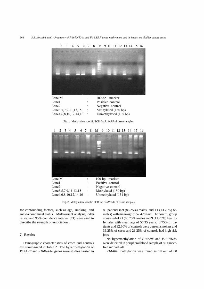
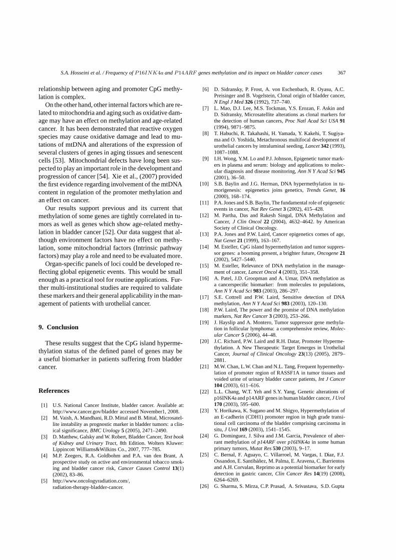
The reaction was carried out in a final volume of50 µL containing 50 ng of bisulphite treated DNA.1.5 mmol/L of MgCl2, 0.2 mmol/L of each dNTP (Fer-mentas, USA), 200 µM of each of the primers and2.5 unit of Taq polymerase (Fermentas, USA). DNAwas amplified during 30 cycles with an initial denatura-tion of 5 minutes at 95◦C and a final extension of 5 min-utes at 72◦C. The cycling program consisted of 30 secdenaturation at 95◦C, 30 sec annealing at 61 and 65◦C(P14ARF and P16INK4α) respectively, and 30 sec ex-tension at 72◦C. PCR products were electrophoresed ona 2% agarose gel. P14ARF methylated and unmethy-lated were recognized by 160 and 165 bp respective-ly, while for P16INK4a were identified by 150 bp formethylated and 151 bp for unmethylated (Figs 1, 2). Inorder to avoid misinterpretation of methylated and un-methylated product, reaction mixture for methylatedand un-methylated primers were prepared separately.
6. Statistical analysis
Statistical analysis was carried out with SPSS 11.5and EPI info 3.2 software programs. ORs were adjusted
364 S.A. Hosseini et al. / Frequency of P16INK4a and P14ARF genes methylation and its impact on bladder cancer cases
1 2 3 4 5 6 7 8 M 9 10 11 12 13 14 15 16
Lane M : 100-bp marker Lane1 : Positive control Lane2 : Negative controlLane3,5,7,9,11,13,15 : Methylated (160 bp) Lane4,6,8,10,12,14,16 : Unmethylated (165 bp)
Fig. 1. Methylation specific PCR for P14ARF of tissue samples.
1 2 3 4 5 6 7 8 M 9 10 11 12 13 14 15 16
Lane M : 100-bp marker Lane1 : Positive control Lane2 : Negative controlLane3,5,7,9,11,13,15 : Methylated (150 bp) Lane4,6,8,10,12,14,16 : Unmethylated (151 bp)
Fig. 2. Methylation specific PCR for P16INK4a of tissue samples.
for confounding factors, such as age, smoking, andsocio-economical status. Multivariant analysis, oddsratios, and 95% confidence interval (CI) were used todescribe the strength of association.
7. Results
Demographic characteristics of cases and controlsare summarized in Table 2. The hypermethylation ofP14ARF and P16INK4α genes were studies carried in
80 patients (69 (86.25%) males, and 11 (13.75%) fe-males) with mean age of 57.42 years. The control groupconsisted of 71 (88.75%) males and 9 (11.25%) healthyfemales with mean age of 56.35 years. 8.75% of pa-tients and 32.50% of controls were current smokers and36.25% of cases and 21.25% of controls had high riskjobs.
No hypermethylation of P14ARF and P16INK4αwere detected in peripheral blood sample of 80 cancer-free individuals.
P14ARF methylation was found in 18 out of 80

S.A. Hosseini et al. / Frequency of P16INK4a and P14ARF genes methylation and its impact on bladder cancer cases 365
Table 2Demographic characteristic of cases with bladder cancer andhealthy controls
Variable Cases Controls
Age (year)Mean(± SD) 57.42 (± 12.59) 56.35 (± 10.13)Median range 25–86 30–85Sex–male 69 (86.25%) 71(88.75%)–female 11 (13.75%) 9 (11.25%)Smoking–current smokers 31(38.75%) 26(32.50%)–non smokers 49(61.25%) 54(67.50%)Job–high risks 29(36.25%) 17(21.25%)–low risks 51(63.75%) 63(78.75%)Alcohol–drinkers 35 (43.75%) 29 (36.25%)–non drinkers 45 (56.25%) 51 (63.75%)Stage� T1 (superficial) 63(78.75%)� T2 (invasive) 17(21.25%)
(22.5%) tissue samples of bladder cancer patients.Among 18 cases with P14ARF methylation in tumourtissues, MS-PCR was able to detect the same change inthe matching blood samples of 8 (44.44%) cases. Forthe other 62 cases with no detected P14ARF methyla-tion in tumour tissue, no signal was obtained on match-ing blood samples by using MS-PCR.
In tissue and matched blood samples of bladder can-cer patients, the incidence of P14ARF hypermethy-lation significantly increased (OR = 0.31, 95%CI =0.12–0.8, P = 0.01) and (OR = 0.0, 95%CI = 0.0–0.62, P = 0.006) respectively with an increase in age.
Clinicopathological analysis revealed that P14ARFhypermethylation in tissue and blood samples was sig-nificantly associated with invasive stage (� T2) (OR =0.21, 95%CI = 0.08–0.51, P = 0.0002) and (OR =0.09, 95%CI = 0.03–0.37, P = 0.00001) respectively(Table 3).
Of the bladder tumour samples amplified, 32(40%) presented P16INK4α promoter hypermethy-lation. Among 32 cases with P16INK4α methyla-tion in tumour tissues, MS-PCR was able to detectthe same change in the matching blood samples of12(37.5%) cases. For the other 48 cases with no detect-ed P16INK4α methylation in tumour tissue, no signalwas obtained on matching blood samples by using MS-PCR. Moreover, 41(51.25%) tumour samples presentedone of the P16INK4α or P14ARF hypermethylation.
Table 4 shows the hypermethylation distribution ofP16INK4α gene along with various clinico-patholog-ical parameters including age, smoking and stage ofcarcinoma in blood samples the patients. Muscle in-
vasive tumour stage (� T2) showed significant asso-ciation with P16INK4α promoter hypermethylation intissue and blood samples of the patients (OR = 0.38,95%CI = 0.17–0.82, P = 0.01) and (OR = 0.13,95%CI = 0.05–0.36, P = 0.00005) respectively.
No association was found between P14ARF andP16INK4α methylation with tobacco, high risk jobsand alcohol consuming in bladder cancer patients.
8. Discussion
There is growing evidence that DNA methylation ofCDKN2A (INK4a/ARF) is an important mechanismof gene inactivation in cancer and its potential as amolecular marker has been regarded. This study hasused methylation – specific PCR for the amplificationof P14ARF and P16INK4a promoter regions in tumourtissue as well as matched blood DNA of cancer patientsand healthy controls. We found promoter methylationin 47.1% of P14ARF and 58.8% of P16INK4a in thetissue samples of bladder cancer cases with invasivestage. These genes are involved in cell cycle regulationmechanisms [33]. Our finding have shown that methy-lation of these genes might be related with advancedstage and tumour metastasis. On the other hand, wefound that hypermethylation at these genes loci detect-ed in blood samples of patients are related to advanceddisease stages. No promoter methylation was found inthe blood DNA of control subjects. Detecting the samealteration in blood and tumour DNA indicated that themethylated DNA released into circulation may havederived from the primary tumour. This is confirmedother literature review which evident that methylationcan occur in a tissue-specific or cancer-specific manner.Cancer-specific methylation is usually detected in tu-mour tissues and seldom observed in blood circulationunless in invasive stage [34,35].
We detected DNA methylation of blood samples ofonly metastatic cancer cases. Therefore blood DNAwill not be useful for early detection of bladder cancerby these markers while urine sediment DNA might bemore helpful in this regard. However, our results areindicating that blood DNA particularly might be usefulfor predicting patient survival and tumor progressionfrom noninvasive to invasive stage. This was previous-ly reported by other authors on the different type ofcancers [36,37].
Moreover, our findings have provided strong evi-dence regarding the diagnostic potential of emergingepigenetic markers in bladder cancer patients. These

366 S.A. Hosseini et al. / Frequency of P16INK4a and P14ARF genes methylation and its impact on bladder cancer cases
Table 3The relationship between P14ARF promoter methylation and clinicopathological parameters of bladder cancer blood and tissue samples
Characteristic Overall Methylated UnMethylated P Valueblood tissue blood tissue blood tissue blood tissue
Age (n = 80)� 50 22 0 (0.0%) 2 (9%) 22 (100%) 20 (91%) 0.006 0.01> 50 58 8 (13.8%) 16 (27.58%) 50 (86.2%) 42 (72.42%)Smoking (n = 80)Smoker 31 5 (16.1%) 10 (32.2%) 26 (83.9%) 21 (67.8%) 0.2 0.1Non smoker 49 3 (6.1%) 8 (16.3%) 46 (93.9%) 41 (83.4%)Stages (n = 80)superficial � T1 63 2 (3.2%) 10 (15.9%) 61 (96.8%) 53 (84.1%) 0.00001 0.0002invasive � T2 17 6 (35.3%) 8 (47.1%) 11 (64.7%) 9 (52.9%)
Table 4The relationship between P16INK4a promoter methylation and clinicopathological parameters of bladder cancer blood and tissueSamples
Characteristic Overall Methylated UnMethylated P Valueblood tissue blood tissue blood tissue blood tissue
Age (n = 80)� 50 22 3 (13.6%) 9 (41%) 19 (86.4%) 13 (59%) 0.95 0.95> 50 58 9 (15.5%) 23 (40%) 49 (85.5%) 35 (60%)Smoking (n = 80)Smoker 31 6 (19.4%) 14 (45.2%) 25 (80.68%) 17 (54.8%) 0.57 0.53Non smoker 49 6 (12.25%) 18 (36.7%) 43 (87.75%) 31 (63.3%)Stages (n = 80)superficial � T1 63 4 (6.3%) 22 (34.9%) 59 (93.7%) 41 (65.1%) 0.00005 0.01invasive � T2 17 8 (47.1%) 10 (58.8%) 9 (52.9%) 7 (41.2%)
molecular assays may help developing more appropri-ate therapeutic interventions and management of clini-cal outcome. Inclusion of the other relevant tumor sup-pressor genes in the current panel of markers may in-crease the sensitivity of the assayOn the other hand,it is possible that some bladder tumour may not carryany epigenetic alteration and, thus, should be detectedby other approaches, such as mutation analysis, LOHanalysis, or detection of homozygous deletions [38–40].
Aging is one of the main risk factors for cancer andthe highest cancer rates exist among people over theage of 70 [41]. Epigenetic alterations closely relatedto the aging process have also been studied alreadyand are proposed to be part of the primary processescontributing to age-related diseases such as cancer [42].
An increase in de novo methylation is an age-relatedevent that occurs in normal tissue and may progress toregional hypermethylation, leading to silencing of spe-cific genes important in the suppression of tumor for-mation [43]. There are some environmental and mito-chondrial factors (intrinsic pathway of apoptosis) whichcan affect age-related methylation and cancer.
Environmental factors known to contribute to methy-lation alterations include carcinogen exposures, inflam-mation, and diet. Several carcinogen exposures such
as tobacco, alcohol, arsenic, and asbestos have beenassociated with methylation-induced gene-inactivationin various human cancers including bladder cancer,head and neck squamous cell carcinoma, and mesothe-lioma [45–49].
It is therefore reasonable to suggest that variousand potentially accumulating exposures throughout lifemay directly or indirectly lead to methylation alter-ations and impact disease susceptibility. In the presentstudy, no significant association was found between therisk of tobacco, high risk job, alcohol consumption andP14ARF and P16INK4A hypermethylation. Contro-versially, Methylation of CpG islands in tobacco- asso-ciated cancers occurs in CDKN2A and directly or indi-rectly is induced by exposure to tobacco smoke in non-small-cell lung carcinoma [50]. Moreover, Wolff et al.(2008) observed that the prevalence of methylation atRUNX3 increased as a function of age at diagnosis anda history of smoking in bladder tumour.
Moreover, we found that the incidence of P14ARFhypermethylation significantly increased in the tissueand blood samples of bladder cancer patients with anincrease in age. A study by Brock et al., (2009) hassupported our findings and, in addition, they showedthat age-related alterations in CpG loci were tissue-dependent. More importantly, they suggested that the
S.A. Hosseini et al. / Frequency of P16INK4a and P14ARF genes methylation and its impact on bladder cancer cases 367
relationship between aging and promoter CpG methy-lation is complex.
On the other hand, other internal factors which are re-lated to mitochondria and aging such as oxidative dam-age may have an effect on methylation and age-relatedcancer. It has been demonstrated that reactive oxygenspecies may cause oxidative damage and lead to mu-tations of mtDNA and alterations of the expression ofseveral clusters of genes in aging tissues and senescentcells [53]. Mitochondrial defects have long been sus-pected to play an important role in the development andprogression of cancer [54]. Xie et al., (2007) providedthe first evidence regarding involvement of the mtDNAcontent in regulation of the promoter methylation andan effect on cancer.
Our results support previous and its current thatmethylation of some genes are tightly correlated in tu-mors as well as genes which show age-related methy-lation in bladder cancer [52]. Our data suggest that al-though environment factors have no effect on methy-lation, some mitochondrial factors (Intrinsic pathwayfactors) may play a role and need to be evaluated more.
Organ-specific panels of loci could be developed re-flecting global epigenetic events. This would be smallenough as a practical tool for routine applications. Fur-ther multi-institutional studies are required to validatethese markers and their general applicability in the man-agement of patients with urothelial cancer.
9. Conclusion
These results suggest that the CpG island hyperme-thylation status of the defined panel of genes may bea useful biomarker in patients suffering from bladdercancer.
References
[1] U.S. National Cancer Institute, bladder cancer. Available at:http://www.cancer.gov/bladder accessed November1, 2008.
[2] M. Vaish, A. Mandhani, R.D. Mittal and B. Mittal, Microsatel-lite instability as prognostic marker in bladder tumors: a clin-ical significance, BMC Urology 5 (2005), 2471–2490.
[3] D. Matthew, Galsky and W. Robert, Bladder Cancer, Text bookof Kidney and Urinary Tract, 8th Edition. Wolters Kluwer:Lippincott Williams&Wilkins Co., 2007, 777–785.
[4] M.P. Zeegers, R.A. Goldbohm and P.A. van den Brant, Aprospective study on active and environmental tobacco smok-ing and bladder cancer risk, Cancer Causes Control 13(1)(2002), 83–86.
[5] http://www.oncologyradiation.com/,radiation-therapy-bladder-cancer.
[6] D. Sidransky, P. Frost, A. von Eschenbach, R. Oyasu, A.C.Preisinger and B. Vogelstein, Clonal origin of bladder cancer,N Engl J Med 326 (1992), 737–740.
[7] L. Mao, D.J. Lee, M.S. Tockman, Y.S. Erozan, F. Askin andD. Sidransky, Microsatellite alterations as clonal markers forthe detection of human cancers, Proc Natl Acad Sci USA 91(1994), 9871–9875.
[8] T. Habuchi, R. Takahashi, H. Yamada, Y. Kakehi, T. Sugiya-ma and O. Yoshida, Metachronous multifocal development ofurothelial cancers by intraluminal seeding, Lancet 342 (1993),1087–1088.
[9] I.H. Wong, Y.M. Lo and P.J. Johnson, Epigenetic tumor mark-ers in plasma and serum: biology and applications to molec-ular diagnosis and disease monitoring, Ann N Y Acad Sci 945(2001), 36–50.
[10] S.B. Baylin and J.G. Herman, DNA hypermethylation in tu-morigenesis: epigenetics joins genetics, Trends Genet, 16(2000), 168–174.
[11] P.A. Jones and S.B. Baylin, The fundamental role of epigeneticevents in cancer, Nat Rev Genet 3 (2002), 415–428.
[12] M. Partha, Das and Rakesh Singal, DNA Methylation andCancer, J Clin Oncol 22 (2004), 4632–4642. by AmericanSociety of Clinical Oncology.
[13] P.A. Jones and P.W. Laird, Cancer epigenetics comes of age,Nat Genet 21 (1999), 163–167.
[14] M. Esteller, CpG island hypermethylation and tumor suppres-sor genes: a booming present, a brighter future, Oncogene 21(2002), 5427–5440.
[15] M. Esteller, Relevance of DNA methylation in the manage-ment of cancer, Lancet Oncol 4 (2003), 351–358.
[16] A. Patel, J.D. Groopman and A. Umar, DNA methylation asa cancerspecific biomarker: from molecules to populations,Ann N Y Acad Sci 983 (2003), 286–297.
[17] S.E. Cottrell and P.W. Laird, Sensitive detection of DNAmethylation, Ann N Y Acad Sci 983 (2003), 120–130.
[18] P.W. Laird, The power and the promise of DNA methylationmarkers, Nat Rev Cancer 3 (2003), 253–266.
[19] J. Hayslip and A. Montero, Tumor suppressor gene methyla-tion in follicular lymphoma: a comprehensive review, Molec-ular Cancer 5 (2006), 44–48.
[20] J.C. Richard, P.W. Laird and R.H. Datar, Promoter Hyperme-thylation. A New Therapeutic Target Emerges in UrothelialCancer, Journal of Clinical Oncology 23(13) (2005), 2879–2881.
[21] M.W. Chan, L.W. Chan and N.L. Tang, Frequent hypermethy-lation of promoter region of RASSF1A in tumor tissues andvoided urine of urinary bladder cancer patients, Int J Cancer104 (2003), 611–616.
[22] L.L. Chang, W.T. Yeh and S.Y. Yang, Genetic alterations ofp16INK4a and p14ARF genes in human bladder cancer, J Urol170 (2003), 595–600.
[23] Y. Horikawa, K. Sugano and M. Shigyo, Hypermethylation ofan E-cadherin (CDH1) promoter region in high grade transi-tional cell carcinoma of the bladder comprising carcinoma insitu, J Urol 169 (2003), 1541–1545.
[24] G. Dominguez, J. Silva and J.M. Garcia, Prevalence of aber-rant methylation of p14ARF over p16INK4a in some humanprimary tumors, Mutat Res 530 (2003), 9–17.
[25] C. Bernal, F. Aguayo, C. Villarroel, M. Vargas, I. Dıaz, F.J.Ossandon, E. Santibanez, M. Palma, E. Aravena, C. Barrientosand A.H. Corvalan, Reprimo as a potential biomarker for earlydetection in gastric cancer, Clin Cancer Res 14(19) (2008),6264–6269.
[26] G. Sharma, S. Mirza, C.P. Prasad, A. Srivastava, S.D. Gupta

368 S.A. Hosseini et al. / Frequency of P16INK4a and P14ARF genes methylation and its impact on bladder cancer cases
and R. Ralhan, Promoter hypermethylation of p16INK4A ,p14ARF , CyclinD2 and Slit2 in serum and tumor DNA frombreast cancer patients, Life Science 80 (2007), 1873–1881.
[27] C.S. Chim, Y.L. Kwong and R. Liang, Gene hypermethyla-tion in multiple myeloma: lessons from a cancer pathwayapproach, Clin Lymphoma Myeloma 8(6) (2008), 331–339.
[28] R.T. Williams and C.J. Sherr, The INK4-ARF (CDKN2A/B)Locus in Hematopoiesis and BCR-ABL-induced Leukemias,Cold Spring Harb Symp Quant Biol. 2008 Nov 26. [Epubahead of print].
[29] S.J. Gallagher, J.F. Thompson, J. Indsto, L.L. Scurr, M. Lett,B.F. Gao, R. Dunleavey, G.J. Mann, R.F. Kefford and H. Ri-zos, p16INK4a expression and absence of activated B-RAFare independent predictors of chemosensitivity in melanomatumors, Neoplasia 10(11) (2008), 1231–1239.
[30] C. Fan, L. He, A. Kapoor, A. Gillis, A.P. Rybak, J.C. Cutz andD. Tang, Bmi1 promotes prostate tumorigenesis via inhibitingp16(INK4A) and p14(ARF) expression, Biochim Biophys Acta1782(11) (2008), 642–648.
[31] J.G. Herman, J.R. Graff, S. Myohanen, B.D. Nelkin and S.B.Baylin, Methylation-specific PCR: a novel PCR assay formethylation status of CpG islands, PNAS 93 (1996), 9821–9826.
[32] E.P. Xing, Y. Nie, Y. Song, G.Y. Yang, Y.C. Cai, L.D.Wang and C.S. Yang, Mechanisms of inactivation of p14ARF,p15INK4b, and p16INK4a genes in human esophageal squa-mous cell carcinoma, Clin Cancer Res 5(10) (1999), 2704–2713.
[33] J.F. Viallard, F. Lacombe, F. Belloc, J.L. Pellegrin and J. Reif-fers, Molecular mechanisms controlling the cell cycle: funda-mental aspects and implications for oncology, Cancer Radio-ther 5(2) (2001), 109–129.
[34] K. Yamashita, S. Upadhyay and M. Osada, Pharmacologicunmasking of epigenetically silenced tumor suppressor genesin esophageal squamous cell carcinoma, Cancer Cell 2 (2002),485–495.
[35] C. Jeronimo, R. Henrique and M.O. Hoque, A quantitativepromoter methylation profile of prostate cancer, Clin CancerRes 10 (2004), 8472–8478.
[36] D.R. Yates, I. Rehman, M.F. Abbod, M. Meuth, S.S. Cross,D.A. Linkens, F.C. Hamdy and J.W.F. Catto, Promoter Hyper-methylation Identifies Progression Risk in Bladder, Cancer-Clinical Cancer Research 13 (2007), 2046.
[37] L. Shen, H. Kantarjian, Y. Guo, E. Lin, J. Shan, X. Huang,D. Berry, S. Ahmed, W. Zhu, Sh. Pierce, Y. Kondo, Y. Oki,J.Jelinek, H. Saba, E. Estey and J.J. Issa, DNA MethylationPredicts Survival and Response to Therapy in Patients WithMyelodysplastic Syndromes, Journal of Clinical Oncology28(4) (2010), 605–613.
[38] L. Mao, M.P. Schoenberg, M. Scicchitano, Y.S. Erozan, A.Merlo and D. Schwab, Molecular detection of primary bladdercancer by microsatellite analysis, Science 271 (1996), 659–662.
[39] G. Steiner, M.P. Schoenberg, J.F. Linn, L. Mao and D. Sidran-sky, Detection of bladder cancer recurrence by microsatelliteanalysis of urine, Nat Med 3 (1997), 621–624.
[40] P. Cairns, T.J. Polascik, Y. Eby, K. Tokino, J. Califano and A.Merlo, Frequency of homozygous deletion at p16/CDKN2 inprimary human tumours, Nat Genet 11 (1995),210–212.
[41] M. Esteller, M.F. Fraga, M.F. Paz, E. Campo, D. Colomer,F.J. Novo, M.J. Calasanz, O. Galm, M. Guo, J. Benitez and
J.G. Herman, Cancer epigenetics and methylation, Science297 (2002), 1807–1808.
[42] M.D. Shahbazian and H.Y. Zoghbi, Rett syndrome andMeCP2: linking epigenetics and neuronal function, Am J HumGenet 71 (2002), 1259–1272.
[43] N. Lopatina, J.F. Haskell, L.G. Andrews, J.C. Poole,S.Saldanha and T.O. Tollefsbol, Differential maintenance andde novo methylating activity by three DNA methyltransferas-es in aging and immortalized fibroblasts, J Cell Biochem 84(2002), 324–334.
[44] B.C. Christensen, J.J. Godleski, C.J. Marsit, E.A. Housemanand C.Y. Lopez-Fagundo. Asbestos exposure predicts cell cy-cle control gene promoter methylation in pleural mesothe-lioma, Carcinogenesis 29 (2008), 1555–1559.
[45] C.J. Marsit, E.A. Houseman, A.R. Schned, M.R. Karagas andK.T. Kelsey, Promoter hypermethylation is associated withcurrent smoking, age, gender and survival in bladder cancer,Carcinogenesis 28 (2007), 1745–1751.
[46] C.J. Marsit, M.D. McClean, C.S. Furniss and K.T. Kelsey,Epigenetic inactivation of the SFRP genes is associated withdrinking, smoking and HPV in head and neck squamous cellcarcinoma, Int J Cancer 119 (2006), 1761–1766.
[47] S. Toyooka, R. Maruyama, K.O. Toyooka, D. McLerran and Z.Feng, Smoke exposure, histologic type and geography-relateddifferences in the methylation profiles of non-small cell lungcancer, Int J Cancer 103 (2003), 153–160.
[48] C.M. Lyon, D.M. Klinge, K.C. Liechty, F.D. Gentry and T.H.March, Radiation-induced lung adenocarcinoma is associatedwith increased frequency of genes inactivated by promoterhypermethylation, Radiat Res 168 (2007), 409–414.
[49] B.C. Christensen, E.A. Houseman, J.J. Godleski, C.J. Mar-sit, J.L. Longacker, Epigenetic profiles distinguish pleuralmesothelioma from normal pleura and predict lung asbestosburden and clinical outcome, Cancer Res, 2008.
[50] D.H. Kim, H.H. Nelson, J.K. Wiencke, D.C. Christiani, J.C.Wain, E.J. Mark and K.T. Kelsey, Promoter hypermethylationof DAP-kinase: association with advanced stage in non-smallcell lung cancer, Oncogene, 2001, in press.
[51] E.M. Wolff, G. Liang, C.C. Cortez, Y.C. Tsai, J.E. Castelao,V.K. Cortessis, D.D. Tsao-Wei, S. Groshen and P.A. Jones,RUNX3 Methylation Reveals that Bladder Tumors Are Olderin Patients with a History of Smoking, Cancer Research 68(2008), 6208–6214.
[52] C. Ch. Brock, E.A. Houseman, J.M. Carmen, Sh. Zheng, R.W.Margaret, J.L. Wiemels, H.H. Nelson, M.R. Karagas, J.F.Padbury, R. Bueno, D.J. Sugarbaker, R. Yeh, J.K. Wienckeand K.T. Kelsey, Aging and Environmental Exposures AlterTissue-Specific DNA Methylation Dependent upon CpG Is-land Context, PLoS Genet 5(8) (2009), e1000602.
[53] Y. Wei, S. Wu, Y. Ma and H. Lee, Respiratory Function Declineand DNA Mutation in Mitochondria, Oxidative Stress andAltered Gene Expression during Aging, Chang Gung Med J32 (2009), 113–132.
[54] W.C. Copeland, J.T. Wachsman, F.M. Johnson and J.S. Penta,Mitochondrial DNA alterations in cancer, Cancer Invest 20(2002), 557–569.
[55] C. Xie, A. Naito, T. Mizumachi, T.T. Evans, M.G. Dou-glas, C.A. Cooney, C. Fan and M. Higuchi, Mitochondrialregulation of Cancer Associated Nuclear DNA Methylation,Biochem Biophys Res Commun 364(3) (2007), 656–661.

Submit your manuscripts athttp://www.hindawi.com
Stem CellsInternational
Hindawi Publishing Corporationhttp://www.hindawi.com Volume 2014
Hindawi Publishing Corporationhttp://www.hindawi.com Volume 2014
MEDIATORSINFLAMMATION
of
Hindawi Publishing Corporationhttp://www.hindawi.com Volume 2014
Behavioural Neurology
EndocrinologyInternational Journal of
Hindawi Publishing Corporationhttp://www.hindawi.com Volume 2014
Hindawi Publishing Corporationhttp://www.hindawi.com Volume 2014
Disease Markers
Hindawi Publishing Corporationhttp://www.hindawi.com Volume 2014
BioMed Research International
OncologyJournal of
Hindawi Publishing Corporationhttp://www.hindawi.com Volume 2014
Hindawi Publishing Corporationhttp://www.hindawi.com Volume 2014
Oxidative Medicine and Cellular Longevity
Hindawi Publishing Corporationhttp://www.hindawi.com Volume 2014
PPAR Research
The Scientific World JournalHindawi Publishing Corporation http://www.hindawi.com Volume 2014
Immunology ResearchHindawi Publishing Corporationhttp://www.hindawi.com Volume 2014
Journal of
ObesityJournal of
Hindawi Publishing Corporationhttp://www.hindawi.com Volume 2014
Hindawi Publishing Corporationhttp://www.hindawi.com Volume 2014
Computational and Mathematical Methods in Medicine
OphthalmologyJournal of
Hindawi Publishing Corporationhttp://www.hindawi.com Volume 2014
Diabetes ResearchJournal of
Hindawi Publishing Corporationhttp://www.hindawi.com Volume 2014
Hindawi Publishing Corporationhttp://www.hindawi.com Volume 2014
Research and TreatmentAIDS
Hindawi Publishing Corporationhttp://www.hindawi.com Volume 2014
Gastroenterology Research and Practice
Hindawi Publishing Corporationhttp://www.hindawi.com Volume 2014
Parkinson’s Disease
Evidence-Based Complementary and Alternative Medicine
Volume 2014Hindawi Publishing Corporationhttp://www.hindawi.com