iraqi jms · diastolic blood pressure, tp = total serum protein, sflt-1 = soluble fms like tyrosin...
TRANSCRIPT

15
Serum Soluble fms-like Tyrosine Kinase-1 (sflt-1) Level at Third Trimester of Pregnancy and One Month Following Delivery in
Women with Preeclampsia
Radhwan M. Hussein1 MSc, Najat A. Hasan2 PhD, Bushra J. Al-Rubayae CIBOG
1Minstry of Health,
2Dept. of Chemistry & Biochemistry, College of Medicine, Al-Nahrain University,
3Dept. of
Obstetrics & Gynecology, Babylon Medical College, Iraq
Abstract Background Preeclampsia is characterized by abnormal vascular response to placentation that is associated with
increased systemic vascular resistance, enhanced platelet aggregation, activation of the coagulation system, and endothelial cell dysfunction.
Objective To investigate the serum level of anti angiogenic factor soluble fms-like tyrosine kinase-1 (sFlt-1) in the third trimester of preeclamptic pregnants in relation to the Doppler ultrasonography indices.
Methods One hundred and five pregnant women at the Pediatric and Maternity Teaching Hospital in Hilla City were studied. All of them were in their third trimester and with singleton pregnancy. All participants were reexamined one month following delivery.
Results The body mass index, spot urine protein to creatinine ratios and mean pulsatility index, resistance index), systolic and diastolic blood pressures were increased in preeclamptic groups. Post-delivery systolic and diastolic blood pressures increased in the control pregnant group and significant reduction in the both preeclamptic groups compared with the systolic and diastolic blood pressures at third trimester of pregnancy. sFlt-1 levels were increased in severe than mild preeclampsia and control groups. In post-delivery period, the sflt-1 decreases in the control pregnant, mild and severe preeclamptic groups in comparison to the third trimester. In preeclampsia, the sflt-1 is positively correlated with blood pressure, protein: creatinine ratio, body mass index, resistance index, pulstality index, total serum protein, and negatively correlated with neonatal weight and gestational age at delivery.
Conclusion The changes in anti angiogenic factors sFlt-1 during pregnancy and in post-delivery period stress its role in the pathogenesis of preeclampsia and could be used to predict the disease progression and impact on the outcome of pregnancy.
Keywords Preeclampsia, sFlt-1, Doppler indices.
List of abbreviation: PE = preeclampsia, BMI = body mass index, P/C ratios = spot urine protein to creatinine ratios, PI = pulsatility index, RI = resistance index, sBP = systolic blood pressure, dBP = diastolic blood pressure, TP = total serum protein, sFlt-1 = soluble fms like tyrosin kinase-1, VEGF = vascular endothelial growth factor, ALT = alanine aminotransferase, AST = aspartate aminotransferase, ELISA = enzyme linked immunosorbent assay, ROC = receiver operating characteristic, PlGF = placental growth factor. Introduction
reeclampsia (PE) is a pregnancy associated multisystem disorder that complicates 2%-10 % of pregnancies of
western world. It is accompanied with a neonatal and perinatal mortality rate of 10 % worldwide (1). The clinical features of preeclampsia are proteinuria and hypertension that occur after 20 weeks of gestation in women who were not previously diagnosed to be hypertensive. Other signs and symptoms of PE are edema and headache (2). PE can be divided into mild and severe forms, according to the amount of proteinuria, the severity of P
Iraqi JMS Published by Al-Nahrain College of Medicine
ISSN 1681-6579 Email: [email protected]
http://www.colmed-alnahrain.edu.iq
Hussein et al, Serum sflt-1 Level in Preeclampsia
16
the hypertension, and the degree of effect on other organ systems (3). The etiology of PE is unknown and, actually the entity is remarkably poorly understood. Delivery of the placenta usually halts the disease progression but it does not reverse all of the associated pathophysiological changes of preeclampsia (4). Soluble fms (feline sarcoma virus)-like tyrosine kinase-1 (sFlt-1) is an anti-angiogenic protein. It a shortened splice variant of the vascular endothelial growth factor (VEGF) receptor-1 that signals angiogenesis .The sFlt-1 captures vascular endothelial growth factor thus preventing its interaction with ligands and down regulating the biological effects of VEGF (5). SFlt-1 is upregulated in PE, leading to increased circulating levels. The increased sFlt-1 levels are associated with decreased circulating levels of VEGF and placental growth factor (PlGF), resulting in endothelial dysfunction. Infusing sFlt-1 to pregnant rats provides a convincing animal model of PE (6). The lowered oxygen tension in primary cytotrophoblast culture and villous explants was found to increase the sFlt-1 expression suggesting the role of hypoxia in stimulating sFlt-1 gene expression (7, 8). It has been reported that uteroplacental ischemia, results in a PE-like phenotype accompanied by elevated circulating sFlt-1 (9). Placental ischemia/ hypoxia may alone be sufficient to induce PE through sFlt-1 up-regulation. When the trophoblastic invasion is defective, an enhanced placental vascular resistance is likely to occur as evidenced in abnormal Doppler ultrasound spectrum of the uterine vessels, indicating that the women are at risk for serious pregnancy disorders like PE (10). So, this study was conducted to test the relationship of the antiangiogenic factor sFlt-1 and the Doppler ultrasound finding and to monitor their impact on the neonatal health and the post-delivery hemodynamics. Methods This research was conducted on 105 pregnant women at their third trimester of pregnancy
over a period of fourteen months from May 2014 till Aug. 2015 at the Pediatric and Maternity Teaching Hospital in Hilla city. The practical part was conducted at Research Laboratories in the Department of Chemistry and Biochemistry, College of Medicine, Al-Nahrain University, and at the Department of Radiology and at laboratories of Pediatric and Maternity Hospital in Hilla city. Out of total 105 pregnant, 70 women suffer from PE with a maternal age of 19-41 year and the other 35 were normotensive pregnant (Maternal age range = 18-36 year) who were served as control. All of studied women were carrying single fetus and at their third trimester of pregnancy. The PE group was further subdivided according to the severity of the disease into mild (n=35) and severe PE (n=35) according to the standard criteria (3). The study was approved by the Institute Review Board of the College of Medicine, Al-Nahrain University and a written consent was provided from each participant to be enrolled in this study. All pregnant women were subjected to clinical examination, blood pressure (BP) measurement, and routine ante-natal ultrasonography. Doppler ultras-onography has been used for each pregnant to determine the pulsatility index (PI) and resistance index (RI). Five milliliters of venous blood samples were withdrawn for the spectrophotometric determination of serum creatinine, total serum protein (TP), alanine aminotransferase (ALT), aspartate aminotransferase (AST), and glucose, and the serum sflt-1 concentration by Enzyme linked immunosorbent assay (ELISA). Spot urine samples were used immediately for the measurement of creatinine, and protein. Statistical Analyses: Group comparisons and data correlation were carried out using mean, standard error of mean, analysis of variance and Pearson correlation. Paired t-test was used to compare the pre delivery and post-delivery levels of aforementioned parameters and a P value of less than 0.05 was considered to be statistically

Iraqi JMS 2016; Vol. 14(1)
17
significant. The receiver operating characteristic (ROC) curve was used to demonstrate the cut-off values, sensitivity, specificity and area under the curve of the studied research variables (11). Results The results of maternal age, gestational age at delivery, BMI, neonatal weight, spot urinary protein / creatinine ratio (P/C) and Doppler findings (PI and RI) for both control pregnant group and patients groups (mild and severe preeclampsia) at third trimester are listed in table 1. There were very high significant decreases (p ˂ 0.001) in mean gestational age
at delivery and of neonatal weight in mild and severe preeclampsia group when compared with those of the control pregnant group. There were high significant increase in means of body mass index (BMI), spot urine protein, to creatinine ratios and mean RI in severe PE (p ˂ 0.01) and in mild PE (p < 0.01) in comparison to their respective values of the control group. The elevation in the PI indices in mild PE was less significant as compared to control group (p < 0.05). The means of all of aforementioned parameters were significantly higher in pregnant women with severe PE as compared with pregnant with mild form of PE.
Table 1. Illustrates the demographic and biochemical data the preeclamptic patients and
controls at third trimester of pregnancy
Parameter Control group preeclampsia
Mild Severe
Number 35 35 35
maternal age (year) 25.6 ±0.84 27.43 ±0.99 26.8 ±0.84
gestational age at delivery (weeks) 38.00 ±0.2 36.92±0.26a▼*** 36.17±0.19 a▼***
BMI (Kg/m2) 27.92±0.55 31.23±0.5 a▲** 34.3±0.86 a▲***, ▲b**
neonatal weight (Kg) 2.66 ±0.05 2.45±0.05 a▼** 2.34±0.4 a▼***
urinary spot protein /creatinine ratio
0.1 ±0.003 1.04±0.05 a▲*** 3.55±0.23 a▲***,b▲***
pulsatility index 0.71 ±0.02 0.84±0.153 a▲* 1.02±0.06 a▲*** , b▲**
resistance index 0.56± 0.01 0.65±0.01a▲** 0.72±0.02 a▲*** , b▲* a = ANOVA test between mild, severe preeclampsia group versus control pregnant group: ▼
***= very high significant
decrease (p ˂ 0.001); ▲**
= high significant increase (p ˂ 0.01); ▲***
=very high significant increase (p ˂ 0.001); ▼**
= high significant decrease (p ˂ 0.01). b = ANOVA test between mild and severe preeclampsia groups:
▲**= high significant
increase (p ˂ 0.01); ▲***
=very high significant increase (p ˂ 0.001).
The results of systolic blood pressure (sBP), diastolic blood pressure (dBP), total serum protein (TP) and sflt-1 for both control pregnant group and patients groups (mild and severe preeclampsia) at third trimester and one month following delivery are listed in table 2. There were very high significant increases (p ˂ 0.001) in mean of sBP and dBP in both mild and severe preeclamptic groups when compared with control pregnant group at third trimester.
Comparison of the results of the blood pressure before and after delivery using paired t-test showed very high significant increases (p ˂ 0.001) in the mean of sBP and dBP in the control pregnant group after one month following delivery compared with the sBP and dBP mean value of the same group at third trimester of pregnancy. On the contrary, the mean values of sBP and dBP were significantly lowered in both mild and severe preeclamptic groups after one month following delivery

Hussein et al, Serum sflt-1 Level in Preeclampsia
18
compared with the sBP and dBP mean value of the same groups at third trimester of pregnancy (p ˂ 0.001). The mean post-delivery
sBP and dBP levels were continued to be significantly higher in severe PE above those with mild PE (p < 0.01).
Table 2. Illustrates the differences in systolic and diastolic blood pressure, total serum protein
and soluble fms like tyrosin kinase-1 between preeclamptic patients and controls at third trimester of pregnancy and one month following delivery
Parameter Control No = 35
Preeclampsia
Mild No = 35
Severe No = 35
sBP (mmHg/hr)
Before delivery After delivery
10.92±0.1 11.31±0.11c▲***
13.52±0.18a▲*** 11.48±0.12c▼***
14.78±0.23a▲***,b▲***
12.07±0.19a▲***,b▲**c▼***
dBP (mmHg/hr)
Before delivery After delivery
7.3±0.11 7.51±0.09 ▲***
9.4 ±0.15 a▲*** 7.64 ±0.13 c▼***
10.45±0.22 a▲***, b▲*** 8.15±0.14a▲***,b▲**c▼***
(TSP) (g/dl)
Before delivery After delivery
6.02±0.11 6.72±0.09 c▲***
5.61 ±.09 a▼**
6.43±0.07a▼*c▲*** 5.23±0.08 a▼***,b ▼**
6.19 ±0.07a▼***,b▼*c▲***
Sflt-1 (ng/ml)
Before delivery After delivery
2.15±0.11 1.96±0.1 c▼***
2.78±0.14a▲** 2.15±0.1 c▼***
3.55±0.15 a▲*** ,b▲*** 2.50±0.1a▲***,b▲*,c▼***
a = ANOVA test between mild, severe preeclampsia group versus control pregnant group: ▼***
= very high significant decrease (p ˂ 0.001);
▲**= high significant increase (p ˂ 0.01);
▲***= very high significant increase (p ˂ 0.001);
▼**= high
significant decrease (p ˂ 0.01). b = ANOVA test between mild and severe preeclampsia groups: ▲**
= high significant increase (p ˂ 0.01);
▲***= very high significant increase (p ˂ 0.001). c = paired t-test for control, mild, severe, mean
concentration at third trimester versus one month following delivery mean values: ▲***
= very high significant increase (p ˂ 0.001);
▼***= very high significant decrease (p ˂ 0.001)
There were high significant decreases in the third trimester mean TSP concentration in mild (p ˂ 0.01) and severe preeclamptic (p ˂ 0.001) groups when compared with control pregnant group in the third trimester, with significant decreases (p ˂ 0.01) in severe preeclamptic group when compared with mild PE. The post-delivery mean TSP levels showed significant elevation (p ˂ 0.001) in control, mild and severe PE groups compared with their concentration at late pregnancy. There was significant increase (p ˂ 0.01) in mean serum sflt-1 in mild preeclamptic group and very high significant increase (p ˂ 0.001) in severe preeclamptic group in comparison with those of the control pregnant group at third trimester. In severe PE, the sflt-1 levels were significantly above those in mild PE group (p ˂ 0.05). There were very high significant decreases (p ˂ 0.001) in means of serum sflt-1 levels in the control pregnant, mild and severe preeclamptic groups after one month following
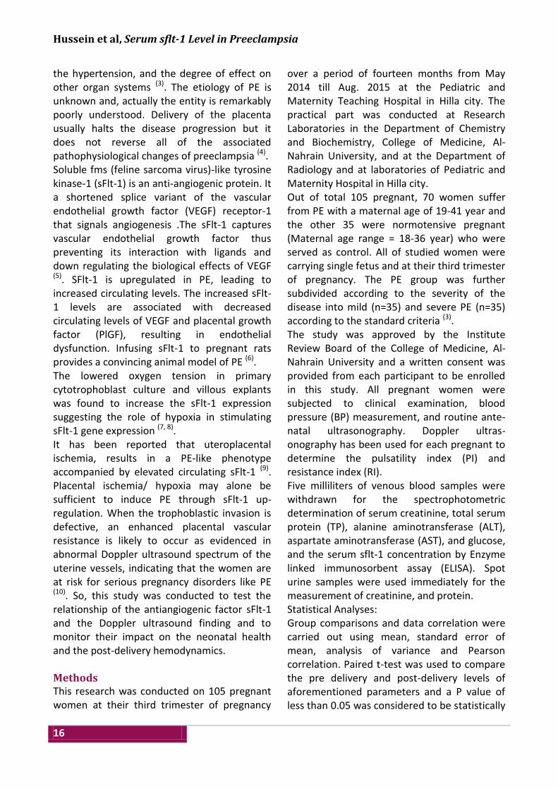
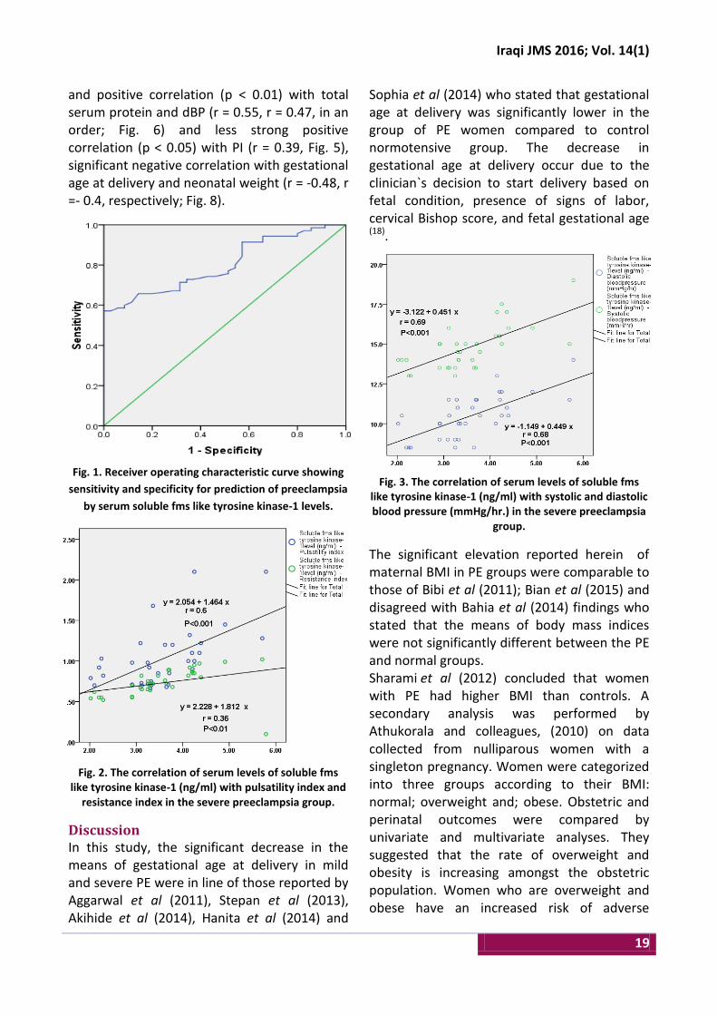
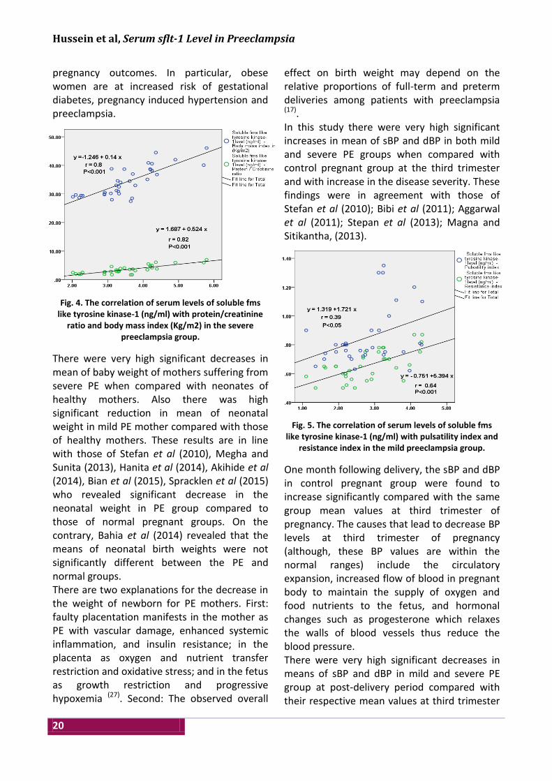
delivery compared with the respective groups mean values at third trimester of pregnancy. Receiver operating characteristic curve (ROC) analyses of serum sflt-1 revealed the ability of this marker to differentiate preeclamptic from normal pregnancies (Fig. 1). In this figure the cut-off value of sflt-1 was 2.74ng / ml with sensitivity and specificity of 67%, and 69%, respectively. In severe PE, the sflt-1 values showed very high positive significant correlation (p ˂ 0.001) with systolic BP (r = 0.69, Fig. 3), diastolic BP (r = 0.68, Fig. 3), P/C ratio (r = 0.82, Fig. 4), BMI (r = 0.8, Fig. 4), PI (r = 0.6, Fig. 2), statistically less but significant positive correlation (p ˂ 0.01) with total serum protein (r = 0.56) and RI (r = 0.36, Fig.2). On the other hand, in mild PE the sFlt-1 concentration exhibited very high significant positive correlation (p ˂ 0.001) with systolic BP (r = 0.62, Fig. 6), P/C ratio (r = 0.68, Fig. 7), BMI, and RI (r = 0.65, r = 0.64, respectively; Fig. 5),
Iraqi JMS 2016; Vol. 14(1)
19
and positive correlation (p ˂ 0.01) with total serum protein and dBP (r = 0.55, r = 0.47, in an order; Fig. 6) and less strong positive correlation (p ˂ 0.05) with PI (r = 0.39, Fig. 5), significant negative correlation with gestational age at delivery and neonatal weight (r = -0.48, r =- 0.4, respectively; Fig. 8). Fig. 1. Receiver operating characteristic curve showing
sensitivity and specificity for prediction of preeclampsia
by serum soluble fms like tyrosine kinase-1 levels.
Fig. 2. The correlation of serum levels of soluble fms like tyrosine kinase-1 (ng/ml) with pulsatility index and
resistance index in the severe preeclampsia group.
Discussion In this study, the significant decrease in the means of gestational age at delivery in mild and severe PE were in line of those reported by Aggarwal et al (2011), Stepan et al (2013), Akihide et al (2014), Hanita et al (2014) and
Sophia et al (2014) who stated that gestational age at delivery was significantly lower in the group of PE women compared to control normotensive group. The decrease in gestational age at delivery occur due to the clinician`s decision to start delivery based on fetal condition, presence of signs of labor, cervical Bishop score, and fetal gestational age (18).
Fig. 3. The correlation of serum levels of soluble fms like tyrosine kinase-1 (ng/ml) with systolic and diastolic blood pressure (mmHg/hr.) in the severe preeclampsia
group.
The significant elevation reported herein of maternal BMI in PE groups were comparable to those of Bibi et al (2011); Bian et al (2015) and disagreed with Bahia et al (2014) findings who stated that the means of body mass indices were not significantly different between the PE and normal groups. Sharami et al (2012) concluded that women with PE had higher BMI than controls. A secondary analysis was performed by Athukorala and colleagues, (2010) on data collected from nulliparous women with a singleton pregnancy. Women were categorized into three groups according to their BMI: normal; overweight and; obese. Obstetric and perinatal outcomes were compared by univariate and multivariate analyses. They suggested that the rate of overweight and obesity is increasing amongst the obstetric population. Women who are overweight and obese have an increased risk of adverse
Hussein et al, Serum sflt-1 Level in Preeclampsia
20
pregnancy outcomes. In particular, obese women are at increased risk of gestational diabetes, pregnancy induced hypertension and preeclampsia.
Fig. 4. The correlation of serum levels of soluble fms like tyrosine kinase-1 (ng/ml) with protein/creatinine
ratio and body mass index (Kg/m2) in the severe preeclampsia group.
There were very high significant decreases in mean of baby weight of mothers suffering from severe PE when compared with neonates of healthy mothers. Also there was high significant reduction in mean of neonatal weight in mild PE mother compared with those of healthy mothers. These results are in line with those of Stefan et al (2010), Megha and Sunita (2013), Hanita et al (2014), Akihide et al (2014), Bian et al (2015), Spracklen et al (2015) who revealed significant decrease in the neonatal weight in PE group compared to those of normal pregnant groups. On the contrary, Bahia et al (2014) revealed that the means of neonatal birth weights were not significantly different between the PE and normal groups. There are two explanations for the decrease in the weight of newborn for PE mothers. First: faulty placentation manifests in the mother as PE with vascular damage, enhanced systemic inflammation, and insulin resistance; in the placenta as oxygen and nutrient transfer restriction and oxidative stress; and in the fetus as growth restriction and progressive hypoxemia (27). Second: The observed overall
effect on birth weight may depend on the relative proportions of full-term and preterm deliveries among patients with preeclampsia (17). In this study there were very high significant increases in mean of sBP and dBP in both mild and severe PE groups when compared with control pregnant group at the third trimester and with increase in the disease severity. These findings were in agreement with those of Stefan et al (2010); Bibi et al (2011); Aggarwal et al (2011); Stepan et al (2013); Magna and Sitikantha, (2013).
Fig. 5. The correlation of serum levels of soluble fms like tyrosine kinase-1 (ng/ml) with pulsatility index and
resistance index in the mild preeclampsia group.
One month following delivery, the sBP and dBP in control pregnant group were found to increase significantly compared with the same group mean values at third trimester of pregnancy. The causes that lead to decrease BP levels at third trimester of pregnancy (although, these BP values are within the normal ranges) include the circulatory expansion, increased flow of blood in pregnant body to maintain the supply of oxygen and food nutrients to the fetus, and hormonal changes such as progesterone which relaxes the walls of blood vessels thus reduce the blood pressure. There were very high significant decreases in means of sBP and dBP in mild and severe PE group at post-delivery period compared with their respective mean values at third trimester

Iraqi JMS 2016; Vol. 14(1)
21
of pregnancy. This occur due to the resumption of the normal physiology of the body and removal of factors that cause the increase in the BP (factors that lead to development of PE) such as presence of the placenta which is central to the pathogenesis of preeclampsia, stress, antiangiogenic agents, immunological factors and decrease in the angiogenic factors.
Fig. 6. The correlation of serum levels of soluble fms like tyrosine kinase-1 (ng/ml) with systolic and diastolic
blood pressure (mmHg/hr.) in the mild preeclampsia group.
There were high significant decreases in mean TP in mild and severe PE groups when compared with those of the control pregnant group and the reduction in serum protein was more significant with the disease severity. Adedeji et al (2012) had observed that the concentration of TP was not significantly decreased in normotensive pregnant women compared to that of non-pregnant women. Olooto et al (2013) showed that TP levels were significantly lowered while urinary protein was significantly elevated in pregnancy related hypertension and PE, respectively as compared to those of the normal pregnant women. The main cause of the relative decrease in the TP in control pregnant group and PE groups (mild and severe PE) is the hemodynamic changes accompanying the expansion of the plasma volume and the increases in cardiac output up to 50% which creates dilution of plasma proteins. As a result, the concentrations of albumin and α1-acid glycoprotein decrease
up to 20-40% at term (30). In PE groups (mild and severe) the loss of glomerular barrier charge and size selectivity and glomerular endotheliosis result in poor filtration and increased urine protein (31). Fig. 7. The correlation between serum levels of soluble
fms like tyrosine kinase-1 (ng/ml) and protein/
creatinine ratio in the mild preeclampsia group.
The post-delivery mean serum protein revealed very high significant elevation in the control pregnant group and PE (mild and severe) as compared with their respective mean values at third trimester period. This can be explained by the hemodynamic changes in which there is a decrease in the expansion of plasma volume post-delivery with a decline in the renal loss of protein especially in PE groups (mild and severe). In this study, there were significant increases in the PI and RI in mild PE with very high significant increases in means of PI in severe PE group compared with those of the control pregnant group. The mean PI and RI were found to be higher with the increase in the severity of the PE. Similar findings were recorded by Dahiana et al (2014), Guedes-Martins et al (2014), Martínez-Ruiz et al (2014), and Bian et al (2015). The present study showed significant increases in the means of sflt-1 in severe PE compared with mild PE and the control pregnant group

Hussein et al, Serum sflt-1 Level in Preeclampsia
22
mean values. Similar observations were reported by many other researchers (20,24,35-38). In 2014, Sophia and co researchers analyzed the serum levels of antiangiogenic factor sflt-1 in addition to the neonatal adverse outcomes in 40 women with established PE and 40 normotensive women. They found that the sFlt-1 levels were significantly higher in the PE women compared to normotensive women. The increased serum values of the anti-angiogenic sFlt-1 were associated with increased rates of late preterm, early term births and very low birth weight. Fig. 8. The correlation between serum levels of soluble
fms like tyrosine kinase-1 (ng/ml) and neonatal weights
(Kg) in the mild preeclampsia group.
Placental overexpression of sFlt-1 specifically in the fetal derived trophoblast cells was implicated as the underlying cause of PE. Fan and colleagues (2014) suggested that the fetal-derived trophoblastic cells over express sflt-1 as a self-defense against excessive VEGFA produced by maternal decidual cells. The results of the significant positive correlation reported herein between the anti-angiogenic factor sFlt-1 with PI and RI and sBP and dBP in pregnant patients with mild or severe PE supported the finding of Sophia et al (2014) who demonstrated a significant contribution of sFlt-1 to the pathogenesis of endothelial dysfunction and the consequent systematic vascular disorder.
According to the results of present study, the higher concentrations of sFlt-1 were negatively correlated with the adverse neonatal outcomes in the form of reduction in the newborn births weight. This can be attributed to the decreased uteroplacental blood flow, and hypoxic stress to the fetus and placenta, also to the poor vascular remodeling and the induced placental and endothelial damage (40). The finding of significant positive correlation between anti-angiogenic factor sFlt-1 concentration and protein/creatinine ratio occur due to direct effects of sflt-1 on renal tissue that might lead to glomerular endotheliosis (41). Aggarwal et al (2011) showed an abnormally increased production of sFlt-1 and sEng, two powerful antiangiogenic molecules, by preeclamptic placentae. The sFlt-1 and sEng are believed to exert their pathogenic effects by limiting the availability of their pro-angiogenic ligands (PlGF, VEGF and transforming growth factor-β) to their native cell surface binding partners on the endothelium. This angiogenic imbalance is believed to induce endothelial dysfunction, systemic vasoconstriction, hypertension and proteinuria. There were very high significant decreases in the means of sflt-1 in preeclamptic groups (mild and severe PE) one month post-delivery compared with the mean sFlt-1 concentration of those groups at late gestational period. This observation can be explained by the postpartum removal of the placenta which represent the major source of sFlt-1 in which placental syncytiotrophoblasts and in particular syncytial knots were identified as a major source of sFlt-1 (42). In conclusion, the association between anti angiogenic factor soluble fms-like tyrosine kinase-1 (sFlt-1) during pregnancy and one month post-delivery period stress its role in the pathogenesis of PE and could be used to predict the disease progression and impact on the outcome of pregnancy, also there were significant association of preeclampsia and sflt-1 with low neonatal weight.

Iraqi JMS 2016; Vol. 14(1)
23
Acknowledgment I would like to express my sincere gratitude to all members of Pediatric and Maternity Teaching Hospital in Hilla City for their assistance in sample collection and our special thanks to Dr. Senna at the Babylon Medical College for her cooperation for gaining an access to some of the required laboratory facilities therein. Author contributions Dr Hasan put the study concept and design; Dr. Al-Rubayae did the physical examination, diagnosis, and the clinical follow up of cases; and Hussein collect blood samples, did the laboratory analyses and preparation of the manuscript. Conflict of interest None Funding None References 1. Osungbade K, Ige O. Public health perspectives of
preeclampsia in developing countries: Implication for health system strengthening. J Preg. 2011 (2011), Article ID 481095:1-6.
2. Chappell C, Bramham K, Shennan A. Short-term prediction of preeclampsia: how close are we? Biomarkers Med. 2014; 8:455-458.
3. Mary L, Nadine T. Preeclampsia. An obstetrician’s prspective. Ad Chr Kidney Dis. 2013; 20:287-296.
4. Cunningham F, Leveno K, Bloom S, et al. Pregnancy Hypertension. 23rd ed. Williams Obstetrics. New York, USA: McGraw-Hill, 2010; Pp. 689-748.
5. Valeria C, Ana C, Ingrid F, et al. Nitric oxide formation Is inversely related to serum levels of antiangiogenic factors soluble fms-like tyrosine kinase-1 and soluble endoglin in preeclampsia. Hypertens. 2008; 52:402-407.
6. Maynard S, Min J, Merchan J, et al. Excess placental soluble fms-like tyrosine kinase 1 (sFlt1) may contribute to endothelial dysfunction, hypertension, and proteinuria in preeclampsia. J Clin Invest. 2003; 111:649-658.
7. Ahmad S, Ahmed A. Elevated placental soluble vascular endothelial growth factor receptor-1 inhibits angiogenesis in preeclampsia. Circ Res. 2004; 95:884-891.
8. Nagamatsu T, Fujii T, Kusumi M, et al. Cytotrophoblasts upregulate soluble fms-like tyrosine kinase-1 expression under reduced oxygen: an implication for the placental vascular development and the pathophysiology of preeclampsia. Endocrinol. 2004; 145:4838-4845.
9. Makris A, Thornton C, Thompson J, et al. Uteroplacental ischemia results in proteinuric hypertension and elevated sFLT-1. Kidney Int. 2007; 71:977-984.
10. Sciscione A, Hayes E. Society for maternal-fetal medicine uterine artery doppler flow studies in obstetric practice. Am J Obstet Gynecol. 2009; 201:121-126.
11. Armitage R, Berry G, Matthews J. Statistical Methods in Medical Research (4
th ed), Blackwell science Ltd.
USA, 2002; Pp. 373-697. 12. Aggarwal P, Chandel N, Jain V, et al. The relationship
between circulating endothelin-1, soluble fms-like tyrosine kinase-1 and soluble endoglin in preeclampsia. J Human Hypertens. 2011; 10:1-6.
13. Stepan H, Richter J, Kley K, et al. Serum levels of growth arrest specific protein 6 are increased in preeclampsia. Regulatory Peptides. 2013; 182:7-11.
14. Akihide O, Chikako H, Kayo T, et al. A trio of risk factors for the onset of preeclampsia in the second and early third trimesters. Pregnancy Hypertens. 2014; 4:224-230.
15. Stepan H, Richter J, Kley K, et al. Serum levels of growth arrest specific protein 6 are increased in preeclampsia. Regulatory Peptides. 2013; 182:7-11.
16. Hanita O, Zaleha A, Azlin M. Serum soluble FMS-like tyrosine kinase 1 and placental growth factor concentration as predictors of preeclampsia in high risk pregnant women. Malaysian J Pathol. 2014; 36:19-26.
17. Sophia M, Ioannis K, Kali M, et al. Biomarkers of endothelial dysfunction in preeclampsia and neonatal morbidity. Eur J Obst Gynecol and Reproduc Biol. 2014; 175:119-123.
18. Coppage K, Sibai B. Preeclampsia and Eclampsia. Women’s Med. 2008; 2:1756-2228.
19. Bibi M, Nuzhat R, Ayesha N, et al. Liver function tests in preeclampsia. J Ayub Med Coll Abbottabad. 2011; 23:1-5.
20. Bian Z, Shixia C, Duan T. First- trimester maternal serum levels of sFLT1, PGF and ADMA predict preeclampsia. PLoS ONE. 2015; 10:1-14.
21. Bahia N, Rosyna A, Sadaf A, et al. Periodontal Disease as a Risk Factor for Preeclampsia. Women’s Health Bull. 2014; 1:1-5.
22. Sharami S, Tangestani A, Faraji R, et al. Role of dyslipidemia in pre-eclamptic overweight pregnant women. Iran J Reprod Med. 2012; 10:105-112.
23. Athukorala C, Rumbold A, Willson K, et al. The risk of adverse pregnancy outcomes in women who are

Hussein et al, Serum sflt-1 Level in Preeclampsia
24
overweight or obese. BMC Pregnancy Childbirth. 2010; 10:55-56.
24. Stefan V, Alberto G, Dietmar S, et al. An automated method for the determination of the sFlt-1/PIGF ratio in the assessment of preeclampsia. Am J Obstet Gynecol. 2010; 202:e1-e11.
25. Megha S, Sunita M. Maternal risk factors and consequences of low birth weight in infants. IOSR j Humanities Social Sci. 2013; 13:39-45.
26. Spracklen C, Ryckman K, Harland K, et al. Effects of smoking and preeclampsia on birth weight for gestational age. J Matern Fetal Neonatal Med. 2015; 28:679-684.
27. Walker C, Krakowiak P, Baker A, et al. Preeclampsia, placental insufficiency, and autism spectrum disorder or developmental delay. JAMA Pediatr. 2015; 169:154-162.
28. Adedeji A, Adedosu O, Afolabi O, et al. Serum protein profile in Nigerian women: an analysis by gestation age. Researcher. 2012; 4:38-42.
29. Olooto W, Amballi A, Adeleye A, et al. Assessment of total protein, albumin, creatinine and aspartate transaminase level in toxaemia of pregnancy. J Med Sci. 2013; 13:791-796.
30. Hyunyoung J. Altered drug metabolism during pregnancy: Hormonal regulation of drug-metabolizing enzymes. Expert Opin Drug Metab Toxicol. 2010; 6:689-699.
31. Asif A, Wenda R. Unravelling the theories of pre-eclampsia: are the protective pathways: the new paradigm J Pharmacol. 2015; 172:1574-1586.
32. Dahiana M, Leona C, Ranjit A, et al. Prediction of preeclampsia by uterine artery Doppler at 20–24 Weeks’ Gestation. Fetal Diagn Ther. 2013; 34:241-247.
33. Guedes-Martins L, Cunha A, Saraiva J, et al. Internal iliac and uterine arteries Doppler ultrasound in the assessment of normotensive and chronic hypertensive pregnant women. Scientific Reports. 2014; 4:1-8.
34. Martínez-Ruiz A, Sarabia-Meseguer M, Vílchez J, et al. Second trimester angiotensing-converting enzyme and uterine artery Doppler as predictors of preeclampsia in a high-risk population. Hypertens Pregnancy. 2015; 34:171-180.
35. Conti E, Zezza L, Ralli E, et al. Growth factors in preeclampsia: A vascular disease model. A failed vasodilation and angiogenic challenge from pregnancy onwards. Cytokine Growth Factor Rev. 2013; 24:411-425.
36. Adamson S. sFLT1 in preeclampsia: trophoblast defense against a decidual VEGFA barrage. J Clin Invest. 2014; 124:4690-4692.
37. Tsiakkas A, Duvdevani N, Wright A, et al. Serum soluble fms-like tyrosine kinase-1 in the three trimesters of pregnancy: effects of maternal characteristics and medical history. Ultrasound Obstet Gynecol. 2015; 45:584-590.
38. Sydney R, Kathy C. Regulation of soluble fms-like tyrosine kinase-1 production in response to placental ischemia/hypoxia: role of angiotensin II. Physiol Rep. 2015; 3:1-6.
39. Fan X, Anshita R, Neeraja K, et al. Endometrial VEGF induces placental sFLT1 and leads to pregnancy complications. J Clin Invest. 2014; 124:4941-4952.
40. Shigeru S, Akitoshi N. A review of the mechanism for poor placentation in early-onset preeclampsia: the role of autophagy in trophoblast invasion and vascular remodeling. J Reprod Immunol. 2014; 101:80-88.
41. Sharon E, Jiang-Yong M, Jaime M, et al. Excess placental soluble fms-like tyrosine kinase 1 (sFlt1) may contribute to endothelial dysfunction, hypertension, and proteinuria in preeclampsia. J Clin Invest. 2003; 111:649-658.
42. Cerdeira A, Karumanchi A. Angiogenic Factors in Preeclampsia and Related Disorders .Cold Spring Harb Perspect Med. 2012; 2:65-85.
Corresponding to Dr. Radhwan M. Hussein
E-mail: [email protected] Received 21
th Sep. 2015: Accepted 12
th Jan. 2016