ir(me)r annual report 2017/18 - cqc.org.uk · care quality commission ir(me)r annual report 2017/18...
TRANSCRIPT
IR(ME)R annual report 2017/18 CQC’s enforcement of the Ionising Radiation (Medical Exposure) Regulations
November 2018

Care Quality Commission IR(ME)R annual report 2017/18 2
Care Quality Commission
Our purpose
The Care Quality Commission is the independent regulator of
health and adult social care in England. We make sure that
health and social care services provide people with safe,
effective, compassionate, high-quality care and we encourage
care services to improve.
Our role
We register health and adult social care providers.
We monitor and inspect services to see whether they are
safe, effective, caring, responsive and well-led, and we
publish what we find, including quality ratings.
We use our legal powers to take action where we identify
poor care.
We speak independently, publishing regional and national
views of the major quality issues in health and social care,
and encouraging improvement by highlighting good practice.
Our values
Excellence – being a high-performing organisation
Caring – treating everyone with dignity and respect
Integrity – doing the right thing
Teamwork – learning from each other to be the best we can
Care Quality Commission IR(ME)R annual report 2017/18 3
CONTENTS
SUMMARY ............................................................................................................... 4
INTRODUCTION ...................................................................................................... 6
CQC’s ACTIVITY IN 2017/18 ................................................................................... 7
SUMMARY OF NOTIFICATIONS ........................................................................... 10
Key themes from all notifications ............................................................................ 13
DIAGNOSTIC IMAGING ......................................................................................... 18
Notifications ............................................................................................................ 18
Inspections .............................................................................................................. 25
Key themes from diagnostic imaging ...................................................................... 26
NUCLEAR MEDICINE ............................................................................................ 28
Notifications ............................................................................................................ 28
Inspections .............................................................................................................. 30
Key themes from nuclear medicine ......................................................................... 30
RADIOTHERAPY ................................................................................................... 34
Notifications ............................................................................................................ 34
Inspections .............................................................................................................. 35
Key themes from radiotherapy ................................................................................ 36
APPENDIX: GLOSSARY OF TERMS .................................................................... 42
REFERENCES ....................................................................................................... 44

Care Quality Commission IR(ME)R annual report 2017/18 4
SUMMARY
In the 2017/18 financial year, more than 40 million diagnostic imaging and nuclear medicine examinations were carried out on NHS patients in England, which is a growth of more than 8% in the last five years. The use of ionising radiation in modern healthcare remains at the forefront of medicine in the diagnosis, monitoring and treatment of a wide variety of health conditions.
The Ionising Radiation (Medical Exposure) Regulations are designed to protect people while undergoing examinations using ionising radiation. CQC receives and investigates notifications of incidents where patients have received an accidental or unintended exposure and we inspect providers to ensure compliance with the regulations.
This year’s report covers the calendar year 2017 as well as the first quarter of 2018. This is because we have changed our data collection and reporting period to the financial year, to bring it into line with CQC’s other reporting. In this period, there were two important issues that had an impact on our regulatory and notification activity:
In January 2017, the Department of Health and Social Care published new guidance defining what constitutes a notifiable incident.
In February 2018, the Ionising Radiation (Medical Exposure) Regulations 2000 were revoked and replaced with IR(ME)R 2017.
In this extended reporting period, we received 1,226 notifications across all modalities. For the first time, we have seen a decrease in the numbers compared with previous years, with a 28% decrease between 2016 and 2017. There were:
975 notifications reported for diagnostic imaging errors, a decrease of around 30% compared with the number reported in the previous reporting period (between the 2016 and 2017 calendar years).
94 notifications relating to errors in nuclear medicine, which is the only modality where notifications increased in 2017 compared with 2016 (by 26%).
157 notifications of radiotherapy errors, a decrease of 35% between 2016 and 2017.
Where the guidance amended the definitions of what constitutes a notifiable incident, it had an impact on the number of notifications; where guidance remained the same, the numbers were comparable with the previous years. This leads us to believe that the overall decrease in notifications is not due to improving practices, but is a direct result of the changes to guidance.
In this reporting period, we carried out 14 IR(ME)R inspections and served two improvement notices.

Care Quality Commission IR(ME)R annual report 2017/18 5
Notifications to CQC show an open and transparent reporting culture and can identify learning for all, which we share throughout this report. Although there was an increase in notifications from nuclear medicine, we can attribute this directly to the revised guidance and not to a deterioration in practice. Overall, for standard nuclear medicine therapy, the frequency and magnitude of notifications of accidental exposures remains reassuringly low.
Action for providers
Most errors can be prevented by making sure that processes are clear and communicated well. Even though most diagnostic imaging departments follow a ‘pause and check’ initiative, we still receive notifications of incidents where a simple ‘stop moment’ could have prevented a patient receiving an unintended or over-exposure.
We understand that a higher demand coupled with staff shortages has increased pressure on departments. However, we reiterate that correctly identifying patients, checking exposure factors and reading requests are fundamental responsibilities of the professionals who carry out these examinations.
We urge employers to clarify and reinforce the responsibilities of all IR(ME)R duty-holders and staff, and to remind them not to become complacent. Speed and efficiency should not come at the expense of vital safety checks for patients.

Care Quality Commission IR(ME)R annual report 2017/18 6
INTRODUCTION
The Ionising Radiation (Medical Exposure) Regulations, known as IR(ME)R, were first established in 2000 to provide a regulatory framework to protect people against the dangers from exposure to ionising radiation.1 The regulations state that medical exposures, such as those used in diagnosis, treatment, research and screening, need to be individually justified and optimised.
In February 2018, IR(ME)R 2000 (as amended) were revoked and replaced with IR(ME)R 20172 to satisfy Council Directive 2013/59/EURATOM.3 The main principles and definitions of the new regulations are the same, but with additions and improved definitions to enhance patient safety.
This is therefore our last annual report on CQC’s activity in enforcing the Ionising Radiation (Medical Exposure) Regulations 2000.
Under IR(ME)R 2000 (between 2006 and 5 February 2018 (when IR(ME)R 2000 was revoked)), we have received and investigated 7,903 notifications of exposures ‘much greater than intended’ (MGTI).
As with our previous reports, we provide a breakdown of the notifications of exposures MGTI that we received across diagnostic imaging, nuclear medicine and radiotherapy (see the appendix for definitions). Under the new 2017 regulations, the concept of ‘much greater than intended’ has been replaced with ‘significant accidental and unintended exposures’.
This report summarises the findings from our inspection and notification activity in 2017, and includes the first quarter of 2018. We now analyse our findings for a financial year rather than calendar year to align with other data collections within CQC. The extended reporting period for this report, along with the changes to guidance and regulations, means that in some areas data for the 2017 annual report is not comparable with previous years. However, all percentage changes represent the same timescales (calendar year 2016 to calendar year 2017) unless otherwise stated.
We also provide preliminary feedback on the implementation of the new regulations. To help employers and healthcare professionals identify areas of poor compliance and procedural failures in their own department, we include some examples of good practice to share learning.
Please note, in some data tables, the sub-total figures of some notifications may have changed compared with previous reports. This is because we re-categorised some notifications following our investigations and reviews when reports became available and the circumstances clarified.

Care Quality Commission IR(ME)R annual report 2017/18 7
CQC’s ACTIVITY IN 2017/18
Guidance on ‘much greater than intended’
In January 2017, the Department of Health and Social Care published new guidance on what constituted ‘much greater than intended’ (MGTI) to clarify what was notifiable under IR(ME)R and ensure consistency in what needed to be notified to CQC.4
We understand that the new guidance resulted in some inconsistencies. This was a result of the interpretation of multiplication factors by dose.
The effects of the MGTI guidance could be seen in the considerable change in the number and type of notifications received, particularly in computed tomography (CT), nuclear medicine and plain film X-ray.
Significant accidental or unintended exposures
The enactment of IR(ME)R 2017 has new and amended requirements for radiation incidents, affecting both providers and CQC.
There have been significant changes in the wording and definitions relating to radiation incidents, replacing MGTI with ‘significant accidental or unintended exposure’ (SAUE). CQC and the devolved administrations have been tasked with reviewing the definitions of what constitutes a SAUE following the Department of Health and Social Care’s consultation. To do this, we have looked at the term ‘significant’, using our experience of investigating notifications under MGTI, and drawing expertise from scientific papers on radiation risks, to determine the type of incidents and information we would require providers to tell us about.
The aim is to publish the new guidance in early 2019. It will be based on recommendations from the International Commission on Radiological Safety (ICRP)5 with the one in 10,000 risk model.
Previously, ‘unintended’ exposures would automatically trigger a notification irrespective of the dose involved. These were mostly referrals made in error for the wrong patient who was not meant to undergo an X-ray examination. We will no longer class these exposures as ‘significant’ unless they reach a threshold where the radiation dose may be considered as significant. Professional bodies expect to publish guidance and definitions relating to ‘clinically significant’ in 2019.
Following the introduction of a dose threshold, we expect to receive substantially fewer notifications. However, through our inspection activity we will still be looking at how a provider analyses radiation incidents that may no longer be notifiable. We may also do this through information requests, where appropriate.
Handling fewer notifications will enable us to carry out more inspections, both under our proactive programmes and more reactive inspections (where we would have previously investigated to conclusion through correspondence).

Care Quality Commission IR(ME)R annual report 2017/18 8
Regulation 9 of IR(ME)R 2017, puts requirements on CQC and its counterparts in the other devolved administrations to:
“…put in place mechanisms enabling the timely dissemination of information, relevant to radiation protection in respect of medical exposures, regarding lessons learned from significant events.”
We have published annual reports and findings from inspection programmes for a number of years, and have now reviewed our own systems of trend analysis. We will continue to publish technical annual reports and, where necessary, shorter detailed reports following inspections or significant events.
CQC’s wider activity in radiology
Comprehensive inspection programmes
Following CQC’s consultation in early 2018 on how we regulate independent healthcare services, we now assess ‘outpatients’ and ‘diagnostic imaging’ as separate services to take account of the distinct nature of these specialties. This allows us to align our inspection method and approach with NHS acute hospitals under the Health and Social Care Act 2008.
From July 2018, CQC also started an inspection programme of independent single specialty diagnostic imaging (and endoscopy) services, which we will be rating for the first time. Further information on our approach to inspecting is on our website.
CQC’s IR(ME)R team is closely involved in inspection activities across the organisation. We have recruited and trained specialist advisors for our wider inspection programmes of health and care services, and produced online training packages to help inspectors to better understand diagnostic imaging services. We also advised on developing CQC’s policy and framework for diagnostic imaging service inspections of acute hospitals.
Reporting snapshot in radiology
We carried out a review of how radiology examinations are reported in NHS acute trusts, in response to serious concerns at a number of trusts about reporting backlogs and delegating clinical evaluations to non-radiology staff.
The review found:
wide variation in the length of time it took to report examinations
a significant number of unreported images in some trusts
variation in governance and monitoring of radiology reporting at a local and board level
variation in arrangements to delegate clinical evaluations to non-radiology staff
a wide variation between trusts in reporting times because of the lack of national guidance on how quickly examinations should be reported.

Care Quality Commission IR(ME)R annual report 2017/18 9
We published our findings and recommendations for improvement in July 2018.6
Following publication, an external working party was established whose aim is to consider the recommendations and implement measures to increase consistency within the NHS.
Under IR(ME)R, all exposures are required to have a clinical evaluation and, as an operator duty, there is a requirement for staff who carry out these clinical evaluations to be ‘adequately trained’, with training records to support.
We ask providers about this area during inspections, but have found that many are still unable to provide evidence of clinical audit, or other assurances that clinical evaluations outside of the radiology department are documented.

Care Quality Commission IR(ME)R annual report 2017/18 10
SUMMARY OF NOTIFICATIONS
In the 2017 calendar year and the first quarter of 2018, we received 1,226 notifications of across all modalities. The total number of notifications (compared with the same period in previous years) has dropped for the first time, showing a 27.8% decrease between 2016 and 2017 (figure 1).
Figure 1: Total number of notifications received by quarter, January 2016 to March 2018
Note: Includes notifications much greater than intended (MGTI) and significant accidental or unintended exposure exposures (SAUE).
We should use caution in comparing this data directly with the figures in previous reports. This is because the new guidance published in January 2017 includes:
change in multiplication factors for high dose examinations (greater than 5mSv)
change in definitions for laterality errors
change in multiplication factors used for timing errors
change in multiplication factor for radiotherapy planning and verification exposures
all incidents where an incorrect radioactive medicinal product has been administered
cases of procedural failure resulting in significant foetal doses.
In addition, the new regulations, enacted on 6 February 2018, include:
under-doses for radiotherapy
a change of the enforcement authority responsible for equipment faults once it is brought into clinical use
a licensing authority for practitioner and employer site licenses for nuclear medicine with enforcement by CQC
the addition of ‘clinically significant’ as a definition.
Care Quality Commission IR(ME)R annual report 2017/18 11
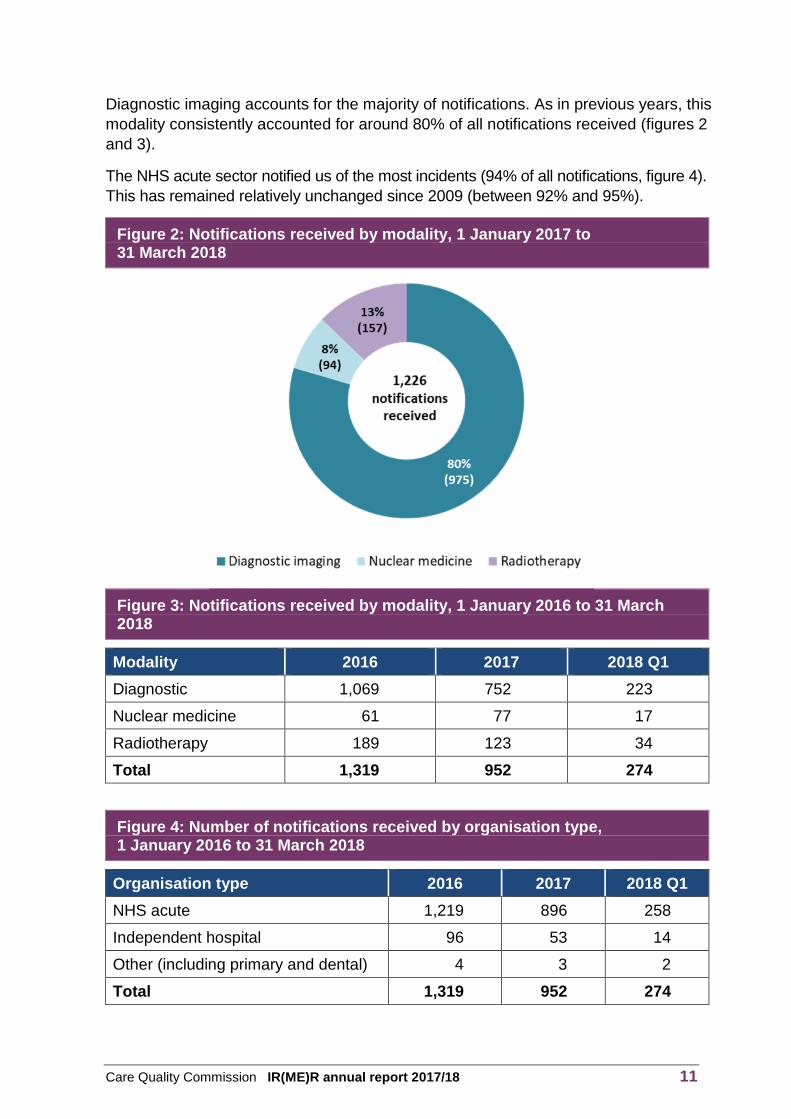
Diagnostic imaging accounts for the majority of notifications. As in previous years, this modality consistently accounted for around 80% of all notifications received (figures 2 and 3).
The NHS acute sector notified us of the most incidents (94% of all notifications, figure 4). This has remained relatively unchanged since 2009 (between 92% and 95%).
Figure 2: Notifications received by modality, 1 January 2017 to 31 March 2018
Figure 3: Notifications received by modality, 1 January 2016 to 31 March 2018
Modality 2016 2017 2018 Q1
Diagnostic 1,069 752 223
Nuclear medicine 61 77 17
Radiotherapy 189 123 34
Total 1,319 952 274
Figure 4: Number of notifications received by organisation type, 1 January 2016 to 31 March 2018
Organisation type 2016 2017 2018 Q1
NHS acute 1,219 896 258
Independent hospital 96 53 14
Other (including primary and dental) 4 3 2
Total 1,319 952 274

Care Quality Commission IR(ME)R annual report 2017/18 12
Comparisons of ‘activity’
Diagnostic imaging, nuclear medicine and radiotherapy play an essential role in the diagnosis, monitoring and treatment of a variety of medical conditions and diseases. The use of ionising radiation and continuous technological developments has meant that these specialties are at the forefront of modern medicine.
The development of minimally invasive techniques, such as using fluoroscopy-guided interventional radiology, has increased in popularity as it can reduce the need for surgical intervention, has lowered infection rates and has shortened recovery times for patients. PET-CT has become more established in diagnosing cancer and monitoring treatment, and NHS proton therapy centres will be treating their first patients in 2019.
The most reliable activity data relating to diagnostic imaging and nuclear medicine services is the Diagnostic Imaging Dataset (DID). This monthly data collection looks at the number of diagnostic imaging and nuclear medicine examinations carried out on NHS patients in England. We are aware that there is no centralised data collection for other areas, such as interventional, cardiology, DXA and non-NHS activities, which makes it difficult for us to analyse data and make comparisons between types of providers.
Over the last five years, the use of diagnostic imaging has grown by 8.3%. There were 41.1 million diagnostic imaging examinations carried out on NHS patients in England during the year from April 2017 to March 2018, of which 28.4 million used ionising radiation.7 Plain film X-rays make up most activity, with 21.8 million examinations in 2017/18 (equivalent to 53.1% of the total activity for NHS patients in a diagnostic imaging department).
Although the number of plain film X-rays carried out each year has remained stable, the number of more complex types of examinations has increased, with computed tomography (CT) increasing by over 30% in the past five years.
Radiotherapy activity
The National Cancer Registration and Analysis Service (NCRAS) is responsible for
the Radiotherapy Dataset (RTDS), which covers all activity within the NHS. This
dataset measures activity in ‘episodes’. A radiotherapy episode is a continuous
period of care for radiotherapy, including all preparation, planning and delivery of
radiotherapy. In 2017/18, there were 133,749 episodes, which is a small decrease
of 0.8% in radiotherapy activity compared with the previous year.
Care Quality Commission IR(ME)R annual report 2017/18 13
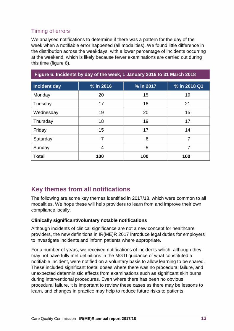
Timing of errors
We analysed notifications to determine if there was a pattern for the day of the week when a notifiable error happened (all modalities). We found little difference in the distribution across the weekdays, with a lower percentage of incidents occurring at the weekend, which is likely because fewer examinations are carried out during this time (figure 6).
Figure 6: Incidents by day of the week, 1 January 2016 to 31 March 2018
Incident day % in 2016 % in 2017 % in 2018 Q1
Monday 20 15 19
Tuesday 17 18 21
Wednesday 19 20 15
Thursday 18 19 17
Friday 15 17 14
Saturday 7 6 7
Sunday 4 5 7
Total 100 100 100
Key themes from all notifications
The following are some key themes identified in 2017/18, which were common to all modalities. We hope these will help providers to learn from and improve their own compliance locally.
Clinically significant/voluntary notable notifications
Although incidents of clinical significance are not a new concept for healthcare providers, the new definitions in IR(ME)R 2017 introduce legal duties for employers to investigate incidents and inform patients where appropriate.
For a number of years, we received notifications of incidents which, although they may not have fully met definitions in the MGTI guidance of what constituted a notifiable incident, were notified on a voluntary basis to allow learning to be shared. These included significant foetal doses where there was no procedural failure, and unexpected deterministic effects from examinations such as significant skin burns during interventional procedures. Even where there has been no obvious procedural failure, it is important to review these cases as there may be lessons to learn, and changes in practice may help to reduce future risks to patients.
Care Quality Commission IR(ME)R annual report 2017/18 14
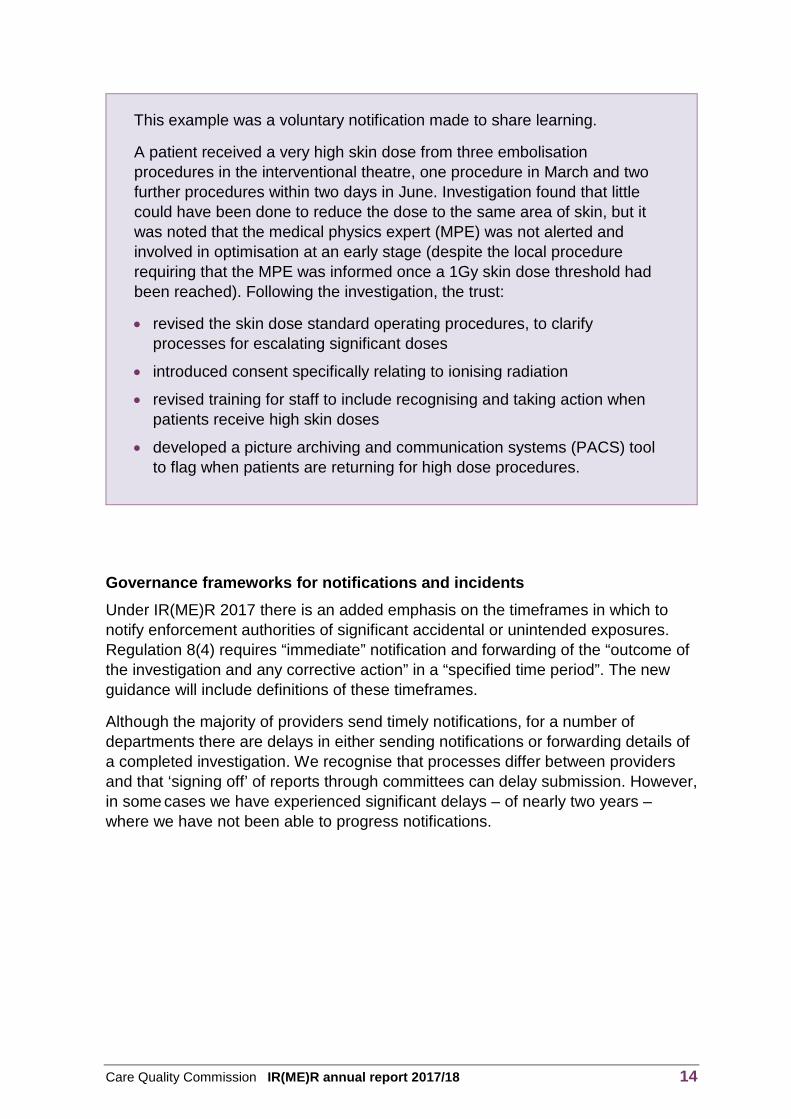
This example was a voluntary notification made to share learning.
A patient received a very high skin dose from three embolisation procedures in the interventional theatre, one procedure in March and two further procedures within two days in June. Investigation found that little could have been done to reduce the dose to the same area of skin, but it was noted that the medical physics expert (MPE) was not alerted and involved in optimisation at an early stage (despite the local procedure requiring that the MPE was informed once a 1Gy skin dose threshold had been reached). Following the investigation, the trust:
revised the skin dose standard operating procedures, to clarify processes for escalating significant doses
introduced consent specifically relating to ionising radiation
revised training for staff to include recognising and taking action when patients receive high skin doses
developed a picture archiving and communication systems (PACS) tool to flag when patients are returning for high dose procedures.
Governance frameworks for notifications and incidents
Under IR(ME)R 2017 there is an added emphasis on the timeframes in which to notify enforcement authorities of significant accidental or unintended exposures. Regulation 8(4) requires “immediate” notification and forwarding of the “outcome of the investigation and any corrective action” in a “specified time period”. The new guidance will include definitions of these timeframes.
Although the majority of providers send timely notifications, for a number of departments there are delays in either sending notifications or forwarding details of a completed investigation. We recognise that processes differ between providers and that ‘signing off’ of reports through committees can delay submission. However, in some cases we have experienced significant delays – of nearly two years – where we have not been able to progress notifications.
Care Quality Commission IR(ME)R annual report 2017/18 15
We inspected a trust following a series of notifications, and found there was little learning from incidents and poor investigations. Following the inspection, the trust reviewed its policies on radiation protection and managing incidents. It implemented actions to improve, including:
a new trust-wide policy to investigate radiation incidents, which clearly stated the duties of all involved
having a named ‘lead’ to oversee all investigations, ensure consistency and act as the main point of contact for CQC’s IR(ME)R team
formatting the reporting template to make sure it includes all relevant information
engaging the trust’s senior management in the process to ensure there is support when it is operationally difficult to contact the referring team.
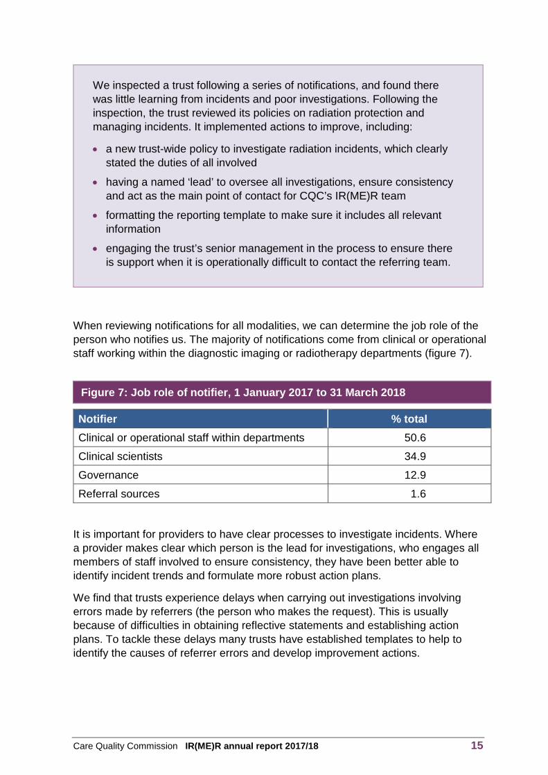
When reviewing notifications for all modalities, we can determine the job role of the person who notifies us. The majority of notifications come from clinical or operational staff working within the diagnostic imaging or radiotherapy departments (figure 7).
Figure 7: Job role of notifier, 1 January 2017 to 31 March 2018
Notifier % total
Clinical or operational staff within departments 50.6
Clinical scientists 34.9
Governance 12.9
Referral sources 1.6
It is important for providers to have clear processes to investigate incidents. Where a provider makes clear which person is the lead for investigations, who engages all members of staff involved to ensure consistency, they have been better able to identify incident trends and formulate more robust action plans.
We find that trusts experience delays when carrying out investigations involving errors made by referrers (the person who makes the request). This is usually because of difficulties in obtaining reflective statements and establishing action plans. To tackle these delays many trusts have established templates to help to identify the causes of referrer errors and develop improvement actions.
Care Quality Commission IR(ME)R annual report 2017/18 16
Following a number of delayed closures on notifications, a trust developed a new process to investigate referrer errors by using a template for the referring clinician to fill in when they have referred the wrong patient. The template helps clinicians to determine where in the pathway procedures were not followed, and any possible causes. It also helps to record people’s reflective statements and reminds referrers of their duties under IR(ME)R by providing information on the trust’s procedures for repeated non-compliance.
Trusts with zero notifications
In our 2016 report, we published the names of NHS trusts that had not made any notifications to CQC of incidents involving exposures ‘much greater than intended’. This had a positive effect as it prompted many providers to review their governance processes and incidents. Four trusts confirmed that notifiable incidents had occurred and the reason for not notifying CQC was because their internal processes had failed.
We believe that in diagnostic imaging departments, with increased demand and higher activity levels, it is highly unlikely that there will have been no notifiable errors. This is a potential risk, and in this report we identify the organisations that had not notified us of any incidents.
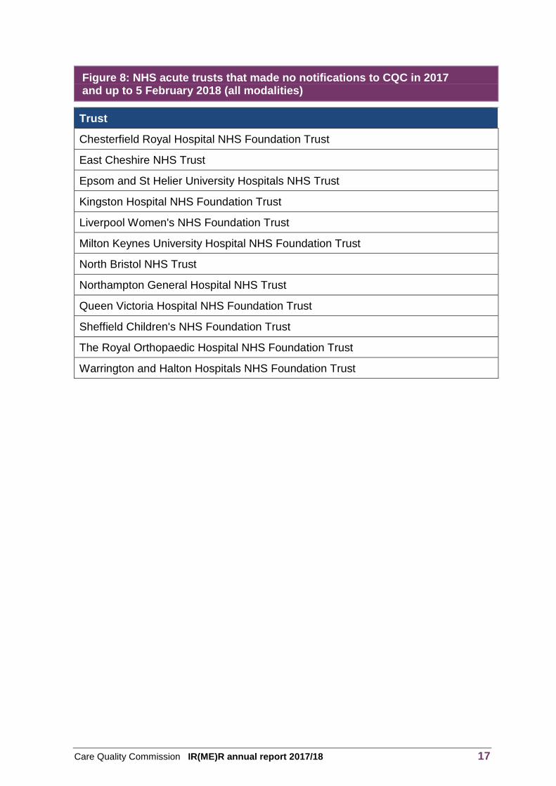
In 2017 and up to 5 February 2018 when IR(ME)R 2017 came into force, 12 NHS trusts had not submitted any notifications (figure 8). The list does not include non-acute NHS organisations or organisations in the independent sector, as they have different commissioning and provider models to the NHS.
As with last year’s report, we emphasise that it is important to take into account the following factors when reviewing the trusts listed:
Some of the trusts had not notified us of any incidents for a number of years and were also mentioned in previous annual reports. These are mostly trusts with departments that carry out a low number of examinations.
A number of these trusts have gone on to make notifications to us since 5 February 2018, after we have discussed their governance and incident management processes.
Four trusts have notified us of incidents retrospectively when they were discovered following internal reviews.
Inspections carried out at three of these trusts have confirmed that there were no notifiable incidents that had been reported internally.
Care Quality Commission IR(ME)R annual report 2017/18 17
Figure 8: NHS acute trusts that made no notifications to CQC in 2017 and up to 5 February 2018 (all modalities)
Trust
Chesterfield Royal Hospital NHS Foundation Trust
East Cheshire NHS Trust
Epsom and St Helier University Hospitals NHS Trust
Kingston Hospital NHS Foundation Trust
Liverpool Women's NHS Foundation Trust
Milton Keynes University Hospital NHS Foundation Trust
North Bristol NHS Trust
Northampton General Hospital NHS Trust
Queen Victoria Hospital NHS Foundation Trust
Sheffield Children's NHS Foundation Trust
The Royal Orthopaedic Hospital NHS Foundation Trust
Warrington and Halton Hospitals NHS Foundation Trust

Care Quality Commission IR(ME)R annual report 2017/18 18
DIAGNOSTIC IMAGING
Notifications
In the 2017 calendar year and the first three months of 2018, we received 975 diagnostic imaging notifications where patients received exposures ‘much greater than intended’ (figure 9). This represented 80% of the total in this period. Notifications from NHS acute trusts accounted for 94% of all diagnostic imaging notifications, with 57 notifications received from other types of provider.
Figure 9: Number of diagnostic imaging notifications received by type of provider, 1 January 2016 to 31 March 2018
Organisation type 2016 2017 2018 Q1
NHS acute 993 709 209
Independent hospital 72 40 12
Other (including primary and dental) 4 3 2
Total 1,069 752 223
We have seen a drop in the number of notifications received in relation to diagnostic imaging. Notifications decreased by 29.6% between 2016 and 2017, which we can attribute to the release of the guidance from the Department of Health and Social Care published in January 2017.
Figure 10: Diagnostic imaging notifications received by sub-modality, 1 January 2016 to 31 March 2018
Sub-modality 2016 2017 2018 Q1
Number % total Number % total Number % total
CT 641 60.0 347 46.1 101 45.3
Plain film X-rays 369 34.5 357 47.5 108 48.4
Mammography 34 3.2 19 2.5 4 1.8
Dental 8 0.7 10 1.3 4 1.8
Fluoroscopy 6 0.6 10 1.3 3 1.3
DXA 3 0.3 5 0.7 1 0.4
Interventional radiology 4 0.4 2 0.3 2 0.9
Cardiac 4 0.4 2 0.3 0 0
Total 1,069 100 752 100 223 100

Care Quality Commission IR(ME)R annual report 2017/18 19
In the 2017 calendar year and the first three months of 2018, the highest number of notifications was from the plain film X-ray sub-modality, whereas previously most notifications were from CT (figure 10).
Effective doses
It is worth noting that the number of plain film X-rays carried out compared with CT scans does not reflect their respective number of notifications. As previously mentioned, plain film X-rays make up the vast majority of examinations carried out in diagnostic imaging departments. But although they comprise 78% of ionising radiation examinations performed on NHS patients, they make up just under 50% of the total diagnostic imaging notifications we receive.
This is due to differences in the multiplication factors when determining whether an incident meets the criteria for notification to us. For example, plain film X-ray errors were notifiable above 10 or 20 times the intended dose (depending on body part) and CT errors, above 2.5 or 10 times (depending on dose). The differences in multiplication factors is risk-based and directly linked to the doses involved in the respective modalities.
There is a notable difference between plain film X-rays and CT examinations in the average doses that patients received in notifications (figure 11).
Figure 11: Average accidental or unintended effective dose received from notifiable incidents for CT and plain film X-ray in milli-Sieverts (mSv)
Sub-modality 2016 2017 2018 Q1
CT 9.9 9.0 10.9
Plain film X-rays 1.2 0.8 1.3
These doses should be viewed in the context of the comparable average dose that the UK population receives annually from natural background radiation (2.7mSv national average and 6.9mSv in Cornwall).8
Although it is extremely difficult to directly compare dose to risk for individual cases, the inferred risk from an exposure that gives an effective dose of 10-100 mSv is considered ‘low’, while that for effective dose in the range of 1-10 mSv is considered ‘very low’.9

Care Quality Commission IR(ME)R annual report 2017/18 20
Types of error
To enable us to analyse incidents we categorise errors by type across all diagnostic imaging sub-modalities (figure 12).
Figure 12: Analysis of errors in notifications from diagnostic imaging departments across all sub-modalities, 1 January 2017 to 31 March 2018
Detailed type of error
2016 2017 2018 Q1
Number % total Number % total Number % total
Referrer error: wrong patient
222 20.8 227 30.2 81 36.3
Operator error: wrong exposure set
156 14.6 91 12.1 21 9.4
Operator error: wrong anatomy/laterality
176 16.5 71 9.4 16 7.2
Operator error: failure to ID patient
101 9.4 130 17.3 34 15.2
Operator error: no check back of previous
94 8.8 40 5.3 11 4.9
Timing error for examination/ booking/NGT timing
86 8.0 34 4.5 14 6.3
Referrer error: no check back
62 5.8 31 4.1 11 4.9
Referrer error: wrong anatomy or modality
55 5.1 34 4.5 8 3.6
Operator error: modality selection
39 3.6 42 5.6 10 4.5
Operator error: other 28 2.6 20 2.7 6 2.7
Operator error: image archive/labelling
28 2.6 13 1.7 1 0.4
Volunteered (or not meeting criteria)
18 1.7 14 1.9 3 1.3
Inadequate supervision 4 0.4 4 0.5 1 0.4
Other 0 0 1 0.1 1 0.4
Equipment failure 0 0 0 0 5 2.2
Total 1,069 100 752 100 223 100

Care Quality Commission IR(ME)R annual report 2017/18 21
In 2017, the majority of root causes were from operator errors, with 56.9% of notifications attributed to these duty holders. A further 41% were identified as referrer errors. The ‘other’ code includes other errors that do not fit other categories, such as foetal doses and equipment errors (before IR(ME)R 2017).
Between 6 February and 31 March 2018, we received five notifications of equipment malfunctions in diagnostic imaging. These involved a range of errors, including automatic exposure control (AEC) failures, and software failures causing the loss of images.
Computed tomography
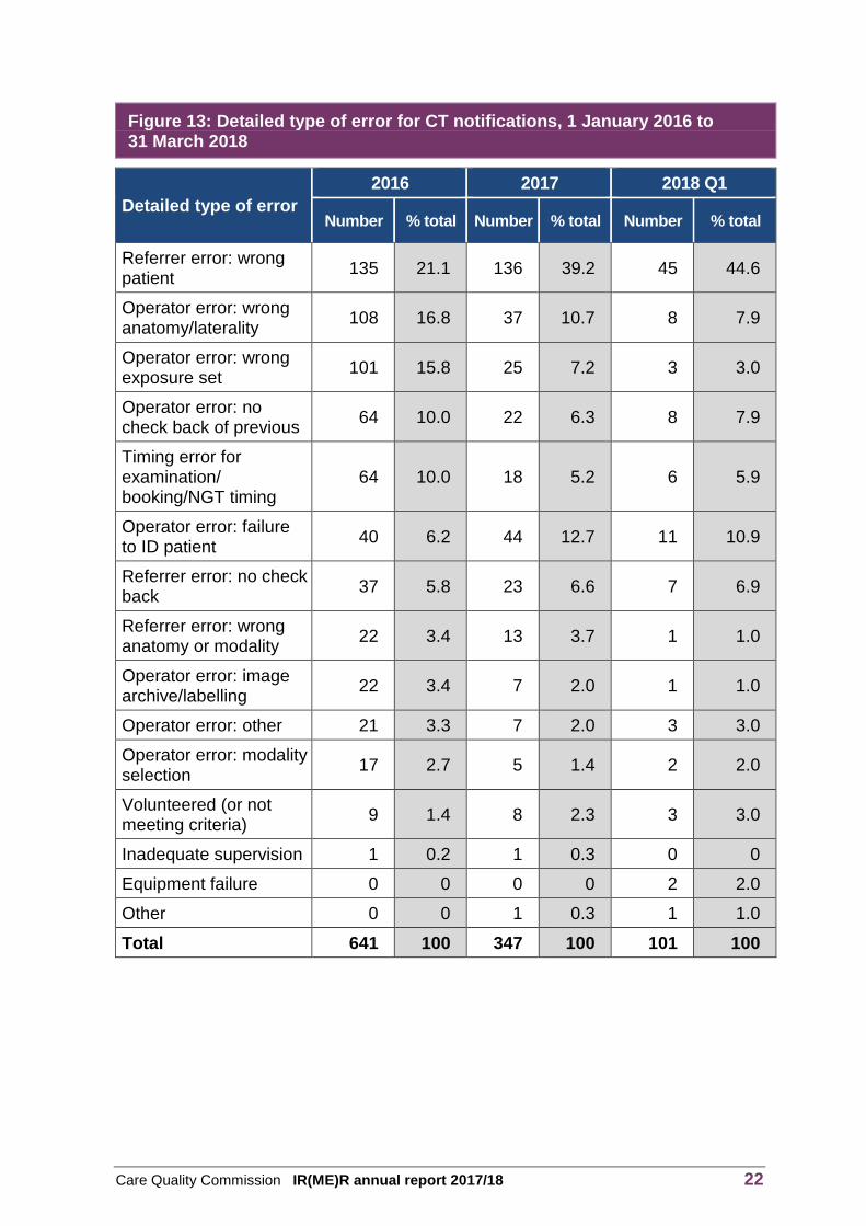
In 2017, the number of notifications from computed tomography (CT) reduced by 45.8%, compared with the previous year. When looking at the detailed type of error, we can see how the amended multiplication factors and definitions in the guidance on MGTI published in January 2017 has directly affected the type, and subsequently number, of notifications received in CT (figure 13). For example, wrong anatomy or laterality scanned reduced by 65.7%.
The change in multiplication factors is particularly evident in ‘wrong exposure set’ and ‘timing errors’, where these categories have seen a reduction of 75.2% and 71.8% respectively.
Where there were no changes to the guidance, the numbers of notifications remained comparable with previous years. Examples include errors by the referrer, (the person who requested a CT scan for the wrong patient), or where operators have failed to identify patients correctly before taking images and subsequently carried out the examination on the wrong patient.
Care Quality Commission IR(ME)R annual report 2017/18 22
Figure 13: Detailed type of error for CT notifications, 1 January 2016 to 31 March 2018
Detailed type of error
2016 2017 2018 Q1
Number % total Number % total Number % total
Referrer error: wrong patient
135 21.1 136 39.2 45 44.6
Operator error: wrong anatomy/laterality
108 16.8 37 10.7 8 7.9
Operator error: wrong exposure set
101 15.8 25 7.2 3 3.0
Operator error: no check back of previous
64 10.0 22 6.3 8 7.9
Timing error for examination/ booking/NGT timing
64 10.0 18 5.2 6 5.9
Operator error: failure to ID patient
40 6.2 44 12.7 11 10.9
Referrer error: no check back
37 5.8 23 6.6 7 6.9
Referrer error: wrong anatomy or modality
22 3.4 13 3.7 1 1.0
Operator error: image archive/labelling
22 3.4 7 2.0 1 1.0
Operator error: other 21 3.3 7 2.0 3 3.0
Operator error: modality selection
17 2.7 5 1.4 2 2.0
Volunteered (or not meeting criteria)
9 1.4 8 2.3 3 3.0
Inadequate supervision 1 0.2 1 0.3 0 0
Equipment failure 0 0 0 0 2 2.0
Other 0 0 1 0.3 1 1.0
Total 641 100 347 100 101 100

Care Quality Commission IR(ME)R annual report 2017/18 23
Plain film X-rays
In 2017, there was a modest decrease (3.3%) in the total number of notifications received for plain film X-ray errors (figure 14). As with CT, the numbers for some types of error changed considerably, some of which we can attribute to changes to the MGTI guidance. However, some changes cannot be directly attributed to this.
For some types of error that were not affected by the 2017 MGTI guidance, the numbers of notifications have remained relatively similar to previous years, for example where referrers have requested X-rays for the wrong patient.
We have seen an unexpected increase in the number of operator errors, where staff have not checked a patient’s identity before the plain film X-ray (33.3% increase) or not checked the exposure factors required before carrying out the examination (33.3% increase). Operators involved in these types of incidents often mention time pressures as being a contributing factor leading to their error. Although we are unable to come to any conclusions to explain the higher number of notifications of this type in 2017, a generally higher percentage in plain film X-ray
could be a result of the higher throughput of patients, as well as a more junior skill mix.
Where the definitions of MGTI have changed, (multiplication factors have not changed for plain film X-ray errors) there is a notable reduction in the number of notifications. For example, the number of laterality errors has decreased by 55.4%.

Care Quality Commission IR(ME)R annual report 2017/18 24
Figure 14: Detailed type of error for plain film X-ray notifications, 1 January 2016 to 31 March 2018
Detailed type of error 2016 2017 2018 Q1
Number % total Number % total Number % total
Referrer error: wrong patient
78 21.1 78 21.8 30 27.8
Operator error: wrong anatomy/ laterality
65 17.6 29 8.1 7 6.5
Operator error: failure to ID patient
60 16.3 80 22.4 19 17.6
Operator error: wrong exposure set
48 13.0 64 17.9 18 16.7
Referrer error: wrong anatomy or modality
29 7.9 19 5.3 7 6.5
Timing error for examination/ booking/NGT timing
19 5.1 14 3.9 8 7.4
Operator error: modality selection
18 4.9 31 8.7 8 7.4
Operator error: no check back of previous
17 4.6 14 3.9 1 0.9
Referrer error: no check back
17 4.6 6 1.7 4 3.7
Volunteered (or not meetingcriteria)
7 1.9 2 0.6 0 0
Operator error: image archive/ labelling
5 1.4 5 1.4 0 0
Operator error: other 3 0.8 12 3.4 2 1.9
Inadequate supervision 3 0.8 3 0.8 1 0.9
Equipment failure 0 0 0 0 3 2.8
Total 369 100 357 100 108 100
Care Quality Commission IR(ME)R annual report 2017/18 25
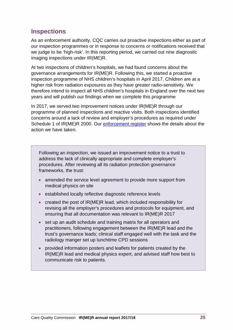
Inspections
As an enforcement authority, CQC carries out proactive inspections either as part of our inspection programmes or in response to concerns or notifications received that we judge to be ‘high-risk’. In this reporting period, we carried out nine diagnostic imaging inspections under IR(ME)R.
At two inspections of children’s hospitals, we had found concerns about the governance arrangements for IR(ME)R. Following this, we started a proactive inspection programme of NHS children’s hospitals in April 2017. Children are at a higher risk from radiation exposures as they have greater radio-sensitivity. We therefore intend to inspect all NHS children’s hospitals in England over the next two years and will publish our findings when we complete this programme
In 2017, we served two improvement notices under IR(ME)R through our programme of planned inspections and reactive visits. Both inspections identified concerns around a lack of review and employer’s procedures as required under Schedule 1 of IR(ME)R 2000. Our enforcement register shows the details about the
action we have taken.
Following an inspection, we issued an improvement notice to a trust to address the lack of clinically appropriate and complete employer’s procedures. After reviewing all its radiation protection governance frameworks, the trust:
amended the service level agreement to provide more support from medical physics on site
established locally reflective diagnostic reference levels
created the post of IR(ME)R lead, which included responsibility for revising all the employer’s procedures and protocols for equipment, and ensuring that all documentation was relevant to IR(ME)R 2017
set up an audit schedule and training matrix for all operators and practitioners, following engagement between the IR(ME)R lead and the trust’s governance leads; clinical staff engaged well with the task and the radiology manger set up lunchtime CPD sessions
provided information posters and leaflets for patients created by the IR(ME)R lead and medical physics expert, and advised staff how best to communicate risk to patients.

Care Quality Commission IR(ME)R annual report 2017/18 26
Key themes from diagnostic imaging
The following are some key themes and case studies that arose from our work in diagnostic imaging, which we hope providers can learn from and improve their own compliance locally.
Pause and check
The majority of diagnostic imaging departments have adopted the ‘pause and check’ initiative, or a locally-derived alternative, following its launch in 2015. However, this concept has not had the impact we thought it might, and as mentioned in the previous three annual reports, we continue to receive notifications of incidents where a simple ‘stop moment’ could have prevented an unintended or over-exposure. We understand that an increase in demand, coupled with staff shortages, has led to increased pressure on departments. However, speed and efficiency should not come at the expense of patient safety.
It is important to remember that correctly identifying patients, checking exposure factors and reading requests are fundamental responsibilities of the professionals who carry out these examinations. The Health and Care Professions Council’s Standards of proficiency state that radiographers and clinical scientists need to conform to standard operating procedures and should be able to operate equipment safely.10 Employers should continue to reinforce this to staff and remind them not to become complacent about checking the identity of patients or to be distracted by other pressures when carrying out these vital safety checks.
Referrer errors
The total number of errors by referrers when requesting examinations of the wrong patient has risen slightly this year. In July 2017, the Society and College of Radiographers launched a referrer ‘pause and check’, which follows a similar concept to the one used by operators.11
Cancellations of requests
Between January 2017 and March 2018, we received more than 50 notifications where an examination had been carried out despite being cancelled. We saw a range of causes for these incidents, such as confusion over who was responsible for the cancellation, and poor communication between referrers and the diagnostic imaging department. We also saw a number of errors by clerical staff, in which they had either not cancelled an examination when requested or had re-instated cancelled requests on the radiology information system (RIS).
It is important to clarify responsibilities to all duty holders, and that clerical staff follow local procedures. Communicating with referrers about the importance of cancelling requests that are no longer required, for example, pre-operative imaging (carried out post op) or post-operative imaging (where operations had been abandoned or changed) is equally important.

Care Quality Commission IR(ME)R annual report 2017/18 27
Failures with interfacing between IT systems are also common, where cancellation messages are not communicated between e-referral systems and RIS. It is important to make sure that there are processes and that these are communicated to all staff where departments have encountered this issue. The issue should also be raised with IT teams, both in-house support and with external IT suppliers, to ensure that they are aware of the risk.
CQC’s IR(ME)R team has discussed our concerns with a number of RIS suppliers and we have made them aware of our concerns around trends we have identified.

Care Quality Commission IR(ME)R annual report 2017/18 28
NUCLEAR MEDICINE
Notifications
In this reporting period (2017 calendar year and the first three months of 2018), we received 94 notifications about nuclear medicine incidents (figure 15). This represented 8% of the total in this period. Notifications from NHS acute trusts accounted for 93% of all nuclear medicine notifications.
Figure 15: Number of nuclear medicine notifications received by type of provider, 1 January 2016 to 31 March 2018
Organisation type 2016 2017 2018 Q1
NHS acute 52 72 15
Independent hospital 9 5 2
Total 61 77 17
Nuclear medicine is the only modality where notifications increased overall (by 26%) compared with the previous year. In previous years, we had reported a mean increase of about four notifications a year. This increase can be directly attributed to the guidance from the Department of Health and Social Care published in January 2017. However, the overall magnitude and risk profile of such incidents remains relatively low and we do not attribute this increase to a deterioration in practice.
The majority of nuclear medicine notifications involved diagnostic exposures (figure 16). Incidents involving therapeutic overexposures only accounted for 5% of nuclear medicine notifications in 2017, of which the majority relate to foetal exposures.
Figure 16: Nuclear medicine notifications received by sub-modality, 1 January 2016 to 31 March 2018
Sub-modality 2016 2017 2018 Q1
Number % total Number % total Number % total
Diagnostic 59 97.0 73 95.0 15 88.0
Therapeutic 2 3.0 4 5.0 2 12.0
Total 61 100 77 100 17 100
Care Quality Commission IR(ME)R annual report 2017/18 29
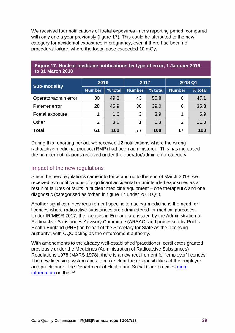
We received four notifications of foetal exposures in this reporting period, compared with only one a year previously (figure 17). This could be attributed to the new category for accidental exposures in pregnancy, even if there had been no procedural failure, where the foetal dose exceeded 10 mGy.
Figure 17: Nuclear medicine notifications by type of error, 1 January 2016 to 31 March 2018
Sub-modality 2016 2017 2018 Q1
Number % total Number % total Number % total
Operator/admin error 30 49.2 43 55.8 8 47.1
Referrer error 28 45.9 30 39.0 6 35.3
Foetal exposure 1 1.6 3 3.9 1 5.9
Other 2 3.0 1 1.3 2 11.8
Total 61 100 77 100 17 100
During this reporting period, we received 12 notifications where the wrong radioactive medicinal product (RMP) had been administered. This has increased the number notifications received under the operator/admin error category.
Impact of the new regulations
Since the new regulations came into force and up to the end of March 2018, we received two notifications of significant accidental or unintended exposures as a result of failures or faults in nuclear medicine equipment – one therapeutic and one diagnostic (categorised as ‘other’ in figure 17 under 2018 Q1).
Another significant new requirement specific to nuclear medicine is the need for licences where radioactive substances are administered for medical purposes. Under IR(ME)R 2017, the licences in England are issued by the Administration of Radioactive Substances Advisory Committee (ARSAC) and processed by Public Health England (PHE) on behalf of the Secretary for State as the ‘licensing authority’, with CQC acting as the enforcement authority.
With amendments to the already well-established ‘practitioner’ certificates granted previously under the Medicines (Administration of Radioactive Substances) Regulations 1978 (MARS 1978), there is a new requirement for ‘employer’ licences. The new licensing system aims to make clear the responsibilities of the employer and practitioner. The Department of Health and Social Care provides more information on this.12

Care Quality Commission IR(ME)R annual report 2017/18 30
Inspections
We carried out two inspections in nuclear medicine in 2017, focusing on the provision of PET-CT in independent services. These were in response to several notifications around the authorisation of examinations under practitioner guidelines. It also coincided with the award of national contracts for PET-CT, which allowed us to understand the special commissioning frameworks involved.
The inspections were focused on the patient pathway from referral to clinical evaluation, looking at the scale of patient throughput, practitioner and operator entitlement, and the support from medical physics experts for the services.
We identified a theme in more than one notification involving referral for a 18F-Fluorocholine scan (Choline) where, during the vetting process, an 18F-Fluorodeoxyglucose (FDG) scan was incorrectly authorised under practitioner guidelines. After speaking with the providers, it was apparent that after referrals are received, the patient pathway for PET-CT differs from that typically found in a hospital’s nuclear medicine department, with ARSAC practitioners ‘delegating’ the
authorisation and reporting of some examinations to other members of staff.
During the inspections, we saw that the provider was rolling out a new electronic-referral system across all its sites to facilitate the management, audit and archiving of data. The system also enabled improved recording of the identity and entitlement of duty holders, in particular the ARSAC-holder, IR(ME)R practitioner and IR(ME)R operators, mostly radiologists, who authorised exposures under the relevant practitioner’s guidelines or reporting scans. Within PET-CT, those with such entitlement are conveniently described in procedure as ‘ARSAC Delegate’ or ‘Reporting Delegate’. The IT platform also helped auditing of patient/scanning data and records, which is useful for demonstrating IR(ME)R compliance more generally.
Key themes from nuclear medicine
The following are some key themes and case studies that arose from our work in nuclear medicine, which we hope providers can learn from and improve their own compliance locally.
Referral errors
In 2017, 39% of the notifications received were due to errors in the referral process. These were mostly because the referrer either selected the wrong patient or occasionally requested the wrong examination. This type of error has remained unchanged by the new guidance or the change in regulations, and the total number is comparable with previous years.

Care Quality Commission IR(ME)R annual report 2017/18 31
One unusual case involving an error with mistaken identity concerned newborn twins. One required a nuclear medicine thyroid scan, but this was performed on the wrong child. Investigations revealed that the twins had been registered by their GP and although they each had the correct NHS patient reference number, their identities were transposed in error at the time of registration. Therefore, the request for a scan on one of the children ended up with the wrong twin presenting on the day of the scan.
The learning here was aimed at ensuring there is no mistaken identity at registration, and the need for careful scrutiny in making requests for twins, especially soon after birth.
Cancellation of referrals
As previously mentioned, failure in the process for cancelling examinations has resulted in unnecessary examinations. Cancellations have usually been made when the referral team realised the wrong patient or pathway had been requested, or the patient’s management, diagnosis or treatment had changed. Of the referrer errors notified, five were the result of the failure to follow local procedures for cancelling a referral, which meant that administration of RMP had unnecessarily continued. Many e-requesting and RIS systems do not allow for a fail-safe cancellation process; therefore nuclear medicine departments tend to have procedures that require a direct phone call into the department. However, referrers regularly need be reminded of such procedures.
Reflective learning
Reflections from staff involved in incidents are valuable in helping to understand the causes and mitigating factors around errors. Where these reflections are carried out in a non-blame, supportive way, they provide opportunities to recognise areas for further training or supervisory support, and to identify environmental factors. We have seen reflective statements that have identified contributory factors, which have then enabled investigations to identify learning points from simple human errors. Examples include distractions that affect concentration levels, interruptions, poor skill mix, and workload pressures or patient queueing.
Wrong radioactive medicinal product injected
In the reporting period, notifications involving operator errors cover a similar spectrum as seen previously, with around a quarter resulting from administering the wrong RMP. Notifications of incidents involving injecting the wrong RMP can be further categorised into:
laboratory practice – where the hospital’s radiopharmacy or external supplier had supplied incorrect or mis-labelled RMP
operator error – the failure to inject the correct RMP into the intended patient.

Care Quality Commission IR(ME)R annual report 2017/18 32
RMP vials or syringes should be correctly labelled, stored and segregated when in use, and perhaps colour-coded and always checked before administration.
We received two notifications where errors meant that four patients underwent scans that had to be repeated. Investigations discovered that ‘free pertechnetate’ and incorrect and mis-labelled RMP had been supplied by the hospital radiopharmacy or external supplier.
Overall, it is essential to pay strict attention to good laboratory organisation, procedure and practice, and to carry out relevant checks, to minimise the risk of staff picking up and administering the wrong RMP. Following incidents, some departments have implemented an independent second check of RMPs before injection.
A check of patient identity is a key step in IR(ME)R procedures, but in nuclear medicine there is also the need to check that the correct RMP and activity is being injected for the examination requested. Local procedures may vary and can occur at different points in the pathway, but a final four-way check, ideally immediately before injection, of patient identity versus protocolled request versus RMP versus dose/activity, can act as a useful ‘fail-safe’.
PET-CT
The number of notifications in PET-CT has reduced from 13 previously to just six in this reporting period. This may be a result of the experience and maturity of manufacturers, providers and operators in using and managing the sophisticated equipment.
Compared with previous years, there have been few practitioner or referrer errors in requesting and authorising FDG versus Choline scans. The notifications mostly describe simple operator errors in RMP administration or equipment, including use of auto-dispenser and selecting incorrect anatomical programmes.
Unintended foetal exposures during therapy
Notifications relating to incidents in radionuclide therapy are by definition relatively high-risk. We received four notifications of unintended foetal doses. None of these involved a breakdown of procedures, but it subsequently came to light that the patients involved were in the early stages of pregnancy at the time of treatment. Therapies included the administration of Iodine-131 for thyroid ablation and treatment of benign thyroid disease, exposing the early pregnancy to direct and indirect exposure from radioactive iodine. Estimated foetal doses were above the 10 mGy threshold for notification and in the range 20-60 mGy.
We note that the frequency of notifications of ‘unknown’ pregnancy during therapy is relatively rare but, despite all checks being made, early pregnancy cannot always be ruled out.
As previously mentioned, it is still worthwhile investigating these incidents, even when there is no breakdown or error, as learning may still be identified.

Care Quality Commission IR(ME)R annual report 2017/18 33
Errors in treatment exposure
Two therapeutic notifications relate to errors in the treatment itself. One notification related to liver radio-embolisation with Yttrium-90 microspheres, and the second case involved Iodine-125 contamination from a damaged radioactive sealed source used in prostate low dose-rate brachytherapy. At the time of writing, the notifications were not closed, so we cannot describe them in detail. However, the former is a very specialised and multidisciplinary technique, which was being conducted as part of a clinical trial, and the latter, iodine seed administration, is a more standardised and well-established practice.
Despite trained and competent IR(ME)R practitioners and operators working to standard protocols, or clinical trial protocols with checks and quality assurance, some interventions can be highly specialised or complicated and may have significant inherent ‘medical’ risks, that are necessarily covered as part of the consent process. We received the notification for the iodine seed contamination incident after IR(ME)R 2017 came into force, which includes ‘equipment fault’ notifications within regulation.
Overall, we can conclude that for standard nuclear medicine therapy, the frequency and magnitude of notifications of accidental exposures is reassuringly low.

Care Quality Commission IR(ME)R annual report 2017/18 34
RADIOTHERAPY
Notifications
In the 2017 calendar year and the first three months of 2018, we received 157 radiotherapy notifications, which represented 13% of the total (figure 18).
Of these notifications, 95% were from NHS acute trusts, which is similar to previous years. Although there is no specific data relating to radiotherapy activity in independent health care, it is well known that the majority of radiotherapy treatments and imaging are carried out in the NHS acute setting, which accounts for the figures. We received eight notifications from three different independent organisations that provide a radiotherapy service.
Figure 18: Number of notifications received in radiotherapy, 1 January 2016 to 31March 2018
Organisation type 2016 2017 2018 Q1
NHS acute 174 115 34
Independent hospital 15 8 0
Total 189 123 34
Between 2016 and 2017, we saw a fall in radiotherapy notifications overall of 34.9%. This is because notifications from planning and verification imaging decreased by 53.4%, while the numbers of brachytherapy and beam therapy notifications have remained largely comparable with previous years (figure 19).
Figure 19: Notifications received in radiotherapy by sub-modality, 1 January 2016 to 31 March 2018
Sub-modality 2016 2017 2018 Q1
Number % total Number % total Number % total
Planning/verification imaging
118 62 55 45 9 26
Beam therapy (radical) 33 17 32 26 16 47
Beam therapy (palliative) 33 17 30 24 9 26
Brachytherapy (radical) 5 3 6 5 0 0
Total 189 100 123 100 34 100

Care Quality Commission IR(ME)R annual report 2017/18 35
Types of error
We categorise notifications in radiotherapy by the type of error. Along with the new definitions of ‘significant accidental and unintended exposures’, we will refine these categories to enable us to carry out more detailed trend analysis. Our new system will include the taxonomy used in guidance from the Royal College of Radiologists Towards safer radiotherapy.
The ‘much greater than intended’ guidance published in January 2017 resulted in a considerable reduction in the number of notifications from radiotherapy imaging (figure 20). This was because multiplication factors were aligned with those in diagnostic imaging CT, which allow for a single ‘repeat’ CT scan in a fraction. However, treatment exposures remained relatively unchanged as the guidance retained the definitions for whole course and individual fraction over-exposures. Refined definitions for geographical misses may account for the slight reduction in notifications that we classify as a treatment error.
Figure 20: Notifications received in radiotherapy, 1 January 2017 to 31 March 2018
Sub-modality 2016 2017 2018 Q1
Number % total Number % total Number % total
Radiotherapy imaging 118 62.4 55 44.7 9 26.5
Treatment error 56 29.6 47 38.2 18 52.9
Planning error 12 6.3 11 8.9 2 5.9
Referral error 1 0.5 5 4.1 3 8.8
Other 2 1.1 5 4.1 2 5.9
Grand total 189 100 123 100 34 100
Inspections
We inspected a number of radiotherapy services in response to high-risk notifications received during 2017 and the first quarter of 2018. Two notifications involved brachytherapy treatments, which we describe in the following examples, and one was in response to treatment delivered to the wrong area.

Care Quality Commission IR(ME)R annual report 2017/18 36
We carried out focused inspections at two trusts in response to incidents involving the delayed removal of brachytherapy eye plaques used to treat ocular tumors. The causes for these over-exposures were established as:
no anaesthetists were available when needed
the dosimetry form was sent outside of normal working hours, when there was no formal medical physics cover for calculating the dose
the removal time was not correctly identified when inserting the plaque.
The trusts’ investigations, and our inspections, identified that the departments had lengthy written protocols and procedures that did not accurately reflect practice or allow staff to follow them effectively. Staff were not working with radiation regularly, therefore radiation protection was not embedded in practice and IR(ME)R was not applied consistently.
Following our inspections, the trusts have:
reviewed theatre arrangements to include a window in theatre lists of the earliest and latest times for removing eye plaques
simplified the written protocols for treatment to clearly define responsibilities
trained staff on the regulatory requirements under IR(ME)R
identified an IR(ME)R lead in the ocular oncology team
reviewed the service level agreement for medical physics experts to ensure clear arrangements for who provides advice on compliance with IR(ME)R.
Key themes from radiotherapy
The following are some themes and case studies from our work in radiotherapy, which we hope clinical departments can learn from and improve their own practice locally.
Referrals for planning scans before confirmed diagnosis
We received a small number of notifications where patients were referred for planning scans before their diagnoses were confirmed through pathology or other diagnostic tests. The results of these tests are intended to determine the correct treatment option for the patients.

Care Quality Commission IR(ME)R annual report 2017/18 37
We received two notifications from a trust of separate unnecessary planning scans that had been requested “pending test results being available”, which were intended to avoid a delay in starting treatment. In one notification, although the test results were made available, they were located on a system that planning radiographers do not routinely access. The results suggested that radiotherapy treatment was no longer the preferred treatment option. The investigation found that although the referrals stated that additional test results were pending, they did not state that the planning scan should not proceed until the results were confirmed.
In response to these incidents, the trust reviewed its referral processes and implemented the following changes:
Referrers are only able to make appointments for pre-treatment scans once the test results are received and assessed.
Electronic action sheets now have a new additional category that makes clear where referrals are awaiting test results. Referrers are encouraged to either not sign action sheets until results are available, or not start them until they have received test results.
In two notifications, the results of the test have led to unnecessary treatment exposures. One of these incidents occurred when a diagnosis was changed following additional pathology tests, but after treatment had already started.
We occasionally receive notifications where the type of treatment for a patient has been amended after a change of professional opinion following a planning scan. While this is not ideal for the patient, and to some extent a waste of resources, we accept it is sometimes unavoidable and in the patient’s best interests to take this new advice. This type of notification shows transparency from the department and can identify learning.
Checking for previous treatments
A number of notifications involved a failure to check for previous radiotherapy treatments. One incident involved a patient who underwent a planning scan and had already received radiotherapy at the same centre earlier that year. In another notification, a planning scan revealed evidence of I-125 brachytherapy seeds that had been implanted years earlier.
These incidents have highlighted the importance of establishing and maintaining summary records of treatment, and making them accessible to clinical oncologists to allow an active check to establish whether the patient has undergone treatment previously. One department introduced a checklist for pre-treatment radiographers, which made specific reference to checking for previous treatments. The department also developed referral documents that explicitly included ‘RETREATMENT’ in the electronic ‘comments’ box, where appropriate, to ensure that the message was clearly visible to the patient’s clinical oncologist.

Care Quality Commission IR(ME)R annual report 2017/18 38
A patient received treatment for a facial-basal cell carcinoma, which was planned and delivered correctly. However, a further lesion was found when the patient attended for follow-up, so an action sheet was completed for the new lesion. When the patient attended for planning, the clinical oncologist was unable to access their notes or the electronic action sheet. They therefore used the most recent letter that they could find to guide planning and treatment. Unfortunately, the clinical oncologist mistakenly thought that the original lesion had yet to be treated and therefore planned and treated the original site rather than the new intended site.
The trust’s comprehensive investigation found a number of contributory factors for this error. After reviewing processes, the trust took action to improve, which included:
revising written procedures in orthovoltage to clearly define responsibilities and procedural steps
introducing a checklist to ensure that all tasks follow the local procedure
auditing clinical practice to ensure compliance with written procedures
extending the allocated time slots for planning
investigating IT issues and ensuring clear contingencies.
Planning scan errors
As with the other modalities, we have received notifications relating to the failure to ‘pause and check’. This results in operators misreading or misinterpreting referrals, and subsequently results in incorrect scan coverage, incorrect use of or lack of contrast media, or incorrect arm positioning. It is important to continue to reinforce the message to staff to read requests fully, follow departmental protocols and, if in doubt, to check with the referring clinical oncologist about the imaging requirements. Staff need to be reminded not to become complacent and not allow themselves to be distracted by other pressures when making these safety checks.
Verification imaging
The guidance on making notifications about verification imaging is now well-understood and established in clinical radiotherapy departments. Importantly, verification imaging has occasionally revealed a ‘queuing’ error identifying that the plan loaded up is not intended for the patient about to undergo imaging and treatment. One simple solution from a radiotherapy centre was to require the ‘operator’ who loads up the plan to also carry out the patient identification procedure with the patient themselves, and to use photo-ID to support the process wherever possible. Although this type of incident means that a patient has to undergo repeat imaging, highlighting a mistake in the run-up to treatment can prevent a potential, more serious error when delivering the treatment.

Care Quality Commission IR(ME)R annual report 2017/18 39
We received three notifications that highlighted a lack of communication between tomotherapy devices and record and verify (R&V) systems, where pre-treatment scans were attached to the wrong patient and the patient needed a repeat scan.
In some cases, these communication errors led to some scans being associated with the wrong patient or treatment phase. In one incident, the operator carried out a cone beam CT (CBCT) scan but attached it to the patient’s phase 1 treatment rather than the current phase 2 reference. Some clinical departments have updated workflow processes to require a re-check of patient identity and scans to help to detect any human errors.
A patient required a repeat scan on the first day of their radiotherapy treatment. As this was at the time of the NHS cyber-attack, the initial planning CT scan had to be uploaded manually, but it was the wrong scan. Having the wrong CT reference data and the matching difficulties led to an additional nine verification imaging episodes over the course of treatment.
The department has amended its local process to ensure that scans are identified correctly throughout the planning/treatment pathway. This involves generating a unique ID for a scan, which is annotated. Each time a scan is exported or imported to other systems, its unique identifier is checked to ensure that it is the correct scan.
Treatment planning
We received 13 notifications relating to planning errors in this reporting period. These involved calculation, prescription or shift errors, and failures to prescribe adequate shielding, as the following examples illustrate.
We were notified of an error that became apparent to clinical staff part way through treatment. A patient was undergoing treatment to their lip using electrons, which required a leaded gum shield and lead ‘splash mask’ to shield adjacent areas to reduce unintended dose to the surrounding ‘healthy’ tissue. The instructions for fitting the gum shield were not clear, it was not fitted correctly and the patient’s lip was not shielded adequately for several fractions.
The detailed local investigation revealed that document QA arrangements, planning procedures and standard operating procedures in the mould room were out of date and had not kept pace with clinical practice. In particular, the clinical mark-up process did not make reference to fitting and location of the gum shield. This led to a review of all aspects of planning superficial treatments, including re-training staff.

Care Quality Commission IR(ME)R annual report 2017/18 40
In another example, following a diagnosis of metastatic cord compression, treatment was planned using a simulator, as the CT scanner was not operational. The image was sub-optimal because of the patient’s discomfort and the thoracic brace they were wearing. Treatment was prescribed and after two fractions were delivered, the patient underwent a CT scan once the scanner was recommissioned. However, this scan showed that the wrong area of spine had been treated. The trust’s investigation found that staff were not confident in using the conventional simulator as it was rarely used.
The trust’s actions included:
ensuring that staff regularly perform a test simulation using a phantom to maintain skill levels
investigating whether CBCT can be used when a CT scanner is not available rather than the simulator.
In one unusual notification, we learned how a radiotherapy department had struggled with an outage of its R&V system during the NHS cyber-attack, which had been intentionally switched off to prevent patients’ images from getting lost. Emergency treatment had only been possible with an ageing linac with no multi leaf collimator capability, which is used to shape the field of treatment. Staff had to plan and treat by making manual calculations but the plan checking arrangements turned out to be inadequate. They had no access to a printed version of its procedures and work instructions to follow during this period.
The patient was treated on their first day using a protocol of 20Gy over five fractions when 20Gy over 10 fractions was intended. The error was detected before their second fraction. The lack of R&V required staff to treat the patient in ‘standby’ mode and verify in ‘maintenance’ mode.
Geographical misses
The most common type of treatment error was geographical misses, with 53 notifications. Of these, 46 (87% of all geographical misses) were for a single fraction only. The majority of these misses can be further sub-categorised into:
aligning to wrong skin markers
on-line image mismatch
intended couch moves not applied, applied in the wrong direction, or proposed moves incorrect.
Care Quality Commission IR(ME)R annual report 2017/18 41
Following these incidents, clinical departments have carried out thorough investigations and implemented a range of actions. Some departments have fed back on more innovative measures they have taken to mitigate future risk to patients. These include:
training in specialist human factors engineering
technical solutions to replace manual tasks to overcome transcription errors
introducing skin rendered imaging
introducing a policy of an additional CBCT scan following any move or any set-up out of tolerance, with the involvement of senior colleagues to assist and check positioning
reinforcing visual checking processes (or ‘sense checks’) after checks under local procedure have been completed
considering indexing systems and pre-treatment verification imaging for patients receiving palliative care
ensuring that two radiographers are on set to make sure that the correct plan is loaded to treat the correct patient
peer review from other providers or from Public Health England.
Treatments during pregnancy
It is well established that radiotherapy departments adopt strict procedures for making enquiries of patients of childbearing potential during consent, before their treatment planning scan, and before the first fraction is delivered. We received six notifications where treatment exposures had been delivered to patients who were pregnant without knowing it. None of these notifications were the result of procedural failures, and in all cases, the consenting and pregnancy procedures had been followed.
Although there had been no procedural breakdowns, department managers nevertheless took the opportunity to review their local arrangements. Changes included ensuring that patients have received both verbal and written advice on the importance of avoiding becoming pregnant, and advising them to take the necessary steps during this time. The other measure was to reinforce with staff the importance of following procedures and document the patient’s response. One trust has introduced a pregnancy test before the planning scan as an additional assurance. However, it is well known that there can be a possibility of false-negative test results particularly during early pregnancy. If a patient is found to be pregnant, it is important for staff to have processes in place to involve medical physics experts and clinical oncologists at the earliest opportunity to carry out dose assessment and help with counselling for the patient.

Care Quality Commission IR(ME)R annual report 2017/18 42
APPENDIX: GLOSSARY OF TERMS
Beam therapy is radiotherapy where a beam of high energy radiation is aimed at the cancer or diseased anatomy. It is typically given as a number of short daily treatments using a machine called a linear accelerator (linac). In all therapy a high dose of radiation is targeted on the tumour but adjacent healthy tissue receiving a small amount of radiation which can be tolerated.
Brachytherapy and nuclear medicine therapy are where radioactive source(s) or material are applied directly to an affected area whether internally or externally.These therapies involve the insertion of small radioactive ‘seeds’ into the cancer, placing radioactive materials (within tubes) directly onto the tumour for a set length of time or injecting radiopharmaceutical which will concentrate naturally in the target organ.
Computed tomography (CT) is a scan that combines a series of X-ray images taken from different angles around the body to create detailed cross-sectional images (slices) of the inside of the body.
Coronary catheterisation refers to the imaging of blood vessels in the heart for both diagnostic and interventional purposes. These procedures can be used in emergencies, such as in a heart attack, or to look at unusual test results, such as stress tests of unexplained heart failure. Throughout this report we use the term Cardiac to describe such procedures.
Dual energy X-ray absorptiometry (DXA) is a special type of X-ray scan that measures bone mineral density (BMD). DXA scans are used to look at osteoporosis.
Fluoroscopy is similar to an X-ray ’movie’. The images are transmitted to a TV-like monitor in real time so that the body part and its motion can be seen in detail. Fluoroscopy is used to look at many body systems, including the digestive, urinary and reproductive systems and provides information on their function as well as anatomy.
Interventional radiology refers to a range of techniques that rely on the use radiological image guidance (fluoroscopy, ultrasound, computed tomography or magnetic resonance imaging) to precisely target therapy. Throughout this report we use this term to describe fluoroscopy-guided interventional radiology (imaging of the blood vessels to look for abnormalities with the use of various contrast media).
Mammography uses X-rays to examine the breast for diagnosis and screening. The goal of mammography is the early detection of breast cancer, through the NHS Breast Screening Programme, or assessing lumps through symptomatic mammograms.

Care Quality Commission IR(ME)R annual report 2017/18 43
Much greater than intended (MGTI). Under Regulation 4(5) of IR(MER) 2000, incidents were required to be investigated and reported when a person undergoing medical exposure was exposed to ionising radiation to an extent that was much greater than intended, regardless of what the dose was, but not as a result of incidents from equipment defect or malfunction.
Nuclear medicine (NM) uses small amounts of radioactive material to diagnose, determine the severity of or treat a variety of diseases, including many types of cancer and heart disease. PET-CT (Positron emission tomography-computed tomography) and SPECT (Single-photon emission computed tomography) are similar but they combine the NM examination with a CT scan.
Plain film X-rays are two-dimensional pictures of the inside of the body. They are good at looking for problems in bones, teeth, the chest and some soft tissue areas, such as the abdomen, and are usually the first (and sometimes only) diagnostic imaging used to diagnose a disease or condition.

Care Quality Commission IR(ME)R annual report 2017/18 44
REFERENCES
1. Department of Health and Social Care, Ionising Radiation (Medical Exposure) Regulations 2017: guidance, June 2018
2. The Ionising Radiation (Medical Exposure) Regulations 2017
3. Directive 2013/59/Euratom - protection against ionising radiation
4. Department of Health and Social Care, Guidance on investigation and notification of medical exposures much greater than intended, January 2017
5. International Commission on Radiological Protection, Radiation and your patient: a guide for medical practitioners
6. Care Quality Commission, Radiology review: A national review of radiology reporting within the NHS in England, July 2018
7. NHS England, Diagnostic Imaging Dataset, Provisional monthly statistics, April 2017 to March 2018, accessed 6 November 2018
8. Department of Health and Social Care, Ionising radiation: dose comparisons, March 2011
9. International Commission on Radiological Protection, Radiation and your patient: a guide for medical practitioners
10. Health & Care Professions Council, Standards of proficiency
11. The Society & College of Radiographers, Have you paused and checked? IR(ME)R referrers checklists
12. Department of Health and Social Care, ARSAC notes for guidance: good clinical practice in nuclear medicine
© Care Quality Commission 2018
Published November 2018
This document may be reproduced in whole or in part in any format or medium for non-commercial purposes, provided that it is reproduced accurately and not used in a derogatory manner or in a misleading context. The source should be acknowledged, by showing the document title and © Care Quality Commission 2018.

Contact us
Call us on: 03000 616161
Email us at: [email protected]
Look at our website: www.cqc.org.uk
Write to us at: Care Quality Commission Citygate Gallowgate Newcastle upon Tyne NE1 4PA
Follow us on Twitter: @CareQualityComm
CQC-426-112018