iron sulfide formation-3-10-2011 - indiana university · although they also occur in non-marine...
TRANSCRIPT

Juergen Schieber, 2011, Iron Sulfide Formation. Encyclopedia of Geobiology, Springer Verlag, J. Reitner & V. Thiel (Eds.), p. 486-502.
1
Iron Sulfide Formation Pathways to sedimentary iron sulfides The building blocks Iron sulfide textures
Single grains Pyrite framboids Polyframboids Small scale pyrite aggregates/clusters Concretions Pyritic layers Hydraulic concentrations (lags)
Iron sulfides and fossil preservation Replacement and encrustation of calcareous shells Soft tissue preservation
Degree of pyritization (DOP) Marcasite and its implications
Background on marcasite Detecting marasite Marcasite in sedimentary rocks Significance of marcasite
Conclusion Bibliography Cross references Figure captions

Juergen Schieber, 2011, Iron Sulfide Formation. Encyclopedia of Geobiology, Springer Verlag, J. Reitner & V. Thiel (Eds.), p. 486-502.
2
IRON SULFIDE FORMATION There has been a great deal of research into the formation of sedimentary iron sulfide minerals because their history is intertwined with the biogeochemical cycles of iron, sulfur, carbon, and oxygen (Berner, 2001). Iron sulfides occur in sediments from a wide range of depositional environments, from the deep sea to the non-marine, and the principal mineral in the rock record is pyrite (FeS2, cubic). Its dimorph marcasite (orthorhombic) is considered metastable and apt to invert to pyrite over geologic time (Murowchick, 1992), but is probably more widespread than commonly acknowledged (Schieber, 2007). Presumed precursor minerals of sedimentary pyrite are mackinawite (FeS1-x) and greigite (Fe3S4), minerals that impart a black coloration to modern reducing sediments (Goldhaber and Kaplan, 1974). Both are metastable and transform over time into pyrite or a mixture of pyrite and pyrrhotite (Berner, 1967). Pyrrhotite, hexagonal FeS, is thermodynamically stable in sediments (Berner, 1967), but extremely rare in modern or ancient sediments (Kobayashi and Nomura, 1972) due to kinetic limitations (Canfield and Raiswell, 1991). Mackinawite and greigite are also known as acid-volatile iron sulfides (AVS), because in contrast to pyrite and marcasite they are readily soluble in HCl. Mineralogically, greigite is the sulfur analog of magnetite and is indeed strongly ferromagnetic (Dekkers and Schoonen, 1996). It can be formed by magnetotactic bacteria and dominates the magnetic properties of some modern sediments and even some ancient sedimentary rocks (e.g. Krs et al., 1990). Although they also occur in non-marine sediments, the bulk of sedimentary iron sulfides forms in marine sediments during early diagenesis via microbial reduction of seawater sulfate. The general process of their formation has been elucidated by a number of classical studies (e.g. Kaplan et al., 1963; Berner, 1970, 1984). From a geobiological perspective iron sulfides are of interest because their presence denotes anoxic and reducing pore waters, because they can preserve minute details of soft tissues (e.g. Briggs et al., 1991; Gabbott et al., 2004; Kammer and Ausich, 2007), because microbial life in pore spaces may be entombed in growing iron sulfide grains (Schieber, 2002), and because the iron sulfide matrix is apparently able to preserve more complex organic molecules (such as amino acids) for long geologic time periods (Nardi et al., 1994). For an in depth discussion of sedimentary sulfide formation and the linkage to the global sulfur cycle the reader is referred to an excellent overview by Goldhaber (2003) in the Treatise on Geochemistry. Pathways to sedimentary iron sulfides The understanding of iron sulfide formation under Earth surface conditions has largely been informed by extensive laboratory studies (see summary by Morse et al., 1987). Multiple potential pathways for sedimentary iron sulfide (pyrite) formation have been recognized. As summarized by Goldhaber (2003), these include (1) direct precipitation of pyrite from polysulfide bearing solutions, (2) progressive conversion of solid iron monosulfides to pyrite (Fig. 1) by polysulfides, (3) reaction of H2S with a solid iron monosulfide to form pyrite (release of H2 gas), and (4) reactions that involve iron loss from a solid iron monosulfide precursor through oxidation.

Juergen Schieber, 2011, Iron Sulfide Formation. Encyclopedia of Geobiology, Springer Verlag, J. Reitner & V. Thiel (Eds.), p. 486-502.
3
Image File: Schieber-Sulfides-Fig-1.tif Figure 1: SEM photomicrograph of pyrite framboids from modern reducing muds of the Santa Barbara Basin, offshore southern California. The sediment was cored, stabilized in Spurr resin, and polished with diamond paste. At lower right a well formed framboid like it might be found in ancient mudstones. In the center a less ordered framboid with bright grains that are pyrite (EDS analysis), and gray grains that have about half the sulfur peak intensity (EDS spectra) of typical pyrite grains. The interpretation is that the gray grains are the Fe-monosulfide precursor material (amorphous FeS, mackinawite, ~ greigite) that has been the subject of geochemical studies (Berner, 1970; Sweeney and Kaplan, 1973; Goldhaber and Kaplan, 1974; Murowchick and Barnes, 1987; Wilkin and Barnes, 1997).

Juergen Schieber, 2011, Iron Sulfide Formation. Encyclopedia of Geobiology, Springer Verlag, J. Reitner & V. Thiel (Eds.), p. 486-502.
4
As indicated by this summation, the body of published literature lives by the assumption that iron monosulfides (generic for mackinawite and/or greigite), are necessary precursors for pyrite formation in sediments. Thus, the normal iron sulfide paragenesis in sediments is presumed to proceed from mackinawite over greigite to pyrite (e.g. Wilkin and Barnes, 1996; Benning et al., 2000). The tacit assumption is that sedimentary pyrite can not form in the absence of acid-volatile iron monosulfides (AVS). Yet, direct evidence for this traditional view is scant. Rickard and Morse (2005) have argued strongly that in numerous studies of modern sedimentary pyrite formation abundant pyrite is produced in the absence of detectable AVS. They argue that many of the equations that describe sedimentary pyrite formation in the literature represent net mass balances and do not describe the actual reaction processes. Rickard and Morse (2005) point out that AVS are not required for reaction pathways that involve polysulfide or H2S (numbers (2) and (3) from above), and that aqueous FeS clusters could be involved instead of solid monosulfide phases. They propose that the products of the diagenetic sulfide machinery, mackinawite, greigite, and pyrite, may form either simultaneously or in no particular order during sediment diagenesis. The building blocks Regardless of how sedimentary iron sulfides are ultimately produced, it is instructive to examine where the requisite iron and sulfur comes from. Because iron is insoluble in the presence of oxygen, the iron content of sea water and freshwater is extremely low (Stumm and Morgan, 1996). It is therefore practical to assume that any iron that is available for iron sulfide formation has to be part of the particulate matter that makes up the sediment. Typical deltaic and marine muds have iron contents that range from 4% to 8% (Calvert, 1976; Chester and Asten, 1976; Aller et al., 1986), but not all of that iron is equally available for diagenetic iron sulfide formation. Although there may be plenty of iron in iron silicates (e.g. biotite, chlorite), release of that iron requires alteration/destruction of the containing silicate, a process that operates more efficiently under the higher temperatures of deep burial diagenesis (Fig. 2). Thus, this iron fraction is not available during early diagenesis when the bulk of sedimentary iron sulfides are formed.

Juergen Schieber, 2011, Iron Sulfide Formation. Encyclopedia of Geobiology, Springer Verlag, J. Reitner & V. Thiel (Eds.), p. 486-502.
5
Image File: Schieber-Sulfides-Fig-2.tif Figure 2: SEM photomicrograph of late diagenetic biotite alteration associated with pyrite formation (backscatter image) in the Devonian Ohio Shale, Kentucky, USA. Pyite (very bright) forms along cleavage planes near the end of a biotite flake because during alteration these are the places where iron is released. The surrounding pore waters must have contained abundant H2S. Iron was precipitated as FeS2 very close to the place where it was released from the biotite lattice, it was not able to migrate away from its source grain.

Juergen Schieber, 2011, Iron Sulfide Formation. Encyclopedia of Geobiology, Springer Verlag, J. Reitner & V. Thiel (Eds.), p. 486-502.
6
The iron fraction that is critical in early diagenesis is in the form of coatings of iron oxyhydroxides on detrital grains (especially on clays; Carroll, 1958), as well as in the form of particulate/colloidal iron oxyhydroxides (e.g. Allard et al., 2004). Of the 4% to 8% of total iron that we can expect in average muddy sediments, only some 10% and under very favorable circumstances up to about half are made available by reducing agents (Gibbs, 1977; Trefrey and Presley, 1982; Canfield, 1989) for formation of early diagenetic iron sulfides. Sulfur, of course, is readily available via seawater sulfate in marine sediments, and for surficial sediments the overlying seawater represents an essentially limitless reservoir. Only upon burial, once consumed sulfate can no longer be replenished by downward diffusion, does sulfur become a limiting commodity. In all sediments, the production of H2S for sulfide formation is accomplished by sulfate reducing prokaryotes that utilize sulfate as an electron acceptor for energy producing chemical reactions. For carbohydrate oxidation this reaction may be written as:
SO42-
+ 2CH2O → H2S + 2HCO3-
Thus, organic matter that can be utilized by sulfate reducing microbes is essential for the operation of the sedimentary sulfide factory. Even in the presence of abundant sulfate, sulfide production will only occur if there is “food” for the microbes. Sulfate reduction equations proposed by Canfield and Raiswell (1991) suggest that it will require about 5 cm3 of microbially “digestible” organic matter (e.g. lactate, ethanol, acetate, sugar etc.) to precipitate 1 cm3 of FeS2. Thus, low organic matter contents in sediments will tend to limit sulfide production and the amount of iron that can be converted to pyrite. Above considerations also have implications with regard to where we most likely will find large quantities of iron sulfides in sedimentary rocks. Because pure carbonates typically accumulate far from the influence of terrigenous clastics, the average carbonate rock tends to contain only minor quantities of iron and thus sedimentary iron sulfides are generally low in abundance. Sandstones, even though they may contain appreciable quantities of iron in the form of iron bearing detrital silicates and as iron oxyhydroxide grain coatings, are comparatively poor in organic matter and thus lack the fuel to drive extensive early diagenetic sulfate reduction. When we do find substantial accumulations of iron sulfides in carbonates or sandstones they are in the majority of cases due to late diagenetic processes that benefited from higher temperatures and constituent bearing formation waters. Exceptions occur when carbonates or sandstones are interbedded with shales and mudstones, because the latter carry the bulk of iron in the sedimentary mass balance (Taylor and McLennan, 1985). The lion’s share of sedimentary iron sulfides is therefore contained in fine grained terrigenous rocks (shales and mudstones). Iron sulfide textures Pyrite, the most commonly reported iron sulfide in sediments, is cubic in structure and thus cubes and octahedra are common morphologies. In addition to these fundamental forms, iron sulfides are also patterned into a wide range of textural types (e.g. single grains, framboids, pore fill cements, concretions, pyrite cemented layers, and pyritic lags) that carry useful information about conditions during deposition and early diagenesis. From a geobiological perspective, concretionary iron sulfide accumulations are of particular interest because their formation was

Juergen Schieber, 2011, Iron Sulfide Formation. Encyclopedia of Geobiology, Springer Verlag, J. Reitner & V. Thiel (Eds.), p. 486-502.
7
likely driven by decomposing organic matter. Their morphology varies widely, and many types have been described, such as pyritized burrow tubes, infilling and encrustation of fossils, replacement of fossil hard parts (carbonate shells, bones), and pyritization of soft tissues.
Single grains As single grains pyrite may occur as a disseminated component, ranging from less than 1 µm to 10’s of µm in size. Yet, while these may indeed be grains that record a single growth episode, etching (for example with HNO3) may reveal internal textures that speak to multi-stage growth.
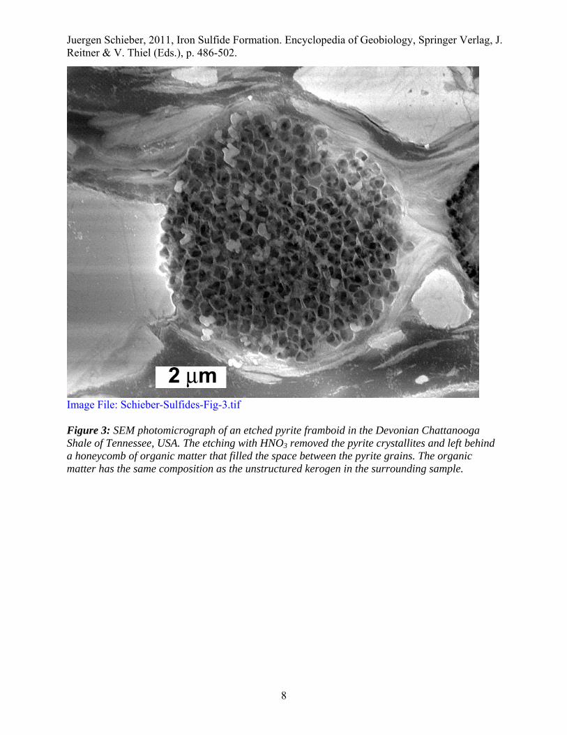
Pyrite Framboids Ubiquitous in modern and ancient sediments, pyrite framboids are typically near-spherical in shape, range in diameter from about 1 μm to tens of μm’s, and are themselves composed of tiny, discrete, and equigranular pyrite crystallites. They have been the subject of numerous studies, and early on there was an assumption that organic matter (Fig. 3) somehow plays a role in their formation (Schneiderhöhn, 1923; Love, 1967). Yet, because they can also be produced abiotically in the laboratory (Berner, 1969; Sweeney and Kaplan, 1973), the role of organic matter remains a matter of debate (e.g. Kalliokoski, 1974). The abundantly noted close association of pyrite framboids and organic matter may well be coincidental because the reducing conditions that promote their formation are best developed in sediments with elevated contents of organic matter.
Juergen Schieber, 2011, Iron Sulfide Formation. Encyclopedia of Geobiology, Springer Verlag, J. Reitner & V. Thiel (Eds.), p. 486-502.
8
Image File: Schieber-Sulfides-Fig-3.tif Figure 3: SEM photomicrograph of an etched pyrite framboid in the Devonian Chattanooga Shale of Tennessee, USA. The etching with HNO3 removed the pyrite crystallites and left behind a honeycomb of organic matter that filled the space between the pyrite grains. The organic matter has the same composition as the unstructured kerogen in the surrounding sample.
Juergen Schieber, 2011, Iron Sulfide Formation. Encyclopedia of Geobiology, Springer Verlag, J. Reitner & V. Thiel (Eds.), p. 486-502.
9
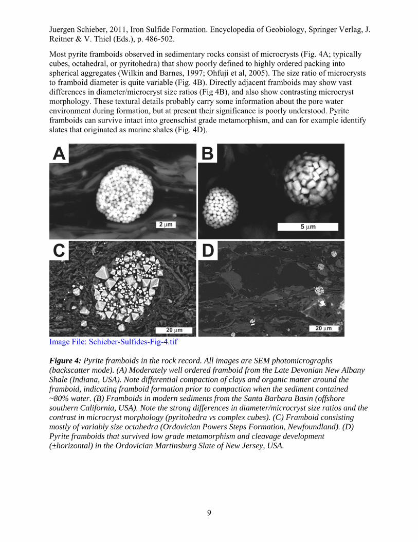
Most pyrite framboids observed in sedimentary rocks consist of microcrysts (Fig. 4A; typically cubes, octahedral, or pyritohedra) that show poorly defined to highly ordered packing into spherical aggregates (Wilkin and Barnes, 1997; Ohfuji et al, 2005). The size ratio of microcrysts to framboid diameter is quite variable (Fig. 4B). Directly adjacent framboids may show vast differences in diameter/microcryst size ratios (Fig 4B), and also show contrasting microcryst morphology. These textural details probably carry some information about the pore water environment during formation, but at present their significance is poorly understood. Pyrite framboids can survive intact into greenschist grade metamorphism, and can for example identify slates that originated as marine shales (Fig. 4D).
Image File: Schieber-Sulfides-Fig-4.tif Figure 4: Pyrite framboids in the rock record. All images are SEM photomicrographs (backscatter mode). (A) Moderately well ordered framboid from the Late Devonian New Albany Shale (Indiana, USA). Note differential compaction of clays and organic matter around the framboid, indicating framboid formation prior to compaction when the sediment contained ~80% water. (B) Framboids in modern sediments from the Santa Barbara Basin (offshore southern California, USA). Note the strong differences in diameter/microcryst size ratios and the contrast in microcryst morphology (pyritohedra vs complex cubes). (C) Framboid consisting mostly of variably size octahedra (Ordovician Powers Steps Formation, Newfoundland). (D) Pyrite framboids that survived low grade metamorphism and cleavage development (±horizontal) in the Ordovician Martinsburg Slate of New Jersey, USA.
Juergen Schieber, 2011, Iron Sulfide Formation. Encyclopedia of Geobiology, Springer Verlag, J. Reitner & V. Thiel (Eds.), p. 486-502.
10
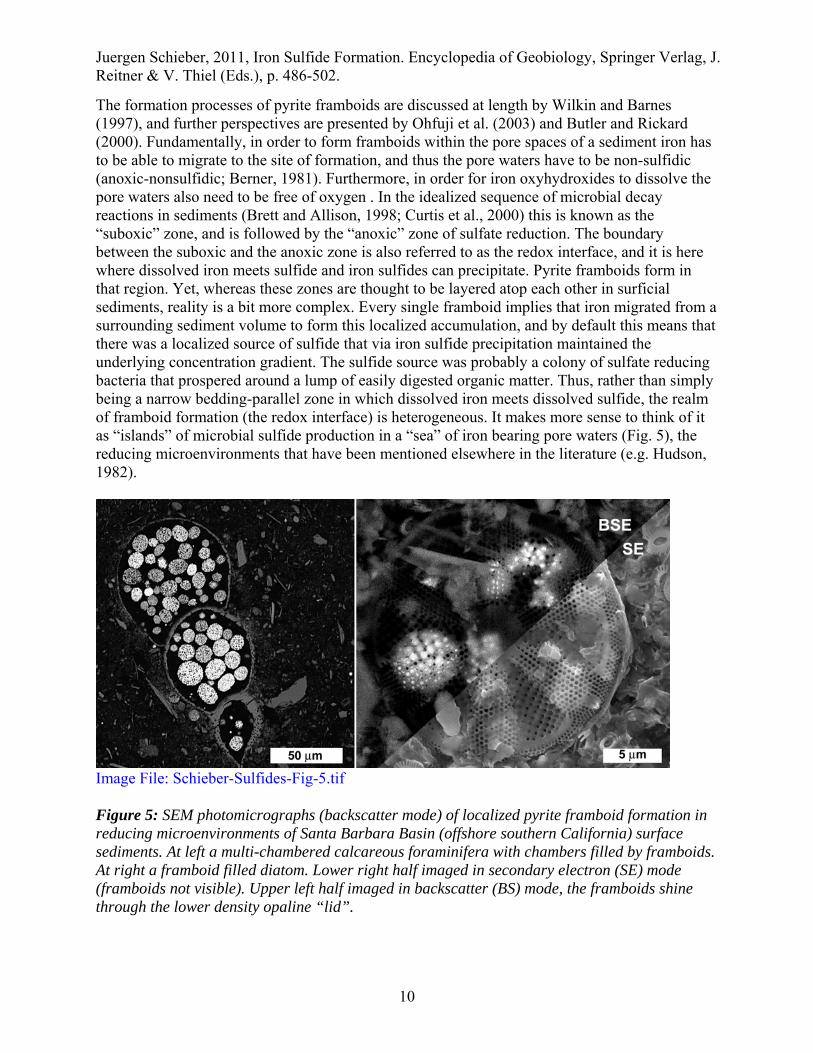
The formation processes of pyrite framboids are discussed at length by Wilkin and Barnes (1997), and further perspectives are presented by Ohfuji et al. (2003) and Butler and Rickard (2000). Fundamentally, in order to form framboids within the pore spaces of a sediment iron has to be able to migrate to the site of formation, and thus the pore waters have to be non-sulfidic (anoxic-nonsulfidic; Berner, 1981). Furthermore, in order for iron oxyhydroxides to dissolve the pore waters also need to be free of oxygen . In the idealized sequence of microbial decay reactions in sediments (Brett and Allison, 1998; Curtis et al., 2000) this is known as the “suboxic” zone, and is followed by the “anoxic” zone of sulfate reduction. The boundary between the suboxic and the anoxic zone is also referred to as the redox interface, and it is here where dissolved iron meets sulfide and iron sulfides can precipitate. Pyrite framboids form in that region. Yet, whereas these zones are thought to be layered atop each other in surficial sediments, reality is a bit more complex. Every single framboid implies that iron migrated from a surrounding sediment volume to form this localized accumulation, and by default this means that there was a localized source of sulfide that via iron sulfide precipitation maintained the underlying concentration gradient. The sulfide source was probably a colony of sulfate reducing bacteria that prospered around a lump of easily digested organic matter. Thus, rather than simply being a narrow bedding-parallel zone in which dissolved iron meets dissolved sulfide, the realm of framboid formation (the redox interface) is heterogeneous. It makes more sense to think of it as “islands” of microbial sulfide production in a “sea” of iron bearing pore waters (Fig. 5), the reducing microenvironments that have been mentioned elsewhere in the literature (e.g. Hudson, 1982).
Image File: Schieber-Sulfides-Fig-5.tif Figure 5: SEM photomicrographs (backscatter mode) of localized pyrite framboid formation in reducing microenvironments of Santa Barbara Basin (offshore southern California) surface sediments. At left a multi-chambered calcareous foraminifera with chambers filled by framboids. At right a framboid filled diatom. Lower right half imaged in secondary electron (SE) mode (framboids not visible). Upper left half imaged in backscatter (BS) mode, the framboids shine through the lower density opaline “lid”.
Juergen Schieber, 2011, Iron Sulfide Formation. Encyclopedia of Geobiology, Springer Verlag, J. Reitner & V. Thiel (Eds.), p. 486-502.
11
Whereas in typical sediments the redox interface is situated somewhat below the sediment water interface, there are situations where the redox interface is actually located in the overlying water column, such as in the Black Sea. In euxinic basins, with the lower portion of the water column anoxic and sulfidic, framboids can grow in the water column near the redox interface and then settle and become part of the accumulating sediments (Wilkin et al., 1996). Wilkin et al. (1997) report that framboids of that origin are rather small (mean framboid diameters < 5 µm, narrow size distribution) when compared to those forming in sediments underlying oxic and dysoxic waters (mean framboid diameters 5-10 µm, broad size distribution). The implication of this is that one can use the framboid size distributions of sediments to determine whether they accumulated beneath an anoxic water column. Wignall and Newton (1998) applied this concept to ancient mudrocks and were able to correlate framboid size distributions with paleoecologically bases reconstructions of bottom water oxygenation. However, framboid size in sediments may also be influenced by the availability of iron during early diagenesis. For example, a study of framboid size distributions in sediments from the Santa Barbara Basin (offshore southern California) indicates that framboid size distributions primarily reflect conditions within the immediate surface sediment (such as availability of readily soluble iron). Framboid size distributions in annual varves (1984 to 2004) from the Santa Barbara Basin suggest three euxinic interludes in the past two decades, even though seasonal bottom water surveys consistently recorded suboxic to dysoxic bottom water conditions (Schieber and Schimmelmann, 2006). It appears therefore that it may not be possible to draw conclusions about water column conditions from pyrite framboid size distributions.
Polyframboids Pyrite framboids may also occur as larger aggregates, so called polyframboids. These have been described from sediments of various ages (Schieber and Baird, 2001) and have a tendency to form in cavities of organic remains (Figs. 5 and 6). Judging from differential compaction around polyframboids (Fig. 6) they must form prior to any compaction, just like single pyrite framboids. They also imply iron migration from a surrounding sediment volume that was anoxic but not sulfidic. The difference between polyframboids and regular pyrite framboids may simply be a higher iron supply from the pore waters, or alternatively that the “islands” of microbial sulfate reduction are more widely spaced and thus focus the available iron onto fewer sites of precipitation. Because most marine sediments start out with comparable amounts of reactive iron, the latter scenario is more likely.

Juergen Schieber, 2011, Iron Sulfide Formation. Encyclopedia of Geobiology, Springer Verlag, J. Reitner & V. Thiel (Eds.), p. 486-502.
12
Image File: Schieber-Sulfides-Fig-6.tif Figure 6: SEM photomicrographs of pyrite polyframboids (backscatter images). Note differential compaction around polyframboid aggregates. (A) Ordovician Powers Steps Formation, Newfoundland, Canada. Note “squeezing” of framboids, suggesting initial formation in a confined space (transported?). (B) Devonian Chattanooga Shale, Tennessee, USA. Here the polyframboid is still enclosed (black line) in an algal cyst (Tasmanites) that formed the initial reducing microenvironment. The space between framboids is filled with secondary, coarser pyrite cement.
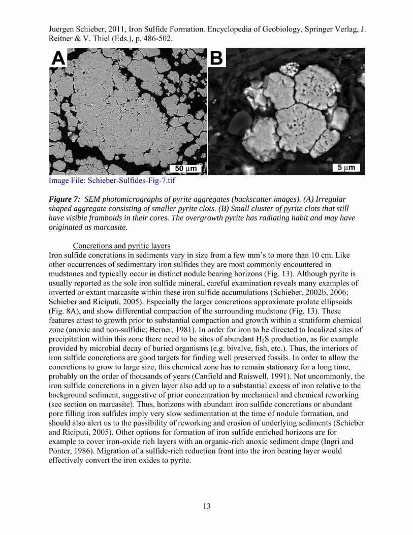
Small scale pyrite aggregates and clusters Pyrite bodies that consist of coalescing, irregular, rounded clots of small pyrite grains have been described as aggregated pyrite by Hudson (1982). The cores of individual clots may be small pyrite crystals or pyrite framboids, and these cores are overgrown by coarser pyrite crystals (Fig. 7). In places these overgrowth grains show a radiating bladed habit. These overgrowths tend to form somewhat later in diagenesis, at a point when downward diffusion of seawater sulfate is increasingly restricted (Strauss and Schieber, 1990). This type of pyrite aggregate ranges in size from tens of microns to several mm’s.
Juergen Schieber, 2011, Iron Sulfide Formation. Encyclopedia of Geobiology, Springer Verlag, J. Reitner & V. Thiel (Eds.), p. 486-502.
13
Image File: Schieber-Sulfides-Fig-7.tif Figure 7: SEM photomicrographs of pyrite aggregates (backscatter images). (A) Irregular shaped aggregate consisting of smaller pyrite clots. (B) Small cluster of pyrite clots that still have visible framboids in their cores. The overgrowth pyrite has radiating habit and may have originated as marcasite.
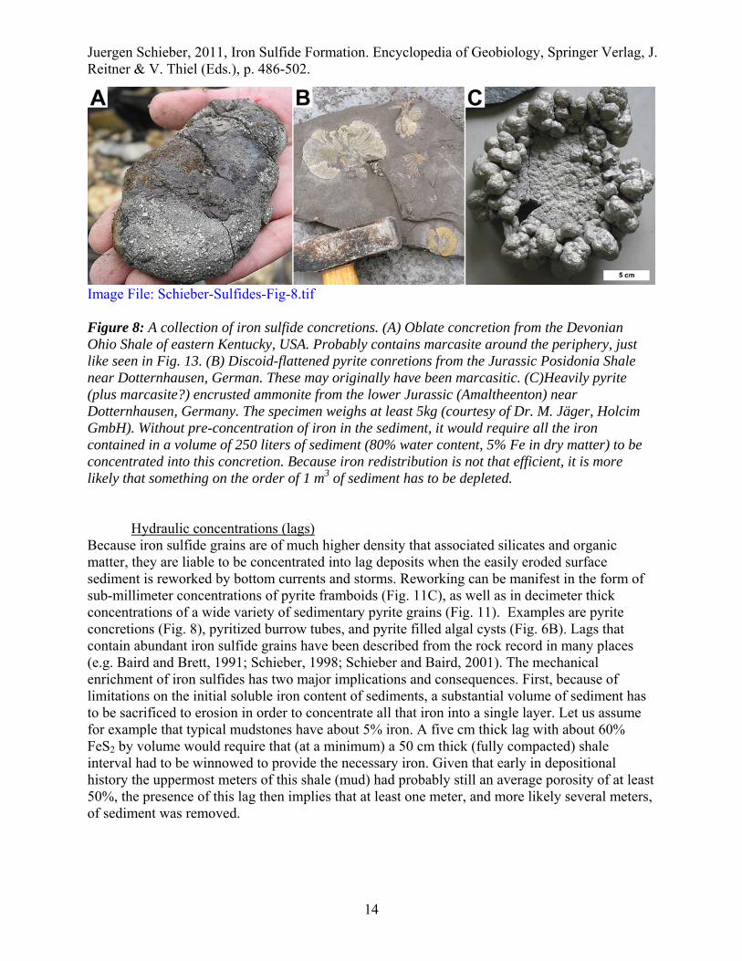
Concretions and pyritic layers Iron sulfide concretions in sediments vary in size from a few mm’s to more than 10 cm. Like other occurrences of sedimentary iron sulfides they are most commonly encountered in mudstones and typically occur in distinct nodule bearing horizons (Fig. 13). Although pyrite is usually reported as the sole iron sulfide mineral, careful examination reveals many examples of inverted or extant marcasite within these iron sulfide accumulations (Schieber, 2002b, 2006; Schieber and Riciputi, 2005). Especially the larger concretions approximate prolate ellipsoids (Fig. 8A), and show differential compaction of the surrounding mudstone (Fig. 13). These features attest to growth prior to substantial compaction and growth within a stratiform chemical zone (anoxic and non-sulfidic; Berner, 1981). In order for iron to be directed to localized sites of precipitation within this zone there need to be sites of abundant H2S production, as for example provided by microbial decay of buried organisms (e.g. bivalve, fish, etc.). Thus, the interiors of iron sulfide concretions are good targets for finding well preserved fossils. In order to allow the concretions to grow to large size, this chemical zone has to remain stationary for a long time, probably on the order of thousands of years (Canfield and Raiswell, 1991). Not uncommonly, the iron sulfide concretions in a given layer also add up to a substantial excess of iron relative to the background sediment, suggestive of prior concentration by mechanical and chemical reworking (see section on marcasite). Thus, horizons with abundant iron sulfide concretions or abundant pore filling iron sulfides imply very slow sedimentation at the time of nodule formation, and should also alert us to the possibility of reworking and erosion of underlying sediments (Schieber and Riciputi, 2005). Other options for formation of iron sulfide enriched horizons are for example to cover iron-oxide rich layers with an organic-rich anoxic sediment drape (Ingri and Ponter, 1986). Migration of a sulfide-rich reduction front into the iron bearing layer would effectively convert the iron oxides to pyrite.
Juergen Schieber, 2011, Iron Sulfide Formation. Encyclopedia of Geobiology, Springer Verlag, J. Reitner & V. Thiel (Eds.), p. 486-502.
14
Image File: Schieber-Sulfides-Fig-8.tif Figure 8: A collection of iron sulfide concretions. (A) Oblate concretion from the Devonian Ohio Shale of eastern Kentucky, USA. Probably contains marcasite around the periphery, just like seen in Fig. 13. (B) Discoid-flattened pyrite conretions from the Jurassic Posidonia Shale near Dotternhausen, German. These may originally have been marcasitic. (C)Heavily pyrite (plus marcasite?) encrusted ammonite from the lower Jurassic (Amaltheenton) near Dotternhausen, Germany. The specimen weighs at least 5kg (courtesy of Dr. M. Jäger, Holcim GmbH). Without pre-concentration of iron in the sediment, it would require all the iron contained in a volume of 250 liters of sediment (80% water content, 5% Fe in dry matter) to be concentrated into this concretion. Because iron redistribution is not that efficient, it is more likely that something on the order of 1 m3 of sediment has to be depleted.
Hydraulic concentrations (lags) Because iron sulfide grains are of much higher density that associated silicates and organic matter, they are liable to be concentrated into lag deposits when the easily eroded surface sediment is reworked by bottom currents and storms. Reworking can be manifest in the form of sub-millimeter concentrations of pyrite framboids (Fig. 11C), as well as in decimeter thick concentrations of a wide variety of sedimentary pyrite grains (Fig. 11). Examples are pyrite concretions (Fig. 8), pyritized burrow tubes, and pyrite filled algal cysts (Fig. 6B). Lags that contain abundant iron sulfide grains have been described from the rock record in many places (e.g. Baird and Brett, 1991; Schieber, 1998; Schieber and Baird, 2001). The mechanical enrichment of iron sulfides has two major implications and consequences. First, because of limitations on the initial soluble iron content of sediments, a substantial volume of sediment has to be sacrificed to erosion in order to concentrate all that iron into a single layer. Let us assume for example that typical mudstones have about 5% iron. A five cm thick lag with about 60% FeS2 by volume would require that (at a minimum) a 50 cm thick (fully compacted) shale interval had to be winnowed to provide the necessary iron. Given that early in depositional history the uppermost meters of this shale (mud) had probably still an average porosity of at least 50%, the presence of this lag then implies that at least one meter, and more likely several meters, of sediment was removed.

Juergen Schieber, 2011, Iron Sulfide Formation. Encyclopedia of Geobiology, Springer Verlag, J. Reitner & V. Thiel (Eds.), p. 486-502.
15
Iron sulfides and fossil preservation
Replacement and encrustation of calcareous shells Encrustation and/or replacement of calcareous shells by iron sulfides is widely observed in the rock record. Typically the assumption is made that the iron sulfide in question is pyrite, but a case can be made that marcasite was involved at least initially (see below). One way to think about calcite/aragonite replacement is that the saturation levels of iron sulfide decline with increasing pH. When carbonates dissolve pH increases and thus forces precipitation of iron sulfide. Chemical modeling suggests, however, that this process is unlikely during early diagenesis (Canfield and Raiswell, 1991). Nonetheless, even the shells of still living mollusks can be pyritized (Srivastava, 1975; Clark and Lutz, 1980), a process favored by pore waters with high levels of dissolved iron and excretion of microbially metabolizable organic substances by the mollusks (Reaves, 1984). In essence, the mollusks supported a sulfidic microenvironment and attracted iron from surrounding (anoxic-nonsulfidic) pore waters. This same basic scenario probably also applies to decomposing organic matter within dead mollusks, leading to shells encrusted or infilled with pyrite (Hudson, 1982). Hudson (1982) distinguished chamber linings (Fig. 9), stalactitic pyrite, and overpyrite (on outer shell surface), mostly consisting of well crystallized equant pyrite. For the potential impact of intermittent re-oxidation of sedimentary iron sulfides on pH levels, carbonate precipitation, and rapid precipitation of the pyrite dimorph marcasite to promote shell encrustation, see the discussion of marcasite formation below.
Image File: Schieber-Sulfides-Fig-9.tif Figure 9: Fossil preservation by iron sulfides, SEM photomicrographs. (A) Chamber lining iron sulfides in an ammonite (secondary electron image), Lias delta (Amaltheenton) near Dotternhausen, Germany. The septa of the ammonite (white arrows) are now calcitic, and the chamber interior is filled with calcite as well. (B) Close-up of the iron sulfide (bright, backscatter image) encrusted septum. The encrusting sulfides are a mixture of pyrite (framboids) and marcasite (overgrowth). (C) Close-up of encrustation near septum (backscatter image). The

Juergen Schieber, 2011, Iron Sulfide Formation. Encyclopedia of Geobiology, Springer Verlag, J. Reitner & V. Thiel (Eds.), p. 486-502.
16
marginal areas of the septum (marked between white arrows) have a different texture than the areas further away. The latter areas contain pyrite framboids (black arrow) in a coarser matrix of blocky to radiating crystals. (D) Close-up of framboids (white arrows) in coarser matrix grains. The latter are anisotropic in reflected light and are either still preserved as marcasite or are marcasite relicts. (E) Detail of the different textured replacement of the septum margin (between white arrows in C). The lamellar texture is probably a relict of the earlier lamellar structure of the septal aragonite.
Soft tissue preservation The preservation of soft tissue by pyritization is another marvel of fossilization, with well known examples from the Hunsrück Slate (Stürmer, 1985), Beecher’s trilobite bed (Cisne, 1973; Briggs et al., 1991), and the Burgess Shale (Conway Morris, 1986). Ongoing research is producing a stream of exceptional soft bodied fossils that are preserved in pyrite, such as worms (Gabbott et al., 2004; Farrell and Briggs, 2007), crinoids (Kammer and Ausich, 2007), and plant tissues (Grimes et al., 2001). Pyritization can occur by infilling of cellular cavities, by preferential replacement of more readily decomposed components, and by pyrite coatings on easily degraded soft parts (Canfield and Raiswell, 1991). The latter process may be aided by microbial coatings (Wuttke, 1983) on the decomposing tissues that help to stabilize chemical gradients around the decomposing material. Framboidal, clustered, and aggregated pyrite are most commonly observed type in this style of preservation. Even microbial cells and textures can be preserved under the right circumstances (Fig. 10). With regard to preservation fidelity, soft tissue preservation by pyrite does not preserve as much detail as preservation in a phosphate matrix.

Juergen Schieber, 2011, Iron Sulfide Formation. Encyclopedia of Geobiology, Springer Verlag, J. Reitner & V. Thiel (Eds.), p. 486-502.
17
Image File: Schieber-Sulfides-Fig-10.tif Figure 10: SEM images of microbial remains preserved in early diagenetic coated iron sulfide grains (ooids and oncoids) from the Ordovician Winnipeg Formation, Saskatchewan, Canada (from Schieber and Riciputi, 2005). A) filamentous structure (white arrows) in slightly etched iron sulfide cortex. Note multiple filaments in upper part of picture. In uppermost part of picture the filaments merge into a sheet-like structure, potentially remnant of a biofilm. B) Typical recrystallized appearance of an outermost cortex surface (unetched). Most crystals are pyrite octahedra. Between these crystal pavements one finds remnant patches of an earlier surface (C, unetched). The latter has a lumpy appearance and consists of tightly packed clusters (a few microns in size) of iron sulfide grains. Closeups of these clusters (D, unetched) show that they actually consist of an ovoid smooth walled structure (black arrow) that is overgrown by iron sulfide grains. These are interpreted as microbes that were preserved in an iron sulfide matrix. Image E (unetched) shows that the smooth surfaces of these ovoids are typically perforated by irregular shaped openings (E, black arrows), comparable to wall structures seen in experimentally pyritized microbes (Bubela and Cloud, 1983).
Juergen Schieber, 2011, Iron Sulfide Formation. Encyclopedia of Geobiology, Springer Verlag, J. Reitner & V. Thiel (Eds.), p. 486-502.
18
Degree of pyritization (DOP) A widely used proxy for paleo-oxygenation, DOP is defined (Raiswell et al., 1988; Raiswell and Canfield, 1998) as
DOP = pyrite Fe/(pyrite Fe + “reactive iron”) An extraction with dithionite or 1N HCl is supposed to mimic reactive iron dissolution in reducing sediments (Canfield, 1989; Leventhal and Taylor, 1990). The highly reactive iron fraction in sediments is defined as the sum of dithionite or 1N HCl leachable iron, pyrite iron, and iron in acid volatile sulfides (AVS). What DOP presumably measures is the completeness of the conversion of reactive iron into pyrite via microbial sulfate reduction during early diagenesis. The underlying premise is that DOP increases as the degree of environmental oxygenation decreases. Because of the need for sufficient quantities of organic matter and reactive iron, measurements of DOP typically focus on carbonaceous mudstones. DOP values below 0.45 are considered indicative of aerobic bottom waters, and those above 0.45 are thought to mark restricted bottom water conditions. Values above 0.75 are considered to indicate anoxic or even euxinic bottom waters (Raiswell et al., 1988). However, the DOP method should not be applied uncritically, because limitations of either metabolizable organic matter or of reactive iron can lead to DOP values that are not reflective of the actual environment (as calibrated by Raiswell et al., 1988). Several other constraints on DOP application, such as outcrop weathering, maturation, and age of rock are summarized by Raiswell et al. (1988). DOP as an environmental indicator can also be invalidated as a consequence of sedimentary processes. Any reworking and winnowing of pyrite bearing sediments will increase the pyrite content of the sediment without simultaneously raising the reactive iron content, and as a result the DOP can rise simply as a result of wave or current reworking. In mudstones, indications of reworking at the mm to cm scale will often go undetected (Fig. 11), and can lead to inflated DOP values that are incompatible with the levels of oxygen restriction suggested by observations of frequent reworking and bioturbation (Schieber, 2001; 2003).

Juergen Schieber, 2011, Iron Sulfide Formation. Encyclopedia of Geobiology, Springer Verlag, J. Reitner & V. Thiel (Eds.), p. 486-502.
19
Image File: Schieber-Sulfides-Fig-11.tif Figure 11: Reworked pyrite in shale successions. Image (A) is a photograph of a polished specimen, all others are SEM photomicrographs (backscatter images). (A) Ripple cross-laminated lag deposit that consists mainly of spherical polyframboid fills of algal cysts (Schieber and Baird, 2001). Devonian Genesee Formation, New York, USA. (B) A horizon with abundant reworked flattened polyframboid clusters (comparable to those in Fig. 6B). Devonian Chattanooga Shale, Tennessee, USA. (C) Lamina with abundant reworked pyrite framboids. Devonian Chattanooga Shale, Tennessee, USA. (D) Detail of framboid enriched lamina from (C). Shows how reworked pyrite framboids are filling spaces between larger silt grains and have been deformed due to compaction. Devonian Chattanooga Shale, Tennessee, USA.

Juergen Schieber, 2011, Iron Sulfide Formation. Encyclopedia of Geobiology, Springer Verlag, J. Reitner & V. Thiel (Eds.), p. 486-502.
20
Marcasite and its implications
Background on marcasite Reports of marcasite in terrigenous clastics are mainly associated with coal deposits (e.g. Nayar, 1946; Read and Cook, 1969; Wiese et al., 1987). Marcasite occurs either in the coal seams themselves, or in the roof shales, prompting Krumbein and Garrels (1952) to propose an acidic bog/peat association as the characteristic environment for marcasite formation. There have, however, been a sufficient number of diagenetic marcasite occurrences reported from normal marine sediments (e.g. Maynard and Lauffenburger, 1978, Rykart, 1983, Jowett et al., 1991; Schieber, 2002b, 2007), as well examples of marcasite that has inverted to pyrite (e.g. Bannister, 1932; Van Horn & Van Horn, 1933), to suggest that it might be more widespread in the marine realm than commonly appreciated. If this is in fact the case, and if acidic conditions are indeed a basic requirement for marcasite formation, we have an obvious problem to reconcile these basic realities with our current understanding of early diagenesis of marine sediments.
As pointed out above, the paragenetic relationships between sedimentary iron sulfide minerals are still poorly understood (Rickard and Morse, 2005). An essential guidepost for thinking about marcasite in sedimentary environments have been experiments conducted by Murowchick and Barnes (1986) that showed that marcasite is the dominant iron disulfide below pH 5. These experiments are consistent with results from much earlier work by Allen et al. (1914) and are also consistent with subsequent work by Schoonen and Barnes (1991a,b) and Benning et al. (2000). Thus, the relative abundance of pyrite vs marcasite in sediments should be a function of pH. Because marine waters have a slightly alkaline pH of approximately 8, it has been presumed that marcasite can not form in marine sediments during early diagenesis (e.g. Rickard et al., 1995). Yet, regardless of this, early diagenetic marcasite unquestionably occurs in marine sedimentary rocks (Maynard and Lauffenburger, 1978; Siesser, 1978; Rykart, 1983; Jowett et al., 1991; Schieber, 2002b; Schieber and Riciputi, 2005; Williams et al., 2003).
Detecting marasite Characteristic morphologies, such as “spearhead” twins and “cockscomb” crystals can be used to identify marcasite. However, morphology does not tell us whether the marcasite has inverted to pyrite and the approach does not work for dense masses of crystals. As long as crystallites are large enough, it is comparatively easy to differentiate marcasite from pyrite with a petrographic microscope in reflected light mode (Ramdohr, 1975), and it is also possible to detect marcasite that has inverted to pyrite (Murowchick, 1992). For micron scale grains a new technique, “electron backscatter diffraction” (EBSD), can be applied for identification with an electron microscope. EBSD allows phase identification of submicron mineral grains and is widely used in material sciences (Prior et al., 1999; Schwartz et al., 2000).
Marcasite in sedimentary rocks Marcasite is probably much more widespread in marine clastics than commonly assumed. It occurs as a cement mineral in marine lag deposits associated with sequence boundaries, in condensed intervals, and even in shales. Lag deposits with marcasite occur for example in Late Devonian shale successions of the eastern US (Schieber, 2007). The Late Devonian Chattanooga and New Albany Shale succession contains laterally continuous erosion surfaces that compartmentalize this black shale succession into a stack of depositional sequences (Schieber,
Juergen Schieber, 2011, Iron Sulfide Formation. Encyclopedia of Geobiology, Springer Verlag, J. Reitner & V. Thiel (Eds.), p. 486-502.
21
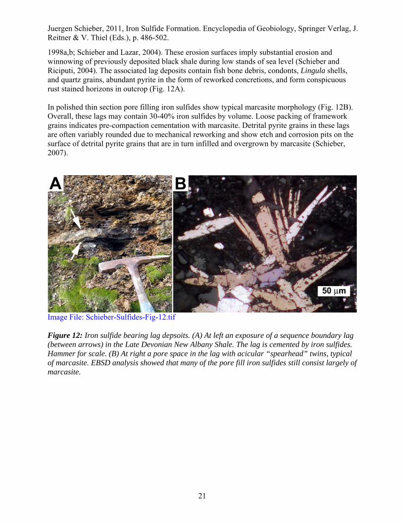
1998a,b; Schieber and Lazar, 2004). These erosion surfaces imply substantial erosion and winnowing of previously deposited black shale during low stands of sea level (Schieber and Riciputi, 2004). The associated lag deposits contain fish bone debris, condonts, Lingula shells, and quartz grains, abundant pyrite in the form of reworked concretions, and form conspicuous rust stained horizons in outcrop (Fig. 12A). In polished thin section pore filling iron sulfides show typical marcasite morphology (Fig. 12B). Overall, these lags may contain 30-40% iron sulfides by volume. Loose packing of framework grains indicates pre-compaction cementation with marcasite. Detrital pyrite grains in these lags are often variably rounded due to mechanical reworking and show etch and corrosion pits on the surface of detrital pyrite grains that are in turn infilled and overgrown by marcasite (Schieber, 2007).
Image File: Schieber-Sulfides-Fig-12.tif Figure 12: Iron sulfide bearing lag depsoits. (A) At left an exposure of a sequence boundary lag (between arrows) in the Late Devonian New Albany Shale. The lag is cemented by iron sulfides. Hammer for scale. (B) At right a pore space in the lag with acicular “spearhead” twins, typical of marcasite. EBSD analysis showed that many of the pore fill iron sulfides still consist largely of marcasite.

Juergen Schieber, 2011, Iron Sulfide Formation. Encyclopedia of Geobiology, Springer Verlag, J. Reitner & V. Thiel (Eds.), p. 486-502.
22
Terrigenous clastic sediments contain at best a few percent reactive iron in the form of iron oxyhydroxide coatings on terrigenous grains (Carroll, 1958, Berner, 1969). Thus, the high iron concentrations in lag deposits with up to 40% iron sulfide require a mechanism for iron enrichment, and in the Phanerozoic at least this iron was most likely reworked from underlying strata (see above). In essence, sedimentary pyrite grains need to be eroded and winnowed in order to add more iron to the surficial layer, a process favored by stratigraphic condensation when net sedimentation rates are negative (Schieber and Riciputi, 2005; Schieber, 2007). After mechanical enrichment, however, a portion of the sulfide iron has to be remobilized to form the observed overgrowths and pore fill cements. Preexisting sulphide grains are destroyed, either wholly or partially, in order to supply dissolved (Fe2+) or readily soluble iron (e.g. Fe(OH)3) for the growth of new iron sulphides. Corrosion features on reworked iron sulphide grains (Schieber and Riciputi, 2005; Schieber, 2007) are evidence for iron sulfide destruction according to the following reaction:
FeS2 + 3.5O2 + H2O Fe2+ + 2SO4
2- + 2H+
This reaction produces acidity and dissolved ferrous iron. Further oxidation of iron (see following equation) produces iron hydroxide and further acidity.
Fe2+ + 0.25O2 + 2.5H2O Fe(OH)3 + 2H+
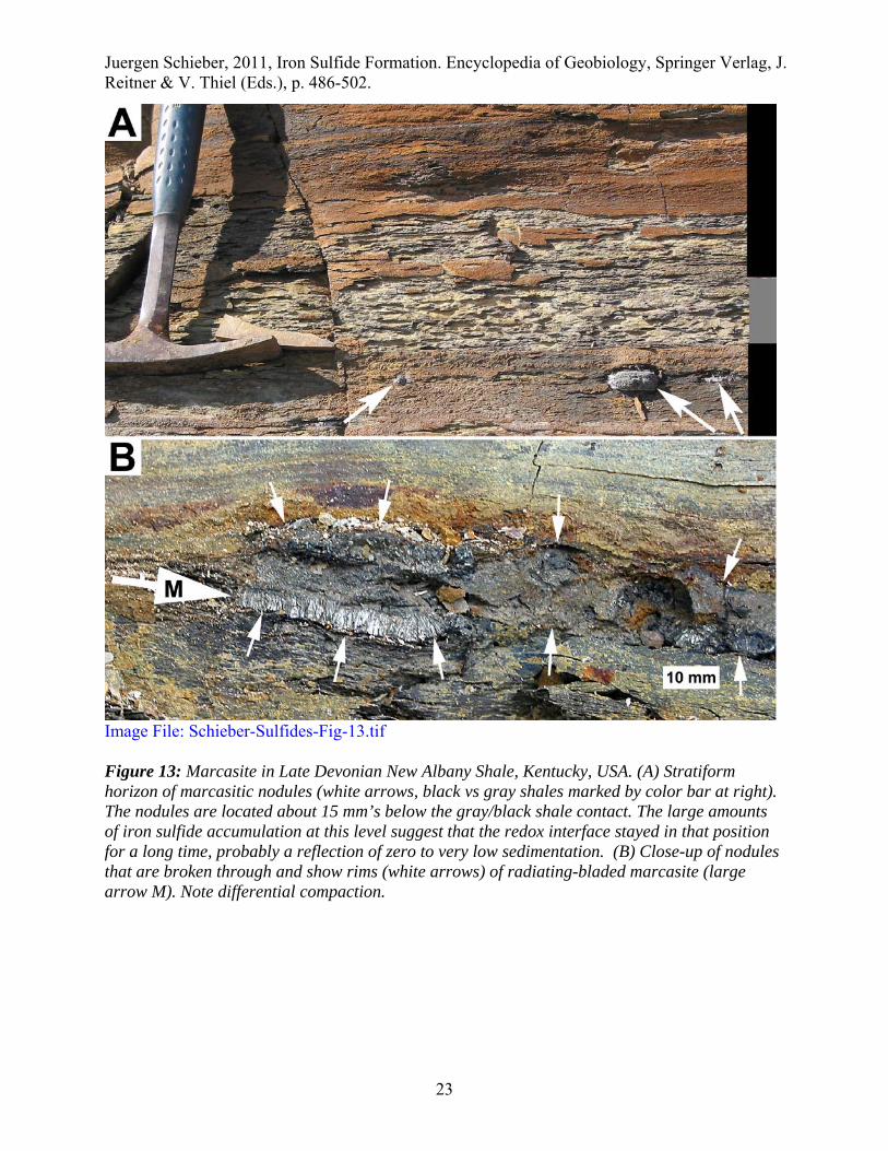
The newly formed iron hydroxides are likely to coat surface sediment grains and would remain in the surficial sediment layer. The associated lowering of pore water pH also promotes large pore water concentrations of Fe2+ (Maynard, 1983). Such a setting favors re-precipitation of marcasite in the presence of H2S influx from underlying sediments (Murowchick and Barnes, 1986; Schoonen and Barnes, 1991a,b; Benning et al., 2000). For sandy lags the presence of marcasite and partial dissolution of reworked pyrite grains is probably a good indicator that a “re-oxidation” model for marcasite formation is applicable (Schieber and Riciputi, 2006; Schieber, 2007). In shales with abundant pyrite a comparable sequence of events may be caused by downward oxidation of previously deposited muds. Potential examples are for example stratiform horizons of iron sulfide concretions in black shale successions (Fig. 13). Where these nodule horizons occur a gray shale bed is typically found just above them (Fig. 13A), and the concretions have a flattened radial fibrous habit, a morphology suggestive of marcasite (Fig. 13B).
Juergen Schieber, 2011, Iron Sulfide Formation. Encyclopedia of Geobiology, Springer Verlag, J. Reitner & V. Thiel (Eds.), p. 486-502.
23
Image File: Schieber-Sulfides-Fig-13.tif Figure 13: Marcasite in Late Devonian New Albany Shale, Kentucky, USA. (A) Stratiform horizon of marcasitic nodules (white arrows, black vs gray shales marked by color bar at right). The nodules are located about 15 mm’s below the gray/black shale contact. The large amounts of iron sulfide accumulation at this level suggest that the redox interface stayed in that position for a long time, probably a reflection of zero to very low sedimentation. (B) Close-up of nodules that are broken through and show rims (white arrows) of radiating-bladed marcasite (large arrow M). Note differential compaction.
Juergen Schieber, 2011, Iron Sulfide Formation. Encyclopedia of Geobiology, Springer Verlag, J. Reitner & V. Thiel (Eds.), p. 486-502.
24
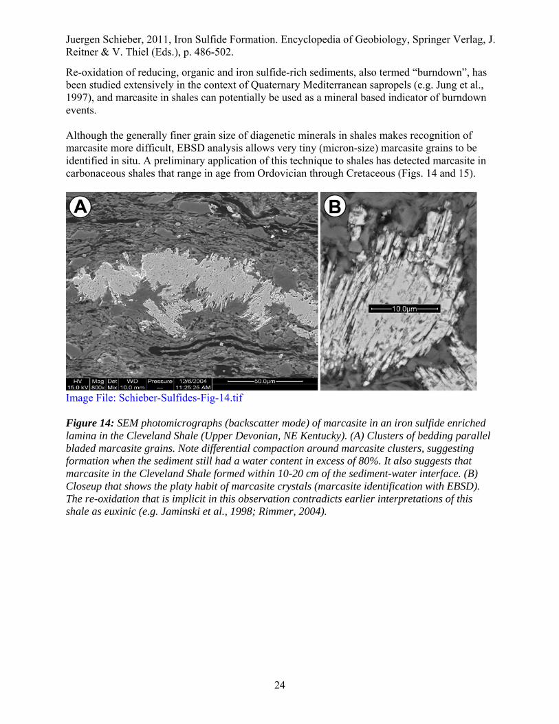
Re-oxidation of reducing, organic and iron sulfide-rich sediments, also termed “burndown”, has been studied extensively in the context of Quaternary Mediterranean sapropels (e.g. Jung et al., 1997), and marcasite in shales can potentially be used as a mineral based indicator of burndown events. Although the generally finer grain size of diagenetic minerals in shales makes recognition of marcasite more difficult, EBSD analysis allows very tiny (micron-size) marcasite grains to be identified in situ. A preliminary application of this technique to shales has detected marcasite in carbonaceous shales that range in age from Ordovician through Cretaceous (Figs. 14 and 15).
Image File: Schieber-Sulfides-Fig-14.tif Figure 14: SEM photomicrographs (backscatter mode) of marcasite in an iron sulfide enriched lamina in the Cleveland Shale (Upper Devonian, NE Kentucky). (A) Clusters of bedding parallel bladed marcasite grains. Note differential compaction around marcasite clusters, suggesting formation when the sediment still had a water content in excess of 80%. It also suggests that marcasite in the Cleveland Shale formed within 10-20 cm of the sediment-water interface. (B) Closeup that shows the platy habit of marcasite crystals (marcasite identification with EBSD). The re-oxidation that is implicit in this observation contradicts earlier interpretations of this shale as euxinic (e.g. Jaminski et al., 1998; Rimmer, 2004).

Juergen Schieber, 2011, Iron Sulfide Formation. Encyclopedia of Geobiology, Springer Verlag, J. Reitner & V. Thiel (Eds.), p. 486-502.
25
Image File: Schieber-Sulfides-Fig-15.tif Figure 15: Pyrite polyframboid cluster in Chattanooga Shale (Tennessee, USA) that formed within an algal cyst (dark rim). The infill was examined with EBSD. Marcasite (m) and quartz (Q) grew in the spaces between pyrite framboids (P). The open arrangement of framboids and the differential compaction around the cyst indicates that cementation happened prior to compaction. The marcasite observed here may well have been caused by intermittent pyrite oxidation in surface sediments. The relief in this image is an artifact, a consequence of oblique imaging (70 degrees tilt) in EBSD mode. Unique fossil beds where preservation in pyrite and marcasite has been observed, such as Beecher’s trilobite bed (Cisne, 1973) and pyritized ammonoids (Hudson, 1982; Seilacher et al., 1985), are another phenomenon where our “burndown” model could find application. The large stratiform iron buildup that at least some of these deposits represent requires a similar level of stratigraphic condensation as seen in some of the examples above. Furthermore, iron needs to be mobile to form the observed mineralized and encrusted fossils. Thus, finding marcasite and dissolution features on reworked pyrite grains in pyritic Lagerstätten would support the view that such deposits may have formed during intermittent reoxygenation events.

Juergen Schieber, 2011, Iron Sulfide Formation. Encyclopedia of Geobiology, Springer Verlag, J. Reitner & V. Thiel (Eds.), p. 486-502.
26
The pH-drop associated with pyrite oxidation during intermittent reoxygenation also explains textural features observed in association with iron sulfide replacement of calcareous shells. Examination of calcareous shell replacement in Carboniferous through Cretaceous examples showed marcasite as a common component, intimately associated with calcite dissolution and precipitation of diagenetic quartz (Fig. 16). This mineralogical triumvirate makes chemical sense because marcasite formation requires low pH, calcite dissolves at low pH, and dissolved silica (from opaline tests) precipitates under low pH conditions.
Image File: Schieber-Sulfides-Fig-16.tif Figure 16: SEM photomicrographs (backscatter) of marcasite replacing biogenic calcite. (A) Clam shell in Boquillas Formation (Cenomanian of West Texas). Skeletal marcasite crystals (marked m) grow as radiating clusters and replace calcite (ca). There is also partial replacement of the shell by quartz (marked qu). (B) Brachiopod shells in Barnett Shale (Mississippian of Texas). Shell in center is replaced by marcasite (marked m) that grows as bladed and sharply pointed crystals. Equant pyrite (py) formed towards end of iron sulfide deposition. Calcite shells marked ca, and replacing quartz marked qu. With regard to carbonate rocks, most published reports of marcasite are from chalks (e.g. Morgan-Jones, 1977; Kelts, 1976). This marcasite is typically in nodule form, measures up to 10 cm across, and shows crystals with marcasite morphology. Nonetheless, XRD analysis frequently shows that this marcasite has inverted to pyrite. There also seems to be an association with phosphate concretions, and thus with reworking, negative net sedimentation, and geochemical "reworking" of iron, just like observed in marcasite occurrences in sandstones and shales. Conclusions The geobiological significance of sedimentary iron sulfides is far reaching. Microbial sulfate reduction as a metabolic pathway is of great antiquity and dates back to at least the early Archean (3.47-Ga; Shen and Buick, 2004). From that time forward, global cycling of sulfur and carbon has been essential for controlling the amount of oxygen in the Earth’s atmosphere

Juergen Schieber, 2011, Iron Sulfide Formation. Encyclopedia of Geobiology, Springer Verlag, J. Reitner & V. Thiel (Eds.), p. 486-502.
27
(Berner, 2001), and some have even speculated that the emergence of life itself is tied to sulfur chemistry on the early Earth (e.g. Russell and Hall, 1997). It also appears that in itself oxygen buildup during the Precambrian led to evolution of increasingly complex biochemical networks among microbes and eventually even to the evolution of complex life forms (Raymond and Segrè, 2006). The iron sulfides that we find in the rock record are a substantial part of the raw data for understanding the complex interaction of life with the atmosphere and the oceans. Every bit of that record, be it in the form of isotope shifts (e.g. Goldhaber, 2003), proportions and distribution of different iron sulfides, sulfide textures, stratal distribution, enclosed and preserved micro- and macrofossils, as well as subsequent alterations of these minerals, is valuable for understanding chemical changes that affected the biosphere on the local, global, and temporal scale. For the rock record, the enduring product of all these processes is the iron sulfide pyrite (FeS2). Yet, the dimorph of pyrite, marcasite, can still be recognized in sedimentary rocks as old as Proterozoic (personal observations) and probably played a much larger role than commonly appreciated. In the minds of many geologists, the presence of iron sulfides in sedimentary rocks is associated with anoxic environmental conditions. In reality, however, once iron sulfides begin to form localized concentrations, be it in the form of micron-size framboids or fist-size concretions, one can imply that oxygen must have been close by. As elaborated on repeatedly above, in order to form even microscopic features like pyrite framboids, iron has to be to be able to migrate through the pore waters, and those conditions only occur near the redox interface that “separates” deeper sulfidic from shallower oxygenated pore waters. Fundamentally this holds true even in the most classical anoxic-euxinic locale of all, the Black Sea. There, pyrite framboids do not form in the sulfidic bottom waters, but again at the redox interface where these waters come in contact with oxygenated surface waters (Wilkin et al., 1996). The bulk of sedimentary iron sulfides in the rock records mark the interface between the anoxic and oxic worlds, they should not be taken as an indicator of environmentally pervasive anoxia.
Juergen Schieber Bibliography Allard, T., Menguy, N., Salomon, J., Calligaro, T., Weber, T., Calas, G. and Benedetti, M.F.
(2004):: Revealing forms of iron in river-borne material from major tropical rivers of the Amazon Basin (Brazil):. Geochimica et Cosmochimica Acta, 68, 3079-3094.
Allen, E.T., Crenshaw, J.L., and Merwin, H.E. (1914): Effect of temperature and acidity in the formation of marcasite (FeS2) and wurzite (ZnS). American Journal of Science, 38, 393-421.
Aller, R.C., Mackin, J.E., and Cox, R.T. (1986): Diagenesis of Fe and S in Amazon inner shelf muds: apparent dominance of Fe reduction and implications for the genesis of ironstones. Continental Shelf Research, 6, 263-289.
Baird, G.C., and Brett, C.E. (1991): Submarine erosion on the anoxic sea floor: Stratinomic, palaeoenvironmental, and temporal significance of reworked pyrite-bone deposits. In Tyson, R.V., and Pearson, T.H. (eds.), Modern and Ancient Continental Shelf Anoxia. Geological Society of London, Special Publication 58, 233–257.
Juergen Schieber, 2011, Iron Sulfide Formation. Encyclopedia of Geobiology, Springer Verlag, J. Reitner & V. Thiel (Eds.), p. 486-502.
28
Bannister, F.A. (1932): The distinction of pyrite from marcasite in nodular growths. Mineralogical Magazine, 23, 179-187.
Benning, L.G., Wilkin, R.T., and Barnes, H.L. (2000): Reaction pathways in the Fe-S system below 100°C. Chemical Geology, 167, 25-51.
Berner, R.A. (1967): Thermodynamic stabilities of sedimentary iron sulfides. American Journal of Science, 265, 773-785.
Berner, R.A. (1969): Migration of iron and sulfur within anaerobic sediments during early diagenesis. American Journal of Science, 267, 19-42.
Berner, R.A. (1970): Sedimentary pyrite formation. American Journal of Science, 268, 1-23. Berner, R.A. (1981): A new geochemical classification of sedimentary environments. Journal of
Sedimentary Petrology, 51, 359-365. Berner, R.A. (1984): Sedimentary pyrite formation: an update. Geochimica et Cosmochimica
Acta, 48, 605-615. Berner, R.A. (2001): Modeling Atmospheric O2 over Phanerozoic Time. Geochimica et
Cosmochimica Acta, 65, 685-694. Brett, C., and Allison, P.A. (1998): Paleontological approaches to the environmental
interpretation of marine mudrocks. In J. Schieber, W. Zimmerle, and P. Sethi (eds.), Shales and Mudstones (vol. 1): Basin Studies, Sedimentology and Paleontology, Schweizerbart’sche Verlagsbuchhandlung, Stuttgart, 301-349.
Briggs, D.E.G., Bottrell, S.H., and Raiswell, R. (1991): Pyritization of soft-bodied fossils; Beecher's Trilobite Bed, Upper Ordovician, New York State. Geology; 9, 1221-1224.
Bubela, B., and Cloud, P. (1983): Sulfide mineralization of microbial cells. Bureau of Mineral Resources, Journal of Australian Geology and Geophysics, 8, 355-357.
Butler, I.B., and Rickard, D. (2000): Framboidal pyrite formation via the oxidation of iron (II) monosulphide by hydrogen sulphide. Geochimica et Cosmochimica Acta, 64, 2665-2672.
Calvert, S.E. (1976): The mineralogy and geochemistry of nearshore sediments. In Chemical oceanography, vol 6, J.P. Ripley and R. Chester (eds.), Academic Press, New York, 187-280.
Canfield, D.E. (1989): Reactive iron in marine sediments. Geochimica et Cosmochimica Acta, 53, 619-632.
Canfield, D.E., and Raiswell, R. (1991): Pyrite formation and fossil preservation. In Allison, P.A., and Briggs, D.E. (eds.), Taphonomy: Releasing the Data Locked in the Fossil Record. New York, Plenum Press, 337-387.
Carroll, D. (1958): Role of clay minerals in the transportation of iron. Geochimica et Cosmochimica Acta, 14, 1191-1206.
Chester, R., and Asten, S.R. (1976): The geochemistry of deep-sea sediments. In Chemical Oceanography, vol 6, J.P. Ripley and R. Chester (eds.), Academic Press, New York, 281-291.
Cisne, J.L. (1973): Anatomy of Triarthrus and the relationships of the Trilobita. Fossils Strata, 4, 45-64.
Clark, G.R., and Lutz, R.A. (1980): Pyritization in the shells of living bivalves. Geology, 8, 268-271.
Conway Morris, S. (1986): The community structure of the Middle Cambrian phyllopod bed (Burgess Shale). Paleontology, 29, 423-458.
Curtis, C.D., Cope, J.C.W., Plant, D., and Macquaker, J.H.S., (2000): “Instantaneous” sedimentation, early microbial sediment strengthening and a lengthy record of chemical diagenesis preserved in Lower Jurassic ammonitiferous concretions from Dorset. J. Geol. Soc. London, 157, 165-172.

Juergen Schieber, 2011, Iron Sulfide Formation. Encyclopedia of Geobiology, Springer Verlag, J. Reitner & V. Thiel (Eds.), p. 486-502.
29
Dekkers, M.J., and Schoonen, M.A.A. (1996): Magnetic properties of hydrothermally synthesized greigite (F3S4): I. Rock magnetic properties at room temperature. Geophys. J. Int., 126, 360-368.
Farrell, U.C., and Briggs, D.E.G. (2007): A pyritized polychaete from the Devonian of Ontario. Proc Biol Sci., 274, 499–504.
Gabbott, S.E., Hou, X.G., Norry, J.N., and Siveter, D.J. (2004): Preservation of Early Cambrian animals of the Chengjiang biota. Geology, 32, 901-904.
Gibbs, R.J. (1977): Transport phases of transition metals in the Amazon and Yukon Rivers. Geological Society of America Bulletin, 88, 829-843.
Goldhaber, M.B. (2003): Sulfur-rich sediments. In Treatise on Geochemistry, vol 7, Sediments, Diagenesis, and Sedimentary Rocks, F.T Mackenzie (ed.), Elsevier, Amsterdam, 257-288.
Goldhaber, M.B., and Kaplan, I.R. (1974): The sulfur cycle. In The Sea, vol 5, E. D. Goldberg (ed.), Wiley, New York, 569-655.
Grimes, T.G., Brock, F., Rickard, D., Davies, K.L., Edwards, D., Briggs, D.E.G., and Parkes, R.J. (2001): Understanding fossilization: Experimental pyritization of plants. Geology; 29, 123-126.
Hudson, J.D. (1982): Pyrite in ammonite-bearing shales from the Jurassic of England and Germany. Sedimentology, 29, 639-667.
Ingri, J., and Ponter, C. (1986): Iron and manganese layering in Recent sediments of the Gulf of Bothnia. Chemical Geology, 56, 105-116.
Jaminski, J., Algeo, T.J., Maynard, J.B., and Hower, J.C. (1998): Climatic origin of dm-scale compositional ciclicity in the Cleveland Member of the Ohio Shale (Upper Devonian):, central Appalachian Basin, U.S.A. In Schieber, J., Zimmerle, W., and Sethi, P. (eds.), Shales and Mudstones I, E. Schweizerbart'sche Verlagsbuchhandlung (Nagele u. Obermiller), 217-242.
Jowett, E.C., Roth, T., Rydzewski, A., and Oszczepalski, S. (1991) "Background" delta 34S values of Kupferschiefer sulphides in Poland: pyrite-marcasite nodules. Mineralium Deposita, 26, 89-98.
Jung, M.; Ilmberger, J.; Mangini, A.; and Emeis, K.-C. (1997): Why some Mediterranean sapropels survived burn-down (and others did not). Marine Geology, 141, 51-60.
Kalliokoski, J. (1974): Pyrite framboids: animal, vegetable, or mineral? Geology, 2, 26-27. Kammer, T.W. and Ausich, W.I. (2007): Soft-tissue preservation of the hind gut in a new genus
of cladid crinoid from the Mississippian (Visean, Asbian) at St. Andrews, Scotland. Palaeontology, 50, 951–959.
Kaplan, I.R., Emery, K.O., and Rittenberg, S.C. (1963): The distribution and isotopic abundance of sulfur in recent marine sediments off southern California. Geochimica et Cosmochimica Acta, 27, 297-331.
Kelts, K.R. (1976): Marcasite in Miocene Calcareous Sediments from Hole 315A. Deep Sea Drilling Project Initial Reports, 33, 867-870.
Kobayashi, K., and Nomura, M. (1972): Iron sulfides in sediment cores from the sea of Japan and their geophysical implications. Earth Planet. Sci. Lett., 16, 200-208.
Krs, M., Krsova, M., Pruner, P., Zeman, A., Novak, F., and Jansa, J. (1990): A petromagnetic study of Miocene rocks bearing micro-organic material and the magnetic mineral greigite (Sokolov and Cheb basins, Czechoslovakia). Phys. Earth Planet Inter., 63, 98-112.
Krumbein, W.C., and Garrels, R.W. (1952): Origin and classification of chemical sediments in terms of pH and oxidation-reduction potentials. The Journal of Geology, 60, 1-33.
Juergen Schieber, 2011, Iron Sulfide Formation. Encyclopedia of Geobiology, Springer Verlag, J. Reitner & V. Thiel (Eds.), p. 486-502.
30
Leventhal, J.S., and Taylor, C. (1990): Comparison of methods to determine degree of pyritization. Geochimica et Cosmochimica Acta, 54, 2621-2625.
Love, L.G. (1967): Early diagenetic iron sulphide in Recent sediments of the Wash (England). Sedimentology, 9, 327-352.
Maynard, J.B. (1983): Geochemistry of Sedimentary Ore Deposits. New York, Springer-Verlag, 305 p.
Maynard, J.B., and Lauffenburger, S.K. (1978): A marcasite layer in prodelta turbidites of the Borden Formation (Mississippian): in eastern Kentucky. Southeastern Geology, 20, 47-58.
Morgan-Jones, M. (1977): Mineralogy of the non-carbonate material from the chalk of Berkshire and Oxfordshire, England. Clay Minerals, 12, 331-344.
Morse, J.W., Millero, F.J., Cornwell, J.C., and Rickard, D. (1987): The chemistry of the hydrogen sulfide and iron sulfide systems in natural waters. Earth Sci. Rev., 24, 1-42.
Murowchick, J.B. (1992): Marcasite inversion and the petrographic determination of pyrite ancestry. Economic Geology, 87, 1141-1152.
Murowchick, J.B., and Barnes, H. (1986): Marcasite precipitation from hydrothermal solutions. Geochimica et Cosmochimica Acta, 50, 2615-2629.
Murowchick, J.B., and Barnes, H.L. (1987): Effects of temperature and degree of supersaturation on pyrite morphology. American Mineralogist, 72, 1241-1250.
Nardi, S., Binda, P.L., Bacelle, L.S., and Concheri, G. (1994): Amino acids of Proterozoic and Ordovician sulfide-coated grains from western Canada: Record of biologically mediated pyrite precipitation. Chemical Geology, 111, 1-15.
Nayar, K.V. (1946): Marcasite in Travancore lignite. Current Science, 5, 229. Ohfuji, H., Butler, I.B., and Rickard, D. (2003): Experimental study of synthetic pyrite framboids
and other morphologies. Geochimica et Cosmochimica Acta, 67, suppl. 1, A351. Ohfuji, H., Boyle, A.P., Prior, D.J., and Rickard, D. (2005): Structure of framboidal pyrite: An
electron backscatter diffraction study. American Mineralogist, 90, 1693-1704. Prior, D.J., Boyle, A.P., Brenker, F., Cheadle, M.C., Day, A., Lopez, G., Peruzzo, L., Potts, G.J.,
Reddy, S.M., Spiess, R., Timms, N.E., Trimby, P.W., Wheeler, J., and Zetterstrom, L. (1999): The application of electron backscatter diffraction and orientation contrast imaging in the SEM to textural problems in rocks. American Mineralogist, 84, 1741-1759.
Raiswell, R., Buckley, F., Berner, R.A., and Anderson, T.F. (1988): Degree of pyritization of iron as a paleoenvironmental indicator of bottom water oxygenation. Journal of Sedimentary Petrology, 58, 812-819.
Raiswell , R., and Canfield, D. E. (1998): Sources of iron for pyrite formation. American Journal of Science, 298, 219-245.
Ramdohr, P. (1975): Die Erzmineralien und ihre Verwachsungen. Akademie Verlag, Berlin, 1277 p.
Raymond, J. and Segrè, D. (2006): The Effect of Oxygen on Biochemical Networks and the Evolution of Complex Life. Science, 311, 1764 – 1767.
Read, H.W., and Cook, A.C. (1969): Note on coals containing marcasite plant petrifactions, Yarrunga creek, Sydney Basin, New South Wales. Journal and Proceedings of the Royal Society of New South Wales, 102, 197-199.
Reaves, C.M. (1984): The migration of iron and sulfur during the early diagenesis of marine sediments. Ph.D. thesis, Yale University, New Haven, Connecticut.
Rickard, D., and Morse, J.W. (2005): Acid volatile sulfide (AVS). Marine Chemistry, 97, 141-198.

Juergen Schieber, 2011, Iron Sulfide Formation. Encyclopedia of Geobiology, Springer Verlag, J. Reitner & V. Thiel (Eds.), p. 486-502.
31
Rimmer, S.M. (2004): Geochemical paleoredox indicators in Devonian-Mississippian black shales, Central Appalachian Basin (USA). Chemical Geology, 206, p. 373-391.
Russell, M.J., and Hall, A.J. (1997): The mergence of life from iron monosulphide bubbles at a submarine hydrothermal redox and pH front. J. Geol. Soc. London, 154, 377-402.
Rykart, R. (1983): "Gilded" petrifications; formation of pyrite and marcasite in bituminous shale. Mineralien Magazin, 7, 280-284.
Schieber, J. (1998a): Sedimentary features indicating erosion, condensation, and hiatuses in the Chattanooga Shale of Central Tennessee: relevance for sedimentary and stratigraphic evolution. In Schieber, J., Zimmerle, W., and Sethi, P. (eds.), Shales and Mudstones I, E. Schweizerbart’sche Verlagsbuchhandlung (Nagele u. Obermiller), 187-215.
Schieber, J. (1998b): Developing a sequence stratigraphic framework for the Late Devonian Chattanooga Shale of the southeastern US: Relevance for the Bakken Shale. In Christopher, J.E., Gilboy, C.F., Paterson, D.F., and Bend, S.L. (eds.), Eighth International Williston Basin Symposium: Saskatchewan Geological Society Special Publication 13, 58-68.
Schieber, J. (2001): Ways in which organic petrology could contribute to a better understanding of black shales. International Journal of Coal Geology, 47, 171-187.
Schieber, J. (2002a): Sedimentary pyrite: A window into the microbial past. Geology, 30, 531-534.
Schieber, J. (2002b): The role of an organic slime matrix in the formation of pyritized burrow trails and pyrite concretions. Palaios, 17, 104-109.
Schieber, J. (2003): Simple gifts and hidden treasures – Implications of finding bioturbation and erosion surfaces in black shales. The Sedimentary Record, 1, 4-8.
Schieber, J. (2007): Oxydation of Detrital Pyrite as a Cause for Marcasite Formation in Marine Lag Deposits from the Devonian of the Eastern US. Deep Sea Research II, 54, 1312-1326.
Schieber, J., and Baird, G. (2001): On the origin and significance of pyrite spheres in Devonian black shales of North America. Journal of Sedimentary Research, 71, 155-166.
Schieber, J., and Lazar, O.R. (2004): Devonian black shales of the eastern U.S. New insights into sedimentology and stratigraphy from the subsurface and outcrops in the Illinois and Appalachian Basins, Indiana Geological Survey Open-File Study 04-05, 90 p.
Schieber, J., and Riciputi, L. (2004): Pyrite ooids in Devonian Black Shales record intermittent Sea level drop and shallow water conditions. Geology, 32, 305-308.
Schieber, J., and Riciputi, L. (2005): Pyrite-marcasite coated grains in the Ordovician Winnipeg Formation, Canada: An intertwined record of surface conditions, stratigraphic condensation, geochemical “reworking”, and microbial activity. Journal of Sedimentary Research, 75, 905-918.
Schieber, J., and Schimmelmann, A. (2006): High resolution pyrite framboid size distribution in Santa Barbara Basin sediments: Implications for the study of black shales. Eos Trans. AGU, 87(36), Ocean Sci. Meet. Suppl., Abstract OS46A-11.
Schneiderhöhn, H. (1923): Chalkographische Untersuchungen des Mansfelder Kupferschiefers. Neues Jahrbuch für Mineralogie, 47, 1-38.
Shen, Y., and Buick, R. (2004): The antiquity of microbial sulfate reduction. Earth-Science Reviews, 64, 243-272.
Schoonen, M.A.A., and Barnes, H.L. (1991a): Reactions forming pyrite and marcasite from solution. I. Nucleation of FeS2 below 100oC. Geochimica et Cosmochimica Acta, 55, 1495-1504.

Juergen Schieber, 2011, Iron Sulfide Formation. Encyclopedia of Geobiology, Springer Verlag, J. Reitner & V. Thiel (Eds.), p. 486-502.
32
Schoonen, M.A.A., and Barnes, H.L. (1991b): Reactions forming pyrite and marcasite from solution. II. Via FeS precursor below 100oC. Geochimica et Cosmochimica Acta, 55, 1505-1514.
Schwartz, A.J., Kumar, M., and Adams, B.L. (2000): Electron Backscatter Diffraction in Materials Science. Berlin, Springer, 350 p.
Seilacher, A., Reif, W.-E., Westphal, F., Riding, R., Clarkson, E.N.K., and Whittington, H.B. (1985): Sedimentological, Ecological and Temporal Patterns of Fossil Lagerstatten [and Discussion]. Philosophical Transactions of the Royal Society of London. Series B, Biological Sciences, Vol. 311, No. 1148, Extraordinary Fossil Biotas: Their Ecological and Evolutionary Significance, 5-24.
Siesser, W.G. (1978): Petrography and geochemistry of pyrite and marcasite in DSDP Leg 40 sediments. Initial Reports of the Deep Sea Drilling Project, Supplement to Volumes 38, 39, 40, and 41, 767-775.
Srivastava, N.K. (1975): early diagenetic changes in recent molluscan shells. Neues Jahr. Geol. Pal. Abh., 148, 380-403.
Stürmer, W. (1985): A small coleoid cephalopod with soft parts from the Lower Devonian discovered using X-ray radiography. Nature, 318, 53-54.
Strauss, H., and Schieber, J. (1989): A sulfur isotope study of pyrite genesis: the Mid-Proterozoic Newland Formation, Belt Supergroup, Montana. Geochimica et Cosmochimica Acta, 54, 197-204.
Stumm W. and Morgan J. J. (1996): Aquatic Chemistry. Wiley, New York, 1022 p., Sweeney, R.E., and Kaplan, I.R. (1973): Diagenetic sulfate reduction in marine sediments.
Economic Geology, 68, 618-634. Taylor, S.R., and McLennan, S.M. (1985): The Continental Crust: Its composition and evolution.
Blackwell, Oxford, 312 p. Trefrey, J.H., and Presley, B.J. (1982): Manganese fluxes from Mississippi Delta sediments.
Geochimica et Cosmochimica Acta, 46, 1715-1726. Van Horn, F.R., and Van Horn, K.R. (1933): X-ray study of pyrite of marcasite concretions in
the rocks of the Cleveland, Ohio, quadrangles. American Mineralogist, 18, 288-294. Wiese, R.G., Powell, M.A., and Fyfe, W.S. (1987): Spontaneous formation of hydrated iron
sulfates on laboratory samples of pyrite- and marcasite-bearing coals. Chemical Geology, 63, 29-38.
Wignall, P.B.; Newton, R. (1998): Pyrite framboid diameter as a measure of oxygen deficiency in ancient mudrocks. American Journal of Science, 298, 537-552.
Wilkin, R.T., and Barnes, H.L. (1996): Pyrite formation by reactions of iron monosulfides with dissolved inorganic and organic sulfur species. Geochimica et Cosmochimica Acta, 61, 323-339.
Wilkin, R.T., Barnes, H.L., and Brantley, S.L. (1996): The size distribution of framboidal pyrite in modern sediments: an indicator of redox conditions. Geochimica et Cosmochimica Acta, 60, 3897-3912.
Wilkin, R. T. and H.L. Barnes (1997): Formation processes of framboidal pyrite. Geochimica et Cosmochimica Acta, 61, 323-339.
Wilkin, R.T., Arthur, M.A., and Dean, W.E. (1997): History of water-column anoxia in the Black Sea indicated by pyrite framboid size distributions. Earth and Planetary Science Letters, 148, 517-525.

Juergen Schieber, 2011, Iron Sulfide Formation. Encyclopedia of Geobiology, Springer Verlag, J. Reitner & V. Thiel (Eds.), p. 486-502.
33
Wuttke, M. (1983): ‘Weichteil-Erhaltung’ durch lithifizierte Mikroorganismen bei mittel-eozänen Vertebraten aus dem Ölschiefern der ‘Grube Messel’ bei Darmstadt. Senckenbergiana Lethaea, 64: 509– 27.