is hypothalamic involvement truly a red flag for multiple ...3)-323.pdf · is hypothalamic...
TRANSCRIPT
323
Is hypothalamic involvement truly a red flag for multiple sclerosis?Chandra Mohan Sharma MD DM, Alok Jain MD, DM , BL Kumawat MD DM, Dinesh Khandelwal MD DM, Deepak Jain MD DM
Department of Neurology, Sawai Man Singh Medical College and Attached Hospitals, Jaipur, Rajasthan, India Abstract
Any hypothalamic disturbance manifesting clinically is considered a major red flag for multiple sclerosis, whereas MRI lesions involving deep grey matter structures are considered an intermediate red flag. However, hypothalamic lesions manifesting clinically with hypersomnia have been described in some patients of multiple sclerosis. We report a case where the first and presenting feature of multiple sclerosis was acute onset hypersomnia with bilateral hypothalamic lesions. On review of recent literature, we also question whether clinical or radiological hypothalamic involvement is really so unusual that it should be considered a red flag for multiple sclerosis.
Neurology Asia 2013; 18(3) : 323 – 325
Address correspondence to: Dr. Alok Jain, Department of Neurology, Sawai Man Singh Medical College and Attached Hospitals,Jaipur, Rajasthan, 302004 India. Tel no: +91 9887808055, Email: [email protected]
INTRODUCTION
Classically, the white matter pathology is said to be predominant in multiple sclerosis (MS), at least in the early stages. In fact, MRI T2 hyper-intensities of the deep grey matter structures including basal ganglia, thalamus and hypothalamus are considered an intermediate red flag for the diagnosis of MS, and any hypothalamic disturbance manifesting clinically is considered a major red flag, i.e. pointing fairly definitively to a non-MS diagnosis.1 Hypothalamic lesions may be the basis of autonomic or endocrine disturbances, as well as disturbances of arousal and sleep, which indeed are rare in MS patients. We describe a case where the first presentation of MS was with acute onset hypersomnia and bilateral hypothalamic lesions. We also review the literature for similar clinical presentations, and for radiological or histopathological evidence of hypothalamic involvement in MS.
CASE REPORT
A 16 year old old girl developed excessive daytime sleepiness over 2 to 3 days, 15 months ago, with spontaneous partial improvement in symptoms over next few weeks. Three months later, she developed unsteadiness of gait and slurring of speech. The MRI brain study done at that time showed T2 hyper-intensities in bilateral hypothalamic regions, lower brainstem, as well as two periventricular lesions along lateral ventricles.
The patient was seen by a General Physician who administered an intravenous steroid pulse, keeping a working diagnosis of demyelinating disease, following which there was considerable improvement in symptoms. She first presented to our centre 8 months after the first event, with acute onset of head nodding movements along with tremulousness of limbs. On examination, bilateral optic disc pallor was noted. She also had cerebellar dysarthria, titubation, limb and gait ataxia. The MRI brain repeated at our centre revealed multiple new supratentorial lesions in juxtacortical and periventricular white matter and pericallosal regions, as well as infratentorial lesions involving bilateral middle cerebellar peduncles. MRI study of spinal cord was normal. CSF analysis showed raised proteins (99mg/dl) with normal glucose and cytology, absent oligoclonal bands and normal levels of angiotensin converting enzyme. The P
100 latencies
of visual evoked potential both sides were prolonged. Routine blood investigations and the endocrine profile were normal. Serum aquaporin-4 antibody was undetectable. Further evaluation by a battery of tests for neurosarcoidosis, autoimmune disorders, vasculitis, neurosyphilis, brucellosis and AIDS was also normal. The patient was treated with intravenous methylprednisolone pulse (1 gram daily for five days), following which there was marked improvement in cerebellar and sleep disturbances. The patient was diagnosed as relapsing remitting MS (RRMS) and counselled
Neurology Asia September 2013
324
for interferon therapy but she refused the same. She presented to us again, after 5 months, with asymmetrical spastic sensori-motor quadriparesis with bladder involvement evolving over a week. MRI showed multiple T2 hyper-intensities in high cervical and dorsal cord (all shorter than three spinal segments). Repeat MRI brain revealed multiple new lesions in juxtacortical white matter with some showing open ring enhancement. There was again a significant clinical recovery with steroid pulse therapy. The patient agreed for interferon therapy this time.
DISCUSSION
The differential diagnosis of the patient included various central nervous system inflammatory diseases (including neurosarcoidosis and collagen vascular diseases) and primary demyelinating disorders including NMO spectrum disorder. Though the initial presentation was odd for MS, later the patient developed other clinical features, as well as MRI lesions considered typical for MS,
both in location and morphology. The McDonald’s criteria (2010) for dissemination in space and time were fulfilled, and alternative diagnosis had been excluded.2 Hence a final diagnosis of RRMS was reached. Acute onset hypersomnia as a manifestation of MS has been reported in some cases so far, all showing bilateral hypothalamic lesions
with decreased CSF hypocretin-1 levels where measured.3-6 On institution of steroid pulse, there was clinical improvement along with regression of hypothalamic lesions, and a rise in hypocretin levels, suggesting a causal relationship between them.3,4 In one of the cases, the patient remained asymptomatic when the MRI showed a lesion in the right part of hypothalamus, but developed acute hypersomnia when he developed bilateral lesions a few weeks later.3 The hypothalamic lesion in our patient also involved the lateral hypothalamic areas, which include the perifornical area, the main site for hypocretin synthesis.7 To the best of our knowledge, this is the first reported case with
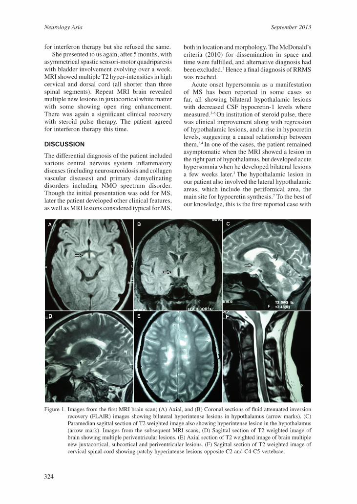
Figure 1. Images from the first MRI brain scan; (A) Axial, and (B) Coronal sections of fluid attenuated inversion recovery (FLAIR) images showing bilateral hyperintense lesions in hypothalamus (arrow marks). (C) Paramedian sagittal section of T2 weighted image also showing hyperintense lesion in the hypothalamus (arrow mark). Images from the subsequent MRI scans; (D) Sagittal section of T2 weighted image of brain showing multiple periventricular lesions. (E) Axial section of T2 weighted image of brain multiple new juxtacortical, subcortical and periventricular lesions. (F) Sagittal section of T2 weighted image of cervical spinal cord showing patchy hyperintense lesions opposite C2 and C4-C5 vertebrae.

325
acute hypersomnia as the initial manifestation of MS. Both MS and narcolepsy are associated with HLA-DQB1*0602-allele. Coexistent cases of MS and narcolepsy have also been reported. However, a recent study which aimed to analyse the difference in Epworth sleepiness scale and CSF hypocretin levels during relapse and remission phase in MS patients found no difference between the two stages.8 The authors concluded that in the absence of hypothalamic lesions, the hypocretin system remains intact and sleepiness is not a typical feature for RRMS. Grey matter involvement in MS is a phenomenon increasingly being recognised. Post-mortem studies have also shown demyelinating lesions and degenerative changes to be common, not just in the cortex but also in deep grey matter structures, especially the thalamus and caudate.9 Regarding hypothalamic lesions, a post mortem study revealed demyelinating lesions of fibre bundles in and adjacent to the hypothalamus in 16 out of the 17 patients studied.10 A MRI based study using T1 relaxation time as a sensitive measure of pathology found higher values in hypothalamus of RRMS patients compared to controls, and the values also correlated well with severity of fatigue.11 Another study using conventional MRI protocols found hypothalamic lesions in 13.3% of MS patients, a frequency higher than previously reported.12
In the light of above mentioned studies providing histopathological and radiological evidence of hypothalamic lesions in MS, and also the clinical cases including ours, we wonder whether hypothalamic involvement is actually that unusual for MS that it should be considered a red flag. We also feel that with the emergence and further refinement of newer MRI techniques like the double inversion recovery technique, appreciation of grey matter involvement in MS may increase, which may change the way we look at MS, and may lead to an overhaul of its diagnostic criteria and the so-called ‘red flags’. ACKNOWLEDGEMENTS
The authors thank the patient and her family for allowing us to report this data. DISCLOSURE
Conflict of interest: None
REFERENCES 1. Miller DH, Weinshenker BG, Filippi M, et al.
Differential diagnosis of suspected multiple sclerosis: a consensus approach. Mult Scler 2008; 14:1157-74.
2. Polman CH, Reingold SC, Banwell B, et al. Diagnostic criteria for multiple sclerosis: 2010 revisions to the McDonald criteria. Ann Neurol 2011; 69:292-302.
3. Kato T, Kanbayashi T, Yamamoto K, et al. Hypersomnia and low CSF hypocretin-1 (orexin-A) concentration in a patient with multiple sclerosis showing bilateral hypothalamic lesions. Intern Med 2003; 42:743-5.
4. Oka Y, Kanbayashi T, Mezaki T, et al. Low CSF hypocretin-1/orexin-A associated with hypersomnia secondary to hypothalamic lesion in a case of multiple sclerosis. J Neurol 2004; 251:885-6.
5. Rao DG, Singhal BS. Secondary narcolepsy in a case of multiple sclerosis. J Assoc Physicians India 1997; 45:321-2.
6. Iseki K, Mezaki T, Oka Y, et al. Hypersomnia in MS. Neurology 2002; 59:2006-7.
7. Swaab DF. Lateral hypothalamic area (LHA), including the perifornical area and intermediate hypothalamic area (IHA). In: Handbook of Clinical Neurology, Volume 79, The human hypothalamus: Basic and clinical aspects — Part I: Nuclei of the human hypothalamus. Elsevier, 2003:281-4.
8. Knudsen S, Jennum PJ, Korsholm K, Sheikh SP, Gammeltoft S, Frederiksen JL Normal levels of cerebrospinal fluid hypocretin-1 and daytime sleepiness during attacks of relapsing-remitting multiple sclerosis and monosymptomatic optic neuritis. Mult Scler 2008; 14:734-8.
9. Vercellino M, Masera S, Lorenzatti M, et al. Demyelination, inflammation, and neurodegeneration in multiple sclerosis deep gray matter. J Neuropathol Exp Neurol 2009; 68:489-502.
10. Huitinga I, De Groot CJ, Van der Valk P, Kamphorst W, Tilders FJ, Swaab DF. Hypothalamic lesions in multiple sclerosis. J Neuropathol Exp Neurol 2001; 60:1208-18.
11. Zellini F, Niepel G, Tench CR, Constantinescu CS. Hypothalamic involvement assessed by T1 relaxation
time in patients with relapsing–remitting multiple sclerosis. Mult Scler 2009; 15:1442-9.
12. Qiu W, Raven S, Wu JS, et al. Hypothalamic lesions in multiple sclerosis. J Neurol Neurosurg Psychiatry 2011; 82:819-22.