is radiography justified for the evaluation of patients ... · is radiography justified for the...
TRANSCRIPT
Is radiography justified for the evaluation of patients presentingwith cervical spine trauma?
Nicholas TheocharopoulosDepartment of Medical Physics, Faculty of Medicine, University of Crete, P.O. Box 2208, Iraklion,71003 Crete, Greece and Department of Natural Sciences, Technological Education Institute of Crete,P.O. Box 140, Iraklion 71004 Crete, Greece
Georgios ChatzakisDepartment of Radiology, Faculty of Medicine, University of Crete, P.O. Box 2208, Iraklion, 71003 Crete,Greece
John Damilakisa�
Department of Medical Physics, Faculty of Medicine, University of Crete, P.O. Box 2208, Iraklion,71003 Crete, Greece
�Received 26 April 2009; revised 4 July 2009; accepted for publication 30 July 2009;published 8 September 2009�
Conventional radiography has been for decades the standard method of evaluation for cervical spinetrauma patients. However, currently available helical multidetector CT scanners allow multiplanarreconstruction of images, leading to increased diagnostic accuracy. The purpose of this study was todetermine the relative benefit/risk ratio between cervical spine CT and cervical spine radiographyand between cervical spine CT and cervical spine radiography, followed by CT as an adjunct forpositive findings. A decision analysis model for the determination of the optimum imaging tech-nique was developed. The sensitivity and specificity of CT and radiography were obtained bydedicated meta-analysis. Lifetime attributable risk of mortal cancer from CT and radiography wascalculated using updated organ-specific risk coefficients and organ-absorbed doses. Patient organdoses from radiography were calculated using Monte Carlo techniques, simulated exposures per-formed on an anthropomorphic phantom, and thermoluminescence dosimetry. A prospective patientstudy was performed regarding helical CT scans of the cervical spine. Patient doses were calculatedbased on the dose-length-product values and Monte Carlo-based CT dosimetry software program.Three groups of patient risk for cervical spine fracture were incorporated in the decision model onthe basis of hypothetical trauma mechanism and clinical findings. Radiation effects were assessedseparately for males and females for four age groups �20, 40, 60, and 80 yr old�. Effective dosefrom radiography amounts to 0.050 mSv and from a typical CT scan to 3.8 mSv. The use of CT ina hypothetical cohort of 106 patients prevents approximately 130 incidents of paralysis in the lowrisk group �a priori fracture probability of 0.5%�, 500 in the moderate risk group �a priori fractureprobability of 2%�, and 5100 in the high risk group �a priori fracture probability of 20%�. Theexpense of this CT-based prevention is 15–32 additional radiogenic lethal cancer incidents. Accord-ing to the decision model calculations, the use of CT is more favorable over the use of radiographyalone or radiography with CT by a factor of 13, for low risk 20 yr old patients, to a factor of 23, forhigh risk patients younger than 80 yr old. The radiography/CT imaging strategy slightly outper-forms plain radiography for high and moderate risk patients. Regardless of the patient age, sex, andfracture risk, the higher diagnostic accuracy obtained by the CT examination counterbalances theincrease in dose compared to plain radiography or radiography followed by CT only for positiveradiographs and renders CT utilization justified and the radiographic screening redundant. © 2009American Association of Physicists in Medicine. �DOI: 10.1118/1.3213521�
Key words: CT, radiography, cervical spine, trauma, decision analysis
I. INTRODUCTION
There is a universal agreement for the necessity of imagingfor the clearance of patients who have sustained traumaticinjury of the cervical spine �CS� but the selection of theoptimal imaging modality remains controversial over theyears. The emergency medicine physician and the orthopedicsurgeon are very often in a dilemma over the appropriateimaging study which will help direct treatment and improve
outcome. Traditionally, plain radiography �three or five4461 Med. Phys. 36 „10…, October 2009 0094-2405/2009/36„1
views�, followed by CT as an adjunct, has been the imagingapproach of choice.1–3
The wide availability of modern multidetector CT�MDCT� scanners currently offers rapid and accurate clear-ance of trauma patients, allow accurate multiplanar reforma-tions, and can obviate the need for plain radiographs. There-fore, nowadays, MDCT may hold the role the singleexamination for the investigation of the CS trauma, which
4
does not necessitate further imaging. However, CT exami-44610…/4461/10/$25.00 © 2009 Am. Assoc. Phys. Med.

4462 Theocharopoulos, Chatzakis, and Damilakis: Cervical spine trauma imaging and justification 4462
nations are associated with a relatively high dose to the pa-tient compared to conventional radiography and thereforebare the potential of late radiation effects such as leukemiaand solid cancers. Hence, CT utilization is justified when thebenefit �diagnostic accuracy� over risk �radiation effects� ra-tio is higher compared to other available imaging modalities.
Although recent epidemiological studies have revived theconcern over the long term carcinogenic effects of ionizingradiation5,6 no data exist in literature on the effectiveness ofradiography and CT performed in cervical spine trauma pa-tients in terms of radiation dose and risks. The aim of thisstudy was to develop and apply a decision analysis model,7,8
incorporating both expected health benefit and potential det-riment from the two imaging modalities for the investigationof the radiation effectiveness of CT relative to radiographyand the subsequent justification of CT usage for CS evalua-tion in trauma patients.
II. MATERIALS AND METHODS
II.A. Decision analysis
For the determination of the optimum imaging technique,we have performed decision analysis based on probability
9
FIG. 1. Decision model for single-modality imaging policy �imaging modaprobabilities �P�.
and Bayesian algebra. Three imaging policies were consid-
weight for paralysis due to cervical spine fracture, P6 is the
Medical Physics, Vol. 36, No. 10, October 2009
ered: �a� Radiography, �b� MDCT, and �c� radiography fol-lowed by CT as an adjunct for positive findings. A positivediagnosis following radiography alone, CT alone, or CT asan adjunct for positive radiography was supposed to be fol-lowed by a surgical or nonsurgical intervention �surgical ormechanical stabilization� and postoperative mortality wasalso accounted for. The major complication following a falsenegative diagnosis was considered to be paralysis. Other pos-sible health outcomes included radiogenic lethal cancer�solid cancer and leukemia�. Every final health state �remis-sion, paralysis, fracture, and death� was assigned with a dis-ability weight �DW�. The disease-specific disability weightshave been introduced by national and international healthorganizations in order to quantify the merit of a health stateand vary from 0 for perfect health to 1 for death. In a deci-sion model, the disability weight of a final health state ismultiplied by the probability of that state to occur given adecision is taken �policy� to yield the total disability of thepolicy. The effectiveness of a policy is inversely proportionalto the corresponding total disability value.
Figure 1 illustrates the decision model for a single-modality imaging policy and the associated DWs and prob-abilities �P�. For the imaging modality A, the total disability
� showing treatment policies and outcomes and the associated DWs and
TDA will be given by the following equation:
TDA = DW1 · �P1� + DW2 · ��1 − P5� · P3 · P2 · �1 − P1�� + DW2 · ��1 − P6� · P5 · P3 · P2 · �1 − P1��
+ DW1 · �P6 · P5 · P3 · P2 · �1 − P1�� + DW3 · ��1 − P7� · �1 − P3� · P2 · �1 − P1�� + DW4 · �P7 · �1 − P3� · P2 · �1
− P1�� + DW2 · ��1 − P5� · P4 · �1 − P2� · �1 − P1�� + DW2 · ��1 − P6� · P5 · P4 · �1 − P2� · �1 − P1��
+ DW1 · �P6 · P5 · P4 · �1 − P2� · �1 − P1�� + DW2 · ��1 − P4� · �1 − P2� · �1 − P1�� , �1�
where DW1 is the disability weight for death, DW2 is thedisability weight for perfect health, DW3 is the disabilityweight for cervical spine fracture, DW4 is the disability
mortality associated with cervical spine surgery, P5 is theprobability of cervical spine surgery following a positive cer-vical spine fracture diagnosis, P7 is the probability of paraly-
lity A
sis following a missed cervical spine fracture, P4 is the

4463 Theocharopoulos, Chatzakis, and Damilakis: Cervical spine trauma imaging and justification 4463
specificity of the imaging modality A, P3 is the sensitivity ofthe imaging modality A, P2 is the a priori fracture probabil-ity �prevalence or patient risk�, and P1 is the radiogenicdeath probability �radiogenic risk�.
Among the variables incorporated in the present studydecision algorithm, the probability values regarding theprevalence of CS fracture, the treatment policy, and the post-operative mortality rates were obtained from the literature.Other determinants of the decision model were the diagnos-tic accuracy of imaging and the radiogenic risk.
II.B. Literature review and meta-analysis
The optimum imaging policy, as derived by the decisionmodel calculations, is strongly dependent on the sensitivityand specificity of the imaging modalities under comparison.Hence, in order to determine the diagnostic accuracy of ra-diography and CT, we have performed dedicated literaturereview and meta-analysis. Literature clearance was con-ducted by two independent reviewers who screened the pub-lication pool for potentially relevant articles. Disagreementsin the selection of suitable publications were solved by dis-cussion. Statistical analysis was performed using the META-
DISC software program.10 A detailed description of the litera-ture search is provided in the Appendix.
II.C. Radiogenic risk evaluation and dosimetry
Lifetime attributable risk of mortal solid cancer and leu-kemia was calculated using Tables 12D-1 and 12D-2 of theBEIR VII report,11 previously published methodology,12 andorgan-absorbed doses obtained by means of Monte Carlotechniques.
Patient organ doses from three-view radiography�anterior-posterior �AP�, lateral �LAT�, and odontoid view�were calculated using the Monte Carlo N-particle code�MCNP4C2, Los Alamos National Laboratory, Los Alamos,NM�. Human anatomy was replicated by a mathematicalphantom constructed with a commercially available softwaretool �BodyBuilder, White Rock Science, NM�.
TABLE I. Health states and corresponding DWs associated with CS trauma.
Health state DW Reference
CS fracture 0.44 9Paralysis due to CS fracture 0.30 15Death 1 DefinitionRemission 0 Definition
TABLE II. Probability ranges and adopted values for the decision model var
VariableProbability
�%�
Fracture probability 0.04–19.7Surgical intervention probability 5.2–32.9Mortality following surgery 0–16.7Paralysis following missed CS fracture 0–30
Medical Physics, Vol. 36, No. 10, October 2009
Projection-specific dose values were calculated by theMonte Carlo simulations on the basis of tube high voltageand filtration and were normalized over the amount of radia-tion incident upon the patient at the beam entry surface. Thisradiation quantity is called entrance surface dose �ESD�, andit is measured using thermoluminescent dosimeters attachedto the examinee’s skin during the exposure.13
For the measurement of ESD, the radiographic projectionswere simulated on an anthropomorphic phantom �Randophantom, Alderson Research Laboratories, Stamford, CT�.The Rando phantom simulates the head and torso of an adultwith height of 1.73 m and weight of 74 kg and consists oftissue equivalent material over a synthetic human skeleton. Ithas been widely used for dosimetric measurements in diag-nostic radiology.13 The radiographic exposures of the phan-tom were performed on a Siemens Polydoros 50 x-ray unit�Siemens, Erlangen, Germany�. It is a conventional screen-film x-ray unit with a total filtration of 3.5 mm aluminum.The phantom was positioned erect against the vertical Buckytable and the focus to film distance was set to 110 cm for allprojections. The AP and LAT radiographs were performed at40 kVp, as used routinely for AP and LAT radiography in ourinstitution, and 40 m As. A high voltage of 60 kVp and atube load of 45 m As were applied for the odontoid projec-tion. Lithium fluoride thermoluminescence dosimeters�TLD-100, Harshaw, OH� were used for recording the ESDresulting from the simulated examinations.
For the determination of patient doses from helical CTscans of the CS, we have performed an 8-month prospectivestudy on an MDCT scanner �Sensation 16, Siemens, Erlan-gen, Germany� which included alert, low, and moderate risktrauma patients referred for CT evaluation. The helical scansof the cervical spine were performed at 120 kVp, a pitchvalue of 0.62, and reconstructed slice thickness of 0.75 mm.The prospective study was approved by the ethics committeeof our institution. Patient consent was not required since thestudy did not alter the routine imaging management oftrauma patients. For each patient in the prospective study,organ doses and effective dose from the helical scan werecalculated based on the dose-length-product �DLP� valuesprovided on a special card of the patient’s record in the pa-tient database of the CT operating console. Scanner-specificDLP to organ dose conversion coefficients were derived forboth sexes using a Monte Carlo-based CT dosimetry soft-ware program.14
.
Reference Model values
16 High risk 20%, moderate risk 2%, low risk 0.5%17 24.6% adults, 7% geriatric �age�65 yr�18 1.6%, 12.2% geriatric �age�65 yr�19–24 5%
iables

4464 Theocharopoulos, Chatzakis, and Damilakis: Cervical spine trauma imaging and justification 4464
III. RESULTS
III.A. Variables for the decision model
Table I shows the disability weights9,15 of the health out-comes associated with CS trauma. The value range of thevariables required for the decision model is provided in TableII. The a priori probability of fracture depends largely on thepatient age and mechanism of trauma.16 Three values of frac-ture prevalence were considered in our model: 0.5% �low�,2% �moderate�, and 20% �high�. The method of fracturetreatment and the prognosis depend on the patient age, se-verity of trauma, and neurologic findings.17–24 In order toaccount for the differences in patient management and radi-osensitivity with age, four age groups were distinctively ex-amined in our model �20, 40, 60 and 80 yr old�.
III.B. Literature review and meta-analysis
The initial literature search returned 212 articles. Title andabstract clearance was conducted and 140 papers were ex-cluded from the study. Detailed reading of the remainingpapers further excluded 20 articles. From the remaining 52
FIG. 2. Forr
FIG. 3. Forrest plots f
Medical Physics, Vol. 36, No. 10, October 2009
articles, 39 were excluded due to incomplete or irrelevantinformation. Complementary manual search yielded threeadditional articles for inclusion. Overall, 16 articles were in-cluded in our meta-analysis.4,25–39
Figures 2 and 3 demonstrate the Forrest plots for sensitiv-ity and specificity for each modality. Sensitivity for CTranged from 98% to 99% and for radiography ranged from25% to 93%. Specificity was 100% for CT and ranged forradiography from 99% to 100%. Pooled sensitivity was 99%�95% CI: 98%–99%� and pooled specificity was 100% �95%CI: 100%–100%� for CT. Pooled sensitivity was 42.8% �95%CI: 39%–46%� and pooled specificity was 99% �95% CI:99%–100%� for radiography. The derived values were usedfor the decision model calculations regarding CT. Because ofthe heterogeneity in the sensitivity and the specificity of ra-diography, the analysis was repeated with three studies in-cluded for the determination of sensitivity4,25,28 and two4,29
for the determination of specificity. Recalculated sensitivitywas 48.3% �95% CI: 41.4%–55.3%, chi-square: 2.02, andp=0.365� and specificity was 98% �95% CI: 95.4%–99.4%,
ots for CT.
est plor radiography.
4465 Theocharopoulos, Chatzakis, and Damilakis: Cervical spine trauma imaging and justification 4465
chi-square: 0.10, and p=0.748�. The latter values were usedfor the decision model calculations regarding the diagnosticaccuracy of radiography.
III.C. Dosimetric results
According to the ESD measurements obtained by thesimulated exposures, the patient effective dose from thethree-view radiographic evaluation of the CS amounts to0.050 mSv �0.034 mSv from the AP projection, 0.006 mSvfrom the LAT, and 0.010 mSv from the odontoid�.
The mean DLP value of the helical CS scans was 512cGy cm for the 50 male and 455 cGy cm for the 36 femalepatients included in our study. Our calculated sex-specificDLP to effective dose conversion coefficients were 0.007 57and 0.007 47 mSv/cGy cm for females and males, respec-tively. As a result, the radiation burdens from a typical heli-cal scan of the CS were estimated to be 3.8 and 3.4 mSv forthe male and female patients, respectively.
III.D. Decision analysis model
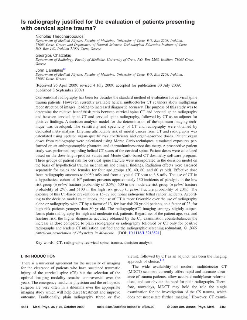
The decision analysis model is illustrated in Fig. 4. Threeimaging options are compared: CT, radiography, and radiog-raphy followed by CT when positive. The relationship be-tween true positive, true negative, false positive, and false
FIG. 4. Decision analysis model showing the three imaging options undercomparison, treatment policies, and outcomes. The relationship between truepositive, true negative, false positive, and false negative diagnosis and thesensitivity/specificity of the imaging modalities is also shown.
negative diagnosis and the sensitivity/specificity of the imag-
Medical Physics, Vol. 36, No. 10, October 2009
ing modalities is also shown. Treatment strategy, postopera-tive mortality, and treatment outcomes �remission or paraly-sis� are stochastic variables with values presented in Table II.
III.E. Effectiveness of the imaging strategies
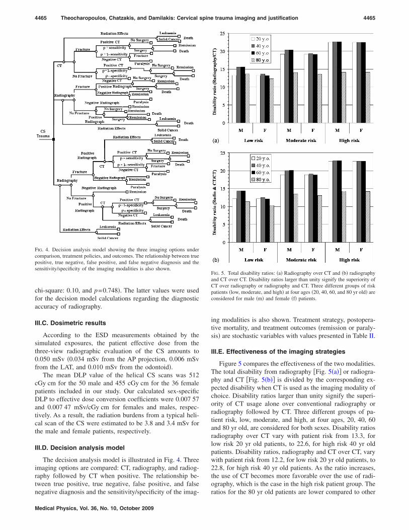
Figure 5 compares the effectiveness of the two modalities.The total disability from radiography �Fig. 5�a�� or radiogra-phy and CT �Fig. 5�b�� is divided by the corresponding ex-pected disability when CT is used as the imaging modality ofchoice. Disability ratios larger than unity signify the superi-ority of CT usage alone over conventional radiography orradiography followed by CT. Three different groups of pa-tient risk, low, moderate, and high, at four ages, 20, 40, 60and 80 yr old, are considered for both sexes. Disability ratiosradiography over CT vary with patient risk from 13.3, forlow risk 20 yr old patients, to 22.6, for high risk 40 yr oldpatients. Disability ratios, radiography and CT over CT, varywith patient risk from 12.2, for low risk 20 yr old patients, to22.8, for high risk 40 yr old patients. As the ratio increases,the use of CT becomes more favorable over the use of radi-ography, which is the case in the high risk patient group. The
FIG. 5. Total disability ratios: �a� Radiography over CT and �b� radiographyand CT over CT. Disability ratios larger than unity signify the superiority ofCT over radiography or radiography and CT. Three different groups of riskpatients �low, moderate, and high� at four ages �20, 40, 60, and 80 yr old� areconsidered for male �m� and female �f� patients.
ratios for the 80 yr old patients are lower compared to other

4466 Theocharopoulos, Chatzakis, and Damilakis: Cervical spine trauma imaging and justification 4466
age groups since this patient cohort, although less radiosen-sitive, presents higher postoperative mortality �Table II�. It isinteresting to notice that the radiography and CT imagingstrategy slightly outperforms the radiography alone optionand only for high and moderate risk patients.
In Fig. 6 we compare the number of fractures missed,leading or not to paralysis, per million examinations. Thenumber of missed fractures increases with the a priori frac-ture probability �patient risk�. Radiography and radiographyand CT yield approximately 50 times more false negativediagnoses than CT alone.
Radiation effects �total mortal cancers per million exami-nations� for both sexes are shown in Fig. 7. The use of CT ina hypothetical cohort of 106 patients prevents approximately130 incidents of paralysis in the low risk group, 500 in themoderate risk group, and 5100 in patients with high a priorifracture probability. The expense of this CT-based preventionis, depending on age, sex, and risk, 15–32 radiogenic lethalcancer incidents additional to those potentially incurred byplain radiography or radiography followed by CT for posi-tive findings.
IV. DISCUSSION
The dramatic increase in the CT usage over the past de-cade and the more accurately quantified radiogenic risk esti-mates have recently restimulated concerns about the clinicalgrounds on which CT examinations are prescribed.5,6 Refer-ring physicians should be able to assess the benefit versuspotential detriment to the examinee and request the mostbeneficial among the available modalities. Hence, justifica-tion is an essential part of developing the imaging strategysince patients should not be deprived of the potential benefitarising from a CT scan.
Regarding the imaging evaluation of CS trauma patients,CT and radiography have been so far compared in two pub-lished studies but only in terms of financial costs, thus ne-glecting the radiation effects.9,40 It was deduced that CT
FIG. 6. Number of fractures missed per million examinees for each imagingstrategy, leading or not to paralysis.
should be the preferred screening modality for high and
Medical Physics, Vol. 36, No. 10, October 2009
moderate risk patients. To the best of our knowledge, nostudies exist that compare CT to radiography followed by CTor that account for radiogenic detriment.
In the present study, a meta-analysis and systematic re-view of the published literature confirmed the superiority ofperformance of MDCT vs plain radiography for the clear-ance of patients suspected with cervical spine injury. More-over, the calculations based on a decision analysis modelestablished that, despite the increased radiation burden to theexaminee, CT is associated with more favorable ratios ofbenefit to risk compared to radiography or radiography andCT, irrespective of risk group stratification �determined bythe mechanism of trauma and the patient age� and patientradiosensitivity �depending on age at irradiation�. It shouldbe stressed that, as shown in Fig. 5, CT outweighs both ra-diography and radiography and CT even for the low riskhighly radiosensitive patients �females of 20 yr old�. Thestrength of this finding was investigated by recalculating thedisability ratios for this patient subgroup for varying valuesof the probability of paralysis and of the sensitivity of radi-ography �sensitivity analysis�. As shown in Fig. 8, the appro-priateness of CT even for the most radiosensitive patient co-hort is not affected by the two abovementioned variables.
FIG. 7. Radiation effects �lethal cancer incidents per million examinees� of�a� CT alone and radiography alone for both sexes and four age groups, and�b� for radiography followed by CT, for both sexes, four age groups, andthree fracture risk levels.
Even at a high hypothetical value of radiography specificity

4467 Theocharopoulos, Chatzakis, and Damilakis: Cervical spine trauma imaging and justification 4467
�99%�, the corresponding sensitivity needed for radiographyto be equivalent CT is 0.997 and for radiography/CT to beequivalent to CT alone is 0.979. Hence, high values of sen-sitivity are needed for equivalent disability scores, which arenot found in literature. Similarly, even at 0.99 specificity anda sensitivity value of 0.85 for radiography, the disability ratiois 4.26 for radiography over CT and 3.86 for radiography andCT over CT, for a probability of paralysis from a missedfracture as low as 0.01 �1%� �Fig. 8�.
Sensitivity analysis was also performed with respect tothe disability weights of paralysis and fracture. Figure 9shows that even for an increased hypothetical diagnostic ac-curacy of radiography �sensitivity of 0.85 and specificity of0.99�, plain radiography or radiography followed by CT isequivalent to CT at a negative disability weight for paralysis,which is unrealistic. Figure 10 shows that even with theabovementioned increased diagnostic accuracy of radiogra-
FIG. 8. �a� Variation in radiography over CT disability ratios �sensitivityanalysis� with sensitivity of radiography �dashed line� for a 0.99 specificityvalue and with probability of paralysis due to a missed CS fracture for 0.99radiography specificity and sensitivity of 0.483 �solid line 1� and of 0.85�solid line 2�. �b� Variation in radiography and CT over CT disability ratios�sensitivity analysis� with sensitivity of radiography �dashed line� for a 0.99specificity value and with probability of paralysis due to a missed CS frac-ture for 0.99 radiography specificity and sensitivity of 0.483 �solid line 1�and of 0.85 �solid line 2�.
phy �sensitivity of 0.85 and specificity 0.99�, and with a re-
Medical Physics, Vol. 36, No. 10, October 2009
duced disability weight of paralysis �0.1�, plain radiographyor radiography followed by CT is equivalent to CT only ifthe facture disability weight is in the range of 0.02–0.04,which is also unrealistic.
The unexpected finding of the very limited superiority ofradiography and CT over plain radiography can be explainedusing Figs. 6 and 7. Implementation of the combined imag-ing protocol calls for 112 600, 29 260 and 22 315 CT scansper million patients of high, medium, and low risk patients,respectively, incurring one to four extraneous radiogenicdeaths. Since only patients with positive radiographs are CTscanned, no false negative findings will be revealed. On thecontrary, because of the 0.99 CT sensitivity, a fraction of thetrue positive radiograph-based diagnoses will be canceledfollowing false negative CT scans, leading to 1126, 293, and223 fractures missed per million high, medium, and low riskpatients, respectively, additional to plain radiography, or 56,14, and 11 cases of paralysis. The actual benefit of CT usage
FIG. 10. Variation in disability ratios �sensitivity analysis� with fracture dis-ability weight for an increased hypothetical diagnostic accuracy of radiog-raphy �sensitivity of 0.85 and specificity of 0.99� and a disability weight for
FIG. 9. Variation in disability ratios �sensitivity analysis� with paralysis dis-ability weight for an increased hypothetical diagnostic accuracy of radiog-raphy �sensitivity of 0.85 and specificity of 0.99�.
paralysis reduced to 0.1.

4468 Theocharopoulos, Chatzakis, and Damilakis: Cervical spine trauma imaging and justification 4468
following positive radiography is the prevention of postop-erative death of examinees without deficit, who would un-dergo surgery due to false positive radiography, i.e., 64, 78,and 80 deaths per million high, medium, and low risk pa-tients, respectively.
In the present study, radiation risk assessment was basedon an up-to-date report derived from credible epidemiologi-cal data.11 Organ-specific coefficients per age group and sexwere combined with the Monte Carlo derived organ-absorbed doses. Radiogenic risk from CT or radiographyoriginates primarily from the irradiation of the thyroid andred bone marrow �RBM�. Male patients are 1.3 times moresusceptible to leukemia compared to female patients and theradiosensitivity of RBM drops by 30% from age 20 to age 80yr. The incidence of thyroid cancer is 4.4 higher on femalepatients in the 20–60 yr old age group and drop to 0 for bothsexes over the age of 80. However, the mortality associatedwith thyroid cancer is as low as 7%.41 Hence, although theradiation burden to the thyroid is four times higher than thatto the RBM, the fatal leukemia risk is 2, for the young ex-aminees, to 200 times higher than the mortality risk of thy-roid cancer. Because of the dose distribution of the specificexamination, the radiogenic risk is low enough to render CTutilization justified even for the low risk patient cohort.
The importance of accurate dose and risk assessment forthe purpose of justification can be highlighted by recalculat-ing the disability ratios for a CT examination of higher bur-den such as coronary angiography. Figure 11 illustrates thatif the dose to the stomach, lung, and thyroid was equal tothat from a coronary angiography scan,12 then the utilizationof CT would not be justified for the low risk patient cohort.Similarly, the importance of applying the organ-specificmethodology for the risk assessment may be shown by recal-culating the risk and the corresponding disability ratios usingthe effective dose to the patient from CT and radiographyand the risk coefficient for adults proposed by ICRP in 1990
−2 −1 42
FIG. 11. Disability ratios �radiography over CT� derived from radiogenicrisks based on organ-absorbed doses associated with CT coronary angiogra-phy. Ratios are provided for three different risk groups �low, moderate, andhigh�, four ages at irradiation �20, 40, 60, and 80 yr old�, and separately formale �m� and female �f� patients.
�5�10 Sv �. Had we used the effective dose based risk
Medical Physics, Vol. 36, No. 10, October 2009
assessment method would have overestimated radiogenicmortality by factors of 5–12 for CT and by factors of 3–32for radiography. As a result, the disability ratios would havebeen 1.5–4 times lower for the low and moderate risk patientgroups �Fig. 12�. It is therefore evident that the process ofjustification regarding the use of CT in the clearance of CStrauma patients had to be performed in the light of the newinformation about the dosimetric characteristics and diagnos-tic performance of the modern MDCT scanners and the ra-diosensitivity of the examinees.
V. CONCLUSIONS
In conclusion, the application of a decision model incor-porating up-to-date organ-specific radiation risk assessmenthas shown that for the evaluation of CS trauma, the higherdiagnostic accuracy obtained by the CT examination coun-terbalances the increase in dose compared to plain radiogra-phy or radiography followed by CT for positive findings andrenders CT utilization dose effective and justified regardless
FIG. 12. Disability ratios: �a� Radiography/CT and b� radiography and CT/CT, derived from radiogenic risks based on the effective dose concept. Ra-tios are provided for male �m� and female �f� patients, three different riskgroups �low, moderate, and high�, and four ages at irradiation �20, 40, 60,and 80 yr old�.
of the patient age, sex, and fracture risk.

4469 Theocharopoulos, Chatzakis, and Damilakis: Cervical spine trauma imaging and justification 4469
ACKNOWLEDGMENT
This work was supported by the European Community, aspecific targeted research project, under the FP6 Specificprogramme for research and training on nuclear energy�Safety and Efficacy of CT, Contract No. FP6/002388�.
APPENDIX: LITERATURE SEARCH
We searched the most popular literature databases �MED-LINE, EMBASE, Web of Science, and Cochrane Database ofSystematic Reviews� for diagnostic cohort studies of patientswith low or intermediate severity cervical spine trauma whowere imaged by means of CT and were published between2000 and 2008, or radiography and were published between1996 and 2008. Our search strategy in MEDLINE was asfollows: Search terms: �cervical spine OR cervical OR neck�AND �trauma� AND �“diagnostic accuracy” OR sensitivity�AND �specificity OR “true positive” OR “true negative” OR“false positive” OR “false negative” OR “true positives” OR“true negatives” OR “false positives” OR “false negatives”OR “predictive value” OR “predictive values” OR “likeli-hood ratio” OR “positive likelihood” OR “negative likeli-hood” OR “receiver operating characteristics” OR “ROC”OR “reference value” OR “reference values” OR “normalvalue” OR “normal values” OR “confidence interval” OR“confidence intervals”� AND �“MDCT” OR tomography�AND has abstract�text� AND English�lang� AND “hu-mans”�MeSH Terms� AND �“2000”�PDAT�: “3000”�PDAT��and limits: only items with abstracts, English, humans, adults��19 yr old�. For the other databases, search strategy wasadapted to the needs of the specific search engine. Auto-mated retrieval was complemented with a manual searchthrough the literature of review articles, for potentially rel-evant articles that were not identified by the original search.
We included in our study �a� original prospective or ret-rospective studies assessing the role of CT or radiography inthe evaluation of the CS injury, �b� articles including twox-ray views of the spine at the minimum, and �c� articlesadequately reporting performance values �true positive, truenegative, false positive, and false negative�. Case reports,review articles, pictorial essays, unpublished data, abstracts,and letters to the editor were excluded from the study. Fur-thermore, publications focusing on topics other than diagnos-tic test assessment and effectiveness analyses, such as man-agement decision issues and technical exhibitions or studiesemploying dynamic radiographic views �flexion, extension,etc� not fulfilling the above mentioned criteria, were alsoexcluded.
a�Author to whom correspondence should be addressed. Electronic mail:[email protected]; Telephone: �30-2810-392569; Fax: �30-2810-542095.
1Royal College of Radiologists, Making the Best Use of a Department ofClinical Radiology, 5th ed. �Royal College of Radiologists, London,2003�.
2J. R. Hoffman, W. R. Mower, A. B. Wolfson, K. H. Todd, and M. I.Zucker, “Validity of a set of clinical criteria to rule out injury to thecervical spine in patients with blunt trauma. National EmergencyX-Radiography Utilization Study Group,” N. Engl. J. Med. 343, 94–99
�2000�.Medical Physics, Vol. 36, No. 10, October 2009
3I. G. Stiell, G. A. Wells, K. L. Vandemheen, C. M. Clement, H. Lesiuk, V.J. De Maio, A. Laupacis, M. Schull, R. D. McKnight, R. Verbeek, R.Brison, D. Cass, J. Dreyer, M. A. Eisenhauer, G. H. Greenberg, I.MacPhail, L. Morrison, M. Reardon, and J. Worthington, “The CanadianC-spine rule for radiography in alert and stable trauma patients,” JAMA,J. Am. Med. Assoc. 286, 1841–1848 �2001�.
4P. T. McCulloch, J. France, D. L. Jones, W. Krantz, T. P. Nguyen, C.Chambers, J. Dorchak, and P. Mucha, “Helical computed tomographyalone compared with plain radiographs with adjunct computed tomogra-phy to evaluate the cervical spine after high-energy trauma,” J. Bone Jt.Surg., Am. Vol. 87, 2388–2394 �2005�.
5D. J. Brenner and E. J. Hall, “Computed tomography:an increasing sourceof radiation exposure,” N. Engl. J. Med. 357, 2277–2284 �2007�.
6E. J. Hall and D. J. Brenner, “Cancer risks from diagnostic radiology,” Br.J. Radiol. 81, 362–378 �2008�.
7J. Bernstein, “Decision analysis,” J. Bone Jt. Surg., Am. Vol. 79, 1404–1414 �1997�.
8M. S. Kocher and D. Zurakowski, “Clinical epidemiology and biostatis-tics: A primer for orthopaedic surgeons,” J. Bone Jt. Surg., Am. Vol. 86,607–620 �2004�.
9C. C. Blackmore, S. D. Ramsey, F. A. Mann, and R. A. Deyo, “Cervicalspine screening with CT in trauma patients: A cost-effectiveness analy-sis,” Radiology 212, 117–125 �1999�.
10J. Zamora, V. Abraira, A. Muriel, K. S. Khan, and A. Coomarasamy,“Meta-DiSc: A software for meta-analysis of test accuracy data,” BMCMed. Res. Methodol. 6, 31–43 �2001�.
11Committee to Assess Health Risks from Exposure to Low Levels of Ion-izing Radiation; Nuclear and Radiation Studies Board, Division on Earthand Life Studies, National Research Council of the National Academies,Health Risks From Exposure to Low Levels of Ionizing Radiation: BEIRVII Phase 2 �The National Academies, Washington, DC, 2006�.
12A. J. Einstein, M. J. Henzlova, and S. Rajagopalan, “Estimating risk ofcancer associated with radiation exposure from 64-slice computed tomog-raphy coronary angiography,” JAMA, J. Am. Med. Assoc. 298, 317–323�2007�.
13K. Faulkner, D. A. Broadhead, and R. M. Harrison, “Patient dosimetrymeasurement methods,” Appl. Radiat. Isot. 50, 113–123 �1999�.
14Imaging Performance Assessment of CT �ImPACT�, St. George’s Hospi-tal, London, UK, CT Patient Dosimetry Calculator �Version 0.99w, June2005, available at http://www.impactscan.org�.
15O. Johnell, B. Jönsson, L. Jönsson, and D. Black, “Cost effectiveness ofalendronate �fosamax� for the treatment of osteoporosis and prevention offractures,” Pharmacoeconomics 21, 305–314 �2003�.
16C. C. Blackmore, S. S. Emerson, F. A. Mann, and T. D. Koepsell, “Cer-vical spine imaging in patients with trauma: Determination of fracturerisk to optimize use,” Radiology 211, 759–765 �1999�.
17Z. N. Irwin, M. Arthur, R. J. Mullins, and R. A. Hart, “Variations ininjury patterns, treatment, and outcome for spinal fracture and paralysis inadult versus geriatric patients,” Spine 29, 796–802 �2004�.
18A. P. Jackson, M. H. Haak, N. Khan, and P. R. Meyer, “Cervical spineinjuries in the elderly: Acute postoperative mortality,” Spine 30, 1524–1527 �2005�.
19D. C. Reid, R. Henderson, L. Saboe, and J. D. Miller, “Etiology andclinical course of missed spine fractures,” J. Trauma 27, 980–986 �1987�.
20J. W. Davis, D. L. Phreaner, D. B. Hoyt, and R. C. Mackersie, “Theetiology of missed cervical spine injuries,” J. Trauma 34, 342–346 �1993�.
21H. C. Sox, M. A. Blatt, M. C. Higgins, and K. I. Marton, Medical Deci-sion Making �Butterworth, Boston, MA, 1988�.
22B. Efron and R. J. Tibshirani, An Introduction to the Bootstrap �Chapmanand Hall, New York, NY, 1993�.
23B. D. Gerrelts, E. U. Petersen, J. Mabry, and S. R. Petersen, “Delayeddiagnosis of cervical spine injuries,” J. Trauma 31, 1622–1626 �1991�.
24A. E. Ajani, D. J. Cooper, C. D. Scheinkestel, J. Laidlaw, and D. V.Tuxen, “Optimal assessment of cervical spine trauma in critically ill pa-tients: A prospective evaluation,” Anaesth. Intensive Care 26, 487–491�1998�.
25P. J. Schenarts, J. Diaz, C. Kaiser, Y. Carrillo, V. Eddy, and J. A. Morris,“Prospective comparison of admission computed tomographic scan andplain films of the upper cervical spine in trauma patients with alteredmental status,” J. Trauma 51, 663–668 �2001�.
26C. M. Bach, I. E. Steingruber, S. Peer, R. Peer-Kühberger, W. Jaschke,and M. Ogon, “Radiographic evaluation of cervical spine trauma. Plain
radiography and conventional tomography versus computed tomogra-
4470 Theocharopoulos, Chatzakis, and Damilakis: Cervical spine trauma imaging and justification 4470
phy,” Arch. Orthop. Trauma Surg. 121, 385–387 �2001�.27M. M. Griffen, E. R. Frykberg, A. J. Kerwin, M. A. Schinco, J. J. Tepas,
K. Rowe, and J. Abboud, “Radiographic clearance of blunt cervical spineinjury: Plain radiograph or computed tomography scan?” J. Trauma 55,222–226 �2003�.
28J. J. Diaz, C. Gillman, J. A. Morris, A. K. May, Y. M. Carrillo, and J.Guy, “Are five-view plain films of the cervical spine unreliable? A pro-spective evaluation in blunt trauma patients with altered mental status,”Trauma 55, 658–663 �2003�.
29S. Widder, C. Doig, P. Burrowes, G. Larsen, R. J. Hurlbert, and J. B.Kortbeek, “Prospective evaluation of computed tomographic scanning forthe spinal clearance of obtunded trauma patients: Preliminary results,” J.Trauma 56, 1179–1184 �2004�.
30J. J. Diaz, J. M. Aulino, B. Collier, C. Roman, A. K. May, R. S. Miller, O.Guillamondegui, and J. A. Morris, “The early work-up for isolated liga-mentous injury of the cervical spine: Does computed tomography scanhave a role?,” J. Trauma 59, 897–903 �2005�.
31G. K. Nguyen and R. Clark, “Adequacy of plain radiography in the diag-nosis of cervical spine injuries,” Emerg. Radiol. 11, 158–161 �2005�.
32C. A. Barba, J. Taggert, A. S. Morgan, J. Guerra, B. Bernstein, M.Lorenzo, A. Gershon, and N. Epstein, “A new cervical spine clearanceprotocol using computed tomography,” J. Trauma 51, 652–656 �2001�.
33K. Brohi, M. Healy, T. Fotheringham, O. Chan, C. Aylwin, S. Whitley,and M. Walsh, “Helical computed tomographic scanning for the evalua-tion of the cervical spine in the unconscious, intubated trauma patient,” J.Trauma 58, 897–901 �2005�.
34C. V. Brown, J. L. Antevil, M. J. Sise, and D. I. Sack, “Spiral computedtomography for the diagnosis of cervical, thoracic, and lumbar spine frac-tures: Its time has come,” J. Trauma 58, 890–895 �2005�.
35B. Sanchez, K. Waxman, T. Jones, S. Conner, R. Chung, and S. Becerra,
Medical Physics, Vol. 36, No. 10, October 2009
“Cervical spine clearance in blunt trauma: Evaluation of a computedtomography-based protocol,” J. Trauma 59, 179–183 �2005�.
36S. C. Gale, V. H. Gracias, P. M. Reilly, and C. W. Schwab, “The ineffi-ciency of plain radiography to evaluate the cervical spine after blunttrauma,” J. Trauma 59, 1121–1125 �2005�.
37R. Mathen, K. Inaba, F. Munera, P. G. Teixeira, L. Rivas, M. McKenney,P. Lopez, and C. J. Ledezma, “Prospective evaluation of multislice com-puted tomography versus plain radiographic cervical spine clearance intrauma patients,” J. Trauma 62, 1427–1431 �2007�.
38J. D. Berne, G. C. Velmahos, Q. El-Tawil, D. Demetriades, J. A. Asensio,J. A. Murray, E. E. Cornwell, H. Belzberg, and T. V. Berne, “Value ofcomplete cervical helical computed tomographic scanning in identifyingcervical spine injury in the unevaluable blunt trauma patient with multipleinjuries: A prospective study,” J. Trauma 47, 896–902 �1999�.
39D. B. Nunez, A. Zuluaga, D. A. Fuentes-Bernardo, L. A. Rivas, and J. L.Becerra, “Cervical spine trauma: How much more do we learn by rou-tinely using helical CT?,” Radiographics 16, 1307–1318 �1996�.
40E. L. Grogan, J. A. Morris, Jr., R. S. Dittus, D. E. Moore, B. K. Poulose,J. J. Diaz, and T. Speroff, “Cervical spine evaluation in urban traumacenters: Lowering institutional costs and complications through helicalCT scan,” J. Am. Coll. Surg. 200, 160–165 �2005�.
41International Commission on Radiological Protection, The 2007 Recom-mendations of the International Commission on Radiological Protection,Publication No. 103 �The International Commission on Radiological Pro-tection, 2007�.
42International Commission on Radiological Protection, 1990 Recommen-dations of the International Commission on Radiological Protection, Pub-lication No. 60 �The International Commission on Radiological Protec-tion, New York, 1991�.