isolation and characterization of biofilm forming bacteria ... d.sonkusale and vidya s... ·...
TRANSCRIPT
Int.J.Curr.Microbiol.App.Sci (2015) Special Issue-2: 118-127
118
Original Research Article
Isolation and Characterization of Biofilm Forming Bacteria from Oral Microflora
Kaustubh D.Sonkusale* and Vidya S.Tale
Department of Microbial Biotechnology, Rajiv Gandhi Institute of IT and Biotechnology, Bharati Vidyapeeth Deemed University, Pune 411046
*Corresponding author
A B S T R A C T
Introduction
The microbes are ubiquitous entities in nature as well as in the human body. The relationship between the host and most microbes as mutually symbiotic (Lee and Mazmanian, 2010; Geuking et al., 2011; Neish, 2009). These microorganisms can be
planktonic or free-living and frequently organize themselves into a consortium of microbes called biofilm that adheres to a favorable support, interacts and produces extracellular matrix (Monroe, 2007). Four proposed explanations behind the formation of biofilms by bacteria during infection are as follows (Jefferson, 2004): (i) protection
ISSN: 2319-7706 Special Issue-2 (May-2015) pp. 118-127 http://www.ijcmas.com
Biofilms are the most innovative and complex architecture of micro-organisms attached to the animate and inanimate objects. The bacterial cells are embedded in the extracellular polysaccharide secreted by bacterial cell. Due to the presence of the exopolysaccharide the bacterial cells gets protected resulting in impaired and slow penetration of the antibacterial agents. Thus, stress response and altered microenvironment becomes a challenge for the pharmaceutical sectors. The dental biofilms are most prevalent cause of oral diseases that colonizes in the gingival and sub-gingival regions of the mouth. The present study aims to isolate the biofilm forming bacteria, characterize it and to screen potential anti biofilm agents. Swabs from oral cavity of healthy individuals were inoculated in Mitis Salivarius and Nutrient broth obtained from healthy individual. The isolates were later inoculated on Mitis Salivarius and Nutrient agar for isolation of Streptococcus and other bacterial species respectively. Five isolates were Gram positive cocci present either in chains or clumps. Three isolates were Gram negative coccobacilli and one of the isolate was Gram negative rod. The isolates were characterized biochemically involving IMViC test, catalase test, sugar fermentation test. Hemolysis was checked using blood agar medium. Biofilm formation assay was carried out by tube assay and microtiter plate assay. Five isolates shows maximum biofilm formation both in static and rotary condition by tube assay. The microtiter plate assay also shows three strong, two moderate and four weak biofilm forming bacteria. The absorbance was considered as an index of bioilm formation.
K e y w o r d s
Biofilm, Exopoly-saccharide, Microflora
Int.J.Curr.Microbiol.App.Sci (2015) Special Issue-2: 118-127
119
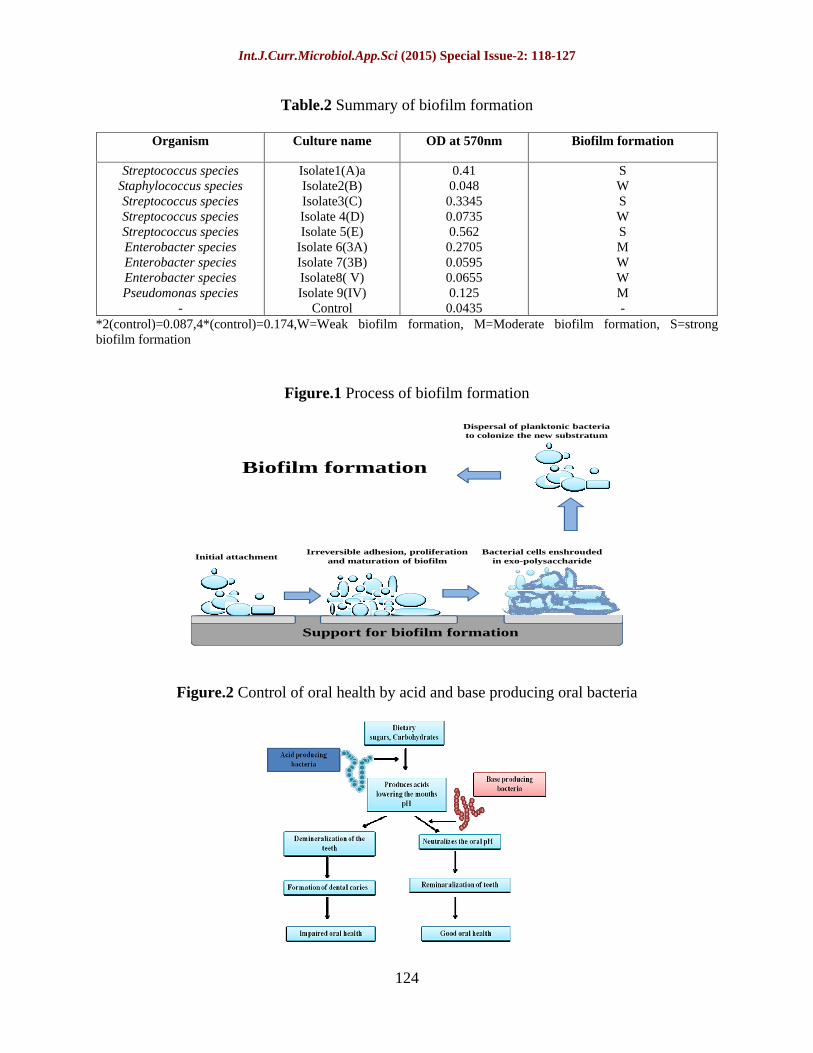
from harmful conditions (defense) (ii) sequestration to a nutrient-rich area (colonization) (iii) utilization of cooperative benefits (community) (4) biofilms normally grows as biofilms and planktonic cultures are an in vitro artifact. Different steps ensue, leading to the formation and maturation of the biofilm (Parsek and Fuqua, 2004; Kierek-Pearson and Karatan, 2005). Biofilm formation can be described in four steps as mention in figure 1. The major features that distinguish biofilm forming bacteria from their planktonic counterparts are their surface attachment ability, high population density, extracellular polymeric substances (EPS) slime and a wide range of physical, metabolic and chemical heterogeneities (Beer and Stoodley., 2006). It is now recognized that biofilm formation is an important aspect of many diseases including native valve endocarditis, osteomyelitis, dental caries, middle ear infections, ocular implant infections, and chronic lung infections in cystic fibrosis patients.
Dental biofilm forming organisms are major sources of the oral diseases like disease of the gums that is initiated or accelerated by subsets of microbes that colonize on or just below the gingival margin (Haffajee et al., 1998).
Oral biofilms mostly consists of multiple bacterial strains include mainly the Streptococcus species i.e. Streptococcus mutans which has major role in the formation of dental caries. In presence of sugar these bacteria ferment the dietary sugars producing acid and lowering the pH of the mouth, leading to demineralization of the teeth which is the initial step in oral biofilm formation. Whereas the mouth acidity is maintained by base producing bacteria like Streptococcus Sangius and Streptococcus Oralis which neutralize the acid produced maintaining the oral health. If
the balance between these two acid and base producing organisms is disturbed the oral health is hampered (Figure 2).
The present study aims at isolation, characterization of oral biofilm forming microorganisms from healthy individuals. The isolates were further characterized for various biochemical tests and biofilm forming ability was checked by Congo red agar method, tube assay and microtiter plate assay. Effect of various physiological parameters like pH, temperature and sucrose concentration was checked against biofilm formation.
Materials and Methods
Sample collection
The samples were collected from nine individuals independent of their age, sex and dietary habits. The swabs were taken from the entire teeth surface, gingival, sub-gingival and buccal cavity. The swab was inoculated in Nutrient broth (HIMEDIA), Mitis Salivarius broth (MitiSalivarius Medium gms/liter- Casein enzymatic hydrolysate 15 gms, Peptic digest of animal tissue 5 gms, Dextrose 1gms, Sucrose 50gms,Dipotassium phosphate 4gms,Trypan blue 0.075gms,Crystal violet 0.0008 gms Final pH 7.0±0.2) and tubes were incubated for 24 hours at 37°C.
Isolation of biofilm forming micro-organisms and morphological characterization
The turbidity was observed after 24 hours and samples were streaked on the nutrient agar and Mitis Salivarius agar. The random colonies were selected independent of their colony characteristics and pure cultures were obtained by frequent subculturing which were maintained on the Nutrient agar
Int.J.Curr.Microbiol.App.Sci (2015) Special Issue-2: 118-127
120
and Mitis Salivarius agar. Isolates were characterized morphologically by Gram s staining.
Biochemical characterization
The isolates were preceded for biochemical characterization including catalase test, IMViC test and sugar fermentation test. The hemolysis pattern was checked on Blood agar (HIMEDIA Blood agar base), supplemented with 5% sheep blood. Identification of pigment producing was performed by using King s B medium.
Detection of biofilm formation
Congo red agar method
This method is based on the characteristic cultural morphology of biofilm-forming bacteria on Congo red medium. The isolates were streaked on the Muller Hinton agar (HIMEDIA) supplemented with 0.8g/l of Congo red dye and incubated for 48 hours at 37°C. The production of black colonies with a dry crystalline consistency indicated biofilm formation and non-biofilm-producing strains develop red colonies (Mathur et al., 2006).
Tube assay
Qualitative assessment of biofilm formation was determined by the tube staining assay (Christensen et al., 1982). Isolates were inoculated (100 l) in 10ml trypticase soy broth (TSB) with 5% sucrose and incubated for 24 hours at 37°C at static and rotary condition. The tubes were decanted and washed with phosphate buffer saline (PBS) (pH 7.3), dried and Stained with 0.1% crystal violet. Excess stain was removed by washing the tubes with deionized water. Formation of biofilm was confirmed with the presence of visible film on the wall and
bottom of the tube. However, the liquid interface did not indicate biofilm formation (Mathur et al., 2006).
Microtiter plate assay
The quantitative estimation of the biofilm formation was done by Microtiter plate assay. The isolates were grown in trypticase soy broth (TSB) with 5% sucrose and incubated overnight at static condition at 37°C to obtain sufficient bacterial growth. The cultures after 24 hours were diluted 100 times with the same medium and 200µl of the culture were inoculated in the 96 well plate in triplicates. The 96 well plate was incubated for 24 hours in static condition at 37°C at static condition. After respective incubation period content of each well was gently removed by slightly tapping the plates. The wells were then washed with phosphate buffer saline (PBS pH 7.3) to remove free-floating planktonic bacteria. The plates were then stained with 0.1% (w/v) crystal violet solution. Excess stain was washed off thoroughly with 95% ethanol and plates were kept for drying. Optical density (OD) was measured using micro ELISA auto reader at wavelength of 570 nm. These OD values were considered as an index of attachment to surface. The experiment was performed in triplicates and average reading was considered.
Microscopic examination of the biofilm
The microscopic examination of the biofilm was done by using a tissue culture plate. The 3ml of trypticase soy broth (TSB) was inoculated with 100µl of overnight grown culture in tissue culture plate. The plates were incubated at 37°C at static condition for 24 hours. After incubation the plates were tapped and the medium was drained off. The plates were washed with phosphate buffer saline (pH 7.3) and stained with
Int.J.Curr.Microbiol.App.Sci (2015) Special Issue-2: 118-127
121
crystal violet; excess of the stain was washed off with deionized water. The plates were dried and examined microscopically.
Effect of pH, temperature and sucrose concentration on biofilm formation
Effect of pH on biofilm formation
Effect of pH was checked by using microtiter plate assay. The overnight grown culture was diluted (1:100) in trypticase soy broth (TSB) medium of various pH 4,7, and 10 and 200µl was inoculated and plates were incubated at 37°C for 24 hours, washed, stained and dried as mentioned earlier. The OD was checked on ELISA plate reader at 570 nm.
Effect of different sucrose concentration on biofilm formation
Effect of sucrose concentration of 2%, 5%, 10% and 15% was checked on biofilm formation. The overnight grown culture was diluted (1:100) in medium supplemented with different sucrose concentration and (200µl) inoculated. The plate was incubated at 37°C for 24 hours, washed, stained and dried. The OD was checked on ELISA plate reader at 570 nm.
Effect of temperature on biofilm formation
Effect of temperature was checked by inoculating 200µl of the diluted culture and incubated in various temperature of range 25°C, 37°C and 50°C for 24 hours. The plate was washed, stained and dried. The OD was checked on ELISA plate reader at 570 nm.
The data obtained were used to classify the strains as high biofilm producers (OD higher than 0.500), moderate (OD between 0.500
and 0.100) or poor producers (OD lower than 0.100).
Results and Discussion
Isolation and morphological characterization of biofilm forming bacteria
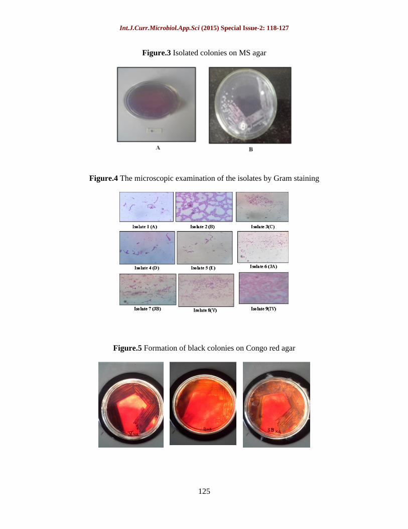
Nine different random colonies grown on both MS medium and nutrient agar were selected for characterization. Colony characteristics of five isolates (A,B,C,D,E) out of nine shows heaped, adherent morphology of smooth, glossy and mucoid colonies on Mitis Salivarius agar probably due to glucan synthesis (Figure 3), While four isolates (3A,3B,V,IV) showed round, flat, sticky colonies and one of the isolate IV shows green color pigment on the nutrient agar.
The microscopic observation revels that the four out of the nine isolates shows Gram positive chains while one of the isolate was Gram positive cluster, where as the other four shows Gram negative short rods (Figure 4).
Biochemical characterization
Results of the biochemical characterization are summarized in table 1. Sugar fermentation test was performed by using three sugars i.e. ribose, lactose and mannitol and was checked for acid and gas production. All the isolates were positive for different sugars for acid and gas production except isolate 9(IV) which shows no acid and gas production in lactose and mannitol.
Four Gram negative bacterial isolates were proceeded for IMViC test. Results are shown in table 1.
Int.J.Curr.Microbiol.App.Sci (2015) Special Issue-2: 118-127
122
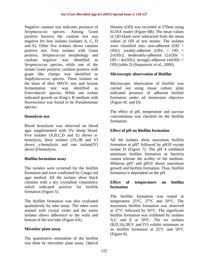
Negative catalase test indicates presence of Streptococcus species. Among Gram positive bacteria the catalase test was negative for four isolates (isolates A, C, D and E). Other five isolates shows catalase positive test. Four isolates with Gram positive, Streptococcus morphology and catalase negative was identified as Streptococcus species, while one of the isolate Gram positive, catalase positive with grape like clumps was identified as Staphylococcus species. Three isolates on the basis of their IMViC test and lactose fermentation test was identified as Enterobacter species. While one isolate indicated growth on King s B medium with fluorescence was found to be Pseudomonas species.
Hemolysis test
Blood hemolysis was observed on blood agar supplemented with 5% sheep blood. Five isolates (A,B,C,D and E) shows -hemolysis, three isolates (3A,3B and V) shows y-hemolysis and one isolate(IV) shows -hemolysis.
Biofilm formation assay
The isolates were screened for the biofilm formation and were confirmed by Congo red agar method. All the isolates show black colonies with a dry crystalline consistency which indicated positive for biofilm formation (Figure 5).
The biofilm formation was also evaluated qualitatively by tube assay. The tubes were stained with crystal violet and the entire isolates shows adherence to the walls and bottom of the test tube (Figure 6A).
Micotiter plate assay
The quantitative estimation of the biofilm was done by microtiter plate assay. Optical
Density (OD) was recorded at 570nm using ELISA reader (Figure 6B). The mean values of OD blank were subtracted from the mean values of OD of test strains. The isolates were classified into: non-adherent (OD < ODc); weakly-adherent (ODc < OD < 2xODc); moderately-adherent (2xODc < OD < 4xODc); strongly-adherent (4xODc < OD) (table 2) (Stepanovic et al., 2000).
Microscopic observation of Biofilm
Microscopic observation of biofilm was carried out using tissue culture plate indicated presence of adherent biofilm formation under oil immersion objective (Figure 6C and D).
The effect of pH, temperature and sucrose concentration was checked on the biofilm formation:
Effect of pH on Biofilm formation
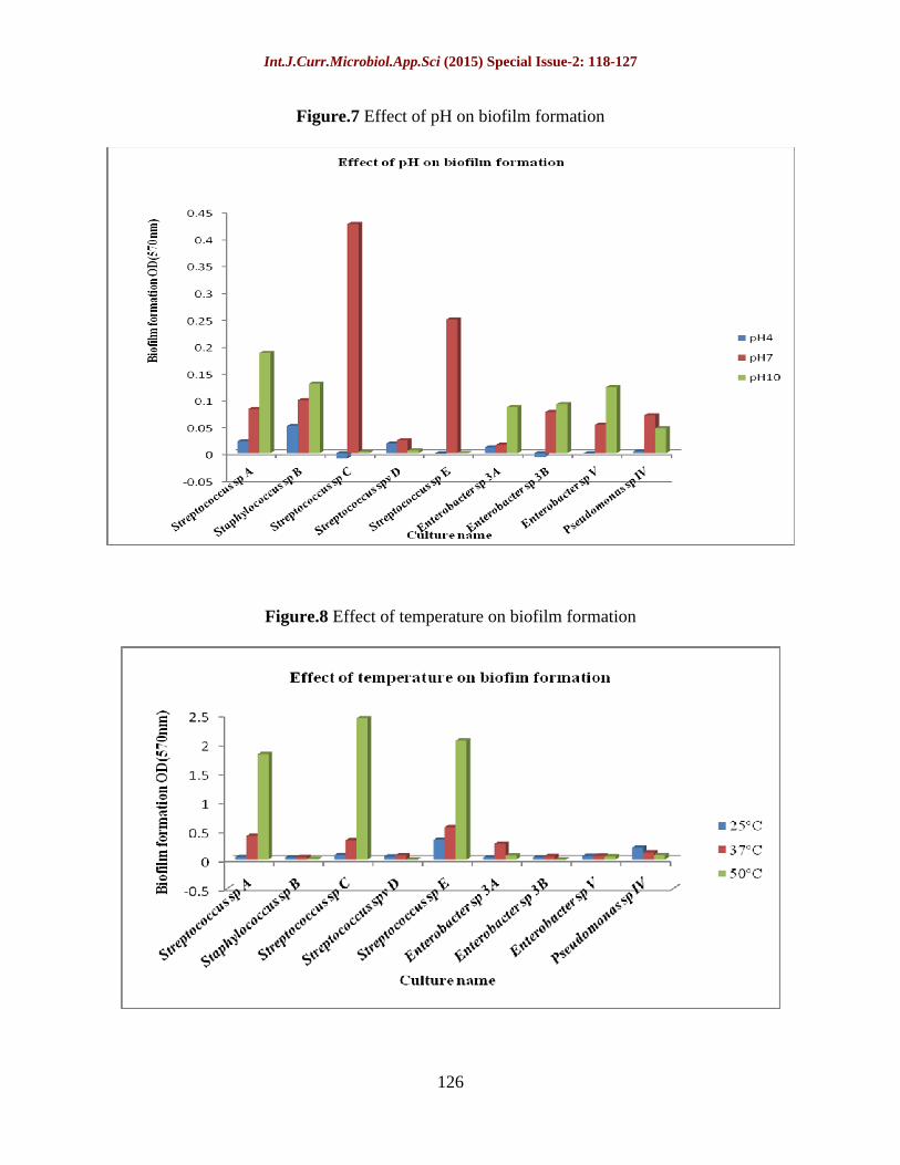
All the isolates show maximum biofilm formation at pH7 followed by pH10 except isolate D (Figure 7). The pH 4 exhibited minimum biofilm formation as bacteria cannot tolerate the acidity of the medium. Whereas pH7 and pH10 shows maximum growth and biofilm formation. Thus, biofilm formation is dependent on the pH.
Effect of temperature on biofilm formation
The biofilm formation was tested at temperature 25°C, 37°C and 50°C. The maximum biofilm formation was observed at 37°C followed by 50°C. The significant biofilm formation was exhibited by isolates A,C and E at 50°C. The six isolates (B,D,3A,3B,V and IV) exhibit minimum or no biofilm formation at 25°C and 50°C (Figure 8).

Int.J.Curr.Microbiol.App.Sci (2015) Special Issue-2: 118-127
123
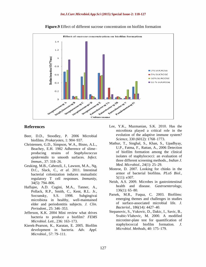
Effect of sucrose concentration on biofilm formation
The sucrose concentration is mainly responsible for the biofilm formation. Thus the effect of 2%, 5%, 10% and 15% sucrose concentration was checked on biofilm formation by the isolates. The strong biofilm formation was observed in three isolates (A, C and E) in all the sucrose concentrations, whereas maximum biofilm formation was in 15% sucrose concentration by these isolates. The five isolates (B, D, 3A, V and IV) exhibited weak to moderate biofilm formation.
The present study focuses on the isolation and characterization of the biofilm forming bacteria from oral microflora. The Streptococcus species is most dominating followed by Enterobacter species out of the total microflora. The qualitative and
quantitative estimation revels that oral microflora contains all four types of biofilm formers i.e. weak, moderate and strong biofilm formers. Results also indicated that the physiological factors like pH, temperature and sucrose concentration are essential for the biofilm formation. Thus, the dental caries can be controlled by changing the physiological conditions of the oral environment up to certain extent. However these bacteria may tolerate adverse conditions and continue to form biofilm formation. Due to the presence of these exo-polysaccharides, the bacterial cells are coated.
Thus, impaired and slow penetration of antibacterial agents becomes a challenge to control biofilms. Futuristic approaches of the study aims to screen the economical and feasible antibiofilm agents.
Table.1 The summery of biochemical characterization of the isolates
Culture name
Sugar fermentation test IMViC Test Catalase Test
Organism
Ribose
G/A
Lactose
G/A
Mannitol
G/A
Indole test
Methyl red test
VP test
Citrate utilization
test
Isolate1(A)
Isolate2(B)
Isolate3(C)
Isolate4(D)
Isolate5(E)
Isolate6(3A)
Isolate7(3B)
Isolate8(V)
Isolate9(IV)
++
++
++
++
++
++
++
++
++
++
++
++
++
++
++
++
++
--
++
++
++
++
++
++
++
++
--
NA
NA
NA
NA
NA
-
-
-
-
NA
NA
NA
NA
NA
-
-
-
-
NA
NA
NA
NA
NA
+
+
+
+
NA
NA
NA
NA
NA
+
+
+
+
-
+
_
_
_
+
+
+
+
Streptococcus species
Staphylococcus species
Streptococcus species
Streptococcus species
Streptococcus species
Enterobacter species
Enterobacter species
Enterobacter species
Pseudomonas species
(*NA=Not applicable)
Int.J.Curr.Microbiol.App.Sci (2015) Special Issue-2: 118-127
124
Table.2 Summary of biofilm formation
Organism Culture name OD at 570nm Biofilm formation
Streptococcus species Staphylococcus species Streptococcus species Streptococcus species Streptococcus species Enterobacter species Enterobacter species Enterobacter species Pseudomonas species
-
Isolate1(A)a Isolate2(B) Isolate3(C) Isolate 4(D) Isolate 5(E)
Isolate 6(3A) Isolate 7(3B) Isolate8( V) Isolate 9(IV)
Control
0.41 0.048
0.3345 0.0735 0.562
0.2705 0.0595 0.0655 0.125
0.0435
S W S W S M W W M -
*2(control)=0.087,4*(control)=0.174,W=Weak biofilm formation, M=Moderate biofilm formation, S=strong biofilm formation
Figure.1 Process of biofilm formation
Support for biofilm formation
Initial attachmentIrreversible adhesion, proliferation
and maturation of biofilmBacterial cells enshrouded
in exo-polysaccharide
Dispersal of planktonic bacteria to colonize the new substratum
Biofilm formation
Figure.2 Control of oral health by acid and base producing oral bacteria
Int.J.Curr.Microbiol.App.Sci (2015) Special Issue-2: 118-127
125
Figure.3 Isolated colonies on MS agar
Figure.4 The microscopic examination of the isolates by Gram staining
Figure.5 Formation of black colonies on Congo red agar
Int.J.Curr.Microbiol.App.Sci (2015) Special Issue-2: 118-127
126
Figure.7 Effect of pH on biofilm formation
Figure.8 Effect of temperature on biofilm formation
Int.J.Curr.Microbiol.App.Sci (2015) Special Issue-2: 118-127
127
Figure.9 Effect of different sucrose concentration on biofilm formation
References
Beer, D.D., Stoodley, P. 2006 Microbial biofilms. Prokaryotes, 1: 904 937.
Christensen, G.D., Simpson, W.A., Bisno, A.L., Beachey, E.H. 1982 Adherence of slimeproducing strains of Staphylococcus epidermidis to smooth surfaces. Infect. Immun., 37: 318 26.
Geuking, M.B., Cahenzli, J., Lawson, M.A., Ng, D.C., Slack, C., et al. 2011. Intestinal bacterial colonization induces mutualistic regulatory T cell responses. Immunity, 34(5): 794 806.
Haffajee, A.D. Cugini, M.A., Tanner, A., Pollack, R.P., Smith, C., Kent, R.L. Jr., Socransky, S.S. 1998. Subgingivai microbiota in healthy, well-maintained elder and periodontitis subjects. J. Clin. Periodont., 25: 346 353.
Jefferson, K.K. 2004 Mini review what drives bacteria to produce a biofilm? FEMS Microbiol. Lett., 236: 163 173.
Kierek-Pearson, K., Karatan, E. 2005. Biofilm development in bacteria. Adv. Appl. Microbiol., 57: 79 111.
Lee, Y.K., Mazmanian, S.K. 2010. Has the microbiota played a critical role in the evolution of the adaptive immune system? Science, 330 (6012): 1768 1773.
Mathur, T., Singhal, S., Khan, S., Upadhyay, U.P., Fatma, F., Rattan, A., 2006 Detection of biofilm formation among the clinical isolates of staphylococci: an evaluation of three different screening methods., Indian J. Med. Microbiol., 24(1): 25 29.
Monroe, D. 2007. Looking for chinks in the armor of bacterial biofilms. PLoS Biol., 5(11): e307.
Neish, A.S. 2009. Microbes in gastrointestinal health and disease. Gastroenterology, 136(1): 65 80.
Parsek, M.R., Fuqua, C. 2003. Biofilms: emerging themes and challenges in studies of surface-associated microbial life. J. Bacteriol., 186(14): 4427 40.
Stepanovic, S., Vukovic, D., Dakic, I., Savic, B., Svabic-Vlahovic, M. 2000. A modified microtiter-plate test for quantification of staphylococcal biofilm formation. J. Microbiol. Methods, 40: 175 179.