isolation and characterization of cellulose nanofibers ... › pubs › ja › 2016 ›...
TRANSCRIPT

Iuu
JTa
b
c
a
ARRAA
KCMBU
1
iitifebmatcg2rh
h0
Carbohydrate Polymers 151 (2016) 725–734
Contents lists available at ScienceDirect
Carbohydrate Polymers
journa l homepage: www.e lsev ier .com/ locate /carbpol
solation and characterization of cellulose nanofibers from bamboosing microwave liquefaction combined with chemical treatment andltrasonication
iulong Xie a,c, Chung-Yun Hse b, Cornelis F. De Hoop c, Tingxing Hu a, Jinqiu Qi a,∗,odd F. Shupe c
College of Forestry, Sichuan Agricultural University, Chengdu, Sichuan 611130, ChinaSouthern Research Station, USDA Forest Service, Pineville, LA 71360, USASchool of Renewable Natural Resource, Louisiana State University Agricultural Center, Baton Rouge, LA 70803, USA
r t i c l e i n f o
rticle history:eceived 14 March 2016eceived in revised form 4 May 2016ccepted 2 June 2016vailable online 3 June 2016
eywords:
a b s t r a c t
Cellulose nanofibers were successfully isolated from bamboo using microwave liquefaction combinedwith chemical treatment and ultrasonic nanofibrillation processes. The microwave liquefaction couldeliminate almost all the lignin in bamboo, resulting in high cellulose content residues within 7 min, andthe cellulose enriched residues could be readily purified by subsequent chemical treatments with lowerchemical charging and quickly. The results of wet chemistry analyses, SEM images, and FTIR and X-rayspectra indicated the combination of microwave liquefaction and chemical treatment was significantly
ellulose nanofibersicrowave liquefaction
ambooltrasonic nanofibrillation
efficient in removing non-cellulosic compounds. Ultrasonication was used to separate the nanofibrilsfrom the purified residues to extract nanofibers. The TEM images confirmed the presence of elementaryfibrils, nano-sized fibril bundles, and aggregated fibril bundles. As evidenced by the TGA analysis, cellu-lose nanofibers isolated by this novel technique had high thermal stability indicating that the isolatednanofibers could possibly be applied as reinforcing elements in biomaterials.
© 2016 Elsevier Ltd. All rights reserved.
. Introduction
Renewable and biodegradable lignocellulosic biomass is of greatnterest because of depleting fossil fuel reserves and increas-ng public concern for environmental stewardship. Cellulose ishe main component of lignocellulosic biomass and has receivedncreasing attention due to its wide existence, excellent per-ormance, biocompatibility, low density, thermal stability, andnvironmental benefits (Fatah et al., 2014). Recently, cellulose haseen considered as a promising resource for reinforcing poly-er matrixes for the preparation of green sustainable materials
nd bioethanol production. Regardless of its potential applica-ions in emerging bio-based materials and bioenergy industries,ellulose consists of a linear homopolysaccharide composed of �-d-lucopyranose units linked together by �-1-4-linkages (Khalil et al.,
014). The cellulose molecules are aggregated into elemental fib-ils with diameter ranges of 2–5 nm by intermolecular forces andydrogen bonds (Hideno, Abe, & Yano, 2014). Therefore, from its∗ Corresponding author.E-mail address: [email protected] (J. Qi).
ttp://dx.doi.org/10.1016/j.carbpol.2016.06.011144-8617/© 2016 Elsevier Ltd. All rights reserved.
unique biological structure, it is reasonable that various nanometersized single fibers that are also referred to as cellulose nanocrys-tals, whiskers, nanowhiskers, cellulose nanofibrils, microfibrillatedcellulose, or nanofibers have been isolated (Khawas & Deka, 2016;Shinoda, Saito, Okita, & Isogai, 2012; Sacui et al., 2014; Wu et al.,2013; Yue et al., 2012).
With the growing demand for sustainable green materials, iso-lation of cellulose nanofibers has been extensively investigated(Khalil et al., 2014; Khalil, Bhat, & Yusra, 2012). Nanofibers havebeen widely used for the preparation of nanostructured biocom-posites (Ansari, Skrifvars, & Berglund, 2015; Awal, Rana, & Sain,2015; Silverio, Neto, Dantas, & Pasquini, 2013), tough hydrogels(Abe, Ifuku, Kawata, & Yano, 2014), membranes (Ma, Burger, Hsiao,& Chu, 2014), and transparent nanopaper or film (Qing et al.,2015; Sun, Wu, Ren, & Lei, 2015) owing to its high crystalline withstrong mechanical performance and outstanding thermal stabil-ity (Tibolla, Pelissari, & Menegalli, 2014). Over the past decades,a large number of lignocellulosic biomass has been explored for
the generation of cellulose nanofibers (Abe, Iwamoto, & Yano,2007). The common raw materials used for the production of cel-lulose nanofibers are wood (Wang, Li, Yano, & Abe, 2014), bamboo(Lu, Lin et al., 2015), wheat straw (Chen et al., 2011), Phormium
7 Polym
tPeMIL
cTSB&T(Tmmgp(s
fehdAlr
cafHatZftncfenwtawrTaiiaatla
ttlrTni
26 J. Xie et al. / Carbohydrate
enax (Fortunati et al., 2013), banana peels (Khawas & Deka, 2016;elissari, Sobral, & Menegalli, 2014), orange peel waste (Hidenot al., 2014), date palm fruit stalks (Hassan, Bras, Hassan, Silard, &auret, 2014), oil palm empty fruit bunch (Fahma, Iwamoto, Hori,
wata, & Takemura, 2010), de-pectinated sugar beet pulp (Li, Wang,i, Cheng, & Adhikari, 2014).
Usually, pretreatment and nanofibrillation are essential pro-esses for the isolation of cellulose nanofibers from plant fibers.he pretreatment process such as acid or alkali treatment (He, Jiang,un, & Xu, 2014; Yue et al., 2015), enzymatic pretreatment (Hassan,ras, Hassan, Silard, & Mauret, 2014; Tibolla et al., 2014; Zhu, Sabo,
Luo, 2011), ionic liquids (Han, Zhou, French, Han, & Wu, 2013),EMPO (2,2,6,6-tetramethylpiperidine-1-oxyl) mediated oxidationJausovec, Vogrincic, & Kokol, 2015; Shimizu, Saito, & Isogai, 2014;akaichi, Saito, Tanaka, & Isogai, 2014), and steam explosion treat-ent (Chirayil et al., 2014) were first used to remove non-cellulosicaterials in plants, and then nanofibrillation technologies such as
rinders (Jang, Lee, Endo, & Kim, 2013; Yousefi et al., 2013), high-ressure homogenizers (Yue et al., 2012), and ultrasonic methodTang, Yang, Zhang, & Zhang, 2014) were used to generate highhear forces to separate the fibrils from the purified cellulose fibers.
Though cellulose nanofibers have been successfully isolatedrom these aforementioned technologies and the results werencouraging, drawbacks still exist such as chemical regent cost,igh energy consumption, time-consuming, and equipment degra-ation has limited these techniques for practical applications.dvanced techniques for production of cellulose nanofibers with
ow cost, environmentally friendly, and time efficiency are stillequired.
As an efficient method, liquefaction has been applied in theonversion of solid woody materials into soluble liquid products,nd the liquid products have shown great potential as alternativesor petroleum to produce value-added bio-based products (Xie, Qi,se, & Shupe, 2014). In recent years, microwave energy has beenpplied in the liquefaction of lignocellulosic biomass to enhancehe biomaterials industry (Xie, Hse, Shupe, Qi, & Pan, 2014; Xie,hai, Hse, Shupe, & Pan, 2015). Compared to conventional lique-action, microwave-assisted liquefaction has advantages such asime efficiency, low chemicals and energy consumption, and eco-omically viability. The products from this technique also showedomparable properties compared to the commercialized ones. Asor the liquefaction of wood residues in the binary of glycerol andthylene glycerol, lignin and hemicellulose in the wood were sig-ificantly decomposed in the initial liquefaction stage (20–40 min)ith the reaction temperature of 160 ◦C, while the cellulose con-
ent in the wood increased (Zhang, Pang, Shi, Fu, & Liao, 2012). Panlso observed that the lignin content in the liquefied Chinese tallowood residue significantly decreased as the liquefaction processes
esulting in enriched cellulose residues (Pan, Shupe, & Hse, 2007).he lignin content in the microwave liquefied bamboo residueslso showed significantly decrease as the reaction temperaturencreased from 75 ◦C to 120 ◦C (Xie, Hse et al., 2014). Therefore,t could be concluded that in the reactions of both conventionalnd microwave-assisted liquefaction of lignocellulosic biomass inn organic solvent with acid as a catalyst, the decomposition of thehree major components (cellulose, lignin, and hemicellulose) inignocellulosic biomass was in the order of lignin, hemicellulose,nd cellulose.
Based on this mechanism, Chen proposed a selective liquefac-ion process for the production of cellulose and biobased resins,he finding in his research showed that large amount of hemicel-ulose and lignin could be liquefied at 100 ◦C in 30 min, and the
etained cellulose had higher susceptibility for enzymatic attack.his approach offered a new approach for the utilizations of lig-ocellulosic biomass in the bioethanol and biobased materialsndustries (Chen, Zhang, & Xie, 2012). In order to extract cellulose
ers 151 (2016) 725–734
for use in both bio-fuels and reinforcing materials, the combinationof bleach and liquefaction processes was described in the researchof Li et al. (2015), in which hemicellulose was selectively liquefiedto get cellulose. From the previous research results, it was obvi-ous that cellulosic fibers could be easily produced by subjecting theraw lignocellulosic biomass to a liquefaction process with reactionsconditions properly controlled.
Therefore, in this study microwave liquefaction was proposedto generate cellulose enriched residues from bamboo. Thereafter,the cellulose enriched residues were chemically purified withlow charging of chemicals. Cellulose nanofibers were isolatedby ultrasonic nanofibrillation of the chemically purified cellulosefibers. Morphology, crystallinity, and thermal stability of the iso-lated nanofibers were determined by employing scanning electronmicroscopy, X-ray diffraction, and thermogravimetry, respectively.This study attempts to achieve an efficient approach for the gen-eration of cellulose nanofibers with the combination of microwaveliquefaction and ultrasonic nanofibrillation.
2. Materials and methods
The major procedures for isolation of nanofiber in this studyincluded reduction of bamboo, microwave liquefaction, anti-solvent fractionation, chemical treatments, and ultrasonication.Fig. 1 depicts the flow chart of the procedures.
2.1. Materials
Three-year-old moso bamboo culms (Phyllostachys pubescens)were harvested from the Kisatchie National Forest, Pineville, La,USA. The moso bamboo was selected because the annual yield ofmoso bamboo was greater than all other kinds of bamboo (about1.8 × 107 tons) and the integrated utilizations of moso bamboo vialiquefaction may provide potential approach for the productionof high-value added bioproducts (Jiang et al., 2012). The bambooculms were reduced to particles using a Thomas Wiley LaboratoryMill (Model 4) equipped with a 2 mm screen. The particles (length:0.05–6.23 mm, diameter: 0.01–1.1 mm) were screened to collectparticles that passed through a 20-mesh sieve and then retained ona 40-mesh sieve. The average length and diameter of the particles of20–40 mesh were 5.06 mm and 0.567 mm, respectively. The weightpercentage of particles between 20 mesh and 40 mesh was 25.20%,and that for particles in the range of 10–20 mesh, 40–60 mesh,60–80 mesh, 80–100 mesh, and >100 mesh were 0.22%, 44.42%,10.08%, 5.86%, and 14.22%, respectively. The particles were driedto a constant weight in an oven maintained at 80 ◦C. The dried par-ticles were stored in polyethylene bags and used without furthertreatment. All chemicals including acids, glycerol, and methanolwere of reagent grade and obtained from commercial sources.
2.2. Microwave liquefaction
Microwave liquefaction of bamboo was carried out in a Mile-stone laboratory microwave oven (Ethos EX, 1200 W maximummicrowave power, 100 bar maximum operating pressure, ASM-400 magnetic stirrer for homogenous mixing of samples, 300 ◦Cmaximum operating temperature, fiber-optic temperature sensor)equipped with 100 mL sealed Teflon reaction vessels that are actu-ally a system of components that consist of a vessel that containsthe sample, a vessel cover, a safety shield, a vent indicator ring, anda pressure adapter plate. The Teflon vessels used in this study have
excellent properties with regard to temperature, acid, and pres-sure resistance, which was suitable for the chemicals used in thisresearch. The application of this vessel system also enables that themicrowave system could provide a closed, clean environment, pre-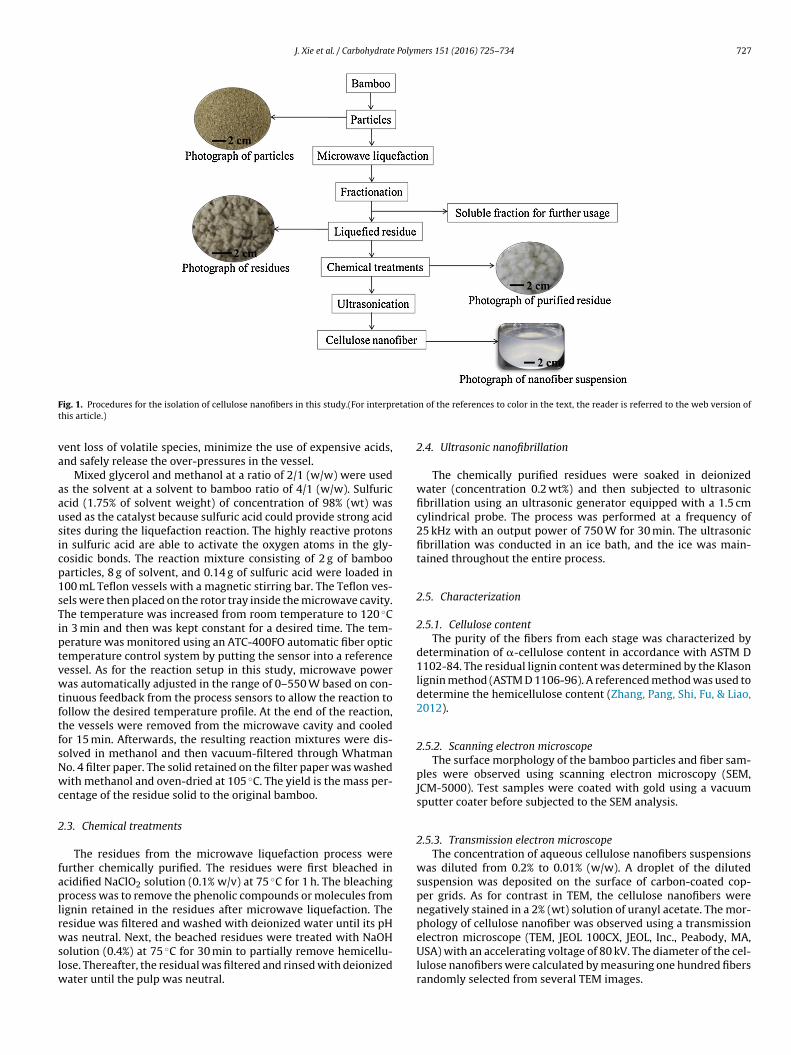
J. Xie et al. / Carbohydrate Polymers 151 (2016) 725–734 727
F etatiot
va
aausicp1sTiptvwtftfsNwc
2
faplrwslw
ig. 1. Procedures for the isolation of cellulose nanofibers in this study.(For interprhis article.)
ent loss of volatile species, minimize the use of expensive acids,nd safely release the over-pressures in the vessel.
Mixed glycerol and methanol at a ratio of 2/1 (w/w) were useds the solvent at a solvent to bamboo ratio of 4/1 (w/w). Sulfuriccid (1.75% of solvent weight) of concentration of 98% (wt) wassed as the catalyst because sulfuric acid could provide strong acidites during the liquefaction reaction. The highly reactive protonsn sulfuric acid are able to activate the oxygen atoms in the gly-osidic bonds. The reaction mixture consisting of 2 g of bambooarticles, 8 g of solvent, and 0.14 g of sulfuric acid were loaded in00 mL Teflon vessels with a magnetic stirring bar. The Teflon ves-els were then placed on the rotor tray inside the microwave cavity.he temperature was increased from room temperature to 120 ◦C
n 3 min and then was kept constant for a desired time. The tem-erature was monitored using an ATC-400FO automatic fiber opticemperature control system by putting the sensor into a referenceessel. As for the reaction setup in this study, microwave poweras automatically adjusted in the range of 0–550 W based on con-
inuous feedback from the process sensors to allow the reaction toollow the desired temperature profile. At the end of the reaction,he vessels were removed from the microwave cavity and cooledor 15 min. Afterwards, the resulting reaction mixtures were dis-olved in methanol and then vacuum-filtered through Whatmano. 4 filter paper. The solid retained on the filter paper was washedith methanol and oven-dried at 105 ◦C. The yield is the mass per-
entage of the residue solid to the original bamboo.
.3. Chemical treatments
The residues from the microwave liquefaction process wereurther chemically purified. The residues were first bleached incidified NaClO2 solution (0.1% w/v) at 75 ◦C for 1 h. The bleachingrocess was to remove the phenolic compounds or molecules from
ignin retained in the residues after microwave liquefaction. Theesidue was filtered and washed with deionized water until its pH
as neutral. Next, the beached residues were treated with NaOHolution (0.4%) at 75 ◦C for 30 min to partially remove hemicellu-ose. Thereafter, the residual was filtered and rinsed with deionized
ater until the pulp was neutral.
n of the references to color in the text, the reader is referred to the web version of
2.4. Ultrasonic nanofibrillation
The chemically purified residues were soaked in deionizedwater (concentration 0.2 wt%) and then subjected to ultrasonicfibrillation using an ultrasonic generator equipped with a 1.5 cmcylindrical probe. The process was performed at a frequency of25 kHz with an output power of 750 W for 30 min. The ultrasonicfibrillation was conducted in an ice bath, and the ice was main-tained throughout the entire process.
2.5. Characterization
2.5.1. Cellulose contentThe purity of the fibers from each stage was characterized by
determination of �-cellulose content in accordance with ASTM D1102-84. The residual lignin content was determined by the Klasonlignin method (ASTM D 1106-96). A referenced method was used todetermine the hemicellulose content (Zhang, Pang, Shi, Fu, & Liao,2012).
2.5.2. Scanning electron microscopeThe surface morphology of the bamboo particles and fiber sam-
ples were observed using scanning electron microscopy (SEM,JCM-5000). Test samples were coated with gold using a vacuumsputter coater before subjected to the SEM analysis.
2.5.3. Transmission electron microscopeThe concentration of aqueous cellulose nanofibers suspensions
was diluted from 0.2% to 0.01% (w/w). A droplet of the dilutedsuspension was deposited on the surface of carbon-coated cop-per grids. As for contrast in TEM, the cellulose nanofibers werenegatively stained in a 2% (wt) solution of uranyl acetate. The mor-phology of cellulose nanofiber was observed using a transmission
electron microscope (TEM, JEOL 100CX, JEOL, Inc., Peabody, MA,USA) with an accelerating voltage of 80 kV. The diameter of the cel-lulose nanofibers were calculated by measuring one hundred fibersrandomly selected from several TEM images.
7 Polym
2
tR4
2
ad(aal
C
w22
2
(2wfl
3
3
maTrsTsltcthtrccTtprf7t
u(rracr8
28 J. Xie et al. / Carbohydrate
.5.4. Fourier transform infrared spectroscopyThe FT-IR analysis was performed by a Nicolet Nexus 670 spec-
rometer equipped with a Thermo Nicolet Golden Gate MKII Singleeflection ATR accessory. Data collection was performed with a
cm−1 spectral resolution and 32 scans were taken per sample.
.5.5. X-ray diffractionCrystallinity of the original bamboo and fiber samples were
nalyzed using wide angle X-ray diffraction (Bruker D 5000). Theata were generated by a diffractormeter with Cu K� radiation� = 1.54 A) at 40 kV and 30 mA over the angular range 2� = 5–40◦
nd a step time of 2.0 s. A focusing powder diffraction method waspplied. The crystallinity index (CrI) was determined using the fol-owing equation:
rI(%) = I002 − Iam
I002× 100 (1)
here I002 is the intensity of the diffraction from the (200) plane at� = 22.1◦, and Iam is the intensity for amorphous material taken at� = 18◦ (French, 2014).
.5.6. Thermogravimetric analysisTG/DTG analysis was conducted with a thermal analyzer, TGA
Q50), to simultaneously obtain thermogravimetric data. About mg of sample was analyzed by the thermal analyzer. Pyrolysisas terminated at 800 ◦C with a heating rate of 20 ◦C/min under aow of 60 mL/min of nitrogen gas.
. Results and discussion
.1. Liquefaction and preparation of cellulose fibers
Cellulose fibers were isolated by the combination of aicrowave liquefaction process combined with bleaching and
lkali treatment prior to subjecting to ultrasonic nanofribillation.able 1 illustrates the yield and chemical compositions of theesidues after the microwave liquefaction and chemical treatmenttages. The residue yield decreased with increasing reaction time.he residual lignin content in the liquefied residues showed aignificant decrease with respect to reaction time, while the cel-ulose content significantly increased. Table 1 also indicated thathe liquefaction of bamboo could also partially eliminate the hemi-ellulose. However, the hemicellulose content first increased andhen decreased with increasing liquefaction time. The increase inemicellulose content in the initial reaction stage was attributedo the significant weight loss of lignin, and thereafter enriched theesidue resulting in higher (relatively) cellulose and hemicelluloseontent, and the decrease in hemicellulose as the liquefaction pro-essing was mainly due to the degradation of hemicellulose itself.he decrease in residue yield induced by prolonging the reactionime is mainly due to the efficient removal of non-cellulosic com-onents such as lignin and hemicellulose from bamboo, and thusesulting in the increase in cellulose content. The residues obtainedrom the liquefaction at 120 ◦C/7 min had a cellulose content of0.74%, which was about 170% of that for the original bamboo, andhe residual lignin content was as low as 1.66%.
However, the residues with high cellulose content from the liq-efaction process at 120 ◦C/7 min showed a light brownish colorFig. 1), indicating that a small amount of lignin was retained in theesidue as confirmed by the above mentioned chemical analysisesults. To further purify the fibers from the liquefaction process
t 120 ◦C/7 min, chemical treatments were conducted in sodiumhlorite and alkali solution (0.4%). After chemical treatment, theesidues became pure white, and the cellulose content increased to3.67%, and the lignin content was as low as 0.13%. The chemicallyers 151 (2016) 725–734
purified residues obtained in this study showed comparable cellu-lose content as that reported in Chen’s report (Chen et al., 2011),i.e., �-cellulose content for chemically purified bamboo fiber was84.4%. Moreover the time required for such purity of cellulose fiberswas mere 1/8 of that in Chen’s study. Chemically purified celluloseas reported in Chen’s study was obtained from a process involvingdewaxing, bleaching, alkali treatment in 16 h. Furthermore, afterliquefaction, the chemical usage for further purifying residues wasmuch lower than that used in previously reported methods (Wuet al., 2013), in which dewaxed samples were bleached three timein 1.4% acidified NaClO2 followed by alkali treatment with 5% KOHfor 4 h.
3.2. Morphological observation
SEM images of the raw bamboo particles and samples frommicrowave liquefaction and chemical treatments are presented inFig. 2. The original bamboo showed large fiber bundles and intactstructures. Some small fragments were attached on the uneven sur-faces (Fig. 2a and b). The diameter of the raw bamboo sample was567 ± 149 �m. The morphological structure of the residues frommicrowave liquefaction differed significantly from that of the orig-inal bamboo. After 7 min of reaction time, the fiber bundles wereseparated into individual micro-sized fibers with a diameter of17 ± 6.9 �m. The separation of fasciculus induced by microwaveliquefaction is mainly attributed to the removal of lignin in themiddle lamella, resulting in the collapse of the compact bamboostructure. It was obvious that the microwave liquefaction was effi-cient in defibrillation of bamboo by dissociating and dissolving thecementing components. As can be seen in Fig. 2d, small granules(1–2 �m) were found on the surface of the residues. These gran-ules were ascribed to lignin substrates. Similar granules were alsoobserved on the residues from microwave liquefaction of bamboofor biopolyols, and the granules were confirmed as lignin substratesby FTIR analysis (Xie, Hse et al., 2014; Xie, Huang, Qi, Hse, & Shupe,2014). This result indicated that a small amount of lignin was stillretained on the residues, which was consistent with results fromthe wet chemistry analysis as discussed above.
After bleaching due to the complete elimination of lignin onthe fiber surface and in the fiber cell wall, the micro-sized fiberswere opened as evidenced by fissures (Fig. 2e). Meanwhile the fibersurface became rough, and microfibrils bundles (0.5–1.0 �m) thatwere composed of nano-sized fibrils were obviously visible andbegan to peel off from the micro-sized fibers (Fig. 2f). Compared tothe fibers from the bleached residues, fibers from the alkali treatedresidues became irregular and were much rougher because of thepartial removal of hemicellulose (Fig. 2g). Curled and flat fiber cellswith many fibril bundles attached were observed in the SEM imagesof the alkali treated residues (Fig. 2g). From the SEM images of thealkali treated fibers, it seemed that the nano-sized fibril bundlesattached to each other without any non-cellulosic cements fillingin the space (Fig. 2h). The ultrasonic treatments resulted in thebreakdown of the hydroxyl bonds and nano-sized fibril bundlesand individual nano fibrils were released as evident from the TEMimages.
The obtained cellulose nanofiber aqueous suspension wasdiluted and characterized by TEM. The TEM images confirmed thepresence of individual nanofibers (Fig. 3a). As shown in Fig. 3b, indi-vidual long fiber-like nano fibrils were entangled together forminga web-like structure. An interconnected network of nanofiberscould provide great reinforcing capability for composite applica-tions (Bhatnagar & Sain, 2005); therefore, the nanofibers obtained
in this study are of particular interest for future research in thisregard. It is interesting to note that an opening of a fibril bundle(19 nm) is clearly shown in Fig. 3c. The diameters of the split singlefibrils in the opening position were 2–5 nm, which was in the diam-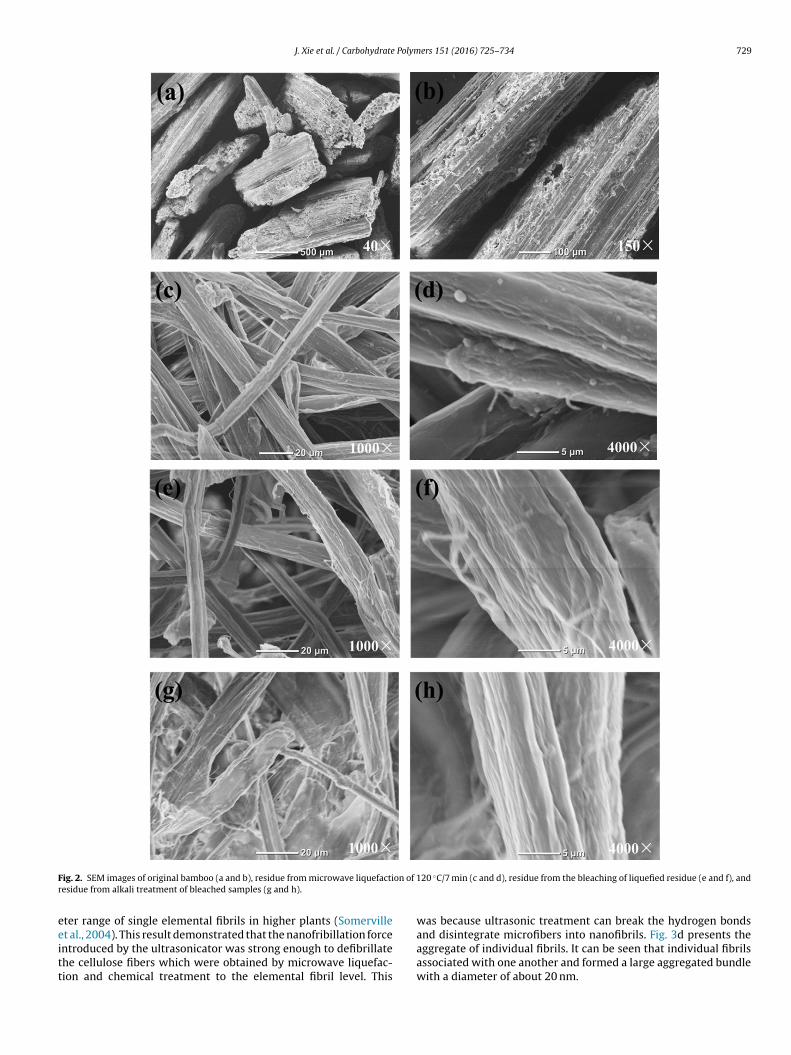
J. Xie et al. / Carbohydrate Polymers 151 (2016) 725–734 729
F on of 1r
eeitt
ig. 2. SEM images of original bamboo (a and b), residue from microwave liquefactiesidue from alkali treatment of bleached samples (g and h).
ter range of single elemental fibrils in higher plants (Somerville
t al., 2004). This result demonstrated that the nanofribillation forcentroduced by the ultrasonicator was strong enough to defibrillatehe cellulose fibers which were obtained by microwave liquefac-ion and chemical treatment to the elemental fibril level. This20 ◦C/7 min (c and d), residue from the bleaching of liquefied residue (e and f), and
was because ultrasonic treatment can break the hydrogen bonds
and disintegrate microfibers into nanofibrils. Fig. 3d presents theaggregate of individual fibrils. It can be seen that individual fibrilsassociated with one another and formed a large aggregated bundlewith a diameter of about 20 nm.
730 J. Xie et al. / Carbohydrate Polymers 151 (2016) 725–734
F re, (c)
(wo
ig. 3. TEM images of (a) individual/elemental fibril, (b) nanofiber network structu
The TEM images were further processed with analysis softwareImageJ) to determine the diameter. The diameter of the nanofibersas in the range of 2–30 nm. For comparison, the nanofibers
btained in this study had a similar diameter to the cellulose
opening of fibril bundle, (d) aggregated bundle, and (e) diameter distribution.
nanofibers isolated from bamboo in other studies; the diameterfor nanofibers prepared by using mechanochemical and chemical-ultrasonic processes were 15–30 nm and 10–40 nm, respectively(Chen et al., 2011; Lu, Tang et al., 2015). Fig. 3e presents the

J. Xie et al. / Carbohydrate Polymers 151 (2016) 725–734 731
Table 1Yield and chemical compositions of residues at each stage of the treatments.
Samples Residue yield (%) Cellulose (%) Hemicellulose (%) Klason lignin (%)
Original bamboo 100.00 ± 0.00 41.72 ± 2.37 22.86 ± 2.19 20.91 ± 0.24120 ◦C/3 min 60.95 ± 3.42 52.85 ± 3.16 25.49 ± 3.06 7.44 ± 0.61120 ◦C/5 min 45.99 ± 1.87 66.74 ± 2.09 19.90 ± 1.58 3.82 ± 0.05120 ◦C/7 min 42.28 ± 2.66 70.74 ±
Bleaching 37.51 ± 1.47 75.30 ±
Alkali treatment 34.17 ± 1.66 83.67 ±
Fb
dawtrfib
pwtipmsos
3
finr2C
fataam
ig. 4. FTIR spectra of (a) original bamboo, (b) microwave liquefied residue, (c)leached residue, (d) alkali treated residue, and (e) cellulose nanofiber.
iameter distribution of the nanofibers. About 70% of fibers have diameter within 2–10 nm, 22% of the fibers had a diameterithin 12–20 nm, and only 9% of the fibers had a diameter greater
han 20 nm. The TEM images and the diameter distribution resultsevealed that the aqueous suspension contained nanometer scalebers comprised of a large number of elemental fibrils, some fibrilundles, and a small amount of aggregated bundles.
There was no precipitate formed in the nanofiber aqueous sus-ension after setup for 24 h, indicating that the nanofiber samplesere still stable after setup for 24 h (Fig. 1). This may be attributed
o the strong fibrillation forces induced by high-intensity ultrason-cation. The introduction of sulphate groups during the liquefactionrocess that was confirmed by the FTIR spectra as discussed lateray also contribute to the stability of the nanofiber suspensions
ince the sulphate groups could provide negative electrostatic layern the surface of the nanofibers resulting in the homogeneous andtable aqueous suspensions (Bondeson & Oksman, 2007) .
.3. FTIR spectroscopy analysis
The FTIR spectra of the original bamboo, microwave lique-ed residue, bleached residue, alkali treated residue, and celluloseanofiber are presented in Fig. 4. The absorbance peak at 3330 cm−1
epresents the stretching vibration of OH and the intensity peak at890 cm−1 was attributed to the asymmetric stretching vibration ofH2 in cellulose, hemicellulose, and lignin were found in all spectra.
The spectrum of the original bamboo showed a significant dif-erence from that of the microwave liquefied residue. The peakst 1735 cm−1 (attributed to the acetyl and uronic ester groups or
he ester linkage of the carboxylic group of ferulic and p-coumariccid of hemicellulose), 1596 cm−1 and 1506 cm−1 (arising from theromatic skeletal vibration), 1456 cm−1 (assigned to C H defor-ation combined with aromatic ring vibration), and 1230 cm−11.78 18.85 ± 1.74 1.66 ± 0.151.31 18.53 ± 1.43 0.29 ± 0.022.69 13.97 ± 1.67 0.13 ± 0.06
(corresponding to methoxyl groups of lignin) were all shown tobe strong peaks in the spectra of the original bamboo. The peakat 1735 cm−1 was weakened in the spectrum of microwave lique-fied residue, indicating the cleavage of the linkages between ferulicacid or p-coumaric acid or (p-) hydroxycinnamic acids and ligninin the fibers (Sun & Chen, 2008). The dissociation of the key esterlinkages between lignin and carbohydrates promoted the disso-lution of lignin into the solvents. The absorbance bands at 1596and 1456 cm−1 disappeared and the bands at 1506 and 1230 cm−1
became small shoulders. This result indicates that lignin functionalgroups such as aromatic rings were almost completely dissoci-ated and dissolved with only a small amount of associated ligninretained on the residues. The retained lignin was confirmed bythe aforementioned SEM images, in which lignin residues wereobserved on the exterior surface of the fibers from the residues.The absorbance band at 1203 cm−1 attributing to S O vibrationappeared in the spectra except for that of the original bamboo (Lu &Hsieh, 2010), revealing that sulphate groups were introduced dur-ing the microwave liquefaction process since sulfuric acid was usedas the catalyst.
All of the four characteristic absorbance bands of lignin (1596,1506, 1456, and 1230 cm−1) were absent in the spectrum ofthe bleached residues (Fig. 4, spectrum c), suggesting the com-plete removal of lignin from the residue. The peak at 1735 cm−1,attributed to hemicellulose, disappeared in the spectrum of thealkali treated residue. This disappearance indicated that theremoval of hemicellulose was achieved by the diluted sodiumhydroxide solution (Fig. 4, spectrum d). No significant differencewas found between the spectra of the alkali treated residue and thenanofibers indicated that the ultrasonic nanofribillation process didnot change the chemical structures of the fibers.
3.4. Crystallinity analysis
Fig. 5 shows the WXRD spectra of the original bamboo,microwave liquefied residue, bleached residue, alkali treatedresidue, and cellulose nanofibers. All of the XRD patters displayedpeaks at 2� = 14.9◦, 16.1◦, 22.1◦, and 34.5◦, corresponding to the(11̄0), (110), (200), and (004) crystallographic planes, respectively(French, 2014). These peaks were typical signatures of cellulose Icrystalline structure, which was performed by repeating �-(1 → 4)– d-glucopyranose units, and building blocks of parallel glucanchains (Paakko et al., 2007).
The calculated crystallinity index (CrI) values were 52.3 and70.6% for the original bamboo and microwave liquefied residue,respectively. The bamboo crystallinity was improved by 135% afterliquefying for 7 min. This significant increase (p < 0.05) of crys-tallinity in the microwave liquefied residues with comparison tothe original bamboo was attributed to the efficient dissolution andremoval of non-cellulosic materials such as lignin and extractivesfrom the amorphous regions and enrichment in cellulose content inthe liquefied residues during microwave liquefaction. This was in
good agreement with the chemical analyses results (Table 1). Thisresult also indicated that microwave liquefaction could be used asan efficient pre-purification process for the isolation of cellulosefibers.
732 J. Xie et al. / Carbohydrate Polymers 151 (2016) 725–734
Fr
tet7talltdvdtcw(
3
flb5lA2oarodccT
tcfoiL
ig. 5. X-ray diffraction spectra of (a) original bamboo, (b) microwave liquefiedesidue, (c) bleached residue, (d) alkali treated residue, and (e) cellulose nanofiber.
The crystallinity was further increased to 72.5% by the applica-ion of a bleaching treatment. This was mainly due to the completelimination of retained lignin in the liquefied residues. The alkalireatment also improved the crystallinity of the bleached residue to4.2% by partially removing the hemicellulose. However the crys-allinity of the nanofibers was found to be lower than that for thelkali treated residue; the CrI for the nanofiber was 67.4%. This wasikely due to the breakdown of the hydrogen bonds of the cellu-ose fibers during the ultrasonic nanofribillation process, revealinghat the ultrasonication treatment caused damage to the crystallineomain of the nanofibers. This result was in accordance with a pre-ious study in which Lu found that ultrasonication treatment couldamage the crystalline region of cellulose resulting in a decrease inhe crystallinity index (Lu, Tang et al., 2015). The decrease in therystallinity for nanofibers from cellulose pulp was also reportedhen a high pressure homogenizer was applied for nanofribillation
Fatah et al., 2014).
.5. Thermal stability
The TG and DTG curves of the original bamboo and samplesrom each stage are illustrated in Fig. 6. Three different weightoss processes were observed in the TG curve for the original bam-oo. The initial weight loss was found in the temperature range of0–200 ◦C due to the evaporation and removal of bound water and
oss of extractives existing in bamboo (Qi, Xie, Hse, & Shupe, 2013). dramatic weight loss was shown in the temperature range of20–450 ◦C which was attributed to the thermal depolymerizationf carbohydrates and lignin. The small weight loss in the temper-ture range of 450–650 ◦C contributed to the degradation of ligninesidues from the second stage tar and char. On the DTG curve of theriginal bamboo, a small shoulder at 260 ◦C corresponding to theecomposition of hemicellulose was found. The peaks on the DTGurves were the maximum degradation rate temperature (Tmax)orresponding to the thermal decomposition of cellulose and themax for the original bamboo was 333 ◦C.
The Tmax for the microwave liquefied residue was 25 ◦C lowerhan that for the original bamboo. The reason may be that, theompact bamboo structure was dissociated by microwave lique-
action; the obtained residues had a larger surface area comprisedf small hemicellulose and lignin fragments that could be eas-ly decomposed at lower temperature (Pang, Gaddipatti, Tucker,ester, & Wu, 2014). These substances may initiate more active
Fig. 6. TGA and DTG curves of (a) original bamboo, (b) microwave liquefied residue,(c) bleached residue, (d) alkali treated residue, and (e) cellulose nanofiber.
sites and accelerate the decomposition of the residues. Anotherexplanation may be the introduction of sulphate groups into theresidues during the liquefaction process, which was confirmed bythe FTIR spectra as shown in Fig. 4 and discussed earlier. The sul-phate groups worked as a dehydration catalyst and decreased theactivation energy of cellulose chain degradation (Jahan, Saeed, He,& Ni, 2011). The bleached residue and alkali treated residue showeda higher Tmax than the original bamboo. This was largely due to thecomplete elimination of lignin and partial removal of hemicellu-lose. The highest Tmax was found for nanofiber, 374 ◦C. Meanwhile,the lowest char yield was obtained for the nanofiber, which wasdue to the removal of non-cellulosic components in the nanofibers.Similar results were found in the isolation of nanofibrils from theHelicteres isora plant (Chirayil et al., 2014). The thermogravimet-ric analysis revealed the nanofiber obtained in this study had highthermal stability.
3.6. Formation mechanism
A formation mechanism of cellulose nanofiber from bamboowas proposed based on the aforementioned results and analyses(Fig. 7). At the first step, the ester linkages between the carbohy-drates and lignin were dissociated and lignin in the middle lamella
was depolymerized by microwave liquefaction, which resulted inthe dissociation of the intact raw bamboo into micro-sized residues.The microwave liquefied residues had considerable accessibilityfor subsequent chemical purification. Then, bleaching and alkali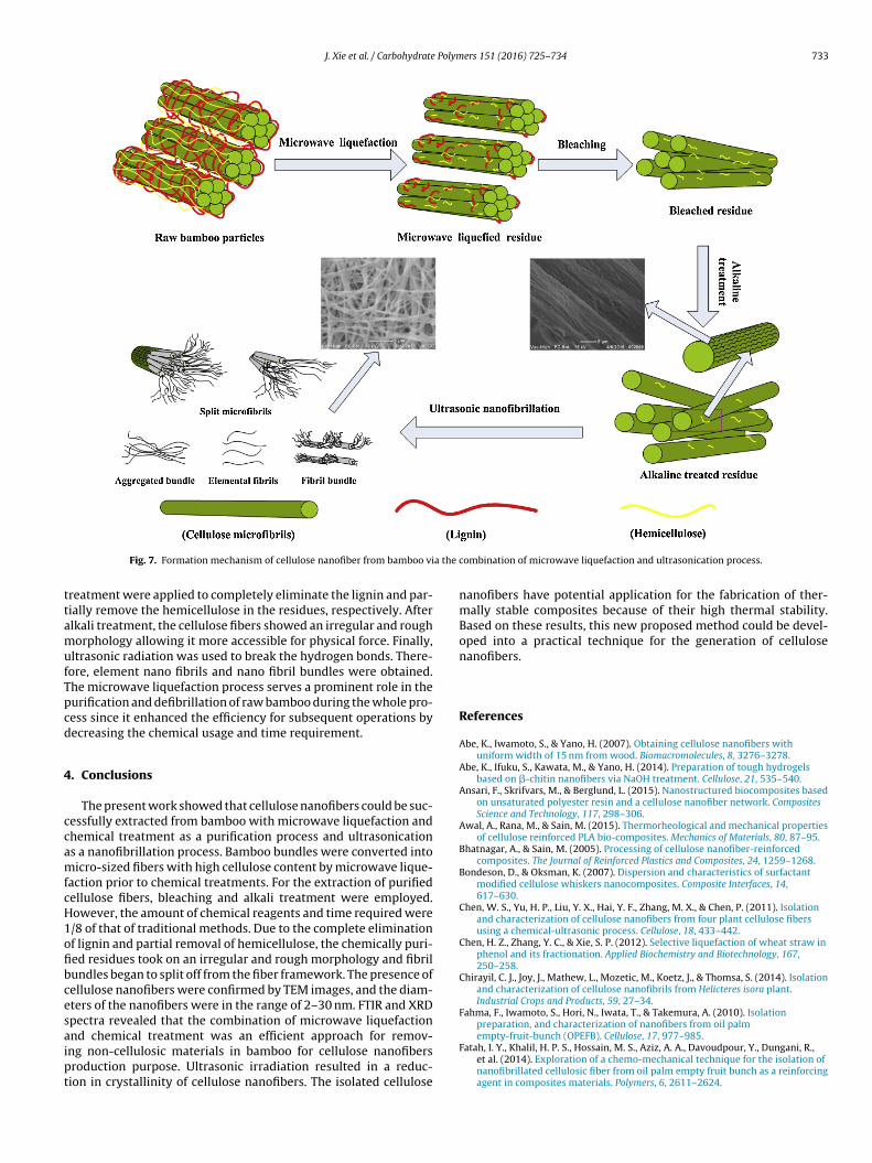
J. Xie et al. / Carbohydrate Polymers 151 (2016) 725–734 733
a the c
ttamufTpcd
4
ccamfcH1ofibcesaipt
Fig. 7. Formation mechanism of cellulose nanofiber from bamboo vi
reatment were applied to completely eliminate the lignin and par-ially remove the hemicellulose in the residues, respectively. Afterlkali treatment, the cellulose fibers showed an irregular and roughorphology allowing it more accessible for physical force. Finally,
ltrasonic radiation was used to break the hydrogen bonds. There-ore, element nano fibrils and nano fibril bundles were obtained.he microwave liquefaction process serves a prominent role in theurification and defibrillation of raw bamboo during the whole pro-ess since it enhanced the efficiency for subsequent operations byecreasing the chemical usage and time requirement.
. Conclusions
The present work showed that cellulose nanofibers could be suc-essfully extracted from bamboo with microwave liquefaction andhemical treatment as a purification process and ultrasonications a nanofibrillation process. Bamboo bundles were converted intoicro-sized fibers with high cellulose content by microwave lique-
action prior to chemical treatments. For the extraction of purifiedellulose fibers, bleaching and alkali treatment were employed.owever, the amount of chemical reagents and time required were/8 of that of traditional methods. Due to the complete eliminationf lignin and partial removal of hemicellulose, the chemically puri-ed residues took on an irregular and rough morphology and fibrilundles began to split off from the fiber framework. The presence ofellulose nanofibers were confirmed by TEM images, and the diam-ters of the nanofibers were in the range of 2–30 nm. FTIR and XRDpectra revealed that the combination of microwave liquefaction
nd chemical treatment was an efficient approach for remov-ng non-cellulosic materials in bamboo for cellulose nanofibersroduction purpose. Ultrasonic irradiation resulted in a reduc-ion in crystallinity of cellulose nanofibers. The isolated celluloseombination of microwave liquefaction and ultrasonication process.
nanofibers have potential application for the fabrication of ther-mally stable composites because of their high thermal stability.Based on these results, this new proposed method could be devel-oped into a practical technique for the generation of cellulosenanofibers.
References
Abe, K., Iwamoto, S., & Yano, H. (2007). Obtaining cellulose nanofibers withuniform width of 15 nm from wood. Biomacromolecules, 8, 3276–3278.
Abe, K., Ifuku, S., Kawata, M., & Yano, H. (2014). Preparation of tough hydrogelsbased on �-chitin nanofibers via NaOH treatment. Cellulose, 21, 535–540.
Ansari, F., Skrifvars, M., & Berglund, L. (2015). Nanostructured biocomposites basedon unsaturated polyester resin and a cellulose nanofiber network. CompositesScience and Technology, 117, 298–306.
Awal, A., Rana, M., & Sain, M. (2015). Thermorheological and mechanical propertiesof cellulose reinforced PLA bio-composites. Mechanics of Materials, 80, 87–95.
Bhatnagar, A., & Sain, M. (2005). Processing of cellulose nanofiber-reinforcedcomposites. The Journal of Reinforced Plastics and Composites, 24, 1259–1268.
Bondeson, D., & Oksman, K. (2007). Dispersion and characteristics of surfactantmodified cellulose whiskers nanocomposites. Composite Interfaces, 14,617–630.
Chen, W. S., Yu, H. P., Liu, Y. X., Hai, Y. F., Zhang, M. X., & Chen, P. (2011). Isolationand characterization of cellulose nanofibers from four plant cellulose fibersusing a chemical-ultrasonic process. Cellulose, 18, 433–442.
Chen, H. Z., Zhang, Y. C., & Xie, S. P. (2012). Selective liquefaction of wheat straw inphenol and its fractionation. Applied Biochemistry and Biotechnology, 167,250–258.
Chirayil, C. J., Joy, J., Mathew, L., Mozetic, M., Koetz, J., & Thomsa, S. (2014). Isolationand characterization of cellulose nanofibrils from Helicteres isora plant.Industrial Crops and Products, 59, 27–34.
Fahma, F., Iwamoto, S., Hori, N., Iwata, T., & Takemura, A. (2010). Isolationpreparation, and characterization of nanofibers from oil palm
empty-fruit-bunch (OPEFB). Cellulose, 17, 977–985.Fatah, I. Y., Khalil, H. P. S., Hossain, M. S., Aziz, A. A., Davoudpour, Y., Dungani, R.,et al. (2014). Exploration of a chemo-mechanical technique for the isolation ofnanofibrillated cellulosic fiber from oil palm empty fruit bunch as a reinforcingagent in composites materials. Polymers, 6, 2611–2624.

7 Polym
F
F
H
H
H
H
J
J
J
J
K
K
K
L
L
L
L
L
M
P
P
P
P
Q
Q
wood residue based on cellulose, hemicellulose, and lignin. Journal of AppliedPolymer Science, 123, 850–856.
Zhu, J. Y., Sabo, R., & Luo, X. L. (2011). Integrated production of nano-fibrillated
34 J. Xie et al. / Carbohydrate
ortunati, E., Puglia, D., Monti, M., Peponi, L., Santulli, C., Kenny, J. M., et al. (2013).Extraction of cellulose nanocrystals from Phormium tenax fibres. Journal ofPolymers and the Environment, 21, 319–328.
rench, A. D. (2014). Idealized powder diffraction patterns for cellulosepolymorphs. Cellulose, 21, 885–896.
an, J. Q., Zhou, C. J., French, A. D., Han, G. P., & Wu, Q. L. (2013). Characterization ofcellulose II nanoparticles regenerated from 1-butyl-3-methylimidazoliumchloride. Carbohydrate Polymers, 94, 773–781.
assan, M. L., Bras, J., Hassan, E. A., Silard, C., & Mauret, E. (2014). Enzyme-assistedisolation of microfibrillated cellulose from date palm fruit stalks. IndustrialCrops and Products, 55, 102–108.
e, W., Jiang, X. C., Sun, F. W., & Xu, X. W. (2014). Extraction and characterizationof cellulose nanofibers from Phyllostachys nidularia Munro via a combinationof acid treatment and ultrasonication. Bioresources, 9, 6876–6887.
ideno, A., Abe, K., & Yano, H. (2014). Preparation using pectinase andcharacterization of nanofibers from orange peel waste in juice factories.Journal of Food Science, 79, 1218–1224.
ahan, M. S., Saeed, A., He, Z. B., & Ni, Y. H. (2011). Jute as raw material for thepreparation of microcrystalline cellulose. Cellulose, 18, 451–459.
ang, J. H., Lee, S. H., Endo, T., & Kim, N. H. (2013). Characteristics of microfibrillatedcellulosic fibers and paper sheets from Korean white pine. Wood Science andTechnology, 47, 925–937.
ausovec, D., Vogrincic, R., & Kokol, V. (2015). Introduction of aldehyde vs.carboxylic groups to cellulose nanofibers using lccase/TEMPO mediatedoxidation. Carbohydrate Polymers, 116, 74–85.
iang, Z. H., Liu, Z. J., Fei, B. H., Cai, Z. Y., Yu, Y., & Liu, X. (2012). The pyrolysischaracteristics of moso bamboo. Journal of Analytical and Applied Pyrolysis, 94,48–52.
halil, H. P. S. A., Bhat, A. H., & Yusra, A. F. I. (2012). Green composites fromsustainable cellulose nanofibrils: a review. Carbohydrate Polymers, 87, 963–979.
halil, H. P. S. A., Davoudpour, Y., Islam, M. N., Mustapha, A., Sudesh, K., Dungani,R., et al. (2014). Production and modification of nanofibrillated cellulose usingvarious mechanical processes: a review. Carbohydrate Polymers, 99, 649–665.
hawas, P., & Deka, S. C. (2016). Isolation and characterization of cellulosenanofibers from culinary banana peel using high-intensity ultrasonicationcombined with chemical treatment. Carbohydrate Polymers, 137, 608–616.
i, M., Wang, L. J., Li, D., Cheng, Y. L., & Adhikari, B. (2014). Preparation andcharacterization of cellulose nanofibers from de-pectinated sugar beet pulp.Carbohydrate Polymers, 102, 136–143.
i, W., Zhang, Y. C., Li, J. H., Zhou, Y. J., Li, R. S., & Zhou, W. (2015). Characterizationof cellulose from banana pseudo-stem by heterogeneous liquefaction.Carbohydrate Polymers, 132, 513–519.
u, P., & Hsieh, Y. L. (2010). Preparation and properties of cellulose nanocrystals:rods spheres, and network. Carbohydrate Polymers, 82, 329–336.
u, Q. L., Lin, W. Y., Tang, L. R., Wang, S. Q., Chen, X. R., & Huang, B. (2015). Amechanochemical approach to manufacturing bamboo cellulose nanocrystals.Journal of Materials Science, 50, 611–619.
u, Q. L., Tang, L. R., Lin, F. C., Wang, S. Q., Chen, Y. D., Chen, X. R., et al. (2015).Preparation and characterization of cellulose nanocrystals viaultrasonication-assisted FeCl3-catalyzed hydrolysis. Cellulose, 21, 3497–3506.
a, H. Y., Burger, C., Hsiao, B. S., & Chu, B. (2014). Fabrication and characterizationof cellulose nanofiber based thin-film nanofibrous composite membranes.Journal of Membrane Science, 454, 272–282.
aakko, M., Ankerfors, M., Kosonen, H., Nykanen, A., Ahola, S., Osterberg, M., et al.(2007). Enzymatic hydrolysis combined with mechanical shearing andhigh-pressure homogenization for nanoscale cellulose fibrils and strong gels.Biomacromolecules, 8, 1934–1941.
an, H., Shupe, T. F., & Hse, C. Y. (2007). Characterization of liquefied wood residuesfrom different liquefaction conditions. Journal of Applied Polymer Science, 105,3739–3746.
ang, C. H., Gaddipatti, S., Tucker, G., Lester, E., & Wu, T. (2014). Relationshipbetween thermal behavior of lignocellulosic components and properties ofbiomass. Bioresource Technology, 172, 312–320.
elissari, F. M., Sobral, P. J. A., & Menegalli, F. C. (2014). Isolation andcharacterization of cellulose nanofibers from banana peels. Cellulose, 21,417–432.
i, J. Q., Xie, J. L., Hse, C. Y., & Shupe, T. F. (2013). Analysis of Phyllostachys
pubescens bamboo residues for liquefaction: chemical components, infraredspectroscopy, and thermogravimetry. Bioresources, 8, 5644–5654.ing, Y., Cai, Z. Y., Wu, Y. Q., Yao, C. H., Wu, Q. L., & Li, X. J. (2015). Facile preparationof optically transparent and hydrophobic cellulose nanofibrils composite films.Industrial Crops and Products, 77, 13–20.
ers 151 (2016) 725–734
Sacui, I. A., Nieuwendaal, R. C., Burnett, D. J., Stranick, S. J., Jorfi, M., Weder, C., et al.(2014). Comparison of the properties of cellulose nanocrystals and cellulosenanofibrils isolated from bacteria, tunicate, and wood processed using acid,enzymatic, mechanical, and oxidative methods. ACS Applied Materials &Interfaces, 6, 6127–6138.
Shimizu, M., Saito, T., & Isogai, A. (2014). Bulky quaternary alkylammoniumcounterions enhance the nanodispersibility of2,2,6,6-tetramethylpiperidine-1-oxyl-oxidized cellulose in diverse solvent.Biomacromolecules, 15, 1904–1909.
Shinoda, R., Saito, T., Okita, Y., & Isogai, A. (2012). Relationship between length anddegree of polymerization of TEMPO-Oxidized cellulose nanofibrils.Biomacromolecules, 13, 842–849.
Silverio, H. A., Neto, W. P. F., Dantas, N. O., & Pasquini, D. (2013). Extraction andcharacterization of cellulose nanocrystals from corncob for application asreinforcing agent in nanocomposites. Industrial Crops and Products, 44,427–436.
Somerville, C., Bauer, S., Brininstool, G., Facette, M., Hamann, T., Milne, J., et al.(2004). Toward a systems approach to understanding plant cell walls. Science,306, 2206–2211.
Sun, F. B., & Chen, H. Z. (2008). Comparison of atmospheric aqueous glycerol andsteam explosion pretreatments of wheat straw for enhanced enzymatichydrolysis. Journal of Chemical Technology and Biotechnology, 83, 707–714.
Sun, X. X., Wu, Q. L., Ren, S. X., & Lei, T. Z. (2015). Comparison of highly transparentall-cellulose nanopaper prepared using sulfuric acid and TEMPO-mediatedoxidation methods. Cellulose, 22, 1123–1133.
Takaichi, S., Saito, T., Tanaka, R., & Isogai, A. (2014). Improvement ofnanodispersibility of oven-dried TEMPO-oxidized celluloses in water. Cellulose,21, 4093–4103.
Tang, Y. J., Yang, S. J., Zhang, N., & Zhang, J. H. (2014). Preparation andcharacterization of nanocrystalline cellulose via low-intensityultrasonic-assisted sulfuric acid hydrolysis. Cellulose, 21, 335–346.
Tibolla, H., Pelissari, F. M., & Menegalli, F. C. (2014). Cellulose nanofibers producedfrom banana peel by chemical and enzymatic treatment. LWT—Food Scienceand Technology, 59, 1311–1318.
Wang, H. Y., Li, D. G., Yano, H., & Abe, K. (2014). Preparation of tough cellulose IInanofibers with high thermal stability from wood. Cellulose, 21, 1505–1515.
Wu, Q., Meng, Y. J., Concha, K., Wang, S. Q., Li, Y. J., Ma, L. F., et al. (2013). Influenceof temperature and humidity on nano-mechanical properties of cellulosenanocrystal films made from switchgrass and cotton. Industrial Crops andProducts, 48, 28–35.
Xie, J. L., Zhai, X. L., Hse, C. Y., Shupe, T. F., & Pan, H. (2015). Polyols from microwaveliquefied bagasse and its application to rigid polyurethane foam. Materials, 8,8496–8509.
Xie, J. L., Hse, C. Y., Shupe, T. F., Qi, J. Q., & Pan, H. (2014). Liquefaction behaviors ofbamboo residues in a glycerol-based solvent using microwave energy. Journalof Applied Polymer Science, http://dx.doi.org/10.1002/APP.40207
Xie, J. L., Huang, X. Y., Qi, J. Q., Hse, C. Y., & Shupe, T. F. (2014). Effect of anatomicalcharacteristics and chemical components on microwave-assisted liquefactionof bamboo wastes. Bioresources, 9, 231–240.
Xie, J. L., Qi, J. Q., Hse, C. Y., & Shupe, T. F. (2014). Effect of lignin derivatives in thebio-polyols from microwave liquefied bamboo on the properties ofpolyurethane foams. Bioresources, 9, 578–588.
Yousefi, H., Faezipour, M., Hedjazi, S., Mousavi, M. M., Azusa, Y., & Heidari, A. H.(2013). Comparative study of paper and nanopaper properties prepared frombacterial cellulose nanofibers and fibers/ground cellulose nanofibers of canolastraw. Industrial Crops and Products, 43, 732–737.
Yue, Y. Y., Zhou, C. J., French, A. D., Xia, G., Han, G. P., Wang, Q. W., et al. (2012).Comparative properties of cellulose nano-crystals from native and mercerizedcotton fibers. Cellulose, 19, 1173–1187.
Yue, Y. Y., Han, J. Q., Han, G. P., Zhang, Q. G., French, A. D., & Wu, Q. L. (2015).Characterization of cellulose I/II hybrid fibers isolated from energycanebagasse during the delignification: morphology, crystallinity and percentageestimation. Carbohydrate Polymers, 133, 438–447.
Zhang, H. R., Pang, H., Shi, J. Z., Fu, T. Z., & Liao, B. (2012). Investigation of liquefied
cellulose and cellulosic biofuel (ethanol) by enzymatic fractionation of woodfibers. Green Chemistry, 13, 1339–1344.