isolation characterization of mouse metallothionein-i … recherchescientifique, unit6 184debiologie...
TRANSCRIPT
Proc. Natl. Acad. Sci. USAVol. 77, No. 11, pp. 6511-6515, November 1980Biochemistry
Isolation and characterization of the mouse metallothionein-I gene(recombinant DNA/DNA sequence/heteroduplex mapping/cadmium)
DIANE M. DURNAM*, FABIENNE PERRINt, FRANK GANNONt, AND RICHARD D. PALMITER**Howard Hughes Medical Institute Laboratory, Department of Biochemistry, University of Washington, Seattle, Washington 98195; and tLaboratoire deG6nktique Mol6culaire des Eucaryotes du Centre National de la Recherche Scientifique, Unit6 184 de Biologie Moleculaire et de G6nie Genetiquede l'Institut National de la Sante et de la Recherche M&dicale, Institut de Chimie Biologique, Faculte de M6decine, Strasbourg 67085, France
Communicated by Earl W. Davie, August 8,1980
ABSTRACT Double-stranded cDNA was synthesized froma mouse liver mRNA fraction enriched for metallothioneinmRNA activity, ligated to restriction site linkers, inserted intopBR322, and used to transform Escherichia coli 1776. Thesequence of the largest plasmid containing DNA that hybridizedto metallothionein mRNA was determined and shown to containa 38base-pair insert that includes the entire coding region and3' untranslated region of metallothionein-I. The metallothi-onein-I insert was nick-translated and used to screen both amouse myeloma and a mouse embryo DNA library in bacte-riophage A A metallothionein-I genomic clone containing 13-15kilobase pairs of mouse DNA was isolated from each library.Both contain a 3.8-kilobase-pair EcoRI fragment that hybridizesto the metallothionein-I probe. The location, size, and orienta-tion of the metallothionein-I gene within the 3.8-kilobase-pairfragment were determined byheteroduplex and restrictionmapping. The gene spans 1.1 kilobase pairs and contains at leasttwo introns.
The quest for a cadmium-binding protein by Margoshes andVallee (1) led to their discovery of a small metal-binding protein(Mr 6000-7000), later called metallothionein (MT), which ischaracterized by having a high cysteine content and no aro-matic amino acids or histidine. It binds several heavy metals,including Cd, Zn, Hg, and Cu, and has been postulated to beinvolved in zinc metabolism and heavy metal detoxification.Most vertebrate tissues contain two forms of MT that differ inamino acid sequence; in the mouse, the two forms are desig-nated MT-I and MT-II (for a recent symposium, see ref. 2).We became interested in studying MT gene regulation be-
cause the amount of translatable MT mRNA increases rapidlyin response to heavy metals or glucocorticoids (3, 4). MT syn-thesis occurs predominantly in liver and kidney in vivo, butnearly all cell types are responsive in culture (unpublishedobservations; refs. 5 and 6). In addition, cell cultures can bemade resistant to Cd, and these lines overproduce MT (7, 8).The molecular mechanisms by which heavy metals and steroidsregulate MT mRNA production are unknown. Because anunderstanding of gene regulation depends upon specific probesfor RNA and DNA sequences, we have initiated our investi-gation by isolating a MT-I cDNA clone and using it to selectgenomic clones containing the MT-I gene.
MATERIALS AND METHODSPreparation of RNA. Swiss Webster mice (25 g) were in-
jected subcutaneously with 1.5 ,umol of CdSO4 and 0.5 ,molof ZnSO4. Six hours later, a 5% (wt/vol) liver homogenate wasprepared in NaDodSO4 and proteinase K, and total nucleicacids were extracted. RNA was precipitated with 2 M LiCl;poly(A)-RNA was selected by oligo(dT)-cellulose chromatog-raphy and sedimented for 20 hr at 280,000 X g on 12-ml, 5-20%sucrose gradients in 10 mM Hepes (pH 7.5) (9). Fractions (0.5
ml) were collected and assayed directly by translation; thosecontaining MT mRNA were pooled and sedimented on a sec-ond, identical gradient. Fractions were assayed for MT mRNAactivity with a nuclease-treated rabbit reticulocyte lysate es-sentially as described (10) except that no exogenous amino acidsother than [85S]cysteine (400 Ci/mmol; 1 Ci = 3.7 X 10"0 bec-querels) were added and the reaction mixtures contained 5 mMdithiothreitol. After translation, samples (5 ,l) were incubatedwith 20,Ml of 100 mM iodoacetate in 0.5 M Tris-HCl (pH 8.8)for 1 hr at 37°C, electrophoresed on a NaDodSO4/polyacryl-amide gel for 14 hr at 10 mA, and analyzed by fluorography.Purified mouse MT (11) was included as a standard.
Preparation of Double-Stranded (ds) cDNA, Insertion intopBR322, and Transformation of X1776. Double-strandedcDNA was prepared (12) and ligated to a 1:1 mixture of Hin-dIII and EcoRI linkers (Collaborative Research, Waltham, MA)prior to being inserted into EcoRI/HindIII-digested pBR322as described (13). Transformation of E. coli X1776 was per-formed as described (14) with the following modifications: (i)the 100 mM CaCl2 buffer was replaced by 70 mM MnCl2/30mM CaCl2/40 mM NaOAc, pH 5.6; (ii) the cells were trans-formed with plasmid at a concentration of 3 ,ug/ml; and (iii)the transformed cells were incubated at 37°C in L broth for 3hr before being spread on ampicillin plates. All recombinantDNA procedures were carried out in a P2 facility in accordancewith the National Institutes of Health guidelines.
Identification of MT Clones. Initial screening for a MTclone was performed by measuring the size of plasmids isolatedfrom 10-ml cultures of the transformants (15) on 0.7% agarosegels. Plasmids containing inserted DNA were immobilized onnitrocellulose filters and hybridized with MT mRNA; RNA thathybridized was eluted and assayed by translation (16).
Screening Genomic Libraries. Bacteriophage X librariescontaining mouse myeloma and mouse embryo DNA weregenerously provided by Davis (17) and Maki (18), respectively.The libraries were screened (19) with the nick-translated (20)Eco/Hind fragment from the cDNA clone (mipEH.4) as ahybridization probe. The stringency of the hybridization wasincreased by including an additional wash step in 5 mM Tris-HCI, pH 7.4/2.5 mM EDTA/0.5% NaDodSO4 at 68°C priorto the formamide washes.
Restriction Mapping. Restriction enzymes were obtainedfrom Bethesda Research Laboratories (Rockville, MD) orBoehringer Mannheim and used according to the protocolssupplied. Products of the reactions were analyzed on agarosegels. DNA in agarose gels was denatured and transferred ontoSartorius nitrocellulose filters (21) and hybridized to nick-translated probes as described (22).
Heteroduplex Analysis.Heteroduplexeswere formed in 70%(vol/vol) deionized formamide/300 mM NaCI/10 mM Tris-HCI, pH 8.5/1 mM EDTA with both DNAs at 1-3, g/ml. The
Abbreviations: MT, metallothionein; ds, double-stranded; bp, basepair(s); kb, kilobase pair(s).
6511
The publication costs of this article were defrayed in part by pagecharge payment. This article must therefore be hereby marked "ad-vertisement" in accordance with 18 U. S. C. §1734 solely to indicatethis fact.
6512 Biochemistry: Durnam et al.
A B A
ov _
B
GB
GBMT * _
MT
MT
7 9 11 13 14 15 16 17 18 19 10 1112 13 14 15 16 17 18FRACTION
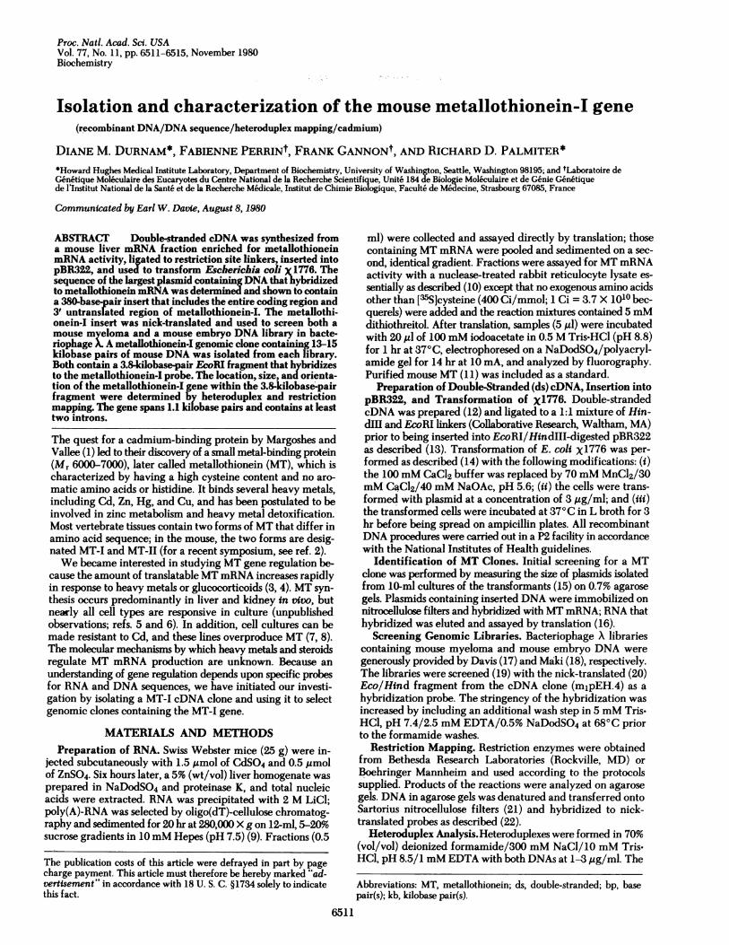
FIG. 1. Fractionation of mouse liver mRNA on sucrose gradients.Aliquots of the gradient fractions were translated in the presence of[35S]cysteine and the products were analyzed by electrophoresis(15-20% gel) and fluorography. The positions of unlabeled MT andglobin (GB) standard detected by staining are indicated. (A) Partialseparation ofMT mRNA from other translatable messages. Fractions16-19 were pooled and further purified on a second identical gradient,the results of which are shown in B. Fractions 14-18 of the secondgradient were pooled and used in all subsequent procedures.
DNAs were denatured at 750C for 5 min, incubated at 250Cfor 30 min, and mounted for microscopy as described (23).Hybrids between the DNA and MT mRNA were formed in thesame buffer by denaturing the DNA at 750C for 5 min andincubating it with 3 jug of gradient-purified MT mRNA per mlat 540C for 4 hr before being mounted for microscopy.
RESULTS AND DISCUSSIONIsolation of a MT-I cDNA Clone. Liver from mice treated
with CdSO4 and ZnSO4 for 6 hr was chosen as the startingmaterial for isolating MT mRNA because the rate of hepaticMT synthesis reaches a maximum 4-6 hr after administrationof heavy metals (unpublished observations; ref. 24). TotalmRNA was isolated and fractionated on 5-20% sucrose gradi-ents. mRNA activity was assayed by translation and the prod-ucts were separated by NaDodSO4/polyacrylamide electro-phoresis. Fig. 1A shows the distribution of translation productsacross a typical gradient. MT mRNA was located primarily infractions 17 and 18. Separation of MT mRNA from othertranslatable mRNAs was achieved by sedimenting the peak MTmRNA fractions on a second sucrose gradient (Fig. 1B). Al-though MT was the only apparent translation product, subse-quent hybridization studies using a MT-I-specific cDNA haveshown that MT mRNA was enriched 550-fold relative to total
V
1 2 3 4 1 2 3 4
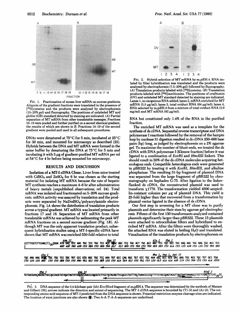
FIG. 2. Hybrid-selection ofMT mRNA by mjpEH.4. RNA iso-lated by filter hybridization was translated and the products were
analyzed by electrophoresis (7.5-20% gel) followed by fluorography.(A) Translation products labeled with [35S]cysteine. (B) Translationproducts labeled with [-6S]methionine. The positions of ovalbumin(OV) and unlabeled MT standard detected by staining are indicated.Lanes 1, no exogenous RNA added; lanes 2, mRNA enriched for MTmRNA (5.5 gg/ml); lanes 3, total oviduct RNA (64 ug/ml); lanes 4,RNA selected by mipEH.4 from a mixture of total oviduct RNA (2.9mg/ml) and MT mRNA (82 ,ug/ml).
RNA but constituted only 1.4% of the RNA in the purifiedfraction.The enriched MT mRNA was used as a template for the
synthesis of ds cDNA. Sequential reverse transcriptase and DNApolymerase I reactions followed by the removal of the hairpinloop by nuclease SI digestion resulted in ds cDNA 250-600 basepairs (bp) long, as judged by electrophoresis on a 2% agarosegel. To maximize the number of blunt ends, we treated the dscDNA with DNA polymerase I (Klenow fraction) before it wasligated to a combination of EcoRI and HindIII linkers. Thisshould result in 50% of the ds cDNA molecules acquiring het-erologous ends. Compatible heterologous ends were generatedin pBR322 by treating it with EcoRI, HindIII, and alkalinephosphatase. The resulting 31-bp fragment of plasmid DNAwas separated from the large fragment of pBR322 by chro-matography on Sephadex G-75. After ligation to the linker-flanked ds cDNA, the reconstructed plasmid was used totransform X1776. The transformation yielded 4500 ampicil-lin-resistant colonies per tig of plasmid DNA. This yield is25-fold higher than that recovered from a transformation byplasmid vector ligated in the absence of ds cDNA.Our first step in screening for a MT clone was to purify
plasmids and determine their sizes by agarose gel electropho-resis. Fifteen of the first 130 transformants analyzed containedplasmids significantly larger than pBR322. These 15 plasmidswere attached to nitrocellulose filters and hybridized to en-
riched MT mRNA. After the filters were thoroughly washed,the attached RNA was eluted in boiling H20 and translated.Visualization of the translation products by electrophoresis on
AaI BIAOuI HfM ICCMCGTCTTCAAG(T)35CGG AT :C MC TGC TCC TGC TCC AXFIWC GGC TCC TGC ACT TGC ACC Aff1CrTrUI TGC MG MC TGC MG TGC ACC TCC
MT ASP PRO ASH CI'S kR CUS KR 21R aY CLY KR CIS IYsR CIS MR SER SER CUS ALA CUS LYS ASH CUS LYS CUS IWR SER
AIu I l ihal Tao! StHTCC MG AAGM TGC TGC TCC TwC TGT CCC GIG GGC TGC TCV MA TGT GCC AG 6GC TGT GTC TGC MA d"C GCG GAC MG TGC ACG TQC TGT GCC TGACIS LY'S LYS SKR CIS CIS KR CI'S CI'S PRO VAL dI CM'SKR LYS cIS ALA CLN CLI CIS VAL CIS LI'S CL ALA ALA ASP LYS CYS TWR CIS CIS ALA STOP
ha I Avar I
rTCCCCCTMA~AAACCC~TTTTTACGAGTCGTAATATAAAGCCGT1TGA~ CYA)20CC1-inrTGTGACGCMGCCACCACGGTAGTAT WCCA-TCTTCCT-AT
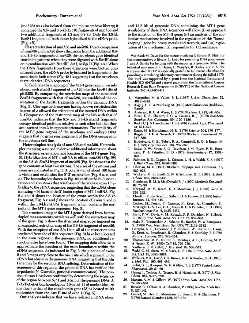
FIG. 3. DNA sequence of the 0.4-kilobase-pair (kb) Eco/Hind fragment of m1pEH.4. The sequence was determined by the methods of Maxamand Gilbert (26); arrows indicate the direction and extent of sequencing. The MT-I cDNA sequence is bounded by (T)-35 and (A)-20. The cor-responding amino acid sequence of MT-I predicted from the DNA sequence is shown. Potential restriction enzyme cleavage sites are indicated.The location of exon junctions are also shown (1). Two A-A-T-A-A sequences are underlined.
Proc. Natl. Acad. Sci. USA 77 (1980)
...-,AMNdlbk.

Proc. Natl. Acad. Sci. USA 77 (1980) 6513
kb B_.9.
,9.2-7.2
-.*1a~.
\0.8
2 3 4 5 6 7 8 9 10 1 2 3 45 678 910FIG. 4. Restriction analysis ofmeX26 and mmX36. Samples were
digested as indicated and analyzed by electrophoresis on a 0.7%agarose gel. (A) Lanes 1 and 10 contain EcoRI-digested P22 as sizemarkers; the sizes of the fragments are indicated. Lanes 2 and 3 showEcoRI digests of meX26 and mmX36, respectively. Lanes 4-9 showthe results of double digests with EcoRI in combination with HindIII(lanes 4 and 5), Sst I (lanes 6 and 7), or Bgl II (lanes 8 and 9). Thedigestion of mmX26 is shown in lanes 4, 6, and 8; the digestion ofmeX36 is shown in lanes 5,7, and 9. (B) Southern blot analysis of thedigestions shown in A. MT-1-containing fragments were identifiedby hybridization to 32P-labeled Eco/Hind fragment of m1pEH.4.
NaDodSO4/polyacrylamide gels followed by fluorographyrevealed that two of the plasmids had hybridized MT mRNA.To check the specificity of the hybridization, we hybridizeda filter containing the larger of the two positive plasmids to amixture of RNA containing MTmRNA and oviduct RNA. Fig.2 shows that the MT plasmid specifically hybridizes MTmRNA. Fig. 2A shows the results obtained when [s5S]cysteine,which constitutes 20 out of 61 amino acids in MT, was used inthe translation. Identical results were obtained with [s5S]me-thionine, represented only once in MT (Fig. 2B).
Characterization of the MT cDNA Clone. In an attemptto determine the size of the insert of the MT cDNA clone, wediscovered that the plasmid was cut by HindlII but was resistantto digestion by EcoRI. Because the cDNA insert was not cleavedby Hae III, we circumvented this problem by isolating theHindIII/Hae III fragment containing the MT cDNA, attachingan EcoRI linker to the blunt end generated by the Hae III di-
3.8 kb
gestion, and repeating the transformation procedure. Cleavageof these transformants with HindIII and EcoRI yielded afragment z400 bp long which included the MT cDNA and 16bp of plasmid DNA acquired during the subcloning. Thissubclone, mjpEH.4, was used in all subsequent procedures.To ascertain whether mjpEH.4 contained MT-I or MT-II
sequences, we determined the sequence of the 400-bp Eco!Hind insert and compared-it to the DNA sequences postulatedfrom the amino acid sequences of MT-I and MT-II (2, 25). Thesequence shown in Fig. 3 indicates that we isolated a MT-Iclone; it confirms the MT-I protein sequence except for thepresence of asparagine at position 23 instead of aspartic acid.The DNA sequence also reveals the following features. (i)mjpEH.4 contains a 132-bp 3' untranslated region and a 3'poly(A) tail in addition to the entire coding region of MT-I. (ii)'The A-A-U-A-A-A sequence located near the 3' end of manymRNAs (27) is also present in the MT-I mRNA. (iii) The choiceof codons is not random. Of the codons that could have a C inthe third position, 89% terminate in C, including all of the-serinecodons and 80% of the cysteine codons. With a random selec-tion, 33% of the serine codons and 50% of the cysteine codonswould have C in the third position. (iv) The original clone hadretained only 1 bp of the EcoRI site which was ligated to 35 bpof poly(dA).poly(dT) located at the 5' end of the MT-I sequence.Determination of the sequence of the genomic DNA indicatesthat this poly(dA)-poly(dT) sequence is not present at the 5' endof the MT-I gene, suggesting that it is an artifact that originatedduring cDNA cloning.
Isolation of Genomic MT Clones. Genomic DNA librarieswere prepared by inserting 10- to 20-kb fragments, resultingfrom partial EcoRI digestion of mouse myeloma or mouseembryo DNA, into bacteriophage X, Charon 4A (18, 19). Ap-proximately 106 plaques of the mouse myeloma stock werescreened by using a nick-translated Eco/Hind fragment frommjpEH.4 as a probe. Initially, the screening conditions usedwere identical to those used in the isolation of a number of eggwhite genes (19, 28). We were surprised on this first screeningto obtain 212 positive clones. Thirty of the clones were purifiedby two sequential screenings, and the DNA from 14 of these wasdigested with EcoRI and analyzed on a 0.7% agarose gel. Eachof these clones had a different EcoRI fragment that hybridizedwith the cDNA probe, suggesting that the cDNA probe has
9.5 kb ,k k34o.91kb kb
ST B11
) long arm
1.0 kb
TATAAAf BII Av Hf Av Hf Xb Hf Av Hf Av Hf P Hf SII A
NII I Nit( I IVI I I~p Hp Hp Hli1'Hp Ha AHh Hpa Tc HIH
Av HfI I
Hh 50 bpI
EXON EXON EXONI1 INTRON A 1 2 lINTRON B 3I3I
76;15 bp 49845 bp 72;13 bp 198 31 bp 22420 bpn.61 n-105 n-147 n-147 n*147
FIG. 5. Restriction map of meX26. Upper restriction map shows the orientation of the EcoRI fragments within the clone. Only the sitesused to orient the fragments are shown. Lower restriction map is an expanded map of the MT-I gene region. Only the sites in proximity to exonsare indicated. The location (ranges are shown) of the exons (dark bars), as.revealed by heteroduplex mapping (Fig. 6), are indicated; n = numberof measurements. SI, Sst I; SII, Sst II; Hh, Hha I; Tc, Tac I; Hp, Hpa II; Hf, Hinfl; BI, Bgl I; BII, Bgl II; Av, Ava II; Xb, Xba I; Tq, Taq I; Hd,HindIII; P, Pst I; Ha, Hae III; Al, Alu I; Bm, BamHI; Xh, Xho I; K, Kpn I.
-A. A
Biochemistry: Durnam et al.
II
-4.0-,;APIMWNW
-2.4 0 A

6514 Biochemistry: Durnam et al.
FIG. 6. Heteroduplex analysis ofmmX36 and meX26. (Insets) The solid line represents the cloned genomic DNA (mmX36 or the subcloned3.8-kb EcoRI fragment of meX26), the dashed line shows the RNA, and the dotted line illustrates the cloned cDNA (the Eco/Hind fragmentor mjpEH.4 linearized by Sal I). 5' and 3' arrowheads indicate the ends of the RNA (b, c, and e); the EcoRI and HindIII ends of the ds cDNAare indicated by e and h, respectively, in d and f. A and B designate the single-stranded introns; 1, 2, and 3 designate the exons. The double whitearrows point to the poly(A) track. (a) Heteroduplex molecule between mmX36 and Sal I-cut m1pEH.4. (b) Hybrid molecule between mmX36and MT mRNA. (c) Hybrid molecule between MT mRNA and the subcloned 3.8-kb fragment of meX26 linearized by BamHI. (d) Heteroduplexbetween the 3.8-kb fragment of meX26 and the Eco/Hind fragment of m1pEH.4. (e) Hybrid molecule between the 1.0-kb PstlPst fragment ofthe genomic DNA and MT mRNA. (f) Heteroduplex molecule between the Pst/Pst genomic fragment and the Eco/Hind fragment of mjpEH.4.At the top is seen a typical molecule revealing the hybridization of exons 2 and 3 as well as a short unhybridized DNA tail; at the bottom is seentwo such heteroduplex molecules hybridized together. (Bar = 0.1 am.)
sequences that are homologous to a large number of regions inthe genomic DNA. To overcome this problem, we incorporateda more stringent wash step (see Materials and Methods);mjpEH.4 hybrids were stable under these conditions whereasall 30 X hybrids were unstable. The remaining 182 clones from
the original screening were pooled and screened under the morestringent hybridization conditions to ultimately yield one clone,designated mmX36. EcoRI digestion of mmX36 yielded frag-ments of 3.8 and 9.5 kb in addition to the long and short armsof X. Under the stringent hybridization conditions, one clone
Proc. Natl. Acad. Sci. USA 77 (1980)
Proc. Natl. Acad. Sci. USA 77 (1980) 6515
(meX26) was also isolated from the mouse-embryo library, itcontained the 9.5- and 3.8-kb EcoRI fragments of mmX36 andtwo additional fragments of 1.3 and 0.9 kb. Only the 3.8-kbEcoRI fragment of both clones hybridized to the cDNA probe(Fig. 4B).
Characterization of mmX36 and meX26. Direct comparisonof mmX36 and meX26 shows that, aside from the additional 0.9-and 1.3-kb fragments of meX26, the two clones gave identicalrestriction patterns when they were digested with EcoRI aloneor in combination with HindIII, Sst I, or Bgl II (Fig. 4A). Whenthe DNA fragments from these digestions were blotted ontonitrocellulose, the cDNA probe hybridized to fragments of thesame size in both clones (Fig. 4B), suggesting that the two clonesshare identical DNA sequences.To facilitate the mapping of the MT-I gene region, we sub-
cloned each EcoRI fragment of meX26 into the EcoRI site ofpBR322. By comparing the restriction maps of the subelonedEcoRI fragments with that of meX26, we established the or-ientation of the EcoRI fragments within the genomic DNA(Fig. 5). Cleavage with enzymes having known restriction sitesin arms of X allowed the orientation of the inserted DNA withinX. Comparison of the restriction map of meX26 with that ofmmX36 indicates that the 9.5- and 3.8-kb EcoRI fragmentsoccupy identical positions relative to each other but that theyare inserted into X in opposite orientations. The similarity ofthe MT-I gene regions of the myeloma and embryo DNAsuggests that no gross rearrangement of the DNA occurred inthe myeloma cell line.Heteroduplex Analysis of mmX36 and meX26. Heterodu-
plex mapping was used to derive additional information aboutthe structure, orientation, and location of the MT-I gene (Fig.6). Hybridization of MT-I mRNA to either mmX36 (Fig. 6b)or the 3.8-kb EcoRI fragment of meX26 (Fig. 6c) shows that thegene contains at least two introns. The sizes of the introns andexons are indicated in Fig. 5. A poly(A) tail of about 100 basesis visible and establishes the 5'-3' orientation (Fig. 6 b, c, ande). The heteroduplex shown in Fig. 6a verifies the 5'-3' orien-tation and also shows that -t40 bases of the 76-base exon 1 hy-bridize to the cDNA sequence, suggesting that the cDNA cloneis missing -'36 bases of the 5' leader region of MT-I mRNA. Fig.6 c and d shows the location of the exons within the 3.8-kbfragment; Fig. 6 e and f shows the location of exons 2 and 3within the 1.0-kb Pst/Pst fragment, which contains the ma-jority of the MT-I gene sequence (Fig. 5).The structural map of the MT-I gene derived from hetero-
duplei measurements correlates well with the restriction mapof the gene. Fig. 5 shows the structural map superimposed onan expanded restriction map of the 3.8-kb fragment of meX26.With the exception of one Alu I site, all of the restriction sitespredicted from the cDNA sequence (Fig. 3) have been locatedin the exon regions in the genomic DNA; no additional re-striction sites have been found. The mapping data allow us toapproximate the location of the exon boundaries within thecDNA sequence. As indicated in Fig. 3, the junction of exons2 and 3 maps very close to the Alu I site which is present in thecDNA but absent in the genomic DNA, suggesting that this AluI site may be the result of RNA splicing. Determination of thesequence of this region of the genomic DNA has verified thishypothesis (N. Glanville, personal communication). The posi-tion of exon 1 has been confirmed by determining the sequenceof the region between Sst I and Xba I of the genomic DNA. AT-A-T-A-A-A box homologous (10 out of 11 of nucleotides areidentical) to that of the conalbumin gene (28) is located mlOOnucleotides from the Ava II site of exon 1 (Fig. 5).Our analyses indicate that we have isolated a cDNA clone
and -15.5 kb of genomic DNA containing the MT-I gene.Availability of these DNA sequences will allow: (i) an approachto the isolation of the MT-II gene; (ii) an analysis of the mo-lecular mechanisms involved in the regulation of this "house-keeping" gene by heavy metals and steroids; and (iii) explo-ration of the mechanism(s) responsible for Cd resistance.
We thank M. Davis for the mouse myeloma X library, R. Maki forthe mouse embryo X library, L. Loeb for providing DNA polymeraseI, and A. Sarthy for helping with the mapping of genomic DNA. Thetechnical assistance of L. Hager, C. Wasylyk, B. Boulay, and E. Sittleris appreciated. R.D.P. is especially grateful to Dr. Pierre Chambon forproviding a stimulating laboratory environment during the fall of 1979.This work was supported by a grant from the National Institutes ofHealth (HD-09172) and a travel grant from the International CancerResearch Data Bank Programme (ICRETT) of the National CancerInstitute (N01-CO-65341).
1. Margoshes, M. & Vallee, B. L. (1957) J. Am. Chem. Soc. 79,4813-4814.
2. Kigi, J. H. R. & Nordberg, M. (1979) Metallothionein (Birkhaser,Basel).
3. Anderson, R. D. & Weser, U. (1978) Biochem. J. 175,841-852.4. Etzel, K. R., Shapiro, S. G. & Cousins, R. J. (1979) Biochem.
Biophys. Res. Commun. 89,1120-1126.5. Rudd, C. J. & Herschman, H. R. (1978) Toxicol. Appl. Pharmacol.
44,511-521.6. Karin, M. & Herschman, H. R. (1979) Science 204, 176-177.7. Rugstad, H. E. & Norseth, T. (1978) Biochem. Pharmacol. 27,
647-650.8. Hildebrand, C. E., Tobey, R. A., Campbell, E. Q. & Enger, M.
D. (1979) Exp. Cell Res. 124, 237-246.9. Rowe, D. W., Moen, R. C., Davidson, J. M., Byers, P. H., Born-
stein, P. & Palmiter, R. D. (1978) Biochemistry 17, 1581-1590.
10. Palmiter, R. D., Gagnon, J., Ericsson, L. H. & Walsh, K. A. (1977)J. Biol. Chem. 252,6386-6393.
11. Cherian, M. G. (1974) Biochem. Biophys. Res. Commun. 61,920-926.
12. Wickens, M. P., Buell, G. N. & Schimke, R. T. (1978) J. Biol.Chem. 253, 2483-2495.
13. Goodman, H. M. & MacDonald R. J. (1979) Methods Enzymol.68,75-90.
14. Norgard, M. V., Keem, K. & Monahan, J. J. (1978) Gene 3,279-292.
15. Elwell, L. P., de Graaf, J., Seibert, D. & Falkow, S. (1975) Infect.Immun. 12,404-410.
16. Cochet, M., Perrin, F., Gannon, F., Krust, A., Chambon, P.,McKnight, G. S., Lee, D. C., Mayo, K. E. & Pahniter, R. D. (1979)Nucleic Acids Res. 6, 2435-2452.
17. Early, P. W., Davis, M. M., Kaback, D. B., Davidson, N. & Hood,L. (1979) Proc. Natl. Acad. Sci. USA 76,857-861.
18. Maki, R., Traunecker, A., Sakano, H., Roeder, W. & Tonegawa,S. (1980) Proc. Natl. Acad. Sci. USA 77,2138-2142.
19. Garapin, A. C., Lepennec, J. P., Roskam, W., Perrin, F., Cami,B., Krust, A., Breathnach, R., Chambon, P. & Kourilsky, P. (1978)Nature (London) 273,349-354.
20. Thomashow, M. F., Nutter, R., Montoya, A. L., Gordon, M. P.& Nester, E. W. (1980) Cell 19,729-739.
21. Southern, E. M. (1975) J. Mol. Biol. 98,503-517.22. Wahl, G. M., Stern, M. & Stark, G. R. (1979) Proc. Natl. Acad.
Sci. USA 76,3683-3687.23. Wellauer, P. K., David, I. B., Brown, D. D. & Reeder, R. H. (1976)
J. Mol. Biol. 105,461-486.24. Probst, G. S., Bousquet, W. F. & Miya, T. S. (1977) Toxicol. Appl.
Pharmacol. 39, 51-60.25. Huang, I., Yoshida, A., Tsunoo, H. & Nakajima, H. (1977) J. Biol.
Chem. 252, 8217-8221.26. Maxam, A. M. & Gilbert, W. (1977) Proc. Natl. Acad. Sci. USA
74,560-564.27. Benoist, C., O'Hare, K. & Chambon, P. (1980) Nucleic Acids Res.
8, 127-142.28. Cochet, M., Hen, R., Maroteaux, L., Perrin, F. & Chambon, P.
(1979) Nature (London) 282,567-574.
Biochemistry: Durnam et al.