isolation characterization two hemolytic phenotypes …jcm.asm.org/content/21/3/302.full.pdf ·...
TRANSCRIPT
JOURNAL OF CLINICAL MICROBIOLOGY, Mar. 1985, p. 302-3060095-1137/85/030302-05$02.00/0
Isolation and Characterization of Two Hemolytic Phenotypes ofVibrio damsela Associated with a Fatal Wound Infection
JILL E. CLARRIDGE' 2,3* AND SONIA ZIGHELBOIM-DAUM3
Laboratory Service, Veterans Administration Medical Center,l and Departments of Pathology2 and Microbiology andImmunology,3 Baylor College of Medicine, Houston, Texas 77211
Received 8 August 1984/Accepted 16 November 1984
Two hemolytic phenotypes of Vibrio damsela, isolated from the tissue of a patient with a fatal woundinfection, were characterized. The patient had underlying disease, and the wound was associated with an injuryinflicted during the handling of a catfish. The phenotypes were morphologically and biochemically similarexcept for their lecithinase, lipase, and hemolytic activities. When grown on rabbit blood agar, one phenotype(LZ) produced a large zone of hemolysis (10 mm) around the colony, whereas the other type (SZ) produced onlya small zone (1 to 2 mm). On sheep blood agar, the differences in hemolytic activity were more subtle. By amodified CAMP test in which V. damsela was streaked perpendicularly to Staphylococcus aureus, it wasdetermined that a factor elaborated by the LZ phenotype (but not the SZ phenotype) protected sheeperythrocytes from the hemolysis normally caused by S. aureus toxins. Cell-free filtrates of broth cultures ofeach phenotype had the same effects on erythrocytes as did the organisms themselves.
Vibrio damsela is a newly described species which hasbeen associated with the marine environment and fish (16,21). Most of the reported human wound infections due to V.damsela have occurred after handling fish or after exposureto seawater (7, 20). These infections, although sometimesserious enough to require debridement, have not had a fataloutcome. We report here the isolation and characterizationof two hemolytic phenotypes of V. damsela from the tissueof a patient with underlying disease who sustained a super-ficial wound which led to the rapid onset of disease anddeath.
CASE REPORTOn the evening of 9 May 1984, an alcoholic 61-year-old
male with a history of pancreatitis and insulin-dependentadult onset diabetes slightly injured his left hand whilecleaning a catfish reportedly caught in a local freshwaterlake. A few hours later he noted that his hand had becometender, swollen, and painful. At 2 p.m. the next day, whenhe sought medical attention at a community hospital, hisleukocyte count was 18,400 cells per ml with a shift to theleft, the platelet count was 115,000, and his hand wasedematous with black discoloration. He was transferred tothe Houston Veterans Administration Medical Center, wherehe appeared to be in acute distress with marked edema of theleft forearm and bullae formation on the hand. The blackdiscoloration had extended up to the elbow with contiguouserythema. The fingertips of the left hand appeared necrotic.A disarticulation of the left shoulder was performed. Tissuefrom the hand and fluid from the bullae were taken forculture. No other cultures were performed. Amikacin, clin-damycin, and penicillin therapy was started. On the morningof 11 May, a few hours after surgery, it was noted that theshoulder area around the incision was erythematous andpossibly necrotic. Extensive debridement of the left shoul-der and chest with extension into the right anterior chest wasperformed; the scapula and clavicle were resected. Thesubsequent course of the patient was complicated by con-
tinued disseminated intravascular coagulation, acute renal
* Corresponding author.
failure, and hypercalcemia, and he subsequently died of acardiac arrest 9 days after the initial injury. No autopsy wasperformed.
MATERIALS AND METHODS
Isolation and characterization of the organism. The tissuefrom the hand and fluid from the bullae were each cultured onthe following media: 5% sheep blood Trypticase soy agar(SBA) (BBL Microbiology Systems), chocolate agar, andColumbia colistin-nalidixic acid agar, which were incubatedin 6 to 8% C02; MacConkey agar, which was incubated inair; and Schaedler sheep blood agar, CDC anaerobe bloodagar with phenylethyl alcohol, Schaedler kanamycin-van-comycin agar with 5% sheep blood, and bacteroides-bile-esculin agar, which were incubated anaerobically. The bio-chemical and morphological identification was accomplishedby the API 20E system (Analytab Products, Plainview,N.Y.), the DMS-rapid NFT system (API System, S.A., LaBalme les Grottes, France), and standard methods (14).
Hemolytic activity. The hemolytic activities of the pheno-type colonies were determined after 24 h of growth on SBAor Casman rabbit blood agar (RBA). We tested the filtratesfor hemolytic activity by dropping 5 to 50 FlI of each filtrateon either SBA or RBA and measuring the size and intensityof the hemolytic zones at 24 h and 1 week. Both filtrates andcolonies were examined by a modified CAMP test (14) inwhich the organisms or filtrates were streaked perpendicu-larly to Staphylococcus aureus ATCC 25923.
Cell-free filtrates. Cell-free filtrates were prepared from V.damsela cultures grown in brain heart infusion broth with0.5, 3, or 6% NaCl at 25 and 35°C. After 8 h, when a samplewas removed to determine the number of CFU per milliliter,the broth was centrifuged and the supernatant was sterilizedby passage through a filter (pore size, 0.22 ,um). The effect ofcell-free filtrate was also assessed by growing the cells on amembrane filter (0.45 ,um; Millipore Corp.) placed on anSBA plate. After 24 h, the filter was removed and the extentof hemolysis was determined.Temperature studies. Filtrates in 0.3-ml aliquots were
heated in glass tubes at 45°C for 30 min, 60°C for 30 min, or
100°C for 10 min in static water baths. The small volume of
302
Vol. 21, No. 3
on October 1, 2018 by guest
http://jcm.asm
.org/D
ownloaded from
TWO HEMOLYTIC PHENOTYPES OF VIBRIO DAMSELA 303
1% V-~~% it.
O
1; l
fb** '2&
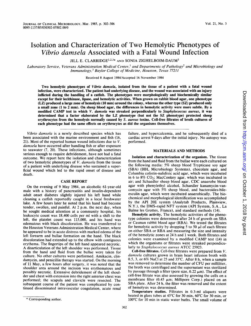
FIG. 1. Gram stain of ground tissue from hand, with bands ofheavy and light staining on the single organism.FIG. 2. Gram stain of organisms grown on an SBA plate. The
elongate form was unusual.
filtrate and large volume of water at the given temperaturesensured rapid equilibration. Controls were incubated at 4 or35°C for 30 min.
V. damsela reference organisms. V. damsela ATCC 33538(CDC 983-79) was obtained from the American Type CultureCollection. Strains CDC 1471-82, CDC 1421-81, and CDC727-82 were obtained from the Centers for Disease Control.
RESULTSNo organisms were seen on Gram stain of the bullae fluid,
and only two colonies grew on a single SBA plate. However,all the blood-containing plates showed hemolysis whereverthe specimen had been streaked, even in the absence ofgrowth in those areas. At several stabs there were no
colonies, but a double zone of hemolysis was observed. TheGram stain of ground tissue showed large, vacuolated,noncurved gram-negative rods (Fig. 1). The tissue inoculumyielded heavy growth of a single organism on all platesexcept on the Columbia colistin-nalidixic acid, Shaedlersheep blood-phenethyl alcohol, and kanamycin-vancomycinagars. Two colony types with slightly different zones ofhemolysis around the colony were observed on SBA andwere subsequently isolated in pure culture. The large-zonecolony type (LZ) and the small-zone colony type (SZ) wereboth identified as V. damsela by the criteria listed in Table 1(24). All tests were conducted under standard conditions andin 2 to 3% NaCI. Of interest is that the urea hydrolasereaction was negative when assayed with 3% NaCI. Theseisolates differed from those in another study (16) in that, on
primary isolation, they grew equally well at 25 and 35°C onSBA and grew poorly on MacConkey agar (no growth at 24h and colonies of about 1 mm at 48 h). On casein-tyrosine-starch plates (usually used for Nocardia identification) andon DNase plates, the organisms grew only when NaCl was
added and gave negative results. The triple sugar-iron-agarreaction was alkaline over acid with gas and no H2S produc-tion. The API 20E system (Analytab) codes at both 25 and35°C incubation were 2014004 and 2015004, which were
interpreted with the API data base as Pseudomonas sp.fluorescence group. The DMS-rapid NFT system code5300244 was not in this data base. The identification as V.damsela was confirmed by the Houston City Health Depart-ment laboratory.On Gram stain, organisms from a colony were observed as
large, pleomorphic, and evenly stained gram-negative rods
(Fig. 2). The colony size (about 3 mm at 24 h) and morphol-ogy of LZ and SZ were the same on both RBA and SBA, andthe hemolytic zones were only slightly different on SBA(about 1 mm for LZ and 0.5 mm for SZ). However, on RBAand LZ had a hemolytic zone of about 10 mm, and the SZhemolytic zone was about 1 to 2 mm. In addition, the LZshowed a line of increased hémolysis on SBA at the mid-point between two colonies, whereas the SZ did not (Fig. 3).Both the SZ and LZ strains grew to 1 x 10⁸, to 3 x 10⁸CFU/ml in 8 h at 25 and 35°C and in 0.5 and 3% NaCl. Theextent of organism growth in 6% NaCl was considerablyless. The SZ filtrates from broth cultures grown at 25°C in3% NaCl, at 35°C in 3% NaCI, at 25°C in 0.5% NaCl, and at35°C in 0.5% NaCI all caused slightly less partial hemolysison SBA than on RBA. Under the same four conditions, theLZ filtrates caused complete hemolysis on RBA and slightlymore hemolysis on SBA than had the SZ filtrates. Duringprolonged incubation (3 days) at 25 or 35°C, the LZ filtratesproduced concentric rings of hemolysis on RBA.A modified CAMP test was used to examine the synergy
between the hemolysins of V. damsela and those of S.
TABLE 1. Test results for the identification of V. damselaisolates
Test method(s) Positive tests Negative tests
API 20E system Urea hydrolysis,a Beta-galactosidase,(350C) arginine, dihydro- lysine, and orni-
lase, glucose utili- thine decarboxyl-zation, nitrate re- ase; indole, aceto-duction in, and H2S
production; gelatinhydrolysis; trypto-phan deaminase;utilization of man-nitol, inositol sorbi-tol, rhamnose,sucrose melibine,amygdalin, andarabinose
DMA-Rapid NFT Urea hydrolysis,a Esculin hydrolysis;System (30°C) arginine dihydro- assimilation of glu-
lase, glucose fer- cose, arabinose,mentation, beta-ga- mannose, mannitol,lactosidase (weak), N-acetylglucosa-gelatin hydrolysis mine, D-gluconate,(at 3 days), malate caprate, adipate, ci-and maltose assimi- trate, and phenylal-lation anine
Standard methods Urea hydrolysis; gela- Growth at 4 and 42°C;tin hydrolysis; growth in 0% NaCl-growth on TCBSb nutrient agar; in-agar; growth on sal- dole production;monella-shigella phenylalanine, de-agar; motility at 25 aminase; hydrolysisand 35°C; catalase, of casein, tyrosine,oxidase (slow), and starch'maltose, and glu-cose fermentation;growth in 6% NaCl-nutrient broth (vari-able and less thanin 3%); acetoin pro-duction" methyl redtest'; production ofDNase<', lecithin-ase, and lipase (SZ)
In the presence of 3% NaCI the reaction was negative.b TCBS, Thiosulfate-citrate-bile saits-sucrose.' Tests were performed in the presence of 2 to 3% NaCI.
VOL. 21, 1985
.01
t)w 4è. «41b
...k
on October 1, 2018 by guest
http://jcm.asm
.org/D
ownloaded from

304 CLARRIDGE AND ZIGHELBOIM-DAUM
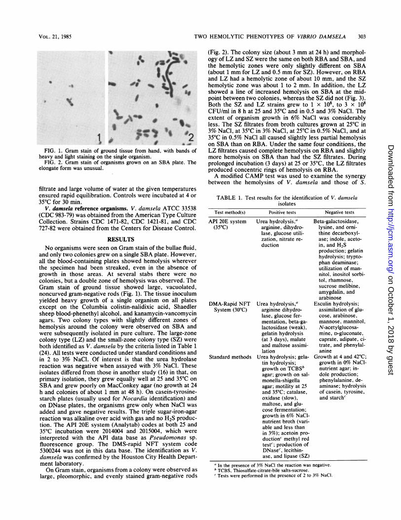
FIG. 3. Thin line of augmented hemolysis between two LZcolonies on SBA.
aureus. The LZ and SZ phenotypes of V. damsela were eachstreaked perpendicularly to a beta-toxin-producing strain ofS. aureus on an SBA plate. Although there was a slightincrease in hemolysis with the SZ strain, the LZ strainprevented the hemolysis of the erythrocytes usually causedby S. aureus (Fig. 4). Similar results were achieved with thecell-free filtrates (Fig. 5) and with organisms grown on top ofa Millipore filter placed on an SBA plate. The prevention ofhemolysis depended upon the LZ filtrate reaching the sheeperythrocytes before the S. aureus toxin. If the LZ filtratewas placed on the SBA plate from 1 to 24 h before the
-~~~~~~~~~~~~.~~~~
FIG. 5. Results of modified CAMP test with cell-free filtrate fromLZ at site 1 and SZ at site 2 placed perpendicularly to the S. aureusstreak.
addition of S. aureus filtrate, the whole area under the dropwas protected from the S. aureus hemolysin. Since V.damsela grew faster and produced toxin sooner than S.aureus, when both organisms were inoculated at the sametime on an SBA plate the usual pattern of protection was anX that crossed at the S. aureus inoculation line (Fig. 4a and4b). If the S. aureus hemolysin was placed on the SBA platefirst, the LZ filtrate could not protect that area. The filtratesshowed no loss of activity after incubation at 4, 35, or 45°Cfor 30 min. However, SZ filtrates lost all activity after the 60and 100°C treatments. After being heated to 100°C, but not
FIG. 4. Results of a modified CAMP test. S. aureus was streaked vertically on the SBA plate, and strains of V. damsela were streakedperpendicularly. (a) Plate after 24 h at 35°C, with augmented hemolysis at sites 3 and 5. (b) The same plate after overnight incubation at 4°C,with an absence of hemolysis at sites 1, 2, and 4. Numbers 1 through 5 refer to V. damsela strains LZ, CDC 737, SZ, CDC 1421, and ATCC33538, respectively.
J. CLIN. MICROBIOL.
on October 1, 2018 by guest
http://jcm.asm
.org/D
ownloaded from
TWO HEMOLYTIC PHENOTYPES OF VIBRIO DAMSELA 305
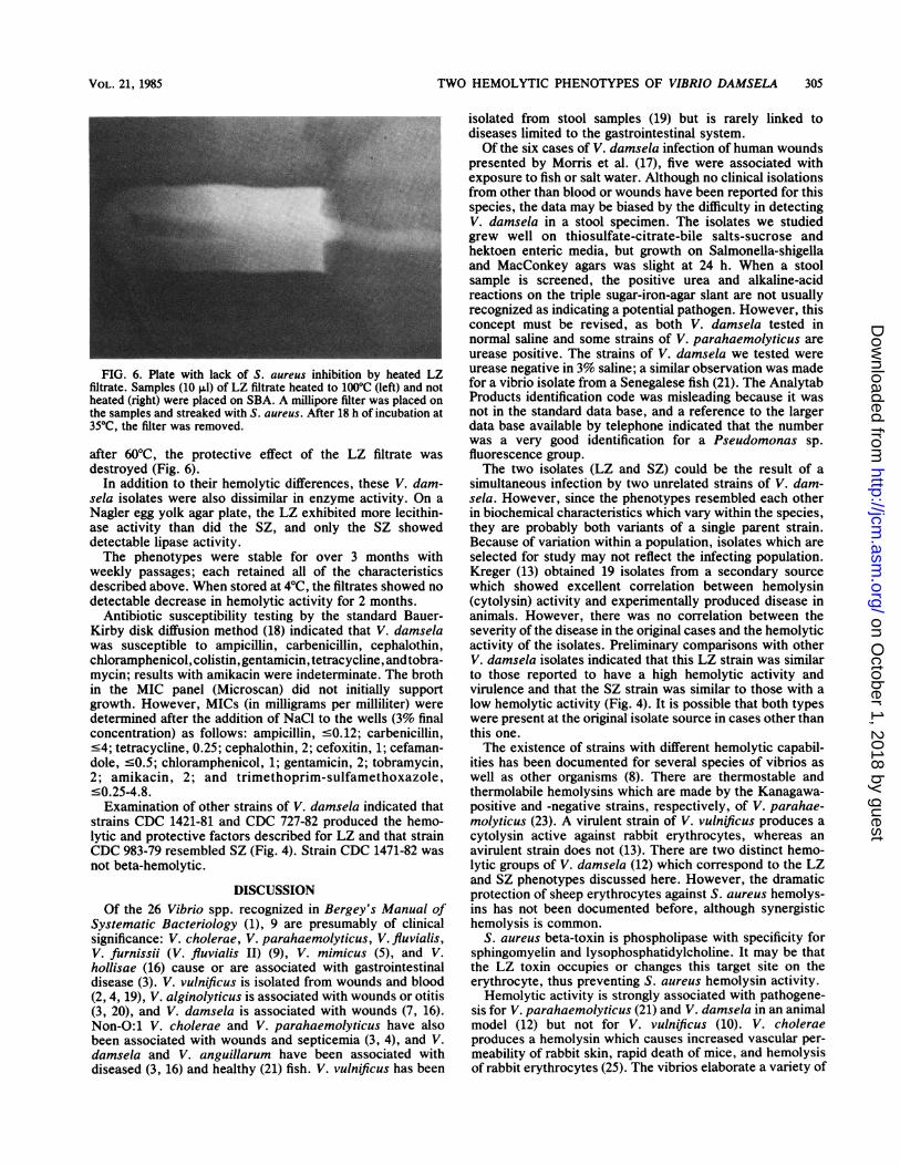
FIG. 6. Plate with lack of S. aureus inhibition by heated LZfiltrate. Samples (10 pul) of LZ filtrate heated to 100°C (left) and notheated (right) were placed on SBA. A millipore filter was placed onthe samples and streaked with S. aureus. After 18 h of incubation at35°C, the filter was removed.
after 60°C, the protective effect of the LZ filtrate wasdestroyed (Fig. 6).
In addition to their hemolytic differences, these V. dam-sela isolates were also dissimilar in enzyme activity. On aNagler egg yolk agar plate, the LZ exhibited more lecithin-ase activity than did the SZ, and only the SZ showeddetectable lipase activity.The phenotypes were stable for over 3 months with
weekly passages; each retained all of the characteristicsdescribed above. When stored at 4°C, the filtrates showed nodetectable decrease in hemolytic activity for 2 months.
Antibiotic susceptibility testing by the standard Bauer-Kirby disk diffusion method (18) indicated that V. damselawas susceptible to ampicillin, carbenicillin, cephalothin,chloramphenicol, colistin, gentamicin, tetracycline, andtobra-mycin; results with amikacin were indeterminate. The brothin the MIC panel (Microscan) did not initially supportgrowth. However, MICs (in milligrams per milliliter) weredetermined after the addition of NaCi to the wells (3% finalconcentration) as follows: ampicillin, <O.12; carbenicillin,<4; tetracycline, 0.25; cephalothin, 2; cefoxitin, 1; cefaman-dole, <0.5; chloramphenicol, 1; gentamicin, 2; tobramycin,2; amikacin, 2; and trimethoprim-sulfamethoxazole,<0.25-4.8.Examination of other strains of V. damsela indicated that
strains CDC 1421-81 and CDC 727-82 produced the hemo-lytic and protective factors described for LZ and that strainCDC 983-79 resembled SZ (Fig. 4). Strain CDC 1471-82 wasnot beta-hemolytic.
DISCUSSIONOf the 26 Vibrio spp. recognized in Bergey's Manual of
Systematic Bacteriology (1), 9 are presumably of clinicalsignificance: V. cholerae, V. parahaemolyticus, V. fluvialis,V. furnissii (V. fluvialis II) (9), V. mimicus (5), and V.hollisae (16) cause or are associated with gastrointestinaldisease (3). V. vulnificus is isolated from wounds and blood(2, 4, 19), V. alginolyticus is associated with wounds or otitis(3, 20), and V. damsela is associated with wounds (7, 16).Non-0:1 V. cholerae and V. parahaemolyticus have alsobeen associated with wounds and septicemia (3, 4), and V.damsela and V. anguillarum have been associated withdiseased (3, 16) and healthy (21) fish. V. vulnificus has been
isolated from stool samples (19) but is rarely linked todiseases limited to the gastrointestinal system.Of the six cases of V. damsela infection of human wounds
presented by Morris et al. (17), five were associated withexposure to fish or salt water. Although no clinical isolationsfrom other than blood or wounds have been reported for thisspecies, the data may be biased by the difficulty in detectingV. damsela in a stool specimen. The isolates we studiedgrew well on thiosulfate-citrate-bile saIts-sucrose andhektoen enteric media, but growth on Salmonella-shigellaand MacConkey agars was slight at 24 h. When a stoolsample is screened, the positive urea and alkaline-acidreactions on the triple sugar-iron-agar slant are not usuallyrecognized as indicating a potential pathogen. However, thisconcept must be revised, as both V. damsela tested innormal saline and some strains of V. parahaemolyticus areurease positive. The strains of V. damsela we tested wereurease negative in 3% saline; a similar observation was madefor a vibrio isolate from a Senegalese fish (21). The AnalytabProducts identification code was misleading because it wasnot in the standard data base, and a reference to the largerdata base available by telephone indicated that the numberwas a very good identification for a Pseudomonas sp.fluorescence group.The two isolates (LZ and SZ) could be the result of a
simultaneous infection by two unrelated strains of V. dam-sela. However, since the phenotypes resembled each otherin biochemical characteristics which vary within the species,they are probably both variants of a single parent strain.Because of variation within a population, isolates which areselected for study may not reflect the infecting population.Kreger (13) obtained 19 isolates from a secondary sourcewhich showed excellent correlation between hemolysin(cytolysin) activity and experimentally produced disease inanimals. However, there was no correlation between theseverity of the disease in the original cases and the hemolyticactivity of the isolates. Preliminary comparisons with otherV. damsela isolates indicated that this LZ strain was similarto those reported to have a high hemolytic activity andvirulence and that the SZ strain was similar to those with alow hemolytic activity (Fig. 4). It is possible that both typeswere present at the original isolate source in cases other thanthis one.The existence of strains with different hemolytic capabil-
ities has been documented for several species of vibrios aswell as other organisms (8). There are thermostable andthermolabile hemolysins which are made by the Kanagawa-positive and -negative strains, respectively, of V. parahae-molyticus (23). A virulent strain of V. vulnificus produces acytolysin active against rabbit erythrocytes, whereas anavirulent strain does not (13). There are two distinct hemo-lytic groups of V. damsela (12) which correspond to the LZand SZ phenotypes discussed here. However, the dramaticprotection of sheep erythrocytes against S. aureus hemolys-ins has not been documented before, although synergistichemolysis is common.
S. aureus beta-toxin is phospholipase with specificity forsphingomyelin and lysophosphatidylcholine. It may be thatthe LZ toxin occupies or changes this target site on theerythrocyte, thus preventing S. aureus hemolysin activity.Hemolytic activity is strongly associated with pathogene-
sis for V. parahaemolyticus (21) and V. damsela in an animalmodel (12) but not for V. vulnificus (10). V. choleraeproduces a hemolysin which causes increased vascular per-meability of rabbit skin, rapid death of mice, and hemolysisof rabbit erythrocytes (25). The vibrios elaborate a variety of
VOL. 21, 1985
on October 1, 2018 by guest
http://jcm.asm
.org/D
ownloaded from
306 CLARRIDGE AND ZIGHELBOIM-DAUM
extracellular toxins and enzymes, many of which couldcause cell damage (6, 11-13, 15, 22, 23, 25). The exactmechanism by which these factors cause the rapid andremarkable clinical manifestations documented here is notknown.
ACKNOWLEDGMENTSThe excellent secretarial assistance of Lori Shell, the expert
technical skills of Bobbye Simon, and the cooperation of the Mi-crobiology Laboratory of the Veterans Administration Medical Cen-ter are appreciated.
LITERATURE CITED
1. Baumann, P., and R. H. Schubert. 1984. Vibrionaceae, p.516-517. In N. R. Krieg and J. G. Holt (ed.), Bergey's manualof systematic bacteriology, vol. 1. The Williams & Wilkins Co.,Baltimore.
2. Blake, P. A., H. A. Merson, R. E. Weaver, D. G. Hollis, andP. L. Heublein. 1979. Disease caused by marine vibrio: clinicalcharacteristics and epidemiology. N. Engl. J. Med. 300:1-5.
3. Blake, P. A., R. E. Weaver, and D. G. Hollis. 1980. Disease ofhumans (other than cholera) caused by vibrios. Annu. Rev.Microbiol. 34:341-367.
4. Bonner, J. R., A. S. Coker, C. R. Berryman, and H. M. Pollock.1983. Spectrum of vibrio infections in a gulf coast community.Ann. Intern. Med. 99:464-469.
5. Davis, B. R., G. R. Fanning, J. M. Madden, A. G. Steigerwalt,H. B. Bradford, Jr., H. L. Smith, Jr., and D. J. Brenner. 1981.Characterization of biochemically atypical Vibrio choleraestrains and designation of a new pathogenic species, Vibriomimicus. J. Clin. Microbiol. 14:631-639.
6. Desmond, E. P., J. M. Janda, F. I. Adams, and E. J. Bottone.1984. Comparative studies and laboratory diagnosis of Vibriovulnificus, an invasive Vibrio sp. J. Clin. Microbiol. 19:122-125.
7. Fernandez, C. R., and G. A. Pankey. 1975. Tissue invasion byunnamed marine vibrios. J. Am. Med. Assoc. 233:1173-1176.
8. Freer, J. H., and J. P. Arbuthnott. 1983. Toxins of Staphylo-coccus aureus. Pharmacol. Ther. 19:55-106.
9. Hickman-Brenner, F. W., D. J. Brenner, A. G. Steigerwalt, M.Schreiber, S. D. Holmberg, L. M. Baldy, C. S. Lewis, N. M.Pickens, and J. J. Farmer III. 1984. Vibrio fluvialis and Vibriofurnissii isolated from a stool sample of one patient. J. Clin.Microbiol. 20:125-127.
10. Johnson, D. S., and F. M. Calia. 1981. Hemolytic reaction ofclinical and environmental strains of Vibrio vulnificus. J. Clin.Microbiol. 14:457-459.
11. Kabir, S., N. Ahmad, and S. Ali. 1984. Neuraminidase produc-tion by Vibrio cholerae O1 and other diarrheageneic bacteria.Infect. Immun. 44:747-749.
12. Kreger, A. S. 1984. Cytolytic activity and virulence of Vibriodamsela. Infect. Immun. 44:326-331.
13. Kreger, A., and D. Lockwood. 1981. Detection of extracellulartoxin(s) produced by Vibrio vulnificus. Infect. Immun.33:583-590.
14. Lennette, E. H., A. Balows, W. J. Hausler, Jr., and J. P. Truant(ed.). 1980. Manual of clinical microbiology, 3rd ed. AmericanSociety for Microbiology, Washington, D.C.
15. Lockwood, D. E., A. S. Kreger, and S. H. Richardson. 1982.Detection of toxins produced by Vibriofluvialis. Infect. Immun.35:702-708.
16. Love, M., D. Teebken-Fisher, J. E. Hose, J. J. Farmer III, F. W.Hickman, and G. R. Fanning. 1981. Vibrio damsela, a marinebacterium, causes skin ulcers on the damselfish Chromis punc-tipinnis. Science 214:1139-1140.
17. Morris, J. G., Jr., R. Wilson, D. G. Hollis, R. E. Weaver, H. G.Miller, C. O. Tacket, F. W. Hickman, and P. A. Blake. 1982.Illness caused by Vibrio damsela and Vibrio hollisae. Lanceti:1294-1297.
18. National Committee for Clinical Laboratory Standards. 1983.Performance standards for antimicrobic disk susceptibility tests.Tentative Standards. National Committee for Clinical Labora-tory Standards, Villanova, Pa.
19. Pollak, S. J., E. F. Parrish III, T. J. Barrett, R. Bretler, andJ. G. Morris. 1983. Vibrio vulnificus septicemia. Isolation oforganisms from stool and demonstration of antibodies by directimmunofluorescence. Arch. Intern. Med. 143:837-838.
20. Ryan, W. J. 1976. Marine vibrios associated with superficialseptic lesions. J. Clin. Pathol. 29:1014-1015.
21. Schandevyl, P., E. Van Dyck, and P. Piot. 1984. HalophilicVibrio species from seafish in Senegal. Apple. Environ. Mi-crobiol. 48:236-238.
22. Smith, G. C., and J. R. Merkel. 1982. Collagenolytic activity ofVibrio vulnificus: potential contribution to its invasiveness.Infect. Immun. 35:1155-1156.
23. Takeda, Y. 1983. Thermostable direct hemolysin of Vibrioparahaemolyticus. Pharmacol. Ther. 19:123-146.
24. Weaver, R. E., D. G. Hollis, W. A. Clark, and P. Riley. 1983.Revised tables from the identification of unusual pathogenicgram-negative bacteria. Centers for Disease Control, Atlanta,Georgia.
25. Yamamoto, K., M. AI-Omani, T. Honda, Y. Takeda, and T.Miwatani. 1984. Non-01 Vibrio cholerae hemolysin: purifica-tion, partial characterization, and immunological relatedness toEl Tor hemolysin. Infect. Immun. 45:192-196.
J. CLIN. MICROBIOL.
on October 1, 2018 by guest
http://jcm.asm
.org/D
ownloaded from