isolation of anaerobic bacteria from human gingiva mouse … · the chamber was heated to 37 c and...
TRANSCRIPT

APPLIED MICROBIOLOGY, Apr. 1969, p. 568-576Copyright ( 1969 American Society for Microbiology
Vol. 17, No. 4Printed in U.S.A.
Isolation of Anaerobic Bacteria from Human Gingivaand Mouse Cecum by Means of a Simplified Glove
Box Procedure'ALEXANDER ARANKI,2 SALAM A. SYED, ERNEST B. KENNEY, AND ROLF FRETER
Department of Microbiology, The University of Michigan, Ann Arbor, Michigan 48104
Received for publication 7 February 1969
An anaerobic glove box constructed of clear flexible vinyl plastic is described. It issufficiently inexpensive and simple in operation to be used not only in research butalso in a clinical laboratory by technicians without special training. Conventionalbacteriological techniques may be used inside the glove box for culturing and trans-ferring anaerobic bacteria. The box may be heated to 37 C and thus serve as an
anaerobic incubator as well, permitting inspection of cultures at any time. Mediamay be prepared and agar plates may be poured on the laboratory bench in theconventional manner. An overlay of trace amounts of palladium black catalyst overplated agar media reduces the medium to an oxidation-reduction (0-R) potentialof -300 mv within 2 days after introduction into the glove box. In spite of its greatersimplicity, the system matched or excelled the roll tube method with respect to allparameters tested, including O-R potential obtainable in the media, 02 concentra-tion in the gas phase, and efficiency in isolating anaerobic bacteria from the mousececum. Comparative studies indicate that the conventional anaerobic jar methodwas inadequate for the isolation of strict anaerobes from human gingival specimensand from the mouse cecum. This was due to the exposure of specimens and media toair during plating on the open laboratory bench. Anaerobic jars were adequate formaintaining the proper conditions for growth of anaerobic bacteria once these hadbeen established in the glove box.
As reviewed in an earlier publication (14),anaerobic bacteria are now recognized as thepredominant component of the intestinal flora ofman and mammals. Human oral flora also con-tains a large proportion of anaerobes (8).
It is also well known that the majority of theseanaerobic bacteria cannot be cultured with con-ventional anaerobic technique (10, 14, 15). Re-cent studies in this laboratory (14) have shownthat a significant improvement in the recovery ofanaerobes from the mouse cecum may be obtainedif care is taken to avoid even momentary expo-sure of the specimen and of the medium to atmos-pheric oxygen. These latter studies were carriedout by use of Hungate's (4) roll tube technique.
In Hungate's method, exclusion of air isachieved by simply using rubber-stoppered tubesor flasks for the preparation and tubing of media.During transfer of media or specimens, atmos-pheric oxygen is kept out of the tubes by im-
1 A preliminary account of this study was given earlier (Bac-teriol. Proc., p. 94, 1968).
2 Present address: Escuela Dental, Laboratorio Microbiologia,Avenida Santa Maria 571, Santiago, Chile.
mediately introducing a stream of sterile inert gaswhenever the rubber stopper is removed. Isolatedcolonies are obtained from specimens with mixedflora by incorporating serial dilutions of thespecimen into liquid agar medium, which is thenallowed to solidify on the walls of a rubber-stoppered test tube (roll tube). The roll tubetechnique is thus the anaerobic equivalent of the''pour plate" method for enumerating bacteria.There can be no doubt that the Hungate
method has opened up an entire field of micro-biological endeavor, i.e., the ecology of the rumenflora. Nevertheless, when compared to conven-tional bacteriological procedures, such as thoseused for the isolation and identification of Entero-bacteriaceae in a clinical laboratory, the Hungatemethod is extremely complex. The preparationand tubing of media under exclusion of oxygenare time consuming. Since this is a pour platemethod, each specimen has to be serially dilutedfirst (under exclusion of oxygen), and then repli-cate roll tubes have to be prepared from eachdilution. An apparatus for streaking prehardenedroll tubes under a stream of inert gas has been de-
568
on April 22, 2020 by guest
http://aem.asm
.org/D
ownloaded from
ISOLATION OF ANAEROBES IN SIMPLIFIED GLOVE BOX
scribed by Moore (6). This constitutes a con-siderable improvement but is still more complexand less efficient than the standard "triple streak"agar plate known to all bacteriologists. Thesimultaneous handling of rubber stoppers, gasoutlets, and tubes during the transfer of culturesor media requires specially trained technicians.Therefore, we attempted to achieve the desiredexclusion of atmospheric oxygen by some moreconvenient means and thus explored the possi-bility of using oxygen-free glove boxes. Our ob-jective was to devise a method which would besufficiently simple and inexpensive to be used notonly in the research laboratory but also in aroutine diagnostic laboratory by technicians hav-ing no special training beyond routine bacterio-logical methods.
Anaerobic glove boxes for the cultivation ofbacteria have been described by Socransky et al.(13), Rosebury and Reynolds (9), Gravens et al.(Bacteriol. Proc., 1967, p. 77), and Draser (1).Those of Rosebury and Reynolds and Gravenset al. were complex and expensive metal chambersconsiderably beyond the technical competence ofthe average bacteriological technician and beyondthe budget of many laboratories. The other gloveboxes described in the literature require elaborateset-up procedures before each use, and for thisreason are unsuitable for prolonged or routineoperation. There is no data available in the litera-ture concerning the relative performance of gloveboxes in relation to other techniques. Socranskyet al. (13) and Rosebury (9) stated that spiro-chetes may be grown in the box as surface colonies.Drasar (1) reported that he can recover 1011bacteria from 1 g of human feces.
Described below is an inexpensive glove boxwhich is simple to operate and which can beassembled entirely from components that arecommercially available (Fig. 1). [Those interestedin using a glove box may obtain a list of all neces-sary components, supplies, commercial sources,and drawings by writing to one of us (R. F.).The entire outfit may be purchased for less than$1,000.] Data on the efficiency of this device inrecovering anaerobic bacteria is given and is re-
lated to the performance of other methods for thecultivation of anaerobic bacteria.
MATERIALS AND METHODS
The glove box is constructed of flexible 20-milclear vinyl plastic with 25-mil neoprene gloves. Thisis the type of chamber which is now widely availablein a great variety of sizes for germ-free animal work.A chamber 2 by 2 by 3 feet (60.9 by 60.9 by 91.4 cm)wide is sufficient if only the plating and transferring ofcultures are to be carried out in the chamber. In thiscase, the cultures must be incubated in anaerobic jarsoutside the glove box. The work described below was
carried out in a glove box 2 by 2 by 7 feet (60.9 by60.9 by 213.3 cm) wide with two pairs of gloves in thefront panel. The chamber was heated to 37 C andhalf of it was used simply as an anaerobic incubator.This eliminates the need for anaerobic jars and hasthe further advantage that cultures may be observedat any time during incubation.The plastic chamber is clamped with a worm-drive
steel clamp to a round steel airlock, 18 inches (45.7cm) long by 12 inches (30.5 cm) in diameter. Thelock has an airtight acrylic plastic door at either endwhich may be closed by tightening a single bolt. Ontop of the lock are two valves, one connecting to thegas tank, the other to a vacuum pump. A vacuumgauge on the top of the airlock completes the list ofcontrols. Inside the chamber, a blower located at oneend of the glove box forces the interior atmospherethrough a plastic hose into a rectangular box, locatedat the opposite end. The top of the box is open andholds a removable tray of stainless-steel screeningwhich is covered with catalyst pellets [palladium-coated alumina, Engelhard; the same material wasfirst used in a glove box by Rosebury and Reynolds(9)]. This system continuously circulates the chamberatmosphere through the catalyst. It has no connec-tion to the outside.
If one wishes to use part of the glove box as ananaerobic incubator, a cone-type heating element isplaced into the box below the catalyst tray. The heateris controlled by a bimetallic thermostat located at theblower intake. This arrangement holds the temperaturein the chamber to within -4l C. Closer temperaturecontrol may be achieved by placing an insulatingblanket of styrene foam on top of the glove box.The atmosphere in the chamber used in the experi-
ments described below was a mixture of 10% H2 and5% CO2 in N2. We also used 10% H2 in 90% CO2which, in preliminary studies, gave no better recoveryof anaerobes from our specimens than the abovemixture. Gases may be obtained premixed from mostsuppliers. Any mixture can be used as long as it con-tains some H2 to react with oxygen in the presence ofthe catalyst.
During development of the glove box technique,oxygen levels were determined with a trace oxygenanalyzer (model GP; Lockwood & McLorie, Hor-sham, Pa.). Precise oxygen analysis is not requiredin routine use, because the proper function of theglove box can be controlled by adding oxidation-reduction (O-R) indicator dye to the media.
Electric leads are introduced into the plastic cham-ber through a rubber stopper which fits into a plasticnipple in the rear wall. The bacteriological loops usedin the glove box have metal handles (Matheson no.51847-10). The tip of the handle is attached via analligator clip to a thin flexible electric cord leading toa 6-v transformer. The second lead from the trans-former connects to a piece of sheet metal [1 by 1 inch(2.54 by 2.54 cm)] mounted on a suitable stand. Whenthe tip of the loop touches this metal, the loop wireheats up to the proper (red-hot) temperature forsterilization. A coil of Nichrome wire is connectedvia a foot switch to the 6-v transformer. When the
569VOL. 17, 1969
on April 22, 2020 by guest
http://aem.asm
.org/D
ownloaded from
570 ARANK
switch is depressed, the coil heats up to red heat andserves to "flame" the rim of test tubes and flasks.A maximum-minimum indicating thermometer
and a hygrometer are kept in the glove box for controlpurposes. A tray containing 2 lb of reusable silica geldesiccant (Tel-Tale, Davison Commercial Chemical,Baltimore, Md.) serves to keep the relative humidityin the glove box between 30 and 50%. At higherhumidities, the drying of agar plates is impaired andthe problem of swarming bacteria becomes serious.When the glove box is set up for the first time, it is
evacuated by opening the inside door of the lock andturning on the vacuum pump. The chamber thensimply collapses, draping itself tightly around anyitems which may already be inside. (Articles too largeto pass through the lock, such as shelves, may beintroduced before the plastic chamber is clamped tothe lock.) After evacuation, the valve leading to thepump is closed, and the chamber is filled with the gasmixture by opening the gas valve on top of the lock.After the chamber is inflated, the inner door of thechamber is closed. A tray with catalyst is then in-troduced into the box in the usual manner and placedon top of the catalyst box. Within 1 to 2 days, theoxygen concentration drops to below 10 ppm and theglove box is ready to be used indefinitely. In normaluse, it is not necessary to add gas to the chamberatmosphere.To introduce dry materials into the glove box,
these materials are placed in the lock via the outsidedoor, keeping the inside door locked. The lock isthen evacuated to 1 mm of Hg. After closing thevalve to the vacuum pump, the lock is filled with gasmixture from the tank, the inside door is opened, andthe materials are taken into the chamber. With avacuum pump with a free air capacity of 75 liters/min, the entire process takes less than 5 min.
Liquid media and agar plates are introduced in asimilar manner except that the lock is evacuated toonly 4 cm of Hg. The chamber is then filled with gas,and the process of evacuation and filling is repeatedtwice. The items may then be taken into the glovebox by opening the inner door of the lock. With apump with a free air capacity of 75 liters/min, evacua-tion of the lock to 4 cm ofHg takes only a few seconds,and the entire process of introducing liquids can becompleted in 5 min. For reasons of economy, we usedless expensive nitrogen from a separate tank connectedto the lock to fill the lock after the first two cycles ofevacuation, with the gas mixture only for the finalfilling. Depending on the widely divergent pricescharged for gas mixtures by the various manufacturers,one entry (i.e., one filling of the lock with gas mixture)costs between $0.10 and $0.25.To remove items from the glove box, the inside
door of the lock is opened, the items are placed intothe lock, the inside door is closed, and the items areremoved via the outside door. No gas needs to beexpended for withdrawals from the chamber whenthese operations are carried out alternately withintroduction of materials into the chamber.During routine use of the glove box, the efficiency
of the catalyst decreases in time. It can be rejuvenatedby heating to 160 C for 2 to 4 hr (same conditions as
.I ET AL. APPL. MICROBIOL.
for sterilizing glassware). We have two trays of catalystfor each glove box which are exchanged when theoxygen concentration exceeds 10 ppm. In chamberswithout oxygen analyzer, a routine schedule of ex-change and rejuvenation of catalyst trays twice aweek is recommended. The catalyst should be replacedcompletely every 4 months. This schedule gives anample margin of safety.
Probable reasons for the decrease in catalystefficiency are moisture and poisoning by H2S elab-orated by the bacteria. Both of these are removed onheating to 160 C. To reduce the release of H2S intothe glove box, broth cultures were kept in screw-captubes and flasks. Agar plates were kept in 0.5 gallon(1.9 liter) size freezer-type plastic containers (SearsRoebuck & Co., no. 9704). These containers werefitted with a suspended bottom of galvanized-steelwire mesh. The space under the wire bottom wasfilled with a slurry of PbCO3 in water which absorbedany H2S formed.
Media. For the cultivation of bacteria from toothscrapings, a slight modification of Huntoon's mediumwas used. This is essentially an enriched veal infusionagar and is referred to below as EVIA. It was com-posed of Veal Infusion Agar (Difco), 40 g/liter;menadione, 5 .g/ml; cysteine hydrochloride, 0.05%O;Na2CO3, 0.056%; and normal rabbit serum, 10%.The ingredients were sterilized and plated as de-scribed below for enriched Trypticase Soy Agar(ETSA).ETSA was used for the cultivation of bacteria from
the mouse cecum. It was composed of trypticaseSoy Broth (TSB; BBL), 30 g/liter; hemin, 1 /Ag/ml;menadione, 0.5 ,ug/ml; cysteine hydrochloride,0.05%; Na2CO3, 0.042%; and agar (Difco), 2%. Thebase medium was autoclaved in a cotton-stopperedErlenmeyer flask at 120 C for 20 min and cooled to 56C in a water bath. Menadione was made up in alcoholicstock solution, sterilized by filtration, and added to theautoclaved base medium. Cysteine hydrochloride andNa2CO3 were autoclaved separately in aqueous solu-tion and added to the base medium after autoclaving.Hemin was kept in alkaline alcoholic stock solutionand was autoclaved with the base medium. Rabbitserum was sterilized by filtration and added to thesterile EVIA medium at 56 C. The concentration ofNa2CO3 had been adjusted in preliminary experimentsto give a neutral medium after equilibration in theglove box atmosphere containing 5% CO2.
Agar media were poured into glass petri dishescovered with metal covers carrying an absorbent disc(BBL no. 05-264-A). A small amount (0.05 ml of a0.1% solution) of indigo carmine or phenosafranineindicator dye was placed into the bottom of two petridishes and mixed with the agar as it was poured. Theseserved as control plates. The media were placed intothe glove box as soon as possible after hardening. Theywere used as soon as the dye in the control plates hadnoticeably faded or had become entirely colorless.The control plate was then removed from the glovebox, in which case the original color returned imme-diately. Reduction of indigo carmine was usually com-plete within 2 days.
Media to be plated with a palladium black overlay
on April 22, 2020 by guest
http://aem.asm
.org/D
ownloaded from

VOL. 17, 1969 ISOLATION OF ANAEROBES IN SIMPLIFIED GLOVE BOX
were autoclaved in two portions. The first portionwas plated as usual in a shallow layer (15 to 20 mlin a dish with an inner diameter of 9 cm). When thislayer had solidified, the second portion of mediumwas mixed with palladium black (Fisher no. P-3)which had been dry sterilized in a test tube for 2 hrat 160 C. Sufficient palladium black to make a 1 mg/mlsuspension was used, and 10 ml of this was placedon top of the primary layer of medium. Controlplates with phenosafranine were prepared as describedabove, by mixing the dye with the lower layer ofmedium.Two days after introduction into the glove box,
all agar plates were placed into closed plastic con-tainers, as described above, in order to minimizedrying and to trap H2S from those plates which hadalready been inoculated.
Diluting fluid was composed of TSB (BBL), 30g/liter; Na2CO3, 0.042%; and cysteine hydrochloride,0.05%. The three ingredients were autoclaved sepa-rately and mixed afterwards as described for agarmedia above. The fluid was then dispensed in 9-mlamounts into sterile screw-cap test tubes (for makingserial dilutions before quantitative culture) or in 100-ml amounts in Erlenmeyer flasks (for use in theWaring Blendor). The broth was introduced into theglove box immediately after dispensing and was usedonly after it had remained there for at least 2 days.O-R potential. Measurements were taken with a
Beckman model G pH meter located inside the glovebox. In liquid media, the reading of three freshlyburnished platinum electrodes (Beckman no. 39273)against a Calomel electrode (Beckman no. 39170) weretaken. The average of the three readings was correctedfor the voltage of the Calomel electrode to give theEh. The potentials of the three electrodes agreedclosely. The system was tested against media contain-ing various O-R indicator dyes and gave the theoreticalpotentials during the progressive reduction of theseindicators. O-R potentials on the surface of agarplates were measured by pressing the platinum elec-trodes against the surface and reading potentialsagainst the reference part of a surface combinationelectrode (Beckman no. 39182). In all instances, thepH was measured with a Beckman glass electrode.It ranged between pH 6.9 and 7.1, and no correctionsfor pH were made in the Eh values presented in thispaper.
Gingival flora. Specimens were taken from a healthyvolunteer, who had his 32 teeth, no open cavities, andno periodontal pockets. His mouth had been cleanedin our School of Dentistry 6 months before this study.The samples were taken according to the criteria ofSocransky et al. (12), before breakfast and beforebrushing of the teeth. The teeth were isolated withcotton rolls and all 32 teeth were scraped with abacteriological loop. The material on the loop wasplaced into a rubber-stoppered tube of diluting fluidwhich had been removed from the glove box justbefore the experiment. While the stopper was opened,a stream of nitrogen was directed into the tube througha sterile 18-gauge hypodermic needle which was at-tached to a short piece of rubber tubing containingsterile cotton.
The specimen was then placed in the glove boxand blended for 30 sec in 100 ml of sterile dilutingfluid in a Waring Blendor at low speed (with a semi-micro jar, A. H. Thomas no. 4282-F). From thishomogenate, serial 10-fold dilutions were made indiluting fluid, with a Fisher no. 13-681-50 pipettefiller to operate the pipettes. Portions (0.1 ml) of theappropriate dilution were finally transferred to agarplates and spread evenly over the surface with asterile glass rod. Ten replicate plates were inoculatedfrom each dilution.
Microscopic counts were made from the originalhomogenate in a Petroff-Hausser chamber as describedearlier (14).
Inoculated media were incubated inside the glovebox, and the number of colonies appearing werecounted daily. When incubation was to be in anaerobicjars, these were filled inside the glove box. Heatedpalladium asbestos catalyst (5 g per jar) was includedin each jar. We used glass jars (desiccators kept closedby a slight vacuum), stainless-steel jars (Torbal jar),and inexpensive airtight jars made in our shop fromacrylic plastic tubing [wall, V8 inch (0.96 cm)] andacrylic plastic sheet [0.5 inch (1.27cm)]. (Anaerobicjars used in conjunction with the glove box do notrequire valves or gauges because no evacuation andgas filling is required.) There was no difference in theperformance of these various jars. For incubation ofcultures, the jars were removed from the glove boxto a conventional incubator at 37 C.When plating of the specimen was to be done out-
side the glove box (to simulate the conventionalanaerobic jar technique), a small amount of thehomogenate was taken from the blendor jar and re-moved from the chamber. Serial dilutions were thenmade in ordinary TSB (BBL) which was freshly pre-pared but had never been inside the glove box. Plat-ing was carried out as described above, except thatit was done outside the glove box. The agar platesused for this type of culture were of the same batchas used for plating inside the glove box. They wereremoved from the glove box 30 min before plating.The inoculated plates were incubated in anaerobicjars.
Mouse cecum. The animals used were strain CD-1from Charles River Laboratories and strain BALB/Wm maintained in this Department by WilliamMurphy. The animals were killed by cervical disloca-tion and immediately thereafter introduced into theglove box. The cecum was then removed underaseptic conditions and its contents were washed into asterile Waring Blendor jar containing 100 ml ofdiluting fluid. Subsequent steps were as describedabove for tooth scrapings.
Statistical analysis. The significance of the dif-ference between mean plate counts was analyzed bymeans of the t test with Table 3 of Fisher and Yates (2).
RESULTSOxygen tension in the glove box. During the
experiments described below, the 02 tension in theglove box was maintained between 5 and 10 ppm.In a chamber 2 by 2 by 7 feet (60.9 by 60.9 by
571
on April 22, 2020 by guest
http://aem.asm
.org/D
ownloaded from

ARANKI ET AL.
213.3 cm) wide and made of 20-mil vinyl plastic;this required 200 g of catalyst spread in a singlelayer over 5.5 by 10.5 inch (13.9 by 26.6 cm) wiremesh tray. With twice this amount of catalyst in a21 by 5.5 inch (53.3 by 13.9 cm) tray, the 02tension was maintained between 2 and 5 ppm. Wehave not as yet determined whether this lower 02concentration results in a higher rate of recoveryof anaerobes from our specimens. Vinyl chambersof thicker material (40 mil) were also used, butwith these we did not obtain significantly lower02 concentrations. It seems probable that boththe plastic and the rubber gloves are permeable tooxygen. When the catalyst was removed from anestablished glove box [2 by 2 by 7 feet wide (60.9by 60.9 by 213.3 cm), 20-mil vinyl] having aninitial 02 concentration of 5 to 10 ppm and aninternal temperature of 37 C, the O2 concentrationrose at a rate of 1 ppm per min. In the same boxkept at room temperature (29 C), the rise was0.6 ppm per min.The 02 concentration in an established glove
box is thus in a steady state determined by therate of inward diffusion of 02 and the rate of itsremoval by the catalyst. Theoretically this shouldresult in a lowering of the H2 concentration in thechamber over a period of several weeks. This is,however, not a practical consideration becausewith every entry of materials into the chamberfresh gas (approximately 4% of the total chambervolume) is mixed in with the gas already in theglove box.O-R potential of media. To evaluate various
methods for preparing media, TSB buffered with0.056%7 Na2CO3 was autoclaved for 20 min in500-ml amounts in cotton-stoppered Erlenmeyerflasks, cooled rapidly, and introduced into theglove box. A second batch of the same mediumwas prepared in a similar manner, except that theflasks were closed with a check valve which per-mitted escape of air during autoclaving but notits reintroduction after removal from the auto-clave. These flasks were cooled rapidly after auto-claving and were introduced into the glove boximmediately afterward. The medium was thuskept entirely free from contact with atmosphericoxygen. At periodic intervals of time beginningimmediately after introduction (time-zero), theO-R potential was measured in samples taken fromthe flasks. Broth autoclaved with a check valveshowed a potential of -280 mv immediatelyafter introduction into the glove box and this re-mained constant thereafter. Broth autoclaved incotton-stoppered flasks was at first quite oxidized(+65 mv), but after 1 day had reached the sameO-R potential as that autoclaved with a checkvalve. This was a constant observation in num-
erous repetitions of this experiment with this andother media. In all instances, media autoclaved incotton-stoppered flasks reached, after 1 to 2 daysin the glove box, the same O-R potential assimilar media autoclaved with a check valve.The above experiment was repeated with TSB
to which Na2CO3 and cysteine hydrochloride hadbeen added (i.e., our "diluting fluid"). Whenthis medium was autoclaved in cotton-stopperedflasks and introduced into the glove box, itsinitial O-R potential was already quite low (-200mv). Surprisingly, however, the final potential(-280 mv) was not lower than that of TSB with-out added cysteine. The reducing activity of thecysteine could only be demonstrated when themedium was poised by the addition of 0.0005%indigo carmine. In this case, reduction of the dyeoccurred several hours earlier in broth containingcysteine as compared to broth without this re-ducing agent. O-R potentials were also measuredon the surface of standard ETSA plates with andwithout paladium black overlay. The O-R po-tential in the presence of paladium catalyst wassignificantly more negative (-290 mv) than thatfound on plain ETSA plates (-250 mv).
Phenosafranine added to broth or regular agarmedia never became reduced. In contrast, agarplates with palladium black overlay consistentlyreduced phenosafranine, and this reaction is nowused routinely by us to control the proper prep-aration of agar media.
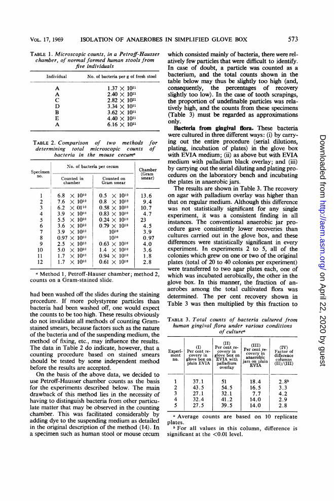
Microscopic bacterial counts. To relate thepresent results to those of others, our standardmethod of making Petroff-Hausser chambercounts was compared with a method involvingquantitation of a Gram-stained smear. In thismethod, the specimen was mixed with a suspen-sion of polystyrene particles of known concentra-tion as described by Socransky et al. (12). Aconventional Gram-stained slide was then pre-pared from this mixture. The total count ofbacteria in the original specimen was calculatedfrom the proportion of bacteria to polystyreneparticles in the average microscopic field. Sincehuman feces have been the standard of referencefor many authors, we used this type of specimenfirst. The counts obtained (Table 1) ranged from1.37 X 101" to 6.16 X 10¹¹ bacteria per g of freshfeces. These values are of the same order ofmagnitude as those reported by Van Houte andGibbons (15).
Table 2 shows comparative counts of bacteriain the mouse cecum obtained by the two methodsmentioned above. As may be seen, the counts ofGram-stained smears were consistently lower.We interpreted this as indicating that some bac-teria (and perhaps some polystyrene particles)
572 APPL. MICROBIOL.
on April 22, 2020 by guest
http://aem.asm
.org/D
ownloaded from
VOL. 17, 1969 ISOLATION OF ANAEROBES IN SIMPLIFIED GLOVE BOX
TABLE 1. Microscopic counts, in a Petroff-Hausserchamber, of normal formed human stools from
five individuals
Individual No. of bacteria per g of fresh stool
A 1.37 X 10¹¹A 2.40 X 10¹¹C 2.82 X 10'1D 3.34 X 101B 3.62 X 10'1E 4.40 X 10¹¹A 6.16 X 10¹¹
TABLE 2. Comparison of two methodsdetermining total microscopic counts
bacteria in the mouse cecum"
forof
No. of bacteria per cecum ChamberSpecimen -(Gram
no.
Counted in Counted on smear)chamber Gram smear
1 6.8 X 10¹⁰ 0.5 X 10¹⁰ 13.62 7.6 X 10¹⁰ 0.8 X 10¹⁰ 9.43 6.2 X 0110 0.58 X 10¹⁰ 10.74 3.9 X 10¹⁰ 0.83 X 10¹⁰ 4.75 5.5 X 10¹⁰ 0.24 X 10¹⁰ 236 3.6 X 10¹⁰ 0.79 X 10¹⁰ 4.57 3.9 X 10¹⁰ 1010 3.98 0.97 X 10¹⁰ 1010 0.979 2.5 X 10¹⁰ 0.63 X 10¹⁰ 4.010 5.0 X 10¹⁰ 1.4 X 10¹⁰ 3.611 1.7 X 10¹⁰ 0.94 X 10¹⁰ 1.812 1.7 X 101° 0.61 X 101° 2.8
a Method 1, Petroff-Hauser Chamber; method 2,Counts On a Gram-stained slide.
had been washed off the slides during the stainingprocedure. If more polystyrene particles thanbacteria had been washed off, one would expectthe counts to be too high. These results obviouslydo not invalidate all methods of counting Gram-stained smears, because factors such as the natureof the bacteria and of the suspending medium, themethod of fixing, etc., may influence the results.The data in Table 2 do indicate, however, that acounting procedure based on stained smearsshould be tested by some independent methodbefore the results are accepted.On the basis of the above data, we decided to
use Petroff-Hausser chamber counts as the basisfor the experiments described below. The maindrawback of this method lies in the necessity ofhaving to distinguish bacteria from other particu-late matter that may be observed in the countingchamber. This was facilitated considerably byadding dye to the suspending medium as detailedin the original description of the method (14). Ina specimen such as human stool or mouse cecum
which consisted mainly of bacteria, there were rel-atively few particles that were difficult to identify.In case of doubt, a particle was counted as abacterium, and the total counts shown in thetable below may thus be slightly too high (and,consequently, the percentages of recoveryslightly too low). In the case of tooth scrapings,the proportion of undefinable particles was rela-tively high, and the counts from these specimens(Table 3) must be regarded as approximationsonly.
Bacteria from gingival flora. These bacteriawere cultured in three different ways: (i) by carry-ing out the entire procedure (serial dilutions,plating, incubation of plates) in the glove boxwith EVIA medium; (ii) as above but with EVIAmedium with palladium black overlay; and (iii)by carrying out the serial diluting and plating pro-cedures on the laboratory bench and incubatingthe plates in anaerobic jars.The results are shown in Table 3. The recovery
on agar with palladium overlay was higher thanthat on regular medium. Although this differencewas not statistically significant for any singleexperiment, it was a consistent finding in allinstances. The conventional anaerobic jar pro-cedure gave consistently lower recoveries thancultures carried out in the glove box, and thesedifferences were statistically significant in everyexperiment. In experiments 2 to 5, all of thecolonies which grew on one or two of the originalplates (total of 20 to 40 colonies per experiment)were transferred to two agar plates each, one ofwhich was incubated aerobically, the other in theglove box. In this manner, the fraction of an-aerobes among the total cultivated flora wasdetermined. The per cent recovery shown inTable 3 was then multiplied by this fraction to
TABLE 3. Total counts of bacteria cultured fromhuman gingival flora under various conditions
of cultures
(II) (I )(I) Per cent re- Per cent re- (VExperi- Per cent re- covery in Factor ofment covery in glove box on covery in differenceno. glove box on EVIA with anaeron l columns
plain EVIA palladium EVIA (11)/(III)overlay
1 37.1 51 18.4 2.8b2 43.5 54.5 16.5 3.33 27.1 32.1 7.7 4.24 32.4 41.2 14.0 2.95 27.5 39.5 14.0 2.8
aAverage counts are based on 10 replicateplates.bFor all values in this column, difference is
significant at the <0.01 level.
573
on April 22, 2020 by guest
http://aem.asm
.org/D
ownloaded from
ARANKI ET AL.
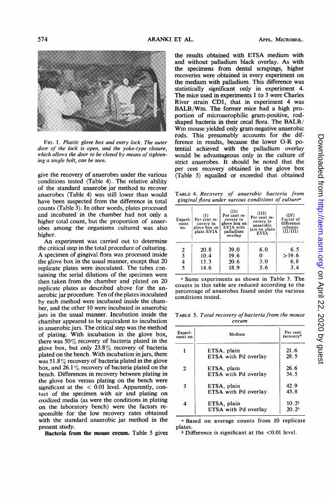
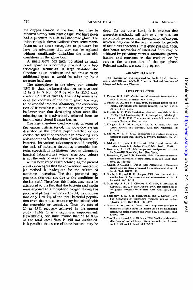
FIG. 1. Plastic glove box and entry lock. The outerdoor of the lock is open, and the yoke-type closure,which allows the door to be closed by means of tighten-ing a single bolt, can be seen.
give the recovery of anaerobes under the variousconditions tested (Table 4). The relative abilityof the standard anaerobe jar method to recover
anaerobes (Table 4) was still lower than wouldhave been suspected from the difference in totalcounts (Table 3). In other words, plates processedand incubated in the chamber had not only a
higher total count, but the proportion of anaer-
obes among the organisms cultured was alsohigher.An experiment was carried out to determine
the critical step in the total procedure of culturing.A specimen of gingival flora was processed insidethe glove box in the usual manner, except that 20replicate plates were inoculated. The tubes con-
taining the serial dilutions of the specimen were
then taken from the chamber and plated on 20replicate plates as described above for the an-
aerobic jar procedure. Ten ofthe plates inoculatedby each method were incubated inside the cham-ber, and the other 10 were incubated in anaerobicjars in the usual manner. Incubation inside thechamber appeared to be equivalent to incubationin anaerobic jars. The critical step was the methodof plating. With incubation in the glove box,there was 50% recovery of bacteria plated in theglove box, but only 23.8% recovery of bacteriaplated on the bench. With incubation in jars, therewas 51.8% recovery of bacteria plated in the glovebox, and 26.1 % recovery of bacteria plated on thebench. Differences in recovery between plating inthe glove box versus plating on the bench were
significant at the < 0.01 level. Apparently, con-tact of the specimen with air and plating on
oxidized media (as were the conditions in platingon the laboratory bench) were the factors re-
sponsible for the low recovery rates obtainedwith the standard anaerobic jar method in thepresent study.
Bacteria from the mouse cecum. Table 5 gives
the results obtained with ETSA medium withand without palladium black overlay. As withthe specimens from dental scrapings, higherrecoveries were obtained in every experiment onthe medium with palladium. This difference wasstatistically significant only in experiment 4.The mice used in experiments 1 to 3 were CharlesRiver strain CD1, that in experiment 4 wasBALB/Wm. The former mice had a high pro-portion of microaerophilic gram-positive, rod-shaped bacteria in their cecal flora. The BALB/Wm mouse yielded only gram-negative anaerobicrods. This presumably accounts for the dif-ference in results, because the lower O-R po-tential achieved with the palladium overlaywould be advantageous only in the culture ofstrict anaerobes. It should be noted that theper cent recovery obtained in the glove box(Table 5) equalled or exceeded that obtained
TABLE 4. Recovery of anaerobic bacteria fromgingivalflora under various conditions of culturea
(II) (I )I) Per cent* re- PereI (IV)Experi- Per cent re- covery in Factor ofment covery in glove box on covery oi differenceno. glove box on EVIA with jars on plain columns
pliVApalladium EVIA (II/III)pliVA overlay
2 20.8 39.0 6.0 6.53 10.4 19.6 0 > 19.64 13.3 20.6 3.0 6.85 14.6 18.9 5.6 3.4
a Same experiments as shown in Table 3. Thecounts in this table are reduced according to thepercentage of anaerobes found under the variousconditions tested.
TABLE 5. Total recovery ofbacteria from the mousececum
Experi- Medium Per centment no. recovery'~
1 ETSA, plain 21.6ETSA with Pd overlay 28.5
2 ETSA, plain 26.6ETSA with Pd overlay 34.5
3 ETSA, plain 42.9ETSA with Pd overlay 45.8
4 ETSA, plain 10.2bETSA with Pd overlay 20.2b
a Based on average counts from 10 replicateplates.
b Difference is significant at the <0.01 level.
574 APPL. MICROBIOL.
on April 22, 2020 by guest
http://aem.asm
.org/D
ownloaded from

VOL. 17, 1969 ISOLATION OF ANAEROBES IN SIMPLIFIED GLOVE BOX
with the same type of specimen in earlier studieswith the roll tube method (14).
DISCUSSIONThe long established standard of anaerobic
bacteriology is Hungate's roll tube method. To beacceptable, newly developed simplified methodsfor the culture of strict anaerobes must be shownto at least equal the conditions obtainable withthis standard. The roll tube method has two(and only these two) critical advantages. (i)Bacteria may be transferred and cultured with-out significant exposure to atmospheric oxygen.(ii) The media used may be kept at a low O-Rpotential at all times, even at the moment ofinoculation.
There can be no doubt that an anaerobic glovebox is superior to any other method in allowingmanipulation of cultures and transfer of bacteriawithout contact with air. In fact, one of theprincipal advantages of a glove box is that thesemanipulations can be accomplished easily and ina "foolproof" manner. The data given in thispaper show that the glove box technique alsoequals or even exceeds the roll tube method inachieving a low O-R potential in the media.As reviewed recently by Hungate et al. (5), amedium able to reduce indigo carmine has aboutthe "right" Eh (-150 mv or lower), whereasreduction of phenosafranin indicates an Eh (-300mv or lower) which is in excess of that requiredto cultivate such fastidious anaerobes as themethanogenic bacteria. In our experience withthe roll tube technique, it was impossible toobtain reduction of phenosafranin in mediaunless potentially toxic reducing agents, such assodium dithionite, were employed. All mediaused in the glove box reduced indigo carmineeven in the absence of cysteine. With the addi-tion of a palladium overlay, plated agar mediaregularly reduced phenosafranin and thus hada lower O-R potential than the roll tube mediaused by most workers. One must realize, ofcourse, that the O-R potential as measured in acomplex bacteriological medium is only oflimited significance (3). Nevertheless, the datashown in Table 5 show that the present methodgave results which were equal to or better thanthose obtained in our earlier work with the rolltubes (14).Very few data are available in the literature
concerning the effect of the oxygen concentra-tion in the gas phase on the growth of strictanaerobes. Hungate (11) noted that the growthof a methanogenic bacterium was inhibited inthe presence of 300 ppm of oxygen and that aslight reduction in growth rate could be notedat levels down to 40 ppm. In view of these data,
the 02 concentration routinely obtainable in theglove box (2 to 5 ppm or 5 to 10 ppm, dependingon the amount of catalyst used) appears to besufficiently low to cultivate strict anaerobes.The principal advantages of the glove box
system are as follows.(i) Anyone who has sufficient technical ability
to evacuate and fill an anaerobic jar can alsooperate the glove box.
(ii) Only standard bacteriological techniquesare required for the isolation and cultivation ofanaerobes, except that the manipulations haveto be done through gloves, which are not amajor hindrance. "Triple streaking" of an agarplate is the most convenient and efficient means ofisolating strains from a mixture, and this ispossible only in a glove box. Moreover, the agarplates may be dried sufficiently to prevent spread-ing of bacteria, a complication which, with ourspecimens, occurred frequently in roll tubeseven when very lean media were used. Also, somediagnostic reactions (e.g., hemolysis and thedisc method of antibiotic sensitivity testing) canonly be carried out on agar plates.
(iii) Media may be prepared in the conven-tional manner on the laboratory bench. This hasbeen possible because of the efficiency of thepalladium catalyst in reducing plated media.Although palladium has been employed beforein roll tubes (7), its use has not been common,most likely because of the dangers inherent inworking with hydrogen on the open laboratorybench. The anaerobic glove box permits the useof this catalyst in a convenient and safe manner.
(iv) Viewing ports may be attached to theplastic chamber to allow operation of a mi-croscope in the glove box for observation ofmotility, etc.
(v) The cost of a complete glove box is notexcessive. A price of less than $1,000 includesitems such as a vacuum pump, which may al-ready be available in many laboratories. Allparts and accessories can be obtained com-mercially.
(vi) Plastic chambers can easily be fabricatedin any desired size or shape. Oversize airlockscan also be obtained commercially. Another useof this type of glove box may therefore be toprovide an anaerobic environment for varioustypes of apparatus, such as continuous culturedevices, chromatographic columns, etc.Without experience in operating a flexible
plastic glove box one may suspect several dis-advantages, such as the possibility of puncturingthe material. We have operated four glove boxesfor as long as 18 months with only one puncturein one of the chambers. Because of the positiveinside pressure, small punctures do not affect
575
on April 22, 2020 by guest
http://aem.asm
.org/D
ownloaded from
576 ARANK
the oxygen tension in the box. They may berepaired simply with plastic tape. We have neverhad a puncture in a 25-mil neoprene glove. Thethinner plastic gloves available from some manu-facturers are more susceptible to puncture buthave the advantage that they can be replacedwithout significantly disturbing the anaerobicconditions in the glove box.A small glove box takes up about as much
bench space as is normally provided for a bac-teriological technician. A large chamber alsofunctions as an incubator and requires as muchadditional space as would be taken up by aseparate incubator.The atmosphere in the glove box contains
10% H2; thus, the largest chamber we have used[2 by 2 by 7 feet (60.9 by 60.9 by 213.3 cm)]contains 2.8 ft5 of pure H2. Even if by some acci-dent the contents of the entire glove box wereto be emptied into the laboratory, the concentra-tion of flammable gas in the air would probablybe lower than that developing when some illu-minating gas is inadvertently released from anincompletely closed Bunsen burner.One may therefore conclude that, in terms of
the various criteria tested, the glove-box systemdescribed in the present paper matched or ex-ceeded the roll tube technique in providing suit-able conditions for the culture of strictly anaerobicbacteria. Its various advantages should simplifythe task of isolating fastidious anaerobic bac-teria, especially in institutions (such as diagnostichospital laboratories) where anaerobic cultureis not the only or even the major activity.As has been emphasized before (14), the present
results show again that the conventional anaerobicjar method is inadequate for the culture offastidious anaerobes. The data presented sug-gest that this was not due to the conditions inthe jar itself. Therefore, this inadequacy must beattributed to the fact that the bacteria and mediawere exposed to atmospheric oxygen during theprocess of plating. Earlier studies (14) have shownthat only 1 to 5% of the total bacterial popula-tion from the mouse cecum may be isolated withthe anaerobic jar technique. Thus, the rate of20 to 45% recovery achieved in the presentstudy (Table 5) is a significant improvement.Nevertheless, one must realize that 55 to 80%of the total cecal flora is still not cultivated.It is possible that some of these bacteria may be
I ET AL. APPL. MICROBIOL.
dead. On the other hand, it is obvious thatanaerobic methods, roll tube or glove box, canaccomplish no more than the exclusion of oxygen,which is only one of the requirements for growthof fastidious anaerobes. It is quite possible, then,that better recoveries of intestinal flora may beachieved by providing various additional growthfactors and nutrients in the medium or byvarying the composition of the gas phase.Relevant studies are now in progress.
ACKNOWLEDGMENT
This investigation was supported by Public Health Servicegrants AI-07328 and AI-07631 from the National Institute ofAllergy and Infectious Diseases.
LITERATURE CITED
1. Drasar, B. S. 1967. Cultivation of anaerobic intestinal bac-teria. J. Pathol. Bacteriol. 94:417-427.
2. Fisher, R. A., and F. Yates. 1963. Statistical tables for bio-logical, agricultural and medical research. Hafner Publish-ing Co., New York.
3. Hewitt, L. F. 1950. Oxidation-reduction potentials in bac-teriology and biochemistry. E. S. Livingstone, Edinburgh.
4. Hungate, R. E. 1950. The anaerobic mesophilic cellulolyticbacteria. Bacteriol. Rev. 14:1-49.
5. Hungate, R. E., M. P. Bryant, and R. A. Mah. 1964. Therumen bacteria and protozoa. Ann. Rev. Microbiol. 18:131-166.
6. Moore, W. E. C. 1966. Techniques for routine culture offastidious anaerobes. Intern. J. System. Bacteriol. 16:174-190.
7. Mylorie, R. L., and R. E. Hungate. 1954. Experiments on themethane bacteria in sludge. Can. J. Microbiol. 1:55-64.
8. Rosebury, T. 1962. Microorganisms indigenous to man.McGraw-Hill Book Co., Inc., New York.
9. Rosebury, T., and J. B. Reynolds. 1964. Continuous anaero-biosis for cultivation of spirochetes. Proc. Soc. Exptl. Biol.Med. 117:813-815.
10. Savage, D. C., and R. Dubos. 1968. Alterations in the mousececum and its flora produced by antibacterial drugs. J.Exptl. Med. 128:97-110.
11. Smith, P. H., and R. E. Hungate. 1958. Isolation and char-acterization of Methanobacterium ruminantium n. sp. J.Bacteriol. 75:713-718.
12. Socransky, S. S., R. J. Gibbons, A. C. Dale, L. Bortnick, E.Rosenthal, and J. B. MacDonald. 1963. The microbiota ofthe gingival crevice area of man. Arch. Oral Biol. 8:275-280.
13. Socransky, S. S., J. B. MacDonald, and S. Sawyer. 1959.The cultivation of Treponema microdentium as surfacecolonies. Arch. Oral Biol. 1:171-172.
14. Spears, R. W., and R. Freter. 1967. Improved isolation ofanaerobic bacteria from the mouse cecum by maintainingcontinuous strict anaerobiosis. Proc. Soc. Exptl. Biol. Med.124:903-909.
15. Van Houte, J., and R. J. Gibbons. 1966. Studies of the cultiv-able flora of normal human feces. Antonie van Leuwen-hook J. Microbiol. Serol. 32:212-222.
. on A
pril 22, 2020 by guesthttp://aem
.asm.org/
Dow
nloaded from