isolation of multi-drug resistant staphylococcus...
TRANSCRIPT

International Journal of Science, Engineering and Technology Research (IJSETR), Volume 5, Issue 2, February 2016
487
ISSN: 2278 – 7798 All Rights Reserved © 2016 IJSETR
Isolation of Multi-Drug Resistant Staphylococcus aureus from the
soil samples of Hyderabad
T. RAGA SUDHA
Department of Microbiology,
Indira Priyadarshini Govt. Degree College (W),
Nampally, Hyderabad, INDIA 5000 01
Mobile no.: 07893812790
Abstract
The use and misuse of antibiotics over the past 50 years in man has lead to the emergence
of various multidrug resistant microorganisms (MDROs). These organisms are of great concern
in discharging health care facilities throughout the world. Staphylococcus aureus (S.aureus) one
of the most important pathogens affecting humans has acquired resistance to various antibiotics.
According to the resistance data given by World Health Organization (WHO), Community
Acquired Methicillin Resistant Staphylococcus aureus (CA-MRSA) has increased significantly
all over the world.
In the current study S.aureus was isolated from the soil samples of the residential and
hospital areas of the Hyderabad city and was evaluated for their sensitivity against twelve
antibiotics clinically used against S.aureus. Methicillin Resistant Staphylococcus aureus
(MRSA) were isolated from the soil samples collected from the residential and hospital areas of
Hyderabad.
Keywords: Antibiotic sensitivity, MDROs, MRSA, soil samples, Staphylococcus aureus
International Journal of Science, Engineering and Technology Research (IJSETR), Volume 5, Issue 2, February 2016
488
ISSN: 2278 – 7798 All Rights Reserved © 2016 IJSETR
Introduction
Though the antimicrobial resistance or drug resistance is not new, the numbers of
resistant organisms are increasing enormously through the length and breadth of geographic
locations day-by-day. Diseases and disease agents that were once thought to be controlled by
antibiotics are returning in new leagues resistant to these therapies [1], [2]. Staphylococcus
aureus (S.aureus), one of the most important pathogens affecting humans has acquired resistance
to various antibiotics and is a leading cause of hospital and community acquired infections,
manifesting from minor skin diseases to life-threatening infections [3], [4]. Drug-resistant strains
initially appeared in hospitals, where most antibiotics were being used. Penicillin resistant
Staphylococcus aureus confronted London civilian hospitals very soon after the introduction of
penicillin in the 1940. Initially, MRSA was mainly a problem in hospital-acquired infections.
Over the past decade, CA-MRSA has increased significantly in a number of countries.
Fortunately, many of these CA-MRSA strains have so far retained susceptibility to a number of
non-beta-lactam antimicrobials, whereas most health-care associated MRSA infections are
caused by difficult-to-treat multi-resistant strains.
The present study is an attempt to isolate and characterize the multi-drug resistance
(MDR) Staphylococcus aureus from soil surrounding houses and hospitals, which was obtained
from various place of Hyderabad. The isolates were investigated for their sensitivity against
twelve antibiotics.

International Journal of Science, Engineering and Technology Research (IJSETR), Volume 5, Issue 2, February 2016
489
ISSN: 2278 – 7798 All Rights Reserved © 2016 IJSETR
Methodology
Chemicals reagents and media: The antibiotic discs, bacteriological media such as nutrient
agar, SIM agar, Simmon’s citrate agar, MRVP broth, nutrient broth, tryptophan broth, Mac
Conkey’s agar, Mannitol agar etc., chemicals required for the preparation of kovac’s reagent,
Barritt’s A and B reagents, stains like crystal violet, methyl red, saffronin, malachite green,
ethanol, iodide etc were procured from Himedia (Mumbai, India).
Collection of sample: The soil samples were collected from the residential areas and the hospital
areas in the city of Hyderabad. Randomly from different parts of the city twenty five soil
samples from the residential areas and twenty five samples from the hospital surroundings were
collected for the experimental. The bacteriological analysis of these samples was done by serial
dilution and agar plate culture techniques. The obtained pure cultures were characterized based
on their morphological and biochemical characteristics as described in Bergey’s Manual for
bacteriology [5].
Serial dilution and agar plating technique: This method is based on the principle that when
soil sample containing bacterial colonies are cultured, every living bacterium develops into a
visible colony on the nutrient agar plate. One gram of the collected soil sample is suspended in
9ml of saline to obtain a 10-1
dilution (10 times dilution). From the above dilution 1ml is
transferred to a fresh 9ml saline solution to obtain a 10-2
dilution. The process is repeated in
order to produce 10-3
, 10-4
, 10-5
, 10-6
and 10-7
serial dilutions. From the dilutions ranging from
10-3
to 10-7
0.1 ml of the suspensions were added to nutrient agar plates (each dilution in 3
replicates) under sterile conditions and incubated at 370C for 24 hours. The number of bacteria in
the testes soil sample can be calculated by the following formula:
International Journal of Science, Engineering and Technology Research (IJSETR), Volume 5, Issue 2, February 2016
490
ISSN: 2278 – 7798 All Rights Reserved © 2016 IJSETR
Organisms per milliliter per gram soil = number of colonies (average of 3 replicates)/ volume
plated (0.1) x dilution
The isolated cultures were differentiated by their morphological characteristics and
transferred to fresh nutrient agar media to produce in pure form. Golden yellow colonies on the
nutrient agar plate were sub cultured and confirmed as S. aureus by performing motility, grams
staining and biochemical tests [6].
Motility: The motility of the native isolates was detected using hanging drop technique, a drop
of bacterial suspension, preferably in mid-logarithmic phase, was placed in the centre of a cover
slip coated with grease. Glass slide having a central depression was placed over the cover slip.
The slide was then inverted without disrupting the drop and examined using 40x optical
microscope (Labomed, Vision 2000, India) for motility of the bacteria [6].
Gram staining of bacteria: The gram-negative bacterial cell wall is thin, complex, multilayered
structure and contains relatively a high lipid contents, in addition to protein and mucopeptides.
The higher amount of lipids is readily dissolved by alcohol, resulting in the formation of large
pores in the cell wall which do not close appreciably on dehydration of cell wall proteins, thus
facilitating the leakage of crystal violet-iodine (CV-I) complex and resulting in the
decolorization of the bacterium which later takes the counter stain and appears pink. In contrast,
the gram positive cell walls are thick and chemically simple, composed mainly of protein and
cross-linked mucopeptides. When it was treated with alcohol, it causes dehydration and closure
of cell wall pores, thereby not allowing the loss of (CV-1) complex and cells remain purple [9].
Heat fix a thin smear of culture on glass slides, cover the smear one by one with crystal violet
(60 seconds), gram’s iodine (60 seconds), 95% ethanol (20 seconds) and safranin (40 seconds).
Air dried the slides after washing with distilled water and observed under microscope [6]. Gram
positive bacteria appear in violet color and the Gram negative bacteria appear in pink color.
International Journal of Science, Engineering and Technology Research (IJSETR), Volume 5, Issue 2, February 2016
491
ISSN: 2278 – 7798 All Rights Reserved © 2016 IJSETR
Biochemical tests:
According to the Bergey’s manual of systematic bacteriology the biochemical characterization of
Staphylococcus aureus was done by the tests shown in the Table I [7].
Antibiotic sensitivity test [8]:
The identified S. aureus cultures (each 20µl) was poured over the basal plates containing
25ml of nutrient agar media in nine sterile petriplates and spread using L-shaped glass rod. The
antibiotic discs used commercially for Gram’s positive bacteria were placed in the plate. The
effect of the each of the organism was tested in triplicate. The plates were incubated at 370C for
24 hr. and there after the zones of inhibition were recorded by measuring the diameter of zone of
inhibition by the following formula:
Zone of inhibition (mm) = D-d
Where,
D= diameter of zone of inhibition
d= diameter of the antibiotic disc (6 mm)
The twelve antibiotics used in the experimental are Pencillin G (P), Amoxicillin (AMX),
Carbenicillin (CB), Methicilin (MET), Azithromycin (AZM), Clindamycin (CD), Roxithromycin
(RO), Lincomycin (L), Vancomycin (VA), Rifampicin (RIF), Teicoplanin (TEI) and Linezolid
(L).
International Journal of Science, Engineering and Technology Research (IJSETR), Volume 5, Issue 2, February 2016
492
ISSN: 2278 – 7798 All Rights Reserved © 2016 IJSETR
Results and Discussion
Among the Gram-positive pathogens, S. aureus continues to cause skin and soft tissue
infections in the community and invasive infections in the hospitalized patients. In a recent
Europe-wide survey, the most common organisms in skin and soft tissue infections were S.
aureus (71% cases) with 22.5 per cent being MRSA [9]. The proportion of infections with
MRSA varied among countries ranging from 0.4 per cent in Sweden to 48.4 per cent in Belgium.
In the United States in a span of 10 years, there was an increase in the overall incidence of S.
aureus and CA-MRSA infections [10].
In the current study 50 soil samples were collected randomly in the city of Hyderabad.
Out of these 50 samples, 25 each were collected from the residential and hospital areas. S.
aureus was isolated from four RS samples and five HS samples. These isolates were identified as
S. aureus by performing biochemical tests.
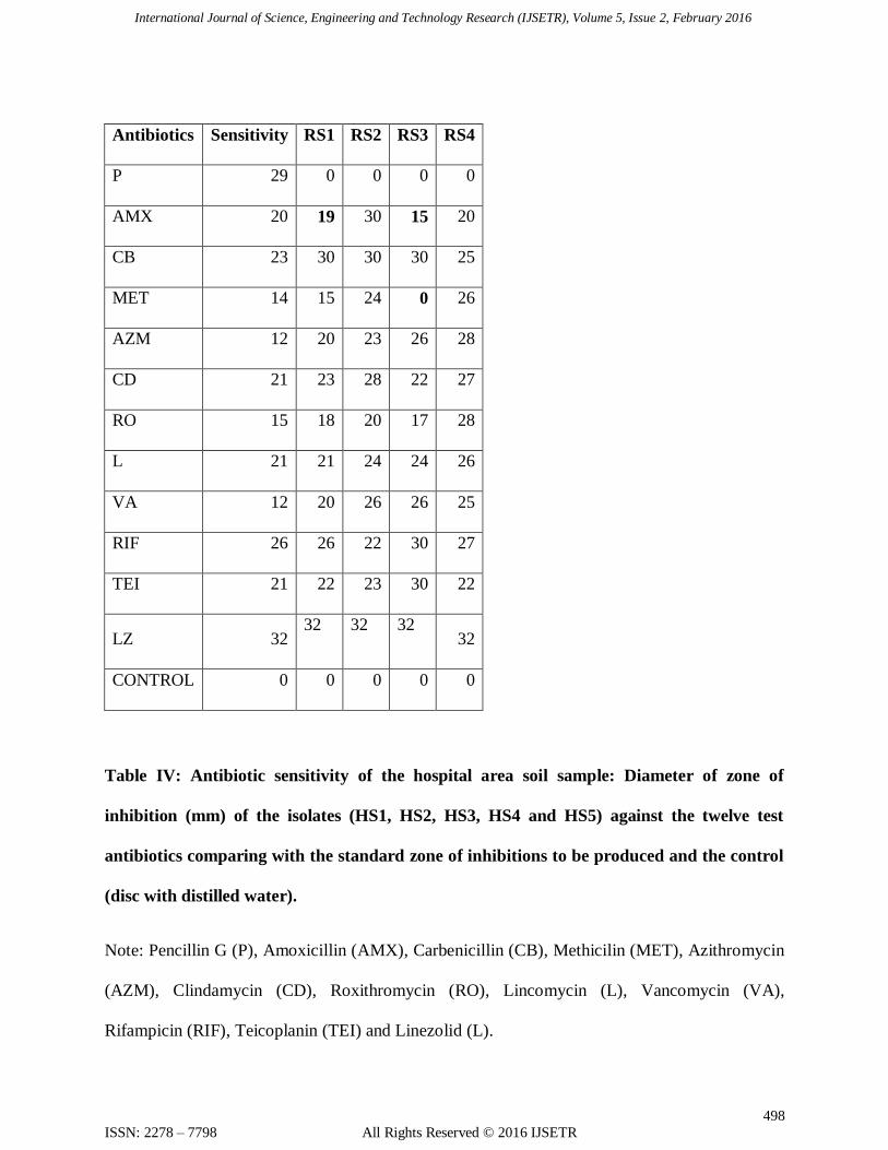
The identified organisms were tested for their sensitivity against 12 antibiotics clinically
used against gram positive bacteria. Among the four S. aureus isolated RS1 is resistant to P and
AMX; RS2 and RS4 are resistant to P; RS3 resistant to P, AMX and MET. Among the five
isolates of the hospital area HS1 is resistant to P, AZM and LZ; HS2 is resistant to P, MET, CD
and LZ; HS3 is sensitive to AZM, RO, VA and RIF; HS4 is sensitive to RO, VA and RIF; HS5
is sensitive to the antibiotics RO and VA only.
Conclusion
Soil is the outermost layer of the earth composed of complex minerals and organic
matter. The microbial profile in a particular portion of the soil is determined by the interaction of
sunlight, rainfall, temperature, moisture, soil pH, vegetation and redox potential. Pathogens may
International Journal of Science, Engineering and Technology Research (IJSETR), Volume 5, Issue 2, February 2016
493
ISSN: 2278 – 7798 All Rights Reserved © 2016 IJSETR
be indigenous or enter by the animal deposits, manure application, sewage, floods or
contaminated water.
The cities are thoroughly populated with human population. The S. aureus being a
commensal of human skin is expected to reach the soil in various forms. Hence, the soil in the
residential areas of the city are analyzed for the presence of S. aureus .The expectation of
isolating drug resistant S. aureus from the hospital zone is high due to the disposal of hospital
waste by the in-patients, out-patients and the hospital authorities. The prevalence of these
MDROs in the soil may have a greater ability to infect susceptible organisms. According to a
survey conducted by the Indian Network for Surveillance of Antimicrobial Resistance (INSAR)
group during the year 2008 and 2009, the percentage of occurrences of MRSA in India is 42 and
40 respectively in the samples collected from the patient wounds [11].
In the current study the percentage of occurrence of S. aureus around the residential and
the hospital areas in the city of Hyderabad is 16 and 20 percentage respectively. The S. aureus
isolated from the residential areas were found to be highly sensitive to most of the antibiotics
compared to the S. aureus isolated from the hospital area which have exhibited a wide range of
resistance to the test antibiotics. All the HS isolates were found to be sensitive to Ro and VA.
The regular surveillance of MRSA is essential for selecting an appropriate antibiotic, for limiting
the use of powerful antibiotics as initial treatment and also to help in postponing the
development of resistant and life-threatening staphylococcal infections.
Acknowledgement
I express my sincere thanks to SERO-UGC, Hyderabad for funding the minor research
project and encouraging budding scientist.

International Journal of Science, Engineering and Technology Research (IJSETR), Volume 5, Issue 2, February 2016
494
ISSN: 2278 – 7798 All Rights Reserved © 2016 IJSETR
I also thank the Principal and the Department of Microbiology, Indira Priyadarshini
Government Degree College, Hyderabad for providing the necessary infrastructure to carry out
the work successfully.
[1] S. Chen, B. Mulgrew, and P. M. Grant, “A clustering technique for digital communications
channel equalization using radial basis function networks,” IEEE Trans. on Neural Networks,
vol. 4, pp. 570-578, July 1993.
References
1. Adegoke, Anthony A., Tom Mvuyo, Okoh Anthony and Jacob Steve, “Studies on
multiple antibiotic resistant bacterial isolated from surgical site infection”, Scientific
Research and Essays, 5(24), 3876-3881, 2010.
2. S.M. Dharmadhikari and S.A.Peshwe, “Molecular level studies on multiple and serum
resistant in UTI pathogen”, Indian Journal of biotechnology, 8, 40-45, 2009.
3. H.K. Tiwari, A. K. Das, D. Sapkota , K. Sivarajan and V.K. Pahwa, “ Methicillin
resistant Staphylococcus aureus: Prevalence and antibiogram in a tertiary care hospital in
western Nepal”, J Infect Dev Ctries, 3:681-4, 2009.
4. A.H. Al-Baidani, W.H. El-Shouny, T.M. Shawa, “ Antibiotic Suseptiblity Pattern of
Methicillin Resistance Staphylococcus aureus in Three Hospitals at Hodeidah City,
Yemen”, Global J. Pharmacol. , 5(2):106-111, 2011.
5. G.M. Garrity, D.R. Boone and R.W. Castenholz, “Bergey’s Manual of Systematic
Bacteriology”, 2nd ed., vol. 1, Springer-Verlag, New York, NY, 2001.
International Journal of Science, Engineering and Technology Research (IJSETR), Volume 5, Issue 2, February 2016
495
ISSN: 2278 – 7798 All Rights Reserved © 2016 IJSETR
6. Cappucinno, G. James and N. Sharman, “Microbiology: A Laboratory Manual”, 3rd
ed.
Additional Wesley Publ. Co. Reading Massachusetts, USA, 1992.
7. R.Cruickshank, “Medical Microbiology”, 11th ed. The English Language Book Society
E. and Livingston Ltd. 1970.
8. A.W. Bauer, W.M. Kirby and J.C. Sherris, ”Antibiotic susceptibility testing by a
standardized single disk method”, Am. J. Clin. Pathol., 45, 493-496, 1966.
9. H.S. Sader, D.J. Farrell, and R.N. Jones, “ Antimicrobial susceptibility of Gram-positive
cocci isolated from skin and skin-structure infections in European medical centres”, Int J
Antimicrobial Agents, 36 : 28-32, 2010.
10. L.A. Tracy, J.P. Furuno, A.D. Harris, M. Singer, P. Langenber and M.C. Roghmann,
“Staphylococcus aureus infections in US veterans, Maryland, USA, 1999-2008”, Emerg
Infect Dis., 17: 441-8, 2011.
11. Methicillin resistant Staphylococcus aureus (MRSA) in India: Prevalence &
susceptibility pattern, Indian Network for Surveillance of Antimicrobial Resistance
(INSAR) group, India. Indian J Med Res, 137, pp 363-369, February 2013,
Table I: Biochemical Characterization of Staphylococcus aureus.
Biochemical test Reaction
Catalase +
Oxidase -
Indole production -
Methyl red +
Voges-Proskauer -

International Journal of Science, Engineering and Technology Research (IJSETR), Volume 5, Issue 2, February 2016
496
ISSN: 2278 – 7798 All Rights Reserved © 2016 IJSETR
Citrate utilization -
Glucose fermentation +
Mannitol fermentation +
Lactose fermentation +
Sucrose fermentation +
Blood hemolysis +
Coagulase +
Table II: Results of morphological and biochemical tests for Staphylococcus aureus.
Note: Mo- motility test, I- indole test, M- methylred, VP- Voges Proskauer’s , C-citrate, O-
oxidase, Ct- catalase, H- hemolysis, Co- coagulase, Mn- mannitol, G- glucose, S- sucrose, L-
lactose
Isolates
no.
Nutrient Agar Media Mo Gram’s
staining
I M VP C O Ct H Co Mn G S L
RS1 Small round golden yellow color
colonies
- + cocci in
clusters.
- + - - - + + + + + + +
RS2 Small round golden yellow color - + cocci in - + - - - + + + + + + +
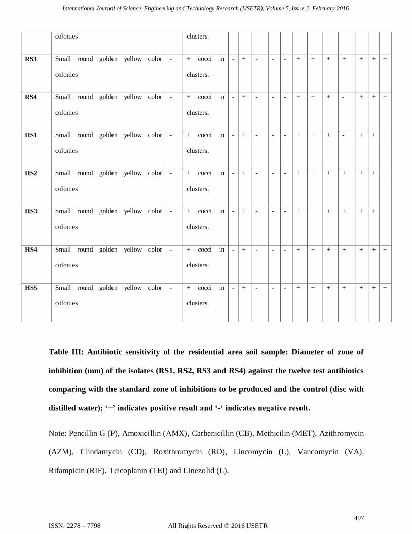
International Journal of Science, Engineering and Technology Research (IJSETR), Volume 5, Issue 2, February 2016
497
ISSN: 2278 – 7798 All Rights Reserved © 2016 IJSETR
colonies clusters.
RS3 Small round golden yellow color
colonies
- + cocci in
clusters.
- + - - - + + + + + + +
RS4 Small round golden yellow color
colonies
- + cocci in
clusters.
- + - - - + + + - + + +
HS1 Small round golden yellow color
colonies
- + cocci in
clusters.
- + - - - + + + - + + +
HS2 Small round golden yellow color
colonies
- + cocci in
clusters.
- + - - - + + + + + + +
HS3 Small round golden yellow color
colonies
- + cocci in
clusters.
- + - - - + + + + + + +
HS4 Small round golden yellow color
colonies
- + cocci in
clusters.
- + - - - + + + + + + +
HS5 Small round golden yellow color
colonies
- + cocci in
clusters.
- + - - - + + + + + + +
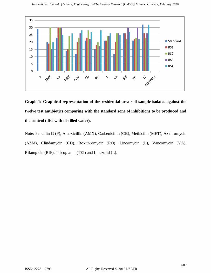
Table III: Antibiotic sensitivity of the residential area soil sample: Diameter of zone of
inhibition (mm) of the isolates (RS1, RS2, RS3 and RS4) against the twelve test antibiotics
comparing with the standard zone of inhibitions to be produced and the control (disc with
distilled water); ‘+’ indicates positive result and ‘-‘ indicates negative result.
Note: Pencillin G (P), Amoxicillin (AMX), Carbenicillin (CB), Methicilin (MET), Azithromycin
(AZM), Clindamycin (CD), Roxithromycin (RO), Lincomycin (L), Vancomycin (VA),
Rifampicin (RIF), Teicoplanin (TEI) and Linezolid (L).
International Journal of Science, Engineering and Technology Research (IJSETR), Volume 5, Issue 2, February 2016
498
ISSN: 2278 – 7798 All Rights Reserved © 2016 IJSETR
Antibiotics Sensitivity RS1 RS2 RS3 RS4
P 29 0 0 0 0
AMX 20 19 30 15 20
CB 23 30 30 30 25
MET 14 15 24 0 26
AZM 12 20 23 26 28
CD 21 23 28 22 27
RO 15 18 20 17 28
L 21 21 24 24 26
VA 12 20 26 26 25
RIF 26 26 22 30 27
TEI 21 22 23 30 22
LZ 32 32 32 32
32
CONTROL 0 0 0 0 0
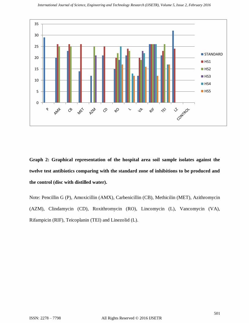
Table IV: Antibiotic sensitivity of the hospital area soil sample: Diameter of zone of
inhibition (mm) of the isolates (HS1, HS2, HS3, HS4 and HS5) against the twelve test
antibiotics comparing with the standard zone of inhibitions to be produced and the control
(disc with distilled water).
Note: Pencillin G (P), Amoxicillin (AMX), Carbenicillin (CB), Methicilin (MET), Azithromycin
(AZM), Clindamycin (CD), Roxithromycin (RO), Lincomycin (L), Vancomycin (VA),
Rifampicin (RIF), Teicoplanin (TEI) and Linezolid (L).
International Journal of Science, Engineering and Technology Research (IJSETR), Volume 5, Issue 2, February 2016
499
ISSN: 2278 – 7798 All Rights Reserved © 2016 IJSETR
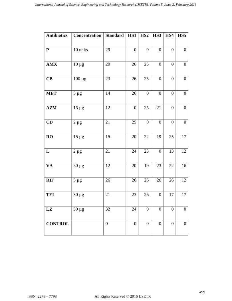
Antibiotics Concentration Standard HS1 HS2 HS3 HS4 HS5
P 10 units 29 0 0 0 0 0
AMX 10 µg 20 26 25 0 0 0
CB 100 µg 23 26 25 0 0 0
MET 5 µg 14 26 0 0 0 0
AZM 15 µg 12 0 25 21 0 0
CD 2 µg 21 25 0 0 0 0
RO 15 µg 15 20 22 19 25 17
L 2 µg 21 24 23 0 13 12
VA 30 µg 12 20 19 23 22 16
RIF 5 µg 26 26 26 26 26 12
TEI 30 µg 21 23 26 0 17 17
LZ 30 µg 32 24 0 0 0 0
CONTROL 0 0 0 0 0 0
International Journal of Science, Engineering and Technology Research (IJSETR), Volume 5, Issue 2, February 2016
500
ISSN: 2278 – 7798 All Rights Reserved © 2016 IJSETR
Graph 1: Graphical representation of the residential area soil sample isolates against the
twelve test antibiotics comparing with the standard zone of inhibitions to be produced and
the control (disc with distilled water).
Note: Pencillin G (P), Amoxicillin (AMX), Carbenicillin (CB), Methicilin (MET), Azithromycin
(AZM), Clindamycin (CD), Roxithromycin (RO), Lincomycin (L), Vancomycin (VA),
Rifampicin (RIF), Teicoplanin (TEI) and Linezolid (L).
0
5
10
15
20
25
30
35
Standard
RS1
RS2
RS3
RS4
International Journal of Science, Engineering and Technology Research (IJSETR), Volume 5, Issue 2, February 2016
501
ISSN: 2278 – 7798 All Rights Reserved © 2016 IJSETR
Graph 2: Graphical representation of the hospital area soil sample isolates against the
twelve test antibiotics comparing with the standard zone of inhibitions to be produced and
the control (disc with distilled water).
Note: Pencillin G (P), Amoxicillin (AMX), Carbenicillin (CB), Methicilin (MET), Azithromycin
(AZM), Clindamycin (CD), Roxithromycin (RO), Lincomycin (L), Vancomycin (VA),
Rifampicin (RIF), Teicoplanin (TEI) and Linezolid (L).
0
5
10
15
20
25
30
35
STANDARD
HS1
HS2
HS3
HS4
HS5