isolation of plant photosystem ii complexes by fractional ... · mega- and supercomplexes, psii...
TRANSCRIPT

ORIGINAL RESEARCHpublished: 10 December 2015doi: 10.3389/fpls.2015.01100
Edited by:Roman Sobotka,
Czech Academy of Sciences,Czech Republic
Reviewed by:Josef Komenda,
Institute of Microbiology of the ASCR,Czech Republic
Bettina Ughy,Biological Research Centre of theHungarian Academy of Sciences,
Hungary
*Correspondence:Dario Piano
Specialty section:This article was submitted to
Plant Cell Biology,a section of the journal
Frontiers in Plant Science
Received: 23 August 2015Accepted: 22 November 2015Published: 10 December 2015
Citation:Haniewicz P, Floris D, Farci D,
Kirkpatrick J, Loi MC, Büchel C,Bochtler M and Piano D (2015)
Isolation of Plant Photosystem IIComplexes by Fractional
Solubilization.Front. Plant Sci. 6:1100.
doi: 10.3389/fpls.2015.01100
Isolation of Plant Photosystem IIComplexes by FractionalSolubilizationPatrycja Haniewicz1, Davide Floris2, Domenica Farci2, Joanna Kirkpatrick3,Maria C. Loi2, Claudia Büchel4, Matthias Bochtler1,5 and Dario Piano1,2*
1 Laboratory of Structural Biology, Department of Molecular Biology, International Institute of Molecular and Cell Biology,Warsaw, Poland, 2 Laboratory of Photosynthesis and Photobiology, Department of Life and Environmental Sciences,University of Cagliari, Cagliari, Italy, 3 Proteomics Core Facility, European Molecular Biology Laboratory, Heidelberg,Germany, 4 Laboratory of Plant Cell Physiology, Institute of Molecular Biosciences, Goethe-University Frankfurt, Frankfurt amMain, Germany, 5 Department of Bioinformatics, Institute of Biochemistry and Biophysics, Warsaw, Poland
Photosystem II (PSII) occurs in different forms and supercomplexes in thylakoidmembranes. Using a transplastomic strain of Nicotiana tabacum histidine tagged onthe subunit PsbE, we have previously shown that a mild extraction protocol withβ-dodecylmaltoside enriches PSII characteristic of lamellae and grana margins. Here,we characterize residual granal PSII that is not extracted by this first solubilization step.Using affinity purification, we demonstrate that this PSII fraction consists of PSII-LHCIImega- and supercomplexes, PSII dimers, and PSII monomers, which were separatedby gel filtration and functionally characterized. Our findings represent an alternativedemonstration of different PSII populations in thylakoid membranes, and they makeit possible to prepare PSII-LHCII supercomplexes in high yield.
Keywords: photosystem II, PSII-LHCII supercomplex, PSII-LHCII megacomplex, thylakoid membranes, Nicotianatabacum, oligomeric state
INTRODUCTION
Oxygenic photosynthesis is one of the key processes sustaining the life on our planet by providingthe biosphere with oxygen and sugars. Photosystem II (PSII) is a membrane protein complex thatplays an essential role in oxygenic photosynthesis. Sunlight drives the splitting of water into oxygen,electrons, and protons (Cardona et al., 2012), but it also causes photodamage, which is minimizedby photoprotection in conditions of excess light and repaired by PSII turnover in all conditions.The distribution of PSII in different complexes within the membranes reflects PSII assembly andrepair (Pokorska et al., 2009), as well as optimization for efficient usage of light while avoidingor limiting damage (Boekema et al., 1995; Dekker and Boekema, 2005; Takahashi et al., 2009;Watanabe et al., 2009; Pagliano et al., 2011). In lamellae and the marginal grana, PSII is assembledde novo or repaired. Exhausted PSII complexes formed in the grana cores migrate to grana marginsand lamellae, while being replaced by new and fully functional complexes (Edelman and Mattoo,2008; Nixon et al., 2010; Puthiyaveetil et al., 2014; Tomizioli et al., 2014). With respect to PSII,lamellae and grana margins may thus be considered as the “nursery”, while the grana cores act asthe “photochemical plant” of thylakoids.
Five main forms of PSII are thought to occur in vivo: the monomeric (PSIIm or C), the dimeric(PSIId or C2), the incomplete form free of the antenna component CP43 (RC-CP47) and finally the
Frontiers in Plant Science | www.frontiersin.org 1 December 2015 | Volume 6 | Article 1100

Haniewicz et al. PSII Composition Across Thylakoid Regions
two PSII complexes, consisting of several combinations ofC2 with the trimeric Light Harvesting Complex II (LHCII),which may bind C2 strongly (S) or mildly (M) via the so-called minor antenna complexes (CP24, CP26, and CP29). Theirassembly leads to higher photosynthetic units called PSII-LHCIIsupercomplex (PSIIsc or C2S2) and megacomplex (PSIImc orC2S2M2; Boekema et al., 1999; Eshaghi et al., 1999; de Bianchiet al., 2008; Caffarri et al., 2009). The five PSII types are localizedin different regions of the thylakoid membranes (Danielssonet al., 2006). In particular PSIIm and RC-CP47 are typicallylocalized in the lamellae and peripheral part of the grana,being the main constituents of the “nursery” region, whereasPSIId, C2S2, C2S2M2 are localized in the grana cores, beingthe main constituents of the functional side of the thylakoidmembranes.
We have recently reported the purification of an inactiveform of PSIIm that contains the subunit PsbS and appears asone of the main PSII forms associated with the lamellae regionof the thylakoid membranes (Haniewicz et al., 2013). In thiswork, we now describe the fraction that is not solubilized bythe mild-extraction protocol. This fraction, that requires harsherextraction conditions, was characterized in order to revealwhether it may contain the grana complexes opening the way fortheir isolation and characterization. The material solubilized inthe second step is characterized by Blue native polyacrylamide gelelectrophoresis (BN-PAGE) and mass spectrometry (MS), whichtogether provide information on native molecular mass andsubunit composition. After solubilization, granal thylakoids werealso subjected to Immobilized-Metal Affinity Chromatography(IMAC) and subsequent Size Exclusion Chromatography (SEC).In the latter step, PSII complexes, supercomplexes, andmegacomplexes could be separated, further characterized andcompared using several techniques. This procedure allowsthe chromatography isolation of preparative amounts ofhighly pure C, C2, C2S2 particles and represents a stepfurther to their structural and functional characterization.The described procedure also provides a direct biochemicalprobe of PSII organization and distribution in thylakoidmembranes.
MATERIALS AND METHODS
Growth of Tobacco PlantsPlant material was obtained from a transplastomic strain ofNicotiana tabacum that carries a hexa-histidine tag at the 5′end of the gene coding for the PsbE subunit (Fey et al., 2008).The plants were grown for 10–12 weeks with a 50% relativehumidity at a constant temperature of 25◦C and under a lightregime of 12 h/day, with a light intensity of 150–200 μmolphotons/(s m2).
Thylakoid PreparationThylakoid membranes were purified as reported previously byFey et al. (2008), but in the last centrifugation step they wereresuspended in 20 mM MES–NaOH, pH 6.5; 100 mM NaCl;5 mMMgCl2; 10 mM NaHCO3; 12.5% (v/v) glycerol.
Thylakoids Solubilization and PSII CoreComplex Purification by AffinityChromatographyThylakoid fractions were obtained routinely and with highreproducibility. Lamellar fractions were obtained by usingthe procedure reported in Haniewicz et al. (2013). Minimalmodifications were introduced with the aim to ensure a completeremoval of the peripheral grana (and lamellae) from the granacores. Briefly, thylakoids membranes where solubilized for30 min at 4◦C and the final chlorophyll concentration waskept in a range between 2 and 3 mg/ml (SL) without majorchanges. After solubilization, the supernatant was separatedfrom the unsolubilized fraction by spinning at 45000 × g. Theunsolubilized granal fraction was homogenized and subsequentlysolubilized for 10 min at 4◦C at a chlorophyll concentration of1 mg/ml (SG), as reported in Fey et al. (2008) for the isolationof PSII complexes with LHC polypeptides bound. In both casessolubilization was carried out using 20 mM β-dodecylmaltoside(β-DDM).
Photosystem II samples were prepared routinely usingNi affinity chromatography. PSII was isolated following theprocedure reported in Piano et al. (2010) with the only differencethat the washing buffer was free of glycerol and betaine(20 mM MES–NaOH, pH 6.5; 100 mM NaCl; 10 mM NaHCO3;15 mM imidazole) and PSII cores were eluted using 40 mMMES–NaOH, pH 6.5; 20 mM NaCl; 5 mM MgCl2; 1 mMCaCl2; 10 mM NaHCO3; 400 mM imidazole. The washing andthe elution buffers contained 0.01% instead of 0.03% (w/v)β-DDM.
Size Exclusion ChromatographyThe eluted fractions from Ni-NTA chromatography were pooledand concentrated using Vivaspin 20 ultrafiltration membraneswith 100 kDa cutoff until a final volume of 200 μl. The proteinsample was loaded on a home-made column of 80 ml bed volumeSuperose 6 resin (GE Healthcare) with a diameter of 10 mmleading to highly reproducible separations of specific proteincomplexes. Protein separation and column pre-equilibrationwere performed in gel filtration buffer (40 mMMES–NaOH, pH6.5; 20 mM NaCl; 5 mMMgCl2; 1 mM CaCl2; 10 mM NaHCO3;0.01% (w/v) β-DDM).
Absorption Spectroscopy, ChlorophyllDetermination, and Yield CalculationThe protein content in thylakoids and purified complexes wascalculated referring to the Chl a and Chl b concentrationsfrom three independent measurements. The analysis was donephotometrically in 80% (v/v) acetone using a Pharmacia BiotechUltrospec 4000 spectrophotometer and Chl concentrations werecalculated according to Porra et al. (1989). Yield calculation inthylakoids and PSII complexes was expressed in mg or % ofchlorophylls. Measurements were performed on samples from10 independent purifications starting from different thylakoidstocks. For each of these 10 purifications was calculated theamount of thylakoids and the amounts of the specific PSIIcomplexes isolated (C-PsbS, C, C2, C2S2) expressing them in mg
Frontiers in Plant Science | www.frontiersin.org 2 December 2015 | Volume 6 | Article 1100

Haniewicz et al. PSII Composition Across Thylakoid Regions
or % of Chls with respect to the initial thylakoid amount. Finally,the amounts for each of the 5 classes (thylakoids, C-PsbS, C, C2,C2S2) were averaged respect to the 10 independent purifications(Table 4). The values were expressed as means ± standarddeviations.
Polyacrylamide Gel ElectrophoresisBlue native polyacrylamide gel electrophoresis was routinelyused as a reference to cross check the correct SG solubilization,the correct SEC profiles and to select the specific pools tobe used for further structural and functional tests. Accordingto Schägger and von Jagow (1991), native electrophoresis wasperformed using 3–12% (w/v) continuous gradient gels. PSIIcomplexes and thylakoid samples at 0.2 mg Chl/ml were mixedwith 0.25 volumes of Coomassie Blue Solution (5% (v/v) servaBlue G, 750 mM aminocaproic acid, 35% (w/v) sucrose). Theelectrophoresis was carried out at 205 V for 5 h for PSIIcomplexes, while for thylakoids it consisted in a run at 60 Vfor 12 h. In both cases the run was performed at 4◦C. Afterthe electrophoretic run, the gels were stained with Coomassiebrilliant blue G250.
Mass SpectrometryThe BN-PAGE gel bands from the SG samples or from the SECfractions were excised and analyzed. Samples were processed asdescribed in Farci et al. (2014). Protein groups were assigned
to bands, and qualitative estimates of protein abundance werebased on an “index” obtained by dividing unweighted spectralcounts (spectral count) by protein mass (kDa). Proteins wereeither excluded (index below 0.25), or divided into groups withindices between 0.25 and 0.50 (marked with + in Table 3), 0.50and 0.75 (marked with ++ in Table 3) and indices higher than0.75 (marked with +++ in Table 3). Higher indices indicategreater abundance of the protein in the samples.
Oxygen EvolutionOxygen evolution rates under light saturation were measuredusing a Clark-type electrode (Hansatech, England) at 20◦C. Inthe reaction mixture the samples were added to gel filtrationbuffer enriched with freshly prepared electron acceptors (1 mM2,6-dichloro-p-benzoquinone and 1 mM ferricyanide). Themeasurements were carried out at a Chls concentration of100μg/ml for thylakoids and 50 μg/ml for PSII samples. Activitywas tested with three independent measurements on the samepreparation and the values were expressed as means ± standarddeviations.
Electron MicroscopySize Exclusion Chromatography fractions of different PSIIcomplexes from three independent purifications were checked byTransmission Electron Microscopy (TEM). Samples were dilutedin gel filtration buffer and applied on glow-discharged carbon
FIGURE 1 | The solubilized grana cores (SG), when resolved by Blue native polyacrylamide gel electrophoresis (BN-PAGE; A) separated into a patternof bands equivalent to specific thylakoid complexes. The bands were attributed to a given complex on the basis of their mass spectrometry (MS) analysis (seeTable 1). The PSII pools purified by NiNTA affinity chromatography are resolved by BN-PAGE (B). The lane SG is the PSII pool purified from the solubilized granafraction, whereas lane SL represents the pool of PSII purified from solubilized lamellae. In lanes M the molecular marker (M) was loaded. C2S2M2, C2S2, C2 and C arethe PSII-LHCII megacomplexes, PSII-LHCII supercomplexes, PSII dimers and PSII monomers, respectively. ∗The BN-PAGE used must be considered reliable in themass range between 66 and 480 kDa according to the molecular marker (M). Above this range, the heaviest bands appear significantly under estimated in weight.
Frontiers in Plant Science | www.frontiersin.org 3 December 2015 | Volume 6 | Article 1100

Haniewicz et al. PSII Composition Across Thylakoid Regions
TABLE 1 | Mass spectrometry (MS) analysis performed on the bands of the SG samples resolved by BN-PAGE (see Figure 1A).
Band PSI (LHCI) PSII (LHCII) Cyt b6fcomplex
ATP synthase Complexes
1 PsaB, D, E, F, KCAB4, 7, 40
PsbA, B, C, D, E, O, QCP29
– – PSI and PSIIhigher complexes
2 PsaA, B, D, F, KCAB40
PsbA, B, C, D, Q – –
3 PsaA, B, D, F, L,H,KCAB4,16, 21, 40, lhca
PsbA, B, C, D, E, OCAB7, 36, 40, CP24, 26, 29
– atpA, B PSII megacomplex(C2S2M2), PSI-LHCIIATPase
4 PsaA, B, D, E, F, L,CAB4, 25, lhca
PsbA, B, C, D, E, O, SCAB7, 13, 36, 40, CP29, lhcb
– atpA, B, C, E, Fatpα, γ
PSII supercomplex(C2S2),PSI-LHCIIATPase
5 PsaBCAB16, 21, 40
PsbA, B, C, D, E, O, SCAB7, 13, 36, 50, CP26, 29
– – PSII dimer (C2)PSI
6 CAB21, 40 PsbA, B, C, D, E, O, QCAB7, 16, 36, 50, CP26, 29
Cyt. f subunitsubunit IV
– PSII monomer (C),cytb6f complex
7 CAB40 PsbA, B, C, DCAB7, 36, CP24, 26
– – PSII incompletemonomer
8 CAB21, 40 PsbC, D, O, P, Q, SCAB7, 36, 50, CP24, 26, 29
Cyt. f subunit atpβ Free subunits
For a more detailed analysis see Supplementary Table S1.
coated copper grids (400 mesh) followed by negative stainingusing filtered 2% uranyl acetate. Electron microscopy wasperformed in a CM12 electron microscope (Philips, Eindhoven,Netherlands) operated at 80 kV. Images were recorded under lowdose conditions (total dose ∼25e-/Å2) with a ES500W camera(Gatan, Pleasanton, CA, USA) at a magnification of 110 kx.
RESULTS
Composition of SL and SG FractionsIn order to characterize PSII species associated to granalthylakoids, we solubilized membranes in two steps. In the firststep, we used a previously described mild and long extractionprocedure that selectively solubilizes only the peripheral partof the thylakoids (lamellae and grana margins; SL) leading tothe chromatography isolation of monomeric PSII that binds thesubunit PsbS (see protocol B samples in Table 1 of Haniewiczet al., 2013). The fraction of thylakoids not solubilized duringthe preparation of SL was pelleted by centrifugation, resuspendedand finally subjected to a second solubilization, leading tosamples with a PSII content representative for grana cores (SG)that were then subjected to BN-PAGE.
SG samples migrated on BN-PAGE resolving in bands withapparent masses from 1050 to 60 kDa (Figure 1A). The contentin thylakoid complexes of each band was identified by MS.According to this analysis, PSII was mainly present in theC2S2M2, C2S2, C2 and C forms (Table 1, Supplementary TableS1). When incubated on ice for more than 6 h, the SL samples,but not the SG, were characterized by a tendency to precipitate,indicating an insufficient solubilization, which led to difficultiesin their characterization by BN-PAGE (data not shown). Takentogether, these data suggested large differences in the propertiesof lamellae and grana cores of the thylakoid membranes.
Oxygen Evolving Activity of SL and SGFractionsThe oxygen evolution capacity of SL and SG samples was tested.The SL samples had a minimal activity of 19 μmol O2/mgChl h, while the SG samples evolved 10-fold more oxygen(186 μmolO2/mg Chl h), suggesting the presence of abundantand functional PSII (Table 2). These findings are consistentwith the accepted view of the lamellae as the assembly and/orrepair region of thylakoids in which the PSII particles are mainlyinactive, and of the grana in which PSII is in an optimal chemical-physical environment that keeps it very active (Aro et al., 2005).
TABLE 2 | Rates of oxygen evolution of solubilized lamellar (SL) thylakoids, solubilized granal (SG) thylakoids and of PSII-LHCII megacomplexes(C2S2M2), PSII-LHCII supercomplexes (C2S2), PSII dimers (C2), and PSII monomers (C).
Thylakoids∗ PSII purified samples∗
SL SG PSII monomers (C) PSII dimers(C2) PSII-LHCII supercomplexes (C2S2) PSII-LHCII megacomplexes (C2S2M2)
19 ± 2 186 ± 5 960 ± 5 1360 ± 12 1030 ± 10 not determined
For each sample, the activity was tested with three independent measurements and the values obtained were expressed in μmol O2/mg Chl h as means ± standarddeviations. The C2S2M2 complexes were not characterized because of their low amounts and their C2S2 impurities. The activity of the C2S2M2 complexes was notdetermined because of their low amounts and their C2S2 impurities. ∗Values represent means ± standard deviations of three independent measurements.
Frontiers in Plant Science | www.frontiersin.org 4 December 2015 | Volume 6 | Article 1100

Haniewicz et al. PSII Composition Across Thylakoid Regions
FIGURE 2 | Size exclusion chromatography of the PSII pool isolated from SG thylakoids by affinity chromatography (solid lines). All the measurementswere recorded at three different wavelengths: 280 nm (proteins), 664 nm (chlorophyll a), 647 nm (chlorophyll b). In the inset are shown the elution fractions analyzedby BN-PAGE, confirming the partial separation of several oligomeric states of PSII. M is the molecular marker. The mostly monomeric PSII pool isolated from SLthylakoids by affinity chromatography (dotted lines) was used as a mass reference. In the chromatograms and in the inset SG:C2S2M2, SG:C2S2, SG:C2, and SG:Care the PSII-LHCII megacomplexes, PSII-LHCII supercomplexes, PSII dimers, and PSII monomers of grana origin (SG), respectively; SL:C are PSII monomer fromlamellae (SL).
Oligomeric States of PSII Complexesfrom the SG FractionThe SG and SL samples were subjected to further PSII separationsteps after solubilization. The histidine tag on PsbE of thetransplastomic plants made it possible to purify PSII by Ni-NTA (Fey et al., 2008). After purification, the oligomeric profileof the obtained SG-PSII was assessed by BN-PAGE and thecomposition compared with the already characterized SL-PSII(Haniewicz et al., 2013), showing a significant difference betweenthe oligomeric patterns of the two PSII samples (Figure 1B). Asclearly shown in Figure 1B, mainly C and sometimes incompletePSII forms such as RC-CP47 can be obtained from SL, while SGis a mixture of C2 and C2S2 with the frequent presence of smallC2S2M2 amounts.
The Different SG-PSII Species can beSeparated by Size ExclusionChromatographyNext, we attempted to separate the different PSII componentsobtained by the Ni-NTA chromatography from the SG-PSIIsamples by means of SEC. SG-PSII samples resolved into acharacteristic and reproducible profile consisting of a shoulderand two peaks (Figure 2). In contrast, the same procedurefor the SL-PSII samples led to a single peak correspondingto monomeric PSII confirming the BN-PAGE analysis on theNiNTA pool (Figures 2 and 1B) and previous results (Haniewiczet al., 2013). The collected fractions from the SEC analysison the SG-PSII samples were checked by BN-PAGE and thusdivided into four pools corresponding to samples enriched in
Frontiers in Plant Science | www.frontiersin.org 5 December 2015 | Volume 6 | Article 1100

Haniewicz et al. PSII Composition Across Thylakoid Regions
TABLE 3 | Mass spectrometry analysis performed on the bands of the SEC fractions resolved by BN-PAGE.
Name Accession Number Mass(kDa)
Qualitative (unweightedsubunit presence)
Quantitative∗ (weighted subunitpresence)
C C2 C2S2 C2S2M2 C C2 C2S2 C2S2M2
PS
II-LH
CII
(sup
er/m
egac
ompl
exes
)
PS
IIm
onom
ers
and
dim
ers
PSB_A (D1) PSBA_TOBAC 39 + + + + +++ +++ +++ +++PSB_B (CP47) PSBB_TOBAC 56 + + + + +++ +++ +++ +++PCB_C (CP43) PSBC_TOBAC 52 + + + + +++ +++ +++ +++PSB_D (D2) PSBD_TOBAC 40 + + + + +++ +++ +++ +++PSB_E (cytb559) PSBE_TOBAC 9 + + + + +++ +++ +++ +PSB_H PSBH_TOBAC 8 + + + + +++ +++ +++ +++PSB_L PBL_TOBAC 4 – + + – – +++ +++ –
PSB_O (33kDa) Q84QE8 35 + + + + – ++ +++ +PSB_O (33kDa) PSBO_TOBAC 35 – + + + – ++ +++ +PSB_R PSBR_TOBAC 14 – + + + – + + –
CA
Ban
dLh
cIIp
rote
ins
Lhcb1 (CB24) CB24_TOBAC 28 + + + + – – +++ +++Lhcb1 (CB27) CB27_TOBAC 28 – + + + – – +++ +++Lhcb1 (CB22) CB22_TOBAC 28 – – + + – – +++ ++Lhcb1 (CB25) CB25_TOBAC 28 – – + – – – +++ –
Lhcb2 (CB23) CB23_TOBAC (+1) 29 – + + + – – +++ ++Lhcb3 A0A076L1Y1_TOBAC (+1) 29 – – + + – – – –
Lhcb4 (CP29) Q0PWS7_TOBAC 31 + + + + – – +++ +++Lhcb5 (CP26) Q0PWS5_TOBAC 30 – + + + – – +++ +++Lhcb6 (CP24) Q0PWS6_TOBAC 27 – + + + – – + +
Plastidial ATPsynthase
ATP_A (α-subunit) ATPA_TOBAC 55 – + – – – – – –
ATP_B (β-subunit) ATPB_TOBAC (+1) 54 – + – – – – – –
– 37kDa innermembranepolypeptide
Q40501_TOBAC 38 – + + + – – – –
VacuolarH+-ATPase
subunit B Q9M5Z8_TOBAC 54 – – + + – – – –
In the table are shown the protein composition and the relative genes for each of the PSII types separated (Figure 2 inset). C2S2M2, C2S2, C2, C are the PSII-LHCIImegacomplexes, PSII-LHCII supercomplexes, PSII dimers, and PSII monomers, respectively. *Unweighted spectrum count/MASS (kDa) = i; + = 0.25 ≤ i ≤ 0.50;++ = 0.50 ≤ i ≤ 0.75; +++ = i > 0.75 (for details see Materials and Methods).
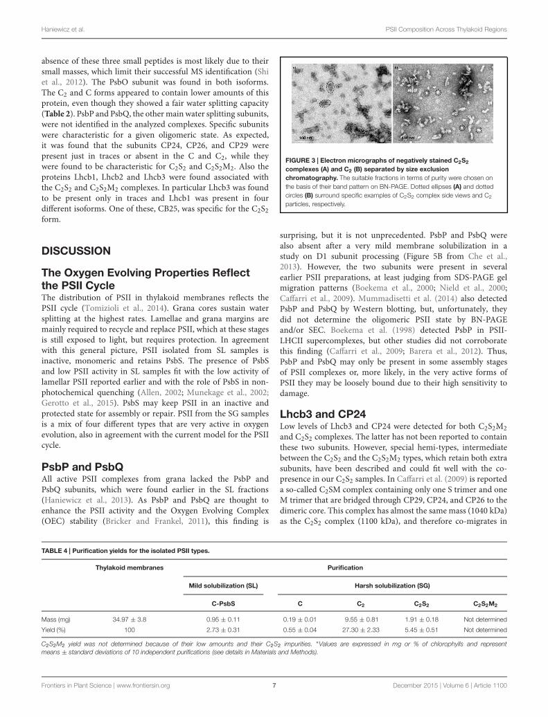
a specific complex primarily identified by its apparent sizeon BN-PAGE (Figure 2 inset). The identity of each complexwas finally confirmed by MS analysis (Table 3, SupplementaryTable S2). Furthermore, the C2S2 and C2 complexes werealso analyzed by TEM (Figure 3), demonstrating the sizesreported for C2 and C2S2 (Aro et al., 2005; Nield and Barber,2006), the latter most easily discernible from the side views(circled in Figure 3A). Unfortunately, C2S2M2 complexes couldnot be analyzed by TEM, because they were obtained inlow amounts and eluted at the beginning of the C2S2 peakin SEC.
The Isolated PSII Types Evolve Oxygen atVery Different RatesThe different PSII complexes obtained by SEC were testedfor their oxygen evolving capacity. The pool of C2S2M2was excluded from this analysis since, as mentioned above,it was not perfectly separated from the C2S2. Each oneof the others pools, represented by a dominant PSII type,were found to be fully functional and characterized by
oxygen evolution rates comparable with other reports inliterature (Kihara et al., 2014; Table 2). As oxygen evolutionper Chl is reported, and because C2S2 binds about threetimes more Chls per reaction center than cores (around210/reaction center as opposed to around 70; Ferreira et al.,2004; Liu et al., 2004; Loll et al., 2005; Barros et al.,2009), the C2S2 complexes are characterized by the highestactivity.
Mass Spectrometry Analysis on the FourPSII Types Showed SignificantDifferences in Subunit CompositionThe BN-PAGE bands obtained from the SEC fractions (Figure 2inset) were excised and directly analyzed by MS to characterizesubunit content (Table 3, Supplementary Table S2). The maincore subunits D1, D2, CP43, CP47, PsbE, PsbH, PsbL, PsbO, andPsbR were found in this analysis. The subunit PsbL, an importantdimerization factor (Suorsa et al., 2004), was absent from theC and C2S2M2 types. PsbR was identified only in the C2 andC2S2 types, while PsbF was not found. The partial or complete
Frontiers in Plant Science | www.frontiersin.org 6 December 2015 | Volume 6 | Article 1100
Haniewicz et al. PSII Composition Across Thylakoid Regions
absence of these three small peptides is most likely due to theirsmall masses, which limit their successful MS identification (Shiet al., 2012). The PsbO subunit was found in both isoforms.The C2 and C forms appeared to contain lower amounts of thisprotein, even though they showed a fair water splitting capacity(Table 2). PsbP and PsbQ, the other mainwater splitting subunits,were not identified in the analyzed complexes. Specific subunitswere characteristic for a given oligomeric state. As expected,it was found that the subunits CP24, CP26, and CP29 werepresent just in traces or absent in the C and C2, while theywere found to be characteristic for C2S2 and C2S2M2. Also theproteins Lhcb1, Lhcb2 and Lhcb3 were found associated withthe C2S2 and C2S2M2 complexes. In particular Lhcb3 was foundto be present only in traces and Lhcb1 was present in fourdifferent isoforms. One of these, CB25, was specific for the C2S2form.
DISCUSSION
The Oxygen Evolving Properties Reflectthe PSII CycleThe distribution of PSII in thylakoid membranes reflects thePSII cycle (Tomizioli et al., 2014). Grana cores sustain watersplitting at the highest rates. Lamellae and grana margins aremainly required to recycle and replace PSII, which at these stagesis still exposed to light, but requires protection. In agreementwith this general picture, PSII isolated from SL samples isinactive, monomeric and retains PsbS. The presence of PsbSand low PSII activity in SL samples fit with the low activity oflamellar PSII reported earlier and with the role of PsbS in non-photochemical quenching (Allen, 2002; Munekage et al., 2002;Gerotto et al., 2015). PsbS may keep PSII in an inactive andprotected state for assembly or repair. PSII from the SG samplesis a mix of four different types that are very active in oxygenevolution, also in agreement with the current model for the PSIIcycle.
PsbP and PsbQAll active PSII complexes from grana lacked the PsbP andPsbQ subunits, which were found earlier in the SL fractions(Haniewicz et al., 2013). As PsbP and PsbQ are thought toenhance the PSII activity and the Oxygen Evolving Complex(OEC) stability (Bricker and Frankel, 2011), this finding is
FIGURE 3 | Electron micrographs of negatively stained C2S2
complexes (A) and C2 (B) separated by size exclusionchromatography. The suitable fractions in terms of purity were chosen onthe basis of their band pattern on BN-PAGE. Dotted ellipses (A) and dottedcircles (B) surround specific examples of C2S2 complex side views and C2
particles, respectively.
surprising, but it is not unprecedented. PsbP and PsbQ werealso absent after a very mild membrane solubilization in astudy on D1 subunit processing (Figure 5B from Che et al.,2013). However, the two subunits were present in severalearlier PSII preparations, at least judging from SDS-PAGE gelmigration patterns (Boekema et al., 2000; Nield et al., 2000;Caffarri et al., 2009). Mummadisetti et al. (2014) also detectedPsbP and PsbQ by Western blotting, but, unfortunately, theydid not determine the oligomeric PSII state by BN-PAGEand/or SEC. Boekema et al. (1998) detected PsbP in PSII-LHCII supercomplexes, but other studies did not corroboratethis finding (Caffarri et al., 2009; Barera et al., 2012). Thus,PsbP and PsbQ may only be present in some assembly stagesof PSII complexes or, more likely, in the very active forms ofPSII they may be loosely bound due to their high sensitivity todamage.
Lhcb3 and CP24Low levels of Lhcb3 and CP24 were detected for both C2S2M2and C2S2 complexes. The latter has not been reported to containthese two subunits. However, special hemi-types, intermediatebetween the C2S2 and the C2S2M2 types, which retain both extrasubunits, have been described and could fit well with the co-presence in our C2S2 samples. In Caffarri et al. (2009) is reporteda so-called C2SM complex containing only one S trimer and oneM trimer that are bridged through CP29, CP24, and CP26 to thedimeric core. This complex has almost the same mass (1040 kDa)as the C2S2 complex (1100 kDa), and therefore co-migrates in
TABLE 4 | Purification yields for the isolated PSII types.
Thylakoid membranes Purification
Mild solubilization (SL) Harsh solubilization (SG)
C-PsbS C C2 C2S2 C2S2M2
Mass (mg) 34.97 ± 3.8 0.95 ± 0.11 0.19 ± 0.01 9.55 ± 0.81 1.91 ± 0.18 Not determined
Yield (%) 100 2.73 ± 0.31 0.55 ± 0.04 27.30 ± 2.33 5.45 ± 0.51 Not determined
C2S2M2 yield was not determined because of their low amounts and their C2S2 impurities. ∗Values are expressed in mg or % of chlorophylls and representmeans ± standard deviations of 10 independent purifications (see details in Materials and Methods).
Frontiers in Plant Science | www.frontiersin.org 7 December 2015 | Volume 6 | Article 1100

Haniewicz et al. PSII Composition Across Thylakoid Regions
BN-PAGE (and also co-sediments in a sucrose gradient; Caffarriet al., 2009). Another hemi-type, called C2S2+, is reported to havesimilar characteristics (Pietrzykowska et al., 2014). It is likely thatthese incomplete forms are assembly intermediates on the way toC2S2M2, or result from its disintegration, either in membranes orduring solubilization or purification.
Isolation of Supercomplexes by CouplingDifferential Solubilization andChromatographyThe organization of the thylakoids and in particular thedistribution of pigment–protein complexes within are verywell characterized (Danielsson et al., 2006). This and otherworks all demonstrate substantial heterogeneity of the PSIIspecies in the thylakoid membranes (Boekema et al., 1995;Dekker and Boekema, 2005; Danielsson et al., 2006; Takahashiet al., 2009; Watanabe et al., 2009; Pagliano et al., 2011).Here, we present a differential solubilization of the thylakoidmembranes, which leads to the isolation of samples with differentoligomeric patterns of PSII and other thylakoid complexes(Figure 1A). Conceptually, our method to access to the granacores fraction, in which is found highly active PSII, is similarto the method described by Berthold et al. (1981), the so-calledBBY preparation, which also removes lamellae and peripheralgranal regions. In contrast to the BBY method, the new methodhas the advantage to avoid the breakdown of granal PSIIcore complexes (C and RC-CP47 are found at most in smallquantities).
CONCLUSION
It has to be remarked that the mild nature of the solubilizationprotocol permits a full separation between PSII monomers anddimers, and makes it possible to prepare C2S2 in larger quantitieswithout density gradients on sucrose or Percoll (Ghanotakis et al.,1987; Caffarri et al., 2004), (Table 4).
AUTHOR CONTRIBUTIONS
DP conceived the study, participated in its design andcoordination, carried out the membranes preparation,participated in the biochemical studies, and drafted themanuscript. PH participated in the design of the study,participated the biochemical studies, and participated inthe membranes preparation. D. Floris carried out themembranes preparation, participated in the biochemicalstudies. D. Farci carried out the membranes preparation,participated in the biochemical studies. JK carried outthe MS analysis. ML participated in the preparation ofmembranes. CB contributed to the electron microscopyanalysis and helped to draft the manuscript. MB participatedin the design of the study and helped to draft themanuscript.
ACKNOWLEDGMENTS
This work was carried out with the support of theprogram Homing Plus (Foundation for Polish Science)grant No: 2012-6/10 co-financed by the European Unionunder the European Regional Development Funds; theprogram PRELUDIUM (National Science Centre) grantnumber DEC-2012/05/05/N/NZ1/01922; the Marie Curieprogram “European Reintegration Grant” (PERG05-GA-2009-247789); the program “FSE SARDEGNA 2007-2013, Legge Regionale 7 agosto 2007, n. 7, Promozionedella ricerca scientifica e dell’innovazione tecnologica inSardegna”.
SUPPLEMENTARY MATERIAL
The Supplementary Material for this article can be found onlineat: http://journal.frontiersin.org/article/10.3389/fpls.2015.01100
REFERENCES
Allen, J. (2002). Photosynthesis of ATP—electrons, proton pumps, rotors, andpoise. Cell 110, 273–276.
Aro, E. M., Suorsa, M., Rokka, A., Allahverdiyeva, Y., Paakkarinen, V., Saleem, A.,et al. (2005). Dynamics of photosystem II: a proteomic approach to thylakoidprotein complexes. J. Exp. Bot. 56, 347–356. doi: 10.1093/jxb/eri041
Barera, S., Pagliano, C., Pape, T., Saracco, G., and Barber, J. (2012). Characterizationof PSII-LHCII supercomplexes isolated from pea thylakoid membrane by one-step treatment with α- and β-dodecyl-D-maltoside. Philos. Trans. R. Soc. Lond.B Biol. Sci. 367, 3389–3399. doi: 10.1098/rstb.2012.0056
Barros, T., Royant, A., Standfuss, J., Dreuw, A., and Kühlbrandt, W. (2009).Crystal structure of plant light-harvesting complex shows the active,energy-transmitting state. EMBO J. 28, 298–306. doi: 10.1038/emboj.2008.276
Berthold, A. D., Babcock, G. T., and Yocum, C. (1981). A highly resolved,oxygen evolving photosystem II preparation from spinach thylakoidmembranes. FEBS Lett. 134, 231–234. doi: 10.1016/0014-5793(81)80608-4
Boekema, E. J., Hankamer, B., Bald, D., Kruip, J., Nield, J., Boonstra, A. F.,et al. (1995). Supramolecular structure of the photosystem II complex from
green plants and cyanobacteria. Proc. Natl. Acad. Sci. U.S.A. 92, 175–179. doi:10.1073/pnas.92.1.175
Boekema, E. J., Nield, J., Hankamer, B., and Barber, J. (1998). Localizationof the 23-kDa subunit of the oxygen-evolving complex of photosystem IIby electron microscopy. Eur. J. Biochem. 252, 268–276. doi: 10.1046/j.1432-1327.1998.2520268.x
Boekema, E. J., van Breemen, J. F. L., van Roon, H., and Dekker, J. P. (2000).Conformational changes in photosystem II supercomplexes upon removal ofextrinsic subunits. Biochemistry 39, 12907–12915. doi: 10.1021/bi0009183
Boekema, E. J., Van Roon, H., Van Breemen, J. F., and Dekker, J. P. (1999).Supramolecular organization of photosystem II and its light-harvesting antennain partially solubilized photosystem II membranes. Eur. J. Biochem. 266,444–452. doi: 10.1046/j.1432-1327.1999.00876.x
Bricker, T. M., and Frankel, L. K. (2011). Auxiliary functions of the PsbO,PsbP and PsbQ proteins of higher plant Photosystem II: a critical analysis.J. Photochem. Photobiol. B Biol. 104, 165–178. doi: 10.1016/j.jphotobiol.2011.01.025
Caffarri, S., Croce, R., Cattivelli, L., and Bassi, R. (2004). A look within LHCII:differential analysis of the Lhcb1-3 complexes building the major trimericantenna complex of higher-plant photosynthesis. Biochemistry 43, 9467–9476.doi: 10.1021/bi036265i
Frontiers in Plant Science | www.frontiersin.org 8 December 2015 | Volume 6 | Article 1100
Haniewicz et al. PSII Composition Across Thylakoid Regions
Caffarri, S., Kouril, R., Kereïche, S., Boekema, E. J., and Croce, R. (2009). Functionalarchitecture of higher plant photosystem II supercomplexes. EMBO J. 28,3052–3063. doi: 10.1038/emboj.2009.232
Cardona, T., Sedoud, A., Cox, N., and Rutherford, A. W. (2012). Charge separationin photosystem II: a comparative and evolutionary overview. Biochim. Biophys.Acta 1817, 26–43. doi: 10.1016/j.bbabio.2011.07.012
Che, Y., Fu, A., Hou, X., McDonald, K., Buchanan, B. B., Huang, W., et al. (2013).C-terminal processing of reaction center protein D1 is essential for the functionand assembly of photosystem II inArabidopsis. Proc. Natl. Acad. Sci. U.S.A. 110,16247–16252. doi: 10.1073/pnas.1313894110
Danielsson, R., Suorsa, M., Paakkarinen, V., Albertsson, P., Styring, S., Aro, E. M.,et al. (2006). Dimeric and monomeric organization of photosystem II. J. Biol.Chem. 281, 14241–14249. doi: 10.1074/jbc.M600634200
de Bianchi, S., Dall’Osto, L., Tognon, G., Morosinotto, T., and Bassi, R. (2008).Minor antenna proteins CP24 and CP26 affect the interactions betweenphotosystem II subunits and the electron transport rate in grana membranesof Arabidopsis. Plant Cell 20, 1012–1028. doi: 10.1105/tpc.107.055749
Dekker, J. P., and Boekema, E. J. (2005). Supramolecular organization of thylakoidmembrane proteins in green plants. Biochim. Biophys. Acta 1706, 12–39. doi:10.1016/j.bbabio.2004.09.009
Edelman, M., and Mattoo, A. K. (2008). D1-protein dynamic in photosystem II:the lingering enigma. Photosynth. Res. 98, 609–620. doi: 10.1007/s11120-008-9342-x
Eshaghi, S., Andersson, B., and Barber, J. (1999). Isolation of a highlyactive PSII-LHCII supercomplex from thylakoid membranes by adirect method. FEBS Lett. 446, 23–26. doi: 10.1016/S0014-5793(99)00149-0
Farci, D., Bowler, M. W., Kirkpatrick, J., McSweeney, S., Tramontano, E.,and Piano, D. (2014). New features of the cell wall of the radio-resistantbacterium Deinococcus radiodurans. Biochim. Biophys. Acta 1838, 1978–1984.doi: 10.1016/j.bbamem.2014.02.014
Ferreira, K. N., Inverson, T. M., Maghlaoui, K., Barber, J., and Iwata, S.(2004). Architecture of the photosynthetic oxygen-evolving center. Science 303,1831–1838. doi: 10.1126/science.1093087
Fey, H., Piano, D., Horn, R., Fischer, D., Schröder, W. P., Bock, R., et al. (2008).Isolation of highly active photosystem II core complexes with a His-tagged Cytb559 subunit from transplastomic tobacco plants. Biochim. Biophys. Acta 1777,1501–1509. doi: 10.1016/j.bbabio.2008.09.012
Gerotto, C., Franchin, C., Arrigoni, G., and Morosinotto, T. (2015). In vivoidentification of Photosystem II Light Harvesting Complexes interactingwith Photosystem II Subunit S. Plant Physiol. 168, 1747–1761. doi:10.1104/pp.15.00361
Ghanotakis, D. F., Demetriou, D. M., and Yocum, C. F. (1987). Isolation andcharacterization of an oxygen-evolving photosystem II reaction center corepreparation and a 28 kDa Chl a binding protein. Biochim. Biophys. Acta 891,15–21. doi: 10.1016/0005-2728(87)90078-8
Haniewicz, P., De Sanctis, D., Büchel, C., Schröder, W. P., Loi, M. C.,Kieselbach, T., et al. (2013). Isolation of monomeric photosystem II thatretains the subunit PsbS. Photosynth. Res. 118, 199–207. doi: 10.1007/s11120-013-9914-2
Kihara, S., Hartzler, D. A., and Savikhin, S. (2014). Oxygen concentrationinside a functioning photosynthetic cell. Biophys. J. 106, 1882–1889. doi:10.1016/j.bpj.2014.03.031
Liu, Z., Yan, H., Wang, K., Kuang, T., Zhang, J., Gui, L., et al. (2004). Crystalstructure of spinach major light-harvesting complex at 2.72 Å resolution.Nature 428, 287–292. doi: 10.1038/nature02373
Loll, B., Kern, J., Saenger,W., Zouni, A., and Biesiadka, J. (2005). Towards completecofactor arrangement in the 3.0 Å resolution structure of photosystem II.Nature438, 1040–1044. doi: 10.1038/nature04224
Mummadisetti, M. P., Frankela, L. K., Bellamyb, H. D., Sallansc, L., Goettertb,J. S., Brylinskia, M., et al. (2014). Use of protein cross-linking andradiolytic footprinting to elucidate PsbP and PsbQ interactions within higherplant Photosystem II. Proc. Natl. Acad. Sci. U.S.A. 111, 16178–16183. doi:10.1073/pnas.1415165111
Munekage, Y., Hojo, M., Meurer, J., Endo, T., Tasaka, M., and Shikanai, T. (2002).PGR5 is involved in cyclic electron flow around photosystem I and II essentialfor photoprotection in Arabidopsis. Cell 110, 361–371. doi: 10.1016/S0092-8674(02)00867-X
Nield, J., and Barber, J. (2006). Refinement of the structural model for thePhotosystem II supercomplex of higher plants. Biochim. Biophys. Acta 1757,353–361. doi: 10.1016/j.bbabio.2006.03.019
Nield, J., Orlova, E. V., Morris, E. P., Gowen, B., van Heel, M., and Barber, J.(2000). 3D map of the plant photosystem II supercomplex obtained bycryoelectron microscopy and single particle analysis. Nat. Struct. Biol. 7, 44–47.doi: 10.1038/71242
Nixon, P. J., Michoux, F., Yu, J., Boehm, M., and Komenda, J. (2010). Recentadvances in understanding the assembly and repair of photosystem II. Ann. Bot.106, 1–16. doi: 10.1093/aob/mcq059
Pagliano, C., Chimirri, F., Saracco, G., Marsano, F., and Barber, J. (2011).Onestep isolation and biochemical characterization of highly active plant PSIImonomeric core. Photosynth. Res. 108, 33–46. doi: 10.1007/s11120-011-9650-4
Piano, D., El Alaoui, S., Korza, H. J., Filipek, R., Sabala, I., Haniewicz, P., et al.(2010). Crystallization of the photosystem II core complex and its chlorophyllbinding subunit CP43 from transplastomic plants of Nicotiana tabacum.Photosynth. Res. 106, 221–226. doi: 10.1007/s11120-010-9597-x
Pietrzykowska, M., Suorsa, M., Semchonok, D. A., Tikkanen, M., Boekema,E. J., Aro, E. M., et al. (2014). The light-harvesting chlorophyll a/bbinding proteins Lhcb1 and Lhcb2 play complementary roles during statetransitions in Arabidopsis. Plant Cell 26, 3646–3660. doi: 10.1105/tpc.114.127373
Pokorska, B., Zienkiewicz, M., Powikrowska, M., Drozak, A., and Romanowska, E.(2009). Differential turnover of the photosystem II reaction centre D1 proteinin mesophyll and bundle sheath chloroplast of maize. Biochim. Biophys. Acta1787, 1161–1169. doi: 10.1016/j.bbabio.2009.05.002
Porra, R. J., Thompson, W. A., and Kriedmann, P. E. (1989). Determination ofaccurate extinction coefficients and simultaneous equations for assayingchlorophylls a and b with four different solvents: verifications of theconcentration of chlorophyll standards by atomic absorption spectroscopy.Biochim. Biophys. Acta 975, 384–394. doi: 10.1016/S0005-2728(89)80347-0
Puthiyaveetil, S., Tsabari, O., Lowry, T., Lenhert, S., Lewis, R. R., Reich, Z.,et al. (2014). Compartmentalization of the protein repair machinery inphotosynthetic membranes. Proc. Natl. Acad. Sci. U.S.A. 111, 15839–15844. doi:10.1073/pnas.1413739111
Schägger, H., and von Jagow, G. (1991). Blue native electrophoresis for isolation ofmembrane protein complexes in enzymatically active form. Anal. Biochem. 199,223–231. doi: 10.1016/0003-2697(91)90094-A
Shi, L. X., Hall, M., Funk, C., and Schröder,W. P. (2012). Photosystem II, a growingcomplex: updates on newly discovered components and low molecular massproteins. Biochim. Biophys. Acta 1817, 13–25. doi: 10.1016/j.bbabio.2011.08.008
Suorsa, M., Regel, R. E., Paakkarinen, V., Battchikova, N., Herrmann, R. G., andAro, E. M. (2004). Protein assembly of photosystem II and accumulation ofsubcomplexes in the absence of low molecular mass subunits PsbL and PsbJ.Eur. J. Biochem. 271, 96–107. doi: 10.1046/j.1432-1033.2003.03906.x
Takahashi, T., Inoue-Kashino, N., Ozawa, S., Takahashi, Y., Kashino, Y., andSatoh, K. (2009). Photosystem II complex in vivo is a monomer. J. Biol. Chem.284, 15598–15606. doi: 10.1074/jbc.M109.000372
Tomizioli, M., Lazar, C., Brugière, S., Burger, T., Salvi, D., Gatto, L., et al. (2014).Deciphering thylakoid sub-compartments using a mass spectrometry-basedapproach. Mol. Cell. Proteomics 13, 2147–2167. doi: 10.1074/mcp.M114.040923
Watanabe, M., Iwai, M., Narikawa, R., and Ikeuchi, M. (2009). Is the photosystemII complex a monomer or a dimer? Plant Cell Physiol. 50, 1674–1680. doi:10.1093/pcp/pcp112
Conflict of Interest Statement: The authors declare that the research wasconducted in the absence of any commercial or financial relationships that couldbe construed as a potential conflict of interest.
Copyright © 2015 Haniewicz, Floris, Farci, Kirkpatrick, Loi, Büchel, Bochtler andPiano. This is an open-access article distributed under the terms of the CreativeCommons Attribution License (CC BY). The use, distribution or reproduction inother forums is permitted, provided the original author(s) or licensor are creditedand that the original publication in this journal is cited, in accordance with acceptedacademic practice. No use, distribution or reproduction is permitted which does notcomply with these terms.
Frontiers in Plant Science | www.frontiersin.org 9 December 2015 | Volume 6 | Article 1100