ispad clinical practice consensus guidelines 2018: diabetic ... · urine output persists because of...
TRANSCRIPT
I S P AD C L I N I C A L P RA C T I C E CON S EN SU S GU I D E L I N E S
ISPAD Clinical Practice Consensus Guidelines 2018: Diabeticketoacidosis and the hyperglycemic hyperosmolar state
Joseph I. Wolfsdorf1 | Nicole Glaser2 | Michael Agus1,3 | Maria Fritsch4 |
Ragnar Hanas5 | Arleta Rewers6 | Mark A. Sperling7 | Ethel Codner8
1Division of Endocrinology, Boston Children's Hospital, Boston, Massachusetts
2Department of Pediatrics, Section of Endocrinology, University of California, Davis School of Medicine, Sacramento, California
3Division of Critical Care Medicine, Boston Children's Hospital, Boston, Massachusetts
4Department of Pediatric and Adolescent Medicine, Medical University of Vienna, Vienna, Austria
5Department of Pediatrics, NU Hospital Group, Uddevalla and Sahlgrenska Academy, Gothenburg University, Uddevalla, Sweden
6Department of Pediatrics, School of Medicine, University of Colorado, Aurora, Colorado
7Division of Endocrinology, Diabetes and Metabolism, Department of Pediatrics, Icahn School of Medicine at Mount Sinai, New York, New York
8Institute of Maternal and Child Research, School of Medicine, University of Chile, Santiago, Chile
Correspondence
Joseph I. Wolfsdorf, Division of Endocrinology, Boston Children's Hospital, 300 Longwood Avenue, Boston, MA.
Email: [email protected]
1 | SUMMARY OF WHAT ISNEW/DIFFERENT
Recommendations concerning fluid management have been modified
to reflect recent findings from a randomized controlled clinical trial
showing no difference in cerebral injury in patients rehydrated at dif-
ferent rates with either 0.45% or 0.9% saline.
2 | EXECUTIVE SUMMARY
The biochemical criteria for the diagnosis of diabetic ketoacidosis
(DKA) are:
• Hyperglycemia (blood glucose >11 mmol/L [≈200 mg/dL])
• Venous pH <7.3 or serum bicarbonate <15 mmol/L
• Ketonemia (blood ß-hydroxybuyrate ≥3 mmol/L) or moderate or
large ketonuria.
The clinical signs of DKA include: Dehydration, tachycardia,
tachypnea, deep sighing respiration, breath smells of acetone, nausea
and/or vomiting, abdominal pain, blurry vision, confusion, drowsiness,
progressive decrease in level of consciousness and, eventually, loss of
consciousness (coma).
Risk factors for DKA in newly diagnosed patients include younger
age, delayed diagnosis, lower socioeconomic status, and residence in a
country with a low prevalence of type 1 diabetes mellitus (T1DM).
Risk factors for DKA in patients with known diabetes include
omission of insulin for various reasons, limited access to medical ser-
vices, and unrecognized interruption of insulin delivery in patients
using an insulin pump.
The following recommendations are based on currently avail-
able evidence and are intended to be a general guide to DKA man-
agement. Because there is considerable individual variability in
presentation of DKA (ranging from mild with only minimal dehydra-
tion to severe with profound dehydration), some patients may
require specific treatment that, in the judgment of the treating phy-
sician, may be within or, occasionally, outside the range of options
presented here. Clinical judgment should always be used to deter-
mine optimal treatment for the individual patient, and timely
adjustments to treatment (electrolyte composition and rate of infu-
sion of rehydration fluids, insulin dose) should be based on ongo-
ing, careful clinical and biochemical monitoring of the patient's
response.
Emergency assessment should follow the general guidelines
for Pediatric Advanced Life Support (PALS) and includes: Imme-
diate measurement of blood glucose, blood or urine ketones,
serum electrolytes, blood gases and complete blood count;
assessment of severity of dehydration, and level of
Received: 11 April 2018 Accepted: 31 May 2018
DOI: 10.1111/pedi.12701
© 2018 John Wiley & Sons A/S. Published by John Wiley & Sons Ltd
Pediatric Diabetes October 2018; 19 (Suppl. 27): 155–177. wileyonlinelibrary.com/journal/pedi 155
consciousness (E). A second peripheral intravenous (IV) catheter
should be inserted (E).
Management should be conducted in a center experienced in
the treatment of DKA in children and adolescents and where vital
signs, neurological status and laboratory results can be monitored
frequently (E). Where geographic constraints require that manage-
ment be initiated in a center with less experience and with fewer
resources, there should be arrangements in place for telephone or
videoconference support from a physician with expertise in
DKA (E).
Meticulous monitoring of the clinical and biochemical response
to treatment is necessary so that timely adjustments in treatment
can be made when indicated by the patient's clinical or laboratory
data (E).
Goals of therapy are to correct dehydration, correct acidosis and
reverse ketosis, gradually restore hyperosmolality and blood glucose
concentration to near normal, monitor for complications of DKA and
its treatment, and identify and treat any precipitating event.
Fluid replacement should begin before starting insulin therapy.
Expand volume using crystalloids, as required, to restore peripheral
circulation (E). Calculate the subsequent rate of fluid administration,
including the provision of maintenance fluid requirements, aiming to
replace the estimated fluid deficit over 24 to 48 hours (A).
Insulin therapy: begin with 0.05 to 0.1 U/kg/h at least 1 hour
AFTER starting fluid replacement therapy (B).
Potassium: If the patient is hyperkalemic, defer potassium replace-
ment therapy until urine output is documented. Otherwise, begin with
40 mmol potassium/L (or 20 mmol potassium/L if the patient is
receiving fluid at a rate ≥10 mL/kg/h) (E).
Bicarbonate administration is not recommended except for
treatment of life-threatening hyperkalemia or unusually severe
acidosis (vpH <6.9) with evidence of compromised cardiac
contractility (C).
Warning signs and symptoms of cerebral edema include: Onset
of headache after beginning treatment or progressively worsening or
severe headache, slowing of heart rate not related to sleep or
improved intravascular volume, change in neurological status (restless-
ness, irritability, increased drowsiness, confusion, incontinence), spe-
cific neurological signs (eg, cranial nerve palsies), rising blood pressure,
and decreased oxygen saturation (C).
In patients with multiple risk factors for cerebral edema (ele-
vated serum urea nitrogen concentration, severe acidosis, severe
hypocapnia), have mannitol or hypertonic saline at the bedside
and the dose calculated (E). If neurologic status deteriorates
acutely, hyperosmolar fluid therapy should be given immedi-
ately (C).
Prevention: Management of an episode of DKA is not complete
until an attempt has been made to identify and treat the cause.
DKA without a preceding febrile illness or gastroenteritis in a
patient with known diabetes is almost always the result of psychoso-
cial problems and failure to appropriately administer insulin.
In new onset diabetes, DKA is frequently the consequence of a
delay in diagnosis (E).
The criteria for hyperglycemic hyperosmolar state (HHS) include:
• Plasma glucose concentration >33.3 mmol/L (600 mg/dL)
• Venous pH >7.25; arterial pH >7.30
• Serum bicarbonate >15 mmol/L
• Small ketonuria, absent to mild ketonemia
• Effective serum osmolality >320 mOsm/kg
• Altered consciousness (eg, obtundation, combativeness) or sei-
zures (in approximately 50%)
In HHS, the goals of initial fluid therapy are to expand the intra-
and extravascular volume, restore normal renal perfusion and promote
a gradual decline in corrected serum sodium concentration and serum
osmolality.
In HHS, begin insulin administration at a dose of 0.025 to
0.05 U/kg/h once plasma glucose is decreasing less than 3 mmol/L
(50 mg/dL) per hour with fluid alone (C).
DKA results from deficiency of circulating insulin and increased
levels of the counterregulatory hormones: catecholamines, glucagon,
cortisol, and growth hormone.1,2 Severe insulin deficiency occurs in
previously undiagnosed T1DM and when treated patients deliber-
ately or inadvertently do not take insulin, especially the long-acting
component of a basal-bolus regimen, or markedly reduce the doses
of insulin, for example, in association with an intercurrent illness
such as gastroenteritis. Patients who use an insulin pump can rap-
idly develop DKA when insulin delivery fails for any reason.3 Rela-
tive insulin deficiency occurs when the concentrations of
counterregulatory hormones markedly increase in response to stress
in conditions such as sepsis, trauma, or febrile illness, which over-
whelm homeostatic mechanisms and lead to metabolic decompensa-
tion despite the patient taking the usual recommended dose of
insulin.
The combination of absolute or relative insulin deficiency and
high counterregulatory hormone concentrations causes an acceler-
ated catabolic state with increased glucose production by the liver
and kidney (via glycogenolysis and gluconeogenesis) and impaired
peripheral glucose utilization, which result in hyperglycemia and
hyperosmolality. Insulin deficiency and high counterregulatory hor-
mone concentrations also increase lipolysis and ketogenesis and
cause ketonemia and metabolic acidosis. Hyperglycemia exceeding
the usual renal threshold of approximately 10 mmol/L (180 mg/dL)
together with hyperketonemia cause osmotic diuresis, dehydration,
and obligatory loss of electrolytes, often aggravated by vomiting
associated with severe ketosis. These changes stimulate further
stress hormone production, which induces more severe insulin resis-
tance and worsening hyperglycemia and hyperketonemia. Lactic aci-
dosis from hypoperfusion or sepsis may contribute to the acidosis
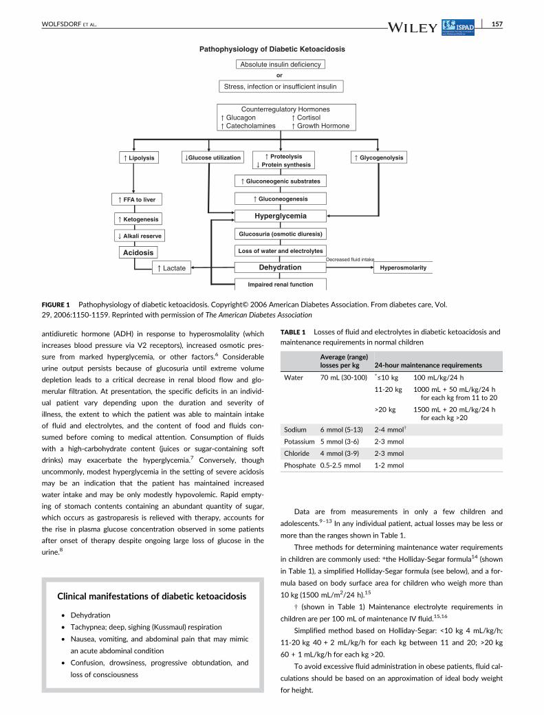
(Figure 1).4
If this cycle is not interrupted by exogenous insulin together with
fluid and electrolyte therapy, fatal dehydration and metabolic acidosis
will ensue.
DKA is characterized by severe depletion of water and electro-
lytes from both the intra- and extracellular fluid (ECF) compart-
ments5; the typical range of losses is shown in Table 1. Despite
their dehydration, patients generally continue to maintain normal
blood pressure or even have high blood pressure,6 possibly due to
elevated plasma catecholamine concentrations, increased release of
156 WOLFSDORF ET AL.
antidiuretic hormone (ADH) in response to hyperosmolality (which
increases blood pressure via V2 receptors), increased osmotic pres-
sure from marked hyperglycemia, or other factors.6 Considerable
urine output persists because of glucosuria until extreme volume
depletion leads to a critical decrease in renal blood flow and glo-
merular filtration. At presentation, the specific deficits in an individ-
ual patient vary depending upon the duration and severity of
illness, the extent to which the patient was able to maintain intake
of fluid and electrolytes, and the content of food and fluids con-
sumed before coming to medical attention. Consumption of fluids
with a high-carbohydrate content (juices or sugar-containing soft
drinks) may exacerbate the hyperglycemia.7 Conversely, though
uncommonly, modest hyperglycemia in the setting of severe acidosis
may be an indication that the patient has maintained increased
water intake and may be only modestly hypovolemic. Rapid empty-
ing of stomach contents containing an abundant quantity of sugar,
which occurs as gastroparesis is relieved with therapy, accounts for
the rise in plasma glucose concentration observed in some patients
after onset of therapy despite ongoing large loss of glucose in the
urine.8
Clinical manifestations of diabetic ketoacidosis
• Dehydration
• Tachypnea; deep, sighing (Kussmaul) respiration
• Nausea, vomiting, and abdominal pain that may mimic
an acute abdominal condition
• Confusion, drowsiness, progressive obtundation, and
loss of consciousness
Data are from measurements in only a few children and
adolescents.9–13 In any individual patient, actual losses may be less or
more than the ranges shown in Table 1.
Three methods for determining maintenance water requirements
in children are commonly used: *the Holliday-Segar formula14 (shown
in Table 1), a simplified Holliday-Segar formula (see below), and a for-
mula based on body surface area for children who weigh more than
10 kg (1500 mL/m2/24 h).15
† (shown in Table 1) Maintenance electrolyte requirements in
children are per 100 mL of maintenance IV fluid.15,16
Simplified method based on Holliday-Segar: <10 kg 4 mL/kg/h;
11-20 kg 40 + 2 mL/kg/h for each kg between 11 and 20; >20 kg
60 + 1 mL/kg/h for each kg >20.
To avoid excessive fluid administration in obese patients, fluid cal-
culations should be based on an approximation of ideal body weight
for height.
Pathophysiology of Diabetic Ketoacidosis
↑ FFA to liver
↑ Ketogenesis
↑ Lipolysis
Acidosis
↓ Alkali reserve
Absolute insulin deficiency
Stress, infection or insufficient insulin
Counterregulatory Hormones ↑ Glucagon ↑ Cortisol ↑ Catecholamines ↑ Growth Hormone
↑ Proteolysis
↓ Protein synthesis
↓Glucose utilization
↑ Gluconeogenic substrates
↑ Glycogenolysis
Glucosuria (osmotic diuresis)
Hyperosmolarity↑ Lactate
Decreased fluid intake
Hyperglycemia
↑ Gluconeogenesis
or
Impaired renal function
Loss of water and electrolytes
Dehydration
FIGURE 1 Pathophysiology of diabetic ketoacidosis. Copyright© 2006 American Diabetes Association. From diabetes care, Vol.
29, 2006:1150-1159. Reprinted with permission of The American Diabetes Association
TABLE 1 Losses of fluid and electrolytes in diabetic ketoacidosis and
maintenance requirements in normal children
Average (range)losses per kg 24-hour maintenance requirements
Water 70 mL (30-100) *≤10 kg 100 mL/kg/24 h
11-20 kg 1000 mL + 50 mL/kg/24 hfor each kg from 11 to 20
>20 kg 1500 mL + 20 mL/kg/24 hfor each kg >20
Sodium 6 mmol (5-13) 2-4 mmol†
Potassium 5 mmol (3-6) 2-3 mmol
Chloride 4 mmol (3-9) 2-3 mmol
Phosphate 0.5-2.5 mmol 1-2 mmol
WOLFSDORF ET AL. 157
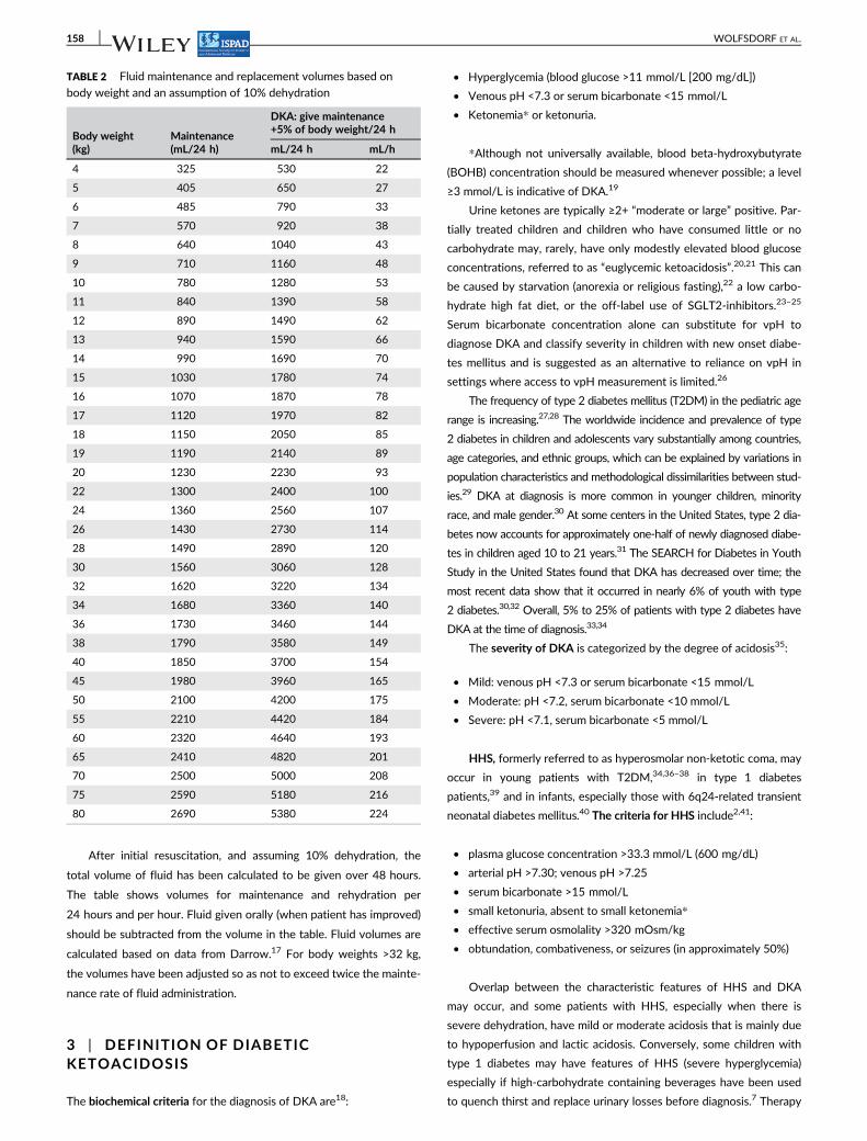
After initial resuscitation, and assuming 10% dehydration, the
total volume of fluid has been calculated to be given over 48 hours.
The table shows volumes for maintenance and rehydration per
24 hours and per hour. Fluid given orally (when patient has improved)
should be subtracted from the volume in the table. Fluid volumes are
calculated based on data from Darrow.17 For body weights >32 kg,
the volumes have been adjusted so as not to exceed twice the mainte-
nance rate of fluid administration.
3 | DEFINITION OF DIABETICKETOACIDOSIS
The biochemical criteria for the diagnosis of DKA are18:
• Hyperglycemia (blood glucose >11 mmol/L [200 mg/dL])
• Venous pH <7.3 or serum bicarbonate <15 mmol/L
• Ketonemia* or ketonuria.
*Although not universally available, blood beta-hydroxybutyrate
(BOHB) concentration should be measured whenever possible; a level
≥3 mmol/L is indicative of DKA.19
Urine ketones are typically ≥2+ “moderate or large” positive. Par-
tially treated children and children who have consumed little or no
carbohydrate may, rarely, have only modestly elevated blood glucose
concentrations, referred to as “euglycemic ketoacidosis”.20,21 This can
be caused by starvation (anorexia or religious fasting),22 a low carbo-
hydrate high fat diet, or the off-label use of SGLT2-inhibitors.23–25
Serum bicarbonate concentration alone can substitute for vpH to
diagnose DKA and classify severity in children with new onset diabe-
tes mellitus and is suggested as an alternative to reliance on vpH in
settings where access to vpH measurement is limited.26
The frequency of type 2 diabetes mellitus (T2DM) in the pediatric age
range is increasing.27,28 The worldwide incidence and prevalence of type
2 diabetes in children and adolescents vary substantially among countries,
age categories, and ethnic groups, which can be explained by variations in
population characteristics and methodological dissimilarities between stud-
ies.29 DKA at diagnosis is more common in younger children, minority
race, and male gender.30 At some centers in the United States, type 2 dia-
betes now accounts for approximately one-half of newly diagnosed diabe-
tes in children aged 10 to 21 years.31 The SEARCH for Diabetes in Youth
Study in the United States found that DKA has decreased over time; the
most recent data show that it occurred in nearly 6% of youth with type
2 diabetes.30,32 Overall, 5% to 25% of patients with type 2 diabetes have
DKA at the time of diagnosis.33,34
The severity of DKA is categorized by the degree of acidosis35:
• Mild: venous pH <7.3 or serum bicarbonate <15 mmol/L
• Moderate: pH <7.2, serum bicarbonate <10 mmol/L
• Severe: pH <7.1, serum bicarbonate <5 mmol/L
HHS, formerly referred to as hyperosmolar non-ketotic coma, may
occur in young patients with T2DM,34,36–38 in type 1 diabetes
patients,39 and in infants, especially those with 6q24-related transient
neonatal diabetes mellitus.40 The criteria for HHS include2,41:
• plasma glucose concentration >33.3 mmol/L (600 mg/dL)
• arterial pH >7.30; venous pH >7.25
• serum bicarbonate >15 mmol/L
• small ketonuria, absent to small ketonemia*
• effective serum osmolality >320 mOsm/kg
• obtundation, combativeness, or seizures (in approximately 50%)
Overlap between the characteristic features of HHS and DKA
may occur, and some patients with HHS, especially when there is
severe dehydration, have mild or moderate acidosis that is mainly due
to hypoperfusion and lactic acidosis. Conversely, some children with
type 1 diabetes may have features of HHS (severe hyperglycemia)
especially if high-carbohydrate containing beverages have been used
to quench thirst and replace urinary losses before diagnosis.7 Therapy
TABLE 2 Fluid maintenance and replacement volumes based on
body weight and an assumption of 10% dehydration
Body weight(kg)
Maintenance(mL/24 h)
DKA: give maintenance+5% of body weight/24 h
mL/24 h mL/h
4 325 530 22
5 405 650 27
6 485 790 33
7 570 920 38
8 640 1040 43
9 710 1160 48
10 780 1280 53
11 840 1390 58
12 890 1490 62
13 940 1590 66
14 990 1690 70
15 1030 1780 74
16 1070 1870 78
17 1120 1970 82
18 1150 2050 85
19 1190 2140 89
20 1230 2230 93
22 1300 2400 100
24 1360 2560 107
26 1430 2730 114
28 1490 2890 120
30 1560 3060 128
32 1620 3220 134
34 1680 3360 140
36 1730 3460 144
38 1790 3580 149
40 1850 3700 154
45 1980 3960 165
50 2100 4200 175
55 2210 4420 184
60 2320 4640 193
65 2410 4820 201
70 2500 5000 208
75 2590 5180 216
80 2690 5380 224
158 WOLFSDORF ET AL.
must be appropriately modified to address the pathophysiology and
particular biochemical disturbances of the individual patient (see
below). See below regarding specific therapy of HHS.
4 | FREQUENCY OF DKA
4.1 | At disease onset
There is wide geographic variation in the frequency of DKA at onset
of diabetes; rates inversely correlate with the regional incidence of
type 1 diabetes. Frequencies range from approximately 15% to 70%
in Europe and North America.30,42–49 DKA at diagnosis is more com-
mon in younger children (especially <2 years of age), including infants
with both transient and permanent neonatal diabetes (overall fre-
quency 66%), often the consequence of diagnostic error or delayed
treatment.50–53 It is also more common in ethnic minority groups, and
in children whose families do not have ready access to medical care
for social or economic reasons.21,32,46,51,54,55
4.2 | In children with established diabetes
The risk of DKA in established type 1 diabetes is 1% to 10% per
patient per year3,56–61:
Risk is increased in59:
• Children who omit insulin58
• Children with poor metabolic control or previous episodes of DKA
• Gastroenteritis with persistent vomiting and inability to maintain
hydration
• Children with psychiatric disorders, including those with eating
disorders
• Children with difficult or unstable family circumstances (eg, paren-
tal abuse)
• Peripubertal and adolescent girls
• Binge alcohol consumption62
• Children with limited access to medical services
In the early days of insulin pump therapy, DKA was more common
than in patients using injection therapy (only rapid- or short-acting
insulin is used in pumps; therefore, interruption of insulin delivery for
any reason rapidly leads to insulin deficiency).3,63 However, a recent
matched comparison of patients using insulin pump therapy with mul-
tiple daily injections showed that DKA occurred less frequently (3.64
vs 4.26 per 100 patient-years) in patients using pump therapy.64
In recurrent DKA, insulin omission or failure to follow sick day or
pump failure management guidelines accounts for almost all episodes.
5 | MANAGEMENT OF DKA
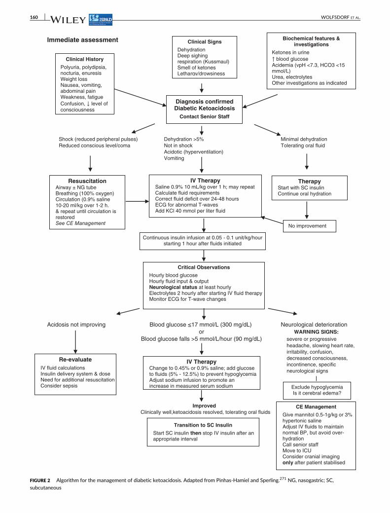
Figure 2 shows an algorithm for the management of DKA.
5.1 | Emergency assessment
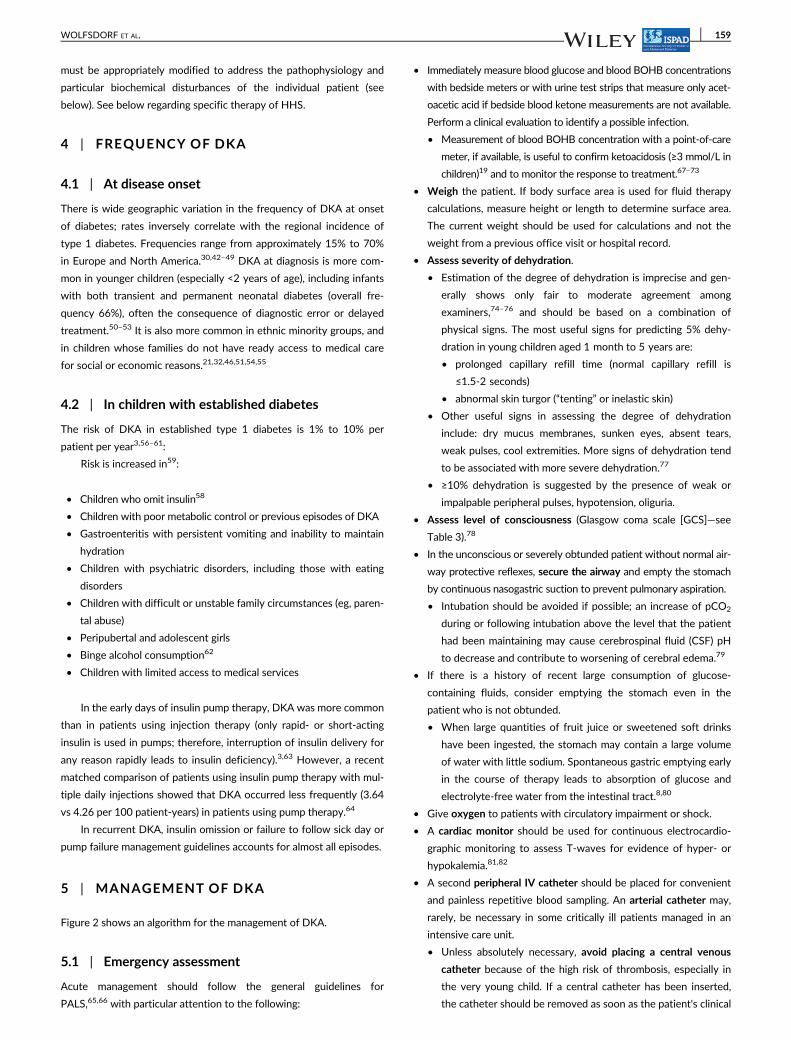
Acute management should follow the general guidelines for
PALS,65,66 with particular attention to the following:
• Immediately measure blood glucose and blood BOHB concentrations
with bedside meters or with urine test strips that measure only acet-
oacetic acid if bedside blood ketone measurements are not available.
Perform a clinical evaluation to identify a possible infection.
• Measurement of blood BOHB concentration with a point-of-care
meter, if available, is useful to confirm ketoacidosis (≥3 mmol/L in
children)19 and to monitor the response to treatment.67–73
• Weigh the patient. If body surface area is used for fluid therapy
calculations, measure height or length to determine surface area.
The current weight should be used for calculations and not the
weight from a previous office visit or hospital record.
• Assess severity of dehydration.
• Estimation of the degree of dehydration is imprecise and gen-
erally shows only fair to moderate agreement among
examiners,74–76 and should be based on a combination of
physical signs. The most useful signs for predicting 5% dehy-
dration in young children aged 1 month to 5 years are:
• prolonged capillary refill time (normal capillary refill is
≤1.5-2 seconds)
• abnormal skin turgor (“tenting” or inelastic skin)
• Other useful signs in assessing the degree of dehydration
include: dry mucus membranes, sunken eyes, absent tears,
weak pulses, cool extremities. More signs of dehydration tend
to be associated with more severe dehydration.77
• ≥10% dehydration is suggested by the presence of weak or
impalpable peripheral pulses, hypotension, oliguria.
• Assess level of consciousness (Glasgow coma scale [GCS]—see
Table 3).78
• In the unconscious or severely obtunded patient without normal air-
way protective reflexes, secure the airway and empty the stomach
by continuous nasogastric suction to prevent pulmonary aspiration.
• Intubation should be avoided if possible; an increase of pCO2
during or following intubation above the level that the patient
had been maintaining may cause cerebrospinal fluid (CSF) pH
to decrease and contribute to worsening of cerebral edema.79
• If there is a history of recent large consumption of glucose-
containing fluids, consider emptying the stomach even in the
patient who is not obtunded.
• When large quantities of fruit juice or sweetened soft drinks
have been ingested, the stomach may contain a large volume
of water with little sodium. Spontaneous gastric emptying early
in the course of therapy leads to absorption of glucose and
electrolyte-free water from the intestinal tract.8,80
• Give oxygen to patients with circulatory impairment or shock.
• A cardiac monitor should be used for continuous electrocardio-
graphic monitoring to assess T-waves for evidence of hyper- or
hypokalemia.81,82
• A second peripheral IV catheter should be placed for convenient
and painless repetitive blood sampling. An arterial catheter may,
rarely, be necessary in some critically ill patients managed in an
intensive care unit.
• Unless absolutely necessary, avoid placing a central venous
catheter because of the high risk of thrombosis, especially in
the very young child. If a central catheter has been inserted,
the catheter should be removed as soon as the patient's clinical
WOLFSDORF ET AL. 159
Immediate assessment
Shock (reduced peripheral pulses) Dehydration >5% Minimal dehydration
Reduced conscious level/coma Not in shock Tolerating oral fluid
Acidotic (hyperventilation)
Vomiting
Acidosis not improving Blood glucose ≤17 mmol/L (300 mg/dL) Neurological deterioration
or WARNING SIGNS:
Blood glucose falls >5 mmol/L/hour (90 mg/dL) severe or progressive
headache, slowing heart rate,
irritability, confusion,
decreased consciousness,
incontinence, specific
neurological signs
Improved
Clinically well,ketoacidosis resolved, tolerating oral fluids
ResuscitationAirway ± NG tubeBreathing (100% oxygen)Circulation (0.9% saline10-20 ml/kg over 1-2 h. & repeat until circulation is restored See CE Management
IV TherapySaline 0.9% 10 mL/kg over 1 h; may repeatCalculate fluid requirementsCorrect fluid deficit over 24-48 hoursECG for abnormal T-wavesAdd KCl 40 mmol per liter fluid
TherapyStart with SC insulinContinue oral hydration
Continuous insulin infusion at 0.05 - 0.1 unit/kg/hour starting 1 hour after fluids initiated
Critical Observations
Hourly blood glucoseHourly fluid input & outputNeurological status at least hourlyElectrolytes 2 hourly after starting IV fluid therapyMonitor ECG for T-wave changes
Re-evaluate
IV fluid calculations Insulin delivery system & dose Need for additional resuscitation Consider sepsis
IV TherapyChange to 0.45% or 0.9% saline; add glucose to fluids (5% - 12.5%) to prevent hypoglycemiaAdjust sodium infusion to promote an increase in measured serum sodium Exclude hypoglycemia
Is it cerebral edema?
CE Management
Give mannitol 0.5-1g/kg or 3% hypertonic salineAdjust IV fluids to maintain normal BP, but avoid over-hydrationCall senior staffMove to ICUConsider cranial imagingonly after patient stabilised
Transition to SC Insulin
Start SC insulin then stop IV insulin after an appropriate interval
Diagnosis confirmedDiabetic Ketoacidosis
Contact Senior Staff
Clinical History
Polyuria, polydipsia, nocturia, enuresisWeight lossNausea, vomiting, abdominal painWeakness, fatigue
Confusion, ↓ level of consciousness
Clinical Signs
DehydrationDeep sighing respiration (Kussmaul)Smell of ketonesLethargy/drowsiness
Biochemical features & investigations
Ketones in urine
↑ blood glucose Acidemia (vpH <7.3, HCO3 <15 mmol/L)Urea, electrolytes Other investigations as indicated
No improvement
FIGURE 2 Algorithm for the management of diabetic ketoacidosis. Adapted from Pinhas-Hamiel and Sperling.271 NG, nasogastric; SC,
subcutaneous
160 WOLFSDORF ET AL.
status permits.83–85 Mechanical and pharmacologic prophylaxis
(low molecular weight heparin) should be considered especially
in children >12 years.
• Insulin should preferably not be given through a central line
unless it is the only available option because its infusion may
be interrupted when other fluids are given through the
same line.
• Give antibiotics to febrile patients after obtaining appropriate
cultures of body fluids.
• Bladder catheterization usually is not necessary, but if the child is
unconscious or unable to void on demand (eg, infants and very ill
young children) the bladder should be catheterized.
• Obtain a blood sample for laboratory measurement of:
• serum or plasma glucose
• electrolytes (including serum bicarbonate)
• blood urea nitrogen, creatinine
• serum osmolality
• venous pH, pCO2†
• hemoglobin, hematocrit and complete blood count. Note that
an increased white blood cell count in response to stress is
characteristic of DKA and is not indicative of infection.86
• albumin, calcium, phosphate, magnesium concentrations
(if possible)
• Perform a urinalysis for ketones if blood or serum ketones have
not been measured.
• Obtain appropriate specimens for culture (blood, urine, throat),
only if there is evidence of infection (eg, fever).
• If laboratory measurement of serum potassium is delayed, per-
form an electrocardiogram (ECG) for baseline evaluation of potas-
sium status.81,82
• Although not essential for management of DKA per se, HbA1c may
be useful in the evaluation and management of specific patients as
it provides information about the duration of hyperglycemia.
6 | WHERE SHOULD THE CHILD WITH DKABE MANAGED?
After initial life support, the child should receive care in a unit
that has:
• Experienced nursing staff trained in monitoring and management
of DKA in children and adolescents
• Written guidelines or, if unavailable, access to online guidelines
for DKA management in children
• Access to a laboratory that can provide frequent and timely mea-
surements of biochemical variables
Whenever possible, a specialist/consultant pediatrician with train-
ing and expertise in the management of DKA should direct inpatient
management. Where geographic constraints require that management
be initiated in a center with less experience and with fewer resources,
there should be arrangements in place for telephone or videoconfer-
ence support from a physician with expertise in DKA.
Children with severe DKA (long duration of symptoms, compro-
mised circulation, or depressed level of consciousness) or those who
are at increased risk for cerebral edema (eg, <5 years of age, severe
acidosis, low pCO2, high blood urea nitrogen) should be considered
for immediate treatment in an intensive care unit (pediatric if avail-
able) or in a unit that has equivalent resources and supervision, such
as a children's ward specializing in diabetes care.18,87 Transport teams
should be knowledgeable about DKA management (or have access to
a medical control physician who is knowledgeable) and should have
rescue medications available during the transport, including high con-
centration dextrose IV solutions and mannitol or 3% hypertonic saline.
In a child with established diabetes, whose parents have been
trained in sick day management, hyperglycemia and ketosis without
vomiting or severe dehydration can be managed at home or in an out-
patient health care facility (eg, emergency ward), provided an experi-
enced diabetes team supervises the care.35,88,89
7 | CLINICAL AND BIOCHEMICALMONITORING
Successful management of DKA and HHS requires meticulous moni-
toring and recording of the patient's clinical and biochemical response
to treatment so that timely adjustments in treatment can be made
when indicated by the patient's clinical or laboratory data.
There should be documentation on a flow chart of hour-by-hour
clinical observations, IV and oral medications, fluids, and laboratory
results. Monitoring should include the following:
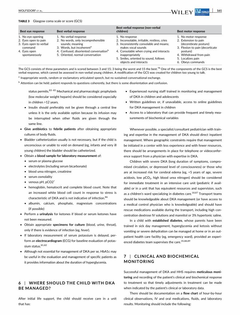
TABLE 3 Glasgow coma scale or score (GCS)
Best eye response Best verbal responseBest verbal response (non-verbalchildren) Best motor response
1. No eye opening2. Eyes open to pain3. Eyes open to verbal
command4. Eyes open
spontaneously
1. No verbal response2. No words, only incomprehensible
sounds; moaning3. Words, but incoherenta
4. Confused, disoriented conversationb
5. Oriented, normal conversation
1. No response2. Inconsolable, irritable, restless, cries3. Inconsistently consolable and moans;
makes vocal sounds4. Consolable when crying and interacts
inappropriately5. Smiles, oriented to sound, follows
objects and interacts
1. No motor response2. Extension to pain
(decerebrate posture)3. Flexion to pain (decorticate
posture)4. Withdrawal from pain5. Localizes pain6. Obeys commands
The GCS consists of three parameters and is scored between 3 and 15; 3 being the worst and 15 the best.78 One of the components of the GCS is the bestverbal response, which cannot be assessed in non-verbal young children. A modification of the GCS was created for children too young to talk.a Inappropriate words, random or exclamatory articulated speech, but no sustained conversational exchange.b Attention can be held; patient responds to questions coherently, but there is some disorientation and confusion.
WOLFSDORF ET AL. 161
• Hourly (or more frequently as indicated) vital signs (heart rate,
respiratory rate, blood pressure)
• Hourly (or more frequently as indicated) neurological observa-
tions (Glasgow coma score; Table 3) for warning signs and symp-
toms of cerebral edema (see below)
• onset of headache after starting DKA treatment or worsening
of headache already present before commencing treatment
• inappropriate slowing of heart rate
• recurrence of vomiting
• change in neurological status (restlessness, irritability,
increased drowsiness, confusion, incontinence) or specific neu-
rologic signs (eg, cranial nerve palsies, abnormal pupillary
responses)
• rising blood pressure
• decreased oxygen saturation
• rapidly increasing serum sodium concentration suggesting loss
of urinary free water as a manifestation of diabetes insipidus
(from interruption of blood flow to the pituitary gland due to
cerebral herniation)
• Amount of administered insulin
• Hourly (or more frequently as indicated) accurate fluid input
(including all oral fluid) and output.
• Capillary blood glucose concentration should be measured hourly
(but must be cross-checked against laboratory venous glucose as
capillary methods may be inaccurate in the presence of poor
peripheral circulation and acidosis, and is limited in measuring
extremely high levels).
• Laboratory tests: serum electrolytes, glucose, blood urea nitrogen,
calcium, magnesium, phosphate, hematocrit, and blood gases
should be repeated 2 to 4 hourly, or more frequently, as clinically
indicated, in more severe cases.
• Blood BOHB concentrations, if available, every 2 to 4 hours.68–72
• Near-patient (also referred to as point-of-care) BOHB mea-
surements correlate well with a reference method up to
3 mmol/L, but are not accurate above 5 mmol/L.70,90
• Lipids and triglycerides can be grossly elevated causing the blood
sample to show a visible rim of lipids, which can interfere with
accuracy of laboratory tests.91
• If the laboratory cannot provide timely results, a portable bio-
chemical analyzer that measures serum electrolytes and blood
gases on fingerstick blood samples at the bedside is a useful
adjunct to laboratory-based determinations. Blood glucose and
blood or urine ketone concentrations can be measured with a
bedside meter while awaiting results from the laboratory.
• Measure body weight each morning.
• Calculations:
• Anion gap = Na − (Cl + HCO3): normal is 12 � 2 mmol/L
• In DKA the anion gap is typically 20 to 30 mmol/L; an anion
gap >35 mmol/L suggests concomitant lactic acidosis (E)
• Corrected sodium = measured Na + 2([plasma glucose −
5.6]/5.6) mmol/L or measured Na + 2([plasma glucose −
100]/100) mg/dL‡
• Effective osmolality (mOsm/kg) = 2 × (plasma Na) + plasma
glucose mmol/L93; normal range is 275 to 295 mOsm/kg
Goals of therapy
• Correct acidosis and reverse ketosis
• Correct dehydration
• Restore blood glucose to near normal
• Monitor for complications of DKA and its treatment
• Identify and treat any precipitating event
7.1 | Fluids and salt
Patients with DKA have a deficit in ECF volume that usually is in the
range 5% to 10% of body weight.9,10 Shock with hemodynamic com-
promise is rare in pediatric DKA. Clinical estimates of the volume defi-
cit are subjective and inaccurate74–76; therefore, in moderate DKA
assume 5% to 7% and in severe DKA 7% to 10% dehydration. The
effective osmolality (formula above) is frequently in the range of
300 to 350 mmol/kg. Increased serum urea nitrogen and hematocrit
or hemoglobin concentration or, alternatively, plasma albumin or total
protein concentration if anemia is suspected94 are useful markers of
the degree of ECF contraction,89,95,96 and should be determined fre-
quently during fluid resuscitation and deficit replacement.97 The
serum sodium concentration is an unreliable measure of the degree of
ECF contraction for two reasons: (1) glucose, largely restricted to the
extracellular space, causes osmotic movement of water into the extra-
cellular space thereby causing dilutional hyponatremia,98,99 and (2) the
low sodium content of the elevated lipid fraction of the serum in
DKA. The latter is not a concern with most modern methods for mea-
suring sodium concentration. It is useful to calculate the corrected
sodium concentration (using the above formula), to help assess the
magnitude of the deficit of sodium and water.5 The “corrected”
sodium represents the expected serum sodium concentration in the
absence of hyperglycemia. Changes in the corrected sodium should
be monitored throughout the course of therapy. As the plasma glu-
cose concentration decreases after administering fluid and insulin, the
measured serum sodium concentration should increase and the
glucose-corrected sodium concentration (formula above) should
slowly decrease or remain in the normal range. It is important to
appreciate that the increase in measured serum sodium concentration
does not indicate a worsening of the hypertonic state. A failure of
measured serum sodium levels to rise or a further decline in serum
sodium levels with therapy is thought to be a potentially ominous sign
of impending cerebral edema.100–102 A rapid and ongoing rise in
serum sodium concentration may also indicate possible cerebral
edema as a result of loss of free water in the urine from diabetes
insipidus.
The objectives of fluid and electrolyte replacement therapy
are to:
• Restore circulating volume
• Replace sodium and the extracellular and intracellular water
deficits
162 WOLFSDORF ET AL.
• Improve glomerular filtration and enhance clearance of glucose
and ketones from the blood
7.2 | Principles of water and salt replacement
Despite much effort to identify the cause of cerebral edema, its path-
ogenesis is incompletely understood and controversy continues con-
cerning the association between the rate of fluid or sodium
administration used in the treatment of DKA and the development of
cerebral edema.103–105 No treatment strategy can be definitively
recommended as being superior to another based on current evi-
dence.104 A recently completed prospective randomized clinical trial
(the PECARN FLUID Study) compared acute and long-term neurologi-
cal outcomes in 1389 episodes of DKA in 1255 children treated with
slower vs more rapid fluid administration using either 0.45% or 0.9%
saline.106 The PECARN FLUID Study showed no significant differ-
ences in the frequency of either altered mental status or clinical diag-
noses of cerebral edema in any of the treatment arms, and long-term
neurocognitive outcomes were similar in all groups. Point estimates
suggested lower frequencies of altered mental status in children rehy-
drated more rapidly with 0.45% saline, but these differences did not
reach statistical significance.106 The results of this study suggest that
an assumed fluid deficit between 5% and 10% of body weight should
be replaced over 24 to 48 hours along with maintenance fluids, using
fluids with a sodium content between 0.45% and 0.9% saline. The risk
of cerebral injury does not appear to be associated with differences in
fluid protocols within these ranges. Therefore, clinicians should not
unnecessarily restrict fluid administration if clinical signs suggest the
need for circulatory volume expansion.
The principles described below are based on the consensus state-
ment from a panel of expert physicians representing the Lawson Wil-
kins Pediatric Endocrine Society (LWPES), the European Society for
Pediatric Endocrinology (ESPE), and the International Society for Pedi-
atric and Adolescent Diabetes (ISPAD)18,107 and incorporate the rec-
ommendations from the PECARN FLUID Study.106
• Water and salt deficits must be replaced.
• IV or oral fluids that may have been given in another facility
before assessment should be factored into calculations of deficit
and replacement volumes.
7.2.1 | Resuscitation fluids
For patients who are volume depleted but not in shock, volume
expansion (resuscitation) should begin immediately with 0.9% saline
to restore the peripheral circulation. The volume administered typi-
cally is 10 mL/kg infused over 30 to 60 minutes; however, if tissue
perfusion is poor the initial fluid bolus is given more rapidly (eg, over
15-30 minutes) and a second fluid bolus may be needed to ensure
adequate tissue perfusion.
• In the rare patient with DKA in shock, rapidly restore circulatory
volume with isotonic saline in 20 mL/kg boluses infused as
quickly as possible through a large bore cannula with reassess-
ment of circulatory status after each bolus.
• Use crystalloid not colloid. There are no data to support the use
of colloid in preference to crystalloid in the treatment of DKA.
7.2.2 | Deficit replacement fluids
Subsequent fluid management (deficit replacement) can be accom-
plished with 0.45% to 0.9% saline or a balanced salt solution (Ringer's
lactate, Hartmann's solution or Plasmalyte).95,100,108–114
• Fluid therapy should begin with deficit replacement plus main-
tenance fluid requirements.
• All children will experience a decrease in vascular volume
when plasma glucose concentrations fall during treatment;
therefore, it is essential to ensure that they receive suffi-
cient fluid and salt to maintain adequate tissue perfusion.
• Deficit replacement should be with a solution that has a tonic-
ity in the range 0.45% to 0.9% saline, with added potassium
chloride, potassium phosphate or potassium acetate (see
below under potassium replacement).95,100,108,112,113,115–117
Decisions regarding use of isotonic vs hypotonic solution for
deficit replacement should depend on clinician judgment based
on the patient's hydration status, serum sodium concentration
and osmolality.
• In addition to providing the usual daily maintenance fluid
requirement, replace the estimated fluid deficit at an even rate
over 24 to 48 hours.18,95,118 Except for severely ill individuals,
oral intake typically begins within 24 hours.118 Although rehy-
dration is generally planned to occur over longer periods, in a
study of 635 episodes of DKA the mean time to correction of
DKA and complete restoration of the circulation was
11.6 � 6.2 hours. At this point, any remaining deficits were
replenished by oral intake once DKA had resolved and patients
were transitioned to subcutaneous insulin.118
• Satisfactory outcomes have also been reported using an alter-
native simplified method: After the initial fluid administration
of 20 mL/kg of normal saline, 0.675% saline (3/4 normal saline,
115.5 mmol sodium) is infused at 2 to 2.5 times the usual
maintenance rate of fluid administration regardless of the
degree of dehydration, and decreased to 1 to 1.5 times the
maintenance rate after 24 hours, or earlier if acidosis
resolved.112
• Clinical assessment of hydration status and calculated effective
osmolality are valuable guides to fluid and electrolyte therapy.
The aim is gradually to reduce serum effective osmolality to
normal.97,118,119 There should be a concomitant increase in
serum sodium concentration as the serum glucose concentra-
tion decreases (sodium should rise by 0.5 mmol/L for each
1 mmol/L decrease in glucose concentration).
• Urinary losses should not routinely be added to the calculation
of replacement fluid, but this may be necessary in rare
circumstances.
• The sodium content of the fluid should be increased if mea-
sured serum sodium concentration is low and does not rise
appropriately as the plasma glucose concentration
falls.100,111,119,120
WOLFSDORF ET AL. 163
• The use of large amounts of chloride-rich fluids (combined
with preferential renal excretion of ketones over chloride)
may be associated with the rapid development of
hyperchloremia121–123 (defined as a ratio of chloride:sodium
[Cl−:Na+] > 0.79124) and hyperchloremic metabolic
acidosis.117,122,125–127
• The acidifying effect of chloride can mask recognition of
resolution of ketoacidosis when total base deficit is used to
monitor biochemical improvement.123
• When hyperchloremia develops, a persisting base deficit or
low bicarbonate concentration can be erroneously inter-
preted as being due to ongoing ketosis.128
• To avoid this misinterpretation, measurement of bedside
BOHB levels will prevent any confusion and can demon-
strate that ketoacidosis has resolved. Hyperchloremic aci-
dosis resolves spontaneously.
• Although the anion gap is useful to track resolution of keto-
sis, it has two limitations in this setting: it is unable to differ-
entiate a mixed metabolic acidosis (hyperchloremic and
ketotic), and the degree of hyperchloremic acidosis is not
quantifiable.
• Normally the difference between the serum sodium and
chloride concentrations is 30 to 35 mmol/L. To partition
the chloride component of the base deficit, the following
formula has been proposed to enable clinicians to track
resolution of ketoacidosis at the bedside: Chloride-
induced base deficit = (plasma sodium − plasma chloride
− 32).123
• The chloride load can be reduced by not giving potas-
sium as potassium chloride (use potassium acetate
instead) and by using fluids such Ringer's lactate or Plas-
malyte in which a portion of the chloride is replaced by
lactate or acetate, respectively.129
7.3 | Insulin therapy
DKA is caused by a decrease in the effective circulating insulin level
associated with increases in counter-regulatory hormone concentra-
tions. Although rehydration alone frequently causes a marked
decrease in blood glucose concentration,130,131 insulin therapy is
essential to restore normal cellular metabolism, to suppress lipolysis
and ketogenesis, and to normalize blood glucose concentrations.132
So-called low dose IV insulin administration is safe and
effective.118,133
• Start insulin infusion at least 1 hour after starting fluid replace-
ment therapy; that is, after the patient has received initial volume
expansion105
• Correction of insulin deficiency
• Dose: 0.05 to 0.1 unit/kg/h (eg, one method is to dilute
50 units regular [soluble] insulin in 50 mL normal saline,
1 unit = 1 mL)134–141
• Route of administration IV
• An IV bolus should not be used at the start of therapy; it is
unnecessary,140,142 may increase the risk of cerebral
edema,105,119,143 can precipitate shock by rapidly decreasing
osmotic pressure, and can exacerbate hypokalemia
• The dose of insulin should usually remain at 0.05 to 0.1 unit/kg/h at
least until resolution of DKA (pH >7.30, serum bicarbonate
>15 mmol/L, BOHB <1 mmol/L, or closure of the anion gap), which
invariably takes longer than normalization of blood glucose concen-
trations.144 Monitor venous pH and serum BOHB concentration
every 2 hours to ensure steady improvement of biochemical param-
eters. If the insulin effect is adequate serum BOHB should decrease
by approximately 0.5 mmol/L/h.72 Increase the insulin dose if the
expected rate of biochemical improvement does not occur.
• If the patient shows marked sensitivity to insulin (eg, some young
children with DKA, patients with HHS, and some older children
with established diabetes), the insulin dose may be decreased pro-
vided that metabolic acidosis continues to resolve. For example, if
a young child is receiving 0.05 unit/kg/h, it may be necessary to
reduce the insulin dose to 0.03 unit/kg/h to prevent hypoglyce-
mia despite the addition of IV glucose.
• For less severe DKA (pH >7.1-7.2), 0.05 U/kg/h (0.03 U/kg/h for age
<5 years with mild DKA) is usually sufficient to resolve the acidosis.
• Uncontrolled retrospective and observational studies have
reported comparable efficacy and safety using 0.05 unit/kg/
h,145,146 and some pediatric centers routinely use this dose for
treatment of DKA. A recent small RCT in children ≤12 years old
showed that low dose (0.05 unit/kg/h) was comparable to stan-
dard dose (0.1 U/kg/h) with respect to rate of blood glucose
decrease and resolution of acidosis; however, there was also no
evidence that the higher dose (0.1 U/kg/h) is harmful.113
• Insulin has an aldosterone-like effect leading to increased urinary
potassium excretion.147–151 High doses administered intrave-
nously for a prolonged period of time may contribute to a
decrease in serum potassium concentration due to increased uri-
nary potassium excretion despite potassium administration.
• Time on IV insulin infusion and dose of insulin should be mini-
mized to avoid severe hypokalemia.152
• During initial volume expansion the plasma glucose concentration
falls steeply.130 Thereafter, and after commencing insulin therapy,
the plasma glucose concentration typically decreases at a rate of
2 to 5 mmol/L/h, depending on the timing and amount of glucose
administration134–137,139,140,153
• To prevent an unduly rapid decrease in plasma glucose concentra-
tion and hypoglycemia, 5% glucose, initially, should be added to
the IV fluid when the plasma glucose falls to approximately 14 to
17 mmol/L (250-300 mg/dL), or sooner if the rate of fall is
precipitous.
• It may be necessary to use 10% or even 12.5% dextrose to
prevent hypoglycemia while continuing to infuse insulin to cor-
rect the metabolic acidosis. These glucose concentrations are
often necessary to prevent hypoglycemia when insulin is
infused at a rate of 0.1 unit/kg/h.
• If BG falls very rapidly (>5 mmol/L/h) after initial fluid expansion,
consider adding glucose even before plasma glucose has
decreased to 17 mmol/L (300 mg/dL).
• If biochemical parameters of DKA (venous pH, anion gap, BOHB
concentration) do not improve, reassess the patient, review
164 WOLFSDORF ET AL.
insulin therapy, and consider other possible causes of impaired
response to insulin; for example, infection, errors in insulin prepa-
ration or route of administration.
• In circumstances where continuous IV administration is not
possible and in patients with uncomplicated DKA, hourly or
2-hourly SC rapid-acting insulin analog (insulin lispro or insulin
aspart) is safe and may be as effective as IV regular insulin
infusion,153–158 but, ideally, should not be used in patients
whose peripheral circulation is impaired. Initial dose SC: 0.3
unit/kg, followed 1 hour later by SC insulin lispro or aspart at
0.1 unit/kg every hour, or 0.15 to 0.20 units/kg every 2 to
3 hours.158
• If blood glucose falls to <14 mmol/L (250 mg/dL) before DKA
has resolved, reduce SC insulin lispro or aspart to 0.05 unit/kg/h
to keep BG ≈11 mmol/L (200 mg/dL) until resolution of DKA.
• Subcutaneous administration of short-acting insulin (regular)
every 4 hours is also a safe and effective alternative to IV insu-
lin infusion in children with pH ≥7.0.159
• A suggested starting dose is 0.8 to 1 unit per kg per 24-hours;
the calculated 24-hour dose is divided by 6 to provide an insu-
lin dose injected every 4 hours. Doses are increased or
decreased by 10% to 20% based on the blood glucose level
before the next insulin injection. For example, if a child weighs
45 kg: 45 × 0.8 = 36 units; starting dose is 6 units.159
7.4 | Potassium replacement
Children with DKA suffer total body potassium deficits on the order of
3 to 6 mmol/kg.9–13 The major loss of potassium is from the intracellular
pool. Intracellular potassium is depleted because of transcellular shifts
caused by hypertonicity (increased plasma osmolality causes solvent drag
in which water and potassium are drawn out of cells), acidosis, and glyco-
genolysis and proteolysis secondary to insulin deficiency also cause
potassium efflux from cells.5 Potassium is lost from the body from vomit-
ing and as a consequence of osmotic diuresis. Volume depletion causes
secondary hyperaldosteronism, which promotes urinary potassium excre-
tion. Total body depletion of potassium occurs; however, at presentation
serum potassium levels may be normal, increased, or decreased.160 Renal
dysfunction, by enhancing hyperglycemia and reducing potassium excre-
tion, contributes to hyperkalemia.160 Administration of insulin and the
correction of acidosis drive potassium back into the cells, decreasing
serum potassium levels.161 The serum potassium concentration may
decrease abruptly, predisposing the patient to cardiac arrhythmias.
Replacement therapy is required regardless of the serum potas-
sium concentration, except if renal failure is present.10,162
• If the patient is hypokalemic, start potassium replacement at the
time of initial volume expansion and before starting insulin ther-
apy. Otherwise, start replacing potassium after initial volume
expansion and concurrent with starting insulin therapy. If the
patient is hyperkalemic, defer potassium replacement therapy until
urine output is documented.
• If immediate serum potassium measurements are unavailable, an
ECG may help to determine whether the child has hyper- or hypo-
kalemia.81,82 Prolongation of the PR interval, T-wave flattening
and inversion, ST depression, prominent U waves, apparent long
QT interval (due to fusion of the T and U waves) indicates hypo-
kalemia. Tall, peaked, symmetrical, T waves and shortening of the
QT interval are signs of hyperkalemia.
• The starting potassium concentration in the infusate should be
40 mmol/L. Subsequent potassium replacement therapy should
be based on serum potassium measurements.
• If potassium is given with the initial rapid volume expansion, a
concentration of 20 mmol/L should be used.
• Potassium phosphate may be used together with potassium chlo-
ride or acetate; for example, 20 mmol/L potassium chloride and
20 mmol/L potassium phosphate or 20 mmol/L potassium phos-
phate and 20 mmol/L potassium acetate (C,E). Administration of
potassium entirely as potassium chloride contributes to the risk of
hyperchloremic metabolic acidosis, whereas administration
entirely as potassium phosphate can result in hypocalcemia.
• Potassium replacement should continue throughout IV fluid
therapy.
• The maximum recommended rate of IV potassium replacement is
usually 0.5 mmol/kg/h.
• If hypokalemia persists despite a maximum rate of potassium
replacement, then the rate of insulin infusion can be reduced.
• Profound hypokalemia (<2.5 mmol/L) in untreated DKA is rare
and necessitates vigorous potassium replacement while delaying
the start of insulin therapy until serum potassium levels are
>2.5 mmol/L to reduce the risk of cardiopulmonary and neuro-
muscular compromise.163
7.5 | Phosphate
Depletion of intracellular phosphate occurs in DKA and phosphate is
lost as a result of osmotic diuresis.5,9–11 Plasma phosphate levels fall
after starting treatment and this is exacerbated by insulin, which pro-
motes entry of phosphate into cells.164–166 Total body phosphate
depletion has been associated with a variety of metabolic
disturbances.167–169 Clinically significant hypophosphatemia may
occur if IV therapy without food consumption is prolonged beyond
24 hours.9–11
• Prospective studies involving relatively small numbers of subjects
and with limited statistical power have not shown clinical benefit
from phosphate replacement.170–175
• Severe hypophosphatemia combined with phosphate depletion
(ie, when not solely due to intracellular phosphate translocation)
is uncommon, but can have severe consequences. Manifestations
depend on the severity and chronicity of the phosphate depletion;
patients usually do not have symptoms until plasma phosphate is
<1 mg/dL (0.32 mmol/L).
• Severe hypophosphatemia can occur during treatment of DKA;
however, symptoms are uncommon because the hypophosphate-
mia usually is acute and there typically is no antecedent chronic
phosphate deficiency.
• Clinical manifestations of hypophosphatemia are largely due to
intracellular phosphate depletion. Decreased intracellular ATP
levels impair cellular functions that depend on energy-rich
WOLFSDORF ET AL. 165
phosphate compounds, and a decrease in 2,3-diphosphoglycerate
(DPG) level increases the affinity of hemoglobin for oxygen and
reduces oxygen release in tissues.175 Many organ systems can be
affected.168,176 Manifestations include:
• Metabolic encephalopathy (irritability, paresthesias, confusion,
seizures, coma); impaired myocardial contractility and respiratory
failure due to weakness of the diaphragm; muscle dysfunction
with proximal myopathy, dysphagia and ileus; rare hematologic
effects include hemolysis, decreased phagocytosis and granulo-
cyte chemotaxis, defective clot retraction, and thrombocytopenia.
Acute hypophosphatemia in a patient with preexisting severe
phosphate depletion can lead to rhabdomyolysis.168,177,178
• Severe hypophosphatemia associated with any of the above
symptoms should be treated.179,180
• Administration of phosphate may induce hypocalcemia.181,182
• Potassium phosphate salts may be safely used as an alternative to
or combined with potassium chloride or acetate, provided that
careful monitoring of serum calcium is performed to avoid hypo-
calcemia (C).181,182
7.6 | Acidosis
Severe acidosis is reversible by fluid and insulin replacement; insulin
stops further ketoacid production and allows ketoacids to be metabo-
lized, which generates bicarbonate. Treatment of hypovolemia
improves tissue perfusion and renal function, thereby increasing the
excretion of organic acids.
Controlled trials have shown no clinical benefit from bicarbonate
administration.183–186 Bicarbonate therapy may cause paradoxical CNS
acidosis187,188 and rapid correction of acidosis with bicarbonate causes
hypokalemia.187,189,190 Bicarbonate administration may be beneficial in
the rare patient with life-threatening hyperkalemia or unusually severe
acidosis (vpH <6.9) that has compromised cardiac contractility.191
• If bicarbonate is considered necessary, cautiously give 1 to
2 mmol/kg over 60 minutes.
Complications of therapy
• Cerebral edema
• Hypokalemia
• Hyperchloremic acidosis
• Hypoglycemia
• Inadequate rehydration
8 | INTRODUCTION OF ORAL FLUIDS ANDTRANSITION TO SC INSULIN INJECTIONS
• Oral fluids should be introduced only when substantial clinical
improvement has occurred (mild acidosis/ketosis may still be present).
• Persistent ketonuria (measurement of urine ketones with test
strips is based on the nitroprusside reaction, which measures
acetoacetate and acetone) characteristically occurs for several
hours after serum BOHB levels have returned to normal.68,72
• Absence of ketonuria should not be used as an end-point for
determining resolution of DKA.
• When ketoacidosis has resolved, oral intake is tolerated, and the
change to SC insulin is planned, a dose of basal (long- or
intermediate-acting) insulin should be administered in addition to
rapid- or short-acting insulin.158 The most convenient time to change
to SC insulin is just before a mealtime. There may, also, be benefits
to earlier administration of a dose of basal insulin while the patient is
still receiving IV insulin infusion. For example, in one uncontrolled
study, 0.3 units/kg of SC insulin glargine was given in the first 6 hours
of management and led to faster resolution of DKA.192 Another ret-
rospective uncontrolled study showed that co-administration of
insulin glargine (approximately 0.4 units/kg) early in the course of
DKA treatment was well tolerated, did not increase the risk of hypo-
glycemia, but was associated with more frequent hypokalemia.193
• To prevent rebound hyperglycemia the first SC injection should
be given 15 to 30 minutes (with rapid-acting insulin) or 1 to
2 hours (with regular insulin) before stopping the insulin infusion
to allow sufficient time for the insulin to be absorbed. With
intermediate- or long-acting insulin, the overlap should be longer
and the rate of IV insulin administration gradually decreased. For
example, for patients on a basal-bolus insulin regimen, the first
dose of basal insulin may be administered in the evening and the
IV insulin infusion is stopped the next morning.
• The regimen, dose and type of SC insulin should be according to
local preferences and circumstances.
• After transitioning to SC insulin, frequent blood glucose monitoring is
required to avoid marked hyperglycemia and hypoglycemia.
8.1 | Morbidity and mortality
In population studies, the mortality rate from DKA in children is 0.15%
to 0.30%194–196 and may be decreasing.196,197 The Centers for Disease
Control and Prevention (CDC) from US National Vital Statistics System
data 1968 to 2009 found that mortality has decreased from an annual
rate of 2.69 per million for the period 1968 to 1969 to a rate of 1.05
per million in 2008 to 2009.197 A number of possible reasons have
been proposed for the reduction in diabetes-related deaths in children,
including improved diabetes care and treatment, increased awareness
of diabetes symptoms, possibly resulting in earlier recognition and
treatment, and advances in education regarding diabetes and manage-
ment of DKA. However, recent data show that DKA is still the leading
cause of death in subjects with T1D diagnosed less than 15 years of
age198 and mortality risk is substantially increased in patients with
chronically poor glycemic control and recurrent DKA.199,200
Cerebral injury is the major cause of mortality and morbidity195,201 and
cerebral edema accounts for 60% to 90% of all DKA deaths.102,202 From
10% to 25% of survivors of cerebral edema have significant residual morbid-
ity.102,202,203 Children without overt neurological symptoms during DKA
treatment may have subtle evidence of brain injury, particularly memory
166 WOLFSDORF ET AL.
deficits, after recovery from DKA.204 Recent studies have shown that
acutely impaired cognition (“mental state”) is common at presentation with
newly diagnosed type 1 diabetes and more likely to occur in children who
present with DKA. Impaired cognition at presentation also is associated with
poorer attention and memory in the week following diagnosis and lower
intelligence quotient at 6 months compared to children with unimpaired
cognition at diagnosis.205 Magnetic resonance imaging (MRI), spectroscopy,
and cognitive assessments at diagnosis and up to 6 months postdiagnosis
show morphologic and functional brain changes that are associated with
adverse neurocognitive outcomes in the medium term.206 Furthermore,
DKA at onset of type 1 diabetes predicts worse long-term glycemic control
independent of demographic and socioeconomic factors.207,208
Other rare causes of morbidity and mortality include:
• Hypokalemia*
• Hypocalcemia, hypomagnesemia
• Severe hypophosphatemia*
• Hypochloremic alkalosis209
• Hypoglycemia
• Other central nervous system complications include dural sinus
thrombosis, basilar artery thrombosis, intracranial hemorrhage,
cerebral infarction210–212
• Venous thrombosis83,84*
• Pulmonary embolism*
• Sepsis
• Rhinocerebral or pulmonary mucormycosis213
• Aspiration pneumonia*
• Pulmonary edema*
• Adult respiratory distress syndrome (ARDS)
• Pneumothorax, pneumomediastinum and subcutaneous emphysema214
• Rhabdomyolysis*
• Ischemic bowel necrosis
• Acute kidney injury including renal failure215*
• Acute pancreatitis216*
*These complications, often with fatality, have been more fre-
quent in HHS (see Reference 41). The pathophysiology and manage-
ment of HHS are discussed below.
8.2 | Cerebral edema
The incidence of clinically overt cerebral edema in national population
studies is 0.5% to 0.9% and the mortality rate is 21% to
24%.102,202,203 Mental status abnormalities (GCS scores <14), how-
ever, occur in approximately 4% to 15% of children treated for DKA
and are associated with evidence of cerebral edema on neuroimag-
ing.217,218 Cerebral edema is rarely seen after adolescence. Neuroim-
aging studies have led to the appreciation that cerebral edema is not a
rare phenomenon in children with DKA, but occurs frequently and
with varying severity.217,219,220 Clinically overt cerebral edema repre-
sents the most severe manifestation of a common phenomenon.221
The cause of cerebral edema is controversial. Some have
explained the pathogenesis as the result of rapid fluid administration
with abrupt changes in serum osmolality.120,222–226 More recent
investigations, however, have found that dehydration and cerebral
hypoperfusion may be associated with DKA-related cerebral
injury,102,227–230 which have led to the formulation of an alternative
hypothesis; namely, that factors intrinsic to DKA may be the cause of
brain injury, which could be worsened during treatment.231,232 It is
noteworthy that the degree of cerebral edema that develops during
DKA correlates with the degree of dehydration and hyperventilation
at presentation, but not with initial osmolality or osmotic changes dur-
ing treatment.218 Disruption of the blood-brain-barrier has been
found in cases of fatal cerebral edema associated with DKA,233,234
which further supports the view that cerebral edema is not simply
caused by a reduction in serum osmolality.
Demographic factors associated with an increased risk of cerebral
edema include:
• Younger age235
• New onset diabetes195,235
• Longer duration of symptoms236
These risk associations may reflect the greater likelihood of
severe DKA.
Epidemiological studies have identified several potential risk fac-
tors at diagnosis or during treatment of DKA. These include:
• Greater hypocapnia at presentation after adjusting for degree of
acidosis102,218,237
• Increased serum urea nitrogen at presentation102,218
• More severe acidosis at presentation105,238,239
• Bicarbonate treatment for correction of acidosis102,240
• A marked early decrease in serum effective osmolality119,239
• An attenuated rise in serum sodium concentration or an early fall
in glucose-corrected sodium during therapy100–102,239
• Greater volumes of fluid given in the first 4 hours105,237,239
• Administration of insulin in the first hour of fluid treatment105
Signs and symptoms of cerebral edemainclude:
• Onset of headache after beginning treatment or pro-
gressively worsening headache.
• Change in neurological status (irritability, confusion,
inability to arouse, incontinence).
• Specific neurological signs (eg, cranial nerve palsies,
papilledema).
• Cushing's triad (rising blood pressure, bradycardia, and
respiratory depression) is a late but important sign of
increased intracranial pressure.
• Decreased O2 saturation.
Clinically significant cerebral edema usually develops within the
first 12 hours after treatment has started but can occur before
treatment has begun102,203,241–244 or, rarely, may develop as late as
24 to 48 hours after the start of treatment.102,235,245 Symptoms and
signs are variable. Although mild to moderate headache at
WOLFSDORF ET AL. 167
presentation may not be unusual, development of a severe head-
ache after commencing treatment is always concerning. A method
of clinical diagnosis based on bedside evaluation of neurological
state is shown below.246 One diagnostic criterion, two major criteria,
or one major and two minor criteria have a sensitivity of 92% and a
false positive rate of only 4%. Signs that occur before treatment
should not be considered in the diagnosis of cerebral edema. Neuro-
imaging is not required for diagnosis of cerebral edema.
Diagnostic criteria
• Abnormal motor or verbal response to pain
• Decorticate or decerebrate posture
• Cranial nerve palsy (especially III, IV, and VI)
• Abnormal neurogenic respiratory pattern (eg, grunting, tachypnea,
Cheyne-Stokes respiration, apneusis)
Major criteria
• Altered mentation, confusion, fluctuating level of consciousness
• Sustained heart rate deceleration (decrease more than 20 beats
per minute) not attributable to improved intravascular volume or
sleep state
• Age-inappropriate incontinence
Minor criteria
• Vomiting
• Headache
• Lethargy or not easily arousable
• Diastolic blood pressure >90 mm Hg
• Age <5 years
A chart with the reference ranges for blood pressure and heart
rate (which vary depending on height, weight, and gender) should be
readily available, either in the patient's chart or at the bedside.
The appearance of diabetes insipidus, manifested by increased
urine output with a concomitant marked increase in the serum sodium
concentration, reflecting loss of free water in the urine, is a sign of cere-
bral herniation causing interruption of blood flow to the pituitary gland.
8.3 | Treatment of cerebral edema
• Initiate treatment as soon as the condition is suspected.
• Adjust fluid administration rate as needed to maintain normal
blood pressure while avoiding excessive fluid administration that
might increase cerebral edema formation. Assiduously avoid
hypotension that might compromise cerebral perfusion pressure.
• Hyperosmolar agents should be readily available at the bedside.
• Give mannitol, 0.5 to 1 g/kg IV over 10 to 15 minutes.247–249 The
effect of mannitol should be apparent after ~15 minutes, and is
expected to last about 120 minutes. If necessary, the dose can be
repeated after 30 minutes.
• Hypertonic saline (3%), suggested dose 2.5 to 5 mL/kg over 10 to
15 minutes, may be used as an alternative to mannitol, or in addi-
tion to mannitol if there has been no response to mannitol within
15 to 30 minutes.250,251
• Hypertonic saline (3%) 2.5 mL/kg is equimolar to mannitol
0.5 g/kg.
• A recent 11-year retrospective cohort study showed that
hypertonic saline has replaced mannitol as the most commonly
used hyperosmolar agent in many US institutions. Although
controversial and further investigation is needed, the data sug-
gest that hypertonic saline may not have benefits over manni-
tol and may be associated with a higher mortality rate.196,252
• Elevate the head of the bed to 30� and keep the head in the mid-
line position.
• Intubation may be necessary for the patient with impending respi-
ratory failure due to severe neurologic compromise.
• After treatment for cerebral edema has been started, cranial imag-
ing may be considered as with any critically ill patient with
encephalopathy or acute focal neurologic deficit. However, treat-
ment of the clinically symptomatic patient should not be delayed
in order to obtain imaging.253 The primary concern that would
warrant neuroimaging is whether the patient has a lesion requir-
ing emergency neurosurgery (eg, intracranial hemorrhage) or a
lesion that may necessitate anticoagulation (eg, cerebrovascular
thrombosis), as suggested by clinical findings of focal or severe,
progressive headache, or focal neurologic deficit.211,254–256
8.4 | Hyperglycemic hyperosmolar state
This syndrome is characterized by extremely elevated serum glucose
concentrations and hyperosmolality without significant ketosis.41 The
incidence of HHS is increasing37,38,257 and a recent study found HHS
in 2% of youth with type 2 diabetes at presentation34; nonetheless, it
is considerably less frequent in children and adolescents than DKA.
Unlike the usual symptoms of DKA (hyperventilation, vomiting, and
abdominal pain), which typically bring children to medical attention, the
gradually increasing polyuria and polydipsia of HHS may go unrecog-
nized resulting in profound dehydration and electrolyte losses at the
time of presentation. In adults, fluid losses in HHS have been estimated
to be twice those of DKA; furthermore, obesity and hyperosmolality
can make the clinical assessment of dehydration challenging. Despite
severe volume depletion and electrolyte losses, hypertonicity preserves
intravascular volume and signs of dehydration may be less evident.
During therapy, decreasing serum osmolality (from enhanced glu-
cosuria and insulin-mediated glucose uptake) results in movement of
water out of the intravascular space resulting in decreased intravascu-
lar volume, and pronounced osmotic diuresis may continue for many
hours in patients with extremely increased plasma glucose concentra-
tions. Early in the course of treatment, urinary fluid losses may be con-
siderable and because intravascular volume may decrease rapidly
during treatment in patients with HHS, more aggressive replacement
of intravascular volume (as compared to treatment of children with
DKA) is required to avoid vascular collapse.
8.5 | Treatment of HHS
There are no prospective data to guide treatment of children and ado-
lescents with HHS. The following recommendations are based on
extensive experience in adults and an appreciation of the pathophysi-
ological differences between HHS and DKA41; see Figure 3. Patients
168 WOLFSDORF ET AL.
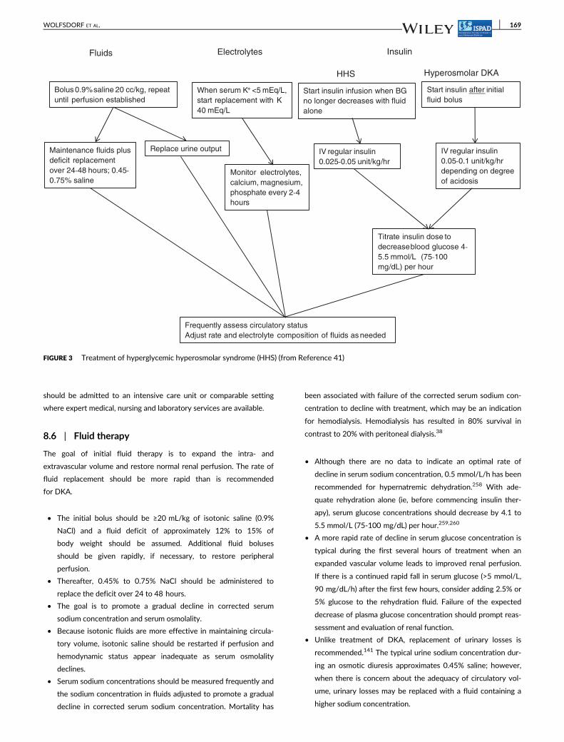
should be admitted to an intensive care unit or comparable setting
where expert medical, nursing and laboratory services are available.
8.6 | Fluid therapy
The goal of initial fluid therapy is to expand the intra- and
extravascular volume and restore normal renal perfusion. The rate of
fluid replacement should be more rapid than is recommended
for DKA.
• The initial bolus should be ≥20 mL/kg of isotonic saline (0.9%
NaCl) and a fluid deficit of approximately 12% to 15% of
body weight should be assumed. Additional fluid boluses
should be given rapidly, if necessary, to restore peripheral
perfusion.
• Thereafter, 0.45% to 0.75% NaCl should be administered to
replace the deficit over 24 to 48 hours.
• The goal is to promote a gradual decline in corrected serum
sodium concentration and serum osmolality.
• Because isotonic fluids are more effective in maintaining circula-
tory volume, isotonic saline should be restarted if perfusion and
hemodynamic status appear inadequate as serum osmolality
declines.
• Serum sodium concentrations should be measured frequently and
the sodium concentration in fluids adjusted to promote a gradual
decline in corrected serum sodium concentration. Mortality has
been associated with failure of the corrected serum sodium con-
centration to decline with treatment, which may be an indication
for hemodialysis. Hemodialysis has resulted in 80% survival in
contrast to 20% with peritoneal dialysis.38
• Although there are no data to indicate an optimal rate of
decline in serum sodium concentration, 0.5 mmol/L/h has been
recommended for hypernatremic dehydration.258 With ade-
quate rehydration alone (ie, before commencing insulin ther-
apy), serum glucose concentrations should decrease by 4.1 to
5.5 mmol/L (75-100 mg/dL) per hour.259,260
• A more rapid rate of decline in serum glucose concentration is
typical during the first several hours of treatment when an
expanded vascular volume leads to improved renal perfusion.
If there is a continued rapid fall in serum glucose (>5 mmol/L,
90 mg/dL/h) after the first few hours, consider adding 2.5% or
5% glucose to the rehydration fluid. Failure of the expected
decrease of plasma glucose concentration should prompt reas-
sessment and evaluation of renal function.
• Unlike treatment of DKA, replacement of urinary losses is
recommended.141 The typical urine sodium concentration dur-
ing an osmotic diuresis approximates 0.45% saline; however,
when there is concern about the adequacy of circulatory vol-
ume, urinary losses may be replaced with a fluid containing a
higher sodium concentration.
Bolus 0.9%saline 20 cc/kg, repeat
until perfusion establishedStart insulin infusion when BG
no longer decreases with fluid
alone
When serum K+ <5 mEq/L,
start replacement with K
40 mEq/L
Start insulin after initial
fluid bolus
Monitor electrolytes,
calcium, magnesium,
phosphate every 2‐4
hours
Frequently assess circulatory status
Adjust rate and electrolyte composition of fluids as needed
Maintenance fluids plus
deficit replacement
over 24‐48 hours; 0.45‐
0.75% saline
Replace urine output
Fluids Electrolytes Insulin
HHS Hyperosmolar DKA
IV regular insulin
0.025‐0.05 unit/kg/hr
IV regular insulin
0.05‐0.1 unit/kg/hr
depending on degree
of acidosis
Titrate insulin dose to
decreaseblood glucose 4‐
5.5 mmol/L (75-100
mg/dL) per hour
FIGURE 3 Treatment of hyperglycemic hyperosmolar syndrome (HHS) (from Reference 41)
WOLFSDORF ET AL. 169
8.7 | Insulin therapy
Whereas tissue hypoperfusion in HHS commonly causes lactic acido-
sis, ketosis usually is minimal. Early insulin administration is unneces-
sary in HHS. Fluid administration alone causes a marked decline in
serum glucose concentration as a result of dilution, improved renal
perfusion leading to increased glucosuria, and increased tissue glucose
uptake with improved circulation. The osmotic pressure exerted by
glucose within the vascular space contributes to the maintenance of
blood volume. A rapid fall in serum glucose concentration and osmo-
lality after insulin administration may lead to circulatory compromise
and venous thrombosis unless fluid replacement is adequate. Patients
with HHS also have extreme potassium deficits; a rapid insulin-
induced shift of potassium to the intracellular space can trigger an
arrhythmia.
• Insulin administration should be initiated when serum glucose
concentration is no longer declining at a rate of at least 3 mmol/L
(~50 mg/dL) per hour with fluid administration alone.
• In patients with more severe ketosis and acidosis, however, insu-
lin administration should be initiated earlier.
• Continuous administration of insulin at a rate of 0.025 to
0.05 units/kg/h can be used initially, with the dosage titrated to
achieve a decrease in serum glucose concentration of 3 to
4 mmol/L (~50-75 mg/dL) per hour.
• Insulin boluses are not recommended.
8.8 | Electrolytes
In general, deficits of potassium, phosphate, and magnesium are
greater in HHS than DKA.
• Potassium replacement (40 mmol/L of replacement fluid) should
begin as soon as serum potassium concentration is within the nor-
mal range and adequate renal function has been established.
• Higher rates of potassium administration may be necessary
after starting an insulin infusion
• Serum potassium concentrations should be monitored every
2 to 3 hours along with ECG monitoring
• Hourly potassium measurements may be necessary if the
patient has hypokalemia
• Bicarbonate therapy is contraindicated; it increases the risk of
hypokalemia and may adversely affect tissue oxygen delivery.
• Severe hypophosphatemia may lead to rhabdomyolysis, hemolytic
uremia, muscle weakness and paralysis. Although administration
of phosphate is associated with a risk of hypocalcemia, an IV solu-
tion that contains a 50:50 mixture of potassium phosphate and
another suitable potassium salt (potassium chloride or potassium
acetate), generally permits adequate phosphate replacement
while avoiding clinically significant hypocalcemia.
• Serum phosphate concentrations should be measured every
3 to 4 hours.
• Patients with HHS frequently have large magnesium deficits, but
there are no data to determine whether replacement of magne-
sium is beneficial.
• Replacement of magnesium should be considered in the occa-
sional patient who experiences severe hypomagnesemia and
hypocalcemia during therapy. The recommended dose is 25 to
50 mg/kg per dose for 3 to 4 doses given every 4 to 6 hours
with a maximum infusion rate of 150 mg/min and 2 g/h.
8.9 | Complications
• Venous thrombosis associated with use of central venous cathe-
ters is a common hazard in HHS.83 Prophylactic use of low-dose
heparin has been suggested in adults but there are no data to
indicate benefit from this practice. Heparin treatment should be
reserved for children who require central venous catheters for
physiologic monitoring or venous access and are immobile for
more than 24 to 48 hours.41 The central venous catheter should
not be used for insulin administration because the large dead
space may cause erratic insulin delivery.
• Rhabdomyolysis may occur in children with HHS resulting in
acute kidney failure, severe hyperkalemia, hypocalcemia, and
muscle swelling causing compartment syndrome.261 The classic
symptom triad of rhabdomyolysis includes myalgia, weakness, and
dark urine; monitoring creatine kinase concentrations every 2 to
3 hours is recommended for early detection.
• For unknown reasons, several children with HHS have had clinical
manifestations consistent with malignant hyperthermia, which is
associated with a high mortality rate.36,262–264 Patients who have
a fever associated with a rise in creatine kinase concentrations
may be treated with dantrolene, which reduces release of calcium
from the sarcoplasmic reticulum and stabilizes calcium metabolism
within muscle cells. Nonetheless, of the three reported patients
with HHS treated with dantrolene only one survived.262,264
• Altered mental status is common in adults whose serum osmolal-
ity exceeds 330 mOsm/kg; however, cerebral edema is rare.38
Among 96 cases of HHS reported in the literature to 2010,
including 32 deaths, there was only one instance of cerebral
edema.38 A decline in mental status after hyperosmolality has
improved with treatment is unusual and should be promptly
investigated.
8.10 | Mixed HHS and DKA
Treatment must take into account potential complications of both
DKA and HHS. Mental status must be closely monitored and frequent
reassessment of circulatory status and fluid balance is necessary to
guide therapy. To maintain an adequate circulatory volume, the rate
of fluid and electrolyte administration usually exceeds that required
for the typical case of DKA. Insulin is necessary to resolve ketosis and
arrest hepatic gluconeogenesis; however, insulin infusion should be
deferred until after the patient has received an initial fluid bolus and
the circulation has been stabilized. Serum potassium and phosphate
concentrations should be carefully monitored as described above
for HHS.
170 WOLFSDORF ET AL.
8.11 | Prevention of recurrent DKA
Management of an episode of DKA is not complete until its cause has
been identified and an attempt made to treat it.
• Insulin omission, either inadvertently or deliberately, is the cause
in most cases.
• The most common cause of DKA in insulin pump users is failure
to take extra insulin with a pen or syringe when hyperglycemia
and hyperketonemia or ketonuria occur.
• Home measurement of blood BOHB concentrations, when com-
pared to urine ketone testing, decreases diabetes-related hospital
visits (both emergency department visits and hospitalizations) by
the early identification and treatment of ketosis.265 Blood BOHB
measurements may be especially valuable to prevent DKA in
patients who use a pump because interrupted insulin delivery rap-
idly leads to ketosis.
• There may be dissociation between urine ketone (sodium
nitroprusside only measures acetoacetate and acetone) and
serum BOHB concentrations, which may be increased to levels
consistent with DKA at a time when a urine ketone test is neg-
ative or shows only trace or small ketonuria.266
• There usually is an important psychosocial reason for insulin
omission.
• an attempt to lose weight in an adolescent girl with an eating
disorder,
• a means of escaping an intolerable or abusive home situation,
• depression or other reason for inability of the patient to man-
age the diabetes unassisted.
• A psychiatric social worker or clinical psychologist should be con-
sulted to identify the psychosocial reason(s) contributing to devel-
opment of DKA.
• An infection is rarely the cause of DKA when the patient/family is
properly educated in diabetes management and is receiving
appropriate follow-up care by a diabetes team with a 24-hour
telephone helpline.267–269
• Insulin omission can be prevented by comprehensive programs
that provide education, psychosocial evaluation and treatment
combined with adult supervision of the entire process of insulin
administration.270
• Parents and patients should learn how to recognize and treat
ketosis and impending DKA with additional rapid- or short-
acting insulin and oral fluids
• Families should have access to a 24-hour telephone helpline
for emergency advice and treatment267
• When a responsible adult administers insulin there may be as
much as a 10-fold reduction in frequency of recurrent DKA.270
ENDNOTES
*Nitroprusside reaction method.†Serum bicarbonate concentration alone can substitute for vpH todiagnose DKA and classify severity in children with new onset diabe-tes mellitus. It is suggested as an alternative to reliance on vpH, espe-cially in settings in which access to vpH measurement is limited.26
‡Some experts use 1.6 rather than 2 as the coefficient in theequation.92
CONFLICT OF INTEREST
J.W. receives royalties as a section editor for UpToDate. M.F. is mem-
ber of Lilly Diabetes, Eli Lilly Ges.m.b. (advisory board); Medtronic
(speaker fee); Ferring pharmaeuticals Ges m.b.h. (prize for best scien-
tific presentation). R.H. receives lecture honoraria from Novo Nordisk,
Medtronic; Advisory Board Novo Nordisk, Medtronic, Insulet; Data
Monitoring Committee Astra Zeneca. M.A.S. was a member of a Sci-
entific Advisory Board for NovoNordisk during 2017 and 2018. A.R.,
N.G., M.A. and E.C. have no conflicts of interest to declare.
ORCID
Maria Fritsch http://orcid.org/0000-0001-5304-673X
Ethel Codner http://orcid.org/0000-0002-2899-2705
REFERENCES
1. Foster DW, McGarry JD. The metabolic derangements and treatmentof diabetic ketoacidosis. N Engl J Med. 1983;309(3):159-169.
2. Kitabchi AE, Umpierrez GE, Murphy MB, Kreisberg RA. Hyperglyce-mic crises in adult patients with diabetes: a consensus statementfrom the American Diabetes Association. Diabetes Care. 2006;29(12):2739-2748.
3. Hanas R, Lindgren F, Lindblad B. A 2-yr national population study ofpediatric ketoacidosis in Sweden: predisposing conditions and insulinpump use. Pediatr Diabetes. 2009;10(1):33-37.
4. Cox K, Cocchi MN, Salciccioli JD, Carney E, Howell M, Donnino MW.Prevalence and significance of lactic acidosis in diabetic ketoacidosis.J Crit Care. 2012;27(2):132-137.
5. Palmer BF, Clegg DJ. Electrolyte and acid-base disturbances inpatients with diabetes mellitus. N Engl J Med. 2015;373(6):548-559.
6. Deeter KH, Roberts JS, Bradford H, et al. Hypertension despite dehy-dration during severe pediatric diabetic ketoacidosis. Pediatr Diabe-tes. 2011;12(4 pt 1):295-301.
7. McDonnell CM, Pedreira CC, Vadamalayan B, Cameron FJ,Werther GA. Diabetic ketoacidosis, hyperosmolarity and hypernatre-mia: are high-carbohydrate drinks worsening initial presentation?Pediatr Diabetes. 2005;6(2):90-94.
8. Carlotti AP, St George-Hyslop C, Guerguerian AM, Bohn D,Kamel KS, Halperin M. Occult risk factor for the development ofcerebral edema in children with diabetic ketoacidosis: possible rolefor stomach emptying. Pediatr Diabetes. 2009;10(8):522-533.
9. Atchley D, Loeb R, Richards D Jr, Benedict E, Driscoll M. On diabeticketoacidosis: a detailed study of electrolyte balances following thewithdrawal and reestablishment of insulin therapy. J Clin Invest.1933;12:297-326.
10. Nabarro J, Spencer A, Stowers J. Metabolic studies in severe diabeticketosis. Q J Med. 1952;82:225-248.
11. Butler A, Talbot N, Burnett C, Stanbury J, MacLachlan E. Metabolicstudies in diabetic coma. Trans Assoc Am Phys. 1947;60:102-109.
12. Danowski T, Peters J, Rathbun J, Quashnock J, Greenman L. Studiesin diabetic acidosis and coma, with particular emphasis on the reten-tion of administered potassium. J Clin Invest. 1949;28:1-9.
13. Darrow D, Pratt E. Retention of water and electrolyte during recov-ery in a patient with diabetic acidosis. J Pediatr. 1952;41:688-696.
14. Holliday MA, Segar WE. The maintenance need for water in paren-teral fluid therapy. Pediatrics. 1957;19(5):823-832.
15. Friedman AL. Pediatric hydration therapy: historical review and anew approach. Kidney Int. 2005;67(1):380-388.
16. Collier S, Gura K, deLoid L, Dalton M. L S. Parenteral Nutrition. In:Sonneville K, Duggan C, eds. Manual of Pediatric Nutrition. 5th
WOLFSDORF ET AL. 171
ed. Shelton, CT: People's Medical Publishing House-USA; 2014:
196-248.17. Darrow DC. The physiologic basis for estimating requirements for
parenteral fluids. Pediatr Clin N Am. 1959;6(1):29-41.18. Dunger DB, Sperling MA, Acerini CL, et al. ESPE/LWPES consensus
statement on diabetic ketoacidosis in children and adolescents. Arch
Dis Child. 2004;89(2):188-194.19. Sheikh-Ali M, Karon BS, Basu A, et al. Can serum
beta-hydroxybutyrate be used to diagnose diabetic ketoacidosis?
Diabetes Care. 2008;31(4):643-647.20. Burge MR, Hardy KJ, Schade DS. Short-term fasting is a mechanism
for the development of euglycemic ketoacidosis during periods of
insulin deficiency. J Clin Endocrinol Metab. 1993;76(5):1192-1198.21. Pinkney JH, Bingley PJ, Sawtell PA, Dunger DB, Gale EA. Presenta-
tion and progress of childhood diabetes mellitus: a prospective
population-based study. The Bart's-Oxford Study Group. Diabetolo-
gia. 1994;37(1):70-74.22. Bas VN, Uytun S, Torun YA. Diabetic euglycemic ketoacidosis in
newly diagnosed type 1 diabetes mellitus during Ramadan fasting. J
Pediatr Endocrinol Metab. 2015;28(3–4):333-335.23. Peters AL, Buschur EO, Buse JB, Cohan P, Diner JC, Hirsch IB. Eugly-
cemic diabetic ketoacidosis: a potential complication of treatment
with sodium-glucose cotransporter 2 inhibition. Diabetes Care. 2015;
38(9):1687-1693.24. Goldenberg RM, Berard LD, Cheng AY, et al. SGLT2
inhibitor-associated diabetic ketoacidosis: clinical review and recom-
mendations for prevention and diagnosis. Clin Ther. 2016;38(12):
2654-2664.e1.25. Misaghian-Xanthos N, Shariff AI, Mekala K, et al. Sodium-glucose
cotransporter 2 inhibitors and diabetic ketoacidosis: a case series
from three academic institutions. Diabetes Care. 2017;40(6):e65-e66.26. von Oettingen J, Wolfsdorf J, Feldman HA, Rhodes ET. Use of serum
bicarbonate to substitute for venous pH in new-onset diabetes. Pedi-
atrics. 2015;136(2):e371-e377.27. Mayer-Davis EJ, Lawrence JM, Dabelea D, et al. Incidence trends of
type 1 and type 2 diabetes among youths, 2002-2012. N Engl J Med.
2017;376(15):1419-1429.28. Ingelfinger JR, Jarcho JA. Increase in the incidence of diabetes and its
implications. N Engl J Med. 2017;376(15):1473-1474.29. Fazeli Farsani S, van der Aa MP, van der Vorst MM, Knibbe CA, de
Boer A. Global trends in the incidence and prevalence of type 2 dia-
betes in children and adolescents: a systematic review and evaluation
of methodological approaches. Diabetologia. 2013;56(7):1471-1488.30. Dabelea D, Rewers A, Stafford JM, et al. Trends in the prevalence of
ketoacidosis at diabetes diagnosis: the SEARCH for diabetes in youth
study. Pediatrics. 2014;133(4):e938-e945.31. American Diabetes Association. Type 2 diabetes in children and ado-
lescents. Pediatrics. 2000;105((3, pt 1)):671-680.32. Rewers A, Klingensmith G, Davis C, et al. Presence of diabetic ketoa-
cidosis at diagnosis of diabetes mellitus in youth: the Search for Dia-
betes in Youth Study. Pediatrics. 2008;121(5):e1258-e1266.33. Gungor N, Hannon T, Libman I, Bacha F, Arslanian S. Type 2 diabetes
mellitus in youth: the complete picture to date. Pediatr Clin N Am.
2005;52(6):1579-1609.34. Klingensmith GJ, Connor CG, Ruedy KJ, et al. Presentation of youth
with type 2 diabetes in the Pediatric Diabetes Consortium. Pediatr
Diabetes. 2016;17(4):266-273.35. Chase HP, Garg SK, Jelley DH. Diabetic ketoacidosis in children and
the role of outpatient management. Pediatr Rev. 1990;11(10):
297-304.36. Morales AE, Rosenbloom AL. Death caused by hyperglycemic hyper-
osmolar state at the onset of type 2 diabetes. J Pediatr. 2004;144(2):
270-273.37. Canarie MF, Bogue CW, Banasiak KJ, Weinzimer SA,
Tamborlane WV. Decompensated hyperglycemic hyperosmolarity
without significant ketoacidosis in the adolescent and young adult
population. J Pediatr Endocrinol Metab. 2007;20(10):1115-1124.38. Rosenbloom AL. Hyperglycemic hyperosmolar state: an emerging
pediatric problem. J Pediatr. 2010;156(2):180-184.
39. Bagdure D, Rewers A, Campagna E, Sills MR. Epidemiology of hyper-glycemic hyperosmolar syndrome in children hospitalized in USA.Pediatr Diabetes. 2013;14(1):18-24.
40. Temple IK, Shield JP. 6q24 transient neonatal diabetes. Rev EndocrMetab Disord. 2010;11(3):199-204.
41. Zeitler P, Haqq A, Rosenbloom A, Glaser N. Hyperglycemic hyperos-molar syndrome in children: pathophysiological considerations andsuggested guidelines for treatment. J Pediatr. 2011;158(1):9-14,14.e1-2.
42. Levy-Marchal C, Papoz L, de Beaufort C, et al. Clinical and laboratoryfeatures of type 1 diabetic children at the time of diagnosis. DiabetMed. 1992;9(3):279-284.
43. Komulainen J, Lounamaa R, Knip M, Kaprio EA, Akerblom HK. Ketoa-cidosis at the diagnosis of type 1 (insulin dependent) diabetes melli-tus is related to poor residual beta cell function. Childhood Diabetesin Finland Study Group. Arch Dis Child. 1996;75(5):410-415.
44. Levy-Marchal C, Patterson CC, Green A. Geographical variation ofpresentation at diagnosis of type I diabetes in children: the EURO-DIAB study. European and Dibetes. Diabetologia. 2001;44((suppl 3)):B75-B80.
45. Hanas R, Lindgren F, Lindblad B. Diabetic ketoacidosis and cerebraloedema in Sweden – a 2-year paediatric population study. DiabetMed. 2007;24(10):1080-1085.
46. Rodacki M, Pereira JR, Nabuco de Oliveira AM, et al. Ethnicity andyoung age influence the frequency of diabetic ketoacidosis at theonset of type 1 diabetes. Diabetes Res Clin Pract. 2007;78(2):259-262.
47. Usher-Smith JA, Thompson M, Ercole A, Walter FM. Variationbetween countries in the frequency of diabetic ketoacidosis at firstpresentation of type 1 diabetes in children: a systematic review. Dia-betologia. 2012;55(11):2878-2894.
48. Fritsch M, Schober E, Rami-Merhar B, Hofer S, Frohlich-Reiterer E,Waldhoer T. Diabetic ketoacidosis at diagnosis in Austrian children: apopulation-based analysis, 1989-2011. J Pediatr. 2013;163:1484-8.e1.
49. Zucchini S, Scaramuzza AE, Bonfanti R, et al. A multicenter retrospec-tive survey regarding diabetic ketoacidosis management in Italianchildren with type 1 diabetes. J Diabetes Res. 2016;2016:5719470.
50. Letourneau LR, Carmody D, Wroblewski K, et al. Diabetes presenta-tion in infancy: high risk of diabetic ketoacidosis. Diabetes Care.2017;40(10):e147-e148.
51. Quinn M, Fleischman A, Rosner B, Nigrin DJ, Wolfsdorf JI. Character-istics at diagnosis of type 1 diabetes in children younger than 6 years.J Pediatr. 2006;148(3):366-371.
52. Bui H, To T, Stein R, Fung K, Daneman D. Is diabetic ketoacidosis atdisease onset a result of missed diagnosis? J Pediatr. 2010;156(3):472-477.
53. Szypowska A, Skorka A. The risk factors of ketoacidosis in childrenwith newly diagnosed type 1 diabetes mellitus. Pediatr Diabetes.2011;12(4, pt 1):302-306.
54. Komulainen J, Kulmala P, Savola K, et al. Clinical, autoimmune, andgenetic characteristics of very young children with type 1 diabetes.Childhood Diabetes in Finland (DiMe) Study Group. Diabetes Care.1999;22(12):1950-1955.
55. Usher-Smith JA, Thompson MJ, Sharp SJ, Walter FM. Factors associ-ated with the presence of diabetic ketoacidosis at diagnosis of diabe-tes in children and young adults: a systematic review. BMJ. 2011;343:d4092.
56. Rosilio M, Cotton JB, Wieliczko MC, et al. Factors associated withglycemic control. A cross-sectional nationwide study in 2,579 Frenchchildren with type 1 diabetes. The French Pediatric Diabetes Group.Diabetes Care. 1998;21(7):1146-1153.
57. Smith CP, Firth D, Bennett S, Howard C, Chisholm P. Ketoacidosisoccurring in newly diagnosed and established diabetic children. ActaPaediatr. 1998;87(5):537-541.
58. Morris AD, Boyle DI, McMahon AD, Greene SA, MacDonald TM,Newton RW. Adherence to insulin treatment, glycaemic control, andketoacidosis in insulin-dependent diabetes mellitus. The DARTS/MEMO Collaboration. Diabetes Audit and Research in Tayside Scot-land Medicines Monitoring Unit. Lancet. 1997;350(9090):1505-1510.
172 WOLFSDORF ET AL.
59. Rewers A, Chase HP, Mackenzie T, et al. Predictors of acute compli-cations in children with type 1 diabetes. JAMA. 2002;287(19):2511-2518.
60. Cengiz E, Xing D, Wong JC, et al. Severe hypoglycemia and diabeticketoacidosis among youth with type 1 diabetes in the T1D exchangeclinic registry. Pediatr Diabetes. 2013;14(6):447-454.
61. Maahs DM, Hermann JM, Holman N, et al. Rates of diabetic ketoaci-dosis: international comparison with 49,859 pediatric patients withtype 1 diabetes from England, Wales, the U.S., Austria, and Germany.Diabetes Care. 2015;38(10):1876-1882.
62. Hermann JM, Meusers M, Bachran R, et al. Self-reported regularalcohol consumption in adolescents and emerging adults with type1 diabetes: A neglected risk factor for diabetic ketoacidosis? Multi-center analysis of 29 630 patients from the DPV registry. Pediatr Dia-betes. 2017;18(8):817-823.
63. Cope JU, Morrison AE, Samuels-Reid J. Adolescent use of insulin andpatient-controlled analgesia pump technology: a 10-year Food andDrug Administration retrospective study of adverse events. Pediat-rics. 2008;121(5):e1133-e1138.
64. Karges B, Schwandt A, Heidtmann B, et al. Association of insulinpump therapy vs insulin injection therapy with severe hypoglycemia,ketoacidosis, and glycemic control among children, adolescents, andyoung adults with type 1 diabetes. JAMA. 2017;318(14):1358-1366.
65. Kleinman ME, Chameides L, Schexnayder SM, et al. Pediatricadvanced life support: 2010 American Heart Association Guidelinesfor Cardiopulmonary Resuscitation and Emergency CardiovascularCare. Pediatrics. 2010;126(5):e1361-e1399.
66. Kleinman ME, de Caen AR, Chameides L, et al. Pediatric basic andadvanced life support: 2010 international consensus on cardiopulmo-nary resuscitation and emergency cardiovascular care science withtreatment recommendations. Pediatrics. 2010;126(5):e1261-e1318.
67. Wiggam MI, O'Kane MJ, Harper R, et al. Treatment of diabetic ketoa-cidosis using normalization of blood 3-hydroxybutyrate concentra-tion as the endpoint of emergency management. A randomizedcontrolled study. Diabetes Care. 1997;20(9):1347-1352.
68. Vanelli M, Chiari G, Capuano C, Iovane B, Bernardini A, Giacalone T.The direct measurement of 3-beta-hydroxy butyrate enhances themanagement of diabetic ketoacidosis in children and reduces timeand costs of treatment. Diabetes Nutr Metab. 2003;16(5–6):312-316.
69. Ham MR, Okada P, White PC. Bedside ketone determination in dia-betic children with hyperglycemia and ketosis in the acute care set-ting. Pediatr Diabetes. 2004;5(1):39-43.
70. Rewers A, McFann K, Chase HP. Bedside monitoring of bloodbeta-hydroxybutyrate levels in the management of diabetic ketoaci-dosis in children. Diabetes Technol Ther. 2006;8(6):671-676.
71. Prisco F, Picardi A, Iafusco D, et al. Blood ketone bodies in patientswith recent-onset type 1 diabetes (a multicenter study). Pediatr Dia-betes. 2006;7(4):223-228.
72. Noyes KJ, Crofton P, Bath LE, et al. Hydroxybutyrate near-patienttesting to evaluate a new end-point for intravenous insulin therapyin the treatment of diabetic ketoacidosis in children. Pediatr Diabetes.2007;8(3):150-156.
73. Klocker AA, Phelan H, Twigg SM, Craig ME. Bloodbeta-hydroxybutyrate vs. urine acetoacetate testing for the preven-tion and management of ketoacidosis in type 1 diabetes: a systematicreview. Diabet Med. 2013;30(7):818-824.
74. Koves IH, Neutze J, Donath S, et al. The accuracy of clinical assess-ment of dehydration during diabetic ketoacidosis in childhood. Diabe-tes Care. 2004;27(10):2485-2487.
75. Sottosanti M, Morrison GC, Singh RN, et al. Dehydration in childrenwith diabetic ketoacidosis: a prospective study. Arch Dis Child. 2012;97(2):96-100.
76. Ugale J, Mata A, Meert KL, Sarnaik AP. Measured degree of dehydra-tion in children and adolescents with type 1 diabetic ketoacidosis.Pediatr Crit Care Med. 2012;13(2):e103-e107.
77. Steiner MJ, DeWalt DA, Byerley JS. Is this child dehydrated? JAMA.2004;291(22):2746-2754.
78. Teasdale G, Jennett B. Assessment of coma and impaired conscious-ness. A practical scale. Lancet. 1974;2(7872):81-84.
79. Tasker RC, Lutman D, Peters MJ. Hyperventilation in severe diabeticketoacidosis. Pediatr Crit Care Med. 2005;6(4):405-411.
80. Davids MR, Edoute Y, Stock S, Halperin ML. Severe degree of hyper-glycaemia: insights from integrative physiology. QJM. 2002;95(2):113-124.
81. Malone JI, Brodsky SJ. The value of electrocardiogram monitoring indiabetic ketoacidosis. Diabetes Care. 1980;3(4):543-547.
82. Soler NG, Bennett MA, Fitzgerald MG, Malins JM. Electrocardiogramas a guide to potassium replacement in diabetic ketoacidosis. Diabe-tes. 1974;23(7):610-615.
83. Gutierrez JA, Bagatell R, Samson MP, Theodorou AA, Berg RA. Fem-oral central venous catheter-associated deep venous thrombosis inchildren with diabetic ketoacidosis. Crit Care Med. 2003;31(1):80-83.
84. Worly JM, Fortenberry JD, Hansen I, Chambliss CR, Stockwell J.Deep venous thrombosis in children with diabetic ketoacidosis andfemoral central venous catheters. Pediatrics. 2004;113(1, pt 1):e57-e60.
85. Chima RS, Hanson SJ. Venous thromboembolism in critical illnessand trauma: pediatric perspectives. Front Pediatr. 2017;5:47.
86. Flood RG, Chiang VW. Rate and prediction of infection in childrenwith diabetic ketoacidosis. Am J Emerg Med. 2001;19(4):270-273.
87. Monroe KW, King W, Atchison JA. Use of PRISM scores in triage ofpediatric patients with diabetic ketoacidosis. Am J Manag Care. 1997;3(2):253-258.
88. Bonadio WA, Gutzeit MF, Losek JD, Smith DS. Outpatient manage-ment of diabetic ketoacidosis. Am J Dis Child. 1988;142(4):448-450.
89. Linares MY, Schunk JE, Lindsay R. Laboratory presentation in dia-betic ketoacidosis and duration of therapy. Pediatr Emerg Care. 1996;12(5):347-351.
90. Yu HY, Agus M, Kellogg MD. Clinical utility of Abbott precisionXceed pro(R) ketone meter in diabetic patients. Pediatr Diabetes.2011;12(7):649-655.
91. Lutfi R, Huang J, Wong HR. Plasmapheresis to treat hypertriglyceri-demia in a child with diabetic ketoacidosis and pancreatitis. Pediat-rics. 2012;129(1):e195-e198.
92. Oh G, Anderson S, Tancredi D, Kuppermann N, Glaser N. Hyponatre-mia in pediatric diabetic ketoacidosis: reevaluating the correction fac-tor for hyperglycemia. Arch Pediatr Adolesc Med. 2009;163(8):771-772.
93. Halperin ML, Goldstein MB. Fluid, Electrolyte, and Acid-Base Physiol-ogy. 3rd ed. Philadelphia, PA: Saunders; 1999.
94. Love AH, Phillips RA. Measurement of dehydration in cholera. J InfectDis. 1969;119(1):39-42.
95. Harris GD, Fiordalisi I. Physiologic management of diabetic ketoaci-demia. A 5-year prospective pediatric experience in 231 episodes.Arch Pediatr Adolesc Med. 1994;148(10):1046-1052.
96. Napolova O, Urbach S, Davids MR, Halperin ML. Assessing thedegree of extracellular fluid volume contraction in a patient with asevere degree of hyperglycaemia. Nephrol Dial Transplant. 2003;18(12):2674-2677.
97. Halperin ML, Maccari C, Kamel KS, Carlotti AP, Bohn D. Strategies todiminish the danger of cerebral edema in a pediatric patient present-ing with diabetic ketoacidosis. Pediatr Diabetes. 2006;7(4):191-195.
98. Katz MA. Hyperglycemia-induced hyponatremia--calculation ofexpected serum sodium depression. N Engl J Med. 1973;289(16):843-844.
99. Hillier TA, Abbott RD, Barrett EJ. Hyponatremia: evaluating the cor-rection factor for hyperglycemia. Am J Med. 1999;106(4):399-403.
100. Harris GD, Fiordalisi I, Harris WL, Mosovich LL, Finberg L. Minimizingthe risk of brain herniation during treatment of diabetic ketoacide-mia: a retrospective and prospective study. J Pediatr. 1990;117:22-31.
101. Hale PM, Rezvani I, Braunstein AW, Lipman TH, Martinez N,Garibaldi L. Factors predicting cerebral edema in young children withdiabetic ketoacidosis and new onset type I diabetes. Acta Paediatr.1997;86(6):626-631.
102. Glaser N, Barnett P, McCaslin I, et al. Risk factors for cerebral edemain children with diabetic ketoacidosis. The Pediatric Emergency Med-icine Collaborative Research Committee of the American Academyof Pediatrics. N Engl J Med. 2001;344(4):264-269.
103. Brown TB. Cerebral oedema in childhood diabetic ketoacidosis: istreatment a factor? Emerg Med J. 2004;21(2):141-144.
WOLFSDORF ET AL. 173
104. Glaser NS, Ghetti S, Casper TC, Dean JM, Kuppermann N. Pediatricdiabetic ketoacidosis, fluid therapy, and cerebral injury: the design ofa factorial randomized controlled trial. Pediatr Diabetes. 2013;14:435-446.
105. Edge JA, Jakes RW, Roy Y, et al. The UK case-control study of cere-bral oedema complicating diabetic ketoacidosis in children. Diabeto-logia. 2006;49(9):2002-2009.
106. Kuppermann N, Ghetti S, Schunk JE, et al. Clinical trial of fluid infu-sion rates for pediatric diabetic ketoacidosis. N Engl J Med. 2018;378:2275-2287.
107. Dunger DB, Sperling MA, Acerini CL, et al. European Society for Pae-diatric Endocrinology/Lawson Wilkins Pediatric Endocrine Societyconsensus statement on diabetic ketoacidosis in children and adoles-cents. Pediatrics. 2004;113(2):e133-e140.
108. Adrogue HJ, Barrero J, Eknoyan G. Salutary effects of modest fluidreplacement in the treatment of adults with diabetic ketoacidosis.Use in patients without extreme volume deficit. JAMA. 1989;262(15):2108-2113.
109. Mel JM, Werther GA. Incidence and outcome of diabetic cerebraloedema in childhood: are there predictors? J Paediatr Child Health.1995;31(1):17-20.
110. Wagner A, Risse A, Brill HL, et al. Therapy of severe diabetic ketoaci-dosis. Zero-mortality under very-low-dose insulin application. Diabe-tes Care. 1999;22(5):674-677.
111. Toledo JD, Modesto V, Peinador M, et al. Sodium concentration inrehydration fluids for children with ketoacidotic diabetes: effect onserum sodium concentration. J Pediatr. 2009;154:895-900.
112. White PC, Dickson BA. Low morbidity and mortality in children withdiabetic ketoacidosis treated with isotonic fluids. J Pediatr. 2013;163(3):761-766.
113. Nallasamy K, Jayashree M, Singhi S, Bansal A. Low-dose vsstandard-dose insulin in pediatric diabetic ketoacidosis: a randomizedclinical trial. JAMA Pediatr. 2014;168:999-1005.
114. Yung M, Letton G, Keeley S. Controlled trial of Hartmann's solutionversus 0.9% saline for diabetic ketoacidosis. J Paediatr Child Health.2017;53(1):12-17.
115. Rother KI, Schwenk WF 2nd. Effect of rehydration fluid with75 mmol/L of sodium on serum sodium concentration and serumosmolality in young patients with diabetic ketoacidosis. Mayo ClinProc. 1994;69(12):1149-1153.
116. Felner EI, White PC. Improving management of diabetic ketoacidosisin children. Pediatrics. 2001;108(3):735-740.
117. Basnet S, Venepalli PK, Andoh J, Verhulst S, Koirala J. Effect of nor-mal saline and half normal saline on serum electrolytes during recov-ery phase of diabetic ketoacidosis. J Intensive Care Med. 2014;29(1):38-42.
118. Fiordalisi I, Novotny WE, Holbert D, Finberg L, Harris GD. An 18-yrprospective study of pediatric diabetic ketoacidosis: an approach tominimizing the risk of brain herniation during treatment. Pediatr Dia-betes. 2007;8(3):142-149.
119. Hoorn EJ, Carlotti AP, Costa LA, et al. Preventing a drop in effectiveplasma osmolality to minimize the likelihood of cerebral edema dur-ing treatment of children with diabetic ketoacidosis. J Pediatr. 2007;150(5):467-473.
120. Duck SC, Wyatt DT. Factors associated with brain herniation in thetreatment of diabetic ketoacidosis. J Pediatr. 1988;113:10-14.
121. Adrogue HJ, Wilson H, Boyd AE 3rd, Suki WN, Eknoyan G. Plasmaacid-base patterns in diabetic ketoacidosis. N Engl J Med. 1982;307(26):1603-1610.
122. Adrogue HJ, Eknoyan G, Suki WK. Diabetic ketoacidosis: role of thekidney in the acid-base homeostasis re-evaluated. Kidney Int. 1984;25(4):591-598.
123. Taylor D, Durward A, Tibby SM, et al. The influence of hyperchlorae-mia on acid base interpretation in diabetic ketoacidosis. IntensiveCare Med. 2006;32(2):295-301.
124. Durward A, Skellett S, Mayer A, Taylor D, Tibby SM, Murdoch IA.The value of the chloride: sodium ratio in differentiating the aetiol-ogy of metabolic acidosis. Intensive Care Med. 2001;27(5):828-835.
125. Oh MS, Carroll HJ, Goldstein DA, Fein IA. Hyperchloremic acidosisduring the recovery phase of diabetic ketosis. Ann Intern Med. 1978;89(6):925-927.
126. Oh MS, Banerji MA, Carroll HJ. The mechanism of hyperchloremicacidosis during the recovery phase of diabetic ketoacidosis. Diabetes.1981;30(4):310-313.
127. Oh MS, Carroll HJ, Uribarri J. Mechanism of normochloremic andhyperchloremic acidosis in diabetic ketoacidosis. Nephron. 1990;54(1):1-6.
128. von Oettingen JE, Rhodes ET, Wolfsdorf JI. Resolution of ketoacido-sis in children with new onset diabetes: evaluation of various defini-tions. Diabetes Res Clin Pract. 2017;135:76-84.
129. Chua HR, Venkatesh B, Stachowski E, et al. Plasma-Lyte 148 vs 0.9%saline for fluid resuscitation in diabetic ketoacidosis. J Crit Care.2012;27(2):138-145.
130. Waldhausl W, Kleinberger G, Korn A, Dudczak R,Bratusch-Marrain P, Nowotny P. Severe hyperglycemia: effects ofrehydration on endocrine derangements and blood glucose concen-tration. Diabetes. 1979;28(6):577-584.
131. Owen OE, Licht JH, Sapir DG. Renal function and effects of partialrehydration during diabetic ketoacidosis. Diabetes. 1981;30(6):510-518.
132. Luzi L, Barrett EJ, Groop LC, Ferrannini E, DeFronzo RA. Metaboliceffects of low-dose insulin therapy on glucose metabolism in diabeticketoacidosis. Diabetes. 1988;37(11):1470-1477.
133. Kitabchi AE. Low-dose insulin therapy in diabetic ketoacidosis: factor fiction? Diabetes Metab Rev. 1989;5(4):337-363.
134. Martin MM, Martin AA. Continuous low-dose infusion of insulin inthe treatment of diabetic ketoacidosis in children. J Pediatr. 1976;89(4):560-564.
135. Edwards GA, Kohaut EC, Wehring B, Hill LL. Effectiveness oflow-dose continuous intravenous insulin infusion in diabetic ketoaci-dosis. A prospective comparative study. J Pediatr. 1977;91(5):701-705.
136. Drop SL, Duval-Arnould JM, Gober AE, Hersh JH, McEnery PT,Knowles HC. Low-dose intravenous insulin infusion versus subcuta-neous insulin injection: a controlled comparative study of diabeticketoacidosis. Pediatrics. 1977;59(5):733-738.
137. Lightner ES, Kappy MS, Revsin B. Low-dose intravenous insulin infu-sion in patients with diabetic ketoacidosis: biochemical effects inchildren. Pediatrics. 1977;60(5):681-688.
138. Perkin RM, Marks JF. Low-dose continuous intravenous insulin infu-sion in childhood diabetic ketoacidosis. Clin Pediatr (Phila). 1979;18(9):540-545-8.
139. Kappy MS, Lightner ES. Low-dose intravenous insulin in the treat-ment of diabetic ketoacidosis. Am J Dis Child. 1979;133(5):523-525.
140. Burghen GA, Etteldorf JN, Fisher JN, Kitabchi AQ. Comparison ofhigh-dose and low-dose insulin by continuous intravenous infusionin the treatment of diabetic ketoacidosis in children. Diabetes Care.1980;3(1):15-20.
141. Kitabchi AE, Umpierrez GE, Fisher JN, Murphy MB, Stentz FB. Thirtyyears of personal experience in hyperglycemic crises: diabetic ketoa-cidosis and hyperglycemic hyperosmolar state. J Clin EndocrinolMetab. 2008;93(5):1541-1552.
142. Lindsay R, Bolte RG. The use of an insulin bolus in low-dose insulininfusion for pediatric diabetic ketoacidosis. Pediatr Emerg Care. 1989;5(2):77-79.
143. Van der Meulen JA, Klip A, Grinstein S. Possible mechanism for cere-bral oedema in diabetic ketoacidosis. Lancet. 1987;2(8554):306-308.
144. Soler NG, FitzGerald MG, Wright AD, Malins JM. Comparative studyof different insulin regimens in management of diabetic ketoacidosis.Lancet. 1975;2(7947):1221-1224.
145. Puttha R, Cooke D, Subbarayan A, et al. Low dose (0.05 units/kg/h)is comparable with standard dose (0.1 units/kg/h) intravenous insulininfusion for the initial treatment of diabetic ketoacidosis in childrenwith type 1 diabetes-an observational study. Pediatr Diabetes. 2010;11:12-17.
146. Al Hanshi S, Shann F. Insulin infused at 0.05 versus 0.1 units/kg/hrin children admitted to intensive care with diabetic ketoacidosis.Pediatr Crit Care Med. 2011;12(2):137-140.
147. Wang J, Barbry P, Maiyar AC, et al. SGK integrates insulin and miner-alocorticoid regulation of epithelial sodium transport. Am J PhysiolRenal Physiol. 2001;280(2):F303-F313.
174 WOLFSDORF ET AL.
148. Tallini NY, Stoner LC. Amiloride-sensitive sodium current in everted
Ambystoma initial collecting tubule: short-term insulin effects.
Am J Physiol Cell Physiol. 2002;283(4):C1171-C1181.149. McCormick JA, Bhalla V, Pao AC, Pearce D. SGK1: a rapid
aldosterone-induced regulator of renal sodium reabsorption. Phys
Ther. 2005;20:134-139.150. Tiwari S, Nordquist L, Halagappa VK, Ecelbarger CA. Trafficking of
ENaC subunits in response to acute insulin in mouse kidney.
Am J Physiol Renal Physiol. 2007;293(1):F178-F185.151. Frindt G, Palmer LG. Effects of insulin on Na and K transporters in
the rat CCD. Am J Physiol Renal Physiol. 2012;302(10):F1227-F1233.152. Carlotti AP, St George-Hyslop C, Bohn D, Halperin ML. Hypokalemia
during treatment of diabetic ketoacidosis: clinical evidence for an
aldosterone-like action of insulin. J Pediatr. 2013;163(1):207-212.e1.153. Fisher JN, Shahshahani MN, Kitabchi AE. Diabetic ketoacidosis:
low-dose insulin therapy by various routes. N Engl J Med. 1977;
297(5):238-241.154. Sacks HS, Shahshahani M, Kitabchi AE, Fisher JN, Young RT. Similar
responsiveness of diabetic ketoacidosis to low-dose insulin by intra-
muscular injection and albumin-free infusion. Ann Intern Med. 1979;
90(1):36-42.155. Umpierrez GE, Latif K, Stoever J, et al. Efficacy of subcutaneous
insulin lispro versus continuous intravenous regular insulin for the
treatment of patients with diabetic ketoacidosis. Am J Med. 2004;
117(5):291-296.156. Umpierrez GE, Cuervo R, Karabell A, Latif K, Freire AX, Kitabchi AE.
Treatment of diabetic ketoacidosis with subcutaneous insulin aspart.
Diabetes Care. 2004;27(8):1873-1878.157. Della Manna T, Steinmetz L, Campos PR, et al. Subcutaneous use of
a fast-acting insulin analog: an alternative treatment for pediatric
patients with diabetic ketoacidosis. Diabetes Care. 2005;28(8):
1856-1861.158. Savoldelli RD, Farhat SC, Manna TD. Alternative management of dia-
betic ketoacidosis in a Brazilian pediatric emergency department.
Diabetol Metab Syndr. 2010;2:41.159. Cohen M, Leibovitz N, Shilo S, Zuckerman-Levin N, Shavit I,
Shehadeh N. Subcutaneous regular insulin for the treatment of dia-
betic ketoacidosis in children. Pediatr Diabetes. 2017;18(4):290-296.160. Adrogue HJ, Lederer ED, Suki WN, Eknoyan G. Determinants of
plasma potassium levels in diabetic ketoacidosis. Medicine
(Baltimore). 1986;65(3):163-172.161. DeFronzo RA, Felig P, Ferrannini E, Wahren J. Effect of graded doses
of insulin on splanchnic and peripheral potassium metabolism in
man. Am J Phys. 1980;238(5):E421-E427.162. Tattersall RB. A paper which changed clinical practice (slowly). Jacob
holler on potassium deficiency in diabetic acidosis (1946). Diabet
Med. 1999;16(12):978-984.163. Davis SM, Maddux AB, Alonso GT, Okada CR, Mourani PM,
Maahs DM. Profound hypokalemia associated with severe diabetic
ketoacidosis. Pediatr Diabetes. 2016;17:61-65.164. Guest G. Organic phosphates of the blood and mineral metabolism in
diabetic acidosis. Am J Dis Child. 1942;64:401-412.165. Guest G, Rapoport S. Electrolytes of blood plasma and cells in dia-
betic acidosis and during recovery. Proc Amer Diab Assoc. 1947;7:
95-115.166. Riley MS, Schade DS, Eaton RP. Effects of insulin infusion on plasma
phosphate in diabetic patients. Metabolism. 1979;28(3):191-194.167. Alberti KG, Emerson PM, Darley JH, Hockaday TD.
2,3-Diphosphoglycerate and tissue oxygenation in uncontrolled dia-
betes mellitus. Lancet. 1972;2(7774):391-395.168. Knochel JP. The pathophysiology and clinical characteristics of
severe hypophosphatemia. Arch Intern Med. 1977;137(2):203-220.169. O'Connor LR, Wheeler WS, Bethune JE. Effect of hypophosphatemia
on myocardial performance in man. N Engl J Med. 1977;297(17):
901-903.170. Gibby OM, Veale KE, Hayes TM, Jones JG, Wardrop CA. Oxygen
availability from the blood and the effect of phosphate replacement
on erythrocyte 2,3-diphosphoglycerate and haemoglobin-oxygen
affinity in diabetic ketoacidosis. Diabetologia. 1978;15(5):381-385.
171. Keller U, Berger W. Prevention of hypophosphatemia by phosphateinfusion during treatment of diabetic ketoacidosis and hyperosmolarcoma. Diabetes. 1980;29(2):87-95.
172. Wilson HK, Keuer SP, Lea AS, Boyd AE 3rd, Eknoyan G. Phosphatetherapy in diabetic ketoacidosis. Arch Intern Med. 1982;142(3):517-520.
173. Becker DJ, Brown DR, Steranka BH, Drash AL. Phosphate replace-ment during treatment of diabetic ketosis. Effects on calcium andphosphorus homeostasis. Am J Dis Child. 1983;137(3):241-246.
174. Fisher JN, Kitabchi AE. A randomized study of phosphate therapy inthe treatment of diabetic ketoacidosis. J Clin Endocrinol Metab. 1983;57(1):177-180.
175. Clerbaux T, Reynaert M, Willems E, Frans A. Effect of phosphate onoxygen-hemoglobin affinity, diphosphoglycerate and blood gasesduring recovery from diabetic ketoacidosis. Intensive Care Med.1989;15(8):495-498.
176. Weisinger JR, Bellorin-Font E. Magnesium and phosphorus. Lancet.1998;352(9125):391-396.
177. Singhal PC, Kumar A, Desroches L, Gibbons N, Mattana J. Prevalenceand predictors of rhabdomyolysis in patients with hypophosphate-mia. Am J Med. 1992;92(5):458-464.
178. Kutlu AO, Kara C, Cetinkaya S. Rhabdomyolysis without detectablemyoglobulinuria due to severe hypophosphatemia in diabetic ketoa-cidosis. Pediatr Emerg Care. 2011;27(6):537-538.
179. Bohannon NJ. Large phosphate shifts with treatment for hyperglyce-mia. Arch Intern Med. 1989;149(6):1423-1425.
180. de Oliveira Iglesias SB, Pons Leite H, de Carvalho WB.Hypophosphatemia-induced seizure in a child with diabetic ketoaci-dosis. Pediatr Emerg Care. 2009;25(12):859-861.
181. Zipf WB, Bacon GE, Spencer ML, Kelch RP, Hopwood NJ,Hawker CD. Hypocalcemia, hypomagnesemia, and transient hypo-parathyroidism during therapy with potassium phosphate in diabeticketoacidosis. Diabetes Care. 1979;2(3):265-268.
182. Winter RJ, Harris CJ, Phillips LS, Green OC. Diabetic ketoacidosis.Induction of hypocalcemia and hypomagnesemia by phosphate ther-apy. Am J Med. 1979;67(5):897-900.
183. Hale PJ, Crase J, Nattrass M. Metabolic effects of bicarbonate in thetreatment of diabetic ketoacidosis. Br Med J (Clin Res Ed). 1984;289(6451):1035-1038.
184. Morris LR, Murphy MB, Kitabchi AE. Bicarbonate therapy in severediabetic ketoacidosis. Ann Intern Med. 1986;105(6):836-840.
185. Okuda Y, Adrogue HJ, Field JB, Nohara H, Yamashita K. Counterpro-ductive effects of sodium bicarbonate in diabetic ketoacidosis. J ClinEndocrinol Metab. 1996;81(1):314-320.
186. Green SM, Rothrock SG, Ho JD, et al. Failure of adjunctive bicarbon-ate to improve outcome in severe pediatric diabetic ketoacidosis.Ann Emerg Med. 1998;31(1):41-48.
187. Assal JP, Aoki TT, Manzano FM, Kozak GP. Metabolic effects ofsodium bicarbonate in management of diabetic ketoacidosis. Diabe-tes. 1974;23(5):405-411.
188. Ohman JL Jr, Marliss EB, Aoki TT, Munichoodappa CS, Khanna VV,Kozak GP. The cerebrospinal fluid in diabetic ketoacidosis. N Engl JMed. 1971;284(6):283-290.
189. Soler NG, Bennett MA, Dixon K, FitzGerald MG, Malins JM. Potas-sium balance during treatment of diabetic ketoacidosis with specialreference to the use of bicarbonate. Lancet. 1972;2(7779):665-667.
190. Lever E, Jaspan JB. Sodium bicarbonate therapy in severe diabeticketoacidosis. Am J Med. 1983;75(2):263-268.
191. Narins RG, Cohen JJ. Bicarbonate therapy for organic acidosis: thecase for its continued use. Ann Intern Med. 1987;106(4):615-618.
192. Shankar V, Haque A, Churchwell KB, Russell W. Insulin glargine sup-plementation during early management phase of diabetic ketoacido-sis in children. Intensive Care Med. 2007;33(7):1173-1178.
193. Harrison VS, Rustico S, Palladino AA, Ferrara C, Hawkes CP. Glargineco-administration with intravenous insulin in pediatric diabeticketoacidosis is safe and facilitates transition to a subcutaneous regi-men. Pediatr Diabetes. 2017;18(8):742-748.
194. Curtis JR, To T, Muirhead S, Cummings E, Daneman D. Recent trendsin hospitalization for diabetic ketoacidosis in Ontario children. Diabe-tes Care. 2002;25(9):1591-1596.
WOLFSDORF ET AL. 175
195. Edge JA, Ford-Adams ME, Dunger DB. Causes of death in children
with insulin dependent diabetes 1990-96. Arch Dis Child. 1999;81(4):
318-323.196. Decourcey DD, Steil GM, Wypij D, Agus MS. Increasing use of
hypertonic saline over mannitol in the treatment of symptomatic
cerebral edema in pediatric diabetic ketoacidosis: an 11-year retro-
spective analysis of mortality. Pediatr Crit Care Med. 2013;14(7):
694-700.197. Saydah SH, Imperatore G, Geiss LS, Gregg E. Diabetes death rates
among youths aged ≤19 years: United States, 1968–2009. Morb
Mortal Wkly Rep. 2012;61(43):869-872.198. Morgan E, Black CR, Abid N, Cardwell CR, McCance DR,
Patterson CC. Mortality in type 1 diabetes diagnosed in childhood in
Northern Ireland during 1989-2012: A population-based cohort
study. Pediatr Diabetes. 2018;19(1):166-170.199. Gibb FW, Teoh WL, Graham J, Lockman KA. Risk of death following
admission to a UK hospital with diabetic ketoacidosis. Diabetologia.
2016;59(10):2082-2087.200. Sperling MA. Diabetes: recurrent DKA – for whom the bell tolls. Nat
Rev Endocrinol. 2016;12(10):562-564.201. Daneman D. Diabetes-related mortality. A pediatrician's view. Diabe-
tes Care. 2001;24(5):801-802.202. Edge JA, Hawkins MM, Winter DL, Dunger DB. The risk and out-
come of cerebral oedema developing during diabetic ketoacidosis.
Arch Dis Child. 2001;85(1):16-22.203. Lawrence SE, Cummings EA, Gaboury I, Daneman D.
Population-based study of incidence and risk factors for cerebral
edema in pediatric diabetic ketoacidosis. J Pediatr. 2005;146(5):
688-692.204. Ghetti S, Lee JK, Sims CE, Demaster DM, Glaser NS. Diabetic ketoa-
cidosis and memory dysfunction in children with type 1 diabetes. J
Pediatr. 2010;156(1):109-114.205. Nadebaum C, Scratch SE, Northam EA, Cameron FJ, Diabetic Ketoa-
cidosis and Brain Injury Study Group. Clinical utility of mental state
screening as a predictor of intellectual outcomes 6 months after
diagnosis of type 1 diabetes. Pediatr Diabetes. 2012;13(8):632-637.206. Cameron FJ, Scratch SE, Nadebaum C, et al. Neurological conse-
quences of diabetic ketoacidosis at initial presentation of type 1 dia-
betes in a prospective cohort study of children. Diabetes Care. 2014;
37(6):1554-1562.207. Shalitin S, Fisher S, Yackbovitch-Gavan M, et al. Ketoacidosis at
onset of type 1 diabetes is a predictor of long-term glycemic control.
Pediatr Diabetes. 2018;19(2):320-328.208. Duca LM, Wang B, Rewers M, Rewers A. Diabetic ketoacidosis at
diagnosis of type 1 diabetes predicts poor long-term glycemic con-
trol. Diabetes Care. 2017;40(9):1249-1255.209. Yasuda K, Hayashi M, Murayama M, Yamakita N. Acidosis-induced
hypochloremic alkalosis in diabetic ketoacidosis confirmed by the
modified base excess method. J Clin Endocrinol Metab. 2016;101(6):
2390-2395.210. Cooper MR, Turner RA Jr, Hutaff L, Prichard R. Diabetic
keto-acidosis complicated by disseminated intravascular coagulation.
South Med J. 1973;66(6):653-657.211. Keane S, Gallagher A, Ackroyd S, McShane MA, Edge JA. Cerebral
venous thrombosis during diabetic ketoacidosis. Arch Dis Child.
2002;86(3):204-205.212. Ho J, Mah JK, Hill MD, Pacaud D. Pediatric stroke associated with
new onset type 1 diabetes mellitus: case reports and review of the
literature. Pediatr Diabetes. 2006;7(2):116-121.213. Moye J, Rosenbloom AL, Silverstein J. Clinical predictors of mucor-
mycosis in children with type 1 diabetes mellitus. J Pediatr Endocrinol
Metab. 2002;15(7):1001-1004.214. Watson JP, Barnett AH. Pneumomediastinum in diabetic ketoacido-
sis. Diabet Med. 1989;6(2):173-174.215. Hursh BE, Ronsley R, Islam N, Mammen C, Panagiotopoulos C. Acute
kidney injury in children with type 1 diabetes hospitalized for dia-
betic ketoacidosis. JAMA Pediatr. 2017;171(5):e170020.216. Haddad NG, Croffie JM, Eugster EA. Pancreatic enzyme elevations in
children with diabetic ketoacidosis. J Pediatr. 2004;145(1):122-124.
217. Glaser NS, Wootton-Gorges SL, Buonocore MH, et al. Frequency ofsub-clinical cerebral edema in children with diabetic ketoacidosis.Pediatr Diabetes. 2006;7(2):75-80.
218. Glaser NS, Marcin JP, Wootton-Gorges SL, et al. Correlation of clini-cal and biochemical findings with diabetic ketoacidosis-related cere-bral edema in children using magnetic resonance diffusion-weightedimaging. J Pediatr. 2008;153(4):541-546.
219. Krane EJ, Rockoff MA, Wallman JK, Wolfsdorf JI. Subclinical brainswelling in children during treatment of diabetic ketoacidosis. N EnglJ Med. 1985;312(18):1147-1151.
220. Hoffman WH, Steinhart CM, el Gammal T, Steele S, Cuadrado AR,Morse PK. Cranial CT in children and adolescents with diabeticketoacidosis. AJNR Am J Neuroradiol. 1988;9(4):733-739.
221. Sperling MA. Cerebral edema in diabetic ketoacidosis: an underesti-mated complication? Pediatr Diabetes. 2006;7(2):73-74.
222. Arieff AI, Kleeman CR. Studies on mechanisms of cerebral edema indiabetic comas. Effects of hyperglycemia and rapid lowering ofplasma glucose in normal rabbits. J Clin Invest. 1973;52(3):571-583.
223. Arieff AI, Kleeman CR. Cerebral edema in diabetic comas. II. Effectsof hyperosmolality, hyperglycemia and insulin in diabetic rabbits. JClin Endocrinol Metab. 1974;38(6):1057-1067.
224. Prockop LD. Hyperglycemia, polyol accumulation, and increasedintracranial pressure. Arch Neurol. 1971;25(2):126-140.
225. Harris GD, Fiordalisi I, Finberg L. Safe management of diabetic ketoa-cidemia. J Pediatr. 1988;113:65-67.
226. Kamel KS, Halperin ML. Acid-base problems in diabetic ketoacidosis.N Engl J Med. 2015;372(6):546-554.
227. Glaser NS, Wootton-Gorges SL, Marcin JP, et al. Mechanism of cere-bral edema in children with diabetic ketoacidosis. J Pediatr. 2004;145(2):164-171.
228. Glaser N, Yuen N, Anderson SE, Tancredi DJ, O'Donnell ME. Cere-bral metabolic alterations in rats with diabetic ketoacidosis: effectsof treatment with insulin and intravenous fluids and effects of bume-tanide. Diabetes. 2010;59(3):702-709.
229. Yuen N, Anderson SE, Glaser N, Tancredi DJ, O'Donnell ME. Cere-bral blood flow and cerebral edema in rats with diabetic ketoacidosis.Diabetes. 2008;57(10):2588-2594.
230. Tasker RC, Acerini CL. Cerebral edema in children with diabeticketoacidosis: vasogenic rather than cellular? Pediatr Diabetes. 2014;15(4):261-270.
231. Rosenbloom AL, Schatz DA, Krischer JP, et al. Therapeutic contro-versy: prevention and treatment of diabetes in children. J Clin Endo-crinol Metab. 2000;85(2):494-522.
232. Glaser N. Cerebral injury and cerebral edema in children with dia-betic ketoacidosis: could cerebral ischemia and reperfusion injury beinvolved? Pediatr Diabetes. 2009;10(8):534-541.
233. Hoffman WH, Stamatovic SM, Andjelkovic AV. Inflammatory media-tors and blood brain barrier disruption in fatal brain edema of dia-betic ketoacidosis. Brain Res. 2009;1254:138-148.
234. Vavilala MS, Richards TL, Roberts JS, et al. Change in blood-brainbarrier permeability during pediatric diabetic ketoacidosis treatment.Pediatr Crit Care Med. 2010;11(3):332-338.
235. Rosenbloom AL. Intracerebral crises during treatment of diabeticketoacidosis. Diabetes Care. 1990;13(1):22-33.
236. Bello FA, Sotos JF. Cerebral oedema in diabetic ketoacidosis in chil-dren [letter]. Lancet. 1990;336(8706):64.
237. Mahoney CP, Vlcek BW, DelAguila M. Risk factors for developingbrain herniation during diabetic ketoacidosis. Pediatr Neurol. 1999;21(4):721-727.
238. Durr JA, Hoffman WH, Sklar AH, el Gammal T, Steinhart CM. Corre-lates of brain edema in uncontrolled IDDM. Diabetes. 1992;41(5):627-632.
239. Durward A, Ferguson LP, Taylor D, Murdoch IA, Tibby SM. The tem-poral relationship between glucose-corrected serum sodium andneurological status in severe diabetic ketoacidosis. Arch Dis Child.2011;96:50-57.
240. Bureau MA, Begin R, Berthiaume Y, Shapcott D, Khoury K,Gagnon N. Cerebral hypoxia from bicarbonate infusion in diabeticacidosis. J Pediatr. 1980;96(6):968-973.
241. Deeb L. Development of fatal cerebral edema during outpatient ther-apy for diabetic ketoacidosis. Pract Diab. 1989;6:212-213.
176 WOLFSDORF ET AL.
242. Glasgow AM. Devastating cerebral edema in diabetic ketoacidosisbefore therapy [letter]. Diabetes Care. 1991;14(1):77-78.
243. Couch RM, Acott PD, Wong GW. Early onset fatal cerebral edema indiabetic ketoacidosis. Diabetes Care. 1991;14(1):78-79.
244. Fiordalisi I, Harris GD, Gilliland MG. Prehospital cardiac arrest in dia-betic ketoacidemia: why brain swelling may lead to death beforetreatment. J Diabetes Complicat. 2002;16(3):214-219.
245. Edge JA. Cerebral oedema during treatment of diabetic ketoacidosis:are we any nearer finding a cause? Diabetes Metab Res Rev. 2000;16(5):316-324.
246. Muir AB, Quisling RG, Yang MC, Rosenbloom AL. Cerebral edema inchildhood diabetic ketoacidosis: natural history, radiographic find-ings, and early identification. Diabetes Care. 2004;27(7):1541-1546.
247. Franklin B, Liu J, Ginsberg-Fellner F. Cerebral edema and ophthalmo-plegia reversed by mannitol in a new case of insulin-dependent dia-betes mellitus. Pediatrics. 1982;69(1):87-90.
248. Shabbir N, Oberfield SE, Corrales R, Kairam R, Levine LS. Recoveryfrom symptomatic brain swelling in diabetic ketoacidosis. Clin Pediatr(Phila). 1992;31(9):570-573.
249. Roberts MD, Slover RH, Chase HP. Diabetic ketoacidosis with intra-cerebral complications. Pediatr Diabetes. 2001;2:109-114.
250. Curtis JR, Bohn D, Daneman D. Use of hypertonic saline in the treat-ment of cerebral edema in diabetic ketoacidosis (DKA). Pediatr Diabe-tes. 2001;2(4):191-194.
251. Kamat P, Vats A, Gross M, Checchia PA. Use of hypertonic saline forthe treatment of altered mental status associated with diabeticketoacidosis. Pediatr Crit Care Med. 2003;4(2):239-242.
252. Tasker RC, Burns J. Hypertonic saline therapy for cerebral edema indiabetic ketoacidosis: no change yet, please. Pediatr Crit Care Med.2014;15(3):284-285.
253. Soto-Rivera CL, Asaro LA, Agus MS, DeCourcey DD. Suspectedcerebral edema in diabetic ketoacidosis: is there still a role for headCT in treatment decisions? Pediatr Crit Care Med. 2017;18(3):207-212.
254. Kanter RK, Oliphant M, Zimmerman JJ, Stuart MJ. Arterial thrombo-sis causing cerebral edema in association with diabetic ketoacidosis.Crit Care Med. 1987;15(2):175-176.
255. Roe TF, Crawford TO, Huff KR, Costin G, Kaufman FR, Nelson MDJr. Brain infarction in children with diabetic ketoacidosis. J DiabetesComplicat. 1996;10(2):100-108.
256. Rosenbloom AL. Fatal cerebral infarctions in diabetic ketoacidosis ina child with previously unknown heterozygosity for factor V Leidendeficiency. J Pediatr. 2004;145(4):561-562.
257. Fourtner SH, Weinzimer SA, Levitt Katz LE. Hyperglycemic hyperos-molar non-ketotic syndrome in children with type 2 diabetes. PediatrDiabetes. 2005;6(3):129-135.
258. Kronan K, Normal ME. Renal and electrolyte emergencies. In:Fleisher GR, Ludwig S, eds. Textbook of Emergency Medicine. 4thed. Philadelphia, PA: Lippincott, Williams and Wilkins; 2000.
259. Matz R. Management of the hyperosmolar hyperglycemic syndrome.Am Fam Physician. 1999;60(5):1468-1476.
260. Delaney MF, Zisman A, Kettyle WM. Diabetic ketoacidosis andhyperglycemic hyperosmolar nonketotic syndrome. Endocrinol MetabClin N Am. 2000;29(4):683-705.
261. Mannix R, Tan ML, Wright R, BaskinM. Acute pediatric rhabdomyolysis:causes and rates of renal failure. Pediatrics. 2006;118(5):2119-2125.
262. Hollander AS, Olney RC, Blackett PR, Marshall BA. Fatal malignanthyperthermia-like syndrome with rhabdomyolysis complicating thepresentation of diabetes mellitus in adolescent males. Pediatrics.2003;111(6, pt 1):1447-1452.
263. Carcillo JA, Tasker RC. Fluid resuscitation of hypovolemic shock:acute medicine's great triumph for children. Intensive Care Med.2006;32(7):958-961.
264. Kilbane BJ, Mehta S, Backeljauw PF, Shanley TP, Crimmins NA.Approach to management of malignant hyperthermia-like syndrome inpediatric diabetes mellitus. Pediatr Crit Care Med. 2006;7(2):169-173.
265. Laffel LM, Wentzell K, Loughlin C, Tovar A, Moltz K, Brink S. Sick daymanagement using blood 3-hydroxybutyrate (3-OHB) compared withurine ketone monitoring reduces hospital visits in young people withT1DM: a randomized clinical trial. Diabet Med. 2006;23(3):278-284.
266. Laffel L. Sick-day management in type 1 diabetes. Endocrinol MetabClin N Am. 2000;29(4):707-723.
267. HoffmanWH, O'Neill P, Khoury C, Bernstein SS. Service and educationfor the insulin-dependent child. Diabetes Care. 1978;1(5):285-288.
268. Drozda DJ, Dawson VA, Long DJ, Freson LS, Sperling MA. Assess-ment of the effect of a comprehensive diabetes management pro-gram on hospital admission rates of children with diabetes mellitus.Diabetes Educ. 1990;16(5):389-393.
269. Grey M, Boland EA, Davidson M, Li J, Tamborlane WV. Copingskills training for youth with diabetes mellitus has long-lasting effectson metabolic control and quality of life. J Pediatr. 2000;137(1):107-113.
270. Golden MP, Herrold AJ, Orr DP. An approach to prevention of recur-rent diabetic ketoacidosis in the pediatric population. J Pediatr. 1985;107(2):195-200.
271. Pinhas-Hamiel O, Sperling M. Diabetic ketoacidosis. In: Hochberg Z,ed. Practical Algorithms in Pediatric Endocrinology. 3rd ed. Basel, Swit-zerland: Karger; 2017:112-113.
How to cite this article: Wolfsdorf JI, Glaser N, Agus M, et al.
ISPAD Clinical Practice Consensus Guidelines 2018: Diabetic
ketoacidosis and the hyperglycemic hyperosmolar state.
Pediatr Diabetes. 2018;19(Suppl. 27):155–177. https://doi.
org/10.1111/pedi.12701
WOLFSDORF ET AL. 177