issn 1309-100x ultrasonic activation on push-out bond
TRANSCRIPT

Journal of International Dental and Medical Research ISSN 1309-100X Ultrasonic Activation on Push-Out Bond Strengths http://www.jidmr.com Stiza Tanita and et al
Volume ∙ 14 ∙ Number ∙ 2 ∙ 2021
Page 600
The Effect of Ultrasonic Activation on Push-Out Bond Strengths of Two Calcium Silicate-Based Sealers to the Root Canal Wall
Stiza Tanita1, Anggraini Margono2, Dewa Ayu Nyoman Putri Artiningsih2
1. Conservative Dentistry Residency Program, Faculty of Dentistry, Universitas Indonesia, Jakarta, Indonesia. 2. Department of Conservative Dentistry, Faculty of Dentistry, Universitas Indonesia, Jakarta, Indonesia.
Abstract The purpose of this study was to analyze the effects of ultrasonic activation on the bond strength of a calcium silicate-based sealer and a calcium silicate pozzolan-based sealer after a root canal procedure. 32 first premolar teeth extracted for orthodontic reasons were divided into four groups. Group 1 had a calcium silicate-based sealer, group 2 had a calcium silicate sealer with ultrasonic activation, group 3 had a calcium silicate pozzolan-based sealer, and group 4 had a calcium silicate pozzolan-based sealer with ultrasonic activation. An ultrasonic scaler and tip was used for the activation. The machine push-out bond strength of each group was measured to determine their connective strength. The stereomicroscope camera was used to determine the failure. Statistical Analysis: A one-way ANOVA test and post-hoc Tamhane test were used to statistically analyze the results (p = 0.05). There was a significant difference between calcium silicate-based sealer and calcium silicate pozzolan-based sealer (p = .015) and the common types failure was mixed failure (62.5%) The bond strength of calcium silicate pozzolan-based sealer was greater compared to calcium silicate-based sealer, and ultrasonic activation increased the bond strength of both types of sealers to the root canal wall.
Experimental article (J Int Dent Med Res 2021; 14(2): 600-605) Keywords: Push-out bond strength, bioceramic sealer, calcium silicate sealer, calcium silicate pozzolan sealer, ultrasonic activation. Received date: 19 January 2021 Accept date: 08 March 2021
Introduction
Root canal treatment consists of access preparation, root canal preparation, and filling the root canal. This filling must eliminate all bacterial communication pathways to the root canal from the oral cavity and periradicular tissue.1 The ideal sealer must be well tolerate by the tissue with a low toxicity, adhesive and stable.2 The filling is successful when the root canal has a hermetic or fluid-tight seal, a reliable and durable seal, and the good sealing properties ofĝ root canal filling materials.3–5 Most endodontic failures (58%) are due to inadequate filling.1 A monoblock system
consists of a primary monoblock, a secondary monoblock, and a tertiary monoblock. The bond between the root canal sealer and dentin is a secondary monoblock concept. There has been increasing interest in developing root canal sealers to achieve secondary monoblock bonds.6,7
Bond strength is the force per unit area required for adhesion between a sealer and the root canal dentin, and calcium silicate-based sealers have a high bond strength because they can form micromechanical tags produced by the biomineralization process.8,9,10
Bioceramic-based sealer, calcium silicate pozzolan-based sealer is a calcium silicate sealer with the addition of alumina silicate which produces Pozzolanic reaction and more cementious particles with faster hardening time than other calcium silicate sealers. The Pozzolanic reaction that occurs between alumina silicate and water produces a gradual decrease of calcium hydroxide (Ca(OH)2). Calcium
*Corresponding author: Dewa Ayu Nyoman Putri Artiningsih Department of Conservative Dentistry, Faculty of Dentistry, Universitas of Indonesia Jl. Salemba Raya no. 4 Jakarta 10430, Indonesia E-mail: [email protected]

Journal of International Dental and Medical Research ISSN 1309-100X Ultrasonic Activation on Push-Out Bond Strengths http://www.jidmr.com Stiza Tanita and et al
Volume ∙ 14 ∙ Number ∙ 2 ∙ 2021
Page 601
hydroxide has a good antibacterial and biological properties, but its solubility is high enough so that the reduction of calcium hydroxide in the Pozzolanic reaction can make the mechanical properties of the sealer more stable and increase the bond strength.14,18 Previous studies have shown that ultrasonic activation can increase the push-out bond strength. However, Parashos et al. (2014) found that excessive use of ultrasonic activation can affect the nature of the sealer and cause voids to form. Ultrasonic activation used with high frequency (25-40 kHz) induces flow into high turbulence in the endodontic sealer and cause the formation of bubble cavitation, increased pressure, and temperature. Then Kim et al. (2018) used ultrasonic vibration frequencies (28-36 kHz) to increase the quality of root canal filling and the outcomes was not increased because of hight frequencies.13,14 So, in this research is using a lower frequencies. Therefore, the purpose of this study was to analyze the effects of ultrasonic vibration on the bond strength of a calcium silicate-based sealer and a calcium silicate pozzolan-based sealer on the strength of their bond to the root canal wall.
Materials and methods
This in vitro study was approved by the appropriate institutional ethics committee with ethical clearance no. 139 / ethical exempted / FKGUI / XII / 2019. A total of 32 extracted human single-rooted mandibular premolars with one root canal and fully formed apex were selected and divided into 4 groups. Group 1 had a calcium silicate-based sealer, group 2 had a calcium silicate sealer with ultrasonic activation, group 3 had a calcium silicate pozzolan-based sealer, and group 4 had a calcium silicate pozzolan-based sealer with ultrasonic activation. All Samples were prepared with the crown down technique using the ProTaper Next instrument (Dentsply, Switzerland). EDTA gel was used as a lubricant during root canal preparation and the root canals were irrigated with as much as 1 ml of 0.9% NaOCl with each change of instruments. After root canal preparation was completed, all canals were irrigated with 17% EDTA solution and allowed to stand for 1 minute. Then they were rinsed with 2.5% NaOCl, rinse saline, and activated with sonic instruments (Endoactivator, Dentsply).
The root canals were filled with gutta-percha with a calcium silicate-based sealer (iRoot SP®, Canada) and a calcium silicate pozzolan-based sealer (Endoseal MTA®, Korea). Then the sealers were activated with an ultrasonic device that vibrated the tweezers used to insert the main gutta-percha (ProTaper Next) into the root canal. The corona was covered with resin-modified glass ionomer cement (RMGIC). Specimens were stored in an incubator at 37 °C with 100% humidity for 24 hours so the sealers hardened completely.
When root canals had been prepared and dried, they were filled with the main con gutta percha and premixed calcium silicate-based sealer (iRoot SP® and Endoseal MTA®). The calcium silicate-based sealer (available in the form of a syringe and ready to use) was inserted into the root canal. Then the sealer was activated with an ultrasonic device (Newtron P5, Satelec) with a vibration magnitude set at 8. It vibrated the tweezers used to insert the main gutta-percha from the con tip (ProTaper Next) into the root canal for 3 seconds.14 The specimens were stored in a 1 gram phosphate buffer saline (PBS) solution at 37 °C at 100% humidity for 2 days to ensure the sealer has hardened.14 All root canals were planted in an acrylic resin block (Technovit 4071, Germany) in a vertical position.
Cutting was done at a distance of 5 to 7 mm from the apical root; a total of 32 specimens per group was obtained.
The bond strength was tested using a Universal Testing Machine (Shimidzu AGS-5kN). The teeth were cut 2 mm thick at 5-7 mm from the apex (middle third) The size of plunger was 0.6 mm. This value was obtained from the formula: Maximum load (newton) divided by the adhesion area of the root canal filling material (mm2) so that the push-out value was obtained in megapascals (Mpa). Then the bond failure test was analyzed with a stereomicroscope camera (Nikon, SMZ800) at a magnification of 30 x. Bond failures were classified according to Skidmore et al.15
The bond strength push-out data were entered into an SPSS program, version 22.0. The first statistical analysis was a data normality test. This test was performed using Saphiro-Wilk with total of sample <50. If the data distribution was normal (parametric), a one-way ANOVA statistical test was followed by a post hoc Tamhane test for each treatment group with a
Journal of International Dental and Medical Research ISSN 1309-100X Ultrasonic Activation on Push-Out Bond Strengths http://www.jidmr.com Stiza Tanita and et al
Volume ∙ 14 ∙ Number ∙ 2 ∙ 2021
Page 602
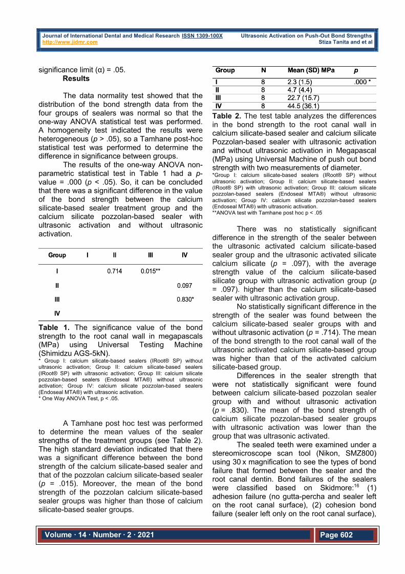
significance limit (α) = .05. Results The data normality test showed that the
distribution of the bond strength data from the four groups of sealers was normal so that the one-way ANOVA statistical test was performed. A homogeneity test indicated the results were heterogeneous (p > .05), so a Tamhane post-hoc statistical test was performed to determine the difference in significance between groups.
The results of the one-way ANOVA non-parametric statistical test in Table 1 had a p-value = .000 (p < .05). So, it can be concluded that there was a significant difference in the value of the bond strength between the calcium silicate-based sealer treatment group and the calcium silicate pozzolan-based sealer with ultrasonic activation and without ultrasonic activation.
Table 1. The significance value of the bond strength to the root canal wall in megapascals (MPa) using Universal Testing Machine (Shimidzu AGS-5kN). * Group I: calcium silicate-based sealers (IRoot® SP) without ultrasonic activation; Group II: calcium silicate-based sealers (IRoot® SP) with ultrasonic activation; Group III: calcium silicate pozzolan-based sealers (Endoseal MTA®) without ultrasonic activation; Group IV: calcium silicate pozzolan-based sealers (Endoseal MTA®) with ultrasonic activation. * One Way ANOVA Test, p < .05.
A Tamhane post hoc test was performed
to determine the mean values of the sealer strengths of the treatment groups (see Table 2). The high standard deviation indicated that there was a significant difference between the bond strength of the calcium silicate-based sealer and that of the pozzolan calcium silicate-based sealer (p = .015). Moreover, the mean of the bond strength of the pozzolan calcium silicate-based sealer groups was higher than those of calcium silicate-based sealer groups.
Table 2. The test table analyzes the differences in the bond strength to the root canal wall in calcium silicate-based sealer and calcium silicate Pozzolan-based sealer with ultrasonic activation and without ultrasonic activation in Megapascal (MPa) using Universal Machine of push out bond strength with two measurements of diameter. *Group I: calcium silicate-based sealers (IRoot® SP) without ultrasonic activation; Group II: calcium silicate-based sealers (IRoot® SP) with ultrasonic activation; Group III: calcium silicate pozzolan-based sealers (Endoseal MTA®) without ultrasonic activation; Group IV: calcium silicate pozzolan-based sealers (Endoseal MTA®) with ultrasonic activation. **ANOVA test with Tamhane post hoc p < .05
There was no statistically significant difference in the strength of the sealer between the ultrasonic activated calcium silicate-based sealer group and the ultrasonic activated silicate calcium silicate (p = .097), with the average strength value of the calcium silicate-based silicate group with ultrasonic activation group (p = .097). higher than the calcium silicate-based sealer with ultrasonic activation group.
No statistically significant difference in the strength of the sealer was found between the calcium silicate-based sealer groups with and without ultrasonic activation (p = .714). The mean of the bond strength to the root canal wall of the ultrasonic activated calcium silicate-based group was higher than that of the activated calcium silicate-based group.
Differences in the sealer strength that were not statistically significant were found between calcium silicate-based pozzolan sealer group with and without ultrasonic activation (p = .830). The mean of the bond strength of calcium silicate pozzolan-based sealer groups with ultrasonic activation was lower than the group that was ultrasonic activated.
The sealed teeth were examined under a stereomicroscope scan tool (Nikon, SMZ800) using 30 x magnification to see the types of bond failure that formed between the sealer and the root canal dentin. Bond failures of the sealers were classified based on Skidmore:16 (1) adhesion failure (no gutta-percha and sealer left on the root canal surface), (2) cohesion bond failure (sealer left only on the root canal surface),

Journal of International Dental and Medical Research ISSN 1309-100X Ultrasonic Activation on Push-Out Bond Strengths http://www.jidmr.com Stiza Tanita and et al
Volume ∙ 14 ∙ Number ∙ 2 ∙ 2021
Page 603
and (3) adhesion-cohesion bond or “mixed” failure (in certain areas, sealer remained on the surface of the root canal but not on other areas).17 Figure 1 shows the types of bond failure observed qualitatively.
Figure 1. Bond failure types: (A) Mixed failure in a calcium silicate-based sealer; (B) Cohesion failure in a calcium silicate-based sealer with ultrasonic activation; (C) Cohesion failure in a calcium silicate pozzolan-based sealer, and (D) Cohesion failure in a calcium silicate pozzolan-based sealer with ultrasonic activation.
Table 3. Bond Failure type of Calcium Silicate-based Sealer of sealer Group I: calcium silicate-based sealers (IRoot® SP) without ultrasonic activation; Group II: calcium silicate-based sealers (IRoot® SP) with ultrasonic activation; Group III: calcium silicate pozzolan-based sealers (Endoseal MTA®) without ultrasonic activation; Group IV: calcium silicate pozzolan-based sealers (Endoseal MTA®) with ultrasonic activation. Note: Bond failure types are based on Skidmore: Failure A: Adhesion failure (no gutta-percha and sealer left on the root canal surface), Failure B: Cohesion (sealer left only on the root canal surface), Failure C: Mixed failure (in certain areas, sealer remaining on the surface of the root canal but not on other areas).
The results showed that there were various types bond failure of the calcium silicate-based sealers (Table 3). The most common type of failure in the calcium silicate-based sealer was
the mixed failure type (62.5%), while cohesion was the most common type of failure in calcium silicate-based sealer with ultrasonic activation (75%), calcium silicate pozzolan-based sealer without ultrasonic activation (62.5%), and calcium silicate-based pozzolan silicate with ultrasonic activation (87.5%).
Discussion The filling of the root canal is successful
when the seal is hermetic or fluid-tight. This can be achieved if the filling material in the root canal can bond to the root canal wall. Bioceramic sealers, especially calcium silicate-based sealers, have good biocompatible properties and bioactivity. Such sealers have a small particle size (1.5 µm), which is smaller than the size of dentinal tubules, which range between 2 and 3 18 As a result, these sealers can enter the dentinal tubules and bond to the root canal wall.1,5
Premixed calcium silicate sealer based on pozzolan has a smaller particle size than premixed calcium silicate-based sealer. That sealer was created through the Pozzolanic reaction that occurs with the addition of aluminum silicate, and it produces a more cementious sealer with a faster hardening time. Kim et al. (2018) used ultrasonic activation to increase the density of calcium silicate based on pozzolan, and they got better results so that its use is recommended by the manufacturer.21,22 Bond strength has an important role in the success of filling the root canal. Calcium silicate-based sealers have high bond strength because they can form micromechanical tags produced by the biomineralization process. The results of this study indicated that the bond strength of calcium silicate based on pozzolan silicate was greater than that of calcium silicate-based sealer, and the difference was statistically significant. The addition of alumina silica in the sealer added to the physical and mechanical properties of the sealer so that the cement is denser and more cementious.8,9,10
In this study it was also found that the effect of ultrasonic activation produced higher bond strength of both types of calcium silicate-based sealer, although it was not statistically significant difference. This higher result was due to ultrasonic vibrations with high frequency (25-30 KHz) which can support the transmission and

Journal of International Dental and Medical Research ISSN 1309-100X Ultrasonic Activation on Push-Out Bond Strengths http://www.jidmr.com Stiza Tanita and et al
Volume ∙ 14 ∙ Number ∙ 2 ∙ 2021
Page 604
cavitation of root canals which will reduce the occurrence of pores in the material and can increase adaptation between surfaces between the sealer and root canal walls. Ultrasonic vibrations for activation of the root canal sealer will also make a better distribution of the sealer in the root canal system. It can be concluded that ultrasonic vibrations in the sealer can cause more effective filling, especially towards irregularity of the root canal and accessory canals and increase the strength of the sealer bond to the root canal wall.16,17
In this study the highest bond strength of the sealer against the root canal wall in 4 sealer groups was achieved by a calcium silicate pozzolan-based sealer with ultrasonic activation with a mean value of 44.5 MPa, followed by a calcium silicate pozzolan-based sealer with a mean value of 22.7 MPa, calcium silicate-based sealer with ultrasonic activation with a mean value of 4.7 MPa, and the smallest value of the bond strength of the sealer was achieved by calcium silicate-based sealer with a mean value of 2.3 MPa. Based on the results of this study, it can be concluded that calcium silicate pozzolan-based sealer with ultrasonic activation can be an alternative to increase the bond strength of the sealer to the root canal wall so the root canal filling becomes more hermetic and prevent leakage in filling the root canal which will cause root canal reinfection by bacteria.
The mix failure type of calcium silicate-based sealer was 62.5%. These results indicate that the bond strength of calcium silicate-based sealer was lowest when compared to calcium silicate-based silicate which was activated ultrasonic and calcium silicate-based silicate pozzolan with and without ultrasonic activation. This was consistent with the results of the significance of the bond strength to the root canal wall.12,21,22
The highest type of cohesion failure was produced by calcium silicate based on pozzolanic silicate with ultrasonic activation at 87.5%. These results indicate that the bond strength of calcium silicate pozzolan-based sealers with the highest ultrasonic activation was higher than that of calcium silicate pozzolan-based sealers without ultrasonic activation and calcium silicate-based sealers with and without ultrasonic activation. Ultrasonic activation makes a good cavitation, and it makes particles of sealer smaller and denser so they are better at filling the
irregularities of root canals and accessory canals. These results show that calcium silicate pozzolan-based sealer with ultrasonic activation had the highest push-out bond strength.14,23,24
Conclusions Ultrasonic activation increases the bond
strength of calcium silicate pozzolan-based sealers compared to calcium silicate pozzolan-based sealers without ultrasonic activation and calcium silicate-based sealers both with and without ultrasonic activation to adhere to the root canal wall. Based on observations with stereomicroscope cameras, the most common type of bond strength failure was cohesion failure. Bond strength of calcium silicate pozzolan-based sealer was better than calcium silicate-based sealer for adhering to the root canal wall. Ultrasonic activation has the same bond strength when compared between calcium silicate-based sealers and calcium silicate pozzolan-based sealers as well as between each sealer.
Acknowledgements The authors would like to thank
Universitas Indonesia for funding this research through PUTI Grant with contact number NKB-1928/UN2 RST/HKP.05.00/2020 and Mr Dudi, Dr Yosi from dental material laboratory for their advice and support.
Declaration of Interest
The authors declare that they have no conflicts of interest to disclose.
References 1. John Ingle, Leif K Bakland, Endodontics, Sixth Edition; 2008 :
541-545 2. Meidyawati R, Suprastiwi E. Comparison sealing ability of MTA
sealer and resin epoxy sealer. J Int Dent Med Res. 2017;10(1):134-138.
3. Tay FR, Pashley DH, Kittur M, et al. The monoblock concept in endodontics. 2018;33(4):101-103. doi:10.18231/2456-8953.2018.0024
4. Shenoy N, Shetty K, Jathanna V. Comparative Evaluation of the Push-Out Bond Strength. J Int Dent Med Res. 2020:13(4):1304-1307.
5. Kelmendi T, Kocani F, Krasniqi B, Kurti A, Kamberi B. Coronal leakage of two different root canal sealers. J Int Dent Med Res. 2020;13(1):128-133.
6. Tay FR, Pashley DH. Monoblocks in Root Canals: A Hypothetical or a Tangible Goal. J Endod. 2007;33(4):391-398. doi:10.1016/j.joen.2006.10.009.

Journal of International Dental and Medical Research ISSN 1309-100X Ultrasonic Activation on Push-Out Bond Strengths http://www.jidmr.com Stiza Tanita and et al
Volume ∙ 14 ∙ Number ∙ 2 ∙ 2021
Page 605
7. Singh H, Markan S, Kaur M, Gupta G. “Endodontic Sealers”: Current Concepts and Comparative Analysis. Dent - Open J. 2015;2(1):32-37. doi:10.17140/doj-2-107
8. Al-Haddad, A. and Aziz, Z.A.C.A. (2016) Bioceramic-Based Root Canal Sealers: A Review. International Journal of Biomaterials, 2016: 1-10. https://doi.org/10.1155/2016/9753210
9. Holland R, Arlindo J, Filho O. Reaction od Rat Connective Tissue to Implanted Dentin Tubes Filled with a White Mineral Trioxide Aggregate. 2005;13:1-4. papers2://publication/uuid/EB29C6CA-FD97-4D03-8C08-0488C9A4E47C.
10. Sirisha K, Ravishankar Y, Ravikumar P, Rambabu T. Validity of bond strength tests: A critical review-Part II. J Conserv Dent. 2014;17(5):420. doi:10.4103/0972-0707.139823
11. https://rootradar.com/blogs/news/the-new-generation-endodontic-bioceramic-sealer-a-welcome-addition. 01 Jul 2020
12. Silva EJNL, Carvalho NK, Prado MC, et al. Push-out Bond Strength of Injectable Pozzolan-based Root Canal Sealer. J Endod. 2016;42(11):1656-1659. doi:10.1016/j.joen.2016.08.009
13. Parashos P, Phoon A, Sathorn C. Effect of Ultrasonication on physical properties of Mineral Trioxide Aggregate. Biomed Res Int. 2014;2014(November):1-4. doi:10.1155/2014/191984
14. Kim JA, Hwang YC, Rosa V, Yu MK, Lee KW, Min KS. Root Canal Filling Quality of a Premixed Calcium Silicate Endodontic Sealer Applied Using Gutta-percha Cone-mediated Ultrasonic Activation. J Endod. 2018;44(1):133-138. doi:10.1016/j.joen.2017.07.023
15. Gade V, Belsare L, Patil S, Bhede R GJ. Evaluation of Push-Out Bond Strength of Endosequence BC Sealer with Lateral Condensation and Thermoplasticized Technique: An in vitro study. J Conserv Dent 2015;18(2)124– 7. 2015.
16. Skidmore LJ, Berzins DW, Bahcall JK. An In Vitro Comparison of the Intraradicular Dentin Bond Strength of Resilon and Gutta-Percha. J Endod. 2006;32(10):963-966. doi:10.1016/j.joen.2006.03.020
17. K L, M W, J C, D P. Adhesion of endodontic sealers to dentin and gutta-percha. J Endod. 2002;28(10):684-688. http://www.embase.com/search/results?subaction=viewrecord&from=export&id=L35524347.
18. Bird DC, Komabayashi T, Guo L, Opperman LA, Spears R. In vitro evaluation of dentinal tubule penetration and biomineralization ability of a new root-end filling material. J Endod. 2012;38(8):1093-1096. doi:10.1016/j.joen.2012.04.017
19. Khandelwal D, Ballal NV. Recent advances in root canal sealers. Int J Clin Dent. 2016;9(3):183-194.
20. Boyadzhieva E, Dimitrova S, Filipov ,I., Zagorchev P. Setting Time And Solubility of Premixed Bioceramic Root Canal Sealer when Applicated with warm Gutta Percha obturation Techniques. IOSR J Dent Med Sci. 2017;16(03):125-129. doi:10.9790/0853-160303125129
21. Yoo YJ, Baek SH, Kum KY, Shon WJ, Woo KM, Lee W. Dynamic intratubular biomineralization following root canal obturation with pozzolan-based mineral trioxide aggregate sealer cement. Scanning. 2016;38(1):50-56. doi:10.1002/sca.21240
22. Jeong D, Lee H. The effect of Pozzolanic reaction under different curing temperatures in strength development of RPC. 2005:1-6.
23. Wiesse PEB, Silva-Sousa YT, Pereira RD, et al. Effect of ultrasonic and sonic activation of root canal sealers on the push-out bond strength and interfacial adaptation to root canal dentine. Int Endod J. 2018;51(1):102-111. doi:10.1111/iej.12794
24. Chadgal sachin, Farooq Riyaz E al. Effect of Ultrasonic Activation of a Bioceramic Sealer on Its Radicular Push out Bond Strength- an in Vitro Study. Int J Res Rev. 2018;5(10):2.