issn 1996 - 0808 volume 10 number 46 14 december, 2016
TRANSCRIPT

African Journal of
Microbiology Research
Volume 10 Number 46 14 December, 2016
ISSN 1996-0808

The African Journal of Microbiology Research (AJMR) is published weekly (one volume per year) by Academic Journals.
provides rapid publication (weekly) of articles in all areas of Microbiology such as: Environmental Microbiology, Clinical Microbiology, Immunology, Virology, Bacteriology, Phycology, Mycology and Parasitology, Protozoology, Microbial Ecology, Probiotics and Prebiotics, Molecular Microbiology, Biotechnology, Food Microbiology, Industrial Microbiology, Cell Physiology, Environmental Biotechnology, Genetics, Enzymology, Molecular and Cellular Biology, Plant Pathology, Entomology, Biomedical Sciences, Botany and Plant Sciences, Soil and Environmental Sciences, Zoology, Endocrinology, Toxicology. The Journal welcomes the submission of manuscripts that meet the general criteria of significance and scientific excellence. Papers will be published shortly after acceptance. All articles are peer-reviewed.
Contact Us
Editorial Office: [email protected]
Help Desk: [email protected]
Website: http://www.academicjournals.org/journal/AJMR
Submit manuscript online http://ms.academicjournals.me/

Editors
Prof. Stefan Schmidt Applied and Environmental Microbiology School of Biochemistry, Genetics and Microbiology University of KwaZulu-Natal Pietermaritzburg, South Africa. Prof. Fukai Bao Department of Microbiology and Immunology Kunming Medical University Kunming, China. Dr. Jianfeng Wu Dept. of Environmental Health Sciences School of Public Health University of Michigan USA. Dr. Ahmet Yilmaz Coban OMU Medical School Department of Medical Microbiology Samsun, Turkey. Dr. Seyed Davar Siadat Pasteur Institute of Iran Pasteur Square, Pasteur Avenue Tehran, Iran. Dr. J. Stefan Rokem The Hebrew University of Jerusalem Department of Microbiology and Molecular Genetics Jerusalem, Israel. Prof. Long-Liu Lin National Chiayi University Chiayi, Taiwan.
Dr. Thaddeus Ezeji Fermentation and Biotechnology Unit Department of Animal Sciences The Ohio State University USA. Dr. Mamadou Gueye MIRCEN/Laboratoire commun de microbiologie IRD-ISRA-UCAD Dakar, Senegal. Dr. Caroline Mary Knox Department of Biochemistry, Microbiology and Biotechnology Rhodes University Grahamstown, South Africa. Dr. Hesham Elsayed Mostafa Genetic Engineering and Biotechnology Research Institute (GEBRI) Mubarak City For Scientific Research Alexandria, Egypt. Dr. Wael Abbas El-Naggar Microbiology Department Faculty of Pharmacy Mansoura University Mansoura, Egypt. Dr. Barakat S.M. Mahmoud Food Safety/Microbiology Experimental Seafood Processing Laboratory Costal Research and Extension Center Mississippi State University Pascagoula, USA. Prof. Mohamed Mahrous Amer Faculty of Veterinary Medicine Department of Poultry Diseases Cairo university Giza, Egypt.

Editors Dr. R. Balaji Raja Department of Biotechnology School of Bioengineering SRM University Chennai, India. Dr. Aly E Abo-Amer Division of Microbiology Botany Department Faculty of Science Sohag University Egypt.
Dr. Haoyu Mao Department of Molecular Genetics and Microbiology College of Medicine University of Florida Florida, USA. Dr. Yongxu Sun Department of Medicinal Chemistry and Biomacromolecules Qiqihar Medical University Heilongjiang P.R. China. Dr. Ramesh Chand Kasana Institute of Himalayan Bioresource Technology Palampur, India. Dr. Pagano Marcela Claudia Department of Biology, Federal University of Ceará - UFC Brazil. Dr. Pongsak Rattanachaikunsopon Department of Biological Science Faculty of Science Ubon Ratchathani University Thailand. Dr. Gokul Shankar Sabesan Microbiology Unit, Faculty of Medicine AIMST University Kedah, Malaysia.
Dr. Kamel Belhamel Faculty of Technology University of Bejaia Algeria. Dr. Sladjana Jevremovic Institute for Biological Research Belgrade, Serbia. Dr. Tamer Edirne Dept. of Family Medicine Univ. of Pamukkale Turkey. Dr. Mohd Fuat ABD Razak Institute for Medical Research Malaysia. Dr. Minglei Wang University of Illinois at Urbana-Champaign USA. Dr. Davide Pacifico Istituto di Virologia Vegetale – CNR Italy. Prof. N. S. Alzoreky Food Science & Nutrition Department College of Agricultural Sciences & Food King Faisal University Saudi Arabia. Dr. Chen Ding College of Material Science and Engineering Hunan University China. Dr. Sivakumar Swaminathan Department of Agronomy College of Agriculture and Life Sciences Iowa State University USA. Dr. Alfredo J. Anceno School of Environment, Resources and Development (SERD) Asian Institute of Technology Thailand. Dr. Iqbal Ahmad Aligarh Muslim University Aligrah, India.

Dr. Juliane Elisa Welke UFRGS – Universidade Federal do Rio Grande do Sul Brazil. Dr. Iheanyi Omezuruike Okonko Department of Virology Faculty of Basic Medical Sciences University of Ibadan Ibadan, Nigeria. Dr. Giuliana Noratto Texas A&M University USA. Dr. Babak Mostafazadeh Shaheed Beheshty University of Medical Sciences Iran. Dr. Mehdi Azami Parasitology & Mycology Department Baghaeei Lab. Isfahan, Iran. Dr. Rafel Socias CITA de Aragón Spain. Dr. Anderson de Souza Sant’Ana University of São Paulo Brazil. Dr. Juliane Elisa Welke UFRGS – Universidade Federal do Rio Grande do Sul Brazil. Dr. Paul Shapshak USF Health Depts. Medicine and Psychiatry & Beh Med. Div. Infect. Disease & Internat Med USA. Dr. Jorge Reinheimer Universidad Nacional del Litoral (Santa Fe) Argentina. Dr. Qin Liu East China University of Science and Technology China. Dr. Samuel K Ameyaw Civista Medical Center USA.
Dr. Xiao-Qing Hu State Key Lab of Food Science and Technology Jiangnan University China. Prof. Branislava Kocic University of Nis School of Medicine Institute for Public Health Nis, Serbia. Prof. Kamal I. Mohamed State University of New York Oswego, USA. Dr. Adriano Cruz Faculty of Food Engineering-FEA University of Campinas (UNICAMP) Brazil. Dr. Mike Agenbag Municipal Health Services, Joe Gqabi, South Africa. Dr. D. V. L. Sarada Department of Biotechnology SRM University Chennai India. Prof. Huaizhi Wang Institute of Hepatopancreatobiliary Surgery of PLA Southwest Hospital Third Military Medical University Chongqing China. Prof. A. O. Bakhiet College of Veterinary Medicine Sudan University of Science and Technology Sudan. Dr. Saba F. Hussain Community, Orthodontics and Peadiatric Dentistry Department Faculty of Dentistry Universiti Teknologi MARA Selangor, Malaysia.

Prof. Zohair I. F. Rahemo Department of Microbiology and Parasitology Clinical Center of Serbia Belgrade, Serbia. Dr. Afework Kassu University of Gondar Ethiopia. Dr. How-Yee Lai Taylor’s University College Malaysia. Dr. Nidheesh Dadheech MS. University of Baroda, Vadodara, India. Dr. Franco Mutinelli Istituto Zooprofilattico Sperimentale delle Venezie Italy. Dr. Chanpen Chanchao Department of Biology, Faculty of Science, Chulalongkorn University Thailand. Dr. Tsuyoshi Kasama Division of Rheumatology, Showa University Japan. Dr. Kuender D. Yang Chang Gung Memorial Hospital Taiwan. Dr. Liane Raluca Stan University Politehnica of Bucharest Department of Organic Chemistry Romania. Dr. Mohammad Feizabadi Tehran University of Medical Sciences Iran. Prof. Ahmed H Mitwalli Medical School King Saud University Riyadh, Saudi Arabia.
Dr. Mazyar Yazdani Department of Biology University of Oslo Blindern, Norway. Dr. Babak Khalili Hadad Department of Biological Sciences Islamic Azad University Roudehen, Iran. Dr. Ehsan Sari Department of Plant Pathology Iranian Research Institute of Plant Protection Tehran, Iran. Dr. Snjezana Zidovec Lepej University Hospital for Infectious Diseases Zagreb, Croatia. Dr. Dilshad Ahmad King Saud University Saudi Arabia. Dr. Adriano Gomes da Cruz University of Campinas (UNICAMP) Brazil Dr. Hsin-Mei Ku Agronomy Dept. NCHU Taichung,Taiwan. Dr. Fereshteh Naderi Islamic Azad University Iran. Dr. Adibe Maxwell Ogochukwu Department of Clinical Pharmacy and Pharmacy Management, University of Nigeria Nsukka, Nigeria. Dr. William M. Shafer Emory University School of Medicine USA. Dr. Michelle Bull CSIRO Food and Nutritional Sciences Australia.

Prof. Márcio Garcia Ribeiro School of Veterinary Medicine and Animal Science- UNESP, Dept. Veterinary Hygiene and Public Health, State of Sao Paulo Brazil. Prof. Sheila Nathan National University of Malaysia (UKM) Malaysia. Prof. Ebiamadon Andi Brisibe University of Calabar, Calabar, Nigeria. Dr. Julie Wang Burnet Institute Australia. Dr. Jean-Marc Chobert INRA- BIA, FIPL France. Dr. Zhilong Yang Laboratory of Viral Diseases National Institute of Allergy and Infectious Diseases, National Institutes of Health USA. Dr. Dele Raheem University of Helsinki Finland. Dr. Biljana Miljkovic-Selimovic School of Medicine, University in Nis, Serbia. Dr. Xinan Jiao Yangzhou University China. Dr. Endang Sri Lestari, MD. Department of Clinical Microbiology, Medical Faculty, Diponegoro University/Dr. Kariadi Teaching Hospital, Semarang Indonesia. Dr. Hojin Shin Pusan National University Hospital South Korea.
Dr. Yi Wang Center for Vector Biology Rutgers University New Brunswick USA. Prof. Natasha Potgieter University of Venda South Africa. Dr. Sonia Arriaga Instituto Potosino de Investigación Científicay Tecnológica/ División de Ciencias Ambientales Mexico. Dr. Armando Gonzalez-Sanchez Universidad Autonoma Metropolitana Cuajimalpa Mexico. Dr. Pradeep Parihar Lovely Professional University Punjab, India. Dr. William H Roldán Department of Medical Microbiology Faculty of Medicine Peru. Dr. Kanzaki, L. I. B. Laboratory of Bioprospection University of Brasilia Brazil. Prof. Philippe Dorchies National Veterinary School of Toulouse, France. Dr. C. Ganesh Kumar Indian Institute of Chemical Technology, Hyderabad India. Dr. Zainab Z. Ismail Dept. of Environmental Engineering University of Baghdad Iraq. Dr. Ary Fernandes Junior Universidade Estadual Paulista (UNESP) Brasil.

Dr. Fangyou Yu The first Affiliated Hospital of Wenzhou Medical College China. Dr. Galba Maria de Campos Takaki Catholic University of Pernambuco Brazil. Dr Kwabena Ofori-Kwakye Department of Pharmaceutics Kwame Nkrumah University of Science & Technology Kumasi, Ghana. Prof. Liesel Brenda Gende Arthropods Laboratory, School of Natural and Exact Sciences, National University of Mar del Plata Buenos Aires, Argentina. Dr. Hare Krishna Central Institute for Arid Horticulture Rajasthan, India. Dr. Sabiha Yusuf Essack Department of Pharmaceutical Sciences University of KwaZulu-Natal South Africa. Dr. Anna Mensuali Life Science Scuola Superiore Sant’Anna Italy. Dr. Ghada Sameh Hafez Hassan Pharmaceutical Chemistry Department Faculty of Pharmacy Mansoura University Egypt.
Dr. Kátia Flávia Fernandes Department of Biochemistry and Molecular Biology Universidade Federal de Goiás Brasil. Dr. Abdel-Hady El-Gilany Department of Public Health & Community Medicine Faculty of Medicine Mansoura University Egypt. Dr. Radhika Gopal Cell and Molecular Biology The Scripps Research Institute San Diego, CA USA. Dr. Mutukumira Tony Institute of Food Nutrition and Human Health Massey University New Zealand. Dr. Habip Gedik Department of Infectious Diseases and Clinical Microbiology Ministry of Health Bakırköy Sadi Konuk Training and Research Hospital Istanbul, Turkey. Dr. Annalisa Serio Faculty of Bioscience and Technology for Food Agriculture and Environment University of Teramo Teramo, Italy.

African Journal of Microbiology Research
Table of Contents: Volume 10 Number 46 14 December, 2016
ARTICLES
Mutations in β-lactamases detected in multidrug resistant gram negative bacteria isolated from community acquired urinary tract infections in Assiut, Egypt 1938 Mohamed Salah, Marwa Azab, Hamada Halaby and Amro Hanora Prevalence and Antibiotic Susceptibility of Escherichia coli and Salmonella spp. isolated from milk of zero grazed cows in Arusha City 1944 Martha M. Sudda, Adelard B. Mtenga, Lughano J. Kusiluka and Neema Kassim Potential control of beans (Phaseolus vulgaris L.) wilt disease using growth regulators, bioagent, antioxidants and essential oils as foliar application under field conditions 1952 Mokhtar M. Abdel-Kader and Nehal S. El-Mougy Antibacterial activity of selected medicinal plants used in South-western Ethiopia 1961 Tadesse Begashawu, Yinebeb Tariku and Ketema Bacha Combination antibiotic-phytochemical effects on resistance adaptation in Staphylococcus aureus 1973 Shuchi Arora, Bhone Myint Kyaw and Chu Sing Lim

Vol. 10(46), pp. 1938-1943, 14 December, 2016
DOI: 10.5897/AJMR2016.8150
Article Number: C325B1562039
ISSN 1996-0808
Copyright © 2016
Author(s) retain the copyright of this article
http://www.academicjournals.org/AJMR
African Journal of Microbiology Research
Full Length Research Paper
Mutations in β-lactamases detected in multidrug resistant gram negative bacteria isolated from
community acquired urinary tract infections in Assiut, Egypt
Mohamed Salah1, Marwa Azab2, Hamada Halaby1 and Amro Hanora2*
1Department of Microbiology and Immunology, Faculty of Pharmacy, Al Azhar University, Assiut, Egypt.
2Department of Microbiology and Immunology, Facultyof Pharmacy, Suez Canal University, Ismailia, Egypt.
Received 8 June, 2016; Accepted 14 October, 2016
The aim of this study was to characterize the beta lactamases genes of bacteria isolated from urinary tract infection (UTI) in Assiut, Egypt. Results revealed that one hundred fifty nine [31.8%] out from 500 urine samples were culture-positive. Escherichia coli was the most common UTI pathogen [61%] followed by Klebsiella pneumoniae [23.3%], Proteus mirabilis [8.2%] and Pseudomonas aeruginosa [7.5%]. Sensitivity of isolates to ampicillin was [15%], amoxicillin/clavulanic acid [43.5%], ceftriaxone [24%], imipenem [95.6%], amikacin [75%], ciprofloxacin [21.4%] and trimethoprim /sulfamethoxazole [37%]. Confirmatory phenotypic detection of extended-spectrum β-lactamases [ESBLs] by ESBL E-test method resulted in [42.7%] isolates were ESBLs producers. Genotypic characterization of ESBLs genes in phenotypically positive isolates resulted in [91.2%] were ESBL producers. The presence of CTX-M type ESBL was [75%] followed by TEM [37%], OXA [24%] and SHV [21%]. Sequencing of ESBLs genes showed that CTX-M-15, OXA [1,116], TEM-1 and SHV [1, 11,111,115] as new ESBL types. Multiple sequence alignment of sequenced genes showed mutation in L31R in SHV-11[Novel SHV-115], E29Q in SHV-1[Novel SHV-111], and P65R in TEM-1 and I97M in OXA-1 [Novel OXA-116]. This study is one from first studies in Egypt that highlights the presence of multiple mutations in ESBLs. Key words: Uropathogens, extended-spectrum β-lactamases (ESBLs), mutation, Egypt.
INTRODUCTION UTIs are ranked among the most common infectious diseases found in either the community or healthcare setting (Nicolle, 2005).
UTIs have been described by the
Egyptians as "sending forth heat from the bladder" since ancient times with the first documented description in the Ebers Papyrus 1550 BC (Al-Achi, 2008). Many
studies reported that Escherichia coli and Klebsiella pneumoniae represented the most common pathogens that caused UTIs in various regions of the world,(Gupta et al., 2011) while Pseudomonas aeruginosa, Proteus mirabilis, Enterobacter, Enterococcus species and Staphylococcus species represented the minority of the
*Corresponding author. E-mail: [email protected] or [email protected]
Author(s) agree that this article remains permanently open access under the terms of the Creative Commons Attribution
License 4.0 International License

detected uropathogens (Thomson et al., 1994).
Emergence of antibiotic resistance in uropathogens increased sharply over the world. It varies according to geographical regions and is directly proportional to the excessive use and misuse of antibiotics (Gupta et al., 2001). Certain microorganisms produce defensive enzymes as ESBLs, which own hydrolytic activity enabling them to attack β-lactam ring of penicillins (Paterson and Bonomo, 2005) β-lactamases possess an active site serine, and generally inhibited by β-lactamase inhibitors such as clavulanic acid, sulbactam or tazobactam (Livermore, 1995).
TEM-1, SHV-1 and TEM-2 enzymes have limited hydrolytic activity, while mutations in these enzyme result in extended-spectrum phenotype [ESBLs] manifested in serious structural alterations within the active site of the protein which potentiate the β-lactamase activity towards the third-generation cephalosporins (Stürenburg and Mack, 2003). Other types of ESBLs including CTXM, OXA, BES, CME, VEB, PER, SFO and GES, which characterized by potent hydrolytic activity, have been emerged which reflecting the abundance of β-lactamase genes that are available in the bacterial gene pool (Ambler et al., 1991; Livermore, 1995; Philippon et al., 2002; Poirel et al., 2002; Stürenburg and Mack, 2003).
In many studies, a remarkable increase in the ESBL rate was reported from all regions of the world (Eisner et al., 2006; Gupta, 2007; Hosoglu et al., 2007). MATERIALS AND METHODS Collection of urine samples A total of 500 clinical samples were collected from Al Azhar university hospital, Assiut, during the period of 1 January 2014 to 1 July 2014, urine samples were collected in a sterile container according to the methods described by Cheesbrough from patients previously clinically diagnosed with UTIs (Cheesbrough, 2006). Isolation and purification of uropathogens Urine samples were centrifuged at 3.000 r.p.m for 5 min and the sediment was streaked on cysteine lactose electrolyte deficient agar [Oxoid, UK] for isolation of different uropathogens. Morphological and Biochemical characterization of isolated bacteria Purified isolates were examined macroscopically and microscopically. Catalase and oxidase tests performed for all isolates. API 20 E and API 20 NE [Biomerieux, France] were used for confirmatory identification of purified isolates from Enterobacteriaceae, and Pseudomonas spp. (Butler et al., 1975; Peladan and Monteil, 1988). Antimicrobial susceptibility testing The antimicrobial susceptibility test was performed using disks [ampicillin 10 µg, amoxicillin/clavulanate 20/10 µg, ceftriaxone 30
Salah et al. 1939 µg, imipenem 10 µg, amikacin 30 µg, ciprofloxacin 5 µg and trimethoprim/ sulfamethoxazole 1.25/ 23.75 µg] diffusion test were performed using the routine discs diffusion procedure described by (Bauer et al., 1966) using Muller-Hinton agar [Oxoid Limited, Hampshire, England] according to the recommendations of Clinical Laboratory Standards Institute [CSLI]. E. coli [ATCC 25922] was used as control strain.
The assay was conducted in duplicate for each organism evaluated. The zone size around each antimicrobial disc was interpreted as susceptible, intermediate or resistant according to interpretative criteria recommended by CLSI (2013). Isolates with inhibition zone ≤ 25 mm to ceftriaxone 30 μg [Oxoid, UK] was considered as ESBLs producers (CLSI, 2013).
The ESBL-E-Test strips [AB biodisc, Solna, Sweden] ceftazidime/ceftazidime + clavulanic acid [TZ/TZL] and cefotaxime/cefotaxime + Clavulanic acid [CT/CTL] were used as per the manufacturer's instructions. An isolate was ESBL positive when the minimum inhibitory concentration [MIC] ratio was ≥ 8 and negative when the MIC ratio was < 8 (Mortensen et al., 2005; CLSI 2013). Genetical analysis
DNA was purified by using WIZARD Genomic DNA Purification Kit (Promega, Germany, catalog No. A1125), following instructions as directed by the manufacturer.
PCR was conducted for detection of specific genes according to the resistance phenotype using forward and reverse primers for the following genes bla-TEM, bla-SHV, bla-CTX-M, and bla-OXA (Oliver et al., 2002; Pagani et al., 2003). PCR reactions was carried out in 50 μl reactions with 2μl forward and reverse primers, 2 μl template DNA, and 10 ml of 5Xof Hot Master Mix [Solis BioDyne -Tartu Estonia]. Thermal cycling consisted of different conditions for amplification of each gene (Table 1).
Agarose gel electrophoresis
The PCR products were visualized using agarose [1%] gel electrophoresis [Biometra-Agarose gel mini, Germany] (Brook, 2005). amplicons sizes were calculated by a comparison with 100 bp to 3kb molecular weight DNA ladder [Solis BioDyne -Tartu Estonia]. PCR products were purified using the Agencourt XP Ampure Beads [Beckam Coulter, USA]. The quality of the final products were assessed using a Bioanalyzer 2100 [Agilent Technologies, USA] and after quantification with a Qubit [Invitrogen, USA]. Sequencing of PCR products β-lactamases were identified by sequencing the purified PCR amplicons using the dideoxynucleotide chain termination method with fluorescent cycle sequencing using dye-labelled terminators [BigDye Terminator version3.1cycle sequencing kit; Applied Biosystems, Grand Island, NY, USA] on an ABI prism 3730 automated DNA sequencer (Sanger et al., 1977).
Sequence assembly, analysis and alignment
The sequences obtained of ESBLs was assembled by [DNA Baser Sequence Assembler v4.32, 2015; Heracle BioSoft, http://www.DnaBaser.com] and compared with published sequences from the same genomic region available in GenBank [BLAST] (McGinnis and Madden, 2004). The multiple sequence alignment was performed by the online software Clustal Omega

1940 Afr. J. Microbiol. Res. Table 1. Universal primers and PCR conditions for β-lactamases.
Expected size [bp]
Reference PCR conditions Oligonucleotide sequence [5` to 3`] Primers Gene
867 Oliver et al. (2002)
1 cycle of 5 min at 96°C; 35 cycles of 1 min at 96°C, 1 min at 43°C , 1 min at 72°C; 1 cycle of 10 min at 72°C
5`- ATGAGTATTCAACATTTCCG- 3`
5`- CTGACAGTTACCAATGCTTA- 3`
TEM-F
TEM-R blaTEM
867 Oliver et al. (2002)
1 cycle of 5 min at 96°C; 35 cycles of 1 min at 96°C, 1min at 48°C, 1min at 72°C; 1 cycle of 10 min at 72°C
5`- GGTTATGCGTTATATTCGCC- 3`
5`- TTAGCGTTGCCAGTGCTC- 3`
SHV-F
SHV-R blaSHV
885 Oliver et al. (2002)
1 cycle of 5 min at 96°C; 35 cycles of 1 min at 96°C, 1 min at 46°C, 2 min at 72°C; 1 cycle of 10 min at 72°C
5`- ACACAATACATATCAACTTCGC- 3`
5`- AGTGTGTTTAGAATGGTGATC- 3`
OXA-F
OXA-R blaOXA
593 Pagani et al., (2003)
1 cycle of 7 min at 94°C ; 35 cycles of 50 s at 94°C , 40 s at 50°C, 1 min at 72°C; 1 cycle of 5 min at 72°C
5`- ATGTGCAGYACCAGTAARGT- 3`
5`- TGGGTRAARTARGTSACCAGA- 3`
CTX-M-F
CTX-M-R blaCTX-M
Table 2. Total antimicrobial susceptibility pattern of UTI isolates results.
Total Pseudomonas
aeruginosa Proteus mirabilis K. pneumoniae E. coli
Isolate
R I S R I S R I S R I S R I S
127 8 24 12 0 0 10 1 2 36 1 0 69 6 22 N Amp
80 5 15 100 0 0 77 7.7 15.3 97.3 2.7 0 71 6 23 %
109 11 39 11 1 0 5 3 5 28 0 9 61 7 29 N CRO
76 7 24 91.7 8.3 0 38.5 23 35.5 75.6 0 24.4 62.8 7.2 30 %
51 39 69 12 0 0 5 2 6 13 12 12 21 25 51 N AMC
32 24.5 43.5 100 0 0 38.5 15.5 46 35.2 32.4 32.4 21.7 25.8 52.5 %
6 1 152 5 0 7 0 0 13 0 0 37 1 1 95 N IMP
3.8 0.6 95.6 41.7 0 58.3 0 0 100 0 0 100 1 1 98 %
64 61 34 1 1 10 3 7 3 21 13 3 39 40 18 N CIP
40.2 38.4 21.4 8.3 0 91.7 23 54 23 57 35 8 40.2 41.3 18.5 %
26 14 119 1 0 11 2 1 10 12 2 23 11 11 75 N AK
16 9 75 8 0 92 15.3 7.7 77 32.4 5.4 62.2 11.5 11.5 77 %
93 7 59 11 0 1 8 1 4 23 1 13 51 5 41 N SXT
58.5 4.5 37 93.7 0 8.3 61.5 7.7 30.8 62.2 2.7 35.1 52.6 5.1 42.3 %
N=number, %=percentage, S=sensitive, I=intermediate, R=resistant, amp=ampicillin, CRO=ceftriaxone, AMC=amoxicillin-clavulanic acid, Imp = imipenem, CIP=ciprofloxacin, AK=amikacin, SXT=sulfamethoxazole-trimethoprim.
[EMBL-EBI, Hinxton, UK] (Sievers et al., 2011). A mutation was considered evident if it resulted in a unique amino acid change when compared with available NCBI sequences TEM-1 [NG_041152],OXA-1 [NG_041621], NG_039554 [SHV-1] and CTXM-15 [NG_037755] [http://www.ncbi.nlm.nih.gov].
Statistical analysis
Data were analyzed using the Statistical Package for the Social Sciences software version 20 [SPSS Inc., Chicago, IL, USA]. A P value of <0.005 for a whole family of tests was considered statistically significant.
RESULTS
Out of the collected samples 31.8% [159/500] were
culture positive. E. coli was the most predominant pathogen 61% [97/159]. Other uropathogens were K. pneumoniae 23.3% [37/159], P. aeruginosa 7.5% [12/159] and P. mirabilis 8.2% [13/159].
The bacteria species showed varying susceptibility patterns to seven of the antimicrobial agents (Table 2). Performance of ESBL phenotypic detection tests All isolates were tested for the production of the ESBLs. Preliminary screening of reduced susceptibility to ceftriaxone resulted in 75.5% [120/159], were ESBLs producers out of them E. coli 70% [68/97], K. pneumoniae 81% [30/37], P. mirabilis 77% [10/13], P.
Salah et al. 1941
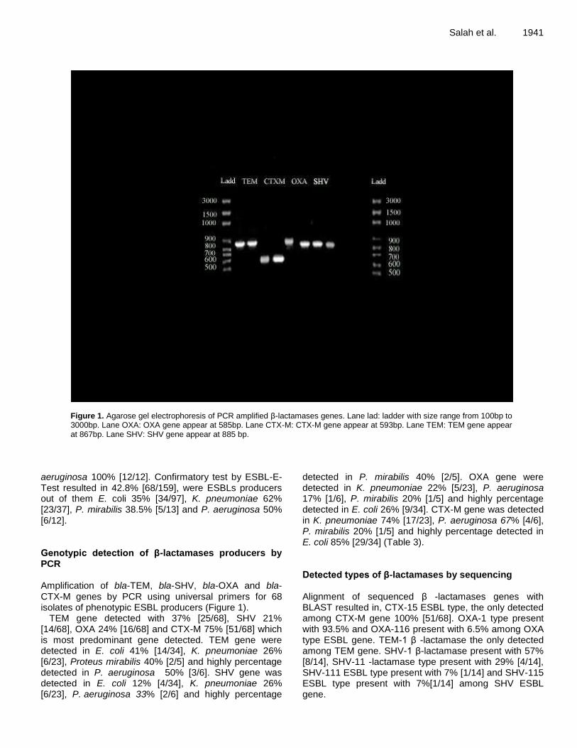
Figure 1. Agarose gel electrophoresis of PCR amplified β-lactamases genes. Lane lad: ladder with size range from 100bp to 3000bp. Lane OXA: OXA gene appear at 585bp. Lane CTX-M: CTX-M gene appear at 593bp. Lane TEM: TEM gene appear at 867bp. Lane SHV: SHV gene appear at 885 bp.
aeruginosa 100% [12/12]. Confirmatory test by ESBL-E-Test resulted in 42.8% [68/159], were ESBLs producers out of them E. coli 35% [34/97], K. pneumoniae 62% [23/37], P. mirabilis 38.5% [5/13] and P. aeruginosa 50% [6/12]. Genotypic detection of β-lactamases producers by PCR Amplification of bla-TEM, bla-SHV, bla-OXA and bla-CTX-M genes by PCR using universal primers for 68 isolates of phenotypic ESBL producers (Figure 1).
TEM gene detected with 37% [25/68], SHV 21% [14/68], OXA 24% [16/68] and CTX-M 75% [51/68] which is most predominant gene detected. TEM gene were detected in E. coli 41% [14/34], K. pneumoniae 26% [6/23], Proteus mirabilis 40% [2/5] and highly percentage detected in P. aeruginosa 50% [3/6]. SHV gene was detected in E. coli 12% [4/34], K. pneumoniae 26% [6/23], P. aeruginosa 33% [2/6] and highly percentage
detected in P. mirabilis 40% [2/5]. OXA gene were detected in K. pneumoniae 22% [5/23], P. aeruginosa 17% [1/6], P. mirabilis 20% [1/5] and highly percentage detected in E. coli 26% [9/34]. CTX-M gene was detected in K. pneumoniae 74% [17/23], P. aeruginosa 67% [4/6], P. mirabilis 20% [1/5] and highly percentage detected in E. coli 85% [29/34] (Table 3). Detected types of β-lactamases by sequencing Alignment of sequenced β -lactamases genes with BLAST resulted in, CTX-15 ESBL type, the only detected among CTX-M gene 100% [51/68]. OXA-1 type present with 93.5% and OXA-116 present with 6.5% among OXA type ESBL gene. TEM-1 β -lactamase the only detected among TEM gene. SHV-1 β-lactamase present with 57% [8/14], SHV-11 -lactamase type present with 29% [4/14], SHV-111 ESBL type present with 7% [1/14] and SHV-115 ESBL type present with 7%[1/14] among SHV ESBL gene.

1942 Afr. J. Microbiol. Res.
Table 3. Frequency distribution of β -lactamases among urinary isolates.
Total Occurrence
Gene Pseudomonas aeruginosa Proteus mirabilis K. pneumonia E. coli
6 0 2 3 1 ZERO
1 0 1 0 0 TEM
1 0 0 1 0 SHV
15 0 0 6 9 CTX-M
2 0 0 0 2 OXA
6 1 0 4 1 CTX-M +TEM
3 0 0 1 2 CTX-M+SHV
2 1 1 0 0 TEM+SHV
11 2 0 3 6 CTX-M+OXA
2 0 0 0 2 OXA+TEM
3 1 0 2 0 OXA+SHV
2 0 0 0 2 CTX-M +TEM+SHV
10 1 0 0 9 CTX-M+OXA+TEM
2 0 1 1 0 CTX-M+OXA+SHV
2 0 0 2 0 CTX-M +TEM +OXA+ SHV
68 6 5 23 34 Total
Table 4. Mutations detected in sequenced β-lactamases genes.
OXA-1 SHV-11 SHV-1 TEM-1 Gene
K. pneumoniae K. pneumoniae E. coli E. coli Microorganism
RAWAN2015 MR1982 esam1980 NORAN2014 Strain
I97M L31R E29Q P65R Mutation
OXA-116 SHV-115 SHV-111 ……….. Novel ESBL
CTX-M,SHV CTX-M, OXA CTX-M CTX-M, OXA Other ESBLs detected
KR780478 KR780481 KR780480 KR632744 Accession number
Mutations in sequenced β-lactamases Multiple sequence alignment of ESBL genes resulted in detection of multiple mutations and development of novel ESBL types (Table 4). DISCUSSION In our results, the leading pathogen causing UTI was E. coli [61%] followed by K. pneumonia [23.3%], P. mirabilis [8.2%] and P. aeruginosa [7.5%] which nearly similar to that reported by Ibrahim et al. (2014) in Egypt. E. coli was the most common pathogen causing UTI in the world this in agreement with our study (Gupta et al., 2011). Blindly treatment of UTI leads to increase the resistance rate of these pathogens to antibiotics especially β–lactam antibiotics due to excessive and misuse of these antibiotics. Imipenem was the most effective antibiotic against UTI and activity more than 95% because of less used due to economic considerations.
In this study production of ESBLs varies from type of isolate to another. P. aeruginosa was the most powerful ESBLs producers [100%], followed by E. coli 97%, K. pneumoniae 82.6% and finally P. mirabilis 82%. In our study sequenced CTX-M showed that most CTX-M genes were CTX-M-15 as that detected in Egypt and middle east area (Amin et al., 2005; Thabit et al., 2011). In our study, multiple mutations detected among β-lactamases; in SHV-11 gene there is mutation in position 31, amino acid Arginine instead of amino acid Leucine [L31R], results in novel SHV-115 K. pneumoniae strain MR1982 ESBL with accession number [KR780481]. L31R mutation in Klebsiella was the first detected in middle east and the second detected in world after Mendonça et al. (2009)
in Portugal from Klebsiella results
in novel SHV 61. TEM-1 gene show mutation in position 65, amino acid Arginine instead of amino acid Proline [P65R] results in E. coli strain NORAN2014 β-lactamase TEM-1with accession number [KR632744.1]. In SHV-1 gene there is mutation in position 29, amino acid Glutamine instead of amino acid Glutamate [E29Q]

results in novel SHV-111 ESBL, E. coli strain esam1980 with accession number [KR780480.1]. In OXA-1 gene there is mutation in position 97, amino acid Methionine instead of amino acid Isoleucine [I97M] results in novel OXA-116, K. pneumoniae strain RAWAN2015 ESBL with accession number [KR780478]. [http://www.ncbi.nlm.nih.gov]. Conclusion Frequent consumption and misuse of antibiotics lead to mutations and the emergence of new genes more aggressive and more resistant to antibiotics, which leads to increased mortality and which calls for the search for new antibiotics and open new horizons in how to address these pathogens. Conflict of Interests The authors have not declared any conflict of interests. REFERENCES Al-Achi A (2008). An Introduction to Botanical Medicines: History,
Science, Uses, and Dangers: History, Science, Uses, and Dangers, ABC-CLIO.
Ambler RA Coulson JM, Frere JM, Ghuysen B, Joris M, Forsman R Levesque G, Tiraby S, Waley (1991). A standard numbering scheme for the class A beta-lactamases. Biochem. J. 276(1):269.
Amin NE, Giske CG, Jalal S, Keijser B, Kronvall G, Wretlind B (2005). Carbapenem resistance mechanisms in Pseudomonas aeruginosa: alterations of porin OprD and efflux proteins do not fully explain resistance patterns observed in clinical isolates. Apmis 113(3):187-196.
Bauer A, Kirby W, Sherris J, turck C, Turck M (1966). Antibiotic susceptibility testing by a standardized single disk method. Am. J. Clin. Pathol. 45(4):493.
Brooks GF, Butel JS, Ornston LN (2005). Microbial Genetics. In: Jawetz and Adelberg’s Medical Microbiology. Lange Medical Book, 20th ed: Pp.86-104.
Butler DA, Lobregat C, Gavan TL (1975). Reproducibility of the analytab (API 20E) system. J. Clin. Microbiol. 2(4):322-326.
Cheesbrough M (2006). District laboratory practice in tropical countries, Cambridge university press.
CLSI (2013). Performance standards for antimicrobial susceptibility testing; Twenty- third informational supplement.CLSI document M100-S23. Wayne, PA, USA.
Eisner A, Fagan EJ, Feierl G, Kessler HH, Marth E, Livermore DM, Woodford N (2006). Emergence of Enterobacteriaceae isolates producing CTX-M extended-spectrum β-lactamase in Austria. Antimicrob. Agents Chemother. 50(2):785-787.
Gupta K, Hooton TM, Naber KG, Wullt B, Colgan R, Miller LG, Moran GJ, Nicolle LE, Raz R, Schaeffer AJ (2011). International clinical practice guidelines for the treatment of acute uncomplicated cystitis and pyelonephritis in women: a 2010 update by the Infectious Diseases Society of America and the European Society for Microbiology and Infectious Diseases. Clin. Infect. Dis. 52(5):e103-e120.
Gupta K, Sahm DF, Mayfield D, Stamm WE (2001). Antimicrobial resistance among uropathogens that cause community-acquired urinary tract infections in women: a nationwide analysis. Clin. Infect. Dis. 33(1): 89-94.
Salah et al. 1943 Gupta V (2007). An update on newer [beta]-lactamases. Indian J. Med.
Res. 126(5):417. Hosoglu S, Gundes S, Kolayli F, Karadenizli A, Demirdag K, Gunaydin
M, Altindis M, Caylan R, Ucmak H (2007). Extended-spectrum beta-lactamases in ceftazidime-resistant Escherichia coli and Klebsiella pneumoniae isolates in Turkish hospitals. Indian J. Med. Microbiol. 25(4):346.
Ibrahim MA, Agban MN, Thabit AG, El-Khamissy TR, Attia AE (2014). Prevalence of Extended-Spectrum β-Lactamase producing Klebsiella pneumoniae by phenotypic and genotypic methods in Assiut University Hospital. Egypt J. Med. Microbiol. 23(4).
Livermore DM (1995). beta-Lactamases in laboratory and clinical resistance. Clin. Microbiol. Rev. 8(4):557-584.
McGinnis S, Madden TL (2004). BLAST: at the core of a powerful and diverse set of sequence analysis tools. Nucleic Acids Res. 32(2):W20-W25.
Mendonça N, Ferreira E, Louro D, Participants A, Caniça M (2009). Molecular epidemiology and antimicrobial susceptibility of extended-and broad-spectrum β-lactamase-producing Klebsiella pneumoniae isolated in Portugal. Int. J. Antimicrob. Agents 34(1):29-37.
Mortensen JE, Bernier M, Gray LD, Dolan S, Hanson ND, Moland ES, Abdalhamid B (2005). New quality control strain for use in routine testing for production of extended-spectrum beta-lactamases by Enterobacteriaceae. J. Clin. Microbiol. 43(5):2545-2545.
Nicolle L (2005). Complicated urinary tract infection in adults. Can. J. Infect. Dis. Med. Microbiol. 16(6):349.
Oliver A, Weigel LM, Rasheed JK, McGowan JE, Raney P, Tenover FC (2002). Mechanisms of decreased susceptibility to cefpodoxime in Escherichia coli. Antimicrob. Agents Chemother. 46(12):3829-3836.
Pagani L, Dell'Amico E, Migliavacca R, D'Andrea MM, Giacobone E, Amicosante G, Romero E, Rossolini GM (2003). Multiple CTX-M-type extended-spectrum β-lactamases in nosocomial isolates of Enterobacteriaceae from a hospital in northern Italy. J. Clin. Microbiol. 41(9):4264-4269.
Paterson DL, Bonomo RA (2005). Extended-spectrum β-lactamases: a clinical update. Clin. Microbiol. Rev. 18(4):657-686.
Peladan F, Monteil H (1988). Identification of Pseudomonas, Flavobacterium, and Alcaligenes with the API 20 NE system. Pathol. Biol. 36(2):187-192.
Philippon A, Arlet G, Jacoby GA (2002). Plasmid-determined AmpC-type β-lactamases. Antimicrob. Agents Chemother. 46(1):1-11.
Poirel L, Gniadkowski M, Nordmann P (2002). Biochemical analysis of the ceftazidime-hydrolysing extended-spectrum β-lactamase CTX-M-15 and of its structurally related β-lactamase CTX-M-3. J. Antimicrob. Chemother. 50(6):1031-1034.
Sanger F, Nicklen S, Coulson AR (1977). DNA sequencing with chain-terminating inhibitors. Proc. Natl. Acad. Sci. U.S.A. 74(12):5463-5467.
Sievers F, Wilm A, Dineen D, Gibson TJ, Karplus K, Li W, Lopez R, McWilliam H, Remmert M, S ding J (2011). Fast, scalable generation of high‐quality protein multiple sequence alignments using Clustal Omega. Mol. Syst. Biol. 7(1).
Stürenburg E, Mack D (2003). Extended-spectrum β-lactamases: implications for the clinical microbiology laboratory, therapy, and infection control. J. Infect. 47(4):273-295.
Thabit AG, El-Khamissy TR, Ibrahim MA, Attia AE (2011). Detection of extendedspectrum beta-lactamase enzymes (ESBLS) produced by Escherichia coli urinary pathogens at assiut university hospital. Bull. Pharm. Sci, Assiut University 34(2):93-103.
Thomson K, Sanders W, Sanders C (1994). USA resistance patterns among UTI pathogens. J. Antimicrob. Chemother. 33(suppl A):9-15.

Vol. 10(46), pp. 1944-1951, 14 December, 2016
DOI: 10.5897/AJMR2016.8209
Article Number: D6B799B62043
ISSN 1996-0808
Copyright © 2016
Author(s) retain the copyright of this article
http://www.academicjournals.org/AJMR
African Journal of Microbiology Research
Full Length Research Paper
Prevalence and Antibiotic Susceptibility of Escherichia coli and Salmonella spp. isolated from milk of zero
grazed cows in Arusha City
Martha M. Sudda1, Adelard B. Mtenga1, 2, Lughano J. Kusiluka1 and Neema Kassim1*
1The School of Life Sciences and Engineering, Nelson Mandela African Institution of Science and Technology, P. O. Box
447 Arusha, Tanzania. 2Tanzania Food and Drugs Authority, P. O. Box 77150, Dar-es -salaam, Tanzania.
Received 12 July 2016, Accepted 21 September, 2016
The present study assessed the antibiotic susceptibility patterns of Escherichia coli and Salmonella isolates of raw milk from zero grazed cows. A total of 65 milk samples were collected for analysis. The standard membrane filtration technique and HiCrome E. coli agar were used in isolation of E. coli from milk samples. Isolation of Salmonella species employed pre-enrichment in buffered peptone water followed by enrichment in Rappaport and Vassilidis broth prior to Xylose lysine deoxychocolate agar as a differential media. The isolates were analyzed for antimicrobial susceptibility to eight different types of antibiotics using disc diffusion method. The prevalence of E. coli was 16 (16.7%) and all the samples tested were negative for Salmonella. The average colony forming unit for E. coli was 2cfu/mL. All E. coli isolates tested were resistant to penicillin (100%) and amoxicillin-clavulanic acid (100%) while 15(93.8%) were sensitive to ciprofloxacin. Resistance was also observed in sulfamethoxazole-trimethoprim (43.8%), chloramphenicol (12.5%), oxytetracycline (68.8%), streptomycin (12.5%) and gentamicin (25%). Of the isolates tested, 14 (87.5%) showed multi-drug resistance pattern. These results confirm that milk from zero grazed cows in Arusha was contaminated with E. coli, and that most of the E. coli strains isolated were resistant to at least one of the antimicrobial agent commonly used in treatment of human diseases. Key words: Salmonella, Escherichia coli, prevalence, antibiotic susceptibility.
INTRODUCTION Milk is considered virtually sterile when secreted into the alveoli of the udder, however; thereafter it may be contaminated in the interior or exterior of the udder. While
the earlier case occurs if the animal is sick, the latter results from inappropriate handling practices and inadequate environmental hygiene and sanitation along
*Corresponding author. E-mail: [email protected].
Author(s) agree that this article remains permanently open access under the terms of the Creative Commons Attribution
License 4.0 International License

the food value chain (Abate et al., 2015). Cow’s milk, being nutritious with high water activity, serves as the best medium for most of microorganisms including pathogenic bacteria such as E. coli and Salmonella, that pose threats to human health (Kanyeka, 2014).
Although Salmonella, Staphyloccocus aureus and E. coli O157:H7 are the bacteria that can be shed through milk (Ogilvie, 1986; Fagundes et al., 2012), Coxiella burnetii, Listeria monocytogenes, Brucella spp, Campylobacter jejuni, Mycobacterium avium subspecies paratuberculosis, Bacillus cereus, Mycobacterium tuberculosis, Mycobacterium bovis and Yersinia enterocolitica are the bacteria commonly contaminating milk (Dhanashekar, 2012). A study by Lubote et al., (2014), in Tanzania reported that milk quality deteriorated along the food value chain; whereby high prevalence rate of Salmonella and E. coli were found in vendors (43.8%) and 8.0 x 10
3 cfu/mL, shops (40%) and 6.6 x 10
3
cfu/mL and from producers (33.3%) and 3.0 x 10³
respectively. Microorganisms isolated from animal products such as raw or unpasteurized milk and meat have long been considered as sources of human infections, where salmonellosis has been reported as one of the common food-borne infections globally (Addis et al., 2011). Salmonella of zoonotic origin has been reported to show increasing rates of resistance to multiple antibiotics (Mijović, 2012). Such resistance is acquired while in the host animal it is spread to humans through the food chain (Carattoli, 2003; Sisak et al., 2006; Kidie et al., 2013). Although E. coli is an enteric commensal bacterium in both animals and humans, pathogenic strains exist and cause different diseases including urinary tract infections, gastroenteritis, septicemia, meningitis and peritonitis (Tadesse et al., 2012).
The increasing use of antibiotics in veterinary practice is suspected to contribute to acceleration of antibiotic resistance in microorganisms found where livestock are kept (Addis et al., 2011). The irrational use of antibiotics in food producing animals could result into antibiotic residues in edible tissues and products (Darwish et al., 2013). It has been reported that, antibiotics used for treatment of human bacterial infections are used for prophylactic, therapeutic and growth promotion in animals too (Phillips et al., 2004). Bacteria that have been exposed to low doses of these antibiotics in tissues and products from these animals may be less susceptible to drugs, and when such bacteria enter the human body through consumption of contaminated foods, they may cause infections that are resistant to many antibiotics (Wang et al., 2011; Clauβen et al., 2013).
A study conducted in Kilosa and Mvomero districts in Morogoro, Tanzania by Kanyeka, (2014) reported antibiotic resistance in bacteria isolated from milking containers and milk products. Example E. coli was reported to be resistant to amoxicillin-clavulanic acid (100%), ampicillin (100%) and amoxicillin (100%) and
Sudda et al. 1945 Salmonella showed resistance to ampicillin (100%) and amoxicillin (100%). Lubote et al. (2014) reported that, milk may contain resistant bacterial strains as a result of cross contamination from containers, humans and the environment. Due to urbanization and limited diversity of pasture, reliance on processed commercial feed mainly cereal and oil seed by-products, zero grazed cows are prone to diseases and prominent use of antibiotics (Shem et al., 2002; Mathews Jr and Johnson, 2013). To this fact, little is known about antimicrobial resistance of bacteria that are shed by the zero grazed cows in the study area. Therefore, the present study aimed at establishing the prevalence and ascertaining the antimicrobial susceptibility pattern of E. coli and Salmonella isolated from raw milk from zero grazed cows in ten wards of Arusha city, Tanzania. MATERIALS AND METHODS Study site The study was purposively conducted in ten wards (Sombetini, Baraa, Engutoto, Moshono, Moivaro, Kimandolu, Sinoni, Lemara, Daraja II and Themi) of the Arusha City where some of the residents practice dairy cattle keeping as a common economic activity (Bukuku, 2013). The Arusha City, which is the headquarter of the Arusha region is situated in the north-eastern corner of Tanzania, between latitudes 2° and 6° South and longitudes 35° and 38° East of the Greenwich (Thadeo, 2014). Sample collection
The sample size was determined using the prevalence rate of 90% from the previous study by Lubote et al. (2014) and the formula used by Addis et al., (2011) which is;
Where; N is the required sample size, Zα, the normal deviation at 5% which is 1.96, P, the estimated prevalence which is 90% and d2, the precision of estimate considered as 0.05. According to the formula, a total of 66 samples should be used in the study. Only 65 samples were analysed for the study as one of the farmer dropped out in the last period of sample collection.
The studied households were selected randomly from the list of dairy keeping households available at the Ward Livestock Offices. From each household, milk samples were collected from only one milked cow that received medication later than others and that the withdraw period for any disease treated was over and seemed apparently health. A total of 65 milk samples each from a single cow were collected from all the teats on the udder of the selected animals. The milk samples were collected during the milking time between 17:00 and 19:00h. The udders and teats of the selected cows were washed thoroughly with warm water and then dried by using towels, then, the fore stream of milk was directed to the household milking container so as to clean the orifice hence prevent contamination by environmental bacteria. Thereafter, a stream of milk was directed to the sterile falcon tubes while avoiding the contact between the sampling container, cow’s teats and the milker’s hands so to prevent contamination of the samples by environmental bacteria. The milk samples were kept in a cool box at
N= (Zα/2)2×P(1−P)/d2;

1946 Afr. J. Microbiol. Res. about 4°C so as to avoid bacterial proliferation. The samples were immediately transported to the Nelson Mandela African Institution of Science and Technology (NM-AIST) laboratory for bacterial culture within five hours (Lubote et al, 2014). All media used in isolation of bacteria were from HiMedia Laboratories Pvt. LTD, Mumbai, India, were of analytical grade and used according to manufacturer’s instructions. Isolation of E. coli The standard membrane filtration technique and HiCrome E. coli agar were used in isolation of E. coli from milk samples. The procedure was carried as described by Robinson and Batt (1999) and Lyimo et al. (2016). Briefly, 10ml of milk sample was diluted into 90ml of double distilled sterile water. Then, 100ml of the diluted sample was filtered through a 47mm membrane filters (cellulose nitrate filters) with pore size of 0.45µm (Sartorius Stedium Biotech GmbH, Goettingen) in a vacuum filtration system. After filtration, each filter membrane was placed on a chromogenic selective agar plate (HiCrome E. coli agar) and then pre-incubated at 37°C for 4h so as to resuscitate the injured or stressed bacteria, followed by incubation for 22h at 44°C. E. coli were picked, preserved in 15% glycerol: 85% Lysogeny broth (LB) and stored at -80ºC for subsequent analysis.
Isolation of Salmonella Isolation of Salmonella was carried out according to the procedures described by Addis et al. (2011). Briefly, 1.0ml of milk sample was pre-enriched with 9.0 ml of buffered peptone water (BPW) for 24h at 37°C. Then, 0.4ml of the non-selective pre-enrichment step was transferred to 10 ml of Rappaport and Vassilidis broth (RVS) and then incubated at 42°C for 24h. Then, a loopful (1µl) of cultured broth from the selective enrichment step were streaked onto Xylose-Lysine Deoxycholate agar (XLD) plates using a sterile wire loop and then incubated at 37°C for 24h. For samples that did not show any growth during 24h, incubation was extended to 48h. Colony forming units The colony forming unit per millilitre (Cfu/ml) was calculated using the formula; Number of colonies *dilution factor /volume plated (Baranzoni, 2014).
Antimicrobial susceptibility testing Sensitivity toward eight different antibacterial agents (streptomycin 300 µg, penicillin 10 µg, tetracycline 10 µg, sulfamethaxazole-trimethoprim 25 µg, oxytetracycline 30 µg, gentamicin 10 µg and amoxicillin-clavulanic acid 3 µg) commonly used for disease treatment in both humans and animals, ciprofloxacin 5µg and chloramphenicol 10µg which are drugs reserved for human disease treatment was carried out. The procedure described by Lalitha (2004) was used. In brief, E. coli cells were resuscitated through incubation on nutrient broth (Liofilchem Bacteriology Products, Roseto) at 37°C for 24 h. The turbidity was adjusted against 0.5 Macfarland concentrations (Remel, Lenexa Kansas) by adding the E. coli culture into sterile normal saline 0.85% (VWR International, West Chester).
Then, sterile swab was used to spread the E. coli cells on the entire surface of the petri dishes that contained Tryptone soy agar (Oxoid ltd Basingstoke, Hampshire). Antibiotic discs were aseptically placed on top of the swabbed petri dishes and the
antibiotics were allowed to diffuse at 24°C for 15 min followed by incubation at 37°C for 24 h. The zones of inhibition were measured by using a vernier calliper into the nearest millimetres in order to establish the susceptibility profile of E. coli. The susceptibility pattern was classified as resistant, intermediate or susceptible according to the Clinical and Laboratory Standards Institute (CLSI). Performance Standards for Antimicrobial Disk and Dilution Susceptibility Tests for Bacteria Isolated From Animals. Clin Lab Stand Inst. 2008; 28: M31–A3. Isolates that were not susceptible to one or more antibacterial agent in three or more different antibiotic classes were considered as multi-drug resistant isolates (Magiorakos et al., 2012).
Data analysis
Data obtained from the antibiotic susceptibility testing were summarized using the Microsoft Excel 2007 and presented in tabular form and bar charts. Standard deviation was also calculated using Microsoft Excel 2007.
RESULTS AND DISCUSSION Isolation of E. coli and Salmonella The present study aimed at assessing the prevalence and antibiotic susceptibility profile of E. coli and Salmonella isolates of zero-grazed cows’ milk. Out of 65 samples, only 7(11%) were positive for E. coli and Salmonella was not detected in any of the samples tested. The highest number of E. coli colonies in E. coli positive samples was four, thus the average prevalence rate of E. coli in the present study was 16 (16.7%). The lowest prevalence rate of 116 (12.9%) E. coli has also been reported by Worku et al, (2012) in Oromia Regional State. Another study by Ekici, et al., (2004) in Turkey reported that neither Salmonella nor E. coli was isolated in all milk samples collected from individual cows while the study by Reta et al. (2016) at Jigjiga City of Somali Regional State reported a higher prevalence of 9 (30%) E. coli and 1 (3.3%) Salmonella isolates. The differences in prevalence rate of E. coli and Salmonella may be attributed to the health status of cows whose milk was sampled. In this study, milk samples were collected from apparently healthy animals and may explain for the low prevalence rates observed. The average colony forming unit for E. coli was 2 cfu/ml. This indicates that, E. coli load in all milk samples were low compared to the previous literature by Marth and Steele (2001); that cows can shed E. coli up to 10
8 cfu/ml. These findings implies
that, raw milk in the study area had low initial bacterial count, probably because milk samples were collected from animals that were considered apparently healthy, this has also been previously observed by Tamime (2009).
The absence of Salmonella in all samples is supported by the previous reports that, Salmonella could be shed through milk only when the animal is suffering from acute clinical salmonellosis and sometimes by carrier animals

Sudda et al. 1947
Table 1. Summary of the diversity of E. coli isolates from zero grazed cow’s milk.
Ward No. of sample
per ward
Number of E. coli positive samples per
ward
Number of
E. coli colonies per
sample
E. coli Cfu/ml per
(100-1
)
Prevalence of E. coli in milk/ward
Number of
Salmonella positive
samples per ward
Sombetini 6 2 1 1 33.3% 0
1 0
Baraa 6 1 3 3 16.7% 0
Moshono 6 1 4 4 16.7% 0
Kimandolu 6 1 4 4 16.7% 0
Themi 6 1 2 2 16.7% 0
Olasity 6 1 1 1 16.7% 0
Moivaro 5 0 0 0 0 0
Lemara 6 0 0 0 0 0
Engutoto 6 0 0 0 0 0
Sinoni 6 0 0 0 0 0
Daraja II 6 0 0 0 0 0
Total 65 7 16 16 0
(Wood et al., 1991; McGuirk and Peek, 2003). Furthermore, the absence of Salmonella in milk suggests that milk is free from bacteria in the interior of the udder only if the animal is healthy (Murphy and Boor, 2000). It has also been reported in study by Abate et al. (2015) that, milk is virtually sterile when secreted into the alveoli of the udder and, after secretion, milk may be contaminated within the udder if the animal is sick or outside the udder as a result of cross contamination. Additionally, presence of E. coli in milk could be due to infection of the teats by environmental E. coli or the milk was contaminated by E. coli from the environment during sampling or because of faulty laboratory procedures (Smith et al., 1985; Smith and Hogan, 1993).
Since milk samples were collected from cows that were considered apparently healthy, but had the history of medication, it could be that, Salmonella isolates were more sensitive whereas E. coli isolates might have been resistant to the administered antimicrobials. On the other hand, Salmonella and E. coli are enteric bacteria which are found in animal’s intestine (Sawant et al., 2007; Ouseph et al., 2009; Tadesse et al., 2012) and their presence in milk could imply that, the animal is a carrier or infected by such bacteria (McGuirk and Peek, 2003). Table 1 shows the diversity of E. coli and Salmonella isolation from milk samples collected from ten wards of Arusha City. Antimicrobial susceptibility testing Among the isolates tested, 56.3% were susceptible to sulfamethoxazole-trimethoprim, chloramphenicol (37.5%), penicillin (0%), oxytetracycline (31.3%),
streptomycin (68.8%), gentamicin (12.5%), ciprofloxacin (93.8%) and amoxicillin-clavulanic acid (0%). The intermediate pattern observed were sulfamethoxazole-trimethoprim (0%), chloramphenicol (50%), penicillin (0%), oxytetracycline (0%), streptomycin (18.8%), gentamicin (62.5%), ciprofloxacin (0%) and amoxicillin-clavulanic acid (0%) and resistance pattern observed were sulfamethoxazole-trimethoprim (43.8%), chloramphenicol (12.5%), penicillin (100%), oxytetracycline (68.8%), streptomycin (12.5%), gentamicin (25%), ciprofloxacin (6.25%) and amoxicillin-clavulanic acid (100%).
Of the selected antibiotics, E. coli were prevalently resistant to penicillin (100%) and amoxicillin-clavulanic acid (100%). The results are similar to the findings by Idriss et al., (2014) who reported that 96% of E. coli isolates were resistant to amoxicillin-clavulanic in Nitra, Slovakia. Similarly, Belayneh et al., (2014) reported that, 65% of the E. coli isolates were resistant to penicillin in East Showa Zone of Akaki District, Ethiopia. The resistance of E. coli isolates to amoxicillin-clavulanic acid observed in the present study is, however, higher than the findings by Čížek et al. (2008) who reported that, 23% of the isolates were resistant to amoxicillin-clavulanic acid. The high resistance to amoxicillin-clavulanic acid and penicillin reported in the present study could be associated with the lack of professionalism in dairy farming which may contribute to misuse of these drugs. As observed in the present study, during the onsite visits, 56.9% of the dairy farmers in Arusha City kept no record of any health interventions made to their animals. Moreover, it could be due to self-medication by using experience, instructions from veterinary input shops or instructions on the label of the respective medicine, a

1948 Afr. J. Microbiol. Res.
Figure 1. Susceptibility profile of E. coli to selected antibiotics. SXT-Sulfamethaxazole-trimethoprim, C-Chloramphenicol, P-Penicillin, TE-Tetracycline, S-Streptomycin, CN-Gentamicin, CIP-Ciprofloxacin, AMC-Amoxillin-clavulanic acid.
practice that may not always result in the correct treatment of the disease. Since in Tanzania, antibiotics are sometimes sold without prescriptions (Van de boogard et al., 2011), the observed resistance could be due to increased use of antibiotics, especially the first line antibiotics which are cheap and easily accessible (Shakya et al., 2013). The susceptibility profile of E. coli to different antibiotics has been summarised in Figure 1 below.
The study has also revealed resistance (12.5%) and intermediate (60%) patterns to chloramphenicol. These findings are in contrast to those reported by Belayneh et al. (2014) in East Showa Zone of Akaki District, Ethiopia, who reported 100% sensitivity of E. coli to chloramphenicol. In this study, a lower proportion (12.5%) of the E. coli isolates were resistant to chloramphenicol compared to the high resistance rate (40%) reported by El-Zubeir and El-Owni (2009). The resistance to chloramphenicol reported in the present study may be associated with indiscriminate use of this abandoned antibiotic. Some studies in Tanzania and Nigeria reported that, chloramphenicol is irrationally used in animals as evidenced by the presence of its residues in poultry and poultry products (Nonga et al., 2010; Darwish et al., 2013). However, it may be due to transfer of resistant genes as a result of cross contamination between humans, animals and the environment (Bischoff et al., 2005;Salehi and Bonab, 2006) or use of other antibiotics belonging to aphenicol group (Ruzauskas et al., 2009). The susceptibility pattern of E. coli from all the sampling sites is summarized in the Table 2.
Of all the isolates tested, 93.8% were susceptible to ciprofloxacin. The prevalence of sensitivity to
ciprofloxacin in this study is lower compared to the results by Lehtolainen (2004) and Persson et al. (2011) who reported 100% susceptibility of the isolates to ciprofloxacin. The higher sensitivity to ciprofloxacin may imply that the drug is not being used in dairy farming to treat animal diseases. This may be attributed by the fact that, the drug is critical for human medicine and prohibited for use in food animals (Boothe et al., 2006; Pallo-Zimmerman et al., 2010).
Among the E. coli isolates tested, 87.5% were multi-drug resistant. Multi-drug resistance pattern of E. coli has also been reported by Haque (2013) in Bangladesh and by Memon et al. (2012) in Eastern China. Of the multi-drug E. coli resistant isolates, 50% showed multi-drug resistance to sulfamethoxazole-trimethoprim, penicillin, tetracycline and amoxicillin-clavulanic acid. The multi-drug resistance pattern observed could be the result of accumulation of resistance genes in the plasmids, each coding for resistance to a specific antibiotic and or multi-drug efflux pump each pumping out more than one antibiotic (Nikaido, 2009). Development of multi-drug resistant bacteria is a threat to public health because it leads to ineffective treatment of infections and poor recovery of the patients (Levy and Marshall, 2004; Magiorakos et al., 2012).
Almost all 16 E. coli isolates showed resistance to at least one antimicrobial agent tested and, more than half, 87.5% (14) showed multi-drug resistance pattern to the tested antibiotics. Although the resistant E. coli were isolated from samples that were not tested for antibacterial residues, there is a possibility that antibacterial residues were present in the milk as it has been reported by other workers in Tanzania (Karimuribo

Sudda et al. 1949
Table 2. Antibacterial susceptibility profile of E. coli isolates of zero grazed cow’s milk to selected antibiotics
WARDS Isolate
No.
Sulfamethaxazole-
trimethoprim
Chloramphenicol
Penicillin Tetracycline Streptomycin
Gentamicin Ciprofloxacin Amoxicillin-
clavulanic acid
Sombetini 1 - +/- - - +/- +/- + -
2 + + - - + + + -
Baraa
3 + +/- - - + +/- + -
4 + + - +/- - +/- + -
5 - + - - +/- +/- + -
Moshono
6 + +/- - + + +/- + -
7 + _ - + + +/- + -
8 + + - - + - + -
9 + +/- - - + - + -
Kimandolu
10 - +/- - - + +/- + -
11 - - - - + - + -
12 - +/- - - - - + -
Themi 13 + +/- - + + +/- - -
14 + + - + + +/- + -
Olasity 15 - +/- - - +/- + + -
16 - + - - + +/- + -
et al., 2005; Kurwijila et al., 2006). Therefore, more extensive research is needed to establish the magnitude of the antimicrobial residues and a concomitant antimicrobial resistance in food animals. The multidrug resistance pattern of E. coli isolates is summarized in Table 3. CONCLUSION AND RECOMMENDATION A total of 16 E. coli isolates were isolated from 65 milk samples examined. Almost all 16 isolates showed resistance to at least one antibacterial agent tested and, more than half (14, 87.5%) showed multi-drug resistance pattern to the tested antibiotics. All 16 E. coli isolates were resistant to
the first line antibiotics, penicillin and amoxicillin-clavulanic acid, probably due to their frequent use in dairy units. However, some of the isolates showed resistance to chloramphenicol and ciprofloxacin drugs that are prohibited for use in food-producing animals. This may be due to either illegal use of the drugs or transfer of resistant genes as a result of interaction with human ecosystem.
Although the resistant E. coli were isolated from milk samples that were not tested for antimicrobial residues, there is a possibility that antimicrobial residues were present in the milk. Therefore, extensive research is proposed to establish the relationship between antimicrobial resistance and antimicrobial residues in food animals as well as
to detect the pathogenic E. coli from the raw cow’s milk.
Furthermore, public health education should be given to the public concerning the prudent use of antibiotics so as to avoid the problem of antibiotic resistance. Additionally, legislation is required to enforce proper use of animal and human medicines to minimize cross-transmission of resistant genes from animals to humans and vice versa. Conflict of Interests The authors have not declared any conflict of interests.

1950 Afr. J. Microbiol. Res.
Table 3. Proportion E. coli that were multi-drug resistance
Combination of drugs to which multidrug resistance was observed
Number of E. coli isolates that showed multidrug resistance to
the combination
Percentage proportion of E. coli that showed multidrug resistance to the
combination (%)
Sulfamethoxazole-trimethoprim,penicillin, Tetracycline and amoxicillin-clavulanic acid
7 50
Penicillin, tetracycline and amoxicillin-clavulanic acid
2 14.3
Chloramphenicol, penicillin and amoxicillin-clavulanic acid
1 7.14
Penicillin, streptomycin and amoxicillin-clavulanic acid
1 7.14
Chloramphenicol, gentamicin and amoxicillin-clavulanic acid
1 7.14
Penicillin, ciprofloxacin and amoxicillin-clavulanic acid
1 7.14
Penicillin, tetracycline, gentamicin and amoxicillin-clavulanic acid
1 7.14
Penicillin, streptomycin and amoxicillin-clavulanic acid
1 7.14
REFERENCES Abate M, Wolde T, Niguse A (2015). Bacteriological Quality and Safety
of Raw Cow’s Milk in and around Jigjiga City of Somali Region, Eastern Ethiopia. IJRBS. 3:48-55.
Addis Z, Kebede N, Sisay Z, Alemayehu H, Wubetie A, Kassa T (2011). Prevalence and antimicrobial resistance of Salmonella isolated from lactating cows and in contact humans in dairy farms of Addis Ababa: a cross sectional study. BMC infect. Dis. 11(1):222.
Baranzoni M (2014). Advances in methods to detect, isolate and quantify foodborne pathogens.
Belayneh R, Belihu K, Tesfaye A (2014). Microbiological study on bacterial causes of bovine mastitis and its antibiotics suscebtibility patterns in East Showa Zone, Akaki district, Ethiopia. J. Vet. Med. Anim. Health, 6(4):116-122.
Bischoff M, White G, Hume E, Poole L, Nisbet J (2005). The chloramphenicol resistance gene cmlA is disseminated on transferable plasmids that confer multiple-drug resistance in swine Escherichia coli. FEMS microbial. Lett. 243(1):285-291.
Boothe M, Boeckh A, Simpson B, Dubose K (2006). Comparison of pharmacodynamic and pharmacokinetic indices of efficacy for 5 fluoroquinolones toward pathogens of dogs and cats. J. Vet. Intern. Med. 20(6):1297-1306.
Bukuku N (2013). Awareness of health risks as a result of consumption of raw milk in Arusha City and Meru District, Tanzania. Sokoine University of Agriculture.
Carattoli A (2003). Plasmid-mediated antimicrobial resistance in Salmonella enterica. Curr. Iss. Mol. Biol. 5(4):113-122.
Čížek A, Dolejská M, Novotná R, Haas D, Vyskočil M (2008). Survey of Shiga toxigenic Escherichia coli O157 and drug‐resistant coliform bacteria from in‐line milk filters on dairy farms in the Czech Republic. J. Appl. Microbiol. 104(3):852-860.
Clauβen M, Bahmann D, Schmidt S (2013). Detection of antibiotic residues in food–pitfalls and optimization of agar diffusion tests in comparison with commercial test kits.
Darwish S, Eldaly A, El-abbasy T, Ikenaka Y, Nakayama S, Ishizuka M (2013). Antibiotic residues in food: the African scenario. Jap. J. Vet. Res. 61:S13-S22.
Dhanashekar R, Akkinepalli S and Nellutla A (2012). Milk borne
infections. An analysis of their potential effect on the milk industry. Germs, 2(3): 101.
Ekici K, Bozkurt H, and Isleyici O (2004). Isolation of some pathogens from raw milk of different milch animals. Pak. J. of Nutr. 3(3): 161-162.
El-zubeir E, El-owni O (2009). Antimicrobial resistance of bacteria associated with raw milk contaminated by chemical preservatives. World J. Dairy Food Sci. 4(1):65-69.
Fagundes H, Corassin C, Tavolaro P, Oliveira C (2012). Milk hygienic practices and occurrence of Staphylococcus aureus and Escherichia coli O157: H7 in small-scale dairy farms in Sao Paulo. Brazil. Afr. J. Microbiol. Res. 6(28):5805-5808.
Haque E (2013). Identification, molecular detection and antibiogram profile of bacteria isolated from california mastitis test positive milk samples of crossbred cows of satkhira district. Int. J. Vet. Sci. 1:1.
Idriss S, Foltys V, Tančin V, Kirchnerová K, Tančinová D, Zaujec K (2014). Mastitis pathogens and their resistance against antimicrobial agents in dairy cows in Nitra, Slovakia. Slov. J. Anim. Sci. 47(1):33-38.
Kanyeka B (2014). Assessment of microbial quality of raw cow's milk and antimicrobial susceptibility of selected milk-borne bacteria in Kilosa and Mvomero Districts, Tanzania. Sokoine University of Agriculture.
Karimuribo E, Mdegela R, Kusiluka L, Kambarage D (2005). Assessment of drug usage and antimicrobial residues in milk on smallholder farms in Morogoro, Tanzania. Bull. Anim. Health Prod. Afr. 53:234-241.
Kidie H, Bae H, Lee J (2013). Prevalence and antimicrobial resistance of Salmonella isolated from poultry slaughterhouses in Korea. Jap. J. Vet. Res. 61(4):129-136.
Kurwijila L, Omore A, Staal S, Mdoe N (2006). Investigation of the risk of exposure to antimicrobial residues present in marketed milk in Tanzania. J. Food Prot. 69(10):2487-2492.
Lalitha M (2004). Manual on Antimicrobial Susceptibility Testing (Under the Auspices of Indian Association of Medical Microbiologists). Vellore, India: CMC
Lehtolainen T (2004). Escherichia coli Mastitis. PhD Thesis. University of Helsinki, Helsinki, Finland.
Levy B, Marshall B (2004). Antibacterial resistance worldwide: Causes,

challenges and responses. Nat. Med. 10: S122-S129. Lubote R, Shahada F, Matemu A (2014). Prevalence of Salmonella spp.
and Escherichia coli in raw milk value chain in Arusha, Tanzania. Amer. J. Res. Comm. 2(9):1-13.
Lyimo B, Buza J, Subbiah M, Temba S, Kipasika H, Smith W, Call R (2016). IncF Plasmids are Commonly Carried by Antibiotic Resistant Escherichia coli Isolated from Drinking Water Sources in Northern Tanzania. Int. J. Microb. 2016(2016).
Magiorakos P, Srinivasan A, Carey R, Carmeli Y, Falagas M, Giske C, Harbarth S, Hindler J, Kahlmeter G, OLsson‐liljequist B (2012). Multidrug‐resistant, extensively drug‐resistant and pandrug‐resistant bacteria: International expert proposal for interim standard definitions for acquired resistance. Clin. Micr. Infect. 18(3):268-281.
Marth EH and Steele J (2001). Applied dairy microbiology, CRC Press. Mathews Jr K, Johnson R (2013). Alternative beef production systems:
Issues and implications. US Department of Agriculture, Economic Research Service, LDPM-218-01.
Mcguirk S, Peek S (2003). Salmonellosis in cattle: A review. In: American Association of Bovine Practitioners 36th Annual Conference.
Memon J, Kashif J, Yaqoob M, Liping W, Yang Y, Hongjie F (2012). Molecular characterization and antimicrobial sensitivity of pathogens from sub-clinical and clinical mastitis in Eastern China. Prevalence. 33(2):170-174
Mijović G (2012). Antibiotic susceptibility of Salmonella spp.: a comparison of two surveys with a 5 years interval: Annual Proceeding Scientific Papers. J. Int. Med. l. Assoc. Bulgaria 18(1):216-219.
Murphy S and Boor K (2000). Sources and causes of high bacteria counts in raw milk: An abbreviated review. Dairy Food Environ. Sanit. 20: 1-4.
Nikaido H (2009). Multidrug resistance in bacteria. Ann. Rev. Biochem. 78:119.
Nonga H, Simon C, Karimuribo E, Mdegela R (2010). Assessment of antimicrobial usage and residues in commercial chicken eggs from smallholder poultry keepers in Morogoro municipality, Tanzania. Zoon. Pub. Health. 57(5):339-344.
Ogilvie H (1986). The persistent isolation of Salmonella typhimurium from the mammary gland of a dairy cow. The Canad. Vet. J. 27(9):329.
Ouseph P, Prasanthan V, Abhilash P, Udayakumar P (2009). Occurrence and distribution of some enteric bacteria along the southern coast of Kerala. Ind. J. Mar. Sci. 38(1): 97.
Pallo-zimmerman M, Byron K, Graves K (2010). Fluoroquinolones: Then and now. Compendium: Cont. Educ. Vet. 9.
Persson Y, Nyman A, Grönlund-andersson U (2011). Etiology and antimicrobial susceptibility of udder pathogens from cases of subclinical mastitis in dairy cows in Sweden. Acta Vet. Scand. 53(1):36.
Phillips I, Casewell M, Cox T, De groot B, Friis C, Jones R, Nightingale C, Preston R, Waddell J (2004). Does the use of antibiotics in food animals pose a risk to human health? A critical review of published data. J. Antimicrob. Chemother. 53(1):28-52.
Reta A, Bereda W and Alemu N (2016). Bacterial contaminations of raw cow’s milk consumed at Jigjiga City of Somali Regional State, Eastern Ethiopia. Inter. J. Food Cont. 3(1): 1-9.
Robinson K, Batt A (1999). Encyclopedia of food microbiology, Academic press, London.
Ruzauskas M, Virgailis M, Siugzdiniene R, Suziedeliene E, Seputiene V, Daugelavicius R, Zienius D, Sengaut J, Pavilonis A (2009). Antimicrobial resistance of Enterococcus species isolated from livestock in Lithuania. Vet. Arch. 79(5): 439-449.
Sawant A, Hegde V, Straley A, Donaldson C, Love C, Knabel J, Jayarao M (2007). Antimicrobial-resistant enteric bacteria from dairy cattle. App. Envir. Micr. 73(1): 156-163.
Shakya P, Barrett P, Diwan V, Marothi Y, Shah H, Chhari N, Tamhankar J, Pathak A, Lundborg S (2013). Antibiotic resistance among Escherichia coli isolates from stool samples of children aged 3 to 14 years from Ujjain, India. BMC Infectious Diseases, 13(1): 477.
Shem M, Mosha F, Machangu R, Kambarage D, Fujihara T (2002). Bovine mastitis in Zebu and crossbred cattle under the extensive management system in Tanzania. Asian Aust. J. Anim. Sci. 15(5):751-756.
Sudda et al. 1951 Smith K, Hogan S (1993). Environmental mastitis. The Veterinary
Clinics of North America. Food Anim. Pract. 9(3):489-498. Smith K, Todhunter D, Schoenberger P (1985). Symposium:
Environmental effects on cow health and performance. J. Dairy Sci. 68:1531-1553.
Sisak F, Havlickova H, Hradecka H, Rychlik I, Kolackova I, Karpiskova R (2006). Antibiotic resistance of Salmonella species isolates from pigs in the Czech Republic. Vet. Med. 51(5):303-310.
Tadesse A, Zhao S, Tong E, Ayers S, Singh A, Bartholomew J, Mcdermott F (2012). Antimicrobial drug resistance in Escherichia coli from humans and food animals, United States, 1950–2002. Emerg. Infect. Dis. 18(5):741-9.
Tamime AY (2009). Dairy powders and concentrated products. John Wiley and Sons.
Thadeo M (2014). Economics of urban households’ cooking fuel consumption in Arusha city, Tanzania. Sokoine University of Agriculture.
Van den boogaard J, Semvua H, Boeree M, Aarnoutse R, Kibiki G (2011). Assessment of antibacterial sale by using the anatomic therapeutic chemical classification and defined daily dose methodology in Moshi Municipality, Northern Tanzania.
Wang J, Macneil D, Kay F (2011). Chemical Analysis of Antibiotic Residues in Food, John Wiley and Sons. P 38
Wood D, Chalmers A, Fenton A, Pritchard J, Schoonderwoerd M, Lichtenberger L (1991). Persistent shedding of Salmonella enteritidis from the udder of a cow. The Canad. Vet. J. 32(12):738.
Worku T, Negera E, Nurfeta A and Welearegay H (2012). Microbiological quality and safety of raw milk collected from Borana pastoral community, Oromia Regional State. Afri. J. Food Sci. Technol. 3:213-222.

Vol. 10(46), pp. 1952-1960, 14 December, 2016
DOI: 10.5897/AJMR2016.8359
Article Number: C11C95962045
ISSN 1996-0808
Copyright © 2016
Author(s) retain the copyright of this article
http://www.academicjournals.org/AJMR
African Journal of Microbiology Research
Full Length Research Paper
Potential control of beans (Phaseolus vulgaris L.) wilt disease using growth regulators, bioagent, antioxidants
and essential oils as foliar application under field conditions
Mokhtar M. Abdel-Kader* and Nehal S. El-Mougy
Department of Plant Pathology, National Research Centre, El-Behoose St., Dokki, 12622, Giza, Egypt.
Received 30 October 2016, Accepted 7 December, 2016
The efficacy of some fungicide alternatives as foliar spray was evaluated against bean wilt incidence under field conditions. The fungicide alternatives were indole acetic acid, gibberllic acid, Trichoderma harzianum, vitamins E and C, lemon grass, moringa leaf and thyme oils. The obtained results showed that the applied fungicide alternatives treatments could suppress the incidence of green bean wilt. In the light of the present study, a thought-provoking outcome of the following investigation was reached when the results clearly indicate that 100% suppression of the disease was reached when the bean plants were sprayed with a combination of indole acetic acid 40 ppm + gibberellic acid 40 ppm + T. harzianum 10
5 cfu/ml. Such an arrest in disease development decreased to 90.5 and 86.4% when the
infected plants were treated with indole acetic acid 20 ppm + gibberellic acid 20 ppm + T. harzianum 105
cfu/ml and T. harzianum was combined with gibberellic acid 40 ppm, respectively. It could be hypothetically suggested that combined treatments between growth regulators with the bioagent as foliar spraying might be used practically for controlling such soilborne diseases replacing fungicides treatments.
Key words: Bean, biocontrol, foliar formulations, fungicide alternatives, wilt disease control.
INTRODUCTION
The oldest known beans have been known since earliest historic times found in the 5th dynasty tombs where they are mentioned in one of Ramses II's paeans. In medicine, ancient Egyptians used beans in remedies against constipation, as a remedy for a sick tongue, treatment for male urinary complaints and when women ate beans on
an empty stomach as a birth control method. Common bean (Phaseolus vulgaris L.) is one of the most widely cultivated food legume species in the world (Baudoin et al., 2001) for local consumption and exportation purposes and is a worldwide food-secure and nutritious worldwide crop to people of all income categories (Pachico, 1993)
*Corresponding author. E-mail: [email protected].
Author(s) agree that this article remains permanently open access under the terms of the Creative Commons Attribution
License 4.0 International LicenseF

especially to the poor as a source of dietary protein because animal protein such as meats and fish is often rare or completely absent from their diets (Beebe et al., 2013). Bean is attacked by certain pathogenic fungi causing wilt, root-rot and leaf spot diseases which seriously affected both plant stand and yield production. These organisms can occur individually throughout the growing season.
Bean plants influenced genuinely by wilt which is broad disease in the globe also is frequently viewed as concerning illustration of major issue of bean production, diminishing as well as decreasing both yield and quality (El-Mougy, 2001; El-Mougy et al., 2007). Yield losses caused as a result of bean wilt disease are more notable in developing countries due to higher abiotic stress were recorded. The main pathogen responsible for wilt incidence of bean was reported to be Fusarium oxysporum f. sp. phaseoli (Burnchara and Camacho, 2000; El-Mougy, 2001).
The control of bean wilt depends on chemical control, biological control, resistant cultivars and control by cultural practices. There is therefore a need to develop tools and procedures that are simple, fast and accurate for the quantification of pathogen populations, particularly, Fusarium species. In order to overcome such hazardous control strategies, scientists, researchers from all over the world paid more attention towards the development of alternative methods which are, by definition, safe in the environment, non-toxic to humans and animals and are rapidly biodegradable. One of such strategy is the use of biocontrol agents (BCAs) to control fungal plant diseases. Among the BCAs, species of the genus Trichoderma is most promising and effective biocontrol agent. Trichoderma as antagonist controlling wide range of microbes (Chet et al., 1977), and their mechanism of mycoparasitism is much more complex, involves nutrient competition, hyperparasitism, antibiosis, space and cell wall degrading enzymes.
A successful disease-control program could involve just a single practice, but the long term reduction of disease losses generally requires the application of several control measures. The best way to ensure success of a disease-management program is to use integrated disease-control measures (Dik et al., 2002). Generally, IPM is regarded as the use of environmentally safe practices to reduce the disease incidence and development or use of multiple control tactics integrated into a single pest control strategy (Zinkernagel et al., 2002). For example, different natural products, that is, biocontrol agents, plant extracts, essential oils, antioxidant, growth regulators and natural compounds were used as an IPM program which takes a somewhat different approach in plant disease control (Bindu and Kumar, 2009; Li et al., 2009; Sharma et al., 2012). A considerable concern in discovering plant-derived anti- microbial agents arised recently (Sagdic et al., 2003) for
Abdel-Kader and El-Mougy 1953 alternative application in the strategy of preventing bacterial and fungal growth (Lanciotti et al., 2004). Volatile compounds and essential oils used as alternatives for anti-bacterial and anti-fungal treatments (Jenny, 2000; Michael, 2000). Furthermore, Juglal et al. (2002) studied the effectiveness of nine essential oils to control the growth of mycotoxins producing moulds and observed that clove, cinnamon and oregano were able to prevent the growth of Aspergillus parasiticus and Fusarium moniliforme. Benkeblia (2004) observed an inhibitory effect of onion essential oil at different concentrations on the growth of F. oxysporum, Aspergillus niger and Penicillium cyclopinum.
In Egypt, the farmers replant the missed holes on the same field. This practice could leads to a high population of the pathogens, causing serious losses which could reach up to 12% (Anonymous, 2012). The Egyptian farmers followed unwise and intensive use of fungicides for disease management. This strategy proved to be an unsatisfactory solution for controlling wilt disease in Egypt. Research over the last few decades for alternative substances with fungicidal properties has revealed that a potentially useful component of integrated disease management (IDM) programmes against foliar fungal pathogens can be spray applications (Fallik et al., 1997; Mann et al., 2004; Mitchell and Walters, 2004). Therefore, recently, great attention has been diverted to the use of fungicide alternatives for suppressing plant diseases (El-Mougy, 1995, 2001, 2009; Siddiqui et al., 2001; El-Gamal et al., 2003; El-Mougy et al., 2004, 2007; Abd-Alla et al., 2009; Abdel-Kader, 1997; Abdel-Kader et al., 2011, 2012; Abdel-Kader and El-Mougy, 2014) to decrease human and animal health risk concerns. Increased use of foliar fungicides has prompted us to explore potential impacts of this change in production practices and determine if foliar non-fungicide use can be justified for use of other than disease management. In the present study, an investigation control method to disease development and to reduce the yield losses caused by wilt using non-fungicidal methods of treatment was performed. In this concern, we aimed to determine the efficacy of some fungicide alternatives growth regulators e.g. indole acetic acid and gibberellic acid individually or combined with the bioagent T. harzianum, lemon grass, moringa leaf and thyme (essential oils) or vitamins C and E (antioxidants) as foliar spray against bean wilt incidence under field conditions. MATERIALS AND METHODS Tested materials Green bean (P. vulgaris) seeds cv. Giza 3 obtained from Vegetables Crop Research Department, Agricultural Research Centre, Giza, Egypt. The growth regulators indole acetic acid (IAA) and gibberellic acid (GA) and antioxidants (Vitamins E and C) were

1954 Afr. J. Microbiol. Res. purchased from Al-Gamhoria Company Ltd. for chemicals and medicinal instruments, Cairo, Egypt. Commercial essential oils of lemon grass (that is, citral, geraniol, borneol and citronellol), moringa leaf (that is, significant source of B vitamins, vitamin C, provitamin A as beta-carotene, vitamin K, manganese, and protein, among other essential nutrients) and thyme (that is, thymol, carvacrol, geraniol, thymol methyl ether, a-pinene) were used in the present work. Essential oils were purchased from Chemical Industrial Development Company (CID), Egypt. The bioagent, used in this study is candidate antagonistic isolate of T. harzianum, obtained from Culture Collection Unit, Department of Plant Pathology, National Research Center, Cairo, Dokki, Egypt. This antagonist was isolated from the rhizosphere of various healthy and wilt or root rot infected leguminous crops, grown in the Delta and Middle Egypt regions, and proved its high antagonistic ability against different pathogens during previous work at the same department. Agents Field experiment was conducted at Researches and Production Station of National Research Centre at Nubaria region, Beheira Governorate, Egypt during 2014 and 2015 two winter growing seasons to evaluate the efficacy of some foliar spray treatments for controlling green bean wilt disease incidence. This field is well known by the authors throughout previous studies as naturally heavily infested with soilborne wilt pathogens. Also, it was observed that soil infestation with wilt pathogen is not equally distributed in all over this field, but it seems to be characterized with semi-homogeneous distribution with wilt pathogens.
In the present work, the applied treatments were designed as follows: Indole Acetic Acid 10 ppm; Indole Acetic Acid 20 ppm; Indole Acetic Acid 40 ppm; Gibberellic Acid 10 ppm; Gibberellic Acid 20 ppm; Gibberellic Acid 40 ppm; Indole Acetic Acid 10 ppm + T. harzianum (105 cfu/mL-1); Indole Acetic Acid 20 ppm + T. harzianum (105 cfu/mL-1); Indole Acetic Acid 40 ppm + T. harzianum (105 cfu/mL-1); Gibberellic Acid 10 ppm + T. harzianum (105 cfu/mL-
1); Gibberellic Acid 20 ppm + T. harzianum (105 cfu/mL-1); Gibberellic acid 40 ppm + T. harzianum (105 cfu/mL-1); Indole Acetic Acid 10 ppm + Gibberellic Acid 10 ppm + T. harzianum (105 cfu/mL-1); Indole Acetic Acid 20 ppm + Gibberellic Acid 20 ppm + T. harzianum (105 cfu/mL-1); Indole Acetic Acid 40 ppm + Gibberellic Acid 40 + T. harzianum (105 cfu/mL-1); T. harzianum (105 cfu/mL); Vitamin E+ lemon grass oil (2% v:v); Vitamin E + Moringa leaf oil (2% v:v); Vitamin E + thyme oil (2% v:v); Vitamin C + lemon grass oil (2% v:v); Vitamin C + Moringa leaf oil (2% v:v); Vitamin C + thyme oil (2% v:v); Untreated control Field study Certain weights or volumes of tested materials were added to 20 L water to obtain the proposed concentration used (Abdel-Kader et al., 2011). The inoculum of the bioagent T. harzianum was used as spore suspension at the rate of 105 cfu/mL-1 (Abdel-Kader et al., 2012).
Stocks solutions at high concentrations (ppm) of tested growth regulators IAA and GA were prepared by dissolving in sterilized distilled water, and then different volumes of the prepared solution were added to 20 L water to obtain the proposed concentrations. The tested essential oils were added individually to 20 L water to obtain the proposed concentration of 2%. A few drops of the emulsifier Tween 20 (Sigma Co.) were added to the essential oil volumes to obtain an emulsion feature. As for antioxidant (vitamin C and vitamin E), certain weight of each were dissolved in 20 L water
to obtain the proposed concentration of 2%.
All foliar spray treatment were applied twice, the first at the emerged stage of bean plants (at two true leaves age) and the second after 15 days interval.
The test field comprised of plots (4×4 m), each comprised of 5 rows (5 holes/1 m and 20 holes/row). Green bean seeds Giza, 3 cv. were sown (at 15 September 2014 and 2015) in all treatments at the rate of one seed/hole. Furthermore, the emerged bean seedlings were thinned to obtain a uniform number of 80 seedling/plot, in order to neglect the incidence of pre-emergence damping-off infection of bean seeds from present work calculation. Concerning illustration five replicates for every specific treatment and in addition untreated plots were taken in consideration. The traditional agricultural practices, that is, soil plowing, fertilization, irrigation, etc., were followed at all experimental plots. Monitoring and scouting for disease incidence in all cultivated plots were preformed weekly (El-Mougy, 2001). Then, bean plants showing wilt symptoms were recorded. The isolated wilt incident was identified as Fusarium oxysporum. Average percent of wilt disease infection was recorded 15 days after each applied spray and the average accumulated disease incidence was calculated at the flowering stage (60 days old) of plant growth.
The field experiments were carried out for two successive winter growing seasons 2014 and 2015 at the same field. The average percent of wilt incidence for the two growing seasons was calculated and recorded as mean disease incidence. Disease incidence and reduction were calculated as following equations: Disease incidence = C-T/T ×100 where C is the number of diseased plants and T is the total number of plants. Disease reduction= D – D1 / D × 100
where D is the number of diseased plants in control and D is the number of diseased plants in treatment. Statistical analysis The obtained data of field experiments were set up in Completely Randomized Design (CRD). The data collected were analyzed by MSTAT-C program (MSTAT-C, 1988). The means differences were compared by Least Significant Difference test (LSD) at 5% level of significance. The statistical analysis procedures were kindly carried out by Statistical Consulting Office, National Research centre, Egypt.
RESULTS
Evaluation of some fungicide alternatives against wilt incidence was carried out under field conditions for two successive growing seasons.
Evaluation of growth regulators
Generally, we observed that the results of the disease control under the effect of Indole Acetic Acid and Gibberellic acid when each was used independently or in combination with the biocontrol agent, T. harzianum depended on their used concentration (Table 1). A dose-

Abdel-Kader and El-Mougy 1955
Table 1. Effect of growth regulators and antioxidants combined with bioagent or essential oils against wilt disease incidence of green bean under field conditions during two successive growing seasons.
Foliar spray treatment Wilt incidence
(%)*
Disease reduction (%)
Indole Acetic Acid 10 ppm 20.0 23.9
Indole Acetic Acid 20 ppm 16.2 38.4
Indole Acetic Acid 40 ppm 12.5 52.4
Indole Acetic Acid 10 ppm + T. harzianum 11.2 57.4
Indole Acetic Acid 20 ppm + T. harzianum 10.0 61.9
Indole Acetic Acid 40 ppm + T. harzianum 7.5 71.4
Gibberellic acid 10 ppm 18.7 28.8
Gibberellic acid 20 ppm 16.2 38.4
Gibberellic acid 40 ppm 13.7 47.9
Gibberellic acid 10 ppm + T. harzianum 11.2 57.4
Gibberellic acid20 ppm + T. harzianum 8.7 66.9
Gibberellic acid40 ppm + T. harzianum 6.2 76.4
Indole Acetic Acid 10 ppm+Gibberellic acid 10 ppm+T. harzianum 5.0 80.9
Indole Acetic Acid 20 ppm+Gibberellic acid 20 ppm+T. harzianum 2.5 90.4
Indole Acetic Acid 40 ppm+Gibberellic acid 40 ppm+T. harzianum 0.0 100
T. harzianum 7.5 71.4
Vitamin E + lemon grass oil 10.0 61.9
Vitamin E + Moringa oil 15.0 42.9
Vitamin E + thyme oil 10.0 61.9
Vitamin C + lemon grass oil 7.5 71.4
Vitamin C + Moringa oil 13.7 47.9
Vitamin C + thyme oil 7.5 71.4
Untreated control 26.3 -
LSD at 0.5% 2.8 -
*Wilt incidence was recorded as the mean percent of disease incidence at the two growing seasons and calculated relatively to the number of emerged bean seedlings= 80.
response experiment was conducted to determine the level of wilt incidence and reduction in the percentage of the disease to obtain a glimpse of a potential control mechanism.
The data revealed that increasing concentration of applied growth regulators showed parallel reduction in disease incidence. Moreover, the growth regulator indole acetic acid was more effective against disease incidence than gibberellic acid. The recorded reduction in wilt incidence ranged between 23.9 and 52.4% for indole acetic acid, meanwhile this range was from 28.8 to 47.9% for gibberellic acid at similar concentrations of 10, 20 and 40 ppm.
Consequently, our observation indicates that the wilt incidence percentage decreased from 23.9 to 38.4% and finally to 52.4% when indole acetic acid was applied while the recorded numbers under the effect of gibberellic acid recorded 28.8, 38.4 and 47.9%.
This indicates that the indole acetic acid when applied at a concentration of 10 and 40 ppm was more effective than gibberellic acid by 4.9 and 4.5%, respectively. At the
concentration of 20 ppm, both agents had the same effect.
Evaluation of the bioagent T. harzianum
T. harzianum when applied during the experimental period at a concentration of 10
5 cfu/mL
-1 caused a fall by
7.5% in the wilt incidence and 71.4% in disease reduction which represents a rise in the control of the wilt disease incidence by 28.5%.
Evaluation of combine effect of growth regulators and the biocontrol agent T. harzianum
The use of the bioagent T. harzianum in a combination with indole acetic acid and gibberellic acid increased the efficacy of the growth regulators against wilt incidence. A combination of T. harzianum with indole acetic acid enhanced the suppressive effect of the growth regulator on the disease with an increase reduction in disease

1956 Afr. J. Microbiol. Res. incidence from (23.9 to 57.4%), (38.4 to 61.9%) and (52.4 to 71.4%) at concentrations of 10, 20 and 40 ppm, respectively.
Similarly, when T. harzianum was combined with gibberellic acid, the suppressive effect of this combination was more evident when the gibberellic acid was used at concentrations of 10, 20 and 40 ppm to cause increase reduction in the disease incidence from 28.8 to 57.9%, 38.4 to 66.9% and 47.9 to 76.4% which could be estimated as 29.1, 28.5 and 28.5%, respectively.
The impact of the three agents when combined together as a single treatment protocol was highly effective when the experiment recorded that the reduction percentage in the disease recorded 80.9 and 90.4% when indole acetic acid and gibberellic acid were used at a concentration of 10 and 20 ppm with 10
5 cfu/mL
-1 T.
harzianum. This propitious effect reached its climax when the plants were treated with indole acetic acid 40 ppm + gibberellic acid 40 ppm+ T. harzianum10
5 cfu/mL
-1 to
protect the plants from wilt disease resulting in 0% infection (that is, a complete suppression in the incidence of green bean wilt). Evaluation of the use of essential oils and antioxidants The obtained results showed that the applied treatments of vitamin C combined with the essential oils of Lemongrass, Moringa leaf and thyme had superior effect on disease incidence recorded as 71.4, 47.9 and 71.4% comparing with vitamin E plus the same essential oils which recorded 61.9, 42.9 and 61.9%, respectively.
Generally, the presented data in Table 1 showed varied numbers of infected bean plants when treated with different treatments. These results could deduced that a percent of 15 and 13.7 infected plants were recorded in Vitamin E + Moringa leaf oil; Vitamin C + Moringa leaf oil treatments, meanwhile, 10.0 and 7.5% infected plants were recorded in both Vitamin E + lemongrass oil and Vitamin C + lemongrass oil treatments, in respective order. Thyme oil showed superior effect for reducing disease incidence when combined with Vitamin C that 7.5% infected plants were recorded when compared with 10.0% infected plants when combined with Vitamin E. Furthermore, it is interesting to note that wilt incidence was reduced by combining the growth regulators and bioagent T. harzianum. Respectively, indole acetic acid at 10, 20 and 40 ppm and gibberlic acid at 10, 20 and 40 ppm showed that 20, 16.2, 12.5% and 18.7, 16.2, 13.7% infected plants. Meanwhile, adding the bioagent T. harzianum reduced the infected plants to be 11.2, 10.0, 7.5% and 11.2, 8.7, and 6.2% in IAA and GA raising concentrations, in respective order. Combined treatments of IAA 10 ppm + GA 10 ppm + T. harzianum and IAA 20 ppm + GA 20 ppm + T. harzianum showed that 5 and
2.5% plants got infected out of 80 grown plants compared to 26.3% (21 plants) in untreated control. However, none of the plants have been infected by wilt disease (0%) when plants were treated with IAA 40 ppm + GA 40 ppm+ T. harzianum. DISCUSSION The need to increase food production by at least 70% in order to match the population increase is highlighted by the United Nations Food and Agriculture Organization. The fact that nearly 20% of the global harvest is lost due to plant diseases (Chakraborty and Newton, 2011) could make this demand more daunting. Chemical control using the application of fungicides could be one of the most efficient ways to combat these diseases. Furthermore, pathogens can develop resistance against the used fungicides which then the higher dose of chemicals are required.
In addition, the environmental and health concerns are associated with the potentially toxic of chemicals which is applied in fields. Therefore, the safer and more sustainable methods of crop protection are demand. A novel area of research in the war against pathogens focuses on enhancing the plant's defense system. If a plant can fight off an infection on its own, we can reduce the amount of pesticides needed. Similar to how children are vaccinated to protect against future diseases, plant pathologists are using the same methodology to enhance plants resistance against pathogens, with the goal of strengthening their defenses mechanism against invaders. For more explanation, the principle of "defense priming" terminology is very similar to how we develop vaccines to treat human diseases. A vaccine works by acting as a pathogen impostor. It tricks the immune system into thinking it is being attacked, which stimulates defense responses, such as the production of antibodies. This procedure creates a defense memory which could allow the immune system to remember a certain pathogen when the body is attacked in the future. It can then respond quickly referring to its primed memory from the vaccine. Furthermore, the plants prepared themselves to be ready for the enemy attacks. The treated plants with induce resistant agents display enhanced tolerance to pathogen infection, which is often characterized by fewer disease symptoms and reduction in pathogen populations within the plant. Although in commercial agriculture, primed plants have not yet been applied on large-scale, scientists are actively conducting their researches on the use of defense priming theory against bacteria, viruses and fungi under both greenhouse and field trials for plant protection (http://phys.org/news/2015-07-pathogens-immune.html#jCp).
This method which cause the induce defense systems could be considered as a safe and effective way to

protect some of the global harvest currently lost due to diseases. The applications of fungicides have potential consequences, not only from the economic perspective, but from the biological impact on fungal populations and to have adverse environmental effects causing health hazards to humans and other non-target organism, including beneficial life forms. Increased use of foliar fungicides has prompted us to explore potential foliar use of others than fungicide use and disease management. This investigation focuses on crop disease management by using natural compounds derived from plants, marine organisms and microbes to represent an ecologically friendly approach for plant diseases control as an alternative to the chemical fungicides with no side effects on humans and animals. We evaluated some fungicide alternatives against wilt incidence under field conditions for two successive growing seasons. Different treatments of growth regulators alone or combined with the bioagent T. harzianum as well as antioxidants individually or combined with essential oils were applied as foliar spray on emerged green bean seedlings. The present study has demonstrated that all treatments tested, some growth regulators, essential oils and antioxidants have antifungal potential activities and could be useful when integrated with bioagents against green bean fungal pathogens.
Hypothetically, we visualize the mechanisms of action of the used agents in our investigation in controlling wilt of beans (P. vulgaris L.) as follows: (i) direct competition with the target organism; (ii) antibiosis; (iii) predation or parasitism of the target organism; (iv) induced resistance of the host plant; and (v) inactivation of the enzymes produced by the pathogen. These five intermingling mechanisms were seen when T. harzianum was used as our biocontrol agent. Recently, biological control of plant pathogens has received great attention as a promising supplement or alternative to chemical control and an attractive proposition to decrease heavy dependence of modern agriculture on costly chemical fungicides, which not only cause environmental pollution but also lead to the development of resistant strains. Commercial biopesticides are now available, and most of which are based on the free living fungi common in soil, associated with plant roots and root ecosystems, fungal genus Trichoderma (Woo et al., 2006) a genus under Deuteromycotina. They are non-plant pathogenic and have gained high importance as the most important and efficient biological control agents against plant pathogens (Lorito et al., 1998; Bokhari and Perveen, 2012). This could be attributed to their high reproductive capacity, ability to survive under unfavourable conditions, capacity to colonize the rhizosphere, strong antagonism against the pathogenic fungi and efficiency in promoting plant growth, defense mechanisms (Hanson and Howell, 2004; Segarra et al., 2007; Tucci et al., 2011; Reglinski et al., 2012) and to control soil borne fungal pathogens (Monte, 2001). Trichoderma spp. are potent mycoparasites
Abdel-Kader and El-Mougy 1957 attacking and parasitizing plant pathogens and also similarly to those of mycorrhizal fungi, whereas they are able to colonize plant roots and producing compounds that stimulate growth and plant defense mechanisms such as alkyl pyrones, isonitriles, polyketides, peptaibols, dikeyopiperazines, sesquiterpenes, and steroids which finally leads to induced systemic resistance (ISR) in the entire plant (Howell, 1998, 2003). An increase in peroxidase activity considered as indicator of plant response (often associated with the production of fungitoxic compounds), an increase in chitinase activity, and the deposition of callose-enriched wall appositions on the inner surface of cell walls. T. harzianum T019 indicates a certain impact on the resistance level for bean plants against R. solani. This strain induces the outflow from claiming plant defense-related genes and produces a larger amount of ergosterol, demonstrating its capacity for development in a higher rate in the soil, which might demonstrate its impacts around plant development and defense in the presence of pathogens (McLean et al., 2004; Mayo et al., 2015). Moreover, Abdel-Kader (1997) reported that T. harzianum introduced to the soil was able to reduce root rot incidence of bean plants significantly more than the fungicide Tolclofos methyl (Rizolex-T). The present work demonstrate that the use of the T. harzianum increased the effectively action of growth regulators against wilt incidence. Similarly in many countries, several workers stated that successful suppression of various plant diseases was achieved by using antagonistic microorganisms as biological controls application (Sivan and Chet, 1986; Sivan et al., 1987; El-Mougy, 2001; Whipps and Lumsden, 2001). These properties of T. harzianum clearly explains its ability to produce the highest degree of plant protection when these properties act in synergism with the properties of growth promoters as seen in our results (Indole Acetic Acid 40 ppm + Gibberellic acid 40 ppm + T. harzianum 10
5 cfu/mL
-1; Indole Acetic Acid 20 ppm + Gibberellic acid
20 ppm + T. harzianum 105
cfu/mL-1
and T. harzianum was combined with gibberellic acid 40 ppm, respectively). When we used the growth promoters, the mechanisms of action of Indole Acetic Acid 40 ppm + Gibberellic acid in controlling wilt of beans (P. vulgaris L.) was attributed to a single distinct mechanism: induced resistance of the host plant. Recent studies on plant-pathogen interactions identify the major naturally occurring auxin (indole-3-acetic acid (IAA)) as a key character in pathogenesis and plant defense (Fu and Wang, 2011). Gibberellins (GA) as well as other plant growth hormones (auxins, cytokinins, abscisic acid, and brassinosteroids) regulating host defense responses triggered by the SA-JA-ET signaling systems and so they may be involved in plant immune expression (Asselbergh et al., 2008; De Vleesschauwer et al., 2010, 2012, 2013; Cao et al., 2011; Pieterse et al., 2012; Naseem et al., 2012; Naseem and Dandekar, 2012; Riemann et al., 2013; De Bruyne et al., 2014).

1958 Afr. J. Microbiol. Res. Plant immune responses have been shown by gibberellins through regulating and modulating JA and SA signaling systems (Navarro et al., 2006; Yang et al., 2008; De Bruyne et al., 2014; Qi et al., 2014). It induces resistance against different fungal and bacterial pathogens (Yang et al., 2008; De Vleesschauwer et al., 2012; Qin et al., 2013). Also, enhancing systemic acquired resistance against pathogens has been shown by GA (Xia et al., 2010) which act as central suppressors of signals causing degradation of DELLA proteins (a class of nuclear growth-repressing proteins) (Navarro et al., 2008). A highly specific blend of these „defense hormones‟, produced by infected plants with the exact combination seemingly depending on the pathogen‟s lifestyle. Increasing concentrations of GA for bean plants treatment enhanced their resistance in a concentration-dependent manner by regulating the plant immunity against Pythium graminicola (De Vleesschauwer et al., 2012).
On the other hand, the essential oils of Lemon grass, Moringa leaf and thyme induced their effect on the wilt as a dual mechanism: (i) direct competition with the target organism; and (v) inactivation of the enzymes produced by the pathogen. Various plant essential oils have been reported to have antifungal activities (Dabur et al., 2007; Bindu and Kumar, 2009). Previous suggestion that plant oils proved their fungitoxic effect which may provide a renewable source of effective antifungal agents either in vitro or in vivo investigations (Ganesan et al., 2015) and to have the potential to replace the synthetic fungicides in the management of root fungi of fruit and vegetables. Generally, several microorganisms including many plants and human pathogenic fungi affected potentially by plant-derived essential oils and extracts which are considered at the same time as nontoxic compounds to plants (Tabassum and Vidyasagar, 2013).
The components of thyme essential oil reaches to 56 compound of which p-cymene (8.41%), γ-terpinene (30.90%) and thymol (47.59%). Geraniol, linalool, gamma-terpineol, carvacrol, thymol and trans-thujan-4-ol/terpinen-4-ol were the major constituents (Amiri, 2012). Lemon grass [Cymbopogon flexuosus (Steud.) Wats, (syn. Andropogon nardus var. flexuosus Hack; A. flexuosus Nees)] is one of the most worldwide grown essential oil plants which have antifungal and antibacterial properties (Zheljazkov et al., 2011) due to its major constituents caryophyllene oxide, t-caryophyllene, geranyl acetate, (E)-citral (geranial), (Z)-citral (neral), and trans-geraniol, citronellyl acetate, citronellol, geraniol and limonene.
The capacity of antioxidant, antimicrobial activity of the essential oil and hydrocarbons of Moringa oleifera (Moringaceae) was attributed to its chemical constants, thymol, (E)-phytol, hexacosane, pentacosane, heptacosane, hexacosane, pentacosane, and heptacosane. Hexanoic acid, acetic acid, nonacosane, 1, 2, 4-trimethyl-benzene, heptacosane, nonacosane,
heptacosane, and pentacosane (1.0 to 6.3%) were among the most abundant constants in the essential oil worldwide (Marrufo et al., 2013; Leone et al., 2015). Moringa contains seven times more vitamin C than oranges. Moringa is a storehouse of vitamin E. The moringa leaves also provide significant amount of antioxidants. Antioxidants as vital to raise host plant resistance, triggering innnate immunity in plants to refine and promote methods of defense to the plant against the pathogens (Madukwe et al., 2013).
When vitamins C and E were used in our endeavor, we kept in mind the fact that, there is no much research yet in this area. The newest research is suggesting and confirming the suggestions that vitamins serve healing purposes for the plants that produce them. Our investigation with vitamins C and E is beginning to show promise. Some plants have shown increased growth and ability to fight off bacterial disease with Vitamin C added to the soil or water (Last et al., 1997; Burkey, 2003; Smirnoff et al., 2007; Li et al., 2009). The antioxidants‟ vitamins C and E play an important role in protecting cells and neutralizing free radicals during infection in addition to crucial roles in defense (DePinto and De Gara, 2004; Sharma et al., 2012).
Conclusion
Disease management strategy research should be largely focused on identifying crop and soil management approaches that have an immediate impact on managing root diseases. It is vital to raise host resistance, to refine and promote methods for the sustainable control of bean wilt simultaneously with the study of pathogenic variation. The information generated will be used as a basis for the development of improved crop management methodologies for sustainable production of beans by resource-poor, smallholder farmers. In this context, the present work emphasizes the importance of Trichoderma as a biological control agent and its application towards management of plant diseases. The present work also revealed a significant reduction in wilt incidence of green bean under field trials using integrated treatment application of growth regulators, essential oils or antioxidants with the bioagent T. harzianum. It may be concluded that such treatments are considered appli-cable, safe and cost-effective method for controlling such root diseases. So, induced resistance needs to find its way into plant diseases protection strategy and crop management programmes.
Conflict of Interests The authors have not declared any conflict of interests.

REFERENCES
Abd-Alla MA, El-Mougy NS, El-Gamal NG (2009).Formulation of Essential oils and Yeast for Controlling Postharvest Decay of Tomato Fruits. Plant Pathol. Bull. 18:23-33.
Abdel-Kader MM (1997). Field application of Trichodermaharzianum as biocide for controlling bean root rot disease. Egypt. J. Phytopathol. 25:19-25.
Abdel-Kader MM, El-Mougy NS (2014).Antagonistic Yeast for Controlling Bean Root Rot Disease under Field Conditions. Int. J. eng & innovate tech. (IJEIT) 4:128-132.
Abdel-Kader MM, El-Mougy NS, Aly MDE, Lashin SM (2012). Different Approaches of Bio-control Agents for Controlling Root Rot Incidence of Some Vegetables Under Greenhouse Conditions. Int. J. Agric. For. 2 (1):115-127.
Abdel-Kader MM, El-Mougy NS, Lashin SM (2011). Essential oils and Trichodermaharzianumas an integrated control measure against faba bean root rot pathogens. J. Pl. Protec. Res. 51:306-313.
Amiri H (2012). Essential Oils Composition and Antioxidant Properties of Three Thymus Species. Evid-Based Complement Alternat. Med. Article ID 728065, 8 pages
Anonymous (2012).Yearbook of Statistics of Ministry of Agricul-ture.Agricultural Economical and Statistical Department, Arab Republic of Egypt, Cairo. (in Arabic), 268 p.
Asselbergh B, De Vleesschauwer D, Höfte M (2008). Global switches and fine-tuning: ABA modulates plant pathogen defense. Mol. Plant Microbe Interact. J. 21:709-719.
Baudoin JP, Camarena F, Lobo M, Mergeai G (2001). Breeding Phaseolus for intercrop combinations in Andean highlands.In: Cooper H.D., Spillane C. & Hodgkin T., eds. Broadening the genetic bases of crop. Oxford, UK: CABI Publishing, pp. 373-384.
Beebe Rao MI, Blair WM, Acosta-Gallegos AJ (2013).Phenotyping common beans for Adaptation to drought. Afr. Crop Sci. J. 4:1-20.
Benkeblia N (2004). Antimicrobial activity of essential oils extracts of various onions (Allium cepa) and garlic (Allium sativum). Lebensmittel Wissenchaft und Technol. 37:263-268.
Bindu S, Kumar P (2009). In vitro antifungal potency of some plant extracts against Fusarium oxysporum. Int. J. Green Pharm. 3:63-65.
Bokhari NA, Perveen K (2012). Antagonistic action of Trichoderma harzianum and Trichoderma viride against Fusarium solani causing root rot of tomato. Afr. J. Microbiol. Res. 6:7193-7197.
Burkey K (2003). Vitamin C Protects Stressed-Out Plants. The United States Department of Agriculture website. USDA, http://www.ars.usda.gov/is/ar/archive/jan03/plant0103.htm.
Burnchara RA, Camacho L (2000). Common bean reaction to Fusariumoxysporumf. sp. phaseoli, the cause of severe vascular wilt in central Afr. J. Phytopathol. 148:39-45.
Cao FY, Yoshioka K,Desveaux D(2011).The roles of ABA in plant–pathogen interactions. J. Plant Res. 124:489-499.
Chakraborty S, Newton AC (2011). Climate change, plant diseases and food security: an overview. Plant Pathol. 60:2-14. Chet I, Inbar J, Hadar I (1997). Fungal antagonists and mycoparasites. In: The Mycota IV: Environmental and Mi-crobial Relationships. Wicklow DT and Soderstorm B, eds. pp. 165-184.
Dabur R, Gupta A, Mandal TK, Singh DD, Bajpai V, Gurav AM, Lavekar GS (2007). Antimicrobial activity of some Indian medicinal plants. Afr. J. Tradit. Complement. Altern. Med. 4:313-318.
De Bruyne L, Höfte M, De Vleesschauwer D (2014).Connecting growth and defense: The emerging roles of brassinosteroids and gibberellins in plant innate immunity. Mol. Plant 7:943-959.
De Vleesschauwer D, Gheysen G, Höfte M (2013). Hormone defense networking in rice: tales from a different world. Trends Plant Sci.10: 555-565.
De Vleesschauwer D, Yang Y, Vera-Cruz C, Höfte M (2010). Abscisicacid-induced resistance against the brown spot pathogen Cochliobolu smiyabeanus in rice involves MAP kinase-mediated repression of ethylene signaling. Plant Physiol. 152:2036-2052.
De Vleesschauwer D, Van Buyten E, Satoh K, Balidion J, Mauleon R, Choi IR, Vera-Cruz C, Kikuchi S, Höfte M (2012). Brassinosteroids antagonize gibberellin- and salicylate-mediated root immunity in rice.
Abdel-Kader and El-Mougy 1959
Plant Physiol. 158:1833-1846. Dik A, Wubben J, Elad Y, Kohi J, Shtienberg D (2002). Combination of
control methods against powdery mildew diseases in glasshouse-grown vegetables and ornamentals. Bull. OIL B-SROP. 25 (10):5-8.
El-Gamal NG, El-Mougy NS, Ismail B (2003). Induction of resistance in bean against root rot and leaf spot diseases incidence under field conditions J. Appl. Sci. 18:47-67.
El-Mougy NS (1995). Studies on wilt and root rot diseases of tomato in Egypt and their control by modern methods. MSc Thesis, Faculty of Agriculture, Cairo University, Egypt, P 162.
El-Mougy NS (2001). Field application of certain biological and chemical approaches on controlling Bean wilt disease. Egypt. J. Phytopathol. 29:69-78.
El-Mougy NS (2009). Effect of some essential oils for limiting early blight (Alternariasolani) development in potato field. J. Plant Protect. Res. 49:57-62.
El-Mougy NS, Abd-El-Karem F, El-Gamal NG, Fotouh YO (2004). Application of fungicides alternatives for controlling cowpea root rot diseases under greenhouse and field conditions. Egypt. J. Phytopathol. 32:23-35.
El-Mougy NS, El-Gamal NG, Abdel-Kader MM (2007). Control of wilt and root rot incidence in Phaseolus vulgaris L. by some plant volatile compounds. J. Plant Protec. Res. 47:255-265.
Fallik E, Ziv O, Grinberg S, Alkalai S, Klein JD (1997). Bicarbonate solutions control powdery mildew (Leveillula taurica) on sweet red pepper and reduce the development of postharvest fruit rotting. Phytoparasitica 25:41-43.
Fu J, Wang S (2011). Insights into Auxin Signaling in Plant–Pathogen Interactions. Front. Plant Sci.2: 74.
Ganesan S, Vadivel K, Jayaraman J (2015). Sustainable Crop Disease Management using Natural Products. CABI Publishing CAB International Wallingford, Oxfordshire, OX10 8DE, UK, 424 p.
Hanson LE, Howell CR (2004). Elicitors of plant defense responses from biocontrol strains of Trichoderma virens. Phytopathology 94: 171-176.
Howell CR (1998). The role of antibiosis in biocontrol. In: G.E. Harman and C.P. Kubicek (Eds.) Trichoderma and Gliocladium, London, Taylor and Francis 2:173-184.
Howell CR (2003). Mechanisms employed by Trichoderma species in the biological control of plant diseases: The history and evolution of current concepts. Plant Dis. 87:4-10.
Jenny J (2000). Essential oils: A new idea for postharvest disease control. Good and Vegetables Magazine, Melbourne, Australia 11(3):50. http://www.postharvest.com.au
Juglal S, Govinden R, Odhav B (2002). Spices oils for the control of co-occuringmycotoxin-producing fungi. J. Food Protect. 65:638-687.
Lanciotti R, Gianotti A, Patrignani N, Belleti N, Guerzoni ME, Gardini F (2004). Use of natural aroma compounds to improve shelf-life of minimally processed fruits. Trends. Food Sci. Technol. 15: 201-208.
Last R, Conkin P, Thompson B, Williams E (1997).Environmental stress sensitivity of an ascorbic acid-deficient Arabidopsis mutant. Proc. Nat. Acad. Sci. USA
Leone A, Spada A, Battezzati A, Schiraldi A, Aristil J, Bertoli S (2015). Cultivation, Genetic, Ethnopharmacology, Phytochemistry and Pharmacology of MoringaoleiferaLeaves: An Overview. Inter. J. Mol. Sci. 16:12791-12835.
Li S, Xue L, Xu S, Feng H, An L (2009). Mediators, genes and signaling in adventitious rooting. Botanical Rev. 75:230-247.
Lorito M, Woo SL, Fernandez IG, Colucci G, Harman GE, Pintor-Toro JA, Filippone E, Muccifora S, Lawrence CB, Zoina A, Tuzun S, Scala F (1998). Genes from mycoparasitic fungi as a source for improving plant resistance to fungal pathogens. Proc. Nat. Acad. Sci. 95:7860-7865.
Madukwe EU, Ezeugwu JO, Eme PE (2013).Nutrient Composition and Sensory Evaluation of Dry Moringa Oleifera Aqueous Extract. Int. J. Basic Appl. Sci. 13:100-102.
Mann RL, Kettlewell PS, Jenkinson R (2004). Effect of foliar-applied potassium chloride on septoria leaf blotch of winter wheat. Plant Pathol. 53:653-659.
Marrufo T, Nazzaro F, Mancini E, Fratianni F, Coppola R, De Martino L,

1960 Afr. J. Microbiol. Res.
Agostinho AB,De Feo V (2013). Chemical Composition and Biological Activity of the Essential Oil from Leaves of Moringa oleifera Lam. Cultivated in Mozambique. Molecules 18:10989-11000.
Mayo S, Gutiérrez S, Malmierca MG, Lorenzana A, Campelo MP, Hermosa R, Casquero PA (2015). Influence of Rhizoctonia solani and Trichoderma spp. in growth of bean (Phaseolus vulgaris L.) and in the induction of plant defense-related genes. Front. Plant Sci .6:685.
McLean KL, Dodd SL, Sleight BE, Hill RA, Stewart A (2004). Comparison of the behavior of a transformed hygromycin resistant strain of Trichoderma atoviride with the wild-type strain. N.Z. Plant Protec. 57:72-76.
Michael D (2000). New Pharmaceutical, Nutraceutical and Industrial Products.The Potential for Australian Agriculture. Publication No. 00/173 Project No. WHP-4A Prepared for Rural Industries Research and Development Corporation by Wondu Holdings Pty Limited.
Mitchell AF, Walters DR (2004). Potassium phosphate induces systemic protection in barley to powdery mildew infection. Pest Manag. Sci. 60: 126-134.
Monte E (2001). Understanding Trichoderma: between biotechnology and microbial ecology. Int. Microbiol. 4:1-4.
MSTAT-C (1988): MSTAT-C, a microcomputer program for the design, arrangement and analysis of agronomic research. Michigan State University, East Lansing.
Naseem M, Dandekar T (2012).The role of auxin-cytokinin antagonism in plant-pathogen interactions. PLOS Pathogens 8:e1003026.
Naseem M, Philippi N, Hussain A, Wangorsch G, Ahmed N, Dandekar T (2012). Integrated systems view on networking by hormones in Arabidopsis immunity reveals multiple crosstalk by cytokinin. Plant Cell 24:1793-1814.
Navarro L, Dunoyer P, Jay F, Arnold B, Dharmasiri N, Estelle M, Voinnet O, Jones JD (2006). A plant miRNA contributes to antibacterial resistance by repressing auxin signaling. Science 312:436-439.
Navarro L, Bari R, Achard P, Lison P, Nemri A, Harberd NP, Jones JD (2008). DELLAs control plant immune responses by modulating the balance of jasmonic acid and salicylic acid signaling. Curr. Biol. 18: 650-655.
Pachico D (1993). The demand for bean technology. In: Henry G, ed. Trends in CIAT‟s Commodities. CIAT working document No. 128. Cali, Colombia: CIAT, pp. 60-74.
Pieterse CMJ, van der Does D, Zamioudis C, Leon-Reyes A, van Wees SCM (2012). Hormonal modulation of plant immunity. Annu. Rev. Cell Dev. Biol.28:489-521.
Qi T, Huang H, Wu D, Qi Y, Song S,Xie D (2014). Arabidopsis DELLA and JAZ proteins bind the WD-repeat/bHLH/MYB complex to modulate gibberellin and jasmonate signaling synergy. Plant Cell 26: 1118-1133.
Qin X, Liu JH, Zhao WS, Chen XJ, Guo XJ, Peng YL (2013). Gibberellin 20-oxidase gene OsGA20ox3 regulates plant stature and disease development in rice. Mol. Plant Microbe Interact.26:227-239.
Reglinski T, Rodenburg N, Taylor JT, Northcott GL, Ah Chee A, Spiers TM, Hill RA (2012). Trichoderma atroviride promotes growth and enhances systemic resistance to Diplodiapinea in radiata pine (Pinusradiata) seedlings. Forest Pathol. 42:75-78.
Riemann M, Haga K, Shimizu T, Okada K, Ando S, Mochizuki S, Nishizawa Y, Yamanouchi U, Nick P, Yano M, Minami E, Takano M, Yamane H, Iino M (2013). Identification of rice Allene Oxide Cyclase mutants and the function of jasmonate for defence against Magnaporthe oryzae. Plant J. 74:226-238.
Sagdic O, Karahan AG, Ozcan M, Ozcan G (2003). Effect of some spices extracts on bacterial inhibition. Int. J. Food Sci. Technol. 9:353-359.
Segarra G, Casanova E, Bellido D, Odena MA, Oliveira E, Trillas I (2007). Proteome, salicylic acid and jasmonic acid changes in cucumber plants inoculated with Trichoderma asperellum strain T34. Proteomics 7:3943-3952.
Sharma P, Jha AB, Dubey RS, Pessarakli M (2012).Reactive Oxygen Species, Oxidative Damage, and Antioxidative Defense Mechanism in Plants under Stressful Conditions. J. Bot. 26 p.
Siddiqui IA, Zaki MJ, Ali NI, Shaukat SS (2001). Survival of
Pseudomonas aeruginosa in various carriers for the inhibition of root rot-root knot disease complex of mungbean. Phytopathol. Mediterr. 40:108-112.
Sivan A, Chet I (1986). Biological control of Fusariumspp. in cotton, wheat and muskmelon by Trichoderma harzianum. J. Phytopathol. 116:39-47.
Sivan A, Ucko O, Chet I (1987). Biological control of Fusarium crown rot of tomato by Trichoderma harzianum under field conditions. Plant Dis. 71:587-592.
Smirnoff N, Dowdle J, Ishikawa T (2007). The role of VTC2 in vitamin C biosynthesis in Arabidopsis thaliana. Comp Biochem Physiol MolIntegr Physiol.146:S250-S250.
Tabassum N, Vidyasagar GM (2013). Antifungal investigations on plant essential oils- A review. Int. J. Pharm. Pharm. Sci. 5:19-28.
Tucci M, Ruocco M, De Masi L, De Palma M, Lorito M (2011).The beneficial effect of Trichoderma spp. on tomato is modulated by the plant genotype. Mol. Plant Pathol.12:341-354.
Whipps JM, Lumsden RD (2001). Commercial use of fungi as plant disease biological control agents: status and prospects. p. 9–22. In: “Fungi as Biocontrol Agents: Progress, Problems and Potential” (Butt TM, Jackson C, Magan N. eds.). CABI Publishing: Wallingford, UK, 390 pp.
Woo SL, Scala F, Ruocco M, Lorito M (2006). The molecular biology of the interactions between Trichoderma spp., phytopathogenic fungi, and plants. Phytopathology 96:181-185.
Xia Y, Navarre D, Seebold K, Kachroo A, Kachroo P (2010). The glabra1 mutation affects cuticle formation and plant responses to microbes. Plant Physiol. 154:833-846.
Yang DL, Li Q, Deng YW, Lou YG, Wang MY, Zhou GX, Zhang YY, He ZH (2008). Altered disease development in the euimutants and Eui over expressors indicates that gibberellins negatively regulate rice basal disease resistance. Mol. Plant Pathol. 1:528-537.
Zinkernagel V, Tischner H, Hausladen H, Habermeyer H, Taborsky V, Polak J, Lebeda A, Kudela V (2002). Practical application of integrated disease management. Plant Prot. Sci. 38: 212-222.
Zheljazkov VD, Cantrell CL, Astatkie T, Cannon JB (2011). Lemongrass Productivity, Oil Content, and Composition as a Function of Nitrogen, Sulfur, and Harvest Time. Agron. J. 103:805-812.

Vol. 10(46), pp. 1961-1972, 14 December, 2016
DOI: 10.5897/AJMR2016.8328
Article Number: 8CC122C62047
ISSN 1996-0808
Copyright © 2016
Author(s) retain the copyright of this article
http://www.academicjournals.org/AJMR
African Journal of Microbiology Research
Full Length Research Paper
Antibacterial activity of selected medicinal plants used in South-western Ethiopia
Tadesse Begashawu1,3, Yinebeb Tariku2 and Ketema Bacha1*
1Department of Biology, College of Natural Sciences, Jimma University, Jimma, Ethiopia.
2Department of Chemistry, College of Natural Sciences, Jimma University, Jimma, Ethiopia.
3Mettu Teachers College, Mettu, Ethiopia.
Received 5 October, 2016; Accepted 18 November, 2016
Traditional medicinal plants claim many traditional uses across different parts of the world. In Ethiopia, it remains the main source of treatment for the majority of human population and livestock. The aim of this study was to evaluate antibacterial activity of crude extracts of some traditional medicinal plants commonly used for traditional human medication in selected localities of Jimma Zone, Southwest Ethiopia. Data on ethno-botanical information and traditional uses of medicinal plants were gathered using semi-structured interview questionnaire involving a total of 30 experienced respondents of the study area. Candidate traditional medicinal plants were collected from Sigmo District of Jimma Zone, South western Ethiopia, labeled, processed, and extracted in accordance with standard procedures and the plant samples were deposited at Herbarium of Jimma University, Ethiopia. Antibacterial activities and minimum inhibitor concentration (MIC) of petroleum ether, chloroform and methanol extracts of leaves and stems of three frequently used plants [Kosteletzkya begonifolia, Leucas martinicensis, and Ranunculus multifidus] were evaluated against Staphylococcus aureus DSM 7346, Pseudomonas aeruginosa DSM 1117, Escherichia coli ATCC 25722 and Salmonella typhimurium ATCC 13311. Phytochemical constituents of the extracts were determined following standard analytical procedures. Results revealed that leaves were the most frequently used parts of the three medicinal plants. They are usually used for the treatment of tooth ache and gastro-intestinal ailments. The highest antimicrobial activities were observed in petroleum ether extract of K. begonifolia stems against S. aureus [Inhibition Zone (IZ), 28.3-30 mm], P. aeruginosa (IZ: 27-28.67 mm), E. coli (IZ: 28.3-31 mm) and S. typhimurium (IZ, 28-30.3)]. The extract displayed activity significantly closer to that of the control antibiotics, ciprofloxacin (IZ, 30-35 mm). Likewise, chloroform extracts of leaves of R. multifidus and methanol
extracts of L. martinicensis displayed strong activities against S. aureus (IZ, 26.670.8 mm) and E. coli
(IZ 26.673.3 mm), respectively. The lowest MIC observed in the current study was 5.6 mg/mL and recorded for both petroleum and chloroform extracts of leaves of R. multifidus, L. martinicensis, and K. begonifolia against S. aureus. The observed antibacterial activities could be accounted to combinations of phytochemical compounds isolated from the test plants including alkaloids, tannins, flavonoids, terpenoids and cardiac glycosides. Leaves of the three traditional medicinal plants evaluated in the current study displayed promising antibacterial activities against bacterial test strain. However, the highest activity was observed in petroleum ether extract of stems of K. begonifolia against all test strains with Inhibition Zone (IZ) diameter ranging between 28-31 mm. Further toxicity and pharmacokinetic study are recommended. Keywords: Medicinal plant, MIC, plant extract, Ethiopia, Jimma, S. aureus, P. aeruginosa, E. coli

1962 Afr. J. Microbiol. Res. INTRODUCTION Plants represent a rich source of antimicrobial agents and have been used medicinally in different parts of the world. Plant based traditional medicine plays an essential role in human medication, with significant numbers of world population relying on traditional medicines for their primary health care (Owolabi et al., 2007). In spite of the great advances achieved in modern medicine, thousands of rural communities in developing countries still dependent on folklore medicine to cure diseases mainly because of economic and cultural factors (Kamatenesi and Oryem-Origa, 2007). However, such plants should be investigated for better understanding of their properties, safety and efficacy to develop alternative antimicrobial drugs (Khulbe and Sati, 2009). Accordingly, the utilization of plants for the production of natural or recombinant compounds of commercial interest has gained increasing attention over the past decades (Canter et al., 2005). Furthermore, in the era of high pressure from emergence of microbial drug resistance and limited therapeutic efficacy of many of the available drugs, search for potent antibacterial drugs with new modes of action should be given emphasis. Local medicinal plants are potential source of novel antimicrobial agents and anti-Quorum sensing substances (Bacha et al., 2016).
According to Abebe (2011), traditional remedies are the most important and sometimes the only source of therapeutics for nearly 80% of the population and 95% of traditional medicinal preparations in Ethiopia. Despite the use of traditional medicine over many centuries, relatively small numbers of plant species have been studied for possible medical applications and the source of information is largely limited to indigenous societies (Cunningham, 1993).
Ethiopia is a country known for its rich plant biodiversity and tradition use of plant based drugs for curing or treating of many human and animal diseases. Reports indicate that more than 35,000 plant species are being used for medicinal purposes all over the world (Lewington, 1993). In Ethiopia alone, 800 plant species are estimated to be in use for traditional medication (Tesema et al., 2002). Likewise, WHO estimates that majority of the population in developing countries, including 90% of African population; rely on traditional medicinal plants for their healthcare (WHO, 2002).
Sigmo District is located in Jimma zone, Oromia Regional state, Ethiopia. Though large part of the district is covered with forest and could serve as potential source of traditional medicinal plants, scientific information on the availability and practice of the use of traditional medicinal plants are lacking. On the other hand, food
borne infections have been one of the major public health concerns in many parts of the world, including the study site, accounting for considerably high cases of illnesses. Among the etiological agents, Pseudomonas aeruginosa, Escherichia coli, Staphylococcus aureus and Salmonella species are the major ones (Branham et al, 2005; Dabassa and Bacha, 2012). The current study was designed to evaluate the antibacterial activities of the frequently used traditional medicinal plants in the study area. Accordingly, three of the traditional medicinal plants labeled as Kosteletzkya begonifolia (Ulbr.) Ulbr, Leucas martinicensis (Jacq.) R.Br. and Ranunculus multifidus), were characterized for their antibacterial activities.
MATERIALS AND METHODS
Ethno-botanical survey was conducted in Sigmo District, Jimma Zone, Southwest Ethiopia. The district was located at an altitude ranging from 2080 to 2490 masl and located at 7° 54’ 641”, N and 36° 06’ 092”, E latitude and longitude, respectively (Figure 1). According to the 2005 census conducted by Central Statistical Agency (CSA) of Ethiopia (CSA, 2005), this district has an estimated total population of 99,998, with male and female share of 50,355 and 49,643, respectively.
Collection of ethno-botanical data
Traditional Medical Practitioners (TMP's) and experienced elders were the main informants during collection of ethno-botanical information of the used traditional medicinal plants in the current study. A total 30 informants (11 TMP's and 19 other community members) were interviewed using semi-structured interview questionnaire. Accordingly, data on the plant parts being used, preparation techniques and administration of plants for management of common food and water borne diseases were collected. To ease the interview processes and get the detailed information without any language barrier, both interview and focus group discussions were conducted using local language having translated content of the interview questions into local language (Afan Oromo).
Collection and preparation of plant material
Having established the commonly used plants in the study area, the stems and leaves of three dominant plant species were collected using plastic bags and transported to Botanical Science Laboratory of Jimma University for identification. Having identified and labeled with the necessary botanical information, voucher specimens were deposited at herbarium of Jimma University, Department of Biology. The collected plant materials (stems and leaves) were dried under shade and grinded to appropriate size using mortar and pestle prior to extraction.
*Corresponding author. E-mail: [email protected]
Author(s) agree that this article remains permanently open access under the terms of the Creative Commons Attribution
License 4.0 International License

Plant extraction
Soxhlet extraction method was used for plant matter extraction (stem and leave). Briefly, 50 g of powdered plant material was weighed and packed into thimble placed in a glass. A reflux condenser and a round bottom flask with 500 ml solvent (starting with petroleum ether) was fitted above and below the thimble holder, respectively. Then, continuous extraction was carried for a period of at least 6 h until the extract becomes colorless. The solvent was removed from each extract by Rotary Evaporator under
reduced pressure and temperature ( 40°C). The concentrated crude extract was then added to 50 ml beaker and the remaining solvent was evaporated under reduced pressure followed by storage in a refrigerator (+4°C). The extraction was continued in the same way using same volume of two other solvents separately (chloroform and methanol) (Mulat et al., 2015). Antibacterial activity assay The antibacterial activity assay was evaluated using agar disc diffusion and micro-dilution broth assay techniques as described in Mackeen et al. (1997) and Komuraiah et al. (2009), respectively. Agar disc diffusion assay Four bacterial strains, including S. aureus DSM 7246, Salmonella typhimurium ATCC 13311, P. aeruginosa DSM 1117 and E. coli ATCC 25722, were used for the assay. The bacterial strains were activated overnight at 37°C prior to testing. Test solutions were prepared by dissolving 0.2, 0.1 and 0.05 g of plant extracts in dimethyl sulfoxide (DMSO) to achieve final stock concentrations of 200, 100 and 50 mg/mL, respectively. Sterile Whatman filter paper discs (6 mm diameter) were evenly placed on Mueller Hinton agar plate surface previously swabbed with an overnight activated culture of the test strains whose turbidity was adjusted to 0.5 McFarland standards. Then, 30 μL each of the test sample was loaded onto duplicate discs. Standard disc of ciprofloxacin (10 μg/disc) and paper disc loaded with 30 μL of DMSO were used as positive and negative controls, respectively.
The plates were then inverted and incubated for 18-24 h at 37°C. After incubation, clear zones around the discs (zones of growth
inhibition) were measured and expressed as Means SD (mm) of two experiments. Determination of minimum inhibitory concentration (MIC) MIC of the crude extracts was determined as described by Komuraiah et al., (2009). Accordingly, extract that exhibited observable activity during antibacterial activity assay was further diluted in nutrient broth (to a concentration of 50 mg/mL, 16.6 mg/mL, 5.66 mg/mL, 1.8 mg/mL, 0.6 µg/mL and 0.2 µg/mL). Then, 0.1 mL (100 μl) of standardized inoculum (1 - 2 × 10⁸ CFU/mL) was added to each test tube. The tubes were incubated aerobically at 37°C for 24 h. Control tubes were included in each test. The lowest concentration of extract that produced no visible bacterial growth (no turbidity) when compared to the control tubes was regarded as MIC.
Determination of minimum bactericidal concentration (MBC)
MBC was determined by sub-culturing the test dilutions onto a fresh agar plate (without extract) and incubated for 24 h. The highest dilution that yielded no single bacterial colony was taken as MBC.
Begashawu et al. 1963 Phytochemical screening A small portion of the powdered plant extracts were used for phytochemical analysis (tested for the presence of plant secondary metabolites including alkaloids, tannins, glycosides, saponins, flavor-noids, steroids and terpenoids) following standard methods described earlier by Trease and Evans (1998).
Data obtained from both survey and experimental activities were analyzed using SPSS software (version 16:00 and Microsoft excel). Values were mean of duplicate experiments and statistical significance within in a category was evaluated using % coefficient of variation with values > 10% considered statistically significant. For mean separation, statistical significances were considered at p<0.05.
RESULTS
Socio-demographic characteristics of respondents
A total of 30 purposively sampled population responded to the questionnaires designed to gather ethno-botanical information and practices associated with traditional medicinal plants of the study area. All of the respondents were familiar with arrays of medicinal plants and have been using 5 to10 types of traditional medicinal plant for more than 5 years mainly for self medication (Tables 1 and 2). The use of traditional medicine has been practiced mostly by elderly males aged between 40-60 years and had more of religious than formal education.
Ethno-botanical description
Among ten traditional medicinal plants rated for their frequency of use, three (R. multifidus, L. martinicensis, and K. begonifolia) were found the most preferred and frequently used traditional medicinal plants of the study site (Figure 2). Leaves of the three plants have been used to treat ailment of the gastrointestinal tracts (GIT) and oral cavity after simple soaking of the leaves in water and filtration (Table 3).
Antibacterial activities of leaves and stems extracts
Mean inhibition zone diameters of duplicate experiments for the three different concentrations of extracts (200, 100 and 50 mg/mL) are as summarized below (Tables 4, and 5). Accordingly, all the tested leave extracts displayed observable activities with mean inhibition zone diameter (IZ) ranging between 14.33 ± 2.33 to 26.67 ± 3.33 mm. Strong inhibitions were recorded for methanol and chloroform extracts of L. martinicensis and R. multifidus against E. coli (26.67 ± 3.33) and S. aureus (26.67 ± 3.33 mm) at 200 mg/mL concentration, respectively. Relatively, similar activity was observed even at 100 mg/mL concentration of petroleum ether extract of L.

1964 Afr. J. Microbiol. Res.
Figure 1. Map of the study site.
martinicensis against P. aeruginosa (24.00 ± 3.50). The least activity was encountered in chloroform extract of K. begonifolia against S. typhimurium (14.33 ± 2.33) both at 50 and 30 µg/ml (Table 4).
Like that of leaves, petroleum, chloroform and methanol extracts of stems of the three plants also showed variable antibacterial activities against the standard bacterial strains. However, exceptional activity comparable to the activity of the control commercial antibiotic ciprofloxacin (IZ, 32-35 mm) were recorded for petroleum ether extract
of stems of K. begonifolia against E. coli (310.5 mm) and S. aureus (30.00 ± 1.50) at 100 mg/ml concentration of the extracts; and S.typhimurium (30.33 ± 0.33) at even lower concentration (50 mg/mL).
The least activity was observed in methanol extract of stem of L. martinicensis against S. typhimurium (20.67±2.83 mm) (Table 4).
MIC and MBC of crude extracts of leaves and stems
Petroleum ether and chloroform extracts of leaves of the three selected plants (R. multifidus, L. martinicensis and K. begonifolia) displayed the lowest MIC and MBC values of 5.6 and 16.6 mg/mL against S. aureus, respectively.
Whereas, all methanol extracts of the same leaves showed similar values for MIC and MBC (16.6 mg/mL each) against S. aureus. For Gram negative test strains, MIC and MBC values recorded for all extracts (petroleum ether, chloroform and methanol) were 16.6 mg/mL and 50 mg/mL, respectively (Table 5).
In the same way, petroleum ether extract of stems of R. multifidus, L. martinicensis, K. begonifolia, and chloroform extract of L. martinicensis showed lowest MIC value of 5.6 mg/mL against S. aureus. However, chloroform and methanol extract of stems of R. multifidus

Begashawu et al. 1965
Table 1. Socio demographic characteristics of Traditional Medicine Practitioners (TMP) of Sigmo District, Jimma Zone, South western Ethiopia, 2012.
Parameter Number of respondents (N=30) %
Sex
Male 28 93.3
Female 2 6.7
Age (years)
21-40 7 23.3
41-60 18 60
>60 5 16.7
Place of living (residence)
Rural 18 60.0
Urban 7 23.3
Dual resident 5 16.7
Educational status
Religious education only 26 86.7
Formal education (grades 1-4) 1 3.3
Completed secondary Education 3 10.0
Occupation
Farmer 19 63.4
Business man 7 23.3
Government employee 4 13.3
Table 2. Traditional Medicine Practitioners’ knowledge and common practices in relation to handling of medicinal plants, Sigmo District, Jimma Zone, South western Ethiopia, 2012.
Parameter Number of respondents (N=30) %
Types of plants known and used by respondents
>5 8 27.0
5-10 17 56.6
>10 5 16.4
Purpose of handling medicinal plants
For self-medication 19 63.3
To serve others without benefit 9 26.7
To serve others with benefit (income generation) 3 10.0
Time of collection of medicinal plants
Morning 24 80.0
At night 6 20.0
How to serve the medicinal plant
After soaking the plant in water 30 100
Chewing of the leave or stem 0 0

1966 Afr. J. Microbiol. Res.
Traditional medicinal plants
Figure 2. Frequency distribution of respondent’s preference to some selected medicinal plant species traditionally used in Sigimo district, Jimma Zone, South western Ethiopia (2012).
Table 3. Ethno-botanical description of the selected medicinal plants, Sigmo District, Jimma Zone, South western Ethiopia, 2012.
Local name Scientific name Family name Main part used Preparation technique Used to treat No. of respondents %
Gubdu caffee Ranunculus multifidus Ranunculaceae Leaf Soaking in water Toothache and GIT 20 66.1
Mata burusa Leucas martinicensis Lamiaceae Leaf Soaking GIT 17 56.6
Ingiccee Kosteletzkya begonifolia Malvaceae Leaf Soaking GIT 16 53.3
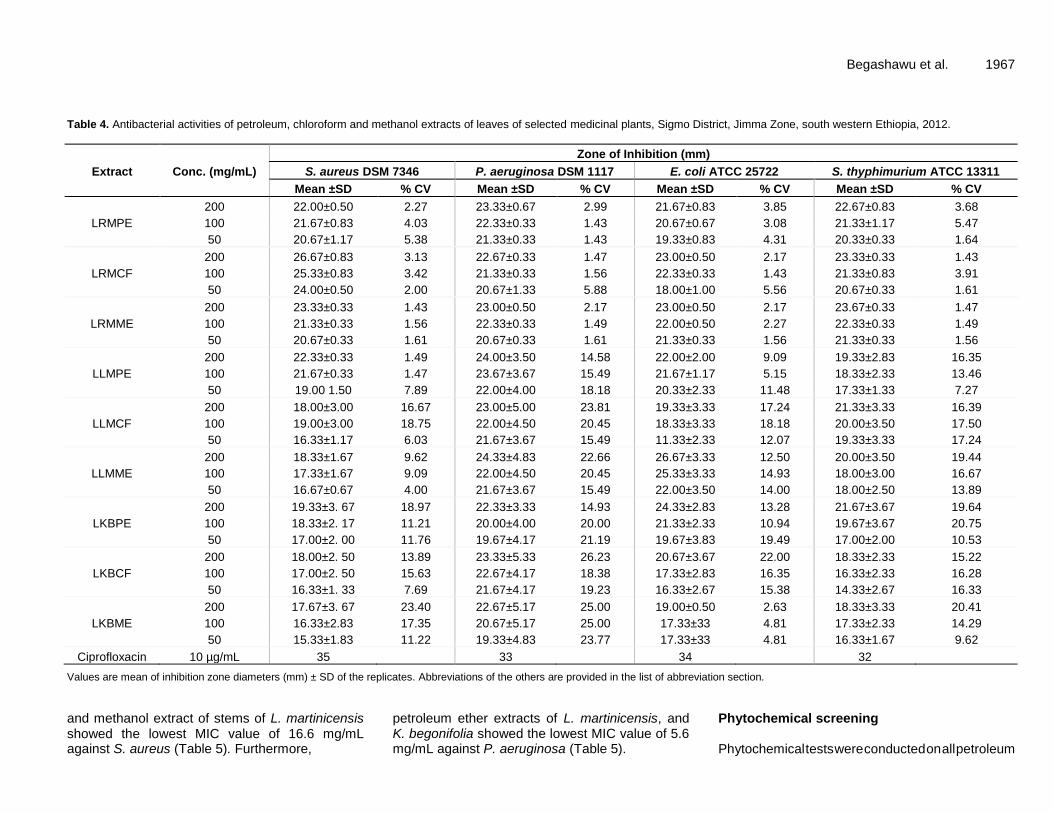
Begashawu et al. 1967 Table 4. Antibacterial activities of petroleum, chloroform and methanol extracts of leaves of selected medicinal plants, Sigmo District, Jimma Zone, south western Ethiopia, 2012.
Extract Conc. (mg/mL)
Zone of Inhibition (mm)
S. aureus DSM 7346 P. aeruginosa DSM 1117 E. coli ATCC 25722 S. thyphimurium ATCC 13311
Mean ±SD % CV Mean ±SD % CV Mean ±SD % CV Mean ±SD % CV
LRMPE
200 22.00±0.50 2.27 23.33±0.67 2.99 21.67±0.83 3.85 22.67±0.83 3.68
100 21.67±0.83 4.03 22.33±0.33 1.43 20.67±0.67 3.08 21.33±1.17 5.47
50 20.67±1.17 5.38 21.33±0.33 1.43 19.33±0.83 4.31 20.33±0.33 1.64
LRMCF
200 26.67±0.83 3.13 22.67±0.33 1.47 23.00±0.50 2.17 23.33±0.33 1.43
100 25.33±0.83 3.42 21.33±0.33 1.56 22.33±0.33 1.43 21.33±0.83 3.91
50 24.00±0.50 2.00 20.67±1.33 5.88 18.00±1.00 5.56 20.67±0.33 1.61
LRMME
200 23.33±0.33 1.43 23.00±0.50 2.17 23.00±0.50 2.17 23.67±0.33 1.47
100 21.33±0.33 1.56 22.33±0.33 1.49 22.00±0.50 2.27 22.33±0.33 1.49
50 20.67±0.33 1.61 20.67±0.33 1.61 21.33±0.33 1.56 21.33±0.33 1.56
LLMPE
200 22.33±0.33 1.49 24.00±3.50 14.58 22.00±2.00 9.09 19.33±2.83 16.35
100 21.67±0.33 1.47 23.67±3.67 15.49 21.67±1.17 5.15 18.33±2.33 13.46
50 19.00 1.50 7.89 22.00±4.00 18.18 20.33±2.33 11.48 17.33±1.33 7.27
LLMCF
200 18.00±3.00 16.67 23.00±5.00 23.81 19.33±3.33 17.24 21.33±3.33 16.39
100 19.00±3.00 18.75 22.00±4.50 20.45 18.33±3.33 18.18 20.00±3.50 17.50
50 16.33±1.17 6.03 21.67±3.67 15.49 11.33±2.33 12.07 19.33±3.33 17.24
LLMME
200 18.33±1.67 9.62 24.33±4.83 22.66 26.67±3.33 12.50 20.00±3.50 19.44
100 17.33±1.67 9.09 22.00±4.50 20.45 25.33±3.33 14.93 18.00±3.00 16.67
50 16.67±0.67 4.00 21.67±3.67 15.49 22.00±3.50 14.00 18.00±2.50 13.89
LKBPE
200 19.33±3. 67 18.97 22.33±3.33 14.93 24.33±2.83 13.28 21.67±3.67 19.64
100 18.33±2. 17 11.21 20.00±4.00 20.00 21.33±2.33 10.94 19.67±3.67 20.75
50 17.00±2. 00 11.76 19.67±4.17 21.19 19.67±3.83 19.49 17.00±2.00 10.53
LKBCF
200 18.00±2. 50 13.89 23.33±5.33 26.23 20.67±3.67 22.00 18.33±2.33 15.22
100 17.00±2. 50 15.63 22.67±4.17 18.38 17.33±2.83 16.35 16.33±2.33 16.28
50 16.33±1. 33 7.69 21.67±4.17 19.23 16.33±2.67 15.38 14.33±2.67 16.33
LKBME
200 17.67±3. 67 23.40 22.67±5.17 25.00 19.00±0.50 2.63 18.33±3.33 20.41
100 16.33±2.83 17.35 20.67±5.17 25.00 17.33±33 4.81 17.33±2.33 14.29
50 15.33±1.83 11.22 19.33±4.83 23.77 17.33±33 4.81 16.33±1.67 9.62
Ciprofloxacin 10 µg/mL 35 33 34 32
Values are mean of inhibition zone diameters (mm) ± SD of the replicates. Abbreviations of the others are provided in the list of abbreviation section.
and methanol extract of stems of L. martinicensis showed the lowest MIC value of 16.6 mg/mL against S. aureus (Table 5). Furthermore,
petroleum ether extracts of L. martinicensis, and K. begonifolia showed the lowest MIC value of 5.6 mg/mL against P. aeruginosa (Table 5).
Phytochemical screening Phytochemical tests were conducted on all petroleum

1968 Afr. J. Microbiol. Res.
Table 5. Antibacterial activity of petroleum, chloroform and methanol extracts of stems of selected medicinal plants, Sigmo District, Jimma Zone, South Western Ethiopia, 2012.
Extract Conc.
(mg/mL)
Zone of Inhibition (mm)
S. aureus DSM 7346 P. aeruginosa DSM 1117 E. coli ATCC 25722 S. typhimurium ATCC 13311
Mean ± SD %CV Mean ± SD %CV Mean ± SD %CV Mean ± SD % CV
SRMPE
200 22.00±0.50 2.27 22.33±0.67 2.99 21.67±0.83 3.85 22.67±0.33 1.47
100 20.67±0.83 4.03 23.33±0.33 1.43 21.67±0.67 3.08 21.00±00 0.00
50 21.67±1.17 5.38 23.33±0.33 1.43 19.33±0.83 4.31 20.33±0.33 1.64
SRMCF
200 26.67±0.83 3.13 22.67±0.33 1.47 23.00±0.50 2.17 23.33±0.33 1.43
100 24.33±0.83 3.42 21.33±0.33 1.56 23.33±0.33 1.43 22.00±1.00 4.55
50 25.00±0.50 2.00 22.67±1.33 5.88 18.00±1.00 5.56 21.00±0.50 2.38
SRME
200 22.67±0.67 2.94 23.00±0.50 2.17 22.33±0.83 3.73 22.67±0.33 1.47
100 21.67±0.33 1.54 21.00±0.50 2.38 21.67±0.83 3.85 21.67±0.67 3.08
50 20.00±0.00 0.00 21.33±0.83 3.91 20.33±0.33 1.64 21.33±0.83 3.91
SLMPE
200 28.67±0.83 2.91 25.33±0.33 1.32 26.00±0.00 0.00 27.67±0.33 1.20
100 28.00±1.00 3.57 24.00±1.00 4.17 24.67±0.33 1.35 27.67±0.33 1.20
50 29.33±0.67 2.27 27.00±0.50 1.85 28.00±0.00 0.00 29.33±1.67 5.68
SLMCF
200 26.00±1.00 3.85 26.33±1.33 5.06 26.00±0.50 1.92 26.33±0.83 3.16
100 26.67±1.83 6.88 25.33±1.67 6.58 26.67±1.33 5.00 24.33±2.17 8.90
50 23.67±1.83 7.75 26.67±1.83 6.88 23.67±0.83 3.52 22.67±1.33 5.88
SLMME
200 27.67±0.33 1.20 25.33±0.33 1.32 25.67±0.83 3.25 25.00±2.50 10.00
100 25.33±0.33 1.32 24.67±0.33 1.35 24.00±1.00 4.17 20.67±2.83 13.71
50 25.00±1.00 4.00 25.67±1.33 5.19 28.00±1.50 5.36 24.33±4.17 17.12
SKBPE
200 28.33±0.83 2.94 28.00±1.50 5.36 28.33±0.83 2.94 29.67±1.33 4.49
100 30.00±1.50 5.00 27.00±1.50 5.56 31.00±0.50 1.61 28.00±1.00 3.57
50 28.67±1.67 5.81 28.67±1.67 5.81 29.00±0.50 1.72 30.33±0.33 1.10
SKBCF
200 27.67±0.83 3.01 27.00±1.50 5.56 25.67±0.83 3.25 23.33±1.17 5.00
100 26.00±1.00 3.85 26.00±2.00 7.69 26.00±1.50 5.77 22.33±1.17 5.22
50 27.33±1.33 4.88 27.67±1.67 6.02 24.67±0.33 1.35 21.33±0.83 3.91
SKBME
200 26.00±0.00 0.00 25.67±1.33 5.19 24.33±0.33 1.37 23.33±1.67 7.14
100 23.67±0.83 3.52 27.00±1.50 5.56 24.33±0.33 1.37 24.67±2.33 9.46
50 25.00±0.50 2.00 27.00±0.50 1.85 25.67±0.33 1.30 23.00±3.00 13.04
Ciprofloxacin 35 33 34 32
Values are mean of inhibition zone diameters (mm) ± SD of the replicates. Abbreviations for the first column are given in the list of abbreviations section.
ether, chloroform and methanol extracts and the findings were summarized and presented below
(Tables 6). All the plant extracts displayed the presence of many bioactive phytochemical
compounds mainly alkaloids, tannins, flavonoid, saponins, steroids and cardiac glycosides (Table

Begashawu et al. 1969 Table 6. MIC and MBC values (mg/mL) of stem extracts against test strains, Jimma Zone, South west, Ethiopia (2012).
Extract S. aureus DSM 7346 P. aeruginosa DSM 1117 E. coli ATCC 25722 S. typhimurium ATCC 13311
MIC MBC MIC MBC MIC MBC MIC MBC
SRMPE 5.6 16.6 16.6 50 16.6 50 5.6 16.6
SRMCF 16.6 50 16.6 50 16.6 50 16.6 50
SRMME 16.6 50 16.6 50 16.6 50 16.6 50
SRMME 5.6 16.6 5.6 16.6 16.6 50 5.6 16.6
SLMCF 5.6 16.6 16.6 50 16.6 50 16.6 50
SLMME 16.6 50 16.6 50 16.6 50 16.6 50
SKBPE 5.6 16.6 5.6 16.6 16.6 50 5.6 16.6
SKBCF 5.6 16.6 16.6 50 16.6 50 16.6 50
SKBME 5.6 16.6 16.6 50 16.6 50 16.6 50
MIC, Minimum Inhibitory Concentration; MBC, Minimum Bactericidal Concentration; Abbreviations of the others are provided in the list of abbreviation section.
6) except for absence of cardiac glycosides in Ranunculus multifidus. DISCUSSION In most developing countries where people are living under poor hygienic conditions, the possibility of contracting infectious diseases could be high. For several reasons, including low access to modern health care systems, high cost of the available commercial drugs and trust developed over long years of traditional practices, large proportion of a population in rural community prefer using traditional medicinal plants in order to manage infectious diseases of different sorts (Giday et al., 2003; Bacha et al., 2016). The current study area is not exceptional. Extensive review made on the status of use of traditional medicinal plants in different parts of Africa (Sofowora, 1993) also indicated that medicinal plants have been widely used among African communities to treat different types of bacterial diseases. Actually, about 85% of world population uses herbal medicines for prevention and treatment of diseases, and the demand is still on the rise both in developed and developing countries (Abramov, 1996; Abera, 2014). Reliance on traditional medicine could be even higher among the pastoral and agro-pastoral communities whose environment were reported to be rich in many of effective medicinal plants (Belayneh et al., 2012).
The survey data shown that knowledge on traditional practices has been acquired from parents to off springs, hence through generations, mainly from elderly religious
men (80%), parents and close relatives (20%) or very rarely through random trial and error method. In agreement with our observation, Tolasa (2007) reported that 91 and 9% of knowledge on the use of traditional medicine were acquired from parents/relatives and through trial and error, respectively, as observed in other part of Ethiopia. One of the major challenges confronting the sustainability of transfer of traditional knowledge and
good practices of the use of traditional medicine is the tendency of keeping the traditional knowledge secrete from others, hence creating a gap in the flow of knowledge across generations. The confidentiality of traditional medical practice is a common phenomenon in other parts of the country, too (Giday et al., 2003), mainly for business interest. In agreement with our observation, the transfer of knowledge to people outside the family circle among the Zay people of Ethiopia takes place only on substantial payment (Giday et al., 2003). When such practice is accompanied by the new generation’s high dependence on modern medication (because of lack of ample information and practical skill), the future fates of traditional medication could be questionable when traditional medicines still have significant contribution to the health care system of larger part of the world population.
In the study area, more than ten traditional medicinal plants have been used for treatment of various ailments including toothache, infection of the GIT, and related diseases. However, the community’s preference to three of the traditional medicinal plants namely, R. multifidus, L. martinicensis, and K. begonifolia, were significantly high with preference rating of 66.6, 56.6 and 53.3%, respectively. The observed high reliance on the three traditional medicinal plants was scientifically sounding as all the three plants were observed to possess various phytochemical compounds of medicinal values including alkaloids, tannins, saponnins, flavonoids, steroids, terpenoids and cardiac glycosides. Flavonoids are found in almost all parts of plants (Ahmed et al., 2015; Cushnine and Lamb, 2005) protect the plant from insect pests and ultraviolet radiations besides imparting color to flowers and fruits. In addition to their antioxidant, anti-depressant and anti-inflammatory role in human body, flavonoids act as bactericidal and bacteriostatic by damaging cytoplasmic membrane, inhibiting energy metabolism and synthesis of nucleic acids in different microorganisms (Ahmed et al., 2015). Earlier report (Kaya et al., 2010) also showed that flavonoids are among the major consti-

1970 Afr. J. Microbiol. Res. tuents isolated from the Ranunculus species and have been considered as the main components of the same plants antioxidant activity besides its antibacterial activity against S. aureus, Bacillus subtilis, B. cereus, E. coli, Helicobacter pylori, P. aeruginosa, P. fluorescens, Enterobacter aerogenes (Ahmed et al., 2015) and infections of Methicillin-resistant S. aureus strains (Alcaraz et al., 2000). Tannins, the other common phytochemical isolated from traditional medicinal plants, inhibit plasma coagulation by S. aureus (Akiyama et al., 2001) and also form chelate with metal ions. The possible antimicrobial mechanisms of tannins could be: induction of complexation with enzymes or substrates, act on the membranes of microorganisms; and toxicity due to complexation with metal ions (Akiyama et al., 2001).
The emergence of multiple drug resistant pathogens, including Multi-Drug Resistant TB (MDR TB), Methicillin-Resistant Staphylococcus aureus (MRSA), and Vancomycin-Resistant Entero-cocci (VRE), is making the treatment and control of infectious diseases more difficult (Bacha et al., 2016), with over 480 000 new cases of MDR-TB only in 2013 (Teklay et al., 2013). According to recent review made in Ethiopia (Mogese et al., 2014), some of the antibiotics have become virtually useless, alarming for the urgent need to apply antibiotic restriction policies as well as measures to prevent further spread of resistant clones. Thus, the pressure due to drug resistance necessitates search for novel antimicrobial substances from plants as the development of resistance to bioactive substance from plants is low, if any (Bacha et al., 2016). Although not evaluated against resistance bacterial strains, our extract could have potent activity against resistance strains as it did against the sensitive test strains.
The antibacterial activity of chloroform extract of leaves of R. multifidus (LRMCF) (conc. 200 mg/mL) against S. aureus (IZ, 26.67 ± 0.83) was comparable to the activity of commercial antibiotic ciprofloxacin (IZ, 35 mm). The same extract displayed almost similar activity (IZ, 25.33 ± 0.83) even at lower concentration (100 mg/mL) against the same strain, with minimal activity against E. coli (IZ, 18.00 ± 1.00) at the lowest concentration (50 mg/mL) used in this study. Likewise, report from different parts of the world (including Turkey and Lebanon) indicated strong antibacterial activities of extracts of Ranunculus bulbosus against S. aureus (Didry et al., 2006), Ranunculus myosuroudes against E. coli and S. typhi with 88.8% susceptibility of the test strains (Barbour et al., 2004), and Ranunculus sceleratus against S. aureus and E. coli (Bissa and Bohra, 2012).
The activities of methanol extract of leaves of L. martinicensis (LLMME) against E. coli at relatively higher concentration (200 mg/mL) (IZ, 26.67 ± 3.33 mm) and even lower concentration (100 mg/mL) (25.33 ± 3.33 mm) were very promising as it had comparable activity to that of commercial antibiotic ciprofloxacin (IZ, 34 mm) against the same tested strain. However, chloroform extract of
leaves of L. martinicensis against S. aureus at relatively lowest concentration (50 mg/mL) had intermediate activity with mean inhibition zone diameter of 16.33 ± 1.17 mm. Besides its antimicrobial activities, L. martinicensis has been used as repellant of mosquito due to minty odor (Imam and Tajuddeen, 2013; Muhammad et al., 2012). The presence of different phytochemical compounds could be responsible for its antibacterial activities as phyto-chemical screening of the leaves extract already revealed the presence of flavonoids, alkaloids and volatile oils (Muhammad et al., 2012).
Chloroform extract of leaves of K. begonifolia also revealed activity (conc. 100 mg/mL) against P. aeruginosa (IZ, 22.67 ± 4.17 mm) followed by petroleum extract (IZ, 22.33 ± 3.33 mm) against S. typhimurium
with activity closer to standard commercial antibiotic ciprofloxacin (IZ, 32 mm) . Leaves of plants are among the commonly used plant parts although there are contradictory reports in this regards (Mesfin et al., 2009; Teklay et al., 2013). Of 114 medicinal plant species identified being used to treat 47 human and 19 livestock diseases (Teklay et al., 2013), leaves parts were the most commonly used section accounting for almost 50% of the total sample analyzed. To the contrary, roots were reported the most commonly used plant parts (35.8%) among seventy-two plant species documented for having medicinal value as reported from Wonago district of South Nations and Nationalities Peoples Region (SNNPR), South Ethiopia (Mesfin et al., 2009).
Similar to leaves crude extracts, the stems extract of the three medicinal plants (R. multifidus, L. martinicensis, K. begonifolia) were evaluated for their antibacterial activities and the results revealed presence of moderate to high activities. Accordingly, chloroform extracts of stems of R. multifidus displayed comparable activity against S. aureus both at 200 mg/mL (IZ, 26.67 ± 0.83 mm) and 50 mg/mL (IZ, 25.00 ± 0.50 mm) although it had lower activity against E. coli (IZ, 18.00 ± 1.00 mm) at the same lower concentration (50 mg/mL). Our findings are in corroboration with related activity observed in roots of Ranunculus repens (Noor et al., 2006) assessed in Pakistan and whole plants of Ranunculus marginatus var. trachycarpus and Ranunculus sprunerianus from Turkey (Noor et al., 2006). The later authors reported that the extracts had both antibacterial as well as antioxidant activities, with the antioxidant activity having positive correlation with the total phenolic and flavonoid contents of the extracts (Kaya et al., 2010).
Likewise, petroleum extracts of stems of L. martinicensis ( SLMPE) revealed strong activity even at lowest concentration (50 mg/mL) against S. aureus, S. typhimurium, E. coli and P. aeruginosa with mean inhibition zone diameter of 29.33 ± 0.67, 29.33 ± 1.67, 28.00 ± 0.00 and 27.00 ± 0.50, respectively. The same extract also showed amazingly maximum activity against S. aureus which (IZ, 28.67 ± 0.83 and 28.00 ± 1.00 mm) at 200 and 100 mg/mL, respectively. As observed from

the above activity, the extract had broad spectrum with inhibitory activity against both Gram positive and Gram negative bacteria as supported by earlier observation made by Pandey et al. (2011). Exceptionally, high activities were observed in petroleum ether extracts of the stems of K. begonifolia (SKBPE) against both Gram positive and Gram negative bacteria as recorded for S. aureus (28.33 ± 0.83 - 30.00 ± 1.50 mm), P. aeruginosa (27.00 ± 1.50 - 28.67 ± 1.67 mm), E. coli (28.33 ± 0.83 - 31.00 ± 0.50 mm) and S. typhimurium (28.00 ± 1.00 - 30.33 ± 0.33) for the three concentrations of the extract (50-200 mg/mL). Both Gram positive and Gram negative bacteria were sensitive to the extracts. In contrast to this finding, some of the earlier study (Pandey et al., 2011) reported that many of the traditional medicinal plants had lesser activities against Gram-negative bacteria as compared to their activities in Gram-positive.
The minimum and maximum MIC values observed in all the three stems extracts using the different solvents were 5.6 mg/mL (in S. aureus, P. aeruginosa, and S. thyphimurium) and 16.6 mg/mL (E. coli). Likewise, most of the isolates had MBC values of 16.6 mg/mL with the exception of 50 mg/mL of all the extracts against E. coli. Usually, plant extracts are bacteriostatic at relatively lower concentrations and bactericidal at higher concentrations (Jaya et al., 2008) although antibacterial activities could depend on the actual concentration of active compounds in the crude extract (Mazumder et al., 2006). In contrary to the current observation, the MIC values of extracts of two other species Raninculus (R. marginatus var. trachycarpus and R. sprunerianus) were reported to be between 128 and 256 μg/mL (Kaya et al., 2010). In the same report, the maximum inhibition zones ranged between 7 and 12 mm, values much lesser than what we observed (19.33-23.67mm) for extracts of Ranunculus multifidus (inclusive of data on stems and leaves extracts). The presence of many bioactive phytochemical compounds mainly alkaloids, tannins, flavonoids, saponins, steroids and cardiac glycosides could be accounted to the antimicrobial activities observed in many of the extracts assessed in this study either individually or possibly in combinations. Likewise, many of the earlier reports made on antibacterial activities of various medicinal plants linked the observed activities to many of these phytochemicals (CSA, 2005; Bissa and Bohra, 2012). The optimal effectiveness of medicinal plants may not be due to one main active constituent, but to the combined action of different compounds originally present in the plants (Farombi. 2003; Gonzalez et al., 1994).
The activities of extracts of the same plant could vary depending on the polarity of solvents used for extraction, the concentration of active compounds. In the present study, petroleum ether extract of stems of K. begonifolia revealed remarkably high inhibition on both Gram positive and Gram negative with significantly high mean inhibition zone diameter (31.00 ± 0.50). Generally, the antibacterial
Begashawu et al. 1971 activities of medicinal plants against the test strains showed that the crude preparations of traditional medicinal plants are among the candidate resources for novel antimicrobial agents and calls for further strengthening of the search for alternative potent antimicrobial agents from the available pool of resources.
Conclusions
The traditional medicinal plants evaluated in the current study have shown promising activities against both Gram negative and Gram positive bacteria. The highest antimicrobial activities were recorded for petroleum ether extract of Kosteletzkya begonifolia stems against S. aureus [Inhibition Zone diameter (IZ), 28.3-30 mm], P. aeruginosa (IZ: 27 to 28.67 mm), E. coli (IZ: 28.3 to 31 mm) and S. typhimurium (IZ, 28 to 30.3)]. The extract displayed activity significantly closer to that of the control antibiotics, ciprofloxacin (IZ, 30 to 35 mm). The finding strengthens the fact that traditional
plants could represent
new sources of antibacterial substances with stable,
biologically active components. As the bioactive substances present in the extracts of the study plants justify the rationale for the use of the same plants in traditional medicine, isolation, purification and structural elucidation of the bioactive constituents are recommended.
Abbreviations
LRMPE, Leaf of Ranunculus multifidus petroleum ether extract; LRMCF, leaf of R. multifidus chloroform extract; LRMME, leaf of R. multifidus methanol extract; LLMPE, leaf of Leucas martinicensis (Jacq.) R. Br petroleum ether extract; LLMCF, leaf of L. martinicensis(Jacq.) R. Br chloroform extract; LLMME, leaf of L. martinicensis (Jacq.) R. Br methanol extract; LKBPE, leaf of Kosteletzkya begonifolia (Ulbr.) Ulbr petroleum ether extract; LKBCF, leaf of K. begonifolia (Ulbr.) Ulbr chloroform extract; LKBME, leaf of K. begonifolia (Ulbr.) Ulbr methanol extract; SRMPE, stem of Ranunculus multifidus petroleum ether extract; SRMCF, stem of R. multifidus chloroform extract; SRMME, stem of R. multifidus methanol extract; SLMPE, stem of Leucas martinicensis (Jacq.) R. Br petroleum ether extract; SLMCF, stem of L. martinicensis (Jacq.)R. Br chloroform extract; SLMME, stem of L. martinicensis (Jacq.)R. Br methanol extract; SKBPE, stem of Kosteletzkya begonifolia (Ulbr.) Ulbr petroleum ether extract; SKBCF, stem of K. begonifolia (Ulbr.) Ulbr chloroform extract; SKBME, stem
of K. begonifolia (Ulbr.) Ulbr methanol extract.
Conflict of interests
The authors have not declared any conflict of interests.
Authors' contributions
TB was fully involved in all phases of the study including

1972 Afr. J. Microbiol. Res. designing of the study, data collection, data analysis and write up. YT supervised data collection and involved in laboratory work, and KB designed the study, supervision the study both in field and laboratory, data analysis and interpretation, and preparation of the manuscript for publication.
ACKNOWLEDGEMENT
The authors would like to thank Jimma University for financial support.
REFERENCES
Abebe D (2011). The Role of Medicinal Plants in Healthcare Coverage of Ethiopia, the possible integration. In: Conservation and Sustainable Use of Medicinal Plants in Ethiopia. Proceeding of The National Workshop on Biodiversity Conservation and Sustainable Use of Medicinal Plants in Ethiopia, held from 28 April-01 May 1998, pp. 6- 21.
Abera B (2014). Medicinal plants used in traditional medicine by Oromo people, Ghimbi District, Southwest Ethiopia. J. Ethnobiol. Ethnomed. 10:40
Abramov V (1996). Traditional medicine. World Health Org. 134:1-3. Ahmed A, Kaleem M, Ahmed Z, Shafiq H (2015). Therapeutic potential
of flavonoids and their mechanism of action against microbial and viral infections - A review. Food Res. Int. 77: 221-235
Akiyama H, Fujii K, Yamasaki O, Oono T, and Iwatsuki K (2001). Antibacterial action of several tannins against Staphylococcus aureus, J. Antimicrobial. Chemother. 48(4):487-491
Alcaraz L, Blanco S, Puig O, Tomas F, Ferretti F (2000): Antibacterial activity of flavonoids against methicillin-resistant Staphylococcus aureus strains, J. Theor. Biol. 205(2):231-240
Bacha K, Tariku Y, Gebreyesus F, Zerihun S, Mohammed A, Weiland-Bräuer N Schmitz R A, Mulat M (2016). Antimicrobial and anti-Quorum Sensing activities of selected medicinal plants of Ethiopia: Implication for development of potent antimicrobial drugs. BMC Microbiol. 16:139
Barbour EK, Al Sharif M, Sagherian VK, Habre AN,Talhouk RS and Talhouk SN (2004). Screening of selected indigenous plants of Lebanon for their antimicrobial activity. J. Ethnopharmacol. 93:1-7.
Belayneh A, Asfaw Z, Demissew S, Bussa NF (2012). Medicinal plants potential and use by pastoral and agro-pastoral communities in Erer Valley of Babile Wereda, Eastern Ethiopia. J. Ethnobiol. Ethnomed. 8:42.
Bissa SH, Bohra A (2012). Evaluation of Antibacterial Potential of Ranunculus sceleratus. Bot. Res. Intern. 5:10-13.
Branham LA, Carr MA, Scott CB, Callaway TR (2005). E. coli O157 and Salmonella spp. in white-tailed deer and livestock. Curr. Issues Intest. Microbiol. 6:25-29.
Canter PH, Thomas WH, and Ernst E (2005). Bringing medicinal plants into cultivation: opportunities and challenges for biotechnology. Trends Biotechnol. 23:180-185.
Central Statistics Agency of Ethiopia (CSA) (2005). National Statistics, Tables B.3 and B.4. 2005
Cunningham AB (1993). African Medicinal Plants: Setting priorities at the interface healthcare between conservation and primary health care. (Sample, A. Ed.). People and Plants Working Paper. Paris, UNESCO. pp. 1-50.
Cushnine T, Lamb AJ (2005). Antimicrobial activity of flavonoids. Int. J. Antimicrob. Agents 26(5):343-356.
Dabassa A, Bacha K (2012). The prevalence and antibiogram of Salmonella and Shigella Isolated from abattoir, Jimma town, South West Ethiopia. Int. J. Pharm. Biol. Res. 3(4):143
Didry N, Dubrenil L, and Pinkas M (2006). Microbiological properties of protoanemonin .isolated from Ranunculus bulbosus. Phytother. Res. 7:21-24.
Farombi EO (2003). African Indigenous Plant with Chemotherape utic Potentials and biotechnological Approach to the production of
bioactive prophylacudiestic agents. Afr. J. Biotechnol. 2(12):662-671 Giday M, Asfaw Z, Elmqvist T, Woldu Z. (2003) An ethnobotanical
study of medicinal plants used by the Zay people in Ethiopia. J. Ethnobiol. Pharm. 85:43-52.
Gonzalez A, Moujir G, Bazzocchi I, Correa IL and Guptha MD (1994). Screening o antimicrobial and cytotoxic activities of Panamanian plants. Phytomedicine 1:149-153.
Imam TS, Tajuddeen UM (2013). Qualitative phytochemical screening and larvicidal potencies of ethanolic extracts of five selected macrophyte species against Anopheles mosquitoes (diptera: culicidae), J. Res. Environ. Sci. Toxicol. 2(6):121-125.
Jaya Prakash Goud M, Komraiah A. Narasimha Rao K, Ragan A, Raju VS and Singara Charya MA (2008). Antibacterial activity of some folklore medicinal plants from south India. Afr. J. Tradit. Complement. Altern. Med. 4:421-426.
Kamatenesi M, Oryem-Origa H (2007). Medicinal plants used to induce labor during childbirth in Western Uganda. J. Ethnopharmacol. 109:1-9
Kaya GI, Somer NU, Konyalioglu S, Yalcin HT, Yavasoglu NU, Sarikaya B and. Onur MA (2010). Antioxidant and antibacterial activities of Ranunculus marginatus var. trachycarpus and Ranunculus sprunerianus. Turk. J. Biol. 34:139-14
Khulbe K, Sati SC (2009). Antibacterial activity of Boenninghausenia albiflora Reichb. (Rutaceae). Afr. J. Biotechnol. 8:6346-6348
Komuraiah A, Bolla K, Rao KN, Ragan A, Raju VS and Singara Charya, MA (2009). Antibacterial studies and phytochemical constituents of South Indian Phyllanthus species. Afr. J. Biotechnol. 8:4991-4995.
Lewington A (1993). Medicinal plants and plant extracts: A Review of their Importation into Europe. Cambridge, UK. P 92.
Mackeen MM, Ali AM, El-Sharkawy SH, Manap MY, Salleh KM, Lajis N H, and Kawazu K (1997). Antimicrobial and Cytotoxic Properties of Some Malaysian Traditional Vegetables (Ulam). Int. J. Pharmacogn., 35(3):174-178
Mazumder A, Mahato A, and Mazumder R (2006). Antimicrobial potentiality of Phyllanthus amarus against drug resistant pathogens. Nat. Prod. Res. 20:323-326
Mesfin F, Demissew S, Teklehaymanot T (2009). An ethnobotanical study of medicinal plants in Wonago Woreda, SNNPR, Ethiopia, J Ethnobiol. Ethnomed. 5:28
Mogese F, Endris M, Mulu A, Tessema B, Belyhun Y, Shiferaw Y, Huruy K, Unakal C, Kassu A (2014). The growing challenges of antibacterial drug resistance in Ethiopia, J. Global Antimicrob. Resist. 2(3):148-154.
Muhammad S, Fatima A, Yahaya MM (2012). The Phytochemical Components of Leucas Martini-censis that Cause Repellence of Adult Mosquito. Int. J. Mod. Bot. 2(1):1-5.
Mulat M, Chali K Tariku Y, Bacha K (2015). Evaluation for in vitro antibacterial activities of selected medicinal plants against food-borne pathogens. Int. J. Pharma. Sci. Rev. Res. 32 (2):45-50.
Noor W, Gul I, Ali, Chaudhary MI (2006). Isolation and antibacterial activity of compounds from Ranunculus repens L. J. Chem. Soc. Pak. Microbiol. 42:361-363..Owolabi J, Omogbai EK and Obasuyi O. (2007). Antifungal and antibacterial activities of the ethanolic and aqueous extract of Kigella africana (Bignoniaceae) stem bark. Afr. J. Biotechnol. 6:1677-1680
Pandey P, Mehta A, and Hajra S (2011). Evaluation of Antimicrobial Activity of Ruta graveolens Stem Extracts by Disc Diffusion Method. J. Philos., 3:92-95.
Sofowora A (1993). Medicinal Plants and Traditional Medicines in Africa. Chic Hester John, Willey & Sons, New York. P 256.
Teklay A, Abera B, Giday M (2013). An ethnobotanical study of medicinal plants used in Kilte Awulaelo District, Tigray Region of Ethiopia. J. Ethnobiol. Ethnomed. 9(1):65.
Tesema T, Mirutse G, Nigusu A (2002). National Biodiversity Strategy and Action Plan Project. Resource base of medicinal plants of Ethiopia, first phase report, Addis Ababa, Ethiopia
Tolasa E (2007). Use, Treat and Conservation of Traditional Medicinal Plants by Indigenous People in Gimbi Woreda, Western Wollega, West Ethiopia. M.Sc. Thesis, Addis Ababa University, Ethiopia.
Trease GE, Evans WC (1998). Pharmacognosy. Brailliar Tiridel and Macmillian Publishers, London.
WHO (2002). Traditional medicine: Growing Needs and Potentials, Geneva.

Vol. 10(46), pp. 1973-1982, 14 December, 2016
DOI: 10.5897/AJMR11.1650
Article Number: 25705E362051
ISSN 1996-0808
Copyright © 2016
Author(s) retain the copyright of this article
http://www.academicjournals.org/AJMR
African Journal of Microbiology Research
Full Length Research Paper
Combination antibiotic-phytochemical effects on resistance adaptation in Staphylococcus aureus
Shuchi Arora1*, Bhone Myint Kyaw2 and Chu Sing Lim2
1School of Chemical and Biomedical Engineering, Nanyang Technological University, 62 Nanyang Drive, 637459,
Singapore. 2School of Mechanical and Aerospace Engineering, Nanyang Technological University, 50 Nanyang Avenue, 639798,
Singapore.
Received 1 November 2016; Accepted 2 December, 2016
Emergence and rapid spread of antibiotic resistance has become one of the leading causes of treatment failures in case of bacterial infections. Antibiotic combinations are generally used to enhance the overall efficacy of therapy with the aim to generate synergistic outcomes. It further helps in reduction of total antibiotic dosage. Phytochemicals are known to have multiple bacterial targets that modulate or modify resistance in bacteria. In the present study, a microchannel-based device and monitoring system was used to demonstrate and investigate short and long term effects of antibiotic-phytochemical combinations in different proportions on Staphylococcus aureus as test organism. Novel and unconventional combinations of antibiotic ciprofloxacin with the phytochemicals, quercetin, rutin, protocatechuic acid and ethyl gallate, were tested. Based on the experimental results, the strains exposed the antibiotic, generated resistant strains in four days, with 8 to 64 fold increase in their minimum inhibitory concentration (MIC) from the parent strain. The strains exposed to antibiotic-phytochemical combinations, however, showed no resistance causing mutations. The results were verified by standard laboratory practices such as disk-diffusion, mutation frequency, population profiling and molecular studies on the exposed strains. The phytochemicals were able to potentiate antibiotic activity; thereby, increasing the antibacterial efficacy and time span of the treatment with a common antibiotic. Key words: Antibiotic combination therapy, antibiotic resistance, ciprofloxacin, microfluidic-device, phytochemical, potentiation, Staphylococcus aureus.
INTRODUCTION Traditionally, phytochemicals and plant extracts have been used in medicine for centuries to treat a number of
diseases and disorders that include high blood pressure, pain, asthma, depression, viral infection, cancer, diabetes,
*Corresponding author. E-mail: [email protected]. Tel: +65-9349-3212. Fax: +65-6792-4062.
Author(s) agree that this article remains permanently open access under the terms of the Creative Commons Attribution
License 4.0 International License

1974 Afr. J. Microbiol. Res. etc (Cragg and Newman, 2005; Fiore et al., 2008; Sarris, 2007). Their intrinsic antibacterial property has been harnessed in a number of occasions to treat bacterial infections (Cushnie and Lamb, 2005; Gibbons, 2004; Raos and Recio, 2005). However, with discovery of antibiotics and other modern medicines, the use of herbal remedies and phytochemicals as therapeutic agents has generally reduced. On the other hand, antibiotic discovery rate has drastically reduced in recent years (Barrett, 2005; Bax et al., 2000; Norrby et al., 2005).
Phytochemicals have been under close and careful scrutiny by many researchers for their antibacterial properties (Alviano and Alviano, 2009; Cowan, 1999; Lewis and Ausubel, 2006; Raos and Recio, 2005; Shibata et al., 2005). These have been used in combination with antibiotics to treat multi-drug resistant or extensively drug resistant (MDR and XDR, respectively) bacterial species. Increasing resistance in bacteria to old and existing antibiotics (Grundmann et al., 2006; Levy and Marshall, 2004), forms the basis for such investigations. The pathogen is either reported to become totally resistant to the drug or is susceptible at a higher dose (8 folds or higher). Particularly, Staphylococcus aureus has been reported as one of the superbugs due to its resistance to many classes of antibiotics (Lindsay and Holden, 2004; Livermore, 2000). Alternative and newer antibacterial solutions are hence, constantly required to check the ever growing resistance in bacterial populations (Bax et al., 2000).
To overcome the fast pace of adaptive resistance in pathogenic bacteria, combination therapy is of significant interest (Cottarel and Wierzbowski, 2007; Moellering Jr, 1983). Screening of drugs in combination with other drugs, and more recently, with herbal-based extracts has thus, become increasingly popular (Cottarel and Wierzbowski, 2007; Sibanda and Okoh, 2007). The main interest behind these studies is to find beneficial combinations that typically occur at sub-minimum inhibitory concentration (MIC) levels of the antibiotic (Sakharkar et al., 2009). Combination of an antibiotic with an appropriate phytochemical may help to reduce the antibiotic dosage as well as harness the benefits of plant-derived antibacterials (Kyaw et al., 2011). They are believed to modify and/or modulate bacterial internal resistance and potentiate the effect of antibiotics when used in combination (Sibanda and Okoh, 2007). Generally, these may thus, increase the efficacy of the treatment by inhibiting the phenomenon of resistance causing elements, such as mutation of genes encoding target enzymes, proteins and efflux pumps (Marquez et al., 2005; Sibanda and Okoh, 2007; Stapleton et al., 2004a; Stapleton et al., 2004b; Tegos et al., 2002).
In this study, selected phytochemicals were shown to delay the antibiotic resistance when used in combination with ciprofloxacin as test antibiotic on S. aureus. A previously validated and tested microchannel-based
system (Arora et al., 2009), was used to perform the long term screening of drug combinations on the pathogen. METHODS AND MATERIALS The current study was performed on a microchannel-based, microfluidic-device and monitoring system. The design, fabrication and validation of the system for drug combination studies have been reported by the authors previously (Arora et al., 2009, 2011). A brief description of the setup and the microchannel device has been explained in the following section. Novel combinations of ciprofloxacin and purified phytochemicals were investigated against two strains of S. aureus. Four phytochemicals were chosen based on their previous antibacterial reports and positive interaction with ciprofloxacin (tests not shown in this study), quercetin and rutin-flavanoids, ethyl gallate - a major tea catechin, and protocatechuic acid - a phenolic antioxidant. Experimental system and microchip The setup consisted of a microfluidic-device (micro-device), designed to simultaneously separate and mix two injected fluids into six varying mixtures including their original concentrations. The same device was also used as a platform for cell culturing (Arora et al., 2009). An incubation chamber was designed to provide a controlled optimum growth environment for the cells and manipulate the micro-device for monitoring. Temperature control and heating was maintained by a programmed microcontroller circuitry (ATmega16 8-bit microcontroller). The wells were monitored using a spectrometer detector at 600 nm wavelength to measure optical density (OD) of the bacteria growing under influence of drug combination at specific time intervals. The OD was translated using a conversion equation to measure bacterial growth into cell count and when compared with the laboratory-based gold standards of microbial plating and counting. The schematic diagram of the incubation chamber, nutrient supply and monitoring system is illustrated in Figure 1. The fluids were injected using pre-programmed syringe pumps into two inlets marked A and B via silicone tubings. The same inlets could be used to extract fluid, as necessary.
Bacterial strains and media S. aureus ATCC 29213 and ATCC 43300 for methecillin sensitive and resistant species (MSSA and MRSA), respectively, were used as test organisms. Both strains were kept suspended in Luria-Bertani (LB) broth containing 40% glycerol (v/v) and stored at -80°C. Iso-sensitist (IS) broth and agar powders were used as liquid and solid media, respectively, procured from Oxoid, Biomedia Bloxwich, Singapore. The bacterial stocks were prepared in IS agar plates and stored at 4°C. Bacteria from the stock was further sub-cultured onto IS agar plates one day before each experiment.
Antibiotics and phytochemicals
Purified powders of ciprofloxacin, quercetin (quer), rutin (rut), protocatechuic acid (PCA or 3,4-dihydroxybenzoic acid) and ethyl gallate (EG) were procured from Sigma-Aldrich, Singapore. Stock solutions were prepared in DI water and the respective diluent to aid dissolution. Ciprofloxacin was dissolved in 40% (v/v) 1 M NaOH to a concentration of 10 mg/mL. Quercetin and rutin were dissolved

Arora et al. 1975
Figure 1. Schematic illustration of the monitoring system. The incubation chamber was connected to a microcontroller circuitry, pre-programmed to maintain the temperature inside the chamber at 37°C (± 0.2°C). The tubing from each syringe was connected on one side to the respective inlet (A or B) of the micro-device and on the other side connected to its respective reservoir.
in 60 and 50% (v/v) 0.1 M NaOH, respectively to a concentration of 102.4 mg/mL. PCA and EG were dissolved in 50 and 100% (v/v) ethanol (99.9%) to a concentration of 102.4 mg/mL. The stock solutions were filtered, aliquoted and stored at -20°C as per recommendations by CLSI (Standards, 2000). MIC and FIC determination The MICs of the drugs were determined in triplicates by broth microdilution method in IS broth as explained by Andrews (2001). The antibiotic concentration ranged from 0.0125 to 128 µg/mL and 8 to 8152 µg/mL for the phytochemicals. The titer plate was inoculated by bacteria of 0.5 Macfarland standards (Standards, 1992) and incubated at 37°C for 24 h aerobically. The MIC endpoint was determined as absence of turbidity in the wells followed by spectrometer analysis at 600 nm wavelength. The fractional inhibitory concentration (FIC) was established to interpret the combination effect of the drug-phytochemical pair under investigation. This was determined by checkerboard broth microdilution method as explained elsewhere (Pillai et al., 2005). The FIC index (FICI) for the drug combination was calculated as the sum of FIC of the two drugs. Antibiotic-phytochemical combination tests The tests were performed separately on MSSA and MRSA with
ciprofloxacin in combination with one of each of the phytochemicals: quercetin, rutin, PCA and EG. A total of four combinations were tested on both strains, viz. ciprofloxacin with quercetin at starting concentrations equal to the MICs determined earlier (abbreviated as Cip1-Quer512, numbers indicating the MIC in µg/mL), ciprofloxacin with rutin (Cip1-Rut4096), ciprofloxacin with PCA (Cip1-PCA4096) and ciprofloxacin with EG (Cip1-EG1024). The combinations studies were performed on the experimental system explained earlier. Growth and sterility controls of the same volumes were also prepared to obtain comparative results. All experiments were performed in triplicates. A translation curve for OD versus bacterial cell number was established using exponential regression to determine the equation relating the two entities. The combinations indicating maximum bacterial growth reduction, with optical density < 0.055 and/or with cell count < 2.5 × 10⁷ CFU/ml, after 24 h (represented as T24) were then tested with the standard methods to obtain the time-kill plots to determine bactericidal activity of the selected antibiotic-phytochemical combination pair (Lorian, 2005). Bactericidal activity was established at T24 from the time-kill assays as greater than 3 Log10-fold decreases from the starting concentration (at T0) (Schwalbe et al., 2007). The procedures for combination screening and time-kill assays are briefly explained in the following paragraphs.
Overnight bacterial culture in IS broth was injected from the two inlets of the microchip to fill the wells to 45 µl. Subsequently, ciprofloxacin and one of the four phytochemicals were simultaneously injected from the microchip inlets. 5 µl of total solution at the required concentration were fed into each well

1976 Afr. J. Microbiol. Res. containing the bacterial suspensions. The end solution in the wells hence, consisted of six concoctions of the two drugs, including the original drug concentration. The resulting volume after drug injection in each well was 50 µl. The wells contained decreasing concentrations of ciprofloxacin in steps of 20% below its MIC and vice-versa for the phytochemicals. The following combinations were obtained, 20% ciprofloxacin with 80% phytochemical, denoted as Cip20-Phy80 (Phy = quer, rut, PCA or EG), similarly, Cip40-Phy60, Cip60-Phy40, Cip80-Phy20 and 100% (=MIC), Cip100 or Phy100. The microchip was incubated at 37°C in the incubation chamber (Figure 1) for 24 h. Bacterial growth was monitored by directly reading the OD of the wells at 600 nm wavelength and later translating to bacterial number using the regression equation, after 2, 4, 6, 8 and 24 h. An identical protocol was used for MRSA.
As a control experiment for comparison of combination efficacy, another experiment was performed on both strains with ciprofloxacin at one inlet and sterile DI water at the other, to check the sole effects of ciprofloxacin diluted at concentrations less than its MIC at 20% step reduction.
After analyzing the results of the above experiments for the combination of drugs and ciprofloxacin at sub-MIC concentrations, a time-kill assay was performed on the combination that suggested maximum growth inhibition indicated by the T24 readings from the micro-device (for cell counts < 2.5 × 10⁷ CFU/mL). The colony counts as CFU/mL (colony forming units) were plotted against time for statistical analysis. The time-kill assay procedure is explained elsewhere (Pillai et al., 2005). Long term behavioral study and mutant selection under drug combination effect After the selection of useful combination mixtures of ciprofloxacin and phytochemicals, a 15-day long study was conducted. Five sterile micro-devices were inoculated with MSSA suspensions (~107-108 CFU/ml). Ciprofloxacin from inlet A and phytochemical or DI water from inlet B at concentrations equal to their respective MICs were then injected into the device. The zero hour reading or T0 was taken at the start of Day 1. The inoculated devices incubated at 37°C for 24 h in the incubation chamber of the system. At the following day at T24, 80% of the fluid in the wells (~40 µl) was extracted directly from the wells for analysis in order for fresh drug combinations and media to be added from the inlets. The extracted fluid was used to conduct disk diffusion test with 5 µg ciprofloxacin disks (Becton Dickinson, Singapore) following the procedure as previously reported (Schwalbe et al., 2007). Each sample was analyzed for ciprofloxacin susceptibility using standard manufacturer guidelines regarding the zone diameters in millimeters. This process was repeated every 24 h.
A part of the T72, T144, T216 and T360 samples was diluted in sterile phosphate buffer saline and plated onto agar plates for storage. These samples were sub-cultured seven times on drug free agar, followed by population analysis profiling (PAP), to study the heterogeneity of the bacterial population after continuous drug exposure (Schwalbe et al., 2007). The PAP was carried out using standard protocol on IS agar plates drugged with ciprofloxacin at concentrations ranging from 0 to 2 µg/mL (0, 0.06, 0.125, 0.25, 0.5, 1 and 2 µg/mL). Together with the disk diffusion and PAP tests, the susceptibility of ciprofloxacin was established for each sample after continuous exposure to ciprofloxacin alone (at MIC and sub-MIC levels) and to ciprofloxacin-phytochemical combinations.
The T360 samples (15th day samples) were stored and sub-cultured at least seven times on drug free IS agar medium and used to determine the mutation frequency and mutation prevention concentration (MPC) of ciprofloxacin after continuous drug exposure. These would reveal information on whether the addition
of phytochemicals delays the emergence of resistance in S. aureus with continuous antibiotic external pressure. The MPC window of mutation selection (window: minimum-MIC and maximum-MPC) for ciprofloxacin on S. aureus would also elucidate the antibiotic potentiation by the addition of phytochemicals. Calculation and determination of mutation frequency was carried out as explained elsewhere (Schwalbe et al., 2007).
For MPC calculation, an inoculum containing 109 CFU/ml was spread on agar plates (100 µl each plate) drugged with 2, 4, 8, 16 times MIC of the sample for ciprofloxacin. They were allowed to incubate for 72-120 h and mutation selection window was determined. The mutants selected at 2× and 4× respective MICs were also assessed further for their MICs to determine the stability of resistance. Five to ten random colonies were taken and streaked on drug free agar at least seven times before their MICs were determined. The MIC microdilution assay was carried out in triplicates to get consistent MIC values. The strains with 8-fold or higher increase in MIC from the parent MSSA ATCC 29213 strain were scrutinized further by molecular analysis to confirm occurrence of genetic mutations, if any.
Ciprofloxacin resistance was confirmed by carrying out molecular studies. Amplification of quinolone resistance-determining regions (QRDR) of gyrA, gyrB, grlA and grlB genes and norA promoter were performed using polymerase chain reaction (PCR), as explained by Sutandar et al. (2008).
RESULTS The results of the antimicrobial susceptibility tests performed on MSSA and MRSA strains are summarized in Table 1. It can be noted from the preliminary micro-dilution studies and checker board analysis that the FICI for the combination of ciprofloxacin with the phyto-chemicals is > 0.5 and ≤ 1.0 that suggest synergistic/ additive outcome of the selected phytochemical with the antibiotic.
The preliminary data from the micro-device experiments provided useful combinations for further tests. In the observations from the control tests that were performed with ciprofloxacin and its dilution with DI water for MIC and sub-MIC levels (0 to 100 % ciprofloxacin), the wells showed significant bacterial growth (CFU/ml > 7 × 10
10)
for concentrations less than MIC up to 20% (0.2, 0.4, 0.6, 0.8 µg/mL) and 0% as the control (data not shown) for both MSSA and MRSA. The addition of phytochemicals in place of DI water showed enhanced bacterial reduction at T24. Quercetin and EG addition had a potentiating effect on ciprofloxacin dose and were able to suppress bacterial growth for 24 h for both MSSA and MRSA. All combinations of ciprofloxacin with quercetin and EG, on the micro-device gave CFU/ml < 2.5 × 10
7 and hence,
were further tested with ciprofloxacin to determine long term usage and bactericidal activity, if present. However, for PCA and rutin on MSSA, only two combinations each were selected for further testing, Cip20-PCA/Rut80 and Cip20-PCA/Rut80. The combinations selected for PCA and rutin on MRSA were Cip20-PCA80, Cip40-PCA60, Cip80-PCA20, Cip20-Rut80 (results not shown in this study).

Arora et al. 1977
Table 1. MIC, FIC and FICI of Ciprofloxacin (cipro) with quercetin (quer), PCA, rutin and EG.
Organism
MIC when single drug was used (µg/ml)
MIC when drug used in combination (µg/ml)
FICI
Cipro Phytochemical Cipro Phytochemical
MSSA
1 Quer - 512 0.25 Quer - 256 0.75
1 Quer - 512 0.5 Quer - 128 0.75
1 PCA - 4096 0.25 PCA - 2048 0.75
1 PCA - 4096 0.5 PCA - 1024 0.75
1 Rutin - 4096 0.5 Rutin - 2048 1.00
1 EG - 1024 0.25 EG - 512 0.75
1 EG - 1024 0.5 EG - 256 0.75
MRSA
1 Quer - 512 0.25 Quer - 256 0.75
1 Quer - 512 0.5 Quer - 128 0.75
1 PCA - 4096 0.25 PCA - 2048 0.75
1 Rutin - 4096 0.5 Rutin - 2048 1.00
1 EG - 1024 0.25 EG - 512 0.75
1 EG - 1024 0.5 EG - 256 0.75
The bactericidal activity of the selected combinations was determined by time-kill assays. The time-kill plots are shown in Figure 2 for combinations against MSSA. From the graphs in Figure 2, it can be observed that for quercetin and EG, three out of four combinations showed total bactericidal activity, and one of the combinations showed bacterial growth suppression or inhibition only. For PCA, both the selected combination percentages showed bactericidal effects. For rutin, out of the two selected combinations from the previous experiments, one of the combinations showed bactericidal effect while the other showed bacteristatic effects.
After the 15-days experiment, the PAP studies of the samples collected at T72, T144, T216 and T360, revealed the presence of subpopulations capable of growing on plates with ≥ 1 µg/ml (MIC) of ciprofloxacin. These sub-populations were not seen in the T0 (or parent strain) PAP. Several samples from the ciprofloxacin combination pairs, including the ciprofloxacin-rutin combination (Cip20-Rut80 and Cip80-Rut20), the ciprofloxacin-quercetin combination (Cip80-Quer20) and the ciprofloxacin-EG combination (Cip20-EG80 and Cip80-EG20) showed growth on plates with 1 µg/mL ciprofloxacin in the T72 PAP, but no growth was observed on 2 µg/mL at T360 (PAP data not shown). The averaged MICs of the T360 samples and their respective mutation frequencies are tabulated in Table 2.
Based on the MICs and mutation frequency calculations, the ciprofloxacin exposed strains of S. aureus obtained at the end of 15 days (T360 sample) showed higher selection of mutants as compared to those exposed to combinations. Selection of mutants was also observed at 4× MIC for the respective ciprofloxacin
exposed strain. Whereas, the MIC of the final strains increased up to 64 folds as compared to the starting MIC, and also compared to the T360 sample of the control strain. The mutation prevention concentration or MPC window for the control strain was determined as 1-4 µg/mL of ciprofloxacin. However, as seen from the table, there were mutants selected from significantly higher concentration of ciprofloxacin (32-64 µg/mL), indicating the development of ciprofloxacin-resistant S. aureus. For the T360 samples exposed to combination, the mutation frequency remained lower than the corresponding ciprofloxacin exposed strains (≤ 10
-6). In some cases,
there were no mutants selected at two and four times MIC. The mutation frequency was hence less than 10
-9.
The MIC of these strains was also maintained at ≤ 2 µg/mL, with Cip80-Quer/EG20 being the exceptions. The MICs of the isolates obtained from plates with respective MIC or twice MIC of ciprofloxacin is summarized in Table 3. Strains exposed to ciprofloxacin at MIC and sub-MIC (strains - Cip100-20) showed resistance to ciprofloxacin with MIC ≥ 32 µg/ml. This resistance was further confirmed by genetic analysis.
For isolates with MIC ≥ 8 µg/ml, genetic evaluation of the QRD region and norA promoter sections of the bacterial genome was performed. For all the ciprofloxacin exposed strains, the gyrA, gyrB, grlA and norA genes sections remained unaltered. However, a point mutation in grlB gene at codon 470 was found, the original asparagine amino acid was substituted by isoleucine. The resultant strain was similar to the one found by Sutandar et al. (2008) where they had exposed the parent S. aureus strain (ATCC strain) to 50% MIC (0.5 µg/mL) of ciprofloxacin for seven days. Interestingly,

1978 Afr. J. Microbiol. Res.
Figure 2. Time-kill plots of ciprofloxacin with a phytochemical in working combinations. (A) Ciprofloxacin in combination with quercetin (1 µg/mL – Cip1, 512 µg/mL – Quer512), (B) with protocatechuic acid (4096 µg/mL – PCA4096), (C) with rutin (4096 µg/mL – Rut4096) and (D) ethyl gallate (1024 µg/mL – EG1024).. All percentage mixtures given in the form of antibiotic to phytochemical percentage (C-Q, C-P, C-R, C-E).
there was no genetic variation found in isolates (MIC ≥ 8 µg/mL) exposed to ciprofloxacin-phytochemical combination. DISCUSSION In this study, the authors used ciprofloxacin that belongs to the fluoroquinolone family. It is a broad spectrum antibiotic that inhibit bacterial enzyme, DNA topoisomerase II, important for DNA replication and cell division. However, due to its increased and over use in the past, many bacterial species including S. aureus have acquired ciprofloxacin resistance (Campion et al., 2004; Nakanishi et al., 1991). Reduction of antibiotic dosages may contribute to minimizing the side effects associated with many antibiotics and in some cases also play a role
in delaying the occurrence of resistance (Amyes et al., 2007). The reduction of antibiotic dose can be augmented by alternative antimicrobial agents in combination with it to give plausible synergy. In the present work, phytochemicals were used in combination with the antibiotic with the aim of facilitating the reduction of synthetic drug dose, thereby reducing toxicity and adverse interactions; at the same time, modulating development of adaptive resistance.
Similar to the quinolones, quercetin and rutin (plant flavanoids) target type II DNA topoisomerases and induces DNA cleavage. These are therefore, also referred to as topoisomerase poisons (Plaper et al., 2003). As seen from the results, the potentiating activity of quercetin with ciprofloxacin can be attributed to the high selectivity of quercetin for prokaryotic DNA gyrase, while ciprofloxacin primarily target topoisomerase IV in S.

Arora et al. 1979
Table 2. Mutation frequency of T360 samples of S. aureus exposed to ciprofloxacin alone and in combination with phytochemicals. The strain names are represented as drug, to which it was exposed to for 15 days, followed by the percentage of its starting MIC. The MICs given in the table were determined after 15 days of drug exposure.
Control Strain
MIC of Ciprofloxacin
(µg/ml)
Mutation frequency
2× MIC 4× MIC
MSSA (ATCC 29213) 1 4×10⁻⁷
< 10-9
Ciprofloxacin only
Ciprofloxacin MIC and sub-MIC concentrations
Cip100 16 10-1
10-6
Cip80 8 10-1
9×10⁻⁷
Cip60 8 10-1
9×10⁻⁷
Cip50 8 10-1
2×10⁻⁷
Cip40 8 10-1
3×10⁻⁷
Cip20 4 10-1
5×10⁻⁷
Ciprofloxacin in combination
With PCA
Cip20-PCA80 1 < 10-9
< 10-9
Cip40-PCA60 1 < 10-9
< 10-9
Cip80-PCA20 2 1.5×10⁻⁶
< 10-9
With Rutin Cip20-Rut80 2 1.6×10
-6 < 10
-9
Cip80-Rut20 2 2×10⁻⁶
< 10-9
With Quercetin
Cip20-Quer80 1 < 10-9
< 10-9
Cip40-Quer60 2 < 10-9
< 10-9
Cip60-Quer40 2 1.6×10⁻⁶
< 10-9
Cip80-Quer20 4 3×10⁻⁶
< 10-9
With EG
Cip20-EG80 2 2.3×10⁻⁶
< 10-9
Cip40-EG60 2 1.5×10⁻⁶
< 10-9
Cip60-EG40 2 < 10-9
< 10-9
Cip80-EG20 4 3×10⁻⁶
< 10-9
aureus (Bearden and Danziger, 2001; Hilliard et al., 1996). This brings about cell lysis due to irreversible DNA damage upon replication and hence, gives a bactericidal effect. This was also observed in the long term bacterial physiological analysis (Figure 2 and Tables 2 to 3). The low mutation frequency and non-mutation selection for some combinations are suggestive of delayed mutation. This delay can be due to the fact that both the drugs work on different and essential targets of the bacterial cell to bring about cell death. Ciprofloxacin resistance in S. aureus is mainly due to altered target, usually mutation in genes coding enzyme DNA gyrase and by multi-drug resistant or MDR efflux pumps (Bearden and Danziger, 2001; Hooper, 2002). As seen in the DNA evaluation of the ciprofloxacin exposed strains, a point mutation was observed in one of the genes coding the topoisomerase II protein sub-units. Quercetin in some studies has also shown to make the bacterial cell wall more permeable, allowing the drugs to enter through the cell wall (Mirzoeva et al., 1997). This explains the potentiating activity of the plant medicine on the antibiotic action.
In the case of ciprofloxacin with rutin, the same potentiation was not observed (Figure 2 and Table 2) even though rutin belongs to the same class of plant flavanoids. Here, the primary target for both drugs is bacterial toposisomerase IV, making it a competitive site (Bernard et al., 1997). Hence, even at high concentrations of rutin (3277 µg/mL or 80% MIC), potentiating activity
was not observed at any combination. The activity of ciprofloxacin with EG was comparable to
that of ciprofloxacin with quercetin, in terms of bactericidal activity, potentiating activity and exhibiting lower mutation frequency. In general, alkyl gallates have an amphipathic molecular structure with a hydrophobic alkyl part (tail) and a hydrophilic pyrogallol moiety (head). They are known to disrupt the fluidity of the lipid bilayer of the bacterial cell membrane. Due to their molecular structure, they act as surfactants and inhibit the electron transport chain of the membrane, thereby inhibiting the bacterial respiratory system (Kubo et al., 2002, 2003). Since they do not have to enter the cell for their action, they are usually not affected by the mechanisms that cause

1980 Afr. J. Microbiol. Res.
Table 3. Ciprofloxacin MIC (µg/mL) for mutants selected from S. aureus drug exposed strains. The mutants were selected from 2× and 4× respective MICs of T360 samples.
Strain × MIC MIC of selected mutant strains (µg/mL)
Control (MSSA ATCC 29213) 1 2
Cip100 2 64
Cip80 2 32
Cip60 2 32
Cip50 2 64
Cip40 1 32
Cip20 1 8
Cip20PCA80 1 2
Cip40PCA60 1 2
Cip80PCA20 2 4
Cip20Rut80 2 8
Cip80Rut20 2 16
Cip20Quer80 - 1
Cip40Quer60 2 2
Cip60Quer40 2 2
Cip80Quer20 1 8
Cip20EG80 2 2
Cip40EG60 2 2
Cip60EG40 1 2
Cip80EG20 2 4
resistance in bacteria. By further addition of ciprofloxacin, the potentiation might have taken place due to disturbance in the fluid membrane by EG, allowing easy access for the antibiotic to its intracellular target. The low mutation frequency and absence of any genetic variation in the T360 sample also supports the potentiation of ciprofloxacin by EG.
The activity of ciprofloxacin with PCA also showed bactericidal effect in both short term and the long term studies. The MIC was consistent with the parent strain throughout the 15 days and no mutants were selected at higher MIC values from these samples. The molecular mechanism of action for PCA and related compounds is not completely understood, but their antibacterial properties have been demonstrated by several researchers (Liu et al., 2005; Rahman et al., 2005).
The results of the present study of antibiotic potentiation by phytochemicals and their role in the delay of adaptive resistance or mutational events in S. aureus suggest a possible incorporation of these ubiquitous elements of nature into the treatment of resistant bacterial infection by common antibiotics. However, more research and in-vivo trials need to be conducted with these combinations. Even though plant-derived antimicrobials have been shown to be effective both in-vitro and in-vivo, and known for their resistance modulating capabilities, they are not yet available in mainstream medicine (Lewis and Ausubel, 2006; Simoes
et al., 2009). Plant antibacterials tend to be less potent as compared to most antibiotics and unable to be used alone for treatment of infections. As also seen in this paper, their MIC is 10 to 12 fold higher than ciprofloxacin. For them to be used in a clinical scenario, the pharmacokinetic and pharmacodynamic (PK/PD) models of the drug must be taken into account. With such high MIC values in vitro, the bioavailability of the drug in vivo as effective dose becomes difficult to reach. Many academic researchers have reviewed attractive potential of plant-derived antimicrobials, and have suggested that they cannot be used alone (Alviano and Alviano, 2009; Lewis and Ausubel, 2006; Sibanda and Okoh, 2007; Simoes et al., 2009). Plant antimicrobials with their narrow spectrum of action can target multiple sites in a bacterial cell, many of which are potentiating targets for a selected antibiotic. Their addition, as resistance modifying agents can be beneficial when used in combination with a strong antibiotic. The bacterial cell acquires resistance against the antibiotic within a few days, but the addition of phytochemicals can make the treatment constructive for long term. Conclusions In this paper, the authors have demonstrated the use of phytochemicals as resistance modulating agents in

combination with test antibiotic, ciprofloxacin against common nosocomial bacteria, S. aureus. It was observed in the-long term study that S. aureus developed ciprofloxacin resistance in as few as four days of continuous exposure to the antibiotic. The T360 samples exposed only to ciprofloxacin were genetically mutated to a similar ciprofloxacin-resistant strain shown by Sutandar and co-workers (2008). This was further validated by disk diffusion tests, PAP tests and mutant selection studies. The mutation frequencies of ciprofloxacin exposed strains were much higher when compared with those of the combinations. The results suggested a delay, if not total reversal, in the process of acquiring ciprofloxacin-resistance with the addition of phytochemicals as modulators or potentiating agents.
The microchannel monitoring system used in the study was capable of expediting the process of simultaneous drug combination investigations. This system may be useful to enhance the study of antimicrobial combinations on bacterial pathogens by taking into consideration other combination ratios not usually investigated in accordance with the existing methods of FICI. ACKNOWLEDGEMENTS The authors of this paper acknowledge the financial support provided by Academic Research Fund (RG 35/06), Ministry of Education, Singapore. They also extend their gratitude to the Biomedical and Pharmaceutical Engineering Cluster and Micromachining Research Center at Nanyang Technological University for providing the facilities to carry out the experiments. REFERENCES
Alviano DS, Alviano CS (2009). Plant extracts: Search for new
alternatives to treat microbial diseases. Curr. Pharm. Biotech. 10(1):106-121.
Amyes SGB, Walsh FM, Bradley JS (2007). Best in class: A good principle for antibiotic usage to limit resistance development? J. Antimicrob. Chemother. 59(5):825-826.
Andrews JM (2001). Determination of minimum inhibitory concentrations. J. Antimicrob. Chemother. 48(SUPPL. 1): 5-16.
Arora S, Baptista C, Lim CS (2011). Maggot metabolites and their combinatory effects with antibiotic on Staphylococcus aureus. Ann. Clin. Microbiol. Antimicrob. 10(1).1-6
Arora S, Lim CS, Foo JY, Sakharkar MK, Dixit P, Liu AQ, Miao JM (2009). Microchip system for monitoring microbial physiological behaviour under drug influences. Proceedings of the Institution of Mechanical Engineers, Part H: J. Eng. Med. 223(6):777-786.
Barrett JF (2005). Can biotech deliver new antibiotics? Curr. Opin. Microbiol. 8(5):498-503.
Bax R, Mullan N, Verhoef J (2000). The millennium bugs - The need for and development of new antibacterials. Int. J. Antimicrob. Agents. 16(1): 51-59.
Bearden DT, Danziger LH (2001). Mechanism of action of and resistance to quinolones. Pharmacotherapy. 21(10 II SUPPL.).
Bernard FX, Sable S, Cameron B, Provost J, Desnottes JF, Crouzet J, topoisomerase IV. Antimicrob. Agents Chemother. 41(5):992-998.
Arora et al. 1981 Campion JJ, McNamara PJ, Evans ME (2004). Evolution of
ciprofloxacin-resistant Staphylococcus aureus in in vitro pharmacokinetic environments. Antimicrob. Agents Chemother. 48(12):4733-4744.
Cottarel G, Wierzbowski J (2007). Combination drugs, an emerging option for antibacterial therapy. Trends Biotechnol. 25(12):547-555.
Cowan MM (1999). Plant products as antimicrobial agents. Clin. Microbiol. Rev. 12(4):564-582.
Cragg GM, Newman DJ (2005). Plants as a source of anti-cancer agents. J. Ethnopharmacol. 100(1-2):72-79.
Cushnie TPT, Lamb AJ (2005). Antimicrobial activity of flavonoids. Int. J. Antimicrob. Agents. 26(5):343-356.
Fiore C, Eisenhut M, Krausse R, Ragazzi E, Pellati D, Armanini D, Bielenberg J (2008). Antiviral effects of Glycyrrhiza species. Phytother. Res. 22(2):141-148.
Gibbons S (2004). Anti-staphylococcal plant natural products. Nat. Prod. Rep. 21(2):263-277.
Grundmann H, Aires-de-Sousa M, Boyce J, Tiemersma E (2006). Emergence and resurgence of meticillin-resistant Staphylococcus aureus as a public-health threat. Lancet. 368(9538):874-885.
Hilliard JJ, Krause HM, Bernstein JI, Fernandez JA, Nguyen V, Ohemeng KA, Barrett JF (1996). A comparison of active site binding of 4-quinolones and novel flavone gyrase inhibitors to DNA gyrase. Adv. Exp. Med. Biol. 390:59-70.
Hooper DC (2002). Fluoroquinolone resistance among Gram-positive cocci. Lancet Infect. Dis. 2(9):530-538.
Kubo I, Fujita KI, Nihei KI (2002). Anti-Salmonella activity of alkyl gallates. J. Agric. Food Chem. 50(23):6692-6696.
Kubo I, Fujita KI, Nihei KI (2003). Molecular design of multifunctional antibacterial agents against methicillin resistant Staphylococcus aureus (MRSA). Bioorg. Med. Chem. 11(19):4255-4262.
Kyaw BM, Arora S, Win KN, Danie LCS (2011). Prevention of emergence of fusidic acid and rifampicin resistance in Staphylococcus aureus using phytochemicals. Afr. J. Microbiol. Res. 5(22):3684-3692.
Levy SB, Marshall B (2004). Antibacterial resistance worldwide: Causes, challenges and responses. Nat. Med. 10(S12):S122-S129.
Lewis K, Ausubel FM (2006). Prospects for plant-derived antibacterials. Nat. Biotechnol. 24(12):1504-1507.
Lindsay JA, Holden MTG (2004). Staphylococcus aureus: Superbug, super genome? Trends Microbiol. 12(8):378-385.
Liu KS, Tsao SM, Yin MC (2005). In vitro antibacterial activity of roselle calyx and protocatechuic acid. Phytother. Res. 19(11): 942-945.
Livermore DM (2000). Antibiotic resistance in staphylococci. Int. J. Antimicrob. Agents. 16(S1):S3-S10.
Lorian V (2005). Antibiotics in Laboratory Medicine PA 19106, Lippincott William and Wilkins.
Marquez B, Neuville L, Moreau NJ, Genet JP, Dos Santos AF, De Andrade MCC, Sant'Ana AEG (2005). Multidrug resistance reversal agent from Jatropha elliptica. Phytochemistry. 66(15):1804-1811.
Mirzoeva OK, Grishanin RN, Calder PC (1997). Antimicrobial action of propolis and some of its components: The effects on growth, membrane potential and motility of bacteria. Microbiol. Res. 152(3):239-246.
Moellering RC (1983). Rationale for use of antimicrobial combinations. Am. J. Med. 75(2 A):4-8.
Nakanishi N, Yoshida S, Wakebe H, Inoue M, Yamaguchi T, Mitsuhashi S (1991). Mechanisms of clinical resistance to fluoroquinolones in Staphylococcus aureus. Antimicrob. Agents Chemother. 35(12):2562-2567.
Norrby SR, Nord CE, Finch R (2005). Lack of development of new antimicrobial drugs: A potential serious threat to public health. Lancet Infect. Dis. 5(2):115-119.
Pillai SK, Moellering RCJ, Eliopoulos GM (2005). Antimicrobial Combinations. Antibiotics in Laboratory Medicine. Lorian V, Lippincott Williams and Wilkins. pp. 365-440.
Plaper A, Golob M, Hafner I, Oblak M, Å olmajer T, Jerala R (2003). Characterization of quercetin binding site on DNA gyrase. Biochem. Biophys. Res. Commun. 306(2):530-536.
Rahman MM, Lopa SS, Sadik G, Harun Or R, Islam R, Khondkar P,

1982 Afr. J. Microbiol. Res.
Alam AHMK, Rashid MA (2005). Antibacterial and cytotoxic compounds from the bark of Cananga odorata. Fitoterapia. 76(7-8):758-761.
Raos JL, Recio MC (2005). Medicinal plants and antimicrobial activity. J. Ethnopharmacol. 100(1-2): 80-84.
Sakharkar MK, Jayaraman P, Soe WM, Chow VTK, Sing LC, Sakharkar KR (2009). In vitro activity of antibiotics and phytochemicals against Pseudomonas aeruginosa. J. Microbiol. Immunol. Infect. 42(5):364-370.
Sarris J (2007). Herbal medicines in the treatment of psychiatric disorders: A systematic review. Phytother. Res. 21(8):703-716.
Schwalbe R, Steele-Moore L, Goodwin AC (2007). Antimicrobial Susceptibility Testing Protocols, CRC Press, Taylor and Francis Group.
Shibata H, Kondo K, Katsuyama R, Kawazoe K, Sato Y, Murakami K, Takaishi Y, Arakaki N, Higuti T (2005). Alkyl gallates, intensifiers of B-lactam susceptibility in methicillin-resistant Staphylococcus aureus. Antimicrob. Agents Chemother. 49(2):549-555.
Sibanda T, Okoh AI (2007). The challenges of overcoming antibiotic resistance: Plant extracts as potential sources of antimicrobial and resistance modifying agents. Afr J Biotechnol. 6(25): 2886-2896.
Simoes M, Bennett RN and Rosa EA (2009). Understanding antimicrobial activities of phytochemicals against multidrug resistant bacteria and biofilms. Nat. Prod. Rep. 26(6):746-757.
Standards NCCL (1992). Methods for determining bactericidal activity of antimicrobial agents. National Committee for Clinical Laboratory Standards: M26-T.
Standards NCCL (2000). Methods for dilution antimicrobial susceptibility
tests for bacteria that grow aerobically, National Committee for Clinical Laboratory Standards: M7-A5.
Stapleton PD, Shah S, Anderson JC, Hara Y, Hamilton-Miller JMT, Taylor PW (2004a). Modulation of B-lactam resistance in Staphylococcus aureus by catechins and gallates. Int. J. Antimicrob. Agents. 23(5):462-467.
Stapleton PD, Shah S, Hamilton-Miller JMT, Hara Y, Nagaoka Y, Kumagai A, Uesato S, Taylor PW (2004b). Anti-Staphylococcus aureus activity and oxacillin resistance modulating capacity of 3-O-acyl-catechins. Int. J. Antimicrob. Agents. 24(4):374-380.
Sutandar A, Lim CS, Hsu LY (2008). System for real-time monitoring of mutation-in-progress. J. Appl. Microbiol. 104(5):1400-1407.
Tegos G, Stermitz FR, Lomovskaya O, Lewis K (2002). Multidrug pump inhibitors uncover remarkable activity of plant antimicrobials. Antimicrob. Agents Chemother. 46(10):3133-3141.

African Journal of
Microbiology Research
Related Journals Published by Academic Journals African Journal of Biotechnology African Journal of Biochemistry Research Journal of Bacteriology Research Journal of Evolutionary Biology Research Journal of Yeast and Fungal Research Journal of Brewing and Distilling