issue 26
Upload: the-meducator-mcmasters-undergraduate-health-sciences-publication
Post on 06-Apr-2016
218 views
DESCRIPTION
ÂTRANSCRIPT

EXPERIMENTAL DRUGSLA S T H O P E O R L I N E C R O S S E D?
CANCER IMMUNOTHERAPYC H I M E R I C A N T I G E N R E C E P TO R S: T H E F U T U R E O F CA N C E R I M M U N OT H E RA P Y
FUELING A PASSION FOR LEARNINGA N I N T E RV I E W W I T H D R. M A H M O O D A K H TA R
DEC 2014| ISSUE 26 WWW.MEDUCATOR.ORG

tabl
e of
con
tent
sM
ED
UC
AT
OR
|
DE
C 2
01
4
1
tabl
e of
con
tent
sM
ED
UC
AT
OR
|
DE
CE
MB
ER
20
14
1
CONTENTStable of
DEC 2014 | I S S U E 2 6 02 INTRODUCTION03 MEDPULSE05 MEDBULLETIN07 PATHOPROFILE08 NEUROABSTRACTS09 OPINION13 FORUMSPACE15 CRITICAL REVIEW23 RESEARCH INSIGHT27 INTERVIEW SPOTLIGHT30 CONTRIBUTORS
OPINION 09 O P I O I D M I S U S E11 V I E W P O I N T S: E X P E R I M E N TA L D R U G S
CRITICAL REVIEW 15 L-A RG I N I N E The metabolism of L-Arginine and its importance in immune function
19 CA N C E R I M M U N OT H E RA P Y Chimeric antigen receptors: the future of cancer immunotherapy
RESEARCH INSIGHT23 B I G B RA I N I M AG I N G A quest to map the most complex organ known to man: the human brain
COVER ART BY ELIYA ZHAO

introductionM
ED
UC
AT
OR
| DE
CE
MB
ER
20
14
2
ISSUE
26
INTR
OD
UCT
ION
dear reader,
GRACE ZHANG
Although humankind has had a long history of emphasizing differences, underneath our skin lies a similar framework of bones, mirrored cellular processes, and twisting coils of DNA. As such, everyone has a stake in improving health care. Medicine is continually diversified into an interdisciplinary field that requires the insights of all faculties; no longer can it be considered solely a science. As such, Issue 26 of The Meducator facilitates a holistic discussion on many branches of the health sciences, including research, ethics, and policy.
The Meducator’s latest initiative, Medpulse, aims to keep readers up to date with worldwide health care issues. To emphasize the holistic nature of modern medicine, the scope of Medpulse expands beyond scientific discovery to include legislation, epidemics, and health policy.
In the Opinion column, Anna Goshua explores medicinal opioid use, examining the balance between preventing abuse and ensuring adequate pain management. Additionally, The Meducator has collaborated with the McMaster Debating Society to bring Viewpoints: a unique opinion piece debating the use of experimental drugs to combat epidemics, in the context of the ongoing Ebola crisis. In the Forumspace, Crystal Chan, Maxwell Tran, and Michelle Kwong offer their insight on the Ebola crisis by discussing the importance of greater transparency during epidemic emergencies. Together, these pieces highlight the importance of health policy in mediating effective health care delivery. For Issue 26, the Interview Spotlight features Dr. Mahmood Akhtar, an instructor in the BHSc program and a researcher with Dr. Tarnopolsky. He discusses his experience investigating mitochondrial dysfunction, a catalyst for a wide variety of disorders. He also shares his approach to teaching, with a focus on developing intrinsic curiosity within his students.
This issue also offers a unique perspective into novel areas of research in immunology and pathology. In their Critical Review, Adam Eqbal and Ben Li explore the emerging field of cancer immunotherapy, through the development of genetically engineered T-cells. Meanwhile, Ben Su et al. shed light on arginine as an important factor in regulating immune system metabolism. The Pathoprofile piece also returns for its second year to analyze the pathology of melanoma; a joint collaboration between our editors and artists, this piece examines one of the deadliest forms of skin cancer in a reader-friendly view.
We are also proud to feature primary research completed by undergraduate students in Issue 26, illustrating students’ potential to contribute to furthering our body of scientific knowledge. Tony Zhang’s Research Insight describes work with the Human Brain Project in Europe, in which he characterized one area of the human brain using high-resolution imaging techniques.
The Meducator is proud to be developing many new initiatives this year, none of which would be possible without the support of our full executive. We’d like to thank Kimia Sorouri, Annie Zhu, and Sebastian Swic for their commitment and dedication in the roles of team managers. This publication is pieced together, letter by letter, pixel by pixel, frame by frame, by the teams of hardworking and diligent students who balance many other commitments on top of their staff responsibilities. In closing, we hope to inspire and fuel you, the reader, with the cooperative and inquisitive nature of the health sciences that our journal strives to represent.
Sincerely,
Bachelor of Health Sciences (Hons.) Class of 2016
RONALD LEUNGBachelor of Health Sciences (Hons.) Class of 2016

taking the pulse of the world.
POST-INFECTION EBOLA THERAPY PROTECTS ILL MONKEYSWINNIPEG, CANADAOn October 13, 2014, the Government of Canada announced that the experimental vesicular stomatitis virus Ebola vaccine (VSV-EBOV), developed by researchers at the Public Health Agency of Canada’s National Microbiology Laboratory (NML), is entering Phase I clinical trials at the Walter Reed Army Institute of Research in the United States. Results from the trial are expected in December 2014.
EFFECTS OF SEROTONIN ON SCRATCHING-INDUCED ITCHINGST. LOUIS, USA Serotonin plays a role in intensifying the itch sensation that is caused by scratching. In response to the mild pain caused by scratching, serotonin is released. Serotonin activates 5-HT1A receptor, which promotes signalling of gastrin-releasing peptide receptor (GRPR). As GRPR carries itch signals, its increased stimulation intensifies the itch sensation.
SUCCESSFUL CULTURING OF INSULIN-PRODUCING STEM CELLSCAMBRIDGE, USAScientists at Harvard University have successfully differentiated large numbers of human embryonic stem cells into insulin-producing pancreatic beta cells which, if transplanted successfully, could replace daily insulin injections for Type 1 diabetics. This breakthrough treatment would restore stricter and finer glucose control than with insulin injections, thereby reducing diabetic complications.
IMPROVED MOUSE MODEL TO DISPLAY EBOLA SYMPTOMSCHAPEL HILL, USAThe first genetic strain of mice that display phenotypic Ebola symptoms has been developed. The model will be important in helping researchers accurately understand the human experience when infected with Ebola, as typical laboratory mice may not produce the severe symptoms. This work will contribute greatly to current Ebola vaccine research.
EXPERIENCING ASTHMA SYMP-TOMS? TAKE SHALLOW BREATHSDALLAS, USAShallow breaths – not deep breaths – are important for managing asthma symptoms. When asthma sufferers hyperventilate, the level of CO2 in their blood is reduced, resulting in perceived shortness of breath. Researchers at Southern Methodist University developed an effective treatment, Capnometry-Assisted Breathing Training, which can track CO2 changes.
Please see www.meducator.org/medpulse/ for citations, article links, and more information on current events in health care
IDENTIFYING GENOMIC RISK FACTORS FOR AUTISMTORONTO, CANADAA team of researchers from the Autism Genome Project have determined that copy-number variation (CNV) is an important risk factor in the development of autism spectrum disorder (ASD). CNVs are mutations of long DNA segments, which can encompass more than one gene. Researchers are calling for CNV testing to be part of the genetic testing involved in ASD diagnosis.

taking the pulse of the world.
NEW BLOOD VESSEL GROWN FROM 25ML OF BLOODGOTHENBURG, SWEDENUsing only 25 mL of blood, stem cells can be obtained to engineer a vein in a timely and painless method. The engineered veins were successfully anastomosed to the superior mesenteric vein and umbilical vein of two young girls who were suffering from lack of circulation to the respective areas.
WOMB TRANSPLANT RESULTS IN SUCCESSFUL BIRTHGOTHENBURG, SWEDENIn Sweden, a woman who had received a womb transplant successfully gave birth—a medical feat observed for the first time. Although other womb transplants have been carried out before, this is the first one that resulted in the birth of a healthy baby. This opens the possibility for women who have lost their uterus to cancer or were born without a uterus to have children.
FIRST-EVER 3D PRINTED VERTEBRAE GIVEN TO PATIENTBEIJING, CHINAA 12-year-old Chinese boy was diagnosed with Ewing’s Sarcoma, a form of rare bone cancer. Beijing scientists used novel 3D printing technologies to create the world’s first printed vertebrae. This technique allows for a greater range of motion and shorter recovery time compared to traditional procedures.
Please see www.meducator.org/medpulse/ for citations, article links, and more information on current events in health care
NOVEL CELL TRANSPLANTS FROM NOSE TREAT PARALYSISLONDON, ENGLANDA man who was paralyzed from the chest down after a knife attack can now walk after a novel transplant treatment which utilized cells from his nose. Olfactory ensheathing cells were transplanted into the patient’s spinal cord and constructed a “nerve bridge” in the spinal column.
LAUNCH OF EMBYONIC STEM CELL TRIAL FOR THE HEARTPARIS, FRANCECardiac progenitor cells derived from human embryonic stem cells are due to be attached to the damaged hearts of six patients. It is anticipated that these cells will then differentiate into cardiomyocytes, which improve left ventricle function and may even stimulate the growth of new cardiac muscle without the detrimental effect of tumour development.
HIDDEN BRAIN SIGNATURES FOUND IN VEGETATIVE PATIENTSCAMBRIDGE, ENGLANDUsing high-density electroencephalographs, researchers at the University of Cambridge have discovered surprising signs of consciousness in vegetative patients. Despite their inability to respond, the subjects maintained extraordinarily complex neural networks. These findings emphasize the necessity of developing more accurate methods of testing awareness and of better understanding the mechanisms underlying brain injuries.
“DEAD” HEARTS SUCCESSFULLY TRANSPLANTED INTO PATIENTSSYDNEY, AUSTRALIASurgeons of St. Vincent’s Hospital have delivered successful transplants to three heart failure patients, using hearts donated after circulatory death. The preservation and reactivation of these hearts were made possible through revolutionary machines that kept them warm and beating. Thirty percent more heart transplant patients are predicted to be saved as a result.
NANOCARRIERS EFFECTIVELY DELIVER ANTI-CANCER DRUGSSINGAPOREA research team from Singapore developed a nanocarrier using compounds derived from epigallocatechin gallate (EGCG), a key antioxidant ingredient in green tea, to deliver chemotherapy drugs to cancer cells. The delivery system not only accurately targets tumour cells without creating adverse side effects, but also possesses inherent anti-cancer effects that boost the drug treatment’s efficacy.
MEDPULSE

med
bulle
tinM
ED
UC
AT
OR
|
DE
CE
MB
ER
20
14
5
MEDBULLETIN
The stage is being set for a world without wheelchairs. On June 12, 2014, 29-year-old paraplegic Jul iano Pinto telekinetical ly control led a robotic exoskeleton to kick off the 2014 World Cup opening ceremonies. Neuroscientist Dr. Miguel Nicolel is, is behind this rising technology, having led the Walk Again Project to its f irst l ive international demonstration.1
In the Walk Again Project , paraplegic patients are fitted with wireless caps implanted with electrodes, which pick up brain waves through electroencephalography (EEG). The process begins with simple thought commands for walking or kicking a bal l . The electric signals are then transmitted to a computer found on the back of the hydraul ic-powered exoskeleton. This step requires the use of Brain Machine Interfaces (BMIs), which enable the computer to read the electrical signals generated by neurons, and instruct the exoskeleton how to move. .2,3
Temperature, pressure, and speed sensors provide feedback for the brain, al lowing the patient to maintain balance and posture. Vibrations are transmitted to the arms of a paraplegic, which mimic the tacti le response that would have been felt by the patient ’s feet . In addition, the feedback al lows for restored spatial awareness.3
These neuroprosthetic suits are powered by hydraul ics, and are composed of various circuit boards.4 Patients require several months of training before extended use is possible.
The development of robotic exoskeletons is undoubtedly a medical breakthrough that wi l l greatly increase the qual ity of l ife for paraplegic individuals.
1. Griggs J. Watch mind-controlled exoskeleton kick off World Cup. NewScientist. 2014 June 13. Available from: http://www.newscientist.com/article/dn25726-watch-mindcontrolled-exoskeleton-kick-off-world-cup.html#.VBNQ4PldWYU [Accessed 11 Sept 2014].
2. Walk Again Project Becomes A Reality. CDN comunicacao corporative. 2014 June 11. Available from: http://www.releasedigital.com.br/aasdap-iinn-els/walk-again-project [Accessed 1 Sept 2014].
3. Nicolelis, M. A Monkey That Controls a Robot with Its Thoughts. TEDMED2012: TED Conferences, LLC. 2012 April. Available from: http://www.ted.com/talks/miguel_nicolelis_a_monkey_that_controls_a_ro-bot_with_its_thoughts_no_really#t-4893 [Accessed 27 Aug 2014].
4. Martins A, Rincon P. Paraplegic in robotic suit kicks off World Cup. BBC News. 2014 June 12. Available from: http://www.bbc.com/news/science-environment-27812218 [Accessed 11 Sept 2014].
5. Juliano Pinto’s Exoskeleton [image from the internet]. 2014 Jun 12. Available from: http://www.edmond-jsafra.org/news/the-edmond-and-lily-safra-international-institute-of-neuroscience-kicks-off-the-2014-fi-fa-world-cup [Accessed 30 Oct 2014].
BIOTECHNOLOGYNEUROPROSTHETIC EXOSKELETONS
ASHLEY LAM
A recent study led by a team at the University of Cal ifornia-Los Angeles has discovered a gene that could significantly influence the process of aging in the human body.
Researchers Walker et al . investigated autophagy and establ ished that the AMPK gene is responsible for the degradation of damaged and senescent cel ls.1 5’ AMP-activated protein kinase (AMPK) plays a role in regulating cel lular energy by breaking down and recycl ing used organel les, which al lows old, deteriorating cel ls to make room for new, healthy cel ls. By manipulating the genome sequence of Drosophi la melanogaster, the team at UCLA confirmed speculations that elevated levels of AMPK delay aging by increasing autophagic activity.2 The researchers were amazed to find that the effects of activating the gene were more complex than simply lengthening a fruit f ly ’s l ifespan. When the AMPK gene was activated in the nervous or digestive systems of the fruit f l ies, autophagy increased and aging slowed down in areas beyond the brain or intestines.3 Various organs were revital ized and this provided anti-aging protection throughout the entire body.
Thus, the findings of this study recognize the potential of decelerating the process of aging, not only on the skin to prevent wrinkles, but also in the brain and heart to prevent diseases such as Parkinson’s disease. Further research into the genetics behind aging may ultimately lead to a dramatic shift in our understanding of age-related i l lnesses and consequently uncover potential solutions to prolong improve the qual ity of l ife.
1. Whiteman H. Gene ‘may slow aging of entire body when activated in key organs.’ [Online] Available from: http://www.medicalnewstoday.com/articles/282245.php [Accessed 9 Sept 2014].
2. Kim J, Kundu M, Viollet B, Guan KL. AMPK and mTOR regulate autophagy through direct phosphoryla-tion of Ulk1. Nature Cell Biology. 2011 Jan 23; 13(2):132-141.
3. Ulgherait M, Rana A, Graniel J, Walker DW et al. AMPK modulates tissue and organismal aging in a non-cell-autonomous manner. Cell Reports. 2014 Sept; 8(5):1-14.
4. Old/Young Woman [image from the internet]. 2014 Sept 22. Available from: http://www.integrated-plasticsurgery.com/stay-forever-young-reverse-aging-face-without-plastic-surgery/ [Accessed 30 Oct 2014].
CELL REGULATIONAMPK TO INCREASE LIFESPAN
VALERIE KIM

medbulletin
ME
DU
CA
TO
R | D
EC
EM
BE
R 2
01
4
6
Multiple sclerosis (MS) is a crippl ing inflammatory disease that affects over 2.5 mi l l ion people worldwide.1 Whi le it currently has no cure, a group of Brit ish, Danish and Austral ian researchers have discovered an unexpected connection between the HIV and MS diagnosis. Results demonstrate that HIV patients have a 40-60% less l ikel ihood of being diagnosed with MS.
Scientists are cal l ing this relationship “the largest protective effect of any factor yet observed in relation to the development of MS.”3 In the abundant cl inical l iterature describing MS and HIV infection, only a single case study reported an individual with concurrent HIV and MS who was also receiving HIV antiretroviral therapies. This patient ’s MS symptoms (including loss of sensitivity, muscle spasms, and speech impediments) decl ined after HIV treatment . Researchers hypothesized that , because MS pathogenesis has previously been l inked to endogenous retroviruses, there may be an interaction in the pathology and treatment of MS and HIV. In a comparative cohort study conducted by Dr. Jul ian Gold et al . the development of MS was found to be much rarer in HIV positive individuals, as compared to controls. Further scrutiny shows an 80% reduced MS diagnosis rate for HIV patients who underwent HIV treatment for over five years.2 These results suggest a protective effect of HIV and HIV therapy against developing MS.
At the moment , the team is uncertain if the decl ine of MS symptoms is a result of HIV’s immunodeficiency or its antiretroviral treatment; however, it is evident that the two conditions are related. Further research wi l l focus on investigating which characteristics of HIV and HIV treatment provide protection, and can potential ly lead to the approval of HIV drugs for MS use..4
1. Multiple Sclerosis Foundation. Facts about MS. http://www.msfocus.org/Facts-About-MS.aspx [Ac-cessed 2 Sept 2014].
2. Gold J, Goldacre R, Maruszak H, Giovannoni G, Yeates D, Goldacre M. HIV and lower risk of multiple scle-rosis: beginning to unravel a mystery using a record-linked database study. Journal of Neurology, Neu-rosurgery & Psychiatry 2014. http://jnnp.bmj.com/content/early/2014/07/16/jnnp-2014-307932 [Accessed 2 Sept 2014].
3. van der Kop M. Does antiretroviral therapy for HIV reduce the risk of developing multiple sclerosis?. Jour-nal of Neurology, Neurosurgery & Psychiatry 2014. http://jnnp.bmj.com/content/early/2014/07/16/jnnp-2014-308297.full [Accessed 2 Sept 2014].
4. National Health Service. Could HIV drugs help treat multiple sclerosis?. \http://www.nhs.uk/news/2014/08August/Pages/HIV-drugs-could-treat-multiplesclerosis.aspx [Accessed 2 Sept 2014].
5. Lab worker. [image from the internet]. 2012 Aug 30. Available from: http://www.drugsdb.com/blog/drug-development-testing-approval-process.html [Accessed 30 Oct 2014].
MS AND HIVA POSSIBLE CORRELATION?
ABIRAMI KIRUBARAJAN
Although xenon gas is typical ly uti l ized as an anesthetic and a tool in diagnostic imaging, a recent study publ ished by researchers from Harvard’s McLean Hospital indicates that it may possess the capacity to address post-traumatic stress disorder (PTSD), among other disorders related to emotional memory.¹ Certain events reminiscent of past trauma act as triggers for individuals affl icted with PTSD causing the brain to perceive the painful memory as if it were new. The administration of xenon may curtai l these effects by inhibit ing memory reconsol idation, or the remembrance and renewed impact of traumatic memories.²
Researchers uti l ized an animal model known as fear conditioning to simulate PTSD rats. Smal l doses of xenon gas were given to the rats upon activation of a fear response el icited by environmental stimul i . Results demonstrated a considerable decrease in these responses during tests conducted up to two weeks after treatment . However, negl igible effects were observed if up to two hours elapsed between the reactivation of the fear memory and the dose of xenon. This suggests that xenon interferes in memory reconsol idation, l ikely due to its abi l ity to block the brain’s NDMA receptors, which are involved in memory formation.³
In contrast to other drugs bearing simi lar functions, xenon circulates through the brain rapidly, enabl ing an immediate response to fear memory reactivation. Another useful impl ication of this qual ity is that xenon gas only needs be administered for a brief duration. This, along with its currently establ ished use in medicine, points toward a promising future for xenon’s effectiveness in treating PTSD. For the mi l l ions suffering from this disorder, this convenient and quick “erasure” of traumatic memories would be nothing short of a wondrous rel ief.
1. O’Brien, S. (2014). Erasing traumatic memories. Harvard Gazette. [online] Available at: http://news.harvard.edu/gazette/story/2014/08/erasing-traumatic-memories/ [Accessed 10 Sep. 2014].
2. Tronson, N. and Taylor, J. (2007). Molecular mechanisms of memory reconsolidation. Nature Reviews Neuroscience 2007; 8(4): 262-275.
3. Meloni E, Gillis T, Manoukian J, and Kaufman M. Xenon Impairs Reconsolidation of Fear Memories in a Rat Model of Post-Traumatic Stress Disorder (PTSD). PLoS ONE 2014, [online] 9(8), p.e106189. Available at: http://dx.doi.org/10.1371/journal.pone.0106189 [Accessed 10 Sept 2014].
4. Distressed Man [image from the internet]. 2013 Oct 30. Available at: http://legionbcyukonblog.org/2013/10/my-friends-with-ptsd/ [Accessed 30 Oct 2014].
BIOCHEMISTRYXENON TO TREAT PTSD
ANNA GOSHUA

1. Tortora GJ. Principles of Anatomy & Physiology, Volume 1. New York: Wiley-Blackwell; 2011.2. Rass K, Reichrath J. UV damage and DNA repair in malignant melanoma and nonmelanoma skin cancer. Adv Exp Med Biol. 2008;624:162–78.3. Pfeifer GP, Besaratinia A. UV wavelength-dependent DNA damage and human non-melanoma and melanoma skin cancer. Photochem Photobiol Sci. 2012 Jan;11(1):90–7.4. Melanoma KnowledgeBase | GNA11 and GNAQ. http://www.cancercommons.org/pa tients-caregivers/melanoma/gna11-and-gnaq/ (accessed 17 October 2014).5. Clark Jr, W. H.et al. (1984). A study of tumor progression: the precursor lesions of superficial spreading and nodular melanoma. Human Pathology, 15(12), 1147-1165.6. Meier, F. et al. (2000). Human melanoma progression in skin reconstructs: biological significance of bFGF. The American Journal of Pathology, 156(1), 193-200.7. Dong, J. et al. (2003). BRAF oncogenic mutations correlate with progression rather than initiation of human melanoma. Cancer Research, 63(14), 3883-3885.8. Hsu, M. Y.et al. (1998). Adenoviral gene transfer of β3 integrin subunit induces conversion from radial to vertical growth phase in primary human melanoma.The American Journal of Pathology, 153(5), 1435-1442.9. Liu, Z., Wang, F., & Chen, X. (2008). Integrin αvβ3‐targeted cancer therapy. Drug Development Research, 69(6), 329-339.10. National Cancer Institute: PDQ® Melanoma Treatment. Bethesda, MD: National Cancer Institute. http://cancer.gov/cancertopics/pdq/treatment/melanoma/Patient (accessed 17 October 2014).
The skin is exposed to excessive UVR, and repair mechanisms are unable to cope. UVB, the type of UVR that is absorbed by
the epidermal layer, causes the DNA to rearrange.2
TC
CA
AC
GTA
GA
C A
GGT TG
CATCTGHigh energy from UVR promotes the formation of covalent bonds between adjacent pyrimidine bases to form a cyclobutane pyrimidine dimer.
This usually occurs as thymine dimers, cytosine dimers, or cytosine-thymine dimers.2
NER failure, or inabil ity to cope with the DNA rearrangement capacity, leads to
uncorrected DNA damage.2 Diªerent genes that may undergo DNA rearrangement to cause prolifer-ation of melanocytes include:
a) The tumour suppressor gene p16/CDKN2 is often seen with the characteristic UVB-induced C to T transition mutation.3
b) The BRAF gene is frequently mutated in melanoma. Though the mutation is a T to A transversion rather than a pyrimidine dimer formation, researchers are sti l l investigating whether this is a sunlight-induced mutation.3 c) Mutations in the GNAQ and GNA11 genes, responsible for regulat-ing cell division, are found to carry the signature of pyrimidine dimer-induced C to T transition.3,4
BENIGN NEVUSMelanocytes grow directly above the basement membrane in
DYSPLASTIC NEVUSAn atypical mole appears, which contains melanocytes with abnor-
PRIMARY MELANOMA RADIAL GROWTH PHASE (RGP)a) Melanocytes grow outwards in
PRIMARY MELANOMA VERTI-CAL GROWTH PHASE (VGP)a) The proliferating melanocytes
METASTATIC MELANOMAIn advanced stage melano-ma, metastases of the prima-
the epidermis, becoming concentrated above the epidermal basement membrane. b) In later stages, sometimes termed ‘the invasive RGP,’ a few non-dividing melanocytes grow in the dermal papillae. An important characteristic of the RGP is its lack of metastatic and tumourigenic activity.5
advance into the dermis. Melanocytes in the VGP produce bFGF, a fibroblast growth factor that can stimulate the migration of RGP melanoma into the VGP.6b) Increased BRAF oncogenic mutation levels in VGP melanoma versus RGP melanoma suggest that this gene mutation could be an indicator of cancer progression.7c) The VGP is associated with an increased expression of the β3 integrin subunit of the αvβ3 vitronectin receptor. This receptor is involved in tumour angiogenesis and can increase tumour growth in vivo.8,9
ry tumour travel through the blood or lymph system to diªerent areas of the body. As the primary tumour invades the dermis during the VGP, drainage into the sentinel lymph node can cause melano-ma cells to travel throughout the lymphatic system to visceral sites of melanoma metastases.10
clusters. Here, the melanocyte cells are normal in structure.6
mal structure and shows a tendency to become malignant.6
1
2
3
4
8
6
7
5
Benign N
evusD
ysplastic Nevus
RG
PVG
PM
etastasis
BASEMENT MEMBRANE
EPIDERMISDERMIS
CLARK CLASSIFICATION OF TUMOR PROGRESSION5
MELANOMAMelanoma is the most dangerous type of skin cancer, occuring due to mal ignant tumours which originate in melanocytes. Melanocytes reside in the basal layer of the epidermis, and produce melanin, a pigment that absorbs ultraviolet radiation (UVR).1 Melanoma is mainly caused by intense UVR exposure in conjunction with genetic mutations. As the skin is exposed to UVR, melanin and repair mechanisms such as nucleotide excision repair (NER) are avai lable to protect the DNA from struc-tural damage. However, in the case of excessive PATHOPROFILEAUTHORS
ARLINDA DENGNICOLE FALZONE
ARTISTMELISSA LEE
exposure to UVR, DNA rearrangements can exceed the repair capacity.2 Current research is investi-gating genes that may be targeted by DNA rearrangement in melanoma, and there is no definite answer as to which genes are mutated by UVR. Recent research suggests that some tumour suppres-sor genes or cel l growth regulation genes may be mutated, leading to neoplastic growth of melano-cytes. This pathophysiology figure includes multiple models undergoing research at this time.

NERVE GROWTH FACTOR RECEPTOR LEVELS IN SUBJECTS WITH TAUOPATHIES: PROGRESSIVE SUPRANUCLEAR PALSY, CORTICOBASAL DEGENERATION, AND PICK’S DISEASEB. KARMUR1, B. MICHALSKI2, E. ROSA3, C. NICOLINI4, M. FAHNESTOCK2 1Undergraduate Student, McMaster University, 2Department of Psychiatry and Behavioural Neurosciences, 3Ph.D. Student, MiNDS GraduateProgram, McMaster University, 4Ph.D. Student, Medical Sciences Graduate Program, McMaster University. Correspondence should be addressed to: [email protected]
RELATIONSHIP BETWEEN ANTI-CYTOSKELETAL ANTIBODIES AND NEUROBEHAVIOURAL ALTERATIONSSARAH TAYLORB.A Combined Hon. Psychology, Neuroscience and Behaviour (PNB) & Sociology, McMaster University. M.A Candidate Health and Social Psychology,Maastricht University. Correspondence should be addressed to: [email protected]
Award-winning abstracts and presentations from the April 2014 NeuroXchange Conference
Systemic lupus erythematosus is an autoimmune disease that can be associated with the development of diverse neurologic and psychiatric manifestations such as depression and epilepsy. A growing number of brain reactive antibodies (BRA) have been implicated as primary pathogenic factors mediating brain damage and behavioural changes. Clinical and experimental studies have suggested a relationship between anti-cytoskeletal antibodies and behavioural alterations. However, it remains unknown whether this subset of antibodies represents a pathogenic BRA class, or merely an epiphenomenon. The focus of the current study is to test this cause-effect relationship by administering anti-cytoskeletal antibodies directly into the brains of healthy mice over a two-week period. 24 male mice (n=12 per treatment) were tested in a behavioural battery
before and after the implantation of a subcutaneous pump delivering experimental and control antibodies. Although group performances were largely comparable, behavioural differences were noted in running wheel activity, which suggests anti-cytoskeletal antibodies induce task-specific behavioural change when administered in a sustained manner. While causing distinctive effects, these antibodies do not seem to account for the complete array of atypical behaviour in human and mouse forms of lupus. These findings necessitate further research to confirm a causal link between BRA and neuropsychiatric manifestations in autoimmune diseases.
The precursor of nerve growth factor (NGF), known as proNGF, is highly expressed in the brain and plays an important role in neuronal survival and regulation. NGF is essential for the survival of basal forebrain cholinergic neurons, which are important for learning and memory. These neurons internalize and retrogradely transport proNGF in signaling endosomes along with its receptors, TrkA and p75NTR. In Alzheimer’s disease, which is characterized by amyloid plaques and tau neurofibrillary tangles, TrkA is lost and proNGF accumulates in the parietal cortex, a basal forebrain target region. ProNGF also accumulates in Pick’s disease, which exhibits abnormal tau protein but no amyloid plaques. In Pick’s disease, but not in other tauopathies, the proNGF accumulation leads us to hypothesize that it
may occur due to decreased TrkA levels. We measured TrkA protein levels in the parietal cortex of three different human tauopathies by Western blotting: Pick’s disease (PiD; n=9), Corticobasal degeneration (CBD; n=12), and Progressive Supranuclear Palsy (PSP; n=13). Those were than compared to controls (n=10). Surprisingly, we found an increase in TrkA levels (normalized to ß-actin) in CBD (p=0.002) and PSP (p<0.001), but no change in TrkA levels in PiD (p=0.428) compared to controls. Accumulation of TrkA in target tissues implies a functional anterograde transport system in the PiD brain. However, retrograde transport may be affected, leaving proNGF to accumulate in target tissues and not be transported to the basal forebrain cell bodies despite the presence of TrkA .
NEUROABSTRACTS
8
ME
DU
CA
TO
R | D
EC
EM
BE
R 2
01
4neuroabstracts

Opioid Misuse and the Undertreatment of Pain:
9
ABSTRACT
In June of this year, Health Canada put forth its proposal to render high-risk drugs tamper-resistant.1 This would entail chemical modifications designed to increase resistance to crushing, chewing, cutting, and dissolution of the tablets.
The primary targets of this policy were prescription opioid medications used to manage acute and chronic pain. Recent studies have demonstrated an alarming prevalence of the misuse of these drugs. In Ontario alone, opioid medication abuse is responsible for the death of 1 in 170 people each year.2
An additional 1 in 8 deaths in individuals aged 25-34 can be attributed to the same cause.2 Although some acknowledge this tentative step taken by the federal government to address the rapidly escalating drug crisis as promising, many feel that this action is insufficient. Moreover, while more can be done to address the issue at hand, it is essential to avoid the stigmatization of administering narcotics. Otherwise, patients who truly need these drugs would be deprived of a satisfactory quality of life.
BACKGROUND
The origin of the opioid lies in the granular substance opium, the source of which is the sap of Southern Asia’s poppy, Papaver somniferum.2 Though the classification of opioids differs based on whether they are natural derivatives of alkaloids, such as morphine or heroin, or synthetic variants like oxycodone, they are collectively referred to as narcotics. Originating from the Greek root “narke,” which indicates a state of numbness, these
drugs induce feelings of euphoria and stupor.3 As such, users are prone to developing physiologic dependencies that may then lead to excessive or prolonged use. Though their solely prescription-based availability appears to restrict opportunities for inappropriate usage, data has recently come to light dispelling the notion that the distribution and ingestion of these drugs are safely regulated. 1 in 170 Ontarians each year experience opioid-related mortality. Furthermore, the deaths of 1 in 8 individuals from the ages of 25 to 34 can be attributed to the use of narcotics.4 In response to this issue, Health Canada has announced its intention to implement legislation that would mandate the chemical modification of products containing high-risk substances, such as controlled-release oxycodone tablets, so that they would be resistant to tampering.4 This would entail employing a tamper-resistant formulation that would render these narcotics resistant to manipulations such as crushing, snorting, or injecting.4
This proposal has been widely acknowledged as positive, but entirely insufficient in properly addressing the scale of the crisis. In the words of Simon Fraser University’s Benedikt Fischer,
“There’s nothing bad about this plan per se, but it is illusionary to assume that [tamper-resistance] will make a significant impact in reducing harm outcomes like death.”5 Many are calling for a drastic reduction in the range of pain for which opioids may be prescribed , citing the abundance of prescriptions given by physicians to be the underlying root of the problem. However, this perspective does not account for the under-treatment of both chronic and acutely severe pain throughout Canada. A cohesive, open-minded approach must be taken in order to address the overarching problem here: the inadequate and oft-misguided treatment of pain.
GETTING TO THE HEART OF THE ISSUE
WHERE LEGILSATION IS LACKINGSince 2000, the prescription of opioids has quadrupled; oxycodone alone has experienced an 850% rise in distribution.6
The acquisition of narcotics has become far easier, so much so that physicians can be described as having “lost respect for the
STRIKING A BALANCEANNA GOSHUABachelor of Health Sciences (Honours), Class of 2018McMaster UniversityCorrespondence: [email protected]
ME
DU
CA
TO
R
| D
EC
EM
BE
R 2
01
4

STRIKING A BALANCE
toxicity of opioids,” according to David Juurlink, the Chair of Clinical Pharmacology at the Sunnybrook Health Sciences Centre.6 Ready accessibility has been linked to increasing prevalence of misuse, especially given the addictive nature of these substances. Health Canada’s proposed legislation targets only a small subset of opioid abusers, hence the reason for widespread dissatisfaction with the intended policy to make tamper-resistant narcotics. The majority of those who have overdosed on a medication such as oxycodone have not tampered with it; rather, it was simply taken in excess in its intended physical form. In corroboration with this fact, PLoS One published a study indicating that less than a fifth of 1300 opioid-related deaths in Ontario from 2006 to 2008 are due to tampering with the prescribed medication.7 Furthermore, the recent development of tamper-resistant formulations raises questions about their efficacy in deterring abuse. Nevertheless, there is merit in this action, although ideally a larger plan should be composed to address the crisis at hand.
THE PAIN PARADOXIt seems strange that the undertreatment of pain is a pressing issue in a country that happens to be one of the world’s largest consumers of opioids, second only to the United States.8 Undertreated pain is commonly cited in in-patient surgical settings, with post-operative patients being given only 30% of their appropriate dosage and leaving 50% in moderate to severe pain. With respect to out-patient care, the insufficient pain treatment infrastructure in Canada has left some Canadians waiting as long as six months to have their chronic pain conditions addressed, a waiting period that has been linked to the development of severe depression.8 Canadians of all ages suffer from persistent pain, including an astounding 15-30% of children, for whom there are only five dedicated pain clinics Canada-wide.9 These statistics point toward the Canadian medical system’s inability to properly treat pain.
The quality of pain treatment suffers in part due to the subjectivity involved in identifying the condition and its severity; miscommunication between the patient and physician is often responsible for undertreated pain. Furthermore, surveys indicate that physicians lack expertise in pain management, particularly where the appropriate administration
of opioids is concerned. In a survey published by the Canadian Family Physician, 1 in 3 family physicians state that, even in the case of severe pain, they would not prescribe narcotics.10 While the fear of opioid prescription inducing addiction in patients is understandable, the refusal to acknowledge situations in which these medications are necessary has significant drawbacks. This brand of ignorance can be attributed to the insufficient time allotted to the assessment and treatment of pain in medical curricula, as well as the fact that less than 1% of funding goes toward research specific to this field. 9 If our own physicians remain uncertain about how to address the issue, it comes as no surprise that the general public is similarly confused. Many myths persist with regards to narcotic-use, such as the notion that physical dependence only develops when taking large doses over a considerable time period, or that dose escalation helps in the case of insufficient pain control.¹¹ With more fiction than reality abounding, patients have a difficult time receiving proper pain treatment and consequently run the risk of becoming victims of the medication that should have benefited them.
THE WAY FORWARD
There are a number of core issues that contribute to the problem at hand, and the best way to address this crisis is by becoming informed. The current lack of comprehensive national data must be remedied as to accurately assess the extent of opioid misuse throughout Canada. In a similar vein, a concerted effort by both the public and physicians must be directed towards improving the understanding of opioids and their use in pain management. The two dichotomous opinions involved – one being an exceedingly negative view of narcotics and refusal to employ them and the other consisting of a lack of awareness of their dangers – result in sects of individuals who either receive inadequate treatment or who become victims of misuse and addiction. Thorough education in this currently neglected field of medicine can curtail volatile fluctuation between extreme perspectives and ensure that patients are able to receive the safe and effective pain management to which they are entitled. ■
10
opinion
1. Hc-sc.gc.ca. Notice of Intent for In-terested Parties: Tamper Resistance under the Controlled Drugs and Sub-stances Act [Internet]. 2014. Available from: http://www.hc-sc.gc.ca/hc-ps/consult/_2014/tamper-resistant-invi-olable/consult-eng.php [Accessed 17 Aug 2014].
2. Mayoclinic.org, (2014). Drug addiction Symptoms - Diseases and Conditions - Mayo Clinic. [Internet] Available at: http://www.mayoclinic.org/diseases-conditions/drug-addiction/basics/symptoms/con-20020970 [Accessed 3 Nov 2014].
3. Stephens E. Medscape: Medscape Access [Internet]. Emedicine.med-scape.com. 2014. Available from: http://emedicine.medscape.com/article/815784-overview [Accessed 17 Aug 2014].
4. Gomes T, Mamdani M, Dhalla I, Pat-erson J, Juurlink D. Opioid dose and drug-related mortality in patients with non malignant pain. Arch Intern Med. 2011;171(7):686--691.
5. Grant K. New opioid standards to tackle widespread, serious abuse in Canada [Internet]. The Globe and Mail. 2014. Available from: http://www.theglobe-andmail.com/life/health-and-fitness/health/new-opioid-standards-to-tackle-widespread-serious-abuse-in-canada/article19486636/ [Accessed 17 Aug 2014].
6. Kiepek N, Hancock L, Toppozini D, Cromarty H, Morgan A, Kelly L. Facili-tating medical withdrawal from opiates in rural Ontario. Rural Remote Health. 2012;12: 2193. [Internet] 2012. Avail-able from: http://www.rrh.org.au.
7. Madadi P, Hildebrandt D, Lauwers A, Koren G. Characteristics of Opioid-Users Whose Death Was Related to Opioid-Toxicity: A Population-Based Study in Ontario, Canada. PLoS One [Internet]. 2013;8(4):e60600. Available from: http://dx.doi.org/10.1371/journal.pone.0060600.
8. Davison C. and Perron M. (2013). First Do No Harm: Responding to Canada’s Prescription Drug Crisis.
9. Lynch M. The need for a Cana-dian pain strategy. Pain Res Manag. 2011;16(2):77.
10. Wenghofer E, Wilson L, Kahan M, Shee-han C, Srivastava A, Rubin A et al. Sur-vey of Ontario primary care physicians’ experiences with opioid prescribing. Can Fam Physician. 2011;57(3):324-332.
11. Mehendale A. Opioids: Myths versus Re-ality, Calling All Physicians. J Palliat Care Med. 2013;03(03). Available from: http://dx.doi.org/10.4172/2165-7386.1000151.EDITED BY MATTHEW YAU I | ART BY MICHAEL SUN
REVIEWED BY DR. NORMAN BUCKLEY
Dr. Norman Buckley is the Chair of the Department of Anesthesia at McMaster University. As the Director of the Pain Management Centre in the Hamilton General Hospital, he has been very involved in the development of the acute post operative pain service for adults and children. In addition to his clinical practice, Dr. Buckley also makes extensive contributions to the improvement of chronic pain management through the dissemination of best practice information.
ME
DU
CA
TO
R | D
EC
EM
BE
R 2
01
4
opin
ion
11
LAST HOPE OR LINE CROSSED?MOHAMED SARRAJ & LEON CHALILBachelor of Health Sciences (Honours), Class of 2016Correspondence: [email protected] & [email protected]
M: In cases of serious epidemics, such as Ebola, gruesome death is a common reality. Every case brings with it not only tragic loss, but also the risk of exposing others. Is it then wrong to agree with a patient’s request for experimental treatment? To deny experimental drugs to this population is to deny them life, simply in deference to the virtue of a complete drug trial. A patient’s wish to embrace a treatment and its uncertainties should be respected — especially if it may protect the individual and society.
L: A knee-jerk reaction to this complex situation is a dangerous approach. To identify drugs that are largely or entirely untested in humans as someone’s “chance at life” is an unjust evaluation of the drug-design process. Moreover, we would be wrong to conjecture about the potential costs or benefits – there is no compelling evidence to support either end of such an argument. The multi-phase structure of therapeutic intervention design exists not to deny people potential life-saving treatments, but to protect them from life-threatening ones. Rather than blundering in the name of urgency, we should maintain stringent guidelines to ensure that we are defending the best interests of everyone involved.
M: Surely the most important part of medical care is allowing a capable patient to determine what care they will receive. The important question here is then: do we believe these patients are capable? Do we actually believe their decisions are coloured too much by desperation to allow for meaningful consent? I think this is a dangerous underestimation of the capacity of the patient. Decisions are made by weighing competing values. “Desperation” may then reflect the extent to which patients value a chance at life over possible side effects or death. On what basis do we decide that they cannot make that decision? Who are we to tell them that they place too much of a value on their life? The same judgements are not made for other terminally ill populations, who can be deemed capable, despite their “desperate” circumstances, to forego effective treatments. To say that a patient is “too desperate” is to make an unwarranted moral judgment. Instead, we should implement strategies to ensure that their consent is reasonably informed.
L: Ultimately, we have to consider not only the patient, but their circumstances as well. In the current state of affairs, Ebola is ravaging entire villages of people. In many of these cases, the populations are undereducated and living in relative poverty. Additionally, broken health care systems in these areas may not employ the adequate sanitation practices that we take for granted, facilitating rapid disease transmission. It is also worth noting that contracting Ebola is likely to be psychologically taxing. People observing bleeding from the gums and eyes might go to any lengths to “cure” themselves. These experimental treatments are not yet proven to work in humans so making it publicly available inadvertently puts many of the patients in a dangerous position, where they might misapply the results of non-human treatments to themselves.Taking this into account, it becomes difficult to see how any case for informed consent could be made. The information is so far from complete (and the circumstances so dire) that we cannot establish informed consent.
M: In a sense, a patient can never provide fully informed consent. Patients are informed of costs and benefits by fallible professionals, who may not have complete information. Side effects cannot be absolutely determined in trials, as the psychological biases of participants in such studies make it difficult to gather accurate information. While the available information on experimental drugs may be particularly limited in extent and conclusiveness, it may still be sufficient to make a reasonable estimate about the effectiveness of certain drugs. This is why pharmaceutical companies are comfortable pouring millions of dollars into drug...
AUTONOMY
INTRODUCTION
1 2
3
5L: When we talk of informed consent and autonomy,
we are certainly not talking about patients making decisions in the same manner as doctors. In fact, aiming for such a thing is counterintuitive to the goal of autonomous decision-making, in which patients make treatment choices after weighing their own values with the associated risks and benefits. However, there must be safeguards to protect the patient in both extremes: when the doctor presses a treatment in spite of the patient’s preference, and when the patient’s wishes might likely lead to their own detriment. The...
6
4
EXPERIMENTAL DRUGS: M
ED
UC
AT
OR
|
DE
CE
MB
ER
20
14

LAST HOPE OR LINE CROSSED?EXPERIMENTAL DRUGS:
12
ME
DU
CA
TO
R | D
EC
EM
BE
R 2
01
4opinion
...development after animal trials. To take a practical example, the experimental drug being recommended for Ebola, ZMapp, has completed a trial with rhesus macaques monkeys with a 100% success rate.1 We are not exactly wandering in the dark, and the risk of death may be greater without these experimental drugs. Given that the patient is informed about the uncertainty inherent in using experimental medicines, their use is justified. Risking safety for the potential success in this case is warranted in pursuing the welfare of the patient and society.
PUBLIC PERCEPTION
CONCLUSIONS
EDITED BY SANA GILL I ART BY BELLE CAO
REVIEWED BY DR. ERIC SEIDLITZDr. Eric Seidlitz is an instructor for the first year Cellular and Molecular Biology course in the Bachelor of Health Sciences Program, and a research scientist in the department of Pathology and Molecular Medicine at McMaster University. Following 8 years of investigating the physiology of patent ductus arteriosis at The Hospital for Sick Children in Toronto, he joined the Singh Lab at the Cancer Centre in Hamilton to study bone metastasis. By studying the mechanism further, Dr. Seidlitz aims to pursue the development of relevant molecularly targeted therapies.
latter scenario is of interest to this discussion.Medical professionals have a duty to respect the autonomy of the patient, but also to evaluate their situation ethically. Are we really benefiting patients by allowing them access to treatments that have not met the stringency requirements expected of drug development?
5
L: Offering not-yet-approved treatments to vulnerable populations converts patients into nothing more than subjects of an experimental study. This can be described as an unethical study design, undermining the patient-doctor relationship and threatening the foundation that medicine is based upon. Such a breach of medical ethics can undermine these countries’ trust in the developed world, which can detrimentally impact humanitarian efforts in the area. We must not endorse research methods that place efficiency over human dignity.
7
L: We must be careful to not commit atrocities under the guise of humanitarian efforts or the pseudo-maximization of patient autonomy. Withholding treatments only recently validated in animal models from a desperate human population is a result of conscientious decision-making for the good of a patient who cannot act autonomously. M:
9 Desperate times call for desperate measures. It is a cliché, but when the risks of inaction are as great as they are in an epidemic, we cannot be paralyzed by the fear of potential consequences. We must not over-exaggerate the possibility of repercussions resulting from intervention. Animal studies provide relevant data and though the risk of side effects is unclear, it is overly-paternalistic and unjust to deny a patient the right to determine their own fate.
10
6
1. Qiu X, Wong G, Audet J, Bello A, Fernando L, Ali-monti J et al. Reversion of advanced Ebola vi-rus disease in nonhuman primates with ZMapp. Nature 514:47 53. Available from: http://dx.doi.org/10.1038/nature13777.
VIEWPOINTS
M: It’s a lose-lose situation. Regardless of which course of action is pursued, the public will resent the actions of developed countries. To withhold experimental drugs is to open ourselves up to accusations of stinginess, racism, or supremacism. To provide the drugs is to invite accusations of attempting to experiment on vulnerable populations. Nevertheless, I think it is the latter accusation that is less detrimental. Consider the fact that drugs will not be received unwillingly. However, if those who are seeking treatment cannot receive it, it may exacerbate their negative views of the medical establishment.
8

13
foru
msp
ace
FORUMSPACE
INTRODUCTION
Following the severe acute respiratory syndrome (SARS) epidemic in 2003, public health authorities vowed to improve the transparency of their communications in future emergencies. In China, any information related to the disease was classified as a state secret during the outbreak.1 Because of this secrecy, the first report formally informing the public about SARS was not released until four months after the first case was diagnosed.1
A World Health Organization (WHO) Working Group was later convened to develop recommendations for transparency in communications during public health emergencies.2 Transparency is characterized by two factors: quality of communication and promotion of trust. For effective epidemic management, public health authorities -- including governmental and non-governmental organizations -- should strive to share information that is up-to-date and evidence-based, while building a trusting relationship with citizens by disclosing all available evidence.
The ongoing Ebola outbreak presents a compelling opportunity to examine the role of transparency during epidemic emergencies, specifically assessing the extent to which quality of communication and promotion of trust have been achieved to date. Ebola is a viral disease that has resulted in approximately 10,000 cases and 5,000 fatalities since the current outbreak began in March 2014.3 The disease can be transmitted from animals and other people through direct contact with infected bodily fluids or contact with contaminated surfaces.4 Currently, the outbreak is concentrated in West Africa, but Ebola has since
arrived in North America.3
QUALITY OF COMMUNICATION
One snapshot of Ebola communications is the infographic, “Facts about Ebola in the U.S.” created by the Centers for Disease Control and Prevention (CDC), which presents six facts in a one-page flyer.5 As one indication that the CDC may have succeeded at making its message about the disease easily accessible to the public, the infographic has been shared 12,000 times on Twitter.6 The broad reach of this message has been attributed to the CDC responding with information about Ebola in less than three hours following the confirmed Ebola case in Dallas. However, while the infographic provides helpful and easy-to-use information, it is also limited in detail. One strategy to further improve the quality of communication could therefore be to provide an accompanying detailed explanation of the facts, including a description of protective measures that citizens can take.
In Sierra Leone, at the epicenter of the disease, the major communication challenge was that public health authorities lacked knowledge regarding common misconceptions about Ebola.8 As a result, authorities were not readily able to correct these common misconceptions that may have been contributing to the continued spread of Ebola.
Several responses emerged to address this challenge. For example, the BBC World Service started an initiative to deliver breaking Ebola news alerts in West Africa via WhatsApp, the most popular instant messaging app in the continent.9 Another example is the recent launch of a crowdsourcing system that facilitates
The McMaster Health Forum strives to be a leading hub for improving health outcomes at the regional and provincial level in Canada. Through problem-solving and discussion, they harness information, convene stakeholders, and prepare action-oriented leaders to meet pressing health issues creatively.
Ebola Calls for Greater Transparency during Epidemic Emergencies
CRYSTAL CHAN1, MAXWELL TRAN2, AND MICHELLE KWONG2
1Bachelor of Life Sciences Program Class of 2016 2 Bachelor of Health Sciences (Honours) ProgramClass of 2017McMaster UniversityCorrespondence: [email protected], [email protected], [email protected]
ME
DU
CA
TO
R
| D
EC
EM
BE
R 2
01
4

CRYSTAL CHAN1, MAXWELL TRAN2, AND MICHELLE KWONG2
14
forumspace
1. Huang Y. The SARS Epidemic and its Aftermath in China: A Political Per-spective. In: Institute of Medicine (US) Forum on Microbial Threats; Knobler S, Mahmoud A, Lemon S, et al., editors. Learning from SARS: Preparing for the Next Disease Outbreak: Workshop Summary. Washington (DC): National Academies Press (US); 2004.
2. O’Malley P, Rainford J, Thompson A. Transparency during public health emergencies: from rhetoric to reality. Bulletin of the World Health Organiza-tion. 2009 Aug 1;87(8):614–8.
3. Ebola Response Roadmap Situation Report Update [Internet]. World Health Organization. 2014 Oct 25 [Cited 2014 Oct 28]. Available from:http://apps.who.int/iris/bit-stream/10665/137185/1/road-mapupdate25Oct14_eng.pdf
4. Study warns swift action needed to curb exponential climb in Ebola outbreak [Internet]. World Health Organization. 2014 Sept 22 [Cited 2014 Oct 28]. Available from: http://www.who.int/mediacentre/news/releases/2014/ebola-study/en/
5. Centres for Disease Control and Prevention. Facts about Ebola in the U.S. [image on the Internet]. 2014 [cited 2014 Oct 28]. Available from: http://www.cdc.gov/vhf/ebola/pdf/infogaphic.pdf
6. 7. Luckerson V. Fear, misinformation, and
social media complicate ebola fight [Internet]. TIME. 2014 Oct 8 [Cited 2014 Oct 20]. Available from: http://time.com/3479254/ebola-social-media/
8. Fredericks B. CDC admits droplets from a sneeze could spread Ebola [Inter-net]. New York Post. 2014 Oct 29 [Cited 2014 Nov 2]. Available from: http://nypost.com/2014/10/29/cdc-admits-droplets-from-a-sneeze-could-spread-ebola/
9. Guilford G. How crowdsourcing and supercomputing are helping Sierra Leone combat its Ebola epidemic [In-ternet]. Quartz. 2014 Oct 27 [Cited 2014 Nov 2]. Available from: http://qz.com/287178/how-crowdsourc-ing-and-supercomputing-are-help-ing-sierra-leone-combat-its-ebola-epidemic/
10. BBC World Service launches chat app Ebola information service [In-ternet]. BBC. 2014 Oct 16 [Cited 2014 Nov 2]. Available from: http://www.bbc.co.uk/mediacentre/lat-estnews/2014/ebola-information-service
11. Ebola tracking system for Sierra Le-one offered by IBM [Internet]. BBC. 2014 Oct 26 [Cited 2014 Nov 2]. Available from: http://www.bbc.com/news/technology-29744081
12. IBM Launches Humanitarian Initia-tives to help Contain Ebola Outbreak in Africa [Internet]. IBM. 2014 Oct 27 [Cited 2014 Nov 2]. Available from: https://www-03.ibm.com/press/us/en/pressrelease/45214.wss
13. Gupta S, Dellorto D. Experimental drug likely saved Ebola patients [Internet]. CNN. 2014 Aug 5 [Cited 2014 Oct 28]. Available from: http://www.cnn.com/2014/08/04/health/experi-mental-ebola-serum/index.html
14. CDC’s misinformation spreads faster than Ebola itself [Internet]. USA News. 2014 Oct 14 [Cited 2014 Nov 3]. Available from: http://www.dailynewsen.com/breaking/cdcs-misinformation-spreads-faster-than-ebola-virus-h2799778.html
15. CDC to revise ebola protocol [In-ternet]. Huffington Post. 2014 Oct 19 [Cited 2014 Nov 3]. Available from: http://www.huffington-p o s t . c o m / 2 0 1 4 / 1 0 / 1 9 /c d c -ebola_n_6011286.html
16. Ebola outbreak: WHO admits it botched early attempt to stop disease. 2014 Oct 17 [Cited 2014 Nov 3]. Available from: http://www.cbc.ca/news/world/ebola-outbreak-who-admits-it-botched-early-attempt-to-stop-disease-1.2802432
communication about Ebola-related issues between the government and citizens through radio, phone calls, and text messaging.8,10,11 This system promotes transparent, high-quality communication by allowing authorities to gauge public understanding and respond to local communities in a timely manner. For example, once implemented, the crowdsourcing system was able to detect confusion about which type of soap would protect against Ebola, and the government subsequently revised its radio broadcasts to explain that any type of soap would work, successfully eliminating the confusion.8
PROMOTION OF TRUST
Insight into how trust should be bolstered can be drawn from an experience in August 2014, when the first instance of fending off Ebola was reported. Two American missionaries, who had contracted the virus in Liberia, received a “secret serum” that improved their conditions.12 Despite this fortunate success, the decision to administer the experimental drug, ZMapp, was shrouded in secrecy. Following the WHO’s policy of transparency, assumptions and evidence about ZMapp should have been released to both the public and, more importantly, health authorities with the responsibility of providing care to citizens.
Transparency related to mistakes made in management is also important for maintaining public trust. Although the CDC consistently claimed over several months that “U.S. hospitals can safely manage patients with Ebola,” two nurses in Texas were infected with the virus.14 In response, the CDC blamed an unknown protocol breach for enabling this transmission, but has not yet revealed to the public the exact nature of this breach.15 In communicating with the public, transparency about the unknown is equally as important as transparency about what is known. As a result of this lack of transparency,
a recent Pew Research poll revealed that only 58% of citizens trust Ebola information from the CDC.13
In contrast, WHO has taken responsibility for its failure to prevent the Ebola outbreak in West Africa during its early stages. In a draft document, the organization acknowledges shortfalls in the competence of its frontline field staff, particularly in failing to recognize that traditional containment methods were inappropriate in a region with under-resourced health systems.15 By taking responsibility for their actions, public health organizations such as WHO can re-establish public trust during an epidemic such as Ebola.
CONCLUSION
The response by public health authorities to the Ebola outbreak illustrates that much still needs to be done to improve communication, pre-, peri-, and post-crises. The onset of Ebola as a global epidemic reinforces the need to mitigate confusion and address concerns among health care providers and members of the community. As such, mobilizing the highest quality research evidence, promoting trust, and focusing on disseminating timely information about what is known and is yet to be fully understood regarding the epidemic and its management in small, digestible portions is necessary and crucial for the collective effort to stop the transmission of Ebola. In striving towards a common goal of epidemic containment and resolution, transparent communication and trust set the stage for effective management of issues surrounding the epidemic. ■
More details on the problem and the suggested approaches to optimizing screening in Canada as identified by the McMaster Health Forum are available in the Forum issue brief, which can be found at http://www.mcmasterhealthforum.org/
ME
DU
CA
TO
R | D
EC
EM
BE
R 2
01
4

The Metabolism of L-Arginine
CRITICAL REVIEW
AND ITS IMPORTANCE IN IMMUNE FUNCTION
ARTIST MICHAEL SUN

AND ITS IMPORTANCE IN IMMUNE FUNCTION
Proper nutrition plays a fundamental role in the maintenance of a healthy lifestyle. However only recently have researchers begun to really understand the importance of metabolic function in all aspects of human health.1,2 The immune system demonstrates the key role of metabolism in the balance between health and disease,2 and the burgeoning field of immunometabolism represents an effort to study the interface between immunology and metabolism.1 Advances in cell biology and metabolomics have been iinstrumental in understanding the role of individual metabolites in immune function.3 The amino acid L-arginine (hereafter referred to simply as “arginine”) in particular has been identified as vital in the proper function of the immune system.4 This review provides an overview of the metabolism of arginine in the body and addresses the role of arginine metabolism in three critical immune functions: nitric oxide synthesis in macrophages, regulation of T-cells by myeloid suppressor cells, and the maturation of B-cells.
METABOLISM OF ARGININE
Arginine is a semi-essential amino acid that is attained through diet or synthesized endogenously.5 Cells of the small intestine convert glutamine into citrulline, which is then released into the blood. First, glutamine is converted to glutamate by a phosphate-dependent glutaminase and reduced to Δ1-pyrroline-5-carboxylate (P5C) by the enzyme P5C synthetase. The deamination of P5C by ornithine aminotransferase (OAT) converts P5C to ornithine. Finally, ornithine is converted to citrulline via ornithine carbamoyltransferase (OCT). Blockage of OAT or OCT activity
results in a severe decrease in the rate of endogenous citrulline synthesis and arginine deficiency.5 Citrulline is then taken up by cells of the kidney proximal tubules and further metabolised to arginine, first through conversion to argininosuccinate by argininosuccinate synthase (ASS) and then to arginine by argininosuccinate lyase (ASL).5
Components of amino acids may then be used for the biosynthesis of other molecules like glucose or fatty acids.6 During amino acid breakdown, the
α-amino group is removed from the carbon backbone via deamination or transamination. This resulting ammonia molecule reacts with aspartate and a bicarbonate ion to feed into the formation of urea , which is less toxic than ammonia, in the urea cycle (Figure 1).6 Arginine acts as a carrier molecule in the urea cycle and may be converted to ornithine or citrulline in the cycle.7 These processes can subsequently feed into several functional processes of the immune system.
ARGININE METABOLISMIN MACROPHAGE FUNCTION
Macrophages are immune cells that play a key role in the clearance of pathogens and the coordination of an adaptive immune response.8 Macrophages are able to destroy pathogens through the generation of superoxide radicals, which are derived from nitric oxide (NO).9 Arginine is the sole metabolic source of NO, and deficiencies in arginine synthesis or arginine transport lead to macrophage dysfunction and an inability to kill pathogens.9
M a c r o p h a g e synthesis of NO has been studied extensively. The nitrogen atom in NO is derived from a terminal amine moiety on the side chain of arginine.10 NO is synthesized from arginine in the NADPH and (6R)-
16
critical review
ABSTRACTThe importance of arginine in maintaining the homeostasis of the immune system is a focus of the emerging field of immunometabolism, and is representative of the growing body of knowledge on the role of metabolites in immune function. Arginine plays a critical role in the function of the immune system, as arginine deficiency in macrophages leads to a defect in nitric oxide production. Consequently, this results in decreased macrophage elimination of pathogens. Additionally, myeloid suppressor cells (MSCs) have been shown to exert regulatory effects on activated T-cells by utilizing arginine metabolism in various different pathways. Decreased levels of arginine have also been observed to interfere with proper B-cell maturation and IgM production, all of which ultimately impair the adaptive immune response. In this review, the importance of arginine in the context of immune system is explored.
BEN SU1, PATRICK RUDAK1, AND CHARLES YIN2,3
1Honours Biochemistry and Biomedical Sciences, Class of 20152Honours Integrated Science, Class of 2014McMaster University3MD/PhD Candidate, Department of Microbiology and ImmunologyWestern UniversityCorrespondence: [email protected]
MITOCHONDRIONCARBAMYL PHOSPHATE
CITRULLINE
CITRULLINEASPARTATE
CITRIN
ASS
ARGININOSUCCINATE
ASL
ARGININEFUMARATE
UREA
ARG
ORNITHINE
ORNITHINE
OTC
OR
NT1
CYTOPLASM
FIGURE 1: Arginine in the urea cycle. The urea cycle is responsible for the conversion of waste nitrogen to urea in the liver. Waste ammonia reacts with a bicarbonate ion to produce carbamyl phosphate, which feeds into the urea cycle. Urea is produced when arginine is converted to urea and ornithine by arginase. Enzymes utilized for arginine breakdown in the urea cycle are also employed by immune cells for critical processes.7
ME
DU
CA
TO
R | D
EC
EM
BE
R 2
01
4

17
criti
cal r
evie
w
tetrahydrobiopterin (BH4) pathway and can induce radicalization of other oxygen species before being converted to either NO2
– or NO3–
and expelled from the body.10
NO synthase (NOS) is the enzyme responsible for the catalysis of NO synthesis, oxidising arginine to oxo-arginine.9 In humans, there are three isoforms of NOS: NOS1, NOS2, and NOS3. NOS1 and NOS3 produce low, baseline levels of NO that play a role in cell-cell communication and vasodilation, respectively. In contrast, NOS2 rapidly produces high levels of NO in response to external stimuli and is thus often referred to as “inducible NOS” (iNOS).9 Macrophages employ iNOS to quickly generate a “burst” of superoxide radicals in response to infection, allowing the macrophage to rapidly eliminate engulfed pathogens.8
Synthesis of NO is regulated by competition of iNOS with the enzyme arginase. Arginase catalyzes the conversion of arginine to urea and ornithine, competing directly with iNOS for substrate.8 Under normal conditions, the reaction rate of arginase exceeds that of the NOS enzymes. Experimental evidence suggests that arginase activity lowers the rate of NO synthesis.8 However, hydroxy-arginine, the first intermediate in the NO synthesis pathway, is an inhibitor of arginase function.8 Therefore, the transient upregulation of iNOS can result in localized upregulation of hydroxy-arginine and inhibit arginase, leading to an overall increase in the rate of NO synthesis.
Macrophages rapidly upregulate iNOS activity following exposure to a pathogen.8 The superoxide radicals produced by iNOS can either be used intracellularly to degrade pathogens that have been taken up by the macrophage, or released into the extracellular environment.8
Several in vitro models of iNOS deficiency have demonstrated a marked defect in pathogen clearance.9,11,12 Inhibition of iNOS activity using both chemical inhibitors and gene disruption methods has shown to significantly worsen disease progression in a number of mouse models of infection, including infection by Mycoplasma tuberculosis, Listeria monocytogenes and Cryptococcus neoformans.9 NO production in macrophages has also been shown to be crucial in the resistance to various viral infections, including
those from the herpesvirus family.11,12
While the NO system is very beneficial, it also has its drawbacks. Although NO is extremely effective in killing a diverse range of pathogens, it also carries tissue-destructive properties.8 Release of excess NO can cause damage to host tissues and result in unproductive inflammation that perpetuates autoimmunity or septic shock. Hence, excessive NO release is now thought to be the underlying cause of a number of chronic inflammatory disorders.8
ARGININE IN T-CELL REGULATION
Myeloid suppressor cells (MSCs) are a diverse class of cells that play a major role in the regulation of adaptive immune responses.13 During periods of immune stress and high T-cell response, MSCs are recruited to lymphoid organs to block T-cell proliferation in an arginine-dependent fashion. Since activated cytotoxic T-cells use relatively large quantities of glucose, MSCs play a role in the maintenance of energy homeostasis.13
One way MSCs suppress T-cell levels is via production of iNOS, whose activity is induced upon MSC interaction with activated T-cells and their associated cytokines such as interferon-γ (IFNγ) and tumor necrosis factor-α (TNFα) (Fig.2a).13 NO produced by iNOS can lead to T-cell apoptosis at elevated or sustained levels, or can block T-cell proliferation by interfering with crucial IL-2 receptor signalling pathways such as the JAK/STAT pathway, which regulates expression of IL-2. In addition, NO blocks the phosphorylation and activation of ERK, a kinase that stabilizes the transcription factor Myc.14 Myc aids in upregulation of glucose uptake and glycolytic enzymes in T-cells, a function that is necessary for their proliferation
IL-13
IL-4
IL-13
IL-4
Arg1L-Arg
L-Arg
CAT-2B
L-ArgL-Arg
depletionTCR
IL-2R
UreaL-ornithine
Polyamines
?
IL-13
IL-4
Arg1L-Arg
UreaL-ornithine
STAT5
Erk
Erk PAkt
Akt PSTAT5
P
STAT5
STAT5
IL-2
L-Arg
L-citrulline
NOS2
NO
IFNγ
TCR
No proliferation
IL-2R
IL-2
(a) (b) (c)
No proliferation Apoptosis
L-citrullineL-citrulline
NOS2
IFNγ
O2
O2-
NO
ONOO-
NO 2
NO 2NO
2
NO2
NO 2
TCR
Signal interference
IL-2R
Cyt C
FIGURE 2: Regulation of T-cell activation by myeloid suppressor cells. Arginine plays an essential role in MSC-mediated suppression of T-cell activation. (a) Production of NO from arginine via iNOS leads to suppression of the STAT5 signaling pathway and blocks production of IL-2, (b) depletion of extracellular arginine through the activity of arginase 1 (Arg1) prevents mTOR-mediated proliferation signaling, (c) arginine depletion by Arg1 leads to the production of peroxynitrite by iNOS, which induces T-cell apoptosis.12
ME
DU
CA
TO
R
| D
EC
EM
BE
R 2
01
4

18
critical review
and cytotoxic activity.15 Furthermore, NO disrupts the activation of Akt, a kinase that upregulates glycolytic activity by downstream activation of HIF-1α.16
Arginase (Arg1) is another enzyme in MSCs that uses arginine metabolism to block T-cell proliferation (Fig.2b).13 Arg1 is involved in the urea cycle and converts arginine into urea and orthinine. Induced by the cytokines IL-4, IL-13, IL-10 and transforming growth factor-β (TGFβ), Arg1 depletes arginine from the extracellular environment surrounding T-cells. Inadequate levels of arginine in the extracellular environment are sensed by mTOR in T-cells, a kinase that regulates nucleic acid, lipid and protein biosynthesis, thus preventing mTOR-mediated T-cell proliferation.16 Arginine depletion due to Arg1 activity also blocks the expression of CD3-ζ, the major signal transduction component of the T-cell receptor. Without CD3-ζ signalling, T-cells cannot sustain activation and will eventually be removed from circulation.13
In MSCs, iNOS and Arg1 are able to regulate each other.13 A byproduct of iNOS activity, hydroxy-arginine, inhibits Arg1, while arginine depletion by Arg1 removes substrate for iNOS. However, in certain conditions, such as when there is a high concentration of lipopolysaccharide, upregulation of both enzymes is observed. One mechanism describing this synergistic effect between both enzymes has been proposed: under conditions of low arginine by Arg1, iNOS may produce the superoxide ion O2– along with NO (Fig.2c). NO and O2– then react to form peroxynitrite (ONOO–), which can induce apoptosis in T-cells by blocking phosphorylation activation of tyrosines or by indirect production of death signals such as cytochrome C.17,18
ARGININE IN B-CELL MATURATION
B- cells play an important role in the adaptive immune system by producing antibodies critical for eliminating invading pathogens. Similar to macrophages, B-cells rely on arginine for its NO production via iNOS, but to a lesser degree of dependence.19,20 In the context of the adaptive immune response, arginine has been shown to significantly affect B-cell maturation and antibody production.21 Several studies have indicated the necessity of arginine in
B- cell maturation. Using transgenic mice with decreased levels of circulating arginine. de Jonge et al. (2002) observed an impaired pro-B to precursor-B-cell transition. 21 This was accompanied by a reduction of B-cell migration from the bone marrow to the periphery and a reduction of serum IgM levels.21 Impaired B-cell maturation and resultant IgM deficiency manifest clinically as increased susceptibility to infection, septicemia, neoplasia and autoimmune diseases.22 Together, these findings suggest that arginine is crucial to B-cell function, rendering it a vital component of the adaptive immune system.
CONCLUSION
Arginine metabolism plays a crucial role in the maintenance of host protection against infection and autoimmunity through several different avenues. In macrophages, arginine serves as the substrate for the production of NO, which is necessary for the generation of free radicals to eliminate pathogens following phagocytosis. Arginine also plays a role in T-cell regulation via iNOS and Arg1 in MSCs. In iNOS-dependent T-cell regulation, T-cell proliferation is suppressed by regulating arginine-dependent iNOS, while Arg-1-dependent T-cell regulation relies on arginine depletion to prevent sustained T-cell activation or mTOR-mediated T-cell proliferation. Therefore, arginine-dependent T-cell regulation by MSCs is essential in regulating the strength and duration of an adaptive immune response. Finally, arginine is required for effective viral resistance, B-cell maturation, and IgM production further demonstrating its essential role in the adaptive immune system.
These research developments highlight the importance of maintaining proper arginine levels in the diet and establishing its role in regulating a strong immune system to effectively control infections while maintaining homeostasis. The importance of arginine has further been established through clinical trials which have shown that immune enhancing dietary supplements containing arginine improved recovery from injuries and lowered the risk of complication following infection, which consequently resulted in reductions in hospital stays, antibiotic use, and overall hospital cost.23 ■
1. Mathis D, Shoelson SE. Immunometabo-lism: an emerging frontier. Nat Rev Immu-nol. 2011 Feb;11(2):81-83
2. Ferrante AW. Macrophages, fat, and the emergence of immunometabolism. J Clin Invest. 2013 Dec 2;123(12):4992–3.
3. Saric J. Interactions between immu-nity and metabolism - contributions from the metabolic profiling of parasite-rodent models. Parasitology. 2010 Aug;137(9):1451–66.
4. Popovic PJ, Zeh HJ, Ochoa JB. Arginine and immunity. J Nutr. 2007 Jun;137(6 Suppl 2):1681S – 1686S.
5. Wu G, Morris SM. Arginine metabolism: nitric oxide and beyond. Biochem J. 1998 Nov;336 ( Pt 1):1–17.
6. Mortimore GE, Pösö AR. Intracellular protein catabolism and its control during nutrient deprivation and supply. Annu Rev Nutr. 1987;7:539–64.
7. Nagamani SCS, Erez A, Lee B. Arginino-succinate lyase deficiency. Genetics in Medicine. 2012 May;14(5):501–7.
8. Murphy K. Janeway’s immunobiol-ogy. 8th ed. New York: Garland Science; 2012. 868 p.
9. MacMicking J, Xie QW, Nathan C. Nitric oxide and macrophage function. Annu Rev Immunol. 1997;15:323–50.
10. Tayeh MA, Marletta MA. Macrophage oxi-dation of L-arginine to nitric oxide, nitrite, and nitrate. Tetrahydrobiopterin is re-quired as a cofactor. J Biol Chem. 1989 Nov 25;264(33):19654–8.
11. Croen KD. Evidence for antiviral effect of nitric oxide. Inhibition of herpes simplex virus type 1 replication. J Clin Invest. 1993 Jun;91(6):2446–52.
12. Benencia, Courreges. Nitric oxide and macrophage antiviral extrinsic activity. Immunology. 1999 Nov;98(3):363–70.
13. Bronte V, Serafini P, Mazzoni A, Segal DM, Zanovello P. L-arginine metabolism in myeloid cells controls T-lymphocyte functions. Trends Immunol. 2003 Jun;24(6):302–6.
14. Sears R, Nuckolls F, Haura E, Taya Y, Tamai K, Nevins JR. Multiple Ras-dependent phosphorylation pathways regulate Myc protein stability. Genes Dev. 2000 Oct 1;14(19):2501–14.
15. Finlay DK, Rosenzweig E, Sinclair LV, Fei-joo-Carnero C, Hukelmann JL, Rolf J, et al. PDK1 regulation of mTOR and hypox-ia-inducible factor 1 integrate metabo-lism and migration of CD8+ T cells. J Exp Med. 2012 Dec 17;209(13):2441–53.
16. Powell JD, Delgoffe GM. The mammalian target of rapamycin: linking T cell differen-tiation, function, and metabolism. Immu-nity. 2010 Sep 24;33(3):301–11.
17. Brito C, Naviliat M, Tiscornia AC, Vuillier F, Gualco G, Dighiero G, et al. Peroxynitrite inhibits T lymphocyte activation and pro-liferation by promoting impairment of ty-rosine phosphorylation and peroxynitrite-driven apoptotic death. J Immunol. 1999 Mar 15;162(6):3356–66.
18. Bronte V, Kasic T, Gri G, Gallana K, Borsel-lino G, Marigo I, et al. Boosting antitumor responses of T lymphocytes infiltrating human prostate cancers. J Exp Med. 2005 Apr 18;201(8):1257–68.
19. Sugimura K, Wada Y, Kimura T, Ohno T, Kobayashi S, Azuma I. Inherited hy-peractivation of L-arginine synthesis in gamma + B lymphocytes of systemic autoimmune MRL mice. Int Immunol. 1990;2(11):1033–8.
20. Mannick JB, Asano K, Izumi K, Kieff E, Stamler JS. Nitric oxide produced by hu-man B lymphocytes inhibits apoptosis and Epstein-Barr virus reactivation. Cell. 1994 Dec;79(7):1137–46.
21. De Jonge WJ, Kwikkers KL, te Velde AA, van Deventer SJH, Nolte MA, Mebius RE, et al. Arginine deficiency affects early B cell maturation and lymphoid organ development in transgenic mice. J Clin Invest. 2002 Nov;110(10):1539–48.
22. Hobbs JR. IgM deficiency. Birth Defects Orig Artic Ser. 1975;11(1):112–6.
23. Kudsk KA, Minard G, Croce MA, Brown RO, Lowrey TS, Pritchard FE, et al. A ran-domized trial of isonitrogenous enteral diets after severe trauma. An immune-enhancing diet reduces septic complica-tions. Ann Surg. 1996 Oct;224(4):531–40; discussion 540–3.
Dr. Chris Verschoor is a post-doctoral fellow in the laboratory of Dr. Dawn Bowdish in the McMaster Immunology Research Centre. His research interests include determining the effect of age on the innate responses of human monocytes and macrophages and investigating the immunlogical bases behind aging and age-related morbidity.
REVIEWED BY DR. CHRIS VERSCHOOR EDITED BY SHARON YEUNG
ME
DU
CA
TO
R | D
EC
EM
BE
R 2
01
4

Chimeric Antigen Receptors
CRITICAL REVIEW
THE FUTURE OF CANCER IMMUNOTHERAPY
ARTIST David Hu

THE FUTURE OF CANCER IMMUNOTHERAPY
20
HISTORY OF CANCER TREATMENT STRATEGIES
For decades, researchers have been striving to develop effective means for treating cancer. Common strategies such as surgery, chemotherapy, and radiotherapy have all been used to treat cancer patients. However, each method has significant drawbacks.3 Surgery effectively removes solid, easily-accessible tumours, but is difficult to employ against metastatic cancers that have spread throughout the body.3 Chemotherapy and radiotherapy can be used to non-invasively reduce the burden of metastatic cancers, but both can cause adverse effects by damaging healthy tissue.3 For this reason, alternative treatment strategies such as immunotherapy are continuously being developed.
THE BEGINNINGS OF CANCER IMMUNOTHERAPY
Cancer immunotherapy is the practice of harnessing the patient’s immune system to fight cancer. The first observation that linked immune function with tumour regression came from the work of Dr. William Coley, an American surgeon and the pioneer of modern cancer immunotherapy.4 Dr. Coley noticed that some cancer patients who developed post-surgical infections after tumour resection surgery surprisingly had better outcomes than those who did not develop post-surgical infections.4 He hypothesized that this was due to the activation of an immune response
against the infection which aided with the clearance of the cancer.4 To test his hypothesis, Dr. Coley developed the earliest form of cancer immunotherapy, which involved injecting bacteria into a cancer patient with the intent of invoking an immune response.4 While Coley’s methods were controversial, they successfully treated almost 1,000 cases. Furthermore, the connection between immune activation and anti-tumour response was critical to the development of the field of cancer immunotherapy.4
MODERN CANCER IMMUNOTHERAPY
Since the work of Dr. Coley, several different approaches to cancer immunotherapy have been developed. One clinically successful method employs the use of antibodies that are specific for antigens on the tumour cell surface.5 An alternate method involves broadly activating the immune system through immune-activating proteins called cytokines.6 Finally, a method which recently demonstrated incredible success in the clinic, and will be further discussed in this review is adoptive T-cell therapy.
ADOPTIVE T-CELL THERAPYAdoptive T-cell therapy involves the transfusion of tumour-specific T-cells into a cancer patient.7 This strategy consists of isolating T-cells from a cancer patient, engineering them to become tumour-specific, growing them to large numbers, and re-infusing them back into the patient (Fig.1).7 One method of generating tumour-specific T-cells involves genetically engineering T-cells to express a chimeric antigen receptor (CAR) on their cell surface.8
CARs are recombinant proteins that can bind
critical review
ABSTRACTChemotherapy, radiation therapy, and surgery are currently the most widely used cancer treatment modalities. Although these therapies have successfully been used to treat various cancers, they are accompanied by significant limitations. Cancer immunotherapy, an emerging field of study named the breakthrough of the year in 2013 is offering hope for an era of new anti-cancer modalities.1 In addition to serving as a safeguard against infectious disease, the immune system also prevents and delays tumour development.. Scientists have developed various immunotherapeutic treatments, each targeting different aspects of the immune system. One therapy involves genetically engineering T lymphocytes with chimeric antigen receptors (CARs) that enhance the ability of T-cells to recognize and eliminate cancer cells. CAR-T-cell therapy has been highly effective in several clinical trials, eliminating detectable tumour mass in several patients with B-cell malignancies and solid tumours.2 This article will provide background into the field of cancer immunotherapy, the structure and function of CARs, and recent developments in CAR-T-cell therapy.
ADAM EQBAL AND BEN LIBachelor of Health Sciences (Biomedical Specialization), Class of 2016 and 2017McMaster UniversityCorrespondence: [email protected] and [email protected]
2. T-CELL ENGINEERING
1. APHERESIS
3. EXPANSION
4. INFUSION
5. RECOGNITION OFT-CELL
6. T CELL ACTIVATION
7. KILLING OFTUMOUR CELLS
T-CELL
CANCER CELL
FIGURE 1: Schematic of
Adoptive T-Cell Therapy from
apheresis to the killing of ovarian
cancer tumour cells.10
ME
DU
CA
TO
R | D
EC
EM
BE
R 2
01
4

21
criti
cal r
evie
wspecifically to a tumour target and induce T-cell activation.8 The genetic modification of T-cells to induce CAR expression is achieved through the use of viral vectors, which have the capacity to insert genetic information into host cells.9 Once a patient has received adoptive transfer of CAR-T-cells, the cells will disseminate systemically throughout the patient, seeking out and eliminating tumour targets.9
STRUCTURE AND FUNCTION OF CHIMERIC ANTIGEN RECEPTORS
The earliest CARs were developed over 25 years ago.11 All CARs are composed of two main functional components.11 The extracellular antigen-recognition domain binds to a specific tumour-associated antigen, while the intracellular signaling domain induces T-cell activation upon tumour-target binding.12 The modern CAR design consists of a single-chain variable fragment (scFv) for antigen-recognition, a hinge region, a transmembrane domain, and an intracellular signalling domain, called the endodomain (Fig.2).13
The scFv consists of the variable regions of the heavy and light chains of an antibody joined together by a flexible peptide linker.14 The variable regions of an antibody are the regions that actually come into contact with antigens and allow the antibody to bind specifically to that target.14 As such, the scFv determines CAR specificity.14 Ideally, the scFv’s used to generate CARs will bind to antigens which are overexpressed on tumour cells and found at low levels on normal cells.14 This allows CAR-T-cells to eliminate cancerous cells with minimal damage to healthy tissue.14
The hinge region connects the extracellular antigen-recognition domain to the transmembrane domain, and influences CAR-T-cell function by generating differences in length and flexibility of the resulting receptor.13 However, it is difficult to accurately predict the impact of different hinge regions on overall CAR functionality, as it largely depends on each individual CAR.15 Therefore, empirical testing of different scFv and hinge combinations combinations is necessary to optimize CAR design.15
The transmembrane domain lies between the hinge and intracellular signalling domains
of the CAR.13 Though initially viewed as an inert structural link between the extracellular and intracellular CAR domains, recent evidence suggests that the transmembrane domain greatly influences cell surface expression of CARs.16
After the extracellular scFv recognizes an antigen and the tumour cell is bound by the CAR-T-cell, the intracellular endodomain sends activation signals to induce T-cell cytotoxicity.13 Endodomains are derived from the signalling portions of the T-cell receptor complex and well-characterized co-stimulatory molecules.13 These co-stimulatory signals are required to ensure that the T-cell becomes fully activated upon tumour-target binding.13 Advanced CARs are engineered to contain multiple different co-stimulatory molecules in its endodomain, further enhancing T-cell effector function.13 However, there are concerns that too many co-stimulatory molecules can lead to CAR-T-cells becoming auto-activated without binding to the specific antigen of interest, causing off-target toxicity.17 Therefore, careful design of the endodomain is essential to maximize anti-tumour efficacy without generating significant toxicity to healthy tissue.17
Overall, CAR design requires careful consideration of the scFv, hinge region, transmembrane domain, and endodomain to ensure optimal CAR-T-cell function. By varying the specificity of the antigen-recognition domain, CARs can be designed to treat different types of cancers which overexpress particular tumour-associated antigens. The hinge region and transmembrane domain can be varied to create a receptor with the ideal structure and high cell surface expression. Finally, the endodomain can be varied to ensure appropriate activation signals are sent to the T-cell upon tumour-target binding. Therefore, CAR-T-cell therapy is highly versatile and, when optimized, can be used to treat a wide range of cancers.
ADVANTAGES OF CHIMERIC ANTIGEN RECEPTORS
Endogenous T-cells contain receptors that only recognize antigens presented on major histocompatibility complex (MHC) molecules on the cell surface.18 Unfortunately, tumours have developed a number of evasion techniques that allow them to escape the endogenous immune response.19 One of these immune evasion techniques is the downregulation of MHC expression, which significantly attenuates the ability of T-cells to recognize tumour cells as there is less processed antigen present on the cell surface.20 CARs, however, bind antigens directly and do not require MHC for antigen recognition.21 Ultimately, this allows CAR-T-cells to overcome this
Tumour Cell
T Lymphocyte
Transmembrane
Endodomain
Native tumour antigenLinker
Hinge Chimeric AntigenReceptor
V LV H
FIGURE 2: A diagram depicting the structure of a CAR, consisting of the heavy (VH) and light (VL) chains of the variable region of an antibody to form the scFv, the hinge, the transmembrane domain, and the endodomain.18
ME
DU
CA
TO
R
| D
EC
EM
BE
R 2
01
4

22
critical review
antibodies for cancer immunotherapy. Lancet. 2009 Mar 21;373(9668):1033–40.
6. Tagawa M. Cytokine therapy for cancer. Curr Pharm Des. 2000 Apr;6(6):681–99.
7. Grupp SA, June CH. Adoptive cellular therapy. Curr Top Microbiol Immunol. 2011;344:149–72.
8. Cheadle EJ, Sheard V, Hombach AA, Chmielewski M, Riet T, Berrevoets C, et al. Chimeric antigen re-ceptors for T-cell based therapy. Methods Mol Biol Clifton NJ. 2012;907:645–66.
9. Han EQ, Li X, Wang C, Li T, Han S. Chimeric anti-gen receptor-engineered T cells for cancer immu-
Jan 15;90(2):720–4. 13. Dotti G, Gottschalk S, Savoldo B, Brenner
MK. Design and development of thera-pies using chimeric antigen receptor-expressing T cells. Immunol Rev. 2014 Jan;257(1):107–26.
14. Ahmad ZA, Yeap SK, Ali AM, Ho WY, Alitheen NBM, Hamid M. scFv An-tibody: Principles and Clinical Ap-plication. J Immunol Res. 2012 Mar 15;2012:e980250.
15. Hudecek M, Lupo-Stanghellini M-T, Ko-sasih PL, Sommermeyer D, Jensen MC, Rader C, et al. Receptor Affinity and Extracellular Domain Modifications Affect Tumor Recognition by ROR1-Specific Chimeric Antigen Receptor T Cells. Clin Cancer Res. 2013 Jun 15;19(12):3153–64.
16. Savoldo B, Ramos CA, Liu E, Mims MP, Keating MJ, Carrum G, et al. CD28 co-stimulation improves expansion and per-sistence of chimeric antigen receptor–modified T cells in lymphoma patients. J Clin Invest. 2011 May 2;121(5):1822–6.
17. Milone MC, Fish JD, Carpenito C, Carroll RG, Binder GK, Teachey D, et al. Chime-ric Receptors Containing CD137 Signal Transduction Domains Mediate En-hanced Survival of T Cells and Increased Antileukemic Efficacy In Vivo. Mol Ther. 2009 Apr 21;17(8):1453–64.
18. Zhang Q, Li H, Yang J, Li L, Zhang B, Li J, et al. Strategies to improve the clinical performance of chimeric antigen recep-tor-modified T cells for cancer. Curr Gene Ther. 2013 Feb;13(1):65–70.
19. Hennecke J, Wiley DC. T Cell Receptor–MHC Interactions up Close. Cell. 2001 Jan 12;104(1):1–4.
20. Drake CG, Jaffee E, Pardoll DM. Mecha-nisms of immune evasion by tumors. Adv Immunol. 2006;90:51–81.
21. Bubeník J. MHC class I down-regulation: tumour escape from immune surveil-lance? (review). Int J Oncol. 2004 Aug;25(2):487–91.
22. Ramos CA, Dotti G. Chimeric Antigen Re-ceptor (CAR)-Engineered Lymphocytes for Cancer Therapy. Expert Opin Biol Ther. 2011 Jul;11(7):855–73.
23. Porter DL, Levine BL, Kalos M, Bagg A, June CH. Chimeric antigen receptor-modified T cells in chronic lymphoid leukemia. N Engl J Med. 2011 Aug 25;365(8):725–33.
24. Grupp SA, Kalos M, Barrett D, Aplenc R, Porter DL, Rheingold SR, et al. Chimeric antigen receptor-modified T cells for acute lymphoid leukemia. N Engl J Med. 2013 Apr 18;368(16):1509–18.
25. Kochenderfer JN, Dudley ME, Kassim SH, Somerville RPT, Carpenter RO, Stetler-Stevenson M, et al. Chemotherapy-Re-fractory Diffuse Large B-Cell Lymphoma and Indolent B-Cell Malignancies Can Be Effectively Treated With Autologous T Cells Expressing an Anti-CD19 Chimeric Antigen Receptor. J Clin Oncol. 2014 Aug 25;JCO.2014.56.2025.
26. Maus MV, Fraietta JA, Levine BL, Kalos M, Zhao Y, June CH. Adoptive immuno-therapy for cancer or viruses. Annu Rev Immunol. 2014;32:189–225.
27. Louis CU, Savoldo B, Dotti G, Pule M, Yvon E, Myers GD, et al. Antitumor activ-ity and long-term fate of chimeric anti-gen receptor–positive T cells in patients with neuroblastoma. Blood. 2011 Dec 1;118(23):6050–6.
28. Beatty GL, Haas AR, Maus MV, Torigian DA, Soulen MC, Plesa G, et al. Mesothe-lin-Specific Chimeric Antigen Recep-tor mRNA-Engineered T Cells Induce Antitumor Activity in Solid Malignan-cies. Cancer Immunol Res. 2014 Feb 1;2(2):112–20.
29. Urruticoechea A, Alemany R, Balart J, Villanueva A, Viñals F, Capellá G. Recent advances in cancer therapy: an overview. Curr Pharm Des. 2010 Jan;16(1):3–10.
30. Zou W. Immunosuppressive networks in the tumour environment and their thera-peutic relevance. Nat Rev Cancer. 2005 Apr;5(4):263–74.
31. Chen G, Emens LA. Chemoimmuno-therapy: reengineering tumor immunity. Cancer Immunol Immunother CII. 2013 Feb;62(2):203–16.
notherapy: progress and challenges. J Hematol OncolJ Hematol Oncol. 2013 Jul 8;6(1):47.
10. Penn Ovarian Cancer Research Center Immuno-therapy Program.
11. Sadelain M, Brentjens R, Rivière I. The Basic Prin-ciples of Chimeric Antigen Receptor Design. Can-cer Discov. 2013 Apr 1;3(4):388–98.
12. Eshhar Z, Waks T, Gross G, Schindler DG. Specific activation and targeting of cytotoxic lymphocytes through chimeric single chains consisting of antibody-binding domains and the gamma or zeta subunits of the immunoglobulin and T-cell receptors. Proc Natl Acad Sci. 1993
1. Couzin-Frankel J. Cancer Immunotherapy. Sci-ence. 2013 Dec 20;342(6165):1432–3.
2. Barrett DM, Singh N, Porter DL, Grupp SA, June CH. Chimeric Antigen Receptor Therapy for Can-cer. Annu Rev Med. 2014;65(1):333–47.
3. Sudhakar A. History of Cancer, Ancient and Modern Treatment Methods. J Cancer Sci Ther. 2009;01(02):i–iv.
4. McCarthy EF. The Toxins of William B. Coley and the Treatment of Bone and Soft-Tissue Sarcomas. Iowa Orthop J. 2006;26:154–8.
5. Weiner LM, Dhodapkar MV, Ferrone S. Monoclonal
Dr. Jonathan Bramson is the Director of the McMaster Immunology Research Centre and a professor in the Department of Pathology and Molecular Medicine. By studying vaccines, viruses, and T-cell therapies, he investigates a diverse range of potential immunological treatments for cancer. With pioneering projects, Dr. Bramson’s lab seeks to find multimodal approaches to recognizing and destroying tumours.
REVIEWED BY DR. JONATHAN BRAMSON
EDITED BY VALERIE KIM
immune evasion strategy employed by cancerous cells. Current biotechnology allows for rapid design and construction of the genetic sequences that code for CARs.21 Furthermore, advancements in cell culture technique allow for the generation of large quantities of tumour-specific T-cells in a relatively short period of time.22 These features make CAR-T-cell therapy very attractive and feasible for use in the clinical setting.22
CURRENT CHALLENGES FOR CAR-T-CELL THERAPY
Despite recent clinical success, CAR-T-cells must overcome significant challenges in order to reach their full therapeutic potential.2 Specifically, researchers need to develop CAR-T-cells that can overcome the immune evasion mechanisms of tumours with minimal toxic side-effects.2
CLINICAL TRIALS FOR CAR-T-CELL THERAPY
The most promising CAR-T-cell clinical trials have targeted hematological malignancies using a CD19-specific CAR.2 CD19 is a molecule expressed exclusively on the surface of B-cells, making it an excellent target for the elimination of B-cell malignancies.2 In a 2011 study, a chronic lymphocytic leukemia patient received a low dose of CD19-specific CAR-T-cells, which proliferated to an amount 1,000 times higher than initial engraftment and persisted for six months in the blood and bone marrow. The patient went into complete remission for ten months after treatment.23 In another 2013 clinical trial, two children with acute lymphoblastic leukemia received CD-19 specific CAR-T-cells, which also expanded to a level 1,000 times higher than initial engraftment and persisted for six months in the blood and bone marrow. Both patients went into complete remission for eleven months after treatment.24
In a recent clinical trial published in August 2014, fifteen patients with diffuse large B-cell lymphoma received CD19-specific CAR-T-cells. Eight achieved complete remissions, four achieved partial remissions, one had stable lymphoma, and two were not evaluable for response.25 These studies demonstrate the potential of CAR-T-cell therapy; infusion of low numbers of CAR-T-cells can result in massive in vivo expansion, long-term persistence of CAR-T-cells, and significant anti-tumour efficacy.2 CD19-specific CAR-T-cells are currently being tested in over 20 different clinical trials worldwide and have shown tremendous success for treating many other types of B-cell malignancies.26
CAR-T-cell therapy has also shown some success in treating solid tumours.9 GD2-specific CAR-T-cells demonstrated strong in vivo persistence and survival, resulting in a durable and complete response in several patients suffering from neuroblastoma.27 Another clinical trial demonstrated that mesothelin-specific CAR-T-cells produced significant anti-tumour responses in patients with malignant pleural mesotheliomas and pancreatic cancer.28
Successful clinical trials provide the impetus for more and larger trials, which are necessary in order to translate CAR-T-cell therapy into regular patient care. It is important to recognize that this is a highly individualized therapy, requiring genetic engineering of patient-specific T-cells. This reduces the risk of immune rejection and allows patients to receive CAR-T-cells.26 In addition, scientists are beginning to realize that cancer is a highly versatile disease that varies drastically in terms of its cellular characteristics and immune evasion strategies from patient to patient.29 As a result, individualized therapy that focuses on treating patients on a case-by-case basis will play an extremely important role in the future of cancer treatment.■
ME
DU
CA
TO
R | D
EC
EM
BE
R 2
01
4

Big Brain Imaging
RESEARCH INSIGHT
A QUEST TO MAP THE MOST COMPLEX ORGAN– THE HUMAN BRAIN
ARTIST ELIYA ZHAO

A QUEST TO MAP THE MOST COMPLEX ORGAN– THE HUMAN BRAIN
24
INTRODUCTION In the past, neuroscientists have worked relatively independently; this is slowly changing with the introduction of multi-national, collaborative research projects that hope to capitalize on the developments in computing power and network capabilities. The Human Brain Project (HBP) is a complex collaborative effort between 135 European research institutions with an ultimate goal of simulating every neuron in a complete human brain.3 The results of the HBP will contribute to the advancement in the diagnosis of mental diseases and subsequent drug testing, via supercomputer simulation.3 The project is divided into 13 diverse sub-projects (SPs).
Forschungszentrum Jülich works with SP Strategic Human Brain Data, which focuses on accumulating immense multi-level and multi-modal data, with the highest possible resolution of human brains.3 BigBrain Atlas is a hallmark of this SP in the HBP, developed from a collaboration between the Institute of Neuroscience and Medicine (INM-1) at Forschungszentrum Jülich and the Montreal Neurological Institute.2
Macroscopic, visually-drawn two-dimensional brain atlases have been readily available for decades. Brodmann’s brain atlas, the first breakthrough cytoarchitectonic map, was introduced in 1909.5 It is based on one hemisphere of one brain, and relied purely on visual inspection of grey matter by experts.4 These atlases have obvious limitations in accounting for inter-individual brain variability
and transferability to three-dimensional space.3 Other imaging-based avenues such as functional MRI are not able to provide the precise localization of brain regions to cellular resolutions.8
To address the inaccuracies in subjective, visually-inspected atlases, as well as the low-resolution of existing atlases, the BigBrain Atlas was created.2, 7 Its three-dimensional capability allows cross-dimensional analysis of brain regions and regional borders.2, 8 The atlas employs computer-mapping techniques that resolve inter-individual examination differences.2,6,7 The BigBrain Atlas’s spatial resolution of just 20-µm represents an astounding improvement from the one-millimetre resolution of the previous highest resolution atlas. The author mapped and looked to confirm existing cytoarchitecture characteristics
of Brodmann’s area 4, the primary motor cortex, in the BigBrain. Area 4 is located anterior to the central sulcus and is the main region involved in motor movements.9 Lesions in this region lead to contralateral paralysis.9
Since the introduction of the BigBrain Atlas, work has been continuously building upon it to conduct structural and functional analysis on different cortices of the brain. Forschungszentrum Jülich has since developed extensive observer-independent, objective brain-mapping techniques, allowing computers to aid in the process of cell density analysis.8
RESEARCH DESIGN Preliminary Methods: The BigBrain was created by meticulously slicing the paraffin-preserved healthy brain of a 65-year-old woman into 20-µm slices using a microtome.2, 8 The resultant 7,404 slices were then stained for cell bodies using the Nissl method and plated in histological sections.2, 6
The histological sections were scanned using flat-bed scanners, digitally enhanced, and stitched together, creating one terabyte of data. 2The digital corrections were completed at the Montreal Neurological Institute, with each section digitally removed of artifacts caused by the slicing process by comparing it to the MRI data taken before the brain was sliced and preserved in slides.2, 8
research insight
ABSTRACT In the past several decades, brain-mapping has emerged as a revolutionary process to further understand the relationship between neural structure and function.1 With increased computing power, computer-mapping in three-dimensional standard brain atlases can now be produced at nearly cel lular resolution. The recent commencement of one of the world’s largest scientif ic projects to date, the Human Brain Project (HBP), aims to synthesize massive data generated by research institutions across Europe. Forschungszentrum Jül ich (Research Centre Juel ich), a member of the HBP, currently has the highest resolution 3D brain atlas in the world, the BigBrain, at 20-micrometer resolution.2 The author outl ines the significance of observer-independent mapping and the different visual and objective techniques used to map the primary motor cortex.
TONY ZHANGBachelor of Health Sciences (Honours), Class of 2016McMaster UniversityCorrespondence: [email protected]
ME
DU
CA
TO
R | D
EC
EM
BE
R 2
01
4

25
rese
arch
insi
ght
Visual Mapping: Visual observations using microscopes were conducted with several other completed atlas brains in Jülich, previously mapped by S. Geyer from University of Düsseldorf. Then, consistent visual criteria of each grey matter layer were established. These criteria were then applied to the BigBrain coronal sections, and using visual inspection, roughly every 50th slide was examined for regions of the cortex that resembled area 4. After visual observations were completed with physical slides of the BigBrain, the region(s) of interest on each slide were digitally scanned using a microscope at 1 µm resolution and saved as TIFF images.
Observer-Independent Mapping: This process is primarily comprised of techniques developed at Forschungszentrum Jülich, and involves statistical probabilistic analysis of cytoarchitecture using computer algorithms. Microscopically scanned brain sections were fed through KS400 image analysis software and in-house developed programs on MatLab (The MathWorks, Inc., Natick, MA), where grey matter regions were isolated using contour lines, and a GLI (Gray Level Index) analysis was performed.6, 8 The GLI calculation process includes segmentation of cell bodies, measuring
the ratio of cell bodies against neuropil in defined measuring fields, and finally, transfer of value to eight-bit grey values as a quantification of the volume fraction of cell bodies in the analyzed region.8 Then, software was used to manually draw perceived visual regions on the images, indicating the contour lines that separates cortical cell layer I and II, VI and white matter. These contour lines allowed the computer to process each region independently, and using a sliding window algorithm, compute cellular densities in similar block sizes (Fig.1).2, 8 From GLI images, profiles that represent the block of interest can be combined and extracted. Each profile contains mean GLI value, centre GLI value, centre of gravity in x-direction, standard deviation, skewness, kurtosis, and analogous parameters of the profile’s first derivatives. The Mahalanobis distance, a statistical tool used to compute differences in distances between cells in adjacent profiles, is then graphed as a function of distance along the cortical ribbon. Local significant peaks indicated likely border locations where there was a significant difference in cellular density. A perpendicular border is drawn on the image, indicating the perceived border location by the program.2, 8 This step completes the observer-independent aspect of cortical analysis.
RESULTS
Area 4 has been characterized by large Betz cells in layer V of the grey matter that project down the brain stem and spinal cord to activate specific motor neurons.9 In addition, area 4 can be further sub-divided into two regions – 4a and 4p.9 The author noted no major visual dissimilarities between the two regions. Nonetheless, area 4a appears to exhibit slightly more distinctively aligned Betz cells, and area 4p exhibits slightly larger Betz cells.The author characterized area 4 with i) Betz cells in layer V, ii) homogeneous layers from layer II to layer VI, iii) a less-prominent layer III of pyramidal cells compared to neighbouring regions such as area 3a (primary somatosensory cortex) and area 6 (premotor cortex), and iv) a less sharp transition from layer I to layer II compared to area 3a. Not all bordering regions exhibited all 4 characteristics, with most regions fulfilling only criteria i, ii, and iii.The three-dimensional model allows the approximation of the location of major landmarks in the brain, including the central gyrus and pre-central gyrus. A volume analysis was conducted and showed no significant difference between the volume of area 4 mapped in the BigBrain compared to the mean volume of area 4 from 10 other S. Geyer brains that were mapped previously.9
FIGURE 1: Computer generated cortical ribbon with profile block sections. The sliding window algorithm shifts the sections to perform calculations of the Mahalanobis distance, surrounding a particular block, with a range of 10-25 profiles across cortical ribbon enclosed by contour lines.8
ME
DU
CA
TO
R
| D
EC
EM
BE
R 2
01
4

26
research insight
DISCUSSION
With resources at Jülich, Brodmann’s area 4 was mapped using visual and observer-independent cytoarchitectonic methods in the BigBrain Atlas, the highest resolution three-dimensional brain atlas currently available, at 20-µm resolution.2 GLI profiles were extracted, generating additional support for the visually inspected criteria. The visual observation revealed no substantial differences compared to previous documentations of area 4 by S. Geyer corroborating the borders.9 Corroborated criteria included sharp borders between somatosensory area 3a and posterior of area 4, larger and slender pyramidal neurons in layer III of area 6, and differences in volume densities from layer II to VI between area 3a and 4. The posterior cingulate cortex, located inferior to area 4 in the coronal plane, plays an important role in the limbic system which contributes to emotions and memory formation.11 The cingulate cortex’s proximity to area 4 and its structural similarity with area 4 was sometimes problematic to observe discrepancies in the lower border of some sections. This will require additional work in modification, moving borders superior in the coronal plane to exclude the cingulate cortex.The 3D reconstruction signifies a work in progress, as borders will be updated with further analysis. In addition, the visual observation was made difficult with staining artifacts that over-stained layer III of the grey matter, making layer III appear more prominent than reality in area 4. Various bordering locations of the cortical ribbon in which bends exist are difficult to replicate on the computer, as the program has a limited capacity for calculating borders in regions where multiple large curves exists. Artifacts were avoided by choosing neighbouring sections without artifacts. Brodmann’s area 4, also known as the primary motor cortex, is essential for voluntary motor movements. A more cohesive understanding of its cytoarchitecture could lead to better functional explanations. The observer-independent mapping method is oblivious to any inter-individual visual observation differences, rendering the borders objective.These mapping techniques will be applied
to additional atlas brains, allowing more visual observation criteria that account for individual variablities.2 The study of area 4 using cytoarchitcture, fibre architecture, and in-vivo mapping approaches may further clarify the functional properties of the primary motor cortex. A completed atlas will give researchers a statistical tool for better developing borders in future brains, and will enable easier analysis of entire cortical regions, rather than portions of one perceived region.
CONCLUSION
The author supports previous area 4 characterization in the mapping of the BigBrain. The atlas, once completed, will give neuroscientists a new toolbox for standardizing features currently debated in neuroanatomy. These atlases will allow the standard comparison of framework with other brains of interest.2 Observing the differences in cellular structure will lead to a thorough understanding of their relationship with respective physiology.
The recognition of the need for standard brain atlases has finally been met with considerable progress. The BigBrain Atlas represents the first atlas usable for cellular differentials at its resolution, and is increasingly serving as an indispensible tool in the neuroscience community. Many more atlases are currently being produced to better understand and account for inter-individual variability in profile analysis. The initial announcement of the HBP has since inspired similar projects, like the BRAIN Initiative10 and other large-scale projects in Asia.11 With the continuous addition of partner institutions, the HBP itself has also been growing steadily. ■
ACKNOWLEDGEMENTSThe research is supported by the Institute of Neuroscience and Medicine (INM-1) at Forschungszentrum Jülich in Germany. The author acknowledges Dr. Sebastian Bludau for his ongoing feedback, Dr. Katrin Amunts for the financial support and guidance, as well as the brain-mapping team in Jülich.
Dr. Sebastian Bludau is currently a postdoctoral researcher in the Institute of Neuroscience and Medicine at Forschungszentrum Jülich in Germany. He is interested in developing probabilistic, cytoarchitectonic maps of Brodmann Area 10 in the human brain based on histological studies using cell-body stained brain sections.
REVIEWED BY DR. SEBASTIAN BLUDAU
EDITED BY MAYLYNN DING
1. Raichle ME. A brief history of human brain mapping. Trends Neurosci [Inter-net]. 2009 Feb;32(2):118–26. Avail-able from: http://www.ncbi.nlm.nih.gov/pubmed/19110322.
2. Amunts K, Lepage C, Borgeat L, Mohlberg H, Dickscheid T, Rousseau M-É, et al. Big-Brain: an ultrahigh-resolution 3D human brain model. Science [Internet]. 2013 Jun 21;340(6139):1472–5. Available from: http://www.ncbi.nlm.nih.gov/pubmed/23788795.
3. Markram H, Meier K, Lippert T, Grill-ner S, Frackowiak R, Dehaene S, et al. Introducing the Human Brain Project. Procedia Comput Sci [Internet]. 2011 Jan;7:39–42. Available from: http://linkinghub.elsevier.com/retrieve/pii/S1877050911006806.
4. Zilles K, Amunts K. Centenary of Brod-mann’s map--conception and fate. Nat Rev Neurosci [Internet]. Nature Publish-ing Group; 2010 Feb;11(2):139–45. Available from: http://www.ncbi.nlm.nih.gov/pubmed/20046193.
5. Brodmann H. Vergleichende Lokalisa-tionslehre der Grosshirnrinde in ihren Prinzipien dargestellt auf Grund des Zel-lenbaues. Barth;1909.
6. Schleicher A, Morosan P, Amunts K, Zilles K. Quantitative architectural analysis: a new approach to cortical mapping. J Autism Dev Disord [Internet]. 2009 Nov;39(11):1568–81. Available from: http://www.ncbi.nlm.nih.gov/pubmed/19582566.
7. Amunts K, Schleicher a, Zilles K. Cytoar-chitecture of the cerebral cortex--more than localization. Neuroimage [Internet]. 2007 Oct 1;37(4):1061–5; discussion 1066–8. Available from: http://www.ncbi.nlm.nih.gov/pubmed/17870622.
8. Bludau S, Eickhoff SB, Mohlberg H, Caspers S, Laird a R, Fox PT, et al. Cy-toarchitecture, probability maps and functions of the human frontal pole. Neuroimage [Internet]. Elsevier Inc.; 2014 Jun;93 Pt 2:260–75. Available from: http://www.ncbi.nlm.nih.gov/pubmed/23702412.
9. Geyer S, Ledberg A, Schleicher A, Kino-mura S, Schormann T, Burgel U, et al. Two different areas within the primary motor cortex of man. Nature [Inter-net]. 1996 Aug 29;382(6594):805–7. Available from: http://dx.doi.org/10.1038/382805a0.
10. Insel TR, Landis SC, Collins FS. The NIH BRAIN Initiative. Science. 2013;340:687–8.
11. Jiang T. Brainnetome: a new-ome to understand the brain and its disorders. Neuroimage [Internet]. Elsevier Inc.; 2013 Oct 15;80:263–72. Available from: http://www.ncbi.nlm.nih.gov/pubmed/23571422.
12. Bush G, Luu P, Posner MI. Cognitive and emotional influences in anterior cingulate cortex. Trends Cogn Sci [Internet]. 2000 Jun;4(6):215–222.
ME
DU
CA
TO
R | D
EC
EM
BE
R 2
01
4
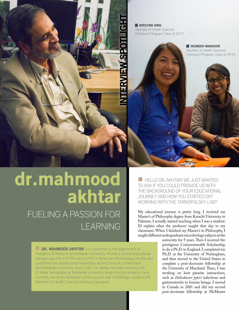
dr.mahmoodakhtar
FUELING A PASSION FOR LEARNING
INTE
RVI
EW S
POTL
IGH
T
DR. MAHMOOD AKHTAR is a researcher in the department of Pediatrics & Medicine at McMaster University. He has a diverse educational background, with a M. Phil. and a Ph.D. in Molecular Microbiology. He has also completed two postdoctoral fellowships at the University of Marylandand McMaster University. Since 2007, Dr. Akhtar has been working with Dr. Mark Tarnopolsky at McMaster University. Aside from his research, he is currently one of the facilitators of the second year Cell Biology course in the Bachelor of Health Sciences (Honours) program.
HELLO DR. AKHTAR! WE JUST WANTED TO ASK IF YOU COULD PROVIDE US WITH THE BACKGROUND OF YOUR EDUCATIONAL JOURNEY AND HOW YOU STARTED OFF WORKING WITH THE TARNOPOLSKY LAB?
My educational journey is pretty long. I received my Master’s of Philosophy degree from Karachi University in Pakistan. I actually started teaching when I was a student: I’d explain what the professor taught that day to my classmates. When I finished my Master’s in Philosophy, I taught different undergraduate microbiology subjects at the
university for 5 years. Then I received the prestigious Commonwealth Scholarship, to do a Ph.D. in England. I completed my Ph.D. at the University of Nottingham, and then moved to the United States to complete a post-doctorate fellowship at the University of Maryland. There, I was working on host parasite interactions, such as Helicobacter pylori infections and gastroenteritis in human beings. I moved to Canada in 2001 and did my second post-doctorate fellowship at McMaster
AVRILYNN DINGBachelor of Health Sciences (Honours) Program, Class of 2017
YASMEEN MANSOORBachelor of Health Sciences (Honours) Program, Class of 2015
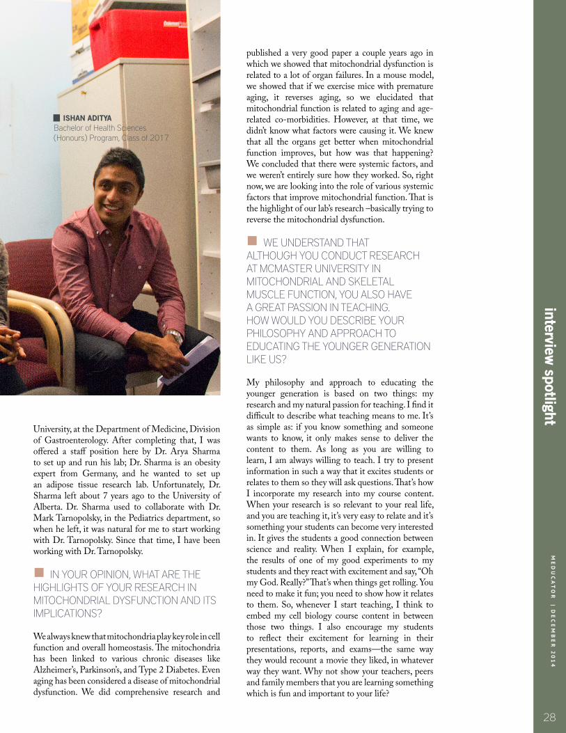
28
interview spotlight
University, at the Department of Medicine, Division of Gastroenterology. After completing that, I was offered a staff position here by Dr. Arya Sharma to set up and run his lab; Dr. Sharma is an obesity expert from Germany, and he wanted to set up an adipose tissue research lab. Unfortunately, Dr. Sharma left about 7 years ago to the University of Alberta. Dr. Sharma used to collaborate with Dr. Mark Tarnopolsky, in the Pediatrics department, so when he left, it was natural for me to start working with Dr. Tarnopolsky. Since that time, I have been working with Dr. Tarnopolsky.
IN YOUR OPINION, WHAT ARE THE HIGHLIGHTS OF YOUR RESEARCH IN MITOCHONDRIAL DYSFUNCTION AND ITS IMPLICATIONS?
We always knew that mitochondria play key role in cell function and overall homeostasis. The mitochondria has been linked to various chronic diseases like Alzheimer’s, Parkinson’s, and Type 2 Diabetes. Even aging has been considered a disease of mitochondrial dysfunction. We did comprehensive research and
published a very good paper a couple years ago in which we showed that mitochondrial dysfunction is related to a lot of organ failures. In a mouse model, we showed that if we exercise mice with premature aging, it reverses aging, so we elucidated that mitochondrial function is related to aging and age-related co-morbidities. However, at that time, we didn’t know what factors were causing it. We knew that all the organs get better when mitochondrial function improves, but how was that happening? We concluded that there were systemic factors, and we weren’t entirely sure how they worked. So, right now, we are looking into the role of various systemic factors that improve mitochondrial function. That is the highlight of our lab’s research –basically trying to reverse the mitochondrial dysfunction.
WE UNDERSTAND THAT ALTHOUGH YOU CONDUCT RESEARCH AT MCMASTER UNIVERSITY IN MITOCHONDRIAL AND SKELETAL MUSCLE FUNCTION, YOU ALSO HAVE A GREAT PASSION IN TEACHING. HOW WOULD YOU DESCRIBE YOUR PHILOSOPHY AND APPROACH TO EDUCATING THE YOUNGER GENERATION LIKE US?
My philosophy and approach to educating the younger generation is based on two things: my research and my natural passion for teaching. I find it difficult to describe what teaching means to me. It’s as simple as: if you know something and someone wants to know, it only makes sense to deliver the content to them. As long as you are willing to learn, I am always willing to teach. I try to present information in such a way that it excites students or relates to them so they will ask questions. That’s how I incorporate my research into my course content. When your research is so relevant to your real life, and you are teaching it, it’s very easy to relate and it’s something your students can become very interested in. It gives the students a good connection between science and reality. When I explain, for example, the results of one of my good experiments to my students and they react with excitement and say, “Oh my God. Really?” That’s when things get rolling. You need to make it fun; you need to show how it relates to them. So, whenever I start teaching, I think to embed my cell biology course content in between those two things. I also encourage my students to reflect their excitement for learning in their presentations, reports, and exams—the same way they would recount a movie they liked, in whatever way they want. Why not show your teachers, peers and family members that you are learning something which is fun and important to your life?
ISHAN ADITYABachelor of Health Sciences (Honours) Program, Class of 2017
ME
DU
CA
TO
R | D
EC
EM
BE
R 2
01
4

tabl
e of
con
tent
sM
ED
UC
AT
OR
|
DE
C 2
01
4
29
IT SEEMS THAT WHEN YOU TEACH, YOU START BY PROMPTING THE STUDENT TO ASK QUESTIONS.
Exactly. If you don’t ask questions, I won’t know what type of job I did, or how I am teaching. When a student asks me a question, I know they are learning. Or if they don’t and tell me, “I didn’t get it; can you explain it again?” Then I will go through it again to make sure you are with me. It’s like student and teacher are holding each other’s hands in this
learning process. If I don’t feel your hand in my hand, if that feel or connection is missing, then I am not an inspiring teacher and I should stay in my lab/office or at home and not with students. Good students make one into a good teacher, so I definitely give some credit to my students in helping me grow. I always tell my students that the sky is the limit.
IN YOUR OPINION, WHAT CHARACTERISTICS DIFFERENTIATE AN EFFECTIVE TEACHER FROM AN INSPIRING TEACHER?
An effective teacher is someone who has knowledge of a subject matter, and they are able to explain the information to their students in such a way that the students understand. The teacher may adopt any strategy of teaching, lecture, PowerPoint presentations,
providing notes, and assessing students through tests. On the other hand, an inspiring teacher is someone with teaching in their blood; they don’t take teaching as a chore. I don’t like people saying “TGIF” on Fridays. Those people should do something else, which they enjoy. I enjoy my research and teaching both. And if you love what you do, you do a far better job. If it was in my control, I would teach all day. I love my research too, but I am a “teacher by nature.” Teaching is like breathing to me. After I finish teaching or meeting with my students, I feel refreshed and have even more energy than what I had to start with. It is unfortunate, today in universities, that there is so much pressure to get
grants, and get publications, which are important too, but some professors are too distracted to do a better job at teaching students. But here is how I see it: When you receive admission to university, you have the right and I have the responsibility that I fulfill my job as a teacher. When you go to class and you don’t get a good grade because you were not taught better, then it’s not on you. It’s on me. Because when you came to school and you say, “I want to learn and get good grades,” and when I am unable to make it happen, then it’s my fault. One hundred percent. That’s the right you have, that I teach you well. But as a student, you need to show that desire to learn; I call it the attitude of a real student that shows that you want to learn. A good teacher will keep teaching if you want to keep learning. I’ll keep teaching, because to me, my students are like my kids; I make sure they learn better. I’m here for them. Otherwise I have no purpose to be with them.
WHAT ADVICE DO YOU HAVE FOR UNDERGRADUATE STUDENTS WHO WANT TO GET INVOLVED IN RESEARCH?
That’s a great question, and I think students should know about this. It is very tough now for a student to get into a lab to do research because a lot of you want to do research. Many of my students, when they finish HTH SCI 2K03, ask me to work in my lab, but it’s impossible to take more than a couple of them. So what they need to do is try to be the best student of the class. If you are my student, right from day one, you show you are interested, you are keen: characteristics that a good student should have. Then I will remember you. I will always remember, “That student always asks good questions,” and now that he/she wants to do research in my lab, I will take him/her. I never forget faces. Your part on how to get into research starts from the first day you take a course in which you are interested. You need to show your interest to your teacher right from day one, not when the course has ended and you send me an e-mail. When there are a lot of people who are interested, then you need to be a little step ahead. Read more about my interest area, read more about what I research, read my publications, and try to understand them better. Work hard on them. Send me an e-mail asking me questions. Then I will be impressed. It’s not that difficult. You just need to work hard and show more interest, and be decisive about the area in which you want to work. You can’t do research if you are interested in ten areas at a time.■
29
inte
rvie
w s
potli
ght
WHEN YOU GO TO CLASS AND YOU DON’T GET A GOOD GRADE, IT’S NOT ON YOU. IT’S ON ME.
BECAUSE YOU COME AND YOU SAY “I WANT TO GET GOOD GRADES. I WANT TO LEARN.” AND WHEN YOU SAY THAT AND I AM
UNABLE TO EXPLAIN IT TO YOU, THEN IT’S MY FAULT. ONE HUNDRED PERCENT.
ME
DU
CA
TO
R
| D
EC
EM
BE
R 2
01
4

30
critical reviewM
EDU
CATOR
| Dec 2
01
4
EDITORS-IN-CHIEF RONALD LEUNG GRACE ZHANG
STRATEGIC DIRECTOR YASMEEN MANSOOR
CREATIVE DIRECTORANNIE ZHU
GRAPHICS AND DESIGNSAMA ANVARIBELLE CAODAVID HU
AVERY LAMMELISSA LEE
ADITYA (DAVE) NIDUMOLULISA ROS-CHOIMICHAEL SUN
ZI (ELIYA) ZHAO
MANAGING EDITORKIMIA SOROURI
EDITORS
MANAGING VIDEO EDITORSEBASTIAN SWIC
VIDEO PRODUCERSFAIZAN BHATIAGEOFFREY LAU
ZHAOYUN (JACK) ZHANG
PRINTINGUNDERGROUND MEDIA AND DESIGN
OUR SPONSORSWe would like to give a special thanks to the following sponsors for their generous support:
30
contributors
BACHELOR OF HEALTH SCIENCES (HONOURS) PROGRAM
MCMASTER STUDENT UNION
ISHAN ADITYAHUMNA AMJADARLINDA DENGAVRILYNN DINGMAYLYNN DING
NICOLE FALZONESANA GILL
ANNA GOSHUA
VALERIE KIMABIRAMI KIRUBARAJAN
ASHLEY LAMFIZZA MANZOORABRAHAM REDDAMAXWELL TRANMATTHEW YAU
SHARON YEUNG
ME
DU
CA
TO
R | D
EC
EM
BE
R 2
01
4
MCMASTER ALUMNI ASSOCIATION
MCAT® is a registered trademark of the Association of American Medical Colleges.

INTERESTED IN WRITING FOR US?VISIT WWW.MEDUCATOR.ORG/GETTING-STARTED
Share your perspective online.
ROUND TABLEWithin the next 25 years,
what will be healthcare’s greatest challenge?
Enrich - Inspire - Discover
Meducator Round Table 2014/2015www. meducator.org/roundtable