itraq-based protein profiling provides insights into the central metabolism changes driving grape...
TRANSCRIPT

RESEARCH ARTICLE Open Access
iTRAQ-based protein profiling provides insightsinto the central metabolism changes drivinggrape berry development and ripeningMaría José Martínez-Esteso1*, María Teresa Vilella-Antón1, María Ángeles Pedreño2, María Luz Valero3
and Roque Bru-Martínez1*
Abstract
Background: Grapevine (Vitis vinifera L.) is an economically important fruit crop. Quality-determining grape componentssuch as sugars, acids, flavors, anthocyanins, tannins, etc., accumulate in the different grape berry development stages.Thus, correlating the proteomic profiles with the biochemical and physiological changes occurring in grape is ofparamount importance to advance in our understanding of berry development and ripening processes.
Results: We report the developmental analysis of Vitis vinifera cv. Muscat Hamburg berries at the protein level from fruitset to full ripening. An iTRAQ-based bottom-up proteomic approach followed by tandem mass spectrometry led to theidentification and quantitation of 411 and 630 proteins in the green and ripening phases, respectively. Two key points indevelopment relating to changes in protein level were detected: end of the first growth period (7mm-to-15mm) andonset of ripening (15mm-to-V100, V100-to-110). A functional analysis was performed using the Blast2GO software basedon the enrichment of GO terms during berry growth.
Conclusions: The study of the proteome contributes to decipher the biological processes and metabolic pathwaysinvolved in the development and quality traits of fruit and its derived products. These findings lie mainly in metabolismand storage of sugars and malate, energy-related pathways such as respiration, photosynthesis and fermentation, and thesynthesis of polyphenolics as major secondary metabolites in grape berry. In addition, some key steps in carbohydrateand malate metabolism have been identified in this study, i.e., PFP-PFK or SuSy-INV switches among others, which mayinfluence the final sugar and acid balance in ripe fruit. In conclusion, some proteins not reported to date have beendetected to be deregulated in specific tissues and developmental stages, leading to formulate new hypotheses on themetabolic processes underlying grape berry development. These results open up new lines to decipher the processescontrolling grape berry development and ripening.
Keywords: Development, Grape berry, iTRAQ, Mesocarp, Proteomics, Quantitative, Vitis vinifera, Functional annotation
BackgroundGrapevine (Vitis vinifera L.) is the most important fruitcrop in the world, and viticulture and enology play animportant role in the economy of many developed andemerging countries. In 2007, the Food and AgricultureOrganization (FAO) of the United Nations estimated thatthe gross world production of grape berries was over 67million tons per year, with land occupancy of 7,272,583 ha
(FAO, 2007 [1]). In the food industry, grape berries arewidely commercialized raw, as a fresh and dried fruit, orare processed mainly as grape juice or wine. Extracts fromgrape berry seeds, skin and also grape plant leaves are usedas commodities in nutraceutical and cosmetic industries.The grape berry is a non climacteric fruit that exhibits a
double sigmoid growth pattern [2]. The first growth phaseafter fruit set is characterized by not only rapid cell div-ision, which increases the number of cells, but also by anexpansion of existing cells. It is followed by a lag phasewith little or no growth. The second growth phase coin-cides with the onset of ripening, called véraison, which is
* Correspondence: [email protected]; [email protected] de Proteómica y Genómica Funcional de Plantas, Dept. Agroquímicay Bioquímica, Facultad de Ciencias, Universidad de Alicante, Apartado 99,E-03080 Alicante, SpainFull list of author information is available at the end of the article
© 2013 Martínez-Esteso et al.; licensee BioMed Central Ltd. This is an open access article distributed under the terms of theCreative Commons Attribution License (http://creativecommons.org/licenses/by/2.0), which permits unrestricted use,distribution, and reproduction in any medium, provided the original work is properly cited.
Martínez-Esteso et al. BMC Plant Biology 2013, 13:167http://www.biomedcentral.com/1471-2229/13/167

characterized by important biochemical and physiologicalchanges such as softening, coloring and engustment ofberry. As grape berries develop, they change in size andcomposition. These transformations range from small,firm and acidic with little sugar, desirable flavors or aroma,to becoming larger, softened, sweet, highly flavored, lessacidic and highly colored fruit. The biochemical changesunderlying grape berry ripening have been recentlyreviewed [3]. The development of these traits is subject togenetic, environmental and viticultural conditions, whichdetermine the final quality of the berry and its derivedproducts. During the first growth period, chlorophyll isthe main pigment present in fruit and cells are rich in or-ganic acids; the most prevalent compounds are tartaricand malic acids, which accumulate mainly in skin andflesh. Compounds vastly increase starting at véraison, andthe major ones are glucose and fructose [4], in addition tophenolic and aromatic compounds [5], while malate con-centration lowers [6]. Flavor development in grapes ispartly due to the acid/sugar balance [7], which is particu-larly important in table grapes. Muscat Hamburg is a clas-sical cultivar of black table grape that is grown in manyparts of Europe, and is greatly appreciated for its pleasantMuscat flavour [8,9].Correct grape ripening is fundamental for both commer-
cial fruit use and wine quality. For this reason, grape berrydevelopment has been investigated by monitoring the ex-pression of quality trait-related genes in a classic targetedapproach [4,10-13], and also by global mRNA expressionprofiling using cDNA or oligonucleotide microarrays[14-19] since genomic resources for Vitis vinifera and re-lated species have been generated on a large scale.In recent years, proteomics-based technologies have been
successfully applied to grapevine to analyze stress responses[20-23] and berry development [24-28]. These studies haveimproved the understanding of changes in grape berries atthe proteome level, but are limited by the intrinsic inaccur-acy of single staining-based quantitative two-dimensionalgel electrophoresis (2-DE) [29]. The application of more ro-bust quantitative proteomic approaches, such as differencegel electrophoresis (DIGE) or MS-based stable isotopic la-beling, should lead to a better coverage and accuracy ofprotein changes. DIGE has been applied to study grapeberry development. It has led to a better coverage and ac-curacy of the protein changes reported from a gel-based ap-proach [30,31]. Lücker et al. [32] studied grape berryripening initiation by the quantitative MS-based iTRAQapproach, and reported a significantly larger number oftotal protein identification and protein changes than single-stain 2-DE-based studies. These authors noticed the diffi-culty of protein identification as no finished genome se-quence data for grapevine were available. Thus, they built aprotein database by combining in-house generated [33] andpublicly available grapevine nucleotide sequences. To date,
this limitation has been greatly reduced thanks to grapevinegenome sequencing initiatives [34,35], which have releasedthe translated ORFs found in the genome to public proteindatabases. Nevertheless, the extent of sequence descriptionand annotation of grapevine proteins, including genome-derived ones, is still far from complete. The exocarp ofgrape berries has been analyzed throughout ripening by theiTRAQ technology to offer better proteome coverage [36]and by making full use of recent grapevine genome sequen-cing initiatives [34,35].Stable isotopic labeling followed by a reverse phase high-
performance liquid chromatography coupled with tandemmass spectrometry (HPLC-MS/MS) analysis is a relativelyrecent quantitative technique, and was first described byRoss et al. [37]. Isobaric tags for relative and absolute pro-tein quantitation (iTRAQ) are currently being used suc-cessfully to characterize and quantify changes in proteinlevels in complex biological samples [38]. The use ofiTRAQ has become a consolidated technique in quantita-tive proteomics since large fold changes of protein expres-sion within broad dynamic ranges of protein abundancecan be measured quite accurately [39]. The iTRAQmethod allows the multiplexed identification and quantita-tion of proteins in four different samples (4-plex) and hasbeen recently scaled up to measure protein changes in upto eight different samples (8-plex) [40].The tissue-specific analysis of grape berry is critical if in-
formation on its individual contribution to final fruit qual-ity is sought. Quality traits relating to the organolepticproperties of berry and wine are linked to specific berrytissue: skin composition plays an important role in deter-mining color, aroma, astringency and bitterness, while thepulp contributes to the sugar/acid balance, crispiness,juiciness and alcoholic potential. The seed also contributesto the astringency and bitterness of wines.Grimplet et al. [41], used a large-scale transcriptomic
analysis to study the differential expression of genesamong main tissues, seed, flesh and skin, and reportedthat 60% of these genes exhibited a significant differentialexpression in at least one of the three major tissue types.Several proteomic analyses at the tissue-specific level havebeen carried out [24,26,31,32,36,42]. Nevertheless on thewhole, a gap still remains either because one only tissue isanalyzed or berry development is not comprehensivelycovered.In this study, the issue of protein expression changes
is addressed in the pericarp/mesocarp tissues in grapeberry and all the developmental phases are covered. Forthe purpose of gaining a better understanding of theprotein composition in grape berry and its changes dur-ing development, seven different stages from fruit set tofully ripened were analyzed. The aim is to improvecoverage and to complement the proteomic analysisdone in previous DIGE experiments [31] with the high-
Martínez-Esteso et al. BMC Plant Biology 2013, 13:167 Page 2 of 20http://www.biomedcentral.com/1471-2229/13/167

throughput proteomic analysis presented herein usingan alternative approach, namely the top-down quantita-tive proteomics iTRAQ technique. The use of annota-tion and analysis tools [43], along with recent literaturefindings, has also led to gain insights into the biologicalsignificance of the proteomic data in the search for use-ful protein biomarkers to track the final quality traitsassociated with grape berry development.
Results and discussionGrape berry development was analyzed by the iTRAQtechnique. One iTRAQ experiment covered four timepoints of the first growth period, which spanned the stagesfrom fruit set to pre-véraison (FS-to-15 mm). AnotheriTRAQ experiment covered three time points of the
second growth period from onset to full ripening (V100-to-140 g/l), and a pre-véraison fourth time point (15 mm),shared with the first experiment. Figure 1 describes the ex-perimental design and workflow for both experiments.
Protein identification and quantitationIn the green stages, 542 proteins were identified, which issimilar to the number of proteins identified with at leasttwo peptides in an iTRAQ experiment of the grape berryexocarp [36] (Additional file 1). Of these, 524 proteinswere quantified and, after applying a p<0.05 filter, the listshortened to 411 (Additional file 2). All the 524 quantifiedsequences were assigned a description and 508 were anno-tated with an average number of five annotations per se-quence and an average annotation level depth of 5.8.
-100 110 g/l 140 g/lReduced/alkylated
Digest Digest Digest
iTRAQlabeling
--V-
Pool samples labeled
FS
Digest Digest Digest Digest
114
4 mm 7 mm 15 mm
31 PRG 115 30 PRG 116 29 PRG 117
Pool samples labeled
114
SCXChromatography
21 fractions
Reverse phase chromatography nanospray ESI
QSTAR ESI-QTOF
Quantitation and identification of proteins with ProteinPilot
Annotation of protein sequences and functional analysis with Blast2GO
SCXChromatography
18 fractions
31 PRG
PRG28 115 30 PRG 116 29 PRG 117 PRG28
Figure 1 Workflow of the iTRAQ experiments for both the first growth period and grape berry ripening. The first growth period,analyzed in the first iTRAQ experiment, covers four developmental stages: fruit set (FS), 4 mm, 7 mm and 15 mm labeled with the 114, 115, 116and 117 iTRAQ tags, respectively. The ripening period covers three developmental stages, 100% véraison (V-100), 110 g/l and 140 g/l, and a fourthpoint, the 15 mm stage, which is common with the previous experiment, and was co-analyzed in a second iTRAQ experiment. For eachexperiment, the extracted proteins were trypsin-digested and peptides were labeled with iTRAQ tags. After labeling, peptides were pooled andfractionated by strong cation exchange (SCX) chromatography. Each fraction was resolved by reverse phase chromatography and was analyzeddirectly by mass spectrometry. Searches and quantitation analysis were carried out using the ProteinPilot v1.0 software (Applied Biosystems).Finally, the identified proteins in the set were annotated and functionally analyzed using the Blast2GO v2.4.0 application [43] based on geneontology (GO) terms.
Martínez-Esteso et al. BMC Plant Biology 2013, 13:167 Page 3 of 20http://www.biomedcentral.com/1471-2229/13/167

In the ripe stages, 1117 proteins were identified, whichis almost twice the number of those identified in the ex-periment for green stages (Additional file 3). Of these,1034 proteins were quantified and, after applying ap<0.05 filter, the list shortened to 630 (Additional file 4).Of the 1034 sequences, 1010 were assigned a descriptionand 977 were annotated with an average number of fiveannotations per sequence and an average annotationlevel depth of 5.5.As a summary of the annotation results, pie charts
for the biological function and cellular componentgene ontology (GO) terms are shown for each experi-ment in Additional file 5, respectively. The resultingannotation files are provided as Additional file 6(green) and Additional file 7 (ripe).
Proteome changes during developmentTo better characterize the most active stages as regardsproteome changes, the relative abundance of the dereg-ulated proteins was computed between contiguous stages(reporter ion ratios 115/114, 116/115 and 117/116 per ex-periment) and these values were used to rank the selectedprotein lists. For each stage transition, two subsets of pro-teins were established: those whose fold change was above1.5 and those with a fold change under −1.5. Figure 2 pro-vides a global picture of the proteome changes betweeneach pair of contiguous sampled time points of the grapeberry formation phase. Figure 2A illustrates the numberof up- and down-regulated proteins passing the filter,whereas Figure 2B depicts the average and the highestfold change of that set of proteins. Of the number ofderegulated proteins, three classes of development stagescan be formed: stages with very few changes (transitionsfrom 4 mm-to-7 mm and from 110 g/l-to-140 g/l); stageswith many changes (transitions from 7 mm-to-15mm andfrom 15 mm-to-V100); stages with an intermediate num-ber of changes (transitions FS-to-4 mm and V100-to-110 g/l). If the amplitude of changes was considered,the average fold change among the paired stages was notso different. However, there were few very major changesat certain transitions that revealed potential marker pro-teins for the developmental stages. The results obtainedherein agree with those reported in a previous proteomicexperiment, which analyzed the same time points usingthe DIGE platform [31]. The additional time point in-cluded in the present study, the transition from V100-to-15 mm, resulted in the development stage where the lar-gest number of proteins underwent changes and wherethe largest fold-changes occurred. These results suggestthat the most important changes at the protein level tookplace at the end of green period and at the onset of ripen-ing, as previous data from genomic [44], oligo/microarraytranscriptomic [14,15] and proteomic [27,31,32] studieshave also indicated.
Functional analysisThe present study contributes to significantly increase thecoverage of the differential proteome of the whole devel-opment of grape berries and confidently allows the detec-tion of putative marker proteins based on a previousproteomic experiment, which revealed that the proteinchanges observed were developmentally associated withthe analyzed biological individuals [31]. The good agree-ment between most of the profiles of the proteins identi-fied in common herein and in Martínez-Esteso et al. [31],serves as validation the present study using the non gel ac-curate quantitative proteomic iTRAQ approach.
Functional annotation analysisA Fischer’s enrichment analysis [45] was carried out bycomparing the frequencies of the annotation terms in theup- and down-regulated protein subsets with those of thewhole list of quantified proteins as reference list. As a re-sult, a series of GO terms, which were statistically overrep-resented in the subset, was obtained (p<0.005). Figure 3shows the result of the Fischer’s enrichment analysis forthe molecular function (F term) (Figure 3A), biologicalprocesses (P term) (Figure 3B) and the cellular compo-nents (C term) (Figure 3C) -selected GO terms whoselevel in most cases was 4 or deeper. Additional file 8 pro-vides a list of the proteins annotated with each enrichedGO term.The proteins annotated with the GO-terms enriched
from the up-regulated list of proteins in FS-to-4 mm weremainly different isoforms of the histones, tubulins and pro-teins involved in cytoskeleton organization and biogenesis.Several ribosomal proteins, which were annotated with theterms ‘intracellular nonmembrane-bound organelle-GO:0019685’ (C term) and ‘cellular component organizationand biogenesis- GO:0016043’ (P term), appeared to be up-regulated in FS-to-4 mm. In contrast, these terms weredown-regulated from 7mm onwards, showing those asleading processes during the early stages of development,when fruit cells were very active in division and expansion,and new proteins were being synthesized. The term ‘hy-drolase activity, acting on glycosyl bonds-GO:0016798’(F term), which appeared to be significantly up-regulated,related to different glycosyl hydrolases and an acid vacuolarinvertase, which started to accumulate from the 4mmstage and was possibly involved in the carbohydrate catab-olism of sucrose unloaded in fruit by acting as a sink organin the vine. The proteins involved in ‘flavonoid biosyntheticprocesses-GO:0009813’ and ‘aromatic amino acid synthesis-GO:0006725’ (P term) appeared to be down-regulated atthis point of development, including the ‘phenylalaninemetabolic process-GO:0006558’ GO term (P term), a pre-cursor of all the phenylpropanoid-derived compounds. Thisfinding reflects an accumulation of flavonoids in earlyfruit development. The 4 mm-to-7 mm transition was
Martínez-Esteso et al. BMC Plant Biology 2013, 13:167 Page 4 of 20http://www.biomedcentral.com/1471-2229/13/167

characterized by a few terms enriched in the down- andup-subsets of proteins, which reflect that most of the pro-cesses involved in development occurred earlier than orafter this time point. Only the term ‘photosynthesis lightharvesting-GO:0009765’ was up-regulated and some termsannotating several isoforms of histones were down-regulated (‘nucleosome-GO:0000786’ (C term) ‘protein-DNA complex assembly-GO:0065004’ (P term) and ‘DNAbinding-GO:0003677’ (F term)), which displayed greater ac-cumulation earlier in grape berry development. In the nexttransition (7 mm-to-15 mm), only the down-regulated sub-set of proteins contained GO-enriched terms. The mostremarkable processes, these being the ‘macromoleculebiosynthetic process-GO:0009059’ (P term) and ‘cellularcomponent organization and biogenesis- GO:0016043’(C term), were linked with proteins like elongation factors,tubulins, histones and ribosomal proteins, which agreeswith the state of cells at this point of development. Theseresults tie in with the fact that fruit entered a lag phase,with little or no division or expansion, and most processeswere switched off before triggering the véraison. Anotherimportant down-regulated term was the phenylpropanoidmetabolism (GO:0009699), involving flavonoids and aminoacid precursors, which included the enzymes of the general
phenylpropanoid pathway. The processes down-regulatedat 7 mm-to-15 mm occurred mainly in the cytosol, unlikethose down-regulated from 15 mm-to-V100, and whichalso occurred mainly in the chloroplast. Several proteinsbelonging to the photosynthetic machinery (‘PhotosystemI-GO:0009522’ (C term), ‘Photosystem II-GO:0009523’(C term)) and those involved in photosynthesis, bothlight (GO:0019684) and dark reactions (GO:0019685)(phosphoribulokinase, rubisco), were strongly down-regulated before véraison was triggered. Some relatedprocesses occurring in the chloroplast such as starchmetabolism (4-alpha-glucanotransferase), redox state(thioredoxin, peroxiredoxin) and protein folding (chap-eronin 60), are linked with the terms enriched in thedown-regulated subset, thus also indicating a decreaseof the photosynthetic-dependent processes. In parallel,a significant number of ribosomal proteins was down-regulated (the term ‘structural molecule activity-GO:0005198’ (F term)). The term ‘integral to membrane-GO:0016021’ (C term) included an aquaporin and plasmamembrane H+-ATPase, indicating a decrease in water-associated specific transporters prior to véraison. Likewise,cell wall-associated processes (endo-xyloglucan tranferase,pectinesterase) appeared to be deregulated, indicating that
-150
-100
-50
0
50
100
FS/4mm 7mm/4mm 15mm/7mm V100/15mm V100/110g/l 140g/l/110g/l
#pro
tein
s qu
anti
fied
>1.
5-fo
ld
up
down
-25
-20
-15
-10
-5
0
5
10
15
FS/4mm 7mm/4mm 15mm/7mm V100/15mm V100/110g/ll 140g/l/110g/l
log2
fol
d-ch
ange
average
max/min
A
B
Figure 2 Overall changes in the protein level throughout berry development. Each value represents (A) the number of sequencesquantified between two consecutive developmental stages, for all the proteins that were up- (dark blue bars) and down-regulated (gray bars),and (B) the fold-change of the proteins, represented as mean (red) and max/min (blue) changes. Transitions were: fruit set to 15 mm (FS-to-15mm), 4 mm to 15 mm (4-to-15 mm), and 7 mm to 15 mm (7-to-15 mm), respectively. The experiment linked to the ripe stages was stated asfollows: V100 to 15 mm (V100-to-15 mm), 110 g/l to 15 mm (110-to-15 mm) and 140 g/l to 15 mm (140-to-15 mm). An arbitrary fold changecutoff of ± 1.5 was used to select the protein subsets.
Martínez-Esteso et al. BMC Plant Biology 2013, 13:167 Page 5 of 20http://www.biomedcentral.com/1471-2229/13/167

A
B
C
Figure 3 (See legend on next page.)
Martínez-Esteso et al. BMC Plant Biology 2013, 13:167 Page 6 of 20http://www.biomedcentral.com/1471-2229/13/167

these profiles play a relevant role at véraison as this stagewas characterized by fruit softening. This is a complexregulated process as inferred by the terms deregulatedat this transition, 15 mm-to-V100, in which some spe-cific isoforms related to cell wall biogenesis (the terms‘cytoplasmic membrane-bound vesicle-GO:0016023’(C term),‘extracellular region-GO:0005576’ (C term), endo-xyloglucantransglycosylase, expansin, polygalacturonase, pectin methyl-esterase, invertase pectin methylesterase inhibitor family)were strongly up-regulated, while other isoforms weredown-regulated at this point. Interestingly, severalstress-related proteins annotated with the term ‘cop-per ion binding-GO:0005507’ (F term) were down-regulated (polyphenol oxidase, diphenol oxidase, super-oxide dismutase). The terms which up-regulated at véraisonwere ‘cytoplasmic membrane-bound vesicle-GO:0016023’ ,‘extracellular region-GO:0005576’ and, apart from theproteins indicated above, they involved ripening-relatedproteins (grip22), plasma membrane proteins and someproteases (subtilisin, cysteine proteinase inhibitor). Never-theless, the most relevant terms during this periodwere ‘cell wall metabolic process-GO:0010382’ (P term),‘chitin catabolic process-GO:0006032’ (P term), invol-ving defense proteins (PR-4, chitinase IV), carbohydrate-related processes, which included the proteins involvedin sucrose metabolism (sucrose synthase, sucrose-phosphate synthase) and, behind the term ‘lyase activity-GO:0016829’ (F term), hexose and pyruvate metabolism(enolase, aldolase, pyruvate decarboxylase) was found.Altogether, this reflects the intense activity aroundcarbohydrate metabolism at the onset of ripening, pre-sumably in the biosynthetic direction, to convert existingpools of malate into sugars for their storage since gly-colysis is supposed to be inhibited after the onset ofripening [6].In the V100-to-110 transition, the down-regulated
process predominated the up-regulated ones. Among theup-regulated ones, the terms ‘chitin catabolic process’ and‘polysaccharide metabolic process-GO:0005976’ (P term),including defense proteins, were kept, and new terms ap-pear such as ‘prefolding complex-GO:0016272’ (C term)(needed for the stabilization of the newly synthetized pro-teins) and ‘cysteine-type endopeptidase activity-GO:0004197’ (F term) (cysteine proteinase, the cysteineprotease component of the protease-inhibitor complexand cathepsin b-like cysteine protease). Among the
down-regulated proteins, the most remarkable featurewas the diminished abundance of proteins synthesis, asinterpreted by the enriched terms ‘intracellular non-membrane-bound organelle’, (C term) ‘cellular componentorganization and biogenesis’ (P term), which included animportant representation of different ribosomal proteins,and the term ‘translation-GO:0006412’ (P term) (t-RNAsynthetases). Apart from these, other terms of the cellularcomponent and molecular function categories associatedwith the ribosomal function were enriched in the down-regulated subset (‘macromolecule biosynthetic process’(P term), ‘structural constituent of ribosome’-GO:0003735(C term)). Another remarkable feature was that the proteinsinvolved in the photosynthetic dark reaction were stronglydown-regulated, which reflects that grape berries lose theirphotosynthetic capability as they ripen. Finally in the 110-to-140 transition, only the term ‘nutrient reservoir activity-GO:0045735’ (F term) was enriched in the up-regulatedsubset of proteins. This term included three seed storageproteins that are considerably accumulated.
Protein profile analysisAs shown in Additional file 9, the behaviour of the timecourse of the functional protein groups, which lie beneathimportant quality traits of the pericarp/mesocarp duringfruit development, was observed in detail. The functionalprotein groups of ‘transporters’ , ‘polyphenols’ , ‘photosyn-thesis, respiration and fermentation’ and ‘carbohydrate andmalate metabolism’ are discussed herein. The remainingfunctional protein groups, including ‘nitrogen and aminoacid (Additional file 10) ‘terpenoid metabolism’ , ‘signalingand hormone’ , ‘stress’ , ‘protein synthesis’ , ‘protein degra-dation’ , ‘protein processing’ , ‘cell division and growth,biogenesis’ ‘defense proteins’ and ‘other proteins of inte-rest’ , are discussed in Additional file 11.
1. TransportersHere we will focus on transport phenomena involved intonoplast and plasma membrane energization for its specialrelevance in sugar transport and accumulation. The vacuoleis a very important organelle in grape berry cells because ofits dominant cytoplasm occupancy [46], its predominantrole as a storage compartment of organic acids, sugars, sec-ondary metabolites and amino acids, and because of itscentral cell function in cytosolic pH regulation. To date,knowledge of the transporter proteins in the tonoplast is
(See figure on previous page.)Figure 3 Enriched GO terms (Molecular function (F term) (A), Biological process (P term) (B) and Cellular component (C term) (C)) forthe sequences annotated in the up- or down-regulated subsets of proteins in each grape berry developmental stage analyzed. Bardiagrams indicate the number of sequences that were up- and down-regulated (X-axis), annotated with each enriched GO term (Y-axis) in theconsecutive paired stages. GO terms descriptions are detailed in Additional file 8. Color code for the consecutive pair stages: FS-to-4 mmtransitions (dark green bars), 4 mm-to-7 mm transitions (medium intensity green), 15 mm-to-7 mm (light green), V100-to-15 mm transitions(yellow), 110 g/l-to-15 mm transitions (light purple), 140 g/l-to-110 g/l (dark purple).
Martínez-Esteso et al. BMC Plant Biology 2013, 13:167 Page 7 of 20http://www.biomedcentral.com/1471-2229/13/167
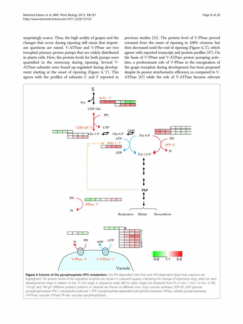
surprisingly scarce. Thus, the high acidity of grapes and thechanges that occur during ripening still mean that import-ant questions are raised. V-ATPase and V-PPase are twotonoplast primary proton pumps that are widely distributedin plants cells. Here, the protein levels for both pumps werequantified in the mesocarp during ripening. Several V-ATPase subunits were found up-regulated during develop-ment starting at the onset of ripening (Figure 4, ‘1’). Thisagrees with the profiles of subunits C and F reported in
previous studies [31]. The protein level of V-PPase provedconstant from the onset of ripening to 100% véraison, butthen decreased until the end of ripening (Figure 4, ‘2’), whichagrees with reported transcript and protein profiles [47]. Onthe basis of V-PPase and V-ATPase proton pumping activ-ities, a predominant role of V-PPase in the energization ofthe grape tonoplast during development has been proposeddespite its poorer stoichometry efficiency as compared to V-ATPase [47] while the role of V-ATPase became relevant
g
Vacuole
S
UDP-Glu
SuSy ‘4’
UDP-GP ‘5’
Glu-6-P Fru-6-P
Fru-1,6-P
G6PI
PFK-1 ‘7’ PFP ‘6’
PEP
-
Glu-1-P
PPi
UTP
PPi
Pi
ATP
ADP
Respiration Biosynthesis
PPiPi
V-PPase ‘2’
ATPADP
V-ATPase ‘1’
PPi
Pi
sPPase ‘3’
Fru
Malate
cf
e
c
b
b
b
Figure 4 Scheme of the pyrophosphate (PPi) metabolism. The PPi-dependent (red line) and ATP-dependent (blue line) reactions arehighlighted. The protein levels of the regulated enzymes are shown in coloured squares, indicating the change of expression (log2 ratio) for eachdevelopmental stage in relation to the 15 mm stage. In sequence order (left to right), stages are displayed from FS, 4 mm, 7 mm, 15 mm, V-100,110 g/l, and 140 g/l. Different proteins isoforms or subunits are shown as different rows. SuSy, sucrose synthase; UDP-GP, UDP-glucosepyrophosphorylase; PFK-1, phosphofructokinase 1; PFP, pyrophosphate-dependent phosphofructokinase; sPPase, soluble pyrophosphatase;V-ATPase, vacuolar ATPase; PP-ase, vacuolar pyrophosphatase.
Martínez-Esteso et al. BMC Plant Biology 2013, 13:167 Page 8 of 20http://www.biomedcentral.com/1471-2229/13/167

throughout the ripening phase [48]. Thus, the protein pro-files found herein for both the H+ pumps were in accord-ance with their reported activities [48]. Although V-PPasewas not detected during the first growth period, the profilesfound showed more abundance at véraison than at ripening;such decrease combined with the emergency of a putativecytosolic sPPase (Figure 4, ‘3’) along ripening, points to pyro-phosphate (PPi) as an energy source to pump protons acrossthe tonoplast during green development, but not duringripening.Furthermore, the induction of ripening is known to be as-
sociated with the shift of phloem unloading from the sym-plastic to the apoplastic pathway [28]. The latter involvesthe participation of several plasma-membrane sugar trans-porters which are induced at véraison [3]. Sugartranslocation is a secondary transport driven by a protongradient established by plasma membrane H+-ATPase(PM-ATPase). Here the abundance of PM-ATPase(Additional file 9A) peaked at the onset of ripening andbecame down-regulated in the following stages, whichis consistent with previous findings [49]. This profile isconsistent with the sugar concentrations in the phloemand berry apoplast; that is, high at véraison and earlyripening, but lower as the berry ripens [50], and it re-sponds to the PM energization need. Thus, PM-ATPasecan play a role relevant in triggering sugar accumula-tion in berries. Unlike PM, the energy demand at thetonoplast level continuously increased for use in bothsugar translocation to the vacuole and the maintenanceof the high sugar gradient concentration between thecytosol and the vacuole. Indeed the induction of proton-sugars antiporters supported vacuolar sugar accumulation[3]. The specific tonoplast antiporter carrier for hexoses,VvHT6, was up-regulated during ripening (Additional file9A) in accordance with reported transcript levels [14,17].In addition, vacuolar H+ pumps must play a key role ingenerating a proton motive force to accumulate sugars. Asdiscussed above, bothV-PPase and V-ATPase can play thisrole, but considering their respective profiles and theprogressive accumulation of soluble PPase, the importanceof the ATP-driven pump increases as the berry ripens.As note above, PPi may play key functions during berry
development although its metabolism is still a largely un-known process. PPi is released as a by-product during sev-eral anabolic reactions, which are highly active duringyoung development, such as the synthesis of DNA, RNA,proteins, carbohydrates, etc., and it can be simply hydro-lyzed by soluble pyrophosphatase (sPPase) to pull up bio-synthetic reactions or used in other processes. Accordingto the abundance profiles of the PPi-utilizing cytosolic en-zymes (Figure 4), in the developing grape berry it mightact as an energy donor per se [51] for tonoplast energiza-tion through the V-PPase pump, but also as a phosphorylgroup donor in sucrose degradation via sucrose synthase
(SuSy) (Figure 4, ‘4’), and UDP-glucose pyrophosphorylase(UDP-GP) (Figure 4, ‘5’) and in glycolysis throughpyrophosphate-dependent phosphofructokinase (PFP)(Figure 4, ‘6’) (reviewed Plaxton, [52]). Besides the pro-files for these enzymes, it is interesting to note that theprofile for sPPase correlates positively to that of phos-phofructokinase 1 (PFK-1) (Figure 4, ‘7’) and V-ATPase,but negatively to that of PPi-utilizing enzymes such asPFP and V-PPase, therefore, sPPase is the candidate tocontrol the cytosolic PPi pools in the ripening berry de-velopment phase.
2. PolyphenolsFrom a wine quality perspective, the major phenoliccompounds in grape berries are non flavonoidhydroxycinnamic acids (HCyAs), and flavonoid anthocy-anins (ACs) and proanthocyanidins (PACs) [53]. Thesynthesis of phenolics share the phenylpropanoid path-way, from phenylalanine (Phe) to p-coumaric acid, fromwhich several branches derive, leading in grapes to lig-nins, hydroxycinnamic acids, stilbenoids and flavonoids[3] (see Figure 5). The obtained results clearly show en-hanced phenylpropanoid synthesis from very early devel-opment as the two first enzymes of the pathway,phenylalanine ammonia lyase (PAL, Figure 5 ‘1’) andcinnamate 4-hydroxylase (C4H, Figure 5 ‘2’), were atleast 4-fold up-regulated toward fruit set in relation to15mm. Likewise, five enzymes of the shikimate pathway(3-deoxy-D-arabino heptulosonate 7-phosphate synthase(DHAPS, Figure 5 ‘3’), dehydroquinate dehydratase-shikimate dehydrogenase (DD-SDH, Figure 5 ‘4’),5-enol-pyruvilshikimate-phosphate synthase (EPSPS, Fig-ure 5 ‘5’) and chorismate synthase (CHOSy, Figure 5‘6’)), leading to aromatic amino acids, had the same pro-file as the phenylpropanoid enzymes during green devel-opment (see Figure 5). Flavonoids constituted asignificant portion of phenolics and included ACs, flavo-nols, flavan-3-ols and PACs or condensed tannins (poly-mers of flavan-3-ols and flavan-3,4-diols), and were themost abundant class of soluble polyphenolics in grapeberries. Common enzymes for the flavonoid pathway(chalcone synthase (CHS, Figure 5 ‘7’), chalcone isomer-ase (CHI, Figure 5 ‘8’), flavonol-3-hydroxylase (F3H, Fig-ure 5 ‘9’), dihydroflavonol reductase (DFR, Figure 5 ‘10’),anthocyanidin synthase/leucoanthocyanidin dioxigenase(ANS/LDOX, Figure 5 ‘11’) and the branching point to-ward PACs biosynthesis (anthocyanidin reductase (ANR,Figure 5 ‘12’)) displayed the same abundance pattern asthe shikimate and phenylpropanoid enzymes. Althoughno enzyme of stilbene or hydroxycinnamic acid synthesisbranches was detected, a resveratrol/hydroxycinnamicacid O-glucosyltransferase (RHCA-GT) with an abun-dance profile like that above was identified. It shared a94% homology with a bi-functional glucosyltransferase
Martínez-Esteso et al. BMC Plant Biology 2013, 13:167 Page 9 of 20http://www.biomedcentral.com/1471-2229/13/167

from Vitis lambrusca cv. Concord (VLRSgt), which pro-duced glycosides of stilbenes and glucose esters ofHCyAs in vitro [54]. Conversely, cinnamyl alcohol de-hydrogenase (CAD, Figure 5 ‘13’), which is involved inlignin biosynthesis, displayed quite an opposite profile. Noother enzymes acting at the branching points of the flavon-oid pathway were detected. Together all these results pro-vide strong evidence that in pre-véraison stages, carbonflows mainly from glycolysis precursors toward not only theproduction of PAC precursors but, and based on weaker evi-dence, also hydroxycinnamic acid and stilbene derivatives.Alternatively, the production of other pathway end products,such as lignins, flavones, flavonols and ACs, seemed blockedin the mesocarp by the absence of the corresponding en-zymes. These results are highly consistent with the type ofPhe-derived secondary metabolites accumulation in the de-veloping grape berry [3,55-60].Besides ANR, leucoanthocyanidin reductase (LAR) also
produces PAC precursors; both were expressed during PACsaccumulation in grapes and were regulated in a temporal-and tissue-specific manner [59]. Although LAR was not
detected in the pericarp of green berries, ANR (Figure 5‘12’) could play a relevant role in driving flavonoid biosyn-thesis in the epicatechin production in the tissue duringearly berry development (see Figure 5). The general trendobserved for flavonoid-derived compound synthesis was thatit was down-regulated in the ripening phase. This stemsfrom the observation that CHI and CHS were stronglydown-regulated at véraison, and remained down-regulateduntil full ripening. Of the remaining flavonoid pathway en-zymes, only DFR was detected and displayed almost un-changed levels from 15 mm to full ripening. In grape berryskins, where ACs heavily accumulated at the onset of ripen-ing, no new synthesis of precursors from p-coumaric acidapparently occurred in the skin, as observed by the decreas-ing profiles of the downsteam enzymes [36]. The presenceof DFR in the mesocarp during ripening would warrant asupply of ACs precursors for synthesis in the skin viaANS and UDP-glucose: flavonoid 3-O-glucosyltransferase(UFGT), as suggested by Martínez-Esteso et al. [36].Although flavonoid compounds were synthesized in
the cytosol, they accumulated in vacuoles. It has been
Figure 5 Scheme of the polyphenol biosynthetic pathways in grape berries during development and ripening. These include shikimatepathway as the phenylalanine (Phe) source, the general phenylpropanoid pathway and the branches of monolignols and flavonoids, includingflavonols, proanthocyanidins (PAs) and anthocyanins (ACs) The protein levels of the regulated enzymes are shown in coloured squares, indicatingthe change of expression (log2 ratio) for each developmental stage in relation to the 15 mm stage. In sequence order (left to right), stages aredisplayed from FS, 4 mm, 7 mm, 15 mm, V-100, 110 g/l, and 140 g/l. Different isoforms or subunits of proteins are shown as different rows. Theenzyme names for each catalytic step are indicated in red if differentially expressed and identified, or in black if not detected. PAL, phenylalanineammonia lyase; C4H, cinnamate-4-hydroxylase; 4CL, 4-coumarate CoA-ligase; CHS, chalcone synthase; CHI, chalcone isomerase; F3H, flavonoid3-hydroxylase; DFR, dihydroflavonol 4-reductase; ANS/LDOX, anthocyanidin reductase/leucoanthocyanidin dioxigenase; UFGT, UDP-glucoseflavonoid 3-O-glucosyltransferase; ANR, anthocyanidin reductase; LAR, leucoanthocyanidin reductase; CCR, cinnamoyl-CoA reductase; CAD,cinnamyl alcohol dehydrogenase; DHAPS, 3-deoxy-d-arabino-heptulosonate 7-phosphate synthase; DD-SDH, dehydroquinate dehydrataseshikimate:nadp oxidoreductase; EPSPS, 5-enol-pyruvylshikimate-phosphate synthase; CHOSy, chorismate synthase.
Martínez-Esteso et al. BMC Plant Biology 2013, 13:167 Page 10 of 20http://www.biomedcentral.com/1471-2229/13/167

thought that the mechanism underlying their transport ismediated through gluthatione-S-transferase (GST) andABC transporters [61,62], although GST-independenttransport also occurs (reviewed by Terrier et al., [63]).High-throughput studies during development in skin-colored cultivars have revealed the expression of a GST as-sociated with ACs accumulation [14,36,64]. Here, sevenGSTs belonging to the tau, phi and lambda classes werefound to be deregulated in flesh during berry develop-ment. As Additional file 12 depicts, only one tau class, as-sociated with AC accumulation, was also detected in berryskin, and none coincided with the four tau class isoformsassociated with stilbenoid accumulation in elicited cell cul-tures [65]. The tau and phi GSTs, in addition to flavonoidbinding, also perform known auxin and cytokinin bindingactivities [66,67], and a role in plant growth and develop-ment has been suggested [62].Three isoenzymes of an isoflavone reductase-like protein
(IFRL) have been reported as being up-regulated duringripening in the mesocarp, which confirms previous resultsobtained in the grape berry mesocarp and skin [31,36]. Thederegulated IFRLs showed a high sequence homologywith Eugenol synthase2 from Clarkia brevweri (CbEGS2)(79%) and with phenylcoumaran benzylic ether reductase(PCBER) from Populus trichocarpa (PtPCBER) (82%). En-zymatic activities reported for CbEGS2 show a preferenceon coniferyl acetate over dehydrodiconiferyl alcohol (DDC)as a substrate [68] which suggests that the sequences cur-rently characterized as PCBER enzymes, as well as otherphylogenetically related sequences, might prefer coniferylacetate or other related substrates than DDC. In fact, in-creased metabolite eugenol during ripening, one of thevolatiles present in grape berries, has been reported inberries of cv. Muscat Hamburg [69]. This increase ranin parallel to our quantitative protein profiles for IFRLs.Further experiments are required to test these hypotheses.
3. Photosynthesis, respiration and fermentationApart from the photosynthates which translocate fromleaves, grape berries are capable of local photosyntheticassimilation. A large number of them was detected andquantified through these iTRAQ experiments, thus pro-viding quite a complete picture of the grape berry photo-synthetic machinery during berry development at theprotein level for the first time (Additional file 13). Almostevery enzyme involved in the Calvin-Benson cycle wasidentified and quantified. They displayed decreasing abun-dance profiles throughout the berry formation and ripen-ing processes, with a sharper decrease from 7-to-15 mmor 15mm-to-V100. Moreover, a broad representation ofthe light reactions proteins was also detected and quanti-fied (Additional file 13). The general profile trend for theseproteins differed for those involved in Calvin-Benson dur-ing berry formation, with light reaction proteins displaying
a moderate increase in abundance during the first growthperiod which leveling off from 7 mm-to-15 mm. Abun-dance reduced during fruit ripening in the same way it didfor the carbon fixation proteins. These results are consist-ent with studies reported at the physiological, transcrip-tional and enzyme activity levels, which indicated that thecapability of grapes to perform photosynthesis diminishedbefore véraison and during the ripening process [47,70,71].Likewise, they can correlate largely with transcriptomicstudies [14,15,17]. The phasing-out of the Calvin-Bensoncycle in relation to light reaction proteins during the firstgrowth period suggests that an important part of the energyand reducing power can be used for processes other thancarbon assimilation, such as the biosynthesis of aromaticamino acids and flavonoids, synthesis of proteins or celldivision, which are highly active during berry formation.Indeed, grape berries cannot achieve a net carbon assimi-lation rate through photosynthesis at any developmentstage, while photosynthesis would serve to balance theloss of CO2 by respiration [72].As noted in the DIGE experiments, oxygen-evolving
enhancer (OEE) proteins were up-regulated from 7mm-to-15mm [31]. Subunits of 33 KDa (OEE33) and 16 KDa(OEE3) increased more markedly than most PSI, PSII,LHCI and LHCII proteins. As hypothesized in a previousproteomic study [31], an increase in the OEE compo-nents could respond to a redirection of unused electronstoward O2 modulating H2O2 formation, which is awidely accepted theory when NADP+ is in short supply[73,74] and may occur at the end of the berry formationphase when many of the aforementioned biosyntheticprocesses slow down, or even stop. Interestingly, twofibrillin isoforms (see Additional file 11, ‘Other proteinsof interest’) displayed that the protein profiles ran in pa-rallel to those of photosynthetic machinery proteins (seeAdditional file 13) with a 1.5- and 2-fold increase foreach one from 7 mm-to-15 mm, which decreased aftervéraison. Fibrillins are lipid-binding proteins involved inthe ABA-mediated photoprotection of PSII [75]. Thefibrillin profile in the grape berry supports the PSIIstabilization theory for the redirection of electrons andH2O2 formation control when it diminishes carbon reduc-tion dramatically through the Calvin cycle. In line withthese findings, antioxidant chloroplastic enzymes weresimultaneously enhanced (see Additional file 11, ‘Stress’).As regards respiratory activity, several proteins involved
in the mitochondrial respiratory chain and oxidative phos-phorylation have been detected to be deregulated duringdevelopment (Additional file 9C). The profiles for theseproteins, along with several subunits for the ATP synthaseand Complex I NADH:UQ reductase, increased duringthe first growth period, but decreased from the onset ofripening, which agrees with the respiratory activities de-scribed during development [71,72]. The adenylate and
Martínez-Esteso et al. BMC Plant Biology 2013, 13:167 Page 11 of 20http://www.biomedcentral.com/1471-2229/13/167

phosphate transporters also displayed similar protein pro-files from the onset of ripening (see Additional file 9A).Other proteins of the respiratory chain from Complex IIIand cytochrome c remained unchanged or only increasedslightly. Profiles were consistent with progressive electrontransport deceleration during ripening. When mitochon-drial respiration is compromised, the ATP synthase is ableto reverse and consume ATP, which serves to maintainthe mitochondrial membrane potential. This activity candeplete ATP and precipitate cell death. This process canbe mitigated by mitochondrial protein IF1, an endogenousATP synthase inhibitor [76]. An ATPase inhibitor protein
was found to be strongly up-regulated from 7mm-to-15 mm and during ripening. Here the results also revealthat the protein levels of NAD-ME (mME, Figure 6 ‘24’ ,see group 4 ‘Carbohydrate metabolism’) peaked at pre-véraison, but declined during ripening. mME was ableto supply high levels of NADH to the mitochondrialmatrix when malate was abundant. This reducing environ-ment could activate non-phosphorylating dehydrogenases(reviewed by Sweetman et al. [77]) when the phosphorylat-ing pathway is limited, which coincides with the oxidativeburst observed at véraison [78]. Instead of the alternativeoxidase as a bypass of proton translocating Complex-I, a
Glu + Fru
PEPCK ’15’
SGlu + Fru
vINV ’23’
Vacuole
Malate
Assimilates from leaves
Sucrose (S)
S
VvHT6 ’29’
Glu + Fru
UDP-Glu +
Fru
SuSy ‘1’S
SuPSy ‘2’
HK ‘6’
UDP-GP ‘4’
Glu-6-PPGM ‘5’
Fru-6-P
Fru-1,6-P
G6PI
FK ‘3’
PFK-1 ‘7’
FBPA ‘9’
PFP ‘8’
GAP DHAPTPI ’10’
1,3 bi-PGA
G3PDH ’11’
3-PGA
PGK ’12’
2-PGA
PEPENO ’14’ PGM ’13’
Piruvate
PPDK ’17’
OAA
PEPC ’20’
cMDH ‘19’Malate
cME ’18’Piruvate
Acetil-CoA
PDH ’27’
Citrate
IsocitrateOAA
Malate
mMDH ’25’
α-ketoglutarate
Succinyl-CoA
Succinate
Fumarate
α-ketoglutarate
Mitochondria
mME ’24’
FH ’26’PDC ’21’
Acetaldehyde
Ethanol
ADH ’22’
-
glutamate
glutamine
GS ’30’
NH4
Hexoses (Glu + Fru)
Fru +Glu
Glu-1-P
S
Hexoses (Glu + Fru)
Glu + Fruab
DTC ‘28’
ba
abc
NH4α-keto glutarateGDH ’31’
CitrateATP-CL’32’
Acetyl-CoA
Lipids/Flavonoids
PK ’16’
Figure 6 Metabolic pathways involved in malate metabolism in grape fruits during development. The protein levels of deregulatedenzymes are shown in coloured squares, indicating the change of expression (log2 ratio) for each developmental stage in relation to the 15mmstage. In sequence order (left to right), stages are displayed from FS, 4 mm, 7 mm, 15 mm, V-100, 110 g/l, and 140 g/l. Different isoforms orsubunits of proteins are shown as different rows. The enzymes names for each biochemical step are indicated in red if differentially expressedand identified, or in black if not detected. Black solid and dashed arrows indicate enzymatic reactions and metabolite movement, respectively.SuSy, sucrose synthase; SuPSy, sucrose phosphate synthase; UDP-GP, UDP- glucose pyrophosphorylase; PFK-1: Phosphofructokinase-1; PFP,pyrophosphate-dependent phosphofructokinase; FK, fructokinase; HK, hexokinase; PGM, phosphoglucomutase; FBPA, fructose bisphosphatealdolase; TPI, triose phosphate isomerase; G3PDH, glyceraldehide-3-phosphate dehydrogenase; PGK, phosphoglycerate kinase; ENO, enolase; PGM,phosphoglycerate mutase; PK, pyruvate kinase; PEPC, phosphoenolpyruvate carboxylase; PEPCK, phosphoenolpyruvate carboxykinase; PPDK,pyruvate orthophosphate dikinase; cMDH, cytosolic malate dehydrogenase; cME, cytosolic malic enzyme; PDC, pyruvate decarboxylase; ADH,alcohol dehydrogenase; vINV, vacuolar acid invertase; VvHT6, hexose transporter 6; PDH, pyruvate dehydrogenase; mMDH, malate dehydrogenasemitochondrial; mME, mitochondrial malic enzyme; FH, fumarate hydratase; ATP-CL, ATP-citrate lyase; GS, glutamine synthase; GDH, glutamatedehydrogenase; DTC, dicarboxylate-tricarboxylate carrier.
Martínez-Esteso et al. BMC Plant Biology 2013, 13:167 Page 12 of 20http://www.biomedcentral.com/1471-2229/13/167

mitochondrial uncoupling protein (MUP) was high at pre-véraison and down-regulated after the onset of ripening,which could contribute to the re-oxidization of coenzymesirrespectively of the energy status of berry cells.These data provide further evidence for the decreasing
respiration rates of the mitochondria as ripening proceeds.In such a situation, the TCA cycle is expected to becomeprogressively inhibited by respiratory control, thus trigger-ing the accumulation of malate in the cytosol. Conse-quently, part of the malate would be diverted towardethanol fermentation, as supported by pyruvate decarb-oxylase accumulation (PDC, Figure 6 ‘21’ , see group 4‘Carbohydrate metabolism’) and alcohol dehydrogenase 2(ADH2) during ripening, (Figure 6 ‘22b’ , see Group 4‘Carbohydrate metabolism’). ADH2 is the isoform respon-sible for ethanol fermentation at ripening [79]. In straw-berry, ethanol fermentation can occur aerobically inripening fruit if acidity increases due to, for instance, lim-ited respiratory activity, with malate as a putative carbonsource [80]. The above situation may lead to a shift in thecytosolic NAD(P)H:NAD(P)+ balance to a reduced form.Interestingly, NADP-dependent FMN quinone reductase(BQR) has been reported to markedly increase by up to 4-fold from V-100-to-110 g/l and to maintain its levels dur-ing ripening. BQR enzymes have not been characterized ingrape berries and only poorly so in plants [81]. BQRs allowfor the two-electron reduction of quinones to the hydro-quinone form to avoid the generation of one-electron re-duced semiquinone, which is known to cause oxidativestress [82], while an isoform in yeast has been shown to actas a NAD(P)-redox sensor for oxidative stress [83]. Hence,BQR becomes an interesting protein to study its putativeimplication in complex redox regulation in the ripeninggrape berry. In addition, BQR has been shown to be one ofthe most abundant proteins in the grape berry mesocarp[31].
4. Carbohydrate and malate metabolismEfforts have been made in recent decades to explain themetabolic enzymes controlling the sugar/acid balanceduring grape development, a major quality trait of grapeberry flesh. Sucrose produced through photosynthesis inthe mesophyll of mature leaves is loaded into thephloem and unloaded into sink berries throughout theirdevelopment and ripening stages (reviewed by Boss andDavies, [7]). From fruit set to véraison, malate is storedin vacuoles as a major end product of imported sucrose;thereafter malate is consumed, while large amounts ofsucrose-derived glucose and fructose are stored in the vacu-ole. As demonstrated by Zhang et al. [50], the unloadingpathway shifts from symplastic to apoplastic at the onset ofripening, and is accompanied by a concominant increase inthe expression and activity of cell wall invertases (cwINV).Proteomic data [31] have provided evidence for a switch
between sucrose synthase (SuSy) and vacuolar acid invertase(vINV) before véraison, thus supporting a supposed func-tional mechanism between SuSy and INV for unloadingsugars during grape berry development and ripening, re-spectively [84], as demonstrated in tomato [85-87]. In thecurrent experiments, cwINV was not identified, but a vINV(Figure 6, ‘23’) was strongly up-regulated (14-fold) from 7-to-15 mm and protein levels were maintained during ripen-ing. The transcripts and protein levels of two vINV(VvGIN1 and VvGIN2) accumulated before véraison anddecreased during fruit ripening [4,17], although vINV activ-ity peaked before véraison and remained constant on a perberry basis [4]. Moreover, the natural depletion of vINV inthe Steuven grapevine hybrid contributed to increase su-crose in the maturing berry [88]. The above cumulative evi-dence indicates that, apart from the important role ofcwINV during ripening [50], vINV also plays a relevant rolein driving the import of sugars during ripening. Further-more, SuSy (Figure 6, ‘1’) has been identified as twoisoforms. One was abundant since the beginning of develop-ment to 7 mm, but then decreased abruptly before véraison,displaying the opposite trend to that of vINV (Figure 6, ‘1a’).The second SuSy isoform (Figure 6 ‘1b’) appeared to be up-regulated at the onset of ripening, while the levels weremaintained thereafter until full ripening. Since the SuSy ex-pression was induced by a sugar-sensing mechanism [89],its decrease before véraison suggests a lower sucrose con-centration in the cytoplasm of sink cells, probably due toplasmodesmata blocking taking place at the shift of theunloading pathway [50]. The first isoform, which was alsodetected in a previous study [31], is a clear candidate to beinvolved in both sugar unloading and metabolism duringthe first growth period, while the second isoform would beresponsible for the function during ripening, together withvINV. The present iTRAQ experiments revealed a third en-zyme involved in sucrose unloading and sink strength, su-crose phosphate synthase (SuPSy) (Figure 6 ‘2’), which wasup-regulated almost 4-fold at the onset of véraison andremained at constant levels during ripening. The joint actionof SuSy, SuPSy and INV would result in dynamic sucrosesynthesis and degradation cycles, referred to as ‘futile cycles’,which occurred in the plant cells regulating flow intensityand direction. Sucrose futile cycles are key mechanisms forunloading and storing sugars into ripening tomato fruit [87]that, as in grapes, stops the symplastic unloading pathway atthe onset of ripening [50].Malate is the main organic acid in grape berries and
shows an accumulation pattern, which peaks in the pre-véraison stage before ripening is triggered [90]. Its metab-olism operates tightly in parallel with sugar fate andcarbohydrate metabolism in grape berry cells in the meso-carp. The abundance profiles for the enzymes involved incarbohydrate metabolism tie in with those found in a pre-vious DIGE proteomic study [31]. Moreover, this iTRAQ
Martínez-Esteso et al. BMC Plant Biology 2013, 13:167 Page 13 of 20http://www.biomedcentral.com/1471-2229/13/167

large-scale study has allowed to increase the coverage ofthe metabolic pathways driving the sugar/acid balanceduring development, and more importantly, also of thoseenzymes controlling the glycolysis/gluconeogenesis andsynthesis/degradation of malate. A previous study hashighlighted the relevance of pyrophosphate-dependentphosphofructokinase (PFP, Figure 6 ‘8’) as the catalyst ofthe phosphorylation of fructose-6 phosphate during greendevelopment. As discussed before (Figure 4), the low levelsof a sPPase and the occurrence of PPi releasing reactionsduring green development support the availability of PPifor PFP. PFP, unlike phosphofructokinase-1 (PFK-1, Figure 6‘7’), is not negatively regulated by end products such as PEPor ATP, thus allowing for the glycolytic flow to proceeddriven simply by substrate availability. Here the profile ofPFP reveals that, after dropping just before véraison, thelevels are no longer recovered. PFK-1, detected in the ripen-ing phase only, was strongly up-regulated at véraison andits levels were held until the end of ripening. Hence, duringripening glycolysis would be controlled strictly by accumu-lated PEP and by the energy status of mesocarp cells.The next key event was the usage of PEP during green
development. Several lines of evidence support two majoruses: conversion into malate via oxalacetate (OAA) forstorage in vacuoles [17,69,91] and respiration [72]. Theprofile of the corresponding enzymes acting on PEP indi-cated that both pathways are open (Figure 6).The two enzymes converting PEP into OAA, PEP carb-
oxylase (PEPC, Figure 6 ‘20’) irreversibly and PEPcarboxykinase (PEPCK, Figure 6 ‘15’) reversibly, weredetected here, but with quite different profiles. One isoformof PEPC was abundant during early development, butdropped before véraison, while another isoform displayedan abundance peak at véraison. Such profiles are consistentwith those for gene expression [14,77,92] and enzyme activ-ity [93,94] in developing grape berries, thus providing evi-dence for its role in malate synthesis at the protein level.Instead, the abundance of PEPCK isoforms started to in-crease in the last green development stages, and their levelwas held until the end of ripening. Despite the detection oftranscripts and enzyme activity in pre-véraison berries[14,77,95], the profiles found herein do not support a keyrole of PEPCK in malate synthesis, as previously suggested[96], rather they would be important during ripening, asdiscussed below. The subsequent conversion of OAA intomalate in the green stages by cytosolic malate dehydrogen-ase (cMDH) is also well supported by the cMDH profile(Figure 6 ‘19’), by the active glycolysis and PEPC in thisphase as continuous sources of NADH and OAA, respect-ively, and by the thermodynamics of the reaction itself. Al-though no individual measurements of cMDH activity havebeen reported, the profiles found herein, as compared tothose of mitochondrial MDH (mMDH), suggest that it maycontribute largely to the high levels of total MDH activity
reported during early development [97]. Taken together,the present results support the notion that a major carbonflow in malate synthesis in the green phase of grape berrydevelopment occurs via PEPC and cMDH until its max-imum accumulation at pre-véraison.In respiratory usage, PEP is directly converted into pyru-
vate through pyruvate kinase (PK, Figure 6 ‘16’) and istransported to mitochondria for complete oxidation. Therespiratory use of PEP is supported by the PK profile, whichis more abundant during early development than at pre-véraison, when it is minimal. The profiles of pyruvate de-hydrogenase (PDH, Figure 6 ‘27’) and MDHm in that phaseare flat. The CO2 evolution profiles in grape berries, higherearly on in development to decrease at the beginning of rip-ening [72], correlate better with glycolytic enzymes, includ-ing PK, than with mitochondrial enzymes PDH andmMDH, which suggests that the respiratory machinery isnot the rate-limiting step. Since both glycolysis and photo-synthesis are operative, the carbon flow through the formerand the intensity of the latter (see the profiles above) maybe major determinant factors of CO2 evolution.Pyruvate might potentially be converted into malate by
the cytosolic NADP-dependent malic enzyme (cME,Figure 6 ‘18’). Yet despite cME catalyzing a reversible reac-tion, malate decarboxylation is thermodynamically favored.So this enzyme is considered to be involved in malate deg-radation during ripening [98]. cME levels rose halfwaythrough green development, while malate accumulated anddropped at mid-ripening. In this sense, cME may not beassigned a unique role in malate degradation during ripen-ing, when large amounts of this compound are releasedfrom the vacuole. One such role can be speculated to formpart of a biochemical pH-stat, which also involves MDH,PEPC and PEPCK [99].At the inception of ripening, net malate accumulation
switches to net degradation [8] due to its release fromthe vacuole and its use in different pathways. Gluconeo-genesis has been suggested to occur in grapes in ripen-ing stages [100]. As mentioned above, cMDH abundancedecreased notably at pre-véraison, while two PEPCKisoforms showed a moderate accumulation from thepre-véraison stage. The increase in the PEPCK tran-scripts [14,77], enzyme activity during ripening [95] andthe proteins found herein supports its role in gluconeo-genesis. However another pathway involving cME andpyruvate diphosphate dikinase (PPDK, Figure 6 ‘17’) mayalso operate in gluconeogenesis in plants. A transcriptencoding a putative PPDK has been seen to increasethroughout berry development, while no changes in theexpression of the ME isoforms has been detected [77].The accumulation profiles of cME found herein, frommid-green development to mid-ripening, and PPDK,which peaked at véraison, are consistent with this role ina transient gluconeogenic phase at the beginning of
Martínez-Esteso et al. BMC Plant Biology 2013, 13:167 Page 14 of 20http://www.biomedcentral.com/1471-2229/13/167

ripening. If we bear in mind the switch of the sugarunloading pathway at pre-véraison [50], it can be hy-pothesized that gluconeogenesis at early ripening stagesis required to compensate the transient decay of sugarsfrom the time that plasmodesmata are blocked to thefull operation of the transporter-mediated apoplasticsugar unloading. The accumulation of PFK-1 and down-stream glycolytic enzymes, including PK, from véraisonto full ripening indicates that the pathway may flow to-ward pyruvate once the sugar import is restored onceagain. Early biochemical and physiological studies pro-vided evidence that gluconeogenesis occurs, but that it isnot a major pathway of malate degradation throughoutwhole berry ripening [90]. Accordingly, the profiles ofcMDH, PEPCK, cME and PPDK support gluconeogene-sis in early, but not late, ripening stages.Respiration during ripening is strongly supported by the
corresponding profiles of the TCA cycle enzymes and, atleast during the first half of the ripening phase, by the pro-files of respiratory complexes (see group 3 above about‘Respiration’). Large quantities of malate released fromvacuoles from véraison and pyruvate, obtained from eithermalate or glycolysis, may be transported directly to mito-chondria to feed the TCA cycle, to produce ATP and tomaintain the respiratory flux in fruit cells. The up-regulation of mMDH (Figure 6 ‘25’) ties in with increasingprotein levels and activity at post-véraison for MDH[97,101]. The use of pyruvate as a respiratory substrate issupported by the increasing PDH profile (Figure 6 ‘27’).This scenario is in agreement with our hypothesis for achange in the malate degradation metabolism from beingsupported mainly by respiration at early ripening stages tothen occur by fermentation in the cytosol at late ripening.In the late ripening stages, the respiration rate may de-crease and induce ethanol synthesis (see group 3 aboveabout ‘Fermentation’).
ConclusionApplying iTRAQ to study grape berry development hasallowed the identification and quantitation of 411 and630 proteins in the green and ripe growing phases, re-spectively. These longer lists of proteins complement aprevious gel-based proteomic study with a better prote-ome coverage and they better connect the two growingphases by analyzing a common time point in develop-ment, particularly 15 mm. This technique allowed thedetection of another key point in development, 15 mm-to-V100, where most of the dramatic changes at theprotein level occurred. The obtained results led to a com-prehensive study of grape berry development that sup-ports and complements a previous proteomic analysis[31]. This large-scale proteomic study as a hypothesis-freeapproach provides quite a complete view of the majorand important pathways which evolve during fruit
development, thus providing a better understanding ofberry development and ripening physiology. These find-ings help provide an understanding of the metabolism andstorage of sugars and malate, energy-related pathwayssuch as respiration, photosynthesis and fermentation, andthe synthesis of polyphenolics as major secondary metabo-lites in grape berries. They all largely determine final berryquality and are of paramount importance for the viticul-tural industry. A similar approach at the protein level isnow feasible to study the effect of other variables of inter-est (environmental factors and cultural practices) on thesepathways. Alternatively, the key steps identified in thisstudy, such as the PFP-PFK or SuSy-INV switches, amongothers, can be targeted under multiple conditions to finelycharacterize their influence on the final sugar/acid balancein ripe fruit. Finally, some proteins underwent majorchanges at specific developmental stages; thus, they can beused as novel protein biomarkers of berry development,which have not been detected to date. Consequently, thismay help open up new lines to explore the parameterscontrolling grape berry development and ripening.
MethodsPlant materialGrape berries (Vitis vinifera L. cv. Muscat Hamburg)were collected from the experimental vineyard at theInstituto Murciano de Investigación y DesarrolloAgrario (Torrepacheco, Murcia, Spain) in 2006. Berrieswere sampled from four selected vines and were col-lected at seven different developmental stages fromfruit set until full ripening during both growing sea-sons. Individual grapes were developmentally stagedaccording to the different berry growth pattern param-eters. Green berries were classified into fruit set (stageFS) and then according to the equatorial diameter of fruitin 4, 7 and 15 mm. The beginning of the second berrygrowth phase, labeled 100% véraison (stage V-100), wasassessed visually as 100% berry surfaces turned pink in.Ripening berries were classified according to their esti-mated density by flotation in different NaCl solutions:stage 110 was berries that sink in 110 g/l, but float in 120g/l, while stage 140 was berries that sink in 140 g/l. Sinceberries in a bunch do not develop uniformly, berries wereharvested from different bunches in the same plant oneach sampling day and were then sorted according to theirdevelopmental stage, considered to be a sample. Four se-lected vines were sampled during berry development andripening to make four biological replicates per stage. Sam-pled berries were immediately frozen in liquid nitrogenafter detaching, except for the 110- and 140-staged berries,which were classified previously according to density. Allsamples were stored at −80°C until use. A parallel set ofsampled berries was refrigerated after detaching and wastransported to the laboratory to determine color index,
Martínez-Esteso et al. BMC Plant Biology 2013, 13:167 Page 15 of 20http://www.biomedcentral.com/1471-2229/13/167

juice pH, total acidity and °Brix, which showed typical pro-files for grapevine berries. The methods and results arereported elsewhere [69].
Protein extractionThe three main berry tissues, seed or endocarp, flesh ormesocarp and skin or exocarp, were differentiated at thedifferent developmental stages, and their dissection wasonly possible after certain stages. Prior to total proteinextraction, seeds were removed from berries from stage4 mm onward, while exocarp tissue was peeled away inberries from stages V-100 onward. Thus, green berryproteins and ripening berries proteins were extractedfrom the pericarp and the mesocarp, respectively. Thetissue of a pool of berries from the same vine wasground to a fine powder in a mortar with liquid nitrogenand 4 g of ground tissue were used to prepare a proteinextract. Protein extracts were obtained as described inMartínez-Esteso et al. [31].The protein was quantified by the Bradford dye-
binding method [102] with bovine serum albumin usedas a standard and an equal amount of protein from eachstaged replicate was pooled for isobaric labeling.
Isobaric peptide labelingFor each developmental stage, a volume corresponding to100 μg of protein was precipitated with 10 volumes of acet-one at −20°C overnight. After centrifugation for 10 min at15300 × g, the protein pellet was dissolved in 60 μl ofiTRAQ dissolution buffer (Applied Biosystems) containing0.2% (w/v) SDS. Proteins were reduced in 3 mM tris-(2-carboxyethyl) phosphine (TCEP) and were incubated for1 h at 60°C. After cooling samples to RT, cysteine residueswere blocked with 2 μl 200 mM methylmethanethiosulfate(MMTS) by incubating at RT for 10 min. Samples were di-luted by adding 250 μl of iTRAQ dissolution buffer. Then,10 μg of proteomics grade modified trypsin (Sigma),dissolved in the same buffer, were added to each vial and di-gestion was allowed to proceed overnight at 37°C. After-ward, a small pellet remained and 5 μg of proteomics grademodified trypsin (Sigma) dissolved in iTRAQ dissolutionbuffer were added to each vial and allowed to digest at37°C for 3 h. The resulting tryptic peptides werevacuum-concentrated and re-suspended in 30 μl of iTRAQdissolution buffer. The labeling reactions were done fol-lowing the manufacturer’s recommendations by addingone iTRAQ reagent, 114.11123, 115.10826, 116.11162 or117.11497, previously dissolved in 70 μl of pure ethanol, toeach protein sample vial. The labeling reaction was stoppedafter 1 h of incubation at RT by adding 1 ml of a buffercontaining 10 mM K2HPO4 and 25% acetonitrile (ACN),pH 2.7, to each vial. In one iTRAQ experiment, reagentswere used to label the FS stages 4 mm, 7 mm and 15 mm,and to label the stages 15 mm, V-100, 110 g/l and 140 g/l
in another. Then, the four samples were pooled and ad-justed to pH 3.0 with concentrated phosphoric acid. As allthese experiments were carried out in a different laboratory,the downstream workflow procedures differed slightly.
Peptide fractionation by strong cation exchangeThe pool of labeled samples was fractionated by strongcation exchange chromatography (SCX). Samples wereseparated using an Äkta Purifier (GE Healthcare) mediumpressure liquid chromatography system equipped with aMono S PC, 1.6 mm × 50 mm column (GE Healthcare) ata flow rate of 0.1 ml/min (green stages), or a BioCADworkstation (Applied Biosystems) utilizing a 100 × 4.6mm polysulfoethyl aspartamide column (PolyLC Inc,Columbia, MD, USA) at a flow rate of 0.5 ml/min (ripestages). First, the sample was loaded into the column, 5-fold diluted in buffer A (10 mM K2HPO4/25% ACN pH2.7). Afterward, the column was washed with buffer A for20 min and peptides were eluted with a two-step gradient:first a linear gradient of 5-35% buffer B (0.5 M KCl in 10mM K2HPO4/25% ACN pH 2.7) for 30 min, followed by alinear gradient of 35-100% buffer B for 60 min. The elu-tion of peptides was monitored at 280 nm and fractionedinto 0.1 ml (green stages) or 214 nm and 0.5 ml (ripestages) throughout the chromatographic run. For thegreen stages, the 77 collected fractions were further re-duced to 21 by pooling sets of three or four consecutivefractions. The 21 resulting fractions were vacuum-concentrated, resuspended in 100 μl 5% ACN/0.5%trifluoroacetic acid (TFA) and desalted with PepClean™C-18 Spin Columns (Thermo Fisher Scientific, Rockford,IL) according to the manufacturer’s recommendations.The peptides eluted in 40 μl 70% ACN/0.1% formic acid(FA) were dried under vacuum and re-suspended in 18 μl0.1% FA. For the ripe stages, 61 fractions were collected,but only 18 containing the eluted labeled peptides mea-sured by optical density monitoring at 214 nm were chosenfor the analysis in a 2-hour LC-MS/MS program. The frac-tionated samples were reduced to 150 μl in a speed-vac(Thermo-Savant, Holbrook, NY, USA) and were transferredto autosampler tubes (LC Packings, Amsterdam, TheNetherlands).
Reverse phase chromatographyAn integrated system consisting of a Famos Autosampler, aSwitchos switching pump and a UltiMate micropump (LCPackings, Amsterdam, Netherlands) was used for the re-verse phase chromatography on the selected SCX fractions.For the green stages, 5 μl of the tryptic peptides werepre-concentrated in a C18 PepMap guard column (300 μmi.d. × 5 mm 5 μm, 100 Å, LC Packings, Amsterdam, TheNetherlands) at 40 μl/min for 3 min in 0.1% FA, followedby elution in a C18 PepMap (75 μm i.d. × 15 cm, 3 μm,100 Å, LC Packings, Amsterdam, The Netherlands) using a
Martínez-Esteso et al. BMC Plant Biology 2013, 13:167 Page 16 of 20http://www.biomedcentral.com/1471-2229/13/167

120 min linear gradient from 15 to 50% solvent B; solventA was 0.1% FA in water and solvent B was 0.1% FA in 95%ACN. For the ripe stages, one fifth of each SCX fractionwas desalted in a C18 PepMap guard column (300 μmi.d. × 5 mm 5 μm, 100 Å, LC Packings, Amsterdam,The Netherlands) at 50 μl/min for 15 minutes. HPLCbuffers consisted of Buffer A −2% ACN, 0.1% FA andBuffer B −98% ACN and 0.1% FA. Peptides were sepa-rated ina manually packed 75 μm × 15 cm C18 column(Magic C18Aq, 5 μm, 100 Å, Michrom BioresourcesInc., Auburn CA, USA) using an 85 min gradient of5%-75% buffer B flowing at 250 nl/min for fractions37–48 and a 35 min gradient of 5%-75% buffer B.
Mass spectrometryThe eluent was sprayed directly into either a QSTAR XLSystem (green stages) or a QSTAR Pulsar I (ripe stages)mass spectrometer equipped with a nanospray source(Applied Biosystems/MDS SCIEX Concord, ON Canada).The QSTAR operating software Analyst QS v1.1 employedan information dependent acquisition (IDA) method foroptimized MS/MS spectra acquisition over a 6-secondcycle, which was repeated throughout gradient duration.The MS-TOF survey scan lasted 1 second over the rangeof 400–1200 m/z targeting ions of charge state 2-4+,which exceeded a threshold of 20 counts. The former tar-get ions within 100 ppm were excluded for the next 180seconds. Each product ion scan lasted 2.5 seconds over arange of 100–1500 m/z. Enhance all was turned on for theproduct ion scans.
Database search and protein quantitationRaw data files were processed using Protein Pilot v1.0with the Paragon Search and ProGroup Algorithms™(Applied Biosystems/MDS Sciex Foster City, CA USA)for both tryptic peptide identification and quantitation.The peptides and corresponding relative abundanceswere obtained in ProteinPilot using a confidence cutoff(called a ‘Prot Score’) of >1.0 (>90%) and >1.3 (>95%) forthe experiments of the green and ripe stages, respect-ively. Database searching for each sample was doneagainst the NCBInr protein database without taxonom-ical restrictions, trypsin, MMTS as a fixed modificationand the iTRAQ label as a variable modification. Onlythe proteins identified with at least 2 different peptidesand p<0.05, and quantified with a ratio of >1.5 andp<0.05, were considered. The former p-value related tothe protein score cutoff in the identification, while thelatter p-value related to the iTRAQ ratio for each quan-tified protein and was computed from the ProGroupAlgorithm in the ProteinPilot software as a measure ofits statistical significance. The sequences the from kera-tins, trypsin and species other than plants were notconsidered.
Bioinformatic functional analysisSince the translated ORFs from the grapevine genome pro-jects [34,35] have been deployed in public databases with-out descriptions and annotations to date, searches oftenmatch an undescribed amino acid sequence. The Blast2GOv2.4.0 application [43] has been used to automatically as-sign protein description and take-up annotations fromhomologous sequences of public databases, which havethen been manually reviewed and enriched, if possible. Afile of the FASTA format sequences of the identified and/orquantified protein set was batch-retrieved from the NCBIwebsite. Blast2GO was fed with the FASTA file and wasrun to: first incorporate the sequence description byperforming a BLASTp search against NCBInr (e-value cut-off of 1 × 10-50, 20 for the retrieved number of BLAST hits,33 for the HSP (highest scoring pair) length cutoff); secondto map the GO, EC and Interpro terms; then to annotatethe sequences (E-Value Hit-Filter of 1 × 10⁻⁶, a Hsp-HitCoverage Cutoff of 0, an Annotation Cutoff of 55, and aGO Weight of 5). The automatic annotation performed byBlast2GO was manually revised to guarantee accurate as-signment. Fisher exact tests [45] were performed to findthe significantly enriched GO terms (FDR <5%). Briefly, thefrequencies of the annotation terms were compared be-tween a reference list containing all the quantified se-quences in the experiment and a subset list of sequencesselected through an arbitrary quantitation ratio cutoff of1.5-fold (ratio <0.6 for the down-regulated and >1.5 for theup-regulated proteins). Subsets were selected at each devel-opmental stage transition analyzed: FS-to-4 mm, 4 mm-to-7 mm, 7 mm-to 15 mm, 15 mm-to-V100, V100-to-110and 110-to-140.
Additional files
Additional file 1: List of proteins identified and quantified for theiTRAQ experiment of the green stages, including the protein andpeptide output files.
Additional file 2: Processed output file (Additional file 1), including theprotein description annotation by Blast2GO v2.4.0 application [43].
Additional file 3: List of proteins identified and quantified for theiTRAQ experiment of the ripe stages, including the protein andpeptide output files.
Additional file 4: Processed output file (Additional file 3), including theprotein description annotation by Blast2GO v2.4.0 application [43].
Additional file 5: Distribution of the annotations of biologicalprocesses and cellular components.
Additional file 6: File containing the GO-terms annotated by theBlast2GO v2.4.0 application [42] for the proteins identified in theiTRAQ experiment of the green stages.
Additional file 7: File containing the GO-terms annotated byBlast2GO v2.4.0 application [42] for the proteins identified in theiTRAQ experiment of the ripe stages.
Additional file 8: The GO terms enriched and sequences annotatedwith such terms in the up- or down-regulated subsets of proteins ineach grape berry developmental stage step analyzed.
Martínez-Esteso et al. BMC Plant Biology 2013, 13:167 Page 17 of 20http://www.biomedcentral.com/1471-2229/13/167

Additional file 9: Profiles of protein functional clusters during berrydevelopment [103].
Additional file 10: Methione synthesis and connection with folatemetabolism in grape berries during development.
Additional file 11: Results and discussion about the functionalgroups ‘Nitrogen and amino acid metabolism’, ‘Terpenoidmetabolism’, ‘Signaling and hormone’, ‘Stress’, ‘Protein synthesis’,‘Protein degradation’, ‘Protein processing’, ‘Cell division andgrowth, biogenesis’, ‘Defense proteins’, ‘Other proteins of interest’.
Additional file 12: Phylogram of the grapevine GSTs identified ingrape berry during development [104].
Additional file 13: Scenario of the photosynthetic machinery ofboth the light and dark phases during grape berry development.
AbbreviationsACs: Anthocyanins; DIGE: Difference gel electrophoresis; FS: Fruit set;V100: Full color change at véraison; GO: Gene ontology; GST: Gluthatione-S-transferase; HSPs: Heat shock proteins; iTRAQ: Isobaric tags for relative andabsolute protein quantitation; HPLC-MS/MS: Liquid chromatography coupledwith tandem mass spectrometry; cMDH: Malate dehydrogenase cytosolic;mMDH: Malate dehydrogenase mitochondrial; PFK-1: Phosphofructokinase 1;PACs: Proanthocyanidins; PFP: Pyrophosphate-dependentphosphofructokinase; sHSP: Small HSP; SCX: Strong cation exchangechromatography; SuSy: Sucrose synthase; TFA: Trifluoroacetic acid;2-DE: Two-dimensional gel electrophoresis; UDP-GP: UDP-glucosepyrophosphorylase; vINV: Vacuolar acid invertase.
Authors’ contributionsMJME contributed to the experimental design, grape sampling, protein extraction,labeling and SCX fractionation, bioinformatic analysis and biological and datainterpretation. MTV contributed to grape sampling, protein extraction and labeling.MLV carried out the mass spectrometric analysis for the protein identification andquantification in the iTRAQ experiment of the green stages. MAP contributed tothe experimental design and data interpretation. RBM defined the work objectivesand technical approach, and contributed to the experimental design, bioinformaticanalysis and data interpretation. This article is part of María José Martínez-Esteso’sPhD Thesis. All authors read and approved the final manuscript.
AcknowledgementsMJME acknowledges a grant from the University of Alicante. The authorsgratefully acknowledge the collaboration of the Instituto Murciano deInvestigación y Desarrollo Agrario Alimentario (IMIDA) in providing the grapeberry material. MJME acknowledges a stay in the UVic Genome BC ProteomicsCentre, with PhD Christoph Borchers as a facility director, in order to performthe iTRAQ experiment of the ripe stages analysis. MJME wish to thank MonicaElliott and Derek Smith for experimental assistance during the stay. The massspectrometry analysis of the iTRAQ experiment of the green stages was carriedout by the Proteomics Unit at the Centro de Investigaciones Príncipe Felipe,directed by Dr. Manuel Sánchez del Pino. The University of Alicante ProteomicsFacility is a member of the Proteored network. This study was supported byGenoma España, the GrapeGen project, as a part of a Genoma España-GenomeCanada joint collaborative research agreement.
Author details1Grupo de Proteómica y Genómica Funcional de Plantas, Dept. Agroquímicay Bioquímica, Facultad de Ciencias, Universidad de Alicante, Apartado 99,E-03080 Alicante, Spain. 2Grupo de Peroxidasas Vegetales, DepartmentFisiología Vegetal, Facultad de Biología, Universidad de Murcia, Campus deEspinardo, E-30100 Murcia, Spain. 3Laboratorio de Proteómica, Centro deInvestigación Príncipe Felipe, Av. Autopista del Saler, 16, 46012 Valencia,Spain.
Received: 25 February 2013 Accepted: 8 October 2013Published: 24 October 2013
References1. FAO:food and agriculture organization of the United Nations; 2007. http://
faostat.fao.org/site/567/DesktopDefault.aspx?PageID¼567#ancor(16 December 2010, date last accessed).
2. Coombe BG: Research on development and ripening of the grape berry.Am J Enol Vitic 1992, 43:101–110.
3. Conde C, da Silva P, Fontes N, Dias ACP, Tavares RM, Sousa MJ, Agasse A,Delrot S, Gerós H: Biochemical changes throughout grape berrydevelopment and fruit and wine quality. Food 2007, 1:1–22.
4. Davies C, Robinson SP: Sugar accumulation in grape berries: cloning oftwo putative vacuolar invertase cDNAs and their expression ingrapevine tissues. Plant Physiol 1996, 111(1):275–283.
5. Kanellis AK, Roubelakis-Angelakis KA: Grape. In Biochemistry of fruit ripening.Edited by Seymour G, Taylor J, Tucker G. London: Chapman and Hall;1993:189–234.
6. Ruffner HP, Hawker JS: Control of glycolysis in ripening berries of Vitisvinifera. Phytochemistry 1977, 16:1171–1175.
7. Boss PK, Davies C: Molecular biology of sugar and anthocyaninaccumulation in grape berries. In Molecular biology and biotechnology ofthe grapevine. Edited by Roubelakis-Angelakis KA. Dordrecht: TheNetherlands: Kluwer Academic Publishers; 2001:1–33.
8. Ribéreau-Gayon P, Boidron JN, Terrier A: Aroma of Muscat grape varieties.J Agr Food Chem 1975, 23:1042–1047.
9. Marino NC, Tamames EL, Jares CMG: Contribution to the study of thearomatic potential of three Muscat Vitis vinifera varieties: identificationof new compounds. Food Sci Technol Int 1995, 1:105–116.
10. Boss PK, Davies C, Robinson SP: Analysis of the expression of anthocyaninpathway genes in developing Vitis vinifera L. cv Shiraz grape berries andthe implications for pathway regulation. Plant Physiol 1996, 111:1059–1066.
11. Tattersall DB, van Heeswijck R, Hoj PB: Identification and characterizationof a fruit-specific thaumatin-like protein which accumulates at very lowlevels in conjunction with the onset of sugar accumulation and berrysoftening in V. vinifera. Plant Physiol 1997, 114:759–769.
12. Davies C, Wolf T, Robinson SP: Three putative sucrose transporters aredifferentially expressed in grapevine tissues. Plant Science 1999, 147:93–100.
13. Nunan KJ, Davies C, Robinson SP, Fincher GB: Expression patterns of cellwall-modifying enzymes during grape berry development. Planta 2001,214:257–264.
14. Terrier N, Glissant GJ, Barrieu F, Abbal P, Coutre C, Ageoges A, Atanassova R,Léon C, Renaudin JP, Dédaldéchamp F, Romieu C, Delrot S, Hamdi S:Isogene specific oligo arrays reveal multifaceted changes in geneexpression during grape berry (Vitis vinifera L.) development.Planta 2005, 222:832–847.
15. Waters DLE, Holton TA, Ablett EM, Lee LS, Henry RJ: cDNA microarrayanalysis of the developing grape (Vitis vinifera cv. Shiraz) berry skin.Funct Integr Genomics 2005, 5:40–58.
16. Waters DLE, Holton TA, Ablett EM, Slade LL, Henry RJ: The ripening winegrape berry skin transcriptome. Plant Sci 2006, 171:132–138.
17. Deluc LG, Grimplet J, Wheatley MD, Tillet RL, Quilici D, Osborne C, SchlauchKA, Schooley DA, Cushman JC, Cramer GR: Transcriptomic and metaboliteanalyses of Cabernet Sauvignon grape berry development.BMC Genomics 2007, 8:429.
18. Fernandez L, Torregrosa L, Terrier N, Sreekantan L, Grimplet J, Davies C,Thomas MR, Romieu C, Ageorges A: Identification of genes associatedwith flesh morphogenesis during grapevine fruit development. Plant MolBiol 2007, 63:307–323.
19. Zenoni S, Ferrarini A, Giacomelli E, Xumerle L, Fasoli M, Malerba G, Bellin D,Pezzotti M, Delledonne M: Characterization of transcriptional complexityduring berry development in Vitis vinifera using RNA-Seq. Plant Physiol2010, 152:1787–1795.
20. Castro AJ, Carapito C, Zorn N, Magne C, Leize E, Van Dorsselaer A, ClementC: Proteomic analysis of grapevine (Vitis vinifera L.) tissues subjected toherbicide stress. J Exp Bot 2005, 56(421):2783–2795.
21. Vincent D, Ergul A, Bohlman MC, Tattersall EAR, Tillett RL, Wheatley MD,Woolsey R, Quilici DR, Joets J, Schlauch K, Schooley DA, Cushman JC,Cramer GR: Proteomic analysis reveals differences between Vitis viniferaL. cv. Chardonnay and cv. Cabernet Sauvignon and their responses towater deficit and salinity. J Exp Bot 2007, 58:1873–1892.
22. Jellouli N, Ben Jouira H, Skouri H, Ghorbel A, Gourgouri A, Mliki: AProteomic analysis of Tunisian grapevine cultivar Razegui under saltstress. J Plant Physiol 2008, 165:471–481.
23. Martínez-Esteso MJ, Selles-Marchart S, Vera-Urbina JC, Pedreño MA, Bru-Martínez R: Changes of defense proteins in the extracellular proteome ofgrapevine (Vitis vinifera cv. Gamay) cell cultures in response to elicitors.J Proteomics 2009, 73:331–341.
Martínez-Esteso et al. BMC Plant Biology 2013, 13:167 Page 18 of 20http://www.biomedcentral.com/1471-2229/13/167

24. Sarry JE, Sommerer N, Sauvage FX, Bergoin A, Rossignol M, Albagnac G,Romieu C: Grape berry biochemistry revisited upon proteomic analysis ofthe mesocarp. Proteomics 2004, 4:201–215.
25. Vincent D, Wheatley MD, Cramer GR: Optimization of protein extraction ofmature grape berry clusters. Electrophoresis 2006, 27:1853–1865.
26. Deytieux C, Geny L, Lapaillerie D, Claverol S, Bonneu M, Donèche B:Proteome analysis of grape skins during ripening. J Exp Bot 2007,58:1851–1862.
27. Giribaldi M, Perugini I, Sauvage FX, Shubert A: Analysis of protein changesduring grape berry ripening by 2-DE and MALDI-TOF. Proteomics 2007,7:3154–3170.
28. Zhang J, Ma H, Feng J, Zeng L, Wang Z, Chen S: Grape berry plasmamembrane proteome analysis and its differential expression duringripening. J Exp Bot 2008, 59:2979–2990.
29. Karp NA, Feret R, Rubtsov DH, Lilley KS: Comparison of DIGE and post-stained gel electrophoresis with both traditional and SameSpots analysisfor quantitative proteomics. Proteomics 2008, 8:948–960.
30. Di Carli M, Zamboni A, Pè ME, Pezzotti M, Lilley KS, Benvenuto E, DesiderioA: Two dimensional differential in gel electrophoresis (2D-DIGE) analysisof grape berry proteome during postharvest withering. J Proteome Res2011, 10(2):429–46.
31. Martínez-Esteso MJ, Sellés-Marchart S, Lijavetzky D, Pedreño MA, Bru-Martínez R: ADIGE-based quantitative proteomic analysis of grape berry fleshdevelopment and ripening reveals key events in sugar and organic acidmetabolism. J Exp Bot 2011, 62(8):2521–2569.
32. Lücker J, Laszczak M, Smith D, Lund ST: Generation of a predicted proteindatabase from EST data and application to iTRAQ analyses in grape (Vitisvinifera cv. Cabernet Sauvignon) berries at ripening initiation.BMC Genomics 2009, 10:1–17.
33. Peng FY, Reid KE, Liao N, Schlosser J, Lijavetzky D, Holt R, Martínez ZapaterJM, Jones S, Marra M, Bohlmann J, Lund ST: Generation of ESTs in Vitisvinifera wine grape (Cabernet Sauvignon) and table grape (MuscatHamburg) and discovery of new candidate genes with potential roles inberry development. Gene 2007, 402(1–2):40–50.
34. Jaillon O, Aury JM, Noel B, et al: The French-Italian public consortium forgrapevine genome characterization: the grapevine genome sequencesuggests ancestral hexaploidization in major angiosperm phyla.Nature 2007, 449:463–467.
35. Velasco R, Zharkikh A, Troggio M, et al: A high quality draft consensussequence of the genome of a heterozygous grapevine variety. PLoS ONE2007, 2:e1326.
36. Martínez-Esteso MJ, Casado-Vela J, Sellés-Marchart S, Elortza F, Pedreño MA,Bru-Martínez R: iTRAQ-based profiling of grape berry exocarp proteinsduring ripening using a parallel mass spectrometric method. Mol Biosyst2011, 7(3):749–65.
37. Ross PL, Huang YN, Marchese JN, Williamson B, Parker K, Hattan S,Khainovski N, Pillai S, Dey S, Daniels S, Purkayastha S, Juhasz P, Martin S,Bartlet-Jones M, He F, Jacobson A, Pappin DJ: Multiplexed proteinquantitation in Saccharomyces cerevisiae using amine-reactive isobarictagging reagents. Mol Cell Proteomics 2004, 3(12):1154–69.
38. Ong SE, Mann M: Mass spectrometry-based proteomics turnsquantitative. Nat Chem Biol 2005, 1:252–262.
39. Casado-Vela J, Martinez-Esteso MJ, Rodríguez E, Borras E, Elortza F, BruMartinez R: iTRAQ-based quantitative analysis of protein mixtures withlarge fold change and dynamic range. Proteomics 2010, 10:343–347.
40. Pierce A, Unwin RD, Evans CA, Griffiths S, Carney L, Zhang L, Jaworska E, LeeCF, Blinco D, Okoniewski MJ, Miller CJ, Bitton DA, Spooncer E, Whetton AD:Eight-channel iTRAQ enables comparison of the activity of sixleukemogenic tyrosine kinases. Mol Cell Proteomics 2008, 7(5):853–863.
41. Grimplet J, Deluc LG, Tillett RL, Wheatley MD, Schlauch KA, Cramer GR,Cushman JC: Tissue-specific mRNA expression profiling in grape berrytissues. BMC Genomics 2007, 8:187.
42. Negri AS, Prinsi B, Rossoni M, Failla O, Scienza A, Cocucci M, Espen L:Proteome changes in the skin of the grape cultivar Barbera amongdifferent stages of ripening. BMC Genomics 2008, 9:378.
43. Götz S, García-Gómez JM, Terol J, Williams TM, Nueda MJ, Robles M, TalónM, Dopazo J, Conesa A: High-throughput functional annotation and datamining with the Blast2GO suite. Nucleic Acids Res 2008, 36:3420–3435.
44. da Silva FG, Iandolino A, Al-Kayal F, Bohlmann MC, Cushman MA, Lim H,Ergul A, Figueroa R, Kabuloglu EK, Osborne C, Rowe J, Tattersall E, Leslie A,Xu J, Baek JM, Cramer JR, Cushman JC, Cook DR: Characterization the
grape transcriptome: analysis of expressed sequence tags from multipleVitis species and development of a compendium of gene expressionduring berry development. Plant Physiol 2005, 239:574–597.
45. Blüthgen N, Brand K, Cajavec B, Swat M, Herzel H, Beule D: Biologicalprofiling of gene groups utilizing gene ontology. Genome Inform 2005,16:106–115.
46. Storey R: Potassium localization in the grape berry pericarp by energy-dispersive X-ray microanalysis. Am J Enol Vitic 1987, 38:301–309.
47. Terrier N, Francois-Xavier S, Ageorges A, Romieu C: Changes in acidity andin proton transport at the tonoplast of grape berries duringdevelopment. Planta 2001, 213:20–28.
48. Terrier N, Romieu C: Inhibition of vacuolar proton pumps by ethanol impairsgrape berry compartmentation. Aust J Grape Wine Res 1998, 4:39–45.
49. Shiratake K, Goto H, Maeshima M, Yamaki S: Fluctuations in the proteinlevels of H+-pumps and water channels of the tonoplast and plasmamembrane during grape berry development. J Japan Soc Hort Sci 2001,70(3):287–293.
50. Zhang X-Y, Wang X-L, Wang X-F, Xia G-H, Pan Q-H, Fan R-C, Wu F-Q, Yu X-C, Zhang D-P: A shift of phloem unloading from symplasmic toapoplasmic pathway is involved in developmental onset of ripening ingrape berry. Plant Physiol 2006, 142:220–232.
51. Davies JM, Poole RJ: Sanders D:the computed free energy change ofhydrolysis of inorganic pyrophosphate and ATP: apparent significancefor inorganic-pyrophosphate-driven reactions of intermediarymetabolism. BBA- Bioenergetics 1993, 1141(1):29–36.
52. Plaxton WC: The organization and regulation of plant glycolysis. Annu RevPlant Phys 1996, 47:185–214.
53. Kennedy JA, Saucier C, Glories Y: Grape and wine phenolics: history andperspective. Am J Enol Vitic 2006, 57:239–248.
54. Hall D, De Luca V: Mesocarp localization of a bi-functional resveratrol/hydroxycinnamic acid glucosyltransferase of Concord grape (Vitislabrusca). Plant J 2007, 49:579–591.
55. Kennedy JA, Matthews MA, Waterhouse AL: Changes in grape seedpolyphenols during fruit ripening. Phytochemistry 2000, 55:77–85.
56. Kennedy JA, Hayasaka Y, Vidal S, Waters EJ, Jones GP: Composition ofgrape skin proanthocyanidins at different stages of berry development.J Agr Food Chem 2001, 49:5348–5355.
57. Downey MO, Harvey JS, Robinson SP: Analysis of tannins in seeds andskins of Shiraz grapes throughout berry development. Aust J Grape WineRes 2003, 9:15–27.
58. Lima MRM, Ferreres F, Dias ACP: Response of Vitis vinifera cell cultures toPhaeomoniella chlamydospora: changes in phenolic production,oxidative state and expression of defence-related genes. Eur J PlantPathol 2012, 132(1):133–146.
59. Bogs J, Downey MO, Harvey JS, Ashton AR, Tanner GJ, Robinson SP:Proanthocyanidin synthesis and expression of genes encodingleucoanthocyanidin reductase in developing grape berries andgrapevine leaves. Plant Physiol 2005, 139:652–663.
60. Zamboni A, Di Carli M, Guzzo F, Stocchero M, Zenoni S, Ferrarini A, TononiP, Toffali K, Desiderio A, Lilley KS, Pè ME, Benvenuto E, Delledonne M,Pezzotti M: Identification of putative stage-specific grapevine berrybiomarkers and omics data integration into networks. Plant Physiol 2010,154:1439–1459.
61. Marrs KA: The functions and regulation of glutathione S-transferases inplants. Annu Rev Plant Phys 1996, 47:127–158.
62. Mueller LA, Walbot V: Models for vacuolar sequestration of anthocyanins. InRegulation of phytochemicals by molecular techniques. 35th edition. Edited byRomeo JT, Saunders JA, Matthews BF. New York: Pergamon; 2001:297–312.
63. Terrier N, Ollé D, Verriès C, Cheynier V: Biochemical and molecular aspectsof flavan-3-ol synthesis during berry development. In Grapevine molecularphysiology and biotechnology. Edited by Roubelakis-Angelakis KA. New York:Springer; 2009:365–388.
64. Ageorges A, Fernandez L, Vialet S, Merdinoglu D, Terrier N, Romieu C: Fourspecific isogenes of the anthocyanin metabolic pathway aresystematically co-expressed with the red colour of grape berries.Plant Sci 2006, 170:372–383.
65. Martínez-Esteso MJ, Sellés-Marchart S, Vera-Urbina JC, Pedreño MA,Bru-Martinez R: DIGE analysis of proteome changes accompanying largeresveratrol production by grapevine (Vitis vinifera cv. Gamay) cellcultures in response to methyl-β-cyclodextrin and methyl jasmonateelicitors. J Proteomics 2011, 74(8):1421–1436.
Martínez-Esteso et al. BMC Plant Biology 2013, 13:167 Page 19 of 20http://www.biomedcentral.com/1471-2229/13/167

66. Bilang J, Macdonald H, King PJ, Sturn A: A soluble auxin-binding proteinfrom Hyoscyamus muticus is a glutathione S-transferase. Plant Physiol1993, 102:29–34.
67. Gonneau J, Mornet R, Laloue M: A Nicotiniana plumbaginifolia proteinlabeled with na azido cytokinin agonist is a glutathione S-transferase.Physiol Plant 1998, 103:114–124.
68. Koeduka T, Louie GV, Orlova I, Kish CM, Ibdah M, Wilkerson CG, BowmanME, Baiga TJ, Noel JP, Dudareva N, Pichersky E: The multiplephenylpropene synthases in both Clarkia breweri and Petunia hybridarepresent two distinct protein lineages. Plant J 2008, 54(3):362–374.
69. Fenoll J, Manso A, Hellin P, Ruiz L, Flores P: Changes in the aromaticcomposition of the Vitis vinifera grape Muscat Hamburg during ripening.Food Chem 2009, 114:420–428.
70. Pandey RM, Farmahan HL: Changes in the rate of photosynthesis andrespiration in leaves and berries of Vitis vinifera grapevines at variousstages of berry development. Vitis 1977, 16:106–111.
71. Famiani F, Walker RP, Técsi L, Chen Z-H, Proietti P, Leegood RC: Animmunohistochemical study of the compartmentation of metabolismduring the development of grape (Vitis vinifera L.) berries. J Exp Bot 2000,51:675–683.
72. Ollat N, Gaudillère J: Carbon balance in developing grapevine berries.Acta Hort. (ISHS) 2000, 526:345–350.
73. Grace S, Pace R, Wydrzynski T: Formation and decay ofmonodehydroascorbate radicals in illuminated thylakoids as determinedby EPR spectroscopy. Biochim Biophys Acta 1995, 1229:155–165.
74. Yang EJ, Oh YA, Lee ES, Park AR, Cho SK, Yoo YJ, Park OK: Oxygen-evolvingenhancer protein 2 is phosphorylated by glycine-rich protein 3/wall-associated kinase 1 in Arabidopsis. Biochem Bioph Res Co 2003, 305:862–868.
75. Yang Y, Sulpice R, Himmelbach A, Minhard M, Christmann A, Grill E: Fibrillinexpression is regulated by abscisic acid response regulators and isinvolved in abscisic acid-mediated photoprotection. Proc Natl Acad SciUSA 2006, 203(15):6061–6066.
76. Campanella M, Parker N, Tan CH, Hall AM, Duchen MR: IF1: setting theplace of the F1 F0-ATP synthase. Trends biochem Sci 2009, 34(7):343–350.
77. Sweetman C, Deluc LG, Cramer GR, Ford CM, Soole KL: Regulation ofmalate metabolism in grape berry and other developing fruits.Phytochemistry 2009, 70:1329–1344.
78. Pilati S, Perazzolli M, Malossini A, Cestaro A, Dematte L, Fontana P, Dal Ri A,Viola R, Velasco R, Moser C: Genome-wide transcriptional analysis ofgrapevine berry ripening reveals a set of genes similarly modulatedduring three seasons and the occurrence of an oxidative burst atvéraison. BMC Genomics 2007, 8:428.
79. Tesnière C, Verriès C: Molecular cloning and expression of c-DNAsencoding alcohol dehydrogenases from Vitis vinifera L. during berrydevelopment. Plant Sci 2000, 157:77–88.
80. Ponce-Valadez M, Watkins CB: Fermentation and malate metabolism inresponse to elevated CO2 concentrations in two strawberry cultivars.Physiol Plant 2008, 134:121–133.
81. ChareonthiphakornN WD: Golan-GoldhirshA Wititsuwannakul R:purification and characterization of NAD(P)H quinone reductase fromthe latex of Hevea brasiliensis Müll.-Arg. (Euphorbiaceae).Phytochemistry 2002, 61(2):123–128.
82. Deller S, Macheroux P, Sollner S: Flavin-dependent quinine reductases.Cell Mol Life Sci 2008, 65:141–160.
83. Sollner S, Schober M, Wagner A, Prem A, Lorkova L, Palfey BA, Grol M,Macheroux P: Quinone reductase acts as a redox switch of the 20S yeastproteasome. EMBO J 2009, 10:65–70.
84. Agasse A, Vignault C, Kappel C, Conde C, Gerós H, Delrot S: Sugar transport andsugar sensing in grape. In Grapevine molecular physiology and biotechnology.Edited by Roubelakis-Angelakis KA. New York: Springer; 2009:105–139.
85. Dali N, Michaud D, Yelle S: Evidence for the involvement of sucrosephosphate synthase in the pathway of sugar accumulation in sucrose-accumulating tomato fruits. Plant Physiol 1992, 99:434–438.
86. D’Aoust MA, Yelle S, Nguyen-Quoc B: Antisense inhibition of tomato fruitsucrose synthase decreases fruit setting and the sucrose unloadingcapacity of young fruit. Plant Cell 1999, 11:2407–2418.
87. Nguyen-Quoc B, Foyer CH: A role of ‘futile cycles’ involving invertase andsucrose synthase in sucrose metabolism of tomato fruit. J Exp Bot 2001,52:881–889.
88. Takayanagi T, Yokotsuka K: Relationship between sucrose accumulationand sucrose-metabolizing enzymes in developing grapes. Am J Enol Vitic1997, 48:403–407.
89. Koch KE, Nolte KD, Duke ER, McCarty DR, Avigne WT: Sugar levelsmodulate differential expression of maize sucrose synthase genes.Plant Cell 1992, 4:59–69.
90. Steffan H, Rapp A, Ullemeyer H, Kupfer G: Uber den reifeabhängigenSaure-Zuker-Stoffwechsel bei beeren von V. Vinifera-Sorten, untersuchtmit 14C Verbindungen. Vitis 1975, 14:181–189.
91. Coombe BG: Distribution of solutes within the developing grape berry inrelation to its morphology. Am J Enol Vitic 1987, 38:120–127.
92. Or E, Baybik J, Sadka A, Saks Y: Isolation of mitochondrial malatedehydrogenase and phosphoenolpyruvate carboxylase cDNA clonesfrom grape berries and analysis of their expression pattern throughoutberry development. J Plant Physiol 2000, 157:527–534.
93. Hawker JS: Changes in the activities of malic enzyme, malatedehydrogenase, phosphopyruvate carboxylase and pyruvatedecarboxylase during the development of a non-climacteric fruit (thegrape). Phytochemistry 1969, 8:19–23.
94. Diakou P, Svanella L, Raymond P, Gaudillere JP, Moing A:Phosphoenolpyruvate carboxylase during grape berry development:protein level, enzyme activity and regulation. Aust J Plant Physiol 2000,27:221–229.
95. Ruffner HP, Hawker JS, Hale CR: Temperature and enzymic control ofmalate metabolism in berries of Vitis vinifera. Phytochemistry 1976,15:1877–1880.
96. Ruffner HP, Kliewer WM: Phosphoenolpyruvate carboxykinase activity ingrape berries. Plant Physiol 1975, 56:67–71.
97. Taureilles-Saurel C, Romieu CG, Robin JP, Flanzy C: Grape (Vitis vinifera L.)malate dehydrogenase: II: Characterization of the major mitochondrial andcytosolic isoforms and their role in ripening. Am J Enol Vitic 1995, 46:29–36.
98. Ruffner HP, Possner D, Brem S, Rast DM: The physiological role of malicenzyme in grape ripening. Planta 1984, 160:444–448.
99. Davies DD: The fine control of cytosolic pH. Physiol Plant 1986, 67:702–706.100. Ruffner HP: Metabolism of tartaric and malic acids in Vitis: a review – Part
B. Vitis 1982, 21:346–358.101. Peruffo ADB, Pallavicini C: Enzymatic changes associated with ripening of
grape berries. J Sci Food Agric 1975, 26:559–566.102. Bradford MM: A rapid and sensitive method for the quantitation of
microgram quantities of protein utilizing the principle of protein-dyebinding. Anal Biochem 1976, 72:248–254.
103. Sturn A, Quackenbush J, Trajanoski Z: Genesis: cluster analysis ofmicroarray data. Bioinformatics 2002, 18(1):207–208.
104. Larkin MA, Blackshields G, Brown NP, Chenna R, McGettigan PA, McWilliamH, Valentin F, Wallace IM, Wilm A, Lopez R, Thompson JD, Gibson TJ,Higgins DG: Clustal W and Clustal X version 2.0. Bioinformatics 2007,23:2947–2948.
doi:10.1186/1471-2229-13-167Cite this article as: Martínez-Esteso et al.: iTRAQ-based protein profilingprovides insights into the central metabolism changes driving grapeberry development and ripening. BMC Plant Biology 2013 13:167.
Submit your next manuscript to BioMed Centraland take full advantage of:
• Convenient online submission
• Thorough peer review
• No space constraints or color figure charges
• Immediate publication on acceptance
• Inclusion in PubMed, CAS, Scopus and Google Scholar
• Research which is freely available for redistribution
Submit your manuscript at www.biomedcentral.com/submit
Martínez-Esteso et al. BMC Plant Biology 2013, 13:167 Page 20 of 20http://www.biomedcentral.com/1471-2229/13/167