j. biol. chem.-2014-sun-12217-31
TRANSCRIPT

Zhang and Wenlong BaiPanida L. Webb, Waise Quarni, Xiaohong Yuefeng Sun, Ravi Kasiappan, Jinfu Tang, Breast Cancer CellsAdaptor in Estrogen Receptor Action in A Novel Function of the Fe65 NeuronalSignal Transduction:
doi: 10.1074/jbc.M113.526194 originally published online March 11, 20142014, 289:12217-12231.J. Biol. Chem.
10.1074/jbc.M113.526194Access the most updated version of this article at doi:
.JBC Affinity SitesFind articles, minireviews, Reflections and Classics on similar topics on the
Alerts:
When a correction for this article is posted•
When this article is cited•
to choose from all of JBC's e-mail alertsClick here
http://www.jbc.org/content/289/18/12217.full.html#ref-list-1
This article cites 47 references, 25 of which can be accessed free at
by guest on May 7, 2014
http://ww
w.jbc.org/
Dow
nloaded from
by guest on May 7, 2014
http://ww
w.jbc.org/
Dow
nloaded from

A Novel Function of the Fe65 Neuronal Adaptor in EstrogenReceptor Action in Breast Cancer Cells*
Received for publication, October 9, 2013, and in revised form, February 28, 2014 Published, JBC Papers in Press, March 11, 2014, DOI 10.1074/jbc.M113.526194
Yuefeng Sun‡, Ravi Kasiappan‡, Jinfu Tang‡, Panida L. Webb‡, Waise Quarni‡, Xiaohong Zhang‡§¶,and Wenlong Bai‡§¶1
From the Departments of ‡Pathology and Cell Biology and §Oncological Sciences, University of South Florida College of Medicineand ¶Programs of Cancer Biology and Evolution, H. Lee Moffitt Cancer Center, Tampa, Florida 33612-4799
Background: Fe65 is a neuronal adaptor with essential roles in neuronal cells.Results: Fe65 regulates promoter recruitments of the estrogen receptor � and growth response of breast cancers to estrogensand tamoxifen.Conclusion: Fe65 is a positive regulator of estrogen actions in breast cancer cells.Significance: The studies define a novel role for a neuronal adaptor in estrogen actions in breast cancer cells.
Fe65 is a multidomain adaptor with established functions inneuronal cells and neurodegeneration diseases. It binds to the Cterminus of the A� amyloid precursor protein and is involved inregulating gene transcription. The present studies show thatFe65 is expressed in breast cancer (BCa) cells and acts as an ER�transcriptional coregulator that is recruited by 17�-estradiolto the promoters of estrogen target genes. Deletion analysesmapped the ER� binding domain to the phosphotyrosine bind-ing domain 2 (PTB2). Ectopic Fe65 increased the transcriptionalactivity of the ER� in a PTB2-dependent manner in reporterassays. Fe65 knockdown decreased, whereas its stable expres-sion increased the transcriptional activity of endogenous ER� inBCa cells and the ability of estrogens to stimulate target geneexpression, ER�, and coactivator recruitment to target genepromoters and cell growth. Furthermore, Fe65 expressiondecreased the antagonistic activity of tamoxifen (TAM), sug-gesting a role for Fe65 in TAM resistance. Overall, the studiesdefine a novel role for the neuronal adaptor in estrogen actionsin BCa cells.
Estrogens are the female sex steroid hormone with estab-lished roles in reproduction and development as well as thebiology and pathogenesis of many tissues including those of thecentral nervous, the cardiovascular, and the skeletal systems,etc. (1, 2). The best defined estrogen target tissues include themammary gland of which both the normal development andepithelial tumorigenesis are subjected to estrogen control. Dueto the retained sensitivity of the majority of human BCa toestrogens, the inhibition of estrogen action at the tumor withsynthetic antagonists such as TAM2 has been the preferred
therapeutic treatment for decades (3, 4). The effects of estro-gens and antiestrogens on mammary epithelial and cancer cellsare predominantly mediated through the ER� that belongs tothe steroid/thyroid nuclear receptor superfamily of ligand-reg-ulated transcription factors (5, 6). The ER� contains an N-ter-minal A/B region, a DNA binding domain composed of twoC2C2 zinc fingers, a hinge region, a ligand binding domain, andan F tail (5). It forms a homodimer and binds to estrogenresponse elements (ERE) to control target gene expression. Inaddition, the ER� also regulates target gene expression through“tethering” to other transcription factors (7, 8). Estrogens bindto the ER� and recruit coactivators (9) to induce the expressionof growth-promoting genes. Estrogen antagonists, on the otherhand, induce a distinct conformation suitable for the binding ofcorepressor complexes, thereby shutting off gene transcription(10, 11). TAM, which aims to block the ER� action, has mixedagonist/antagonist activity and may either stimulate or inhibitER� activity, depending on the tissue and gene context (12).The use of TAM has significantly benefitted women with ER�-positive BCa, but the benefit has been limited by the issue ofresistance.
Besides mammary gland development and tumorigenesis,another known target organ for estrogens is the brain. Estro-gens have been shown to be neuroprotective, and their decreaseafter menopause is believed to contribute to the development ofneurodegenerative diseases such as Alzheimer disease. APPplays important roles in the pathogenesis of Alzheimer disease,and recent studies have shown that its C-terminal fragmentproduced after the cleavage by �-secretase, namely APPct orAICD, forms a multimeric complex with the nuclear adaptorprotein Fe65 and stimulates transcription (13–15). Publishedstudies have shown that 17�-estradiol inhibits the transcrip-tional activity of the APPct complex and impairs the ability ofthe complex to induce neuroblastoma cell apoptosis (16), pro-viding a mechanism explaining the neuronal protective effectsof estrogens. Both in vitro and in vivo immunological analyses
* This work was supported, in whole or in part, by National Institutes of HealthPublic Health Service Grant R01 CA111334. This work was also supportedby an Idea Award from the Breast Cancer Research Program of UnitedStates Department of Defense Grant W81XWH-09-1-0574.
1 To whom correspondence should be addressed: Dept. of Pathology and CellBiology, University of South Florida College of Medicine, 12901 Bruce B.Downs Blvd., MDC 64, Tampa, FL 33612-4799. Tel.: 813-974-0563; Fax: 813-974-5536; E-mail: [email protected].
2 The abbreviations used are: TAM, tamoxifen; ERE, estrogen response ele-ment; MTT, 3-[4,5-dimethylthiazol-2-yl]-2,5 diphenyl tetrazolium bromide;
E2, 17�-estradiol; IB, immunoblotting; IP, immunoprecipitation; IHC,immunohistochemistry; TMA, breast tissue microarray; BCa, breast cancer;APP, amyloid precursor protein; VDR, vitamin D receptor; AR, androgenreceptor.
THE JOURNAL OF BIOLOGICAL CHEMISTRY VOL. 289, NO. 18, pp. 12217–12231, May 2, 2014© 2014 by The American Society for Biochemistry and Molecular Biology, Inc. Published in the U.S.A.
MAY 2, 2014 • VOLUME 289 • NUMBER 18 JOURNAL OF BIOLOGICAL CHEMISTRY 12217
by guest on May 7, 2014
http://ww
w.jbc.org/
Dow
nloaded from

have revealed that the ER� forms a complex with full-lengthAPP or APPct and that the complex formation occurs betweenendogenous proteins in mouse brains, which is increased intransgenic mice expressing mutant presenilin 1 and APP (16).Mechanistic investigations have found that the functionalinteraction between the ER� and APP is indirectly mediatedthrough Fe65, identifying it as a novel ER� interacting protein(16).
Fe65 is a multidomain adaptor protein containing an unde-fined N terminus, a group II tryptophan-tryptophan (WW)domain in the middle, and two C-terminal PTB domains,namely PTB1 and PTB2 (17). Through PTB2, it forms a multi-meric complex with APP or APPct to stimulate transcriptionthrough the recruitment of the transcription factor CP2/LSF/LBP1 and the histone acetyl transferase Tip60 (13–15) to PTB1as well as the nucleosome assembly factor SET to the WWdomain (18). The PTB1 domain also interacts with two cellsurface lipoprotein receptors, the low density lipoproteinreceptor-related protein (19) and ApoEr2 (20), forming tri-meric complexes with APP and establishing a biological linkagebetween APP and the lipoprotein receptors. Besides SET, theWW domain also binds to Mena (21), through which Fe65 reg-ulates actin cytoskeleton, cell motility, and neuronal growthcone formation (22, 23).
There are two Fe65 isoforms produced by the alternativesplicing of a 6-bp mini-exon encoding Arg-Glu dipeptideinserted in the PTB1 domain. The isoform with this mini-exonis expressed exclusively in neurons, whereas the isoform lack-ing the dipeptide exists in non-neuronal cells (24). Besides itsneuronal functions in APP processing and Alzheimer diseasebiology, Fe65 has been reported to regulate other essential cel-lular functions such as DNA damage repair that goes beyondneuronal cells. Fe65 null mice are more sensitive to DNA dam-ages induced by etoposide and ionizing radiations (25). Studieswith Fe65 null mouse embryonic fibroblasts concluded thatFe65 was required for the efficient repair of DNA double-strandbreaks, a function dependent on its interaction with Tip60 andAPPct (26, 27). However, functions of Fe65 in non-neuronalcells are largely undefined, and nothing is known about itsinvolvement in estrogen actions in BCa.
In the present study we demonstrate for the first time thatFe65 is expressed in mammary epithelial cells and that itsexpression is increased in BCa cells and human breast tumorsamples. Fe65 is recruited by estrogens to the promoters ofestrogen target genes in BCa cells and potentiates the recruit-ment of the ER� and its coactivators to the promoters. Itincreases the agonistic activity of 17�-estradiol and decreasesthe antagonistic activity of TAM. The studies define Fe65 as apositive ER� regulator that increases the growth of human BCacells and contributes to TAM resistance.
EXPERIMENTAL PROCEDURES
Reagents and Antibodies—17�-Estradiol, anti-FLAG affinitygels, and 3-[4,5-dimethylthiazol-2-yl]-2,5 diphenyl tetrazoliumbromide (MTT) were purchased from Sigma. Fetal bovineserum (FBS), charcoal stripped FBS, and Lipofectamine 2000were from Invitrogen. Anti-hemagglutinin (anti-HA.11) anti-body was obtained from Covance (Princeton, NJ). Anti-Fe65,
anti-c-Myc, anti-cyclin D1 were from Cell Signaling (Boston,MA). Anti-APPct was from Calbiochem. The following anti-bodies were obtained from Santa Cruz Biotechnology (SantaCruz, CA): anti-ER� (F10), anti-Fe65 (H-260), anti-�-actin(AC-15), anti-HSP60 (H-1), HDAC1 (H-11), anti-Tip60 (N-17),anti-histone H1 (N-16). Fe65 (siFe65) (sequence 5�-CUACGU-AGCUCGUGAUAAG-3�), siER� (sequence 5�-GCCAGCAG-GUGCCCUACUA-3�), and scrambled control (siCtrl) siRNAoligonucleotides were synthesized by Dharmacon/ThermoScientific (Waltham, MA). The ECL Western blotting sub-strates were from Thermo Scientific. Luciferase assay sub-strates were from Promega Corp. (Madison, WI). Chip assaykit (EZ-ChipTM) was from Millipore (Billerica, MA). Breastinvasive ductal carcinoma tissue array slides were purchasedfrom US Biomax Inc. (Rockville, MD), and their usage was incompliance with policies of the institutional review board atUniversity of South Florida.
To construct tagged Fe65, cDNA of the non-neuronal Fe65(Thermo Scientific) was amplified by PCR using forward (GC-GGGATCCATGTCTGTTCCATCATCACTG) and reverse(GAGGTCGACTCATGGGGTATGGGCCCC) primers. Myc-Fe65 was constructed by cloning the amplified cDNA into theBam H1 and Sal1 of p-CMV-3Tag-2a-Myc plasmid (AgilentTechnologies, Santa Clara, CA). To construct the expressionplasmids of HA-Fe65 and deletion constructs, Fe65 cDNA orfragments were amplified by PCR using the Myc-Fe65 as thetemplate, digested with BamH1 and Sal1, and ligated intopCMV-HA plasmid generated by replacing the FLAG tag withHA of pCMV-3Tag-1A (Agilent Technologies). The primersused for the constructions of Fe65 and its deletion mutants areas follows: full-length: GCGGGATCCATGTCTGTTCCATC-ATCACTGAGC (forward) and GAGGTCGACTCATGGGG-TATGGGCCCCAGCCG (reverse); N-terminal 128 amino aciddeletion (dN128): GCGGGATCCATGAACCGAGGCCTAC-GAGGACCT (forward) and GAGGTCGACTCATGGGGTA-TGGGCCCCCAGCCG (reverse); N-terminal 242 amino aciddeletion (dN242): GCGGGATCCATGTTCTGGAACCCCA-ACGCCTTC (forward) and GAGGTCGACTCATGGGGTA-TGGGCCCCCAGCCG (reverse); WW deletion (dWW):TTCACCGGTCAAGAGGAGTCCCAGCTCAC (forward) andCTTACCGGTGTCGGAATCCGTCTCGAAG (reverse); PTB1deletion (dPTB1): CCCAAGAGGAGGAGAAGTGCTTGGTAA-ATGGACT(forward)AGTCCATTTACCAAGCACTTCTCCTC-CTCTTGGG(reverse);PTB2deletion(dPTB2):CTTGTGGATGT-CCCTTTCCAATCCCAGGCCTC (forward) and GAGGCCTGG-GATTGGAAAGGGACATCCACAAG (reverse); C-terminal 182aminoaciddeletion(dC182):GCGGGATCCATGTCTGTTCCAT-CATCACTGAGC (forward) GAGGTCGACTCATTGGAAAGG-GACATCCACAAG (reverse); C-terminal 42 amino acid deletion(dC182): GCGGGATCCATGTCTGTTCCATCATCACTGAGC(forward) and GAGGTCGACACGGGCATCCAGACACTTCT-GGTA (reverse).
FLAG-VDR was generated by sub-cloning VDR cDNA in-frame with 3�FLAG epitope in the pCMV vector. FLAG-ER�plasmid was a gift from Dr. Muyan (28). pGEX-ER�(297–595)was provided by Dr. Corbo (29), and AIB1 was provided by Dr.Meltzer (30). FLAG-AR (31), pLEN�gal (16, 32), EREe1bLuc
Fe65 and Breast Cancer
12218 JOURNAL OF BIOLOGICAL CHEMISTRY VOLUME 289 • NUMBER 18 • MAY 2, 2014
by guest on May 7, 2014
http://ww
w.jbc.org/
Dow
nloaded from

(16, 32), ARE2e1bLuc (31), and p23luc (33) plasmids had beenused in previous studies.
Cell Culture—MCF-7, T47D, MDA-MB-231, and MDA-MB-361 cells were cultured in Dulbecco’s modified Eagle’sMedium (DMEM) containing 2 mM L-glutamine, 100 units/mlpenicillin, 100 �g/ml streptomycin, and 10% FBS. BT474 andZR75–1 cells were cultured in RPMI 1640 medium supple-mented with 2 mM L-glutamine, 100 units/ml penicillin, 100�g/ml streptomycin, and 10% FBS. MCF-10A cells were main-tained in DMEM/F-12 medium containing 10 �g/ml insulin, 20ng/ml EGF, 100 ng/ml cholera toxin, 0.5 �g/ml hydrocortisone,100 units/ml penicillin, 100 �g/ml streptomycin, and 5% FBS.For all experiments involving estrogen and androgen treat-ment, cells were starved in phenol red-free medium containing3% stripped FBS for 3 days to remove steroids in the assay sys-tem, and all steroid treatments were carried out under the samesteroid-free conditions.
Transfections, Fe65 Knockdown, and Reporter Assays—Forreporter assays, cells were steroid-starved for 2 days in DMEMcontaining 3% charcoal stripped FBS, and transfections wereperformed with Lipofectamine 2000. One day post transfec-tions, cells were treated with vehicle (ethanol (EtOH)) or 17�-estradiol (E2) for 48 h. Cellular extracts were prepared bydirectly adding lysis buffer containing 25 mM Tris-phosphate(pH 7.8), 2 mM dithiothreitol, 2 mM 1, 2-diaminocyclohexane-N,N,N�,N�-tetraacetic acid, 10% glycerol, and 0.2% TritonX-100. Luciferase and �-galactosidase (�-gal) activity wasdetermined as previously described (32, 34).
For Fe65 knockdown, cells were transfected with Fe65(siFe65) or control (siCtrl) siRNA, and 24 h post-transfection,Fe65 knockdown was verified by immunoblotting (IB). ForFe65 stable expression, MCF-7 cells were transfected withMyc-Fe65 or empty vectors, and stable clones were selected inthe presence of 1 �g/ml puromycin. Individual clones were iso-lated, and Fe65 expression was confirmed by IB. For ER� targetgene expression, transfected cells were estrogen-starved for 3days and treated with EtOH or E2 for 2 h, and the expression ofselective estrogen target genes was measured by IB and quanti-fied by ImageJ (National Institutes of Health, rsb.info.nih.gov).
GST Pulldown Assays—pGEX-ER�(297–595) in which GSTis fused with ER� ligand binding domain (29) was transformedinto the BL21(DE3) Escherichia coli strain. Transformed bacte-ria were cultured at 37 °C until the optical density at 600 nmreached 0.8. The culturing was continued for 4 h in the presenceof 0.5 mM isopropylthiogalactopyranoside. The bacteria werethen harvested and lysed by sonication in buffer containing 50mM Tris (pH 7.5), 150 mM NaCl, 1 mM EDTA, 6 mM MgCl2, 1mM dithiothreitol, and 1 mM phenylmethylsulfonyl fluoride.GST fusion proteins were purified using glutathione-Sepharose4B beads (GE Healthcare). Then 50 �g of bead-bound GST-ER� were incubated overnight at 4 °C with extracts of 293Tcells transfected with Fe65 constructs and washed 3 times withthe lysis buffer containing 0.5% Nonidet P-40. Fe65 proteinbound to the beads was released by boiling in sample buffer,resolved in an 8% SDS-polyacrylamide gel (SDS-PAGE), anddetected by IB analyses.
Real Time RT-PCR—Total RNA was extracted from cellsusing TRIzol reagent and cDNA template synthesized from 2
�g of RNA using Oligo(dT)12–18 primer and Superscript IIreverse transcriptase. Taqman probes (Invitrogen) were used tomeasure human cyclin D (assay ID number Hs00277039_m1),c-Myc (assay ID number Hs00153408_m1), and GAPDH (assayID number Hs02758991_g1) mRNA expression. PCR reactionswere performed in a 20-�l mixture containing 150 ng of cDNA,10 �l of 2� TaqMan PCR master mixes, and 1 �l of corre-sponding TaqMan probes. The reactions were run as described(35) on the ABI Prism 7900 Fast Real-time PCR system in trip-licate as follows: 95 °C for 10 min, 45 cycles of a 15-s denaturingat 95 °C, and 1 min annealing at 60 °C. Cyclin D and c-MycmRNA expression levels were normalized with cognateGAPDH by subtracting the cycle threshold (Ct) value ofGAPDH from Ct value of the target genes to produce a �Ct.The -fold of induction over vehicle control was calculated basedon the formula 2�[�Ct (17�-estradiol) � �Ct (vehicle)].
Subcellular Fractionation—Subcellular fractionation wascarried out as previously described (36) with minor modifica-tions. Briefly, cell pellets were resuspended in lysis buffer con-taining 10 mM HEPES-NaOH (pH 7.9), 10 mM KCl, 0.1 mM
EDTA, 0.1 mM EGTA, and 1 mM dithiothreitol and incubatedon ice for 15 min. To 400 �l of the suspension solution, 12.5 �lof 10% (v/v) Nonidet P-40 was added followed by agitation for10 s and centrifugation at 13,000 rpm for 1 min. The superna-tant was collected as cytoplasmic proteins, and the pellet (crudenuclear fraction) was washed 3 times with lysis buffer contain-ing 0.625% (v/v) Nonidet P-40 and resuspended in extractionbuffer containing 10 mM HEPES-NaOH (pH 7.9), 400 mM NaCl,1 mM EDTA, and 1 mM EGTA. The suspension was incubatedon ice for 30 min with a brief vortex every 5 min followed bycentrifugation for 5 min at 13,000 rpm. The supernatant wasthen collected as nuclear proteins.
Immunological Analyses—For immunoprecipitations (IPs),cells suspended in lysis buffer containing 20 mM Tris-HCl (pH7.5), 150 mM NaCl, 1 mM EDTA, 1% (v/v) Nonidet P-40, 1 mM
PMSF, and protease inhibitor mixture were lysed by sonicationfor 6 s with two repeats and incubation on ice for 10 min. Aftercentrifugation, cellular extracts were incubated with primaryantibodies overnight at 4 °C and with beads for 2 h. The beadswere washed with cold lysis buffer three times and boiled inSDS-PAGE sample buffer to release the precipitates.
For IB analyses, precipitates or cellular extracts were sepa-rated in SDS-PAGE, transferred to a nitrocellulose membrane,and probed with cognate antibodies. Horseradish peroxidase-linked secondary antibodies and enhanced chemiluminescencereagents (Thermo Scientific, Waltham, MA) were used todetect proteins.
For chromatin IP (ChIP) assays, cells were plated in DMEMmedium containing 10% FBS. One day after plating, themedium was changed to DMEM containing 3% charcoalstripped FBS for estrogen deprivation. For T47D cells, siRNAtransfections were performed with Lipofectamine 2000 24 hafter estrogen depletion. Then cells were cultured in DMEMmedium containing 3% stripped FBS for another 48 h beforeestrogen treatment. For MCF-7 stable clones, cells were estro-gen-starved for 72 h and treated with ethanol or 1 � 10�7 M
17�-estradiol for 45 min before cross-linking with 1% formal-dehyde. All subsequent steps followed the company’s instruc-
Fe65 and Breast Cancer
MAY 2, 2014 • VOLUME 289 • NUMBER 18 JOURNAL OF BIOLOGICAL CHEMISTRY 12219
by guest on May 7, 2014
http://ww
w.jbc.org/
Dow
nloaded from

tions (EMD Millipore, Billerica, MA). ChIP assay primers forcathepsin D, c-Myc, and IGF-1 had been described previously(37). The primers for EBAG9 were ATTGTCTGCCCTTCGC-CGT (forward) and TTTGGAGGCTGCGTGCTTT (reverse).
For ER� and Fe65 immunofluorescence staining and imag-ing, T47D cells in chamber slides were fixed in phosphate-buff-ered saline (PBS) containing 4% paraformaldehyde, permeabi-lized for 10 min with PBS containing 0.25% Triton X-100, andincubated in PBS containing 0.2% Tween 20 and 1% BSA for 30min to block nonspecific binding. Then fixed cells were incu-bated overnight at 4 °C with primary antibodies in the samebuffer in a humidified chamber, washed with PBS 3 times, andthen incubated with secondary antibodies for 1 h at room tem-perature in the dark. After three more washes, the slides weremounted in medium with DAPI, and the coverslip was sealedwith nail polish. Images were observed and captured under aLeica TCS SP2 confocal microscope in the USF Imaging Core.
Fe65 Immunohistochemistry (IHC) and Breast Tissue Micro-array (TMA) Analyses—Fe65 expression was assessed by IHCof breast TMA slides containing paraffin sections from 70 casesof breast invasive ductal carcinomas and 2 cases of normal and3 cases of adjacent breast tissue with duplicate cores per case.The TMA slides were deparaffinized before the antigenretrieval procedure was performed for 30 min in 10 mM citrate(pH 6.0). Endogenous peroxidase activity was quenched byincubating the slides with 3% H2O2 for 20 min. Then the slideswere incubated overnight at 4 °C with primary antibody againstFe65 (Santa Cruz, H-260, SC33155, 1:50 dilution) in a humidi-fied chamber and after 3 washes with biotinylated goat anti-rabbit IgG (Vector Laboratories, Burlingame, CA) at a 1:200dilution for 1 h at room temperature. For detection, peroxidase-based Elite ABC (avidin/biotin complex) kit (Vector Laborato-ries) was used together with liquid 3,3�-diaminobenzidine, sub-strate and color development was monitored under themicroscope. The slides were briefly counterstained in hematox-ylin, dehydrated, cleared, and mounted in Permount medium(Fisher).
For automated quantitation of Fe65 IBC signals, the slideswere scanned using the Aperio™ ScanScope XT system (Vista,CA) with a 200�/0.8 NA objective lens with a 2� doubler(0.265 �m/pixel) at a rate of 10 min/slide via Basler tri-linear-array detection. Whole slide images (.svs) were loaded into Tis-sue Studio v3.0 (Definiens, Munich, Germany) and segmentedinto individual TMA cores using the TMA mapping function-ality. Individual cells were identified using hematoxylin thresh-olding (0.2) and an IHC threshold (0.5). The typical size of thenuclei was set to be 60 �m2, and the cells were grown (cellsimulation at 2 �m) in every direction. The IHC classificationwas divided into negative, weak (�0.56), moderate, and strong(�0.64) staining for each cell and compiled into a summarydata sheet using Microsoft Excel 2010. Percentages of positivenuclei were a sum of the percentage of the nuclei that wereweak, moderate, and strong for Fe65. Statistical analyses wereperformed with Student’s t test.
MTT Assays—Cells were seeded into 96-well plates at a den-sity of 2 � 103 cells per well, estrogen-starved for 3 days, andtreated with ethanol, 1 � 10�8 M E2, or 10�7 M TAM for varioustimes. MTT reagent was added to each well to give a final con-
centration of 0.5 mg/ml and incubated for 3 h. After removingthe medium, 100 �l of DMSO was added, and the absorption at595 nm was determined with a MRX microplate reader(DYNEX Technologies, Chantilly, VA).
RESULTS
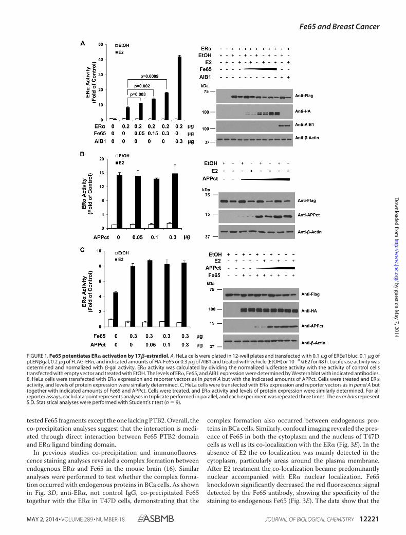
Fe65 Is a Positive ER� Transcriptional Regulator—Publishedstudies have shown that estrogens regulate the transcriptionalactivity of APPct through a Fe65-ER� protein complexdetected both in vitro and in the mouse brain (16). The complexformation makes it possible for Fe65 to regulate ER� activity.To test this idea, the ER� and Fe65 were ectopically expressedin HeLa cells, and ER� transcriptional activity was measuredwith the co-transfected EREe1bluc luciferase reporter gene. Asshown in Fig. 1A, ectopic Fe65 expression in HeLa cellsincreased ER� activity induced by E2 in a dose-dependent man-ner. The increase was statistically significant and about half ofwhat was induced by AIB1, a bona fide ER coactivator. In sim-ilar assays ectopic APPct expression did not increase ER� activ-ity either in the absence (Fig. 1B) or presence (Fig. 1C) of Fe65.IB analyses (Fig. 1, right panels) showed that the ER�, Fe65, andAPPct proteins were expressed in a manner dependent on theamounts of plasmids used in the transfections and that ER�protein expression was not significantly altered by Fe65. Thedata suggest that Fe65 increased ER� activity per molecule. Theanalyses identify Fe65 as a positive ER� regulator that stimu-lates ER� activity in a manner independent of its complex for-mation with APPct.
In similar reporter assays (Fig. 2, left panels), Fe65 exhibitedlittle effects on the transcriptional activity of the androgen (Fig.2B) and vitamin D (Fig. 2C) receptors. The lack of an effect onthe androgen and vitamin D receptors was not due to insuffi-cient Fe65 expression because in parallel analyses (Fig. 2A) thesame ectopic Fe65 expression increased ER� activity to levelscomparable with that presented in Fig. 1, whereas IB analysesrevealed a comparable amount of Fe65 protein expression (Fig.2, right panels). These analyses show that Fe65 may be an ER�-selective transcriptional coactivator.
Fe65 Binds to the ER� and Stimulates Its TranscriptionalActivity through the PTB2 Domain—Previous studies definedthe Fe65 binding domain to the C-terminal ligand bindingdomain of the ER� (16). To define the Fe65 domain mediatingits interaction with the ER�, Fe65 mutants lacking defineddomains were generated (Fig. 3A), and their ability to bind tothe ER� was assessed in co-IP analyses (Fig. 3B) and GST pull-down assays (Fig. 3C). In cells co-transfected with FLAG-ER�with either Myc- or HA-tagged full-length Fe65, Fe65 wasdetected in anti-FLAG precipitates together with the ER�. NoFe65 was detected in the precipitates when cells were trans-fected with Myc-Fe65 and the control vector, showing that thepresence of Fe65 in the FLAG precipitates accurately measuredthe interaction between the ER� and Fe65. Deletion of Fe65regions containing PTB2 eliminated its ability to co-precipitatewith FLAG-ER�, whereas the deletion of other regions had lit-tle effects, showing that the interaction was mediated throughPTB2. This conclusion was reaffirmed by subsequent GST pull-down assays (Fig. 3C), in which GST-ER�-C terminus (GST-ER-C) containing the ligand binding domain precipitated all
Fe65 and Breast Cancer
12220 JOURNAL OF BIOLOGICAL CHEMISTRY VOLUME 289 • NUMBER 18 • MAY 2, 2014
by guest on May 7, 2014
http://ww
w.jbc.org/
Dow
nloaded from
tested Fe65 fragments except the one lacking PTB2. Overall, theco-precipitation analyses suggest that the interaction is medi-ated through direct interaction between Fe65 PTB2 domainand ER� ligand binding domain.
In previous studies co-precipitation and immunofluores-cence staining analyses revealed a complex formation betweenendogenous ER� and Fe65 in the mouse brain (16). Similaranalyses were performed to test whether the complex forma-tion occurred with endogenous proteins in BCa cells. As shownin Fig. 3D, anti-ER�, not control IgG, co-precipitated Fe65together with the ER� in T47D cells, demonstrating that the
complex formation also occurred between endogenous pro-teins in BCa cells. Similarly, confocal imaging revealed the pres-ence of Fe65 in both the cytoplasm and the nucleus of T47Dcells as well as its co-localization with the ER� (Fig. 3E). In theabsence of E2 the co-localization was mainly detected in thecytoplasm, particularly areas around the plasma membrane.After E2 treatment the co-localization became predominantlynuclear accompanied with ER� nuclear localization. Fe65knockdown significantly decreased the red fluorescence signaldetected by the Fe65 antibody, showing the specificity of thestaining to endogenous Fe65 (Fig. 3E). The data show that the
FIGURE 1. Fe65 potentiates ER� activation by 17�-estradiol. A, HeLa cells were plated in 12-well plates and transfected with 0.1 �g of EREe1bluc, 0.1 �g ofpLEN�gal, 0.2 �g of FLAG-ER�, and indicated amounts of HA-Fe65 or 0.3 �g of AIB1 and treated with vehicle (EtOH) or 10�8
M E2 for 48 h. Luciferase activity wasdetermined and normalized with �-gal activity. ER� activity was calculated by dividing the normalized luciferase activity with the activity of control cellstransfected with empty vector and treated with EtOH. The levels of ER�, Fe65, and AIB1 expression were determined by Western blot with indicated antibodies.B, HeLa cells were transfected with ER� expression and reporter vectors as in panel A but with the indicated amounts of APPct. Cells were treated and ER�activity, and levels of protein expression were similarly determined. C, HeLa cells were transfected with ER� expression and reporter vectors as in panel A buttogether with indicated amounts of Fe65 and APPct. Cells were treated, and ER� activity and levels of protein expression were similarly determined. For allreporter assays, each data point represents analyses in triplicate performed in parallel, and each experiment was repeated three times. The error bars representS.D. Statistical analyses were performed with Student’s t test (n � 9).
Fe65 and Breast Cancer
MAY 2, 2014 • VOLUME 289 • NUMBER 18 JOURNAL OF BIOLOGICAL CHEMISTRY 12221
by guest on May 7, 2014
http://ww
w.jbc.org/
Dow
nloaded from

co-localization was mainly determined by ER� localization inthe cells.
Consistent with the mapping analyses, reporter assays showedthat the PTB2 domain is essential for Fe65 to potentiate ER�transcriptional activation. As shown in Fig. 4A, although full-length Fe65 increased ER� activity in a dose-dependent man-ner, the PTB2 deletion mutant did not change ER� activity inparallel analyses. IB analyses showed that the PTB2 deletionmutant was expressed in levels comparable with full-lengthprotein (Fig. 4B), ruling out the possibility that the lack of aneffect of the PTB2 mutant was due to the insufficient proteinexpression. Levels of the ER� protein expression were not sig-nificantly altered by the full-length Fe65 or the PTB2 deletionmutant (Fig. 4B), showing that the reporter analyses reflectedthe effect of Fe65 on the specific activity of the ER� per mole-
cule. The reporter analyses together with the domain mappingallow us to conclude that Fe65 binds to the ER� and potentiatesits activity through its PTB2 domain.
Fe65 Is Expressed in Mammary Epithelial Cells, and theExpression Increased in BCa Cells and Breast Tumor Tissues—The ER� plays an important role in BCa formation and medi-ates the mitogenic activity of estrogens in stimulating BCagrowth. The interaction between the ER� and Fe65 suggests apossible role for Fe65 in estrogen actions in BCa. Consistentwith this idea, Fe65 was detected in the non-tumorigenic MCF-10A human breast epithelial cells, and its expression was foundto be increased in human BCa cells as well as two mouse mam-mary tumor cells: WT145 and VDR-KO (Fig. 5A). Theincreased expression was detected in both ER�-negative andpositive cells with the exception of MCF-7. Subcellular frac-
FIGURE 2. Fe65 selectively increases ER� transcriptional activity. A, HeLa cells were transfected with 0.1 �g EREe1bluc, 0.1 �g of pLEN�gal, and indicatedamounts of FLAG-ER� and HA-Fe65. Cells were treated, and ER� activity and the levels of protein expression were determined with the indicated antibodies asin Fig. 1A. B, HeLa cells were transfected with 0.1 �g of AREe1bluc, 0.1 �g of pLEN�gal, and the indicated amounts of FLAG-AR and HA-Fe65. Cells were treatedwith EtOH or 10�8
M R1881 and AR activity, and the levels of protein expression were determined with the indicated antibodies as in Fig. 1A. C, HeLa cells weretransfected with 0.1 �g of p23luc reporter, 0.1 �g of pLEN�gal, and the indicated amounts of FLAG-VDR and HA-Fe65. Cells were treated with EtOH or 10�8
M
1�,25-dihydroxvitamin D3 (VD3), and VDR activity and levels of protein expression were determined with the indicated antibodies as in Fig. 1A.
Fe65 and Breast Cancer
12222 JOURNAL OF BIOLOGICAL CHEMISTRY VOLUME 289 • NUMBER 18 • MAY 2, 2014
by guest on May 7, 2014
http://ww
w.jbc.org/
Dow
nloaded from

FIGURE 3. Fe65 interacts with the ER� through its PTB2 domain, and the interaction occurs with endogenous proteins in BCa cells. A, schematicrepresentations of the domain structure of full-length Fe65 and its deletion mutants. Numbers of amino acids of full-length Fe65 and its domains are marked.B, 293T cells were transfected with 1.5 �g of FLAG-ER� and Myc-tagged (left panels) or HA-tagged (right panels) Fe65 mutants as indicated. Cellular extractswere subjected to IP analyses followed by IB with indicated antibodies. C, GST and GST-ER�-C proteins produced in and purified from bacteria were incubatedwith lysates of cells transfected with HA-tagged Fe65 deletion mutants. GST pulldown assays were performed, and proteins in the precipitates were detectedby IB with anti-HA antibody (lower panels). Coomassie blue (C.B.) staining was included to show that comparable amounts of GST and GST-ER-C were used inthe pulldown assays. IB analyses of the lysates were also included to show the relative amounts of the Fe65 deletion mutants used in the pulldown assays. D,whole cell lysates of T47D cells were subjected to IP analyses with anti-ER� antibody followed by IB analyses with ER� and Fe65 antibodies as indicated. E, T47Dcells were treated with EtOH or 10�7
M E2 for 1 h and subjected to DAPI (blue) and immunofluorescence staining with anti-ER� (green) and anti-Fe65 (red)antibodies. Images were captured under a confocal fluorescence microscope to show Fe65 and ER� location and co-localization. Images of MDA-MB-361 cellsstably expressing control (Ctrl) or Fe65 (Fe65 KD) shRNA were included as a quality control for Fe65 staining (right panels).
Fe65 and Breast Cancer
MAY 2, 2014 • VOLUME 289 • NUMBER 18 JOURNAL OF BIOLOGICAL CHEMISTRY 12223
by guest on May 7, 2014
http://ww
w.jbc.org/
Dow
nloaded from

tionation revealed the presence of Fe65 in the nucleus of alltested BCa cells, including MCF-7 (Fig. 5B), which appearedabsent in MCF-10A cells. Consistent with the cell line data, IHCstaining of breast tissue sections with anti-Fe65, not IgG con-trol, detected Fe65 expression in both the cytoplasm and thenucleus of epithelial and stromal cells, with a stronger nuclearsignal in the tumor than the control samples (Fig. 5C). Quanti-tative IHC analyses of breast TMA slides containing 140 breasttumor sections and 10 normal and adjacent controls showedthat the nuclear Fe65 in breast tumors was higher than controlsand that the difference was statistically significant (p � 0.0015)(Fig. 5D). The increased Fe65 expression in BCa cells, particu-larly the increase in the nucleus, supports the idea that Fe65 is apositive regulator of ER� actions in tumor cells that may pro-mote breast tumorigenesis. It is important to point out thatalthough the IB analyses in BCa cell lines indicated a higherFe65 expression in ER�-negative than in ER�-positive cells(Fig. 5A), the TMA analyses did not detect a significant differ-ence between ER�-positive and negative tumors (data notshown), showing that the Fe65 expression data from culturedcells did not reflect the situation in tumors. Our data miningand bioinformatics did not reveal a consistent increase (ordecrease) in Fe65 mRNA expression in breast tumors as com-pared with controls (data not shown), indicating that theincreased Fe65 protein expression in tumors may be due to anincrease at the rate of protein synthesis or a decrease in proteindegradation or both.
Fe65 Stimulates the Activity of Endogenous ER� in BCa Cellsand Is Recruited by Estrogens to Target Gene Promoters, Whichin Turn, Facilitates the Recruitment of the ER� and Its Co-activators—To test its effect on endogenous ER�, Fe65 waseither stably expressed or knocked down in ER�-positive BCacells, and the changes in the transcriptional activity of endoge-nous ER� was determined with transfected EREe1bluc report-ers. The transcriptional analyses showed that stable Fe65expression in MCF-7 cells significantly increased E2 stimula-tion of endogenous ER� activity in a dose-dependent manner(Fig. 6A), whereas Fe65 knockdown in both T47D (Fig. 6B) andBT474 (Fig. 6C) cells decreased ER� activity. Consistent withtransient transfection studies, ER� expression was not signifi-cantly altered by Fe65 expression (Fig. 6A), supporting a posi-
tive effect of Fe65 on the specific activity of endogenous ER� permolecule. It is important to note that, under our assay conditions,the activity of endogenous ER� on transfected EREe1bluc reporterwas much weaker than ectopic ER� expressed in HeLa cells, whichmay explain the less dramatic effects of Fe65 on endogenous ER�in BCa cells observed in reporter analyses, which was neverthelessstatistically significant.
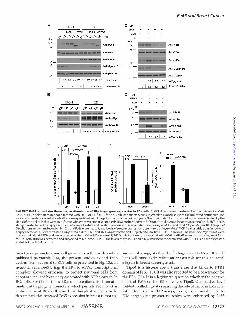
Consistent with the reporter-based transcriptional analyses,transient transfection of Fe65, but not the PTB2 deletionmutant, into MCF-7 cells increased E2 induction of c-Myc andcyclin D1 expression in a dose-dependent manner (Fig. 7A),showing that the positive effect of Fe65 on endogenous ER�observed in reporter assays was translated into changes inestrogen target gene expression and that the effect on the tar-get genes involved PTB2-mediated complex formation withendogenous ER�. Similarly, stable Fe65 expression increased(Fig. 7B), whereas its knockdown in T47D (Fig. 7C) and BT474(Fig. 7D) decreased the ability of E2 to induce c-Myc expres-sion. The regulation of estrogen target genes by Fe65 overex-pression (Fig. 7E) or knockdown (Fig. 7F) was also detected atmRNA levels, supporting a transcriptional control. The levelsof endogenous ER� protein expression were not significantlyaltered by either Fe65 overexpression or knockdown, suggest-ing that the positive effect reflected an increase in ER� activity.The analyses identify Fe65 as a positive regulator of the endog-enous ER� in BCa cells that exerts its positive effect throughPTB2-mediated complex formation with the receptor.
To further characterize the positive effect of Fe65 on endog-enous ER� in BCa cells, ChIP assays were performed to testwhether Fe65 was recruited to the promoters of defined estro-gen target genes and, more importantly, whether changes inlevels of Fe65 expression altered the promoter recruitment ofthe ER� and its coactivators. As shown in Fig. 8A, E2 recruitedFe65 to the promoters of four different estrogen target genes inT47D cells, and the amounts of Fe65 on the promoters weredecreased by Fe65 siRNA. Interestingly, Fe65 knockdown alsoresulted in decreased recruitments of both the ER� and itscoactivator AIB1, showing a role of Fe65 in determining thechromatin recruitment of the ER� and its coactivators. Such arole for Fe65 was further supported by the finding that the sta-ble Fe65 expression in MCF-7 cells increased estrogen recruit-
FIGURE 4. The PTB2 domain is essential for Fe65 to potentiate estrogen stimulation of ER� transcriptional activity. A, HeLa cells were plated andtransfected as in Fig. 1A but together with the indicated amounts of HA-tagged Fe65 or PTB2 deletion mutant. Cells were treated with E2, and ER� activity wassimilarly determined. B, IB analyses were performed in parallel with lysates from transfected cells to show that Fe65 and the PTB2 deletion mutant wereexpressed at comparable levels and did not change ER� protein expression. �-Actin blot was included to show even loading.
Fe65 and Breast Cancer
12224 JOURNAL OF BIOLOGICAL CHEMISTRY VOLUME 289 • NUMBER 18 • MAY 2, 2014
by guest on May 7, 2014
http://ww
w.jbc.org/
Dow
nloaded from

ment of the ER� and AIB1 to target gene promoters (Fig. 8B).More importantly, the ability of TAM to block E2-induced ER�and AIB1 recruitments to the promoters was significantly sup-pressed by Fe65 stable expression, suggesting a potential rolefor Fe65 in the development of TAM resistance.
The increased ER� and AIB1 recruitment to target gene pro-moters might be easily explained if Fe65 promoted estrogen-induced ER� nuclear localization. To test this, cellular distribu-tion of the ER� and Fe65 was analyzed with nuclear andcytosolic extracts prepared from control and Fe65 stable clonesof MCF-7 cells. As shown in Fig. 8C, the stable Fe65 expressionresulted in a significant increase of its presence in both thecytosol and the nucleus but did not alter ER� localization to the
nucleus induced by E2, showing a lack of Fe65 effect on ER�nuclear localization. Overall, the data suggest that the positiveFe65 effect on ER� recruitments to target gene promoters islikely due to a role of Fe65 in facilitating the ER� to interact withchromatin.
To test whether the recruitment of Fe65 to the target genepromoters by E2 depended on the ER�, ChIP assays were per-formed in T47D cells transfected with control or ER� siRNA(Fig. 9). As expected, ER� siRNA decreased ER� proteinexpression (Fig. 9A) as well as the amounts of the ER� and AIB1coactivator on the target gene promoters (Fig. 9B, lower panels).The receptor siRNA did not alter the overall levels of Fe65 pro-tein in the cells (Fig. 9A) but decreased its basal and E2-induced
FIGURE 5. Fe65 is expressed in breast epithelial cells, and the expression is increased in BCa cells and breast tumor tissue samples. A, whole cell lysatesof non-tumorigenic breast epithelial (MCF-10A) and BCa cells were subjected to IB analyses with the indicated antibodies. �-actin blot was included to showeven loading. B, cytoplasmic and nuclear extracts were subjected to IB analyses with indicated antibodies. Hsp60 and HDAC1 blots were included to show theseparation of cytoplasmic and nuclear extracts. C, representative images of normal breast and tumor tissue sections that were immunohistochemically stainedwith control IgG or anti-Fe65 as indicated. Pictures in the lower panels are enlarged (4�) versions of the marked areas of those in the top panels to show thebluish nuclei of normal ductal epithelial cells (middle panel), which indicated weak Fe65 signals, and the strong nuclear staining of breast tumor cells (rightpanels). D, Fe65 expression was assessed by IHC staining and automated quantification of TMA slides containing 140 cores of breast ductal carcinomas (T) and10 cores of normal and adjacent breast tissues (N). The dot plot shows the distribution of nuclear Fe65 signals, whereas the tube plot compares the range andaverage nuclear Fe65 levels between N and T groups.
Fe65 and Breast Cancer
MAY 2, 2014 • VOLUME 289 • NUMBER 18 JOURNAL OF BIOLOGICAL CHEMISTRY 12225
by guest on May 7, 2014
http://ww
w.jbc.org/
Dow
nloaded from

recruitment to the promoters of all tested estrogen target genes(Fig. 9B, top panels). The analyses show that Fe65 recruitmentto estrogen target gene promoters depends on the ER�. Overall,the ChIP analyses support the existence of a mutual positiveregulation between the ER� and Fe65 on their recruitment toestrogen target gene promoters.
Fe65 Promotes Estrogen-induced BCa Cell Growth and Sup-presses the Antagonistic Activity of TAM—Given the fact thatER� activity and the expression of c-Myc and cyclin D1 areoften associated with cell growth in BCa cells, the positiveeffects of Fe65 on ER� activity and target gene expression areexpected to be translated into a positive effect on BCa cellgrowth and its stimulation by estrogens. Indeed, our cell growthanalyses showed that Fe65 knockdown significantly decreasedthe ability of E2 to stimulate the growth of T47D (Fig. 10A) andZR75–1 (Fig. 10B) cells. Consistently, the ability of E2 to stim-ulate MCF-7 cell growth was significantly stronger in Fe65 sta-ble clones than controls (Fig. 10C), and the increase in estrogenstimulation was positively correlated with Fe65 expressionlevels.
Consistent with the ChIP assay data presented in Fig. 8B, theability of TAM to suppress E2-induced cell growth was signifi-cantly reduced by stable Fe65 expression (Fig. 10D). In the con-trol clone, TAM suppressed E2-induced cell growth by 81.6%,
whereas in the Fe65 overexpression clone, it was reduced to61.7%. The data demonstrate that Fe65 promotes estrogen-in-duced cell growth and reduces the antiestrogenic activity ofTAM in suppressing the estrogen stimulation.
DISCUSSION
Fe65 has been well studied in neuronal cells and in the patho-genesis of Alzheimer disease for its functions in mediating thetrafficking and processing of APP as well as its participation intranscriptional regulation (38). Up to now there are essentiallyno reports about the function of Fe65 in cancer cells. The pres-ent studies are the first to clearly establish a role for Fe65 in BCacells. Multiple lines of evidences are presented to support thenovel role of Fe65 as a positive ER� regulator that potentiatesestrogen stimulation of BCa cell growth. First, Fe65 bound tothe ER� through the PTB2 domain and increased ER� activityin reporter assays in a PTB2-dependent manner. Second, Fe65expression was detected in breast epithelial cells, and its expres-sion was found to be increased in human BCa cells and breasttumor tissue samples. Third, E2 recruited Fe65 to the promot-ers of natural estrogen target genes. Finally, Fe65 stable expres-sion increased and its knockdown decreased the activity ofendogenous ER� in BCa cells as well as estrogen-induced targetgene expression, recruitment of the ER� and coactivators to
FIGURE 6. Fe65 potentiates estrogen stimulation of endogenous ER� activity in BCa cells. A, MCF-7 cells stably transfected with empty vector (Ctrl) or Fe65(Fe65 OE Clones) were transiently transfected with 0.3 �g of EREe1bluc and pLEN�Gal. B and C, T47D (panel B) and BT474 (panel C) cells were transfected withcontrol (siCtrl) or Fe65 (siFe65) siRNA and 24 h later re-transfected with 0.3 �g of EREe1bluc and pLEN�Gal. Transfected cells were treated with E2, and luciferaseactivity was determined and normalized as in Fig. 1A. Fe65 overexpression and knockdown as well as its effect on ER� protein expression were monitored byIB analyses with cognate antibodies as indicated.
Fe65 and Breast Cancer
12226 JOURNAL OF BIOLOGICAL CHEMISTRY VOLUME 289 • NUMBER 18 • MAY 2, 2014
by guest on May 7, 2014
http://ww
w.jbc.org/
Dow
nloaded from
target gene promoters, and cell growth. Together with studiespublished previously (16), the present studies extend Fe65actions from neuronal to BCa cells as presented in Fig. 10E. Inneuronal cells, Fe65 brings the ER� to APPct transcriptionalcomplex, allowing estrogens to protect neuronal cells fromapoptosis induced by toxicity associated with APP cleavage. InBCa cells, Fe65 binds to the ER� and potentiates its chromatinbinding at target gene promoters, which permits Fe65 to act asa stimulator of BCa cell growth. Although it remains to bedetermined, the increased Fe65 expression in breast tumor tis-
sue samples suggests that the findings about Fe65 in BCa celllines will most likely reflect an in vivo role for this neuronaladaptor in breast tumorigenesis.
Tip60 is a histone acetyl transferase that binds to PTB1domain of Fe65 (13). It was also reported to be a coactivator forthe ER� (39). It is a legitimate question whether the positiveeffect of Fe65 on the ER� involves Tip60. Our studies haveyielded conflicting data regarding the role of Tip60 in ER� acti-vation by Fe65. In ChIP assays, estrogens recruited Tip60 toER� target gene promoters, which were enhanced by Fe65
FIGURE 7. Fe65 potentiates the estrogen stimulation of ER� target gene expression in BCa cells. A, MCF-7 cells were transfected with empty vector (Ctrl),Fe65, or PTB2 deletion mutant and treated with EtOH or 10�8
M E2 for 2 h. Cellular extracts were subjected to IB analyses with the indicated antibodies. Theexpression levels of cyclin D1 and c-Myc were quantified with ImageJ and normalized with cognate �-actin signals. The normalized signals were divided by thesignal of control cells that were transfected with empty vector or scrambled siRNA and treated with EtOH and are shown at the bottom of the blots. B, MCF-7 cellsstably transfected with empty vector or Fe65 were treated, and levels of protein expression determined as in panel A. C and D, T47D (panel C) and BT474 (panelD) cells transiently transfected with siCrtl or siFe65 were treated, and levels of protein expression determined as in panel A. E, MCF-7 cells stably transfected withempty vector or Fe65 were treated as in panel A but for 1 h. Total RNA was extracted and subjected to real time RT-PCR analyses. The levels of c-Myc mRNA werenormalized with GAPDH and are expressed as -fold of the EtOH control. F, T47D cells transiently transfected with siCrtl or siFe65 were treated as in panel A butfor 1 h. Total RNA was extracted and subjected to real time RT-PCR. The levels of cyclin D1 and c-Myc mRNA were normalized with GAPDH and are expressedas -fold of the EtOH controls.
Fe65 and Breast Cancer
MAY 2, 2014 • VOLUME 289 • NUMBER 18 JOURNAL OF BIOLOGICAL CHEMISTRY 12227
by guest on May 7, 2014
http://ww
w.jbc.org/
Dow
nloaded from

FIGURE 8. Fe65 is recruited to ER� target gene promoters by estrogens and required for optimal recruitment of the ER� and its coactivators. A, T47D cellswere transfected with siCtrl or siFe65 and treated with EtOH or 10�7
M E2 for 45 min. ChIP assays were performed with the indicated antibodies. B, MCF-7 cells stablytransfected with empty vector or Fe65 were treated with EtOH, 10�7
M E2, or 10�7M E2 plus 10�7
M TAM (E2TAM) for 45 min. ChIP assays were performed with theindicated antibodies. C, MCF-7 cells stably transfected with empty vector or Fe65 were treated with EtOH or 10�8
M E2 for 1 h. Cytoplasm and nucleus proteins weresubjected to IB analyses with the indicated antibodies. Hsp60 and HDAC1 blots were included to show the separation of cytoplasmic and nuclear extracts.
FIGURE 9. Fe65 recruitment to estrogen target gene promoters is ER�-dependent. A, T47D cells were transfected with siCtrl or siER�. Cellular extracts weresubjected to IB analyses with indicated antibodies. B, T47D cells were transfected with siCtrl or siER� and treated with EtOH or 10�7
M for 45 min. ChIP assayswere performed with indicated antibodies.
Fe65 and Breast Cancer
12228 JOURNAL OF BIOLOGICAL CHEMISTRY VOLUME 289 • NUMBER 18 • MAY 2, 2014
by guest on May 7, 2014
http://ww
w.jbc.org/
Dow
nloaded from

overexpression (data not shown). The data are consistent withthe fact that Tip60 is a common interacting protein for Fe65and ER�. However, in reporter gene analyses, ectopic expres-sion of Tip60, instead of synergizing with Fe65 in stimulatingER� activity as it would be expected for a coactivator, actuallydecreased the ability of Fe65 to activate the ER� (data notshown). The reporter data are consistent with the fact thatAPPct did not increase ER� activity or potentiate the positiveeffect of Fe65 on the ER� (Fig. 1). Tip60 has been shown to be atumor suppressor of which the expression, particularly theexpression in the nucleus, is decreased in breast tumors (40).Interestingly, it has been shown that estrogen-induced c-Mycand cyclin D1 mRNA expression was not affected by Tip60depletion in MCF-7 cells (41) even though several non-growthrelated ER� target genes were altered, suggesting a selectiveinvolvement of Tip60 in ER� action in BCa cells.
Our data suggest that APPct is not involved in ER� activationby Fe65 (Fig. 1). Questions remain of what signals regulate Fe65nuclear localization in BCa cells and its positive effect on theER�. Under overexpression conditions, Fe65-ER� complex for-mation appeared to be constitutive and not altered by estrogentreatments (data not shown). However, ChIP assays revealedthat Fe65 binding to estrogen target gene promoters wasinduced by E2 (Figs. 8 and 9). The analyses suggest that estro-gens are part of the signals operating in BCa cells to regulate theactions of Fe65-ER� complex in the nucleus. Fe65 overexpres-sion increased both cytoplasmic and nuclear Fe65 levels inMCF-7 cells (Fig. 8C), showing that a simple way of promotingnuclear Fe65-ER� activity is to increase overall Fe65 expres-sion, which naturally occurs in breast tumors as suggested bydata in Fig. 5, C and D. In addition to APP cleavage, Fe65 phos-phorylation had been shown to induce Fe65 nuclear localiza-tion (42). Fe65 has also been reported to interact with the Cterminus of Notch1 (43). It would be interesting to find outwhether these signaling pathways known to operate in BCa cellsregulate ER� activation through Fe65.
Although the present studies have focused on the effect ofFe65 on ER� actions, it is important to point out that Fe65 mayhave a much broad impact on BCa biology beyond estrogenstimulation of cell growth. For example, Fe65 has beendescribed to be required for efficient repair of DNA double-strand breaks, a function that depends on its interaction withTip60 and APPct (26, 27). There are also reports showing thatthe phosphorylated H2A.X is higher in Fe65 null cells than inthe wild type under genotoxic damages (25) and that the phos-phorylation somehow depends on Fe65 accumulation in thenucleus (44 – 46). Thus, through Fe65-ER� complex formation,estrogens may regulate DNA damage repair in breast epithelialcells. Furthermore, the WW domain of Fe65 is known to bind toMena (21), through which it regulates the actin cytoskeleton,cell motility, and neuronal growth cone formation (22, 23).Fe65 thus may also control BCa invasion through Mena andallow estrogens to regulate BCa invasion through the Fe65-ER�complex. In this regard it is important to point out that signif-icant amounts of Fe65 protein were detected in the cytoplasmof BCa cells and that Mena splice isoforms regulate BCa inva-sion (47), which is apparently a cytoplasmic activity mediatedthrough actin filaments.
FIGURE 10. Fe65 promotes estrogen-induced BCa cell growth and sup-presses the antagonistic activity of TAM. T47D (A) and ZR75–1 (B) cellswere estrogen-starved and transfected with siCtrl or siFe65 and 48 h laterre-transfected for another time. 24 h post the second transfection, the cellswere plated in 96-well plates and treated with EtOH or 10�8
M E2 for 3 and 6days. Cell growth was determined in MTT assays. The increase in cell growthrate was calculated by subtracting from and dividing A595 values with A595 ofcontrol cells (time zero). C, control MCF-7 and Fe65 OE clone (2–5) were estro-gen-starved for 72 h and treated with 10�8
M E2 for 0, 3, and 6 days. E2 treat-ment was started at different times to allow the growth of all treated cellsbeing measured in MTT assays at the same time. For example, for 0 day treat-ment, the cells were treated with EtOH for 6 days. The increase in cell growthrate was calculated as in panels A and B. D, control MCF-7 and Fe65 OE clone(2–5) were estrogen-starved for 72 h and treated with either EtOH, 10�7
M E2or 10�7
M E2 plus 1 �M TAM for 3 days. Cell growth was determined in MTTassays. Percentages of TAM inhibition were calculated by subtracting fromthe E2-induced increase in growth rate (over day 0) with that induced by E2plus TAM followed by division with E2-induced increase in growth rate. For allgrowth analyses, each data point represents 18 samples analyzed in parallel.Each experiment was repeated three times. p values were calculated byStudent’s t test. The error bars in all growth analyses represent the S.D. E, amodel describing the role of Fe65 in estrogen actions in neurons and BCacells. See the first paragraph of “Discussion” for details.
Fe65 and Breast Cancer
MAY 2, 2014 • VOLUME 289 • NUMBER 18 JOURNAL OF BIOLOGICAL CHEMISTRY 12229
by guest on May 7, 2014
http://ww
w.jbc.org/
Dow
nloaded from

In summary, the present studies have extended the functionsof the Fe65 neuronal adaptor protein to BCa cells and defined apositive role for Fe65 in estrogen actions and BCa cell growth.The existence of Fe65-ER� complex in BCa cells increases thecomplexity of the mechanisms underlying estrogen actions inBCa cells. For the reason that Fe65 is a multidomain proteinthat interacts with a variety of proteins with diverse functions,its interaction with the ER� may change the receptor activityboth quantitatively and qualitatively by allowing more complexregulation patterns. Our finding that Fe65 expression sup-pressed the antiestrogenic activity of TAM implicates that Fe65may be a new molecular target for BCa intervention and thattargeted disruption of the ER�-Fe65 interaction may representa new strategy to overcome TAM resistance.
Acknowledgments—We thank Dr. JoEllen Welsh for the mammarytumor cells and Drs. M. Muyan, L. Corbo, and P. S. Melzer for pro-viding ER� and AIB1 plasmids, respectively. We also thank Mark C.Lloid and Agnieszka Kasprzak and the pathology core facility of Mof-fitt Cancer Center for the assistance with the TMA analyses.
REFERENCES1. Couse, J. F., and Korach, K. S. (1999) Estrogen receptor null mice. What
have we learned and where will they lead us? Endocr. Rev. 20, 358 – 4172. Le Romancer, M., Poulard, C., Cohen, P., Sentis, S., Renoir, J. M., and
Corbo, L. (2011) Cracking the estrogen receptor’s posttranslational codein breast tumors. Endocr. Rev. 32, 597– 622
3. Early Breast Cancer Trialists’ Collaborative Group (EBCTCG) (2005) Ef-fects of chemotherapy and hormonal therapy for early breast cancer onrecurrence and 15-year survival. An overview of the randomised trials.Lancet 365, 1687–1717
4. Gradishar, W. J. (2005) The future of breast cancer. The role of prognosticfactors. Breast cancer research and treatment. Breast Cancer Res. Treat.89, S17–S26
5. Tsai, M. J., and O’Malley, B. W. (1994) Molecular mechanisms of action ofsteroid/thyroid receptor superfamily members. Annu. Rev. Biochem. 63,451– 486
6. Mangelsdorf, D. J., Thummel, C., Beato, M., Herrlich, P., Schütz, G., Ume-sono, K., Blumberg, B., Kastner, P., Mark, M., Chambon, P., and Evans,R. M. (1995) The nuclear receptor superfamily. The second decade. Cell83, 835– 839
7. Umayahara, Y., Kawamori, R., Watada, H., Imano, E., Iwama, N., Mor-ishima, T., Yamasaki, Y., Kajimoto, Y., and Kamada, T. (1994) Estrogenregulation of the insulin-like growth factor I gene transcription involves anAP-1 enhancer. J. Biol. Chem. 269, 16433–16442
8. Savouret, J. F., Rauch, M., Redeuilh, G., Sar, S., Chauchereau, A., Wood-ruff, K., Parker, M. G., and Milgrom, E. (1994) Interplay between estro-gens, progestins, retinoic acid, and AP-1 on a single regulatory site in theprogesterone receptor gene. J. Biol. Chem. 269, 28955–28962
9. Lonard, D. M., and O’Malley, B. W. (2006) The expanding cosmos ofnuclear receptor coactivators. Cell 125, 411– 414
10. Shang, Y., Hu, X., DiRenzo, J., Lazar, M. A., and Brown, M. (2000) Cofactordynamics and sufficiency in estrogen receptor-regulated transcription.Cell 103, 843– 852
11. Perissi, V., Jepsen, K., Glass, C. K., and Rosenfeld, M. G. (2010) Decon-structing repression. Evolving models of co-repressor action. Nat. Rev.Genet 11, 109 –123
12. Smith, C. L., and O’Malley, B. W. (2004) Coregulator function. A key tounderstanding tissue specificity of selective receptor modulators. Endocr.Rev. 25, 45–71
13. Cao, X., and Südhof, T. C. (2001) A transcriptionally (correction of tran-scriptively) active complex of APP with Fe65 and histone acetyltransferaseTip60. Science 293, 115–120
14. Zambrano, N., Minopoli, G., de Candia, P., and Russo, T. (1998) The Fe65
adaptor protein interacts through its PID1 domain with the transcriptionfactor CP2/LSF/LBP1. J. Biol. Chem. 273, 20128 –20133
15. Kim, H. S., Kim, E. M., Lee, J. P., Park, C. H., Kim, S., Seo, J. H., Chang, K. A.,Yu, E., Jeong, S. J., Chong, Y. H., and Suh, Y. H. (2003) C-terminal frag-ments of amyloid precursor protein exert neurotoxicity by inducing gly-cogen synthase kinase-3� expression. FASEB J. 17, 1951–1953
16. Bao, J., Cao, C., Zhang, X., Jiang, F., Nicosia, S. V., and Bai, W. (2007)Suppression of �-amyloid precursor protein signaling into the nucleus byestrogens mediated through complex formation between the estrogenreceptor and Fe65. Mol. Cell. Biol. 27, 1321–1333
17. McLoughlin, D. M., and Miller, C. C. (2008) The FE65 proteins and Alz-heimer’s disease. J. Neurosci. Res. 86, 744 –754
18. Telese, F., Bruni, P., Donizetti, A., Gianni, D., D’Ambrosio, C., Scaloni, A.,Zambrano, N., Rosenfeld, M. G., and Russo, T. (2005) Transcription reg-ulation by the adaptor protein Fe65 and the nucleosome assembly factorSET. EMBO Rep. 6, 77– 82
19. Trommsdorff, M., Borg, J. P., Margolis, B., and Herz, J. (1998) Interactionof cytosolic adaptor proteins with neuronal apolipoprotein E receptorsand the amyloid precursor protein. J. Biol. Chem. 273, 33556 –33560
20. Hoe, H. S., Magill, L. A., Guenette, S., Fu, Z., Vicini, S., and Rebeck, G. W.(2006) FE65 interaction with the apoE receptor ApoEr2. J. Biol. Chem.281, 24521–24530
21. Ermekova, K. S., Zambrano, N., Linn, H., Minopoli, G., Gertler, F., Russo,T., and Sudol, M. (1997) The WW domain of neural protein FE65 interactswith proline-rich motifs in Mena, the mammalian homolog of Drosophilaenabled. J. Biol. Chem. 272, 32869 –32877
22. Sabo, S. L., Ikin, A. F., Buxbaum, J. D., and Greengard, P. (2001) TheAlzheimer amyloid precursor protein (APP) and FE65, an APP-bindingprotein, regulate cell movement. J. Cell Biol. 153, 1403–1414
23. Sabo, S. L., Ikin, A. F., Buxbaum, J. D., and Greengard, P. (2003) Theamyloid precursor protein and its regulatory protein, FE65, in growthcones and synapses in vitro and in vivo. J. Neurosci. 23, 5407–5415
24. Hu, Q., Hearn, M. G., Jin, L. W., Bressler, S. L., and Martin, G. M. (1999)Alternatively spliced isoforms of FE65 serve as neuron-specific and non-neuronal markers. J. Neurosci. Res. 58, 632– 640
25. Minopoli, G., Stante, M., Napolitano, F., Telese, F., Aloia, L., De Felice, M.,Di Lauro, R., Pacelli, R., Brunetti, A., Zambrano, N., and Russo, T. (2007)Essential roles for Fe65, Alzheimer amyloid precursor-binding protein, inthe cellular response to DNA damage. J. Biol. Chem. 282, 831– 835
26. Stante, M., Minopoli, G., Passaro, F., Raia, M., Vecchio, L. D., and Russo, T.(2009) Fe65 is required for Tip60-directed histone H4 acetylation at DNAstrand breaks. Proc. Natl. Acad. Sci. U.S.A. 106, 5093–5098
27. Murr, R., Loizou, J. I., Yang, Y. G., Cuenin, C., Li, H., Wang, Z. Q., andHerceg, Z. (2006) Histone acetylation by Trrap-Tip60 modulates loadingof repair proteins and repair of DNA double-strand breaks. Nat. Cell Biol.8, 91–99
28. Li, X., Huang, J., Yi, P., Bambara, R. A., Hilf, R., and Muyan, M. (2004)Single-chain estrogen receptors (ERs) reveal that the ER�/� heterodimeremulates functions of the ER� dimer in genomic estrogen signaling path-ways. Mol. Cell. Biol. 24, 7681–7694
29. Le Romancer, M., Treilleux, I., Leconte, N., Robin-Lespinasse, Y., Sentis,S., Bouchekioua-Bouzaghou, K., Goddard, S., Gobert-Gosse, S., andCorbo, L. (2008) Regulation of estrogen rapid signaling through argininemethylation by PRMT1. Mol. Cell 31, 212–221
30. Anzick, S. L., Kononen, J., Walker, R. L., Azorsa, D. O., Tanner, M. M.,Guan, X. Y., Sauter, G., Kallioniemi, O. P., Trent, J. M., and Meltzer, P. S.(1997) AIB1, a steroid receptor coactivator amplified in breast and ovariancancer. Science 277, 965–968
31. Yang, Y., Tse, A. K., Li, P., Ma, Q., Xiang, S., Nicosia, S. V., Seto, E., Zhang,X., and Bai, W. (2011) Inhibition of androgen receptor activity by histonedeacetylase 4 through receptor SUMOylation. Oncogene 30, 2207–2218
32. Lee, H., and Bai, W. (2002) Regulation of estrogen receptor nuclear exportby ligand-induced and p38-mediated receptor phosphorylation. Mol. Cell.Biol. 22, 5835–5845
33. Jiang, F., Li, P., Fornace, A. J., Jr., Nicosia, S. V., and Bai, W. (2003) G2/Marrest by 1,25-dihydroxyvitamin D3 in ovarian cancer cells mediatedthrough the induction of GADD45 via an exonic enhancer. J. Biol. Chem.278, 48030 – 48040
Fe65 and Breast Cancer
12230 JOURNAL OF BIOLOGICAL CHEMISTRY VOLUME 289 • NUMBER 18 • MAY 2, 2014
by guest on May 7, 2014
http://ww
w.jbc.org/
Dow
nloaded from

34. Li, P., Nicosia, S. V., and Bai, W. (2001) Antagonism between PTEN/MMAC1/TEP-1 and androgen receptor in growth and apoptosis of pros-tatic cancer cells. J. Biol. Chem. 276, 20444 –20450
35. Kasiappan, R., Shen, Z., Tse, A. K., Jinwal, U., Tang, J., Lungchukiet, P.,Sun, Y., Kruk, P., Nicosia, S. V., Zhang, X., and Bai, W. (2012) 1,25-Dihy-droxyvitamin D3 suppresses telomerase expression and human cancergrowth through microRNA-498. J. Biol. Chem. 287, 41297– 41309
36. Nakaya, T., and Suzuki, T. (2006) Role of APP phosphorylation in FE65-dependent gene transactivation mediated by AICD. Genes Cells 11,633– 645
37. Shang, Y., and Brown, M. (2002) Molecular determinants for the tissuespecificity of SERMs. Science 295, 2465–2468
38. Bórquez, D. A., and González-Billault, C. (2012) The amyloid precursorprotein intracellular domain-Fe65 multiprotein complexes. A challenge tothe amyloid hypothesis for Alzheimer’s disease? Int. J. Alzheimers Dis.2012, 353145
39. Brady, M. E., Ozanne, D. M., Gaughan, L., Waite, I., Cook, S., Neal, D. E.,and Robson, C. N. (1999) Tip60 is a nuclear hormone receptor coactivator.J. Biol. Chem. 274, 17599 –17604
40. Gorrini, C., Squatrito, M., Luise, C., Syed, N., Perna, D., Wark, L., Marti-nato, F., Sardella, D., Verrecchia, A., Bennett, S., Confalonieri, S., Cesaroni,M., Marchesi, F., Gasco, M., Scanziani, E., Capra, M., Mai, S., Nuciforo, P.,Crook, T., Lough, J., and Amati, B. (2007) Tip60 is a haplo-insufficienttumour suppressor required for an oncogene-induced DNA damage re-sponse. Nature 448, 1063–1067
41. Jeong, K. W., Kim, K., Situ, A. J., Ulmer, T. S., An, W., and Stallcup, M. R.(2011) Recognition of enhancer element-specific histone methylation byTIP60 in transcriptional activation. Nat. Struct. Mol. Biol. 18, 1358 –1365
42. Lee, E. J., Chun, J., Hyun, S., Ahn, H. R., Jeong, J. M., Hong, S. K., Hong,J. T., Chang, I. K., Jeon, H. Y., Han, Y. S., Auh, C. K., Park, J. I., and Kang,S. S. (2008) Regulation Fe65 localization to the nucleus by SGK1 phosphor-ylation of its Ser-566 residue. BMB Rep. 41, 41– 47
43. Fischer, D. F., van Dijk, R., Sluijs, J. A., Nair, S. M., Racchi, M., Levelt, C. N.,van Leeuwen, F. W., and Hol, E. M. (2005) Activation of the Notch path-way in Down syndrome. Cross-talk of Notch and APP. FASEB J. 19,1451–1458
44. Nakaya, T., Kawai, T., and Suzuki, T. (2008) Regulation of FE65 nucleartranslocation and function by amyloid �-protein precursor in osmoticallystressed cells. J. Biol. Chem. 283, 19119 –19131
45. Nakaya, T., Kawai, T., and Suzuki, T. (2009) Metabolic stabilization of p53by FE65 in the nuclear matrix of osmotically stressed cells. FEBS J. 276,6364 – 6374
46. Kawai, T., Nakaya, T., and Suzuki, T. (2010) Roles of the intramolecularregions of Fe65 in its trans-accumulation and in p53 stabilization in thenuclear matrix of osmotically stressed cells. FEBS Lett. 584, 765–769
47. Di Modugno, F., Iapicca, P., Boudreau, A., Mottolese, M., Terrenato, I.,Perracchio, L., Carstens, R. P., Santoni, A., Bissell, M. J., and Nisticò, P.(2012) Splicing program of human MENA produces a previously unde-scribed isoform associated with invasive, mesenchymal-like breast tu-mors. Proc. Natl. Acad. Sci. U.S.A. 109, 19280 –19285
Fe65 and Breast Cancer
MAY 2, 2014 • VOLUME 289 • NUMBER 18 JOURNAL OF BIOLOGICAL CHEMISTRY 12231
by guest on May 7, 2014
http://ww
w.jbc.org/
Dow
nloaded from