j. insect physiol., vol. selectivity of yolk … · comparison of vitellogenins of two insects ......
TRANSCRIPT

809
J. Insect Physiol., 1976, Vol. 22, pp. 809 to 818. Pergamon Press. Printed in Great Britain.
SELECTIVITY OF YOLK PROTEIN UPTAKE:COMPARISON OF VITELLOGENINS OF TWO INSECTS
JOSEPH G. KUNKEL* and M. L. PANt
Yale University, New Haven, Connecticut andUniversity of Massachusetts at Amherst, Amherst, Massachusetts, U.S.A.
(Received 13 November 1975)
Abstract--The vitellogenins of Hyalophora cecropia and Blattella germanica have similar Stokes' radii (69 and75,&), sedimentation coefficients (15.9 and 16.8s) and isoelectric points (pH 5.7 and 5.0), and share similar aminoacid compositions with vitellogenins of other animals. The two vitellogenins show no immunological crossreaction.Blattella oocytes take up their own vitellogenin in vivo at a rapid rate and that of Hyalophora at a low ratecomparable to that of a non-vitellogenic protein. Hyalophora oocytes take up their own vitellogenin rapidly in vitroand Blattella Vitellogenin only at a negligible rate. Molecular weights, shapes and charges of the vitellogenins aresimilar to non-vitellogenins and form no basis for selective uptake.
INTRODUCTION
In the development of the yolky eggs of many animalspecies, certain serum proteins are the major sourceof yolk. These serum proteins, termed vitellogenins,are synthesized and secreted from other maternal tis-sues (the liver in vertebrates, and the fat body in in-sects), and enter the o6cytcs by pinocytosis{WALLACE and DUMONT, 1968; TELFER andSMIm, 1970) to form the vitellin or yolk protein.This uptake is interesting because it is highlyselective; the vitellogenin may be one, or a few outof many serum proteins in the blood. Specializedcoated vesicles may be involved in the uptake of thevitellogenins (ANDERSON, 1970).
One test of the selectivity of the uptakemechanism is whether oocytes of one species cantake up the vitellogenin of another species. Inter-specific uptake of vitellogenins has been observedpreviously between two species of saturniid moth(TELFER, 1960), between closely relatedcockroaches (BELL, 1972) and also, interestinglyenough, between chickens and alligators (SCHJEIDEet al., 1963). The latter might be considered asevidence that the uptake mechanism is rather non-specific since it can span the classes of reptiles andbirds. We present evidence here to the contrary,showing that purified vitellogenins from two speciesof insects of different orders, Dictyoptera and Lepi-doptera, will not support vitellogenesis in eachother's o6cytes, even though they have very similargross chemical properties. The results indicate thatthe selective uptake mechanism involves recognitionof subtle differences in the protein structure.
* Present address: Zoology Department, University ofMassachusetts at Amherst, Amherst, Mass. 01002, U.S.A.
± Present address: Department of Zoology, Universityof Tennessee, Knoxville, Tenn. 37916, U.S.A.
MATERIALS AND METHODS
AnimalsCockroaches, Blattella germanica, were kept in
synchronous culture by controlling their feeding(KUNKEL, 1966), thus providing a source ofuniformly aged adult females for use in experiments.Silkmoths, Hyalophora cecropia, were raised onblack cherry foliage in the field; pupae were chilledto terminate diapause: and pharate adults were stagedaccording to SCHNEIDERMAN and WILLIAMS(1954).
Isolation of vitellogeninsSince the vitellogenins may be modified after
uptake into the oocytes (BROOKES and DEJMAL,1968; WALLACE, 1964), they were isolated fromthe blood of ovariectomised females, in which thevitellogenins accumulate. The vitellogenins of thetwo species and two non-vitellogenic serum proteins,I and II, of Blat-tella were purified by combinationsof DEAL cellulose chromatography and sucrosegradient centrifugation (PAN and WALLACE, 1974;KUNKEL, in prep.).
Labeling of vitellogenins
For Blattella 0.2 µCi of uniformly labeled 14C-amino acid mixture (Schwarz-Mann) was injecteddaily into each of 58 ovariectomised adult females ondays 2 to 5 after the first feeding. On the 6th dayafter feeding, haemolymph was collected in 0.15 MNaC1 buffered with 0.01 M Sodium phosphate, pH7.2 (PBS). The serum was centrifuged to removecells and the vitellogenin was then isolated. ForHyalophora, 4 diapausing pupae were ovariec-tomised according to TELFER (1954) andtransferred to 25°C to initiate pupal adultdevelopment. On day 15 to 16 of the pharate adult

810 Kunkel and Pan
stage, each was injected with 0.1 mCi 3H-leucine(Schwarz-Mann), and held for 4 days to label thevitellogenin. Haemolymph was collected, with a traceof phenylthiourea to prcvcnt melanin formation,centrifuged to remove cells, and pooled. Vitellogeninwas then isolated.
After purification, the specific activities of theproteins were determined by dissolving samples inNCS (Nuclear Chicago), counting by liquidscintillation in toluene scintillator (Packard Tricarb3003), and expressing radioactivity on the basis ofprotein estimated from absorbance at 280 nm. Theamount of radioactive protein taken up by oocytes wasestimated in a similar manner, correcting fordifferences in counting efficiency. Double labelcounting was performed as described by BUSH (1964).
Immunochemical methodsAntisera against the vitellogenins of Blattella and
Hyalophora were obtained by immunizing rabbits withovariectomized female blood and yolk extracts,respectively, emulsified with Freund's complete adju-rant. Antisera thus prepared werc rendered specific forvitellogenins by absorption with larval blood forBlattella and with adult male blood for Hyalophoraantiserum. Figure 1 demonstrates the purity of thevitellogenins as well as the specificity of the antibodies.
Micro-Ouchterlony tests were performed on stan-dard 25 x 75 mm microscope slides covered with 2 mlof 0.5% agarose made in PBS. Wells were cut with a14 gauge needle and the distance from the peripheralwells to the centre well was 5 mm. Immuno-electrophoresis was performed as described previously(PAN and WYATT, 1971). The Oudin test was carriedout as described previously (BECKER et al., 1951).
Biochemical analysesAgarose gel filtration (Biogel A1.5, Bio Rad Labs)
was used for determining molecular (Stokes') radii(ACKERS, 1964, 1967). The elution volume (Ve) of aprotein was determined on a 2.5 x 50cm agarosecolumn. Blue dextran (Pharmacia Co.) was used todetermine the void volume (Vo), and dinitrophenyla-lanine was used to determine the internal volume (vi).The column partition coefficient, o, for a protein wascalculated as (Ve-Vo)/Vi. The a values for three calib-
ration proteins of known Stokes' radii (ACKERS,1964) were determined using multiple independent
runs. The inverse error-frequency-complement (erfc-1)transformation of a (ACKERS, 1967) was calculatedfrom standard tables of the error function. Transfor-mations of sigma were plotted against the knownStokes' radii and a straight line was fitted to the datausing least squares (Fig. 2). The Stokes' radius of anunknown protein with an associated standard error ofestimation was calculated from its elution volume byextrapolation using the standard curve (RAo, 1965).
Amino acid analyses were performed on the purifiedproteins delipidated by three successive chloroform
methanol (2:1) extractions, followed by methanol andanhydrous ether rinses and drying under a continuousflow of dry nitrogen. Multiple samples of 2 mg proteinwere hydrolysed in vacuo in 6 N HCl at 110C for 24,48 and 72 hr and the hydroly-sates were analysed on aBeckman 120C amino acid analyser with automaticintegrator using an accelerated technique(SPACKMAN, 1967). Data were analysed by digitalcomputer, extrapolating ammonia, threonine and serineto zero hydrolysis time and valine and isoleucine toinfinite time (SPACKMAN el al., 1958). Cystine pluscystine was determined as cys-teic acid by performicacid oxidation (HIRS, 1967). Tryptophan wasdetermined spectrophotometricallyby its oxidation with N-bromo succinimide (SPANDEand WITKOP, 1967).
Percent lipid was determined from the chloroform:methanol extracts of the proteins. Carbohydratecontent of the extracted proteins was measured by theanthrone reaction (SPIRO. 1966) and expressed as apercentage as mannose (YAMASAKI, 1973; BARZEVet al. 1975). Partial specific volumes were calculatedfrom composition data (SCHACHMAN, 1966)assuming specific volumes of 0.640 and 1.093respectively for the carbohydrate and lipid components.Molecular weights and frictional ratios, f/fo, of the
native proteins were calculated from the sedimentationcoefficients, Stokes' radii and partial specific volumes(SIEGEL and MONTY, 1966). Subunit mol. wt weredetermined by SDS gel electrophoresis (WEBER et al.,1972). The sedimentation coefficients were estimatedby sucrose gradient sedimentation (MARTIN andAMES, 1961) using human IgG, catalase and cecropiavitellogenin (PAN and WALLACE, 1974) as knownsedimenting markers.
Fig. 2. Gel filtration determination of Stokes' radii of thevitellogcnins of silk moth and cockroach. The Stokes' radii ofstandard proteins were plotted against the inverse errorfrequency complcment (erfc-1) of the column partitioncoefficient, sigma, for each protein, (2.5 x 50 cm, BioGel A1.5).The arrows point to the average elution position for three runs ofeach unknown: Blattella vitellogenin BgV and HyaIophoravitellogenin, HcV.

811
Fig. 1. Immunoelectrophoresis of purified vitellogenin, V, male sera and ovariectomised female sera of Blattella, Bg and Hyalophora, Hc. The antisera used were: a complex antiserum against silk moth yolk,anti-Hc; the same antiserum adsorbed making it specific for vitellogenin, anti-HcV; a complex antiserumagainst Blattella ovariectomised female serum, anti-Bg; the same antiserum adsorbed to make it specific forvitellogenin, anti-BgV. Illustrated is a photographic composite of two slides; on one slide the proteins werestained immediately after electrophoresis, and in a duplicate the antisera were added and immunodiffusionwas allowed to occur before washing and staining.

812
Fig. 4. Subunit composition of purified serum proteins determined by SDS acrylamide gel electro-phoresis (7.5% acrylamide, 0.4 x 8 cm gels, Coomasie Brilliant Blue stain; WEBER et al., 1972) SP = non-vitellogenicserum proteins I and lI of Blattella. V , vitellogenins of Blattella, Bg and Hyalo-phora Hc. STD = a gel withfive known molecular weight protein standards, top to bottom: phos-phorylase a, bovine serum albuminmonomer, pepsin, trypsin and lysozyme. Each of the gels contains an equivalent amount of lysozyme as aninternal mobility standard.

Selectivity of vitellogenesis 813
RESULTS
Physical properties of the vitellogeninsPhysical and chemical properties of the two
vitello-genins and the two Blattella serum proteins aregiven in Table 1. The Stokes' radii of the vitellogeninsof Hyalophora and Blattella, although similar, aresignificantly different (P < 0.001). The two other majorserum proteins in adult Blattella, I and II, have simi-larly large Stokes' radii but Bg I1 despite its large sizehas a low sedimentation coefficient due to its high(53%) lipid content. The similar size of the twovitellogenins is also indicated by their mobility in suc-rose gradients (Fig. 3). They form a single absorbance
peak sedimenting at about 16s, but the 14C labelledBlattella protein is seen to sediment at about 16.8s,
slightly faster than the 3H labelled Hyalophora proteinwhose sedimentation coefficient, 15.9s, has beendetermined in the analytical ultracentrifuge (PAN andWALLACE, 1974). Based on their Stokes' radii, partialspecific volumes and sedimentation coefficients, thecalculated molecular weights and frictional ratios of thefour proteins suggest that they are all similarly largeand spherical (Table 1).Despite similarities in native molecular weight, uniquesubunit structure is revealed by SDS acrylamide gelelectrophoresis, Fig. 4. Both vitellogenins have a largeand a small subunit. The Blattella vitellogenin hasvariable minor bands which may be artefacts of howlong the protein had been in the bloodstream of theovariectomised female. Molecular weights of thesubunits were estimated by running suitable knownmol. wt standard proteins in the same gels with theunknowns and extrapolating on a mobility vs log mol.wt plot of the standard proteins (Table 1). Since molarratios of the subunits cannot be adequately determinedfrom the stainability of the gels it is not possible toaccurately estimate the number of each subunit in thenative 16s vitellogenins. Since Blattella serum protein Ihas only one subunit it was possible to calculate thatthere are six subtraits in the native protein. This along
with its sedimentation coefficient and aminoacidcomposition (Table 2), suggest that it is similar tothe protein calliphorin (MUNN et al., 1972). Blattellaserum protein II has an apparently heterogeneoussubunit composition. A serum protein with similarp r o p e r t i e s h a s b e e n d e s c r i b e d i n
Table 1. Physical and chemical properties of vitellogenic and non-vitellogenic serum proteins
Stokes' MW§ MW% % radius native SDS
Vitellogenins pI CHO lipid Vξ (Å) S20.w (×103) f/fo§ (× 103)
___Blattella 5.0 4.5 15.7 0.784 75.0 -+ 1.6' 16.8 659 1.27 100 germanica 52Hyalophora 5.7 1.0 9.43' 0.758 69.4 + 1.6' 15.9 516 1.29 120 cecropia 43Non-vitellogenins Bg I 5.5 0.0 0.731 59.5 19.0 476 1.16 80 Bg II 6.9 53.2 0.926 63.0 5.4 511 1.10 __* mean ± standard error of the mean for three replicates.t from PAN and WALLACE (1974).ξ calculated from amino acid, CHO and lipid composition (Schachman, 1957).§ calculated from partial specific volume, Stokes' radius and sedimentation coefficient. (SIEGEL and MONTY, 1966).
Fig. 3. Relative sedimentation of 3H-Hyalophora vitello-
genin and 14C-Blattella vitellogenin. Human lgG was usedas a 7S sedimentation marker. Sedimentation is from left toright, 5 to 25'5[, sucrose gradient in PBS (InternationalSB283 rotor, 4l,000 rev/min, 15 hr), fractions were differ-entially counted in Bray's solution (BRAY, 1960).
814 JOSEPH G. KUNKEL AND M. L. PAN
Locusta migratoria (PELED and TIETZ, 1965).Isoelectric points of the two vitellogenins were taken asthe pH of zero mobility in electrophoresis (Fig. 5).There is a difference of approximately 0.7 pH unit inisoelec-tric point which may account for the higherelectro-phoretic mobility of Blattella vitellogenin seenin Fig. 1. Blattella serum protein I has an acidisoelectric point between that of the two vitellogeninswhile serum protein II has an isoelectric point close toneutrality (Table 1).
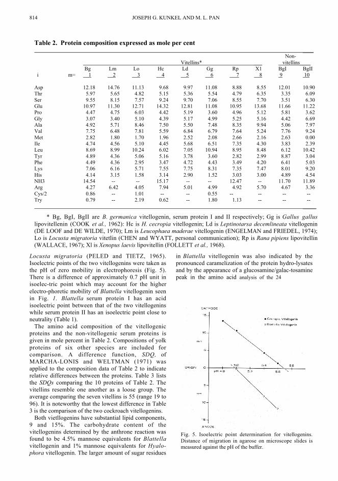
The amino acid composition of the vitellogenicproteins and the non-vitellogenic serum proteins isgiven in mole percent in Table 2. Compositions of yolkproteins of six other species are included forcomparison. A difference function, SDQ, ofMARCHA-LONIS and WELTMAN (1971) wasapplied to the composition data of Table 2 to indicaterelative differences between the proteins. Table 3 liststhe SDQs comparing the 10 proteins of Table 2. Thevitellins resemble one another as a loose group. Theaverage comparing the seven vitellins is 55 (range 19 to96). It is noteworthy that the lowest difference in Table3 is the comparison of the two cockroach vitellogenins.
Both vietllogenins have substantial lipid components,9 and 15%. The carbohydrate content of thevitellogenins determined by the anthrone reaction wasfound to be 4.5% mannose equivalents for Blattellavitellogenin and 1% mannose equivalents for Hyalo-phora vitellogenin. The larger amount of sugar residues
in Blattella vitellogenin was also indicated by thepronounced caramelization of the protein hydro-lysatesand by the appearance of a glucosamine/galac-tosaminepeak in the amino acid analysis of the 24
Table 2. Protein composition expressed as mole per cent ____
Non- Vitellins* ___ vitellins____ Bg Lm Lo Hc Ld Gg Rp X1 BgI BgII
i m= 1 2 3 4 5 6 7 8 9 10
Asp 12.18 14.76 11.13 9.68 9.97 11.08 8.88 8.55 12.01 10.90Thr 5.97 5.65 4.82 5.15 5.36 5.54 4.79 6.35 3.35 6.09Ser 9.55 8.15 7.57 9.24 9.70 7.06 8.55 7.70 3.51 6.30Glu 10.97 11.30 12.71 14.32 12.81 11.08 10.95 13.68 11.66 11.22Pro 4.47 4.75 6.03 4.42 5.19 3.60 4.96 5.12 5.81 3.62Gly 3.07 3.40 5.10 4.39 5.17 4.99 5.25 5.16 4.42 6.69Ala 4.92 5.71 8.46 7.50 5.50 7.48 8.35 9.94 5.06 7.97Val 7.75 6.48 7.81 5.59 6.84 6.79 7.64 5.24 7.76 9.24Met 2.82 1.80 1.70 1.96 2.52 2.08 2.66 2.16 2.63 0.00Ile 4.74 4.56 5.10 4.45 5.68 6.51 7.35 4.30 3.83 2.39Leu 8.69 8.99 10.24 6.02 7.05 10.94 8.95 8.48 6.12 10.42Tyr 4.89 4.36 5.06 5.16 3.78 3.60 2.82 2.99 8.87 3.04Phe 4.49 4.36 2.95 3.47 4.72 4.43 3.49 4.20 6.41 5.03Lys 7.06 6.16 5.71 7.55 7.75 8.31 7.35 7.47 8.01 9.20His 4.14 3.15 1.58 3.14 2.90 1.52 3.03 3.00 4.89 4.54NH3 14.54 -- -- 15.17 -- -- 12.47 -- 11.70 11.89Arg 4.27 6.42 4.05 7.94 5.01 4.99 4.92 5.70 4.67 3.36Cys/2 0.86 -- 1.01 -- -- 0.55 -- -- -- --Try 0.79 -- 2.19 0.62 -- 1.80 1.13 -- -- -- _____
* Bg, BgI, BgII are B. germanica vitellogenin, serum protein I and II respectively; Gg is Gallus galluslipovitellenin (COOK et al., 1962): Hc is H. cecropia vitellogenin; Ld is Leptinotarsa decemlineata vitellogenin(DE LOOF and DE WILDE, 1970); Lm is Leucophaea maderae vitellogenin (ENGELMAN and FRIEDEL, 1974);Lo is Locusta migratoria vitetlin (CHEN and WYATT, personal communication); Rp is Rana pipiens lipovitellin(WALLACE, 1967); Xl is Xenopus laevis lipovitellin (FOLLETT et al., 1968).
Fig. 5. Isoelectric point determination for vitellogenins.Distance of migration in agarose on microscope slides ismeasured against the pH of the buffer.

Selectivity of vitellogenesis 815
hr hydrolysates only. No such caramelization nor peakwas observed for Hyalophora vitellogenin.
To determine if there are detectable antigenicsimilarities of the two vitellogenins, they werecompared immunologically using the Ouchterlonydouble diffusion test as well as the Oudin singlediffusion test. No visible reaction occurred betweeneither protein and the antiserum prepared against theother.
Physiological comparison of vitellogeninsReciprocal experiments were performed to test
whether the two vitellogenins could replace each otherphysiologically. First the two radioactively labeledvitellogenins were tested for their ability to be takenup in vivo from the haemolymph into the ovaries ofBlattella. Four days after molting 60 unfed virginadult female cockroaches were fed simultaneously at30°C and allowed to develop for 98 hr, by which time,active yolk deposition was taking place (KUNKEL,1973). They were then injected with approximately 36
µg of 14C-Blattella vitellogenin (60 dis/rain/g) or 3H-
Hyalophora vitellogenin (150 dis/ min/µg), or 14C-Blattella serum protein II (105 dis/ min/µg) as acontrol for non-specific uptake. At 2, 4, 8, and 19 hrafter injection, five animals were sacrificed for each ofthe proteins injected. The ovaries were dissected out,rinsed in PBS, and the amount of incorporatedradioactivity determined. Figure 6 illustrates theproportion of the injected radioactivity taken up fromthe haemolymph by the ovaries by different times. Theinjected Blattella vitellogenin was taken up rapidly(initial rate of 0.2 to 0.3 µg/oocy-te/hr.) while theHyalophora vitellogenin was taken up at a muchslower rate similar to that for the con-
trol protein, Blattella serum protein II. Therefore, itseems that Blattella oocytes do not recognize Hyalo-phora vitellogenin as a vitellogenic protein.
Uptake of the two vitellogenins into the oocytesof Hyalophora was tested by incubation of o6cytes in
vitro. Equivalent weights of 14C-Blattella vitello-genin and 3H-Hyalophora vitellogenin were added to200 µ1 of medium, making it approximately 1.8%protein. Ovaries were dissected from 18 to 19 daypharate adult moths and individual vitellogenicoocytes with surrounding follicle cells were isolatedand graded in size, Medium sized vitellogenic oocyteswere placed into the incubation medium. Samples ofoocytes were taken from the medium at intervals andrinsed. The diameter of each oocyte was measuredbefore it was dissolved in NCS for counting. Theaverage amount of each protein taken up per oocyte aswell as the counts per min per unit surface area aregiven in Fig. 7. In this incubation of Hyalophoraoocytes, the moth vitellogenin was taken up at a rateof about 0.75µg/oocyte/hr, while the cockroachvitellogenin was virtually excluded.
DISCUSSION
The mechanism by which o6cytes recognizevitello-genin is of interest as an example of how a cellcommunicates with its environment through its cellsurface. The o6cyte surface comes in contact withmany serum proteins during vitellogenesis but onlyvitello-genin is recognized and singled out for rapidpinocy-totic uptake. A previous result (SCHJEIDE etal., 1963) has suggested that the method of recognitionof vitel-1ogenin by an oocyte might not be highlyspecific.
Table 3. Relatedness based on mole per cent* amino acid composition. Departures from identity are measured by the SΔQ §statistic of MARCHALONIS and WELTMAN (1971). ___
Non- Vitellins ___ vitellins ___
Bg Lm Lo Hc Ld Gg Rp Xl BgI BgIIk \ j = 1 2 3 4 5 6 7 8 9 10
1 0 19 82 55 22 39 44 68 76 632 0 71 56 42 39 63 72 82 763 0 96 80 67 67 76 144 1144 0 24 64 58 29 107 1095 0 36 25 65 80 686 0 21 40 101 457 0 24 121 628 0 119 629 0 9910 0
___* For consistency the mole percents of Table 2 were computed ignoring the contribution of NH3, Cys/2 and Try to the
denominator and these groups were not used in the application of the equation for SΔQ.
§ SΔQjk = Σ (Pij-Pik)2, where Pim = mole per cent of amino acid 'i' in protein 'm', 'j' and 'k' index the columns and rows of
Table 3. 'i' and 'm' indexes the amino acids and proteins of Table 2.

816 JOSEPH G. KUNKEL AND M. L. PAN
In testing this hypothesis we have taken two insectvitellogenins and compared their physical, chemicaland physiological properties.
Vitellins and their precursors, vitellogenins tend tobe large macromolecules (8.9 to 28s; COOKE et al.,1962; WALLACE et al., 1967; BROOKES andDEJMAL, 1968; PAN and WALLACE, 1974): thecockroach and silkmoth vitellogenins studied aboveconform to this generality, both sedimenting close to arate of 16s with diameters between 140 and 150Å. Thepartial specific volumes (V) computed fromcomposition data are all higher than those usuallyassumed for simple proteins even for Blattella serumprotein I, which lacks carbohydrate and lipid. Thepartial specific volume computed for silkmothvitellogenin agrees well with a previous determinationby pyncnometry (PAN and WALLACE, 1974). Themol. wt computed from the Stokes' radii, sedimentationcoefficients, and partial specific volumes show that allthe serum proteins studied are large, 476,000 to659,000 mol. wt. The computed frictional ratios, f/fo,show them all to be close to spherical. This largespherical size of the vitellogenins may be an adaptationfor serving as an efficient transport and storage protein.A more general reason that all the major serum proteinsof Blattella may be of large diameter is to avoid filt-ration by the pericardial cells, which have specialisedintracellular filtration junctions to prevent large mol-ecules from reaching the pinocytotic surface (CROSS-LEY, 1972: LIBERTOFF and KUNKEL, in prep.).
Both vitellogenins are heteropolymers with twoapparently different size classes of subunits. However,since the vitellogenins contain a substantial amount of
carbohydrate the determinations of mol. wt may not beaccurate (WEBER et al., 1972). Blattella serum proteinI which lacks substantial carbohydrate has a singlesubunit of about 80,000 mol. wt. This suggests that thenative protein has six subunits. Blattella serum proteinII although homogeneous on sucrose gradients, discelectrophoresis pH 7.5, and immuno-electrophoresispH 6.5, gives multiple bands on SDS gelelectrophoresis. This is similar to a serum lipo-proteinfrom Locusta which also shares an extremely similaramino acid conposition (PELED and TIETZ, 1975).
The isoelectric points of vitellogenins in this studywere low, 5.0 and 5.7, so that at physiological pH theywould have a pronounced negative charge. This couldnot be a sufficient criterion to allow for vitello-genicrecognition since the non-vitellogenic serum proteins ofBlattella would also be negatively charged. Negativecharge would also be an unusual general criterion foruptake since in other model pinocytotic systems mostproteins will only stimulate pinocytosis below theirisoelectric points i.e. when they are positively charged(GIESE, 1973).
The relative chemical composition of vitellogeninsmight give some clue to specificity. The amino acidcomposition of the eight vitellogenins show somesuperficial similarities such as high aspartate and glu-tamate and low methionine which are characteristics ofproteins in general (KING and JUKES, 1969). In theabsence of amino acid sequence data it is difficult
Fig. 6. Uptake of serum proteins into Blattella oocytes in f/fo.
At zero time approximately 36 µg of radioactively labelledprotein was injected into groups of females which had beenfed for 98 hr. The proportion of the injected protein taken up isplotted against time since injection.
Fig. 7. Uptake of vitellogenins into silkmoth oocytes in vitro.Medium-sized vitellogenic oocytes were placed into 200 µ1 of aculture medium (PAN et al., 1967) containing approximately 9mg/ml each of C-14-Blattella vitellogenin and H-3-Hyalophoravitellogenin. The uptake over time with associated standard error(vertical bars) was calculated in counts per min per square mmof oocyte surface and is also displayed in µg of protein enteringthe average sized egg.

Selectivity of vitellogenesis 817to make judgements of the homology of the proteinsbeing compared. We applied the difference function ofMARCHALONIS and WELTMAN (1971) to theamino acid composition data. This function, SΔQ,measures departures from identical mole percentamino acid composition on a scale from zero to20,000. Identical proteins analysed in differentlaboratories are found to have SΔQs of up to 4. Thedistribution of SΔQ for large numbers of comparisonsof related and unrelated proteins has been published(MARCHALONIS and WELTMAN, 1971), providinga background for comparison of the tabulated analysesof vitellogenins. The vitellogenins form a group interms of amino acid composition. None of the 28comparisons of vitellogenins result in SΔQs over 100,while fewer than 2 percent of the 820 crosscomparisons of 41 unrelated proteins have SΔQs lowerthan 100 (MARCHALONIS and WELTMAN, 1971).The uniformly low SΔQs demonstrate a conservatismin amino acid composition of vitellogenins. However,immunological cross-reaction, a measure of aminoacid sequence difference (PRAGER and WILSON,1971), suggests that vitello-genins are notconservative in primary structure. As reported above,the cockroach and silk moth vitello-genins do notshow any immunological crossreaction, andsubsequent work has shown that insect vitello-geninsdo not crossreact far outside the genus level ofrelationship (KUNKEL, JOHNSON, HAGGERTYand SARGENT, in prep.). This suggests markeddivergence in amino acid sequence amongvitellogenins, despite similarity in mole percentcomposition.
The vitellogenins also vary in the amount ofconjugated carbohydrate and lipid that they contain.Blat-tella vitellogenin contains about five percentmannose equivalents compared to one percent inHyalophora vitellogenin and none in Rana piplenslipovitellin. While the gross physical properties ofvitellogenins do not seem sufficient to account forspecificity of uptake, the differences in primarystructure and carbohydrate content may form the basisof that specificity.
Physiological properties of the serum proteinsThe physiological tests showed that the oocytes
of the cockroach and silk moth could not recognizeeach others vitellogenins for selective uptake. Theuptake by Hyalophora oocytes in vitro demonstrates ahigh specificity for their particular vitellogenin: verylittle cockroach vitellogenin entered the oocytes in thesix hours of incubation. Blattella oocytes in vivo overa twenty hr period demonstrate uptake of Hyalophoravitellogenin at a low rate which did not exceed that ofa control non-vitellogenic cockroach serum proteinunder the same conditions. A previous study (SCH-JEIDE et al., 1963), claiming specific uptake ofalligator yolk protein into chicken oocytes, did notcontrol for non-specific uptake. and thus fails toprovide evidence for lack of specificity of the uptake
process. Non-specific uptake at a low rate may occuras an accompanying leakage during rapid pinocytoticuptake of vitellogenin. Alternately or in addition, non-specific uptake could be occurring by way of anindependent set of pinocytotic vesicles as suggestedby ANDERSON (1970).
The possibility of an adapter or recognition mol-ecule produced by the follicle cells (ANDERSON andTELFER, 1969; BELL and SAMS, 1974) althoughintrinsically interesting does not alter the necessity fora recognition site on the vitellogenin. We propose thatthis site depends on subtle differences in the structureof the vitellogenin which allow it to be picked outfrom a mixture of grossly similar serum proteins.
Acknowledgements--We are indebted to G. R. WYATTfor coordination, for advice and encouragement, and for theuse of his laboratory at Yale. We thank J. S. FRUTON for theuse of the amino acid analyser, and S.S. HUSAIN forinstruction in its use. This research was supported by grantsfrom the National Institutes of Health (HD-02176), and theWhitehall Foundation to G. R. WYATT, and by a Universityof Massachusetts Faculty Research Grant, and a grant fromthe National Institutes of Health (AI-11269) to J.G.K. We aregrateful for the fine technical help of R. MCCARTHY.
REFERENCES
ACKERS G. K. (1964) Molecular exclusion and restricteddiffusion processes in molecular sieve chromatography.Biochemistry 3, 723-730.
ACKERS G. K. (1967) A new calibration procedure for gelfiltration columns. J. biol. Chem. 242. 3237-3238.
ANDERSON E. (1970) Two types of coated vesicles inoocyte development. d. de Microscope 8. 721-738.
ANDERSON L. M. and TELFER W. H. (1969) A folliclecell contribution to the yolk spheres of moth oocytes.Tissue Cell 1. 633-644.
BARZEV A., WAJC E., COHEN E., SAPIR L.,APPLEBAUM S. W., and EMMERICH H. (1975)Vitellogenin accumulation in the fat body andhaemolymph of Locusta migratoria in relation to eggmaturation. J. Insect Physiol. 21, 1257-1263.
BECKER E. L., MUNOZ J., LAPRESLE C., and LEBEAUL. J. (1951) Antigen-antibody reactions in agar II. Elemen-tary theory and determination of diffusion coefficients ofantigens. J. Immunol. 67. 501-511.
BELL W. J. (1972) Yolk formation by transplanted oocytes.J. exp. Zool. 181, 41-48.
BELL W. J. and SAMS G. R. (1974) Factors promotingvitel-logenic competence and yolk deposition in the cock-roach ovary: the post ecdysis female. J. Insect Physiol. 21.173 180.
BRAY G. A. (1960) A simple efficient liquid scintillator forcounting aqueous solutions in a scintillation counter.Analyt. Blochem. 1, 279-285.
BROOKES V. J. and DEJMAL R. K. (1968) Yolk protein:structural changes during vitellogenesis in the cockroachLeucophaea maderae. Science, Wash. 160, 999-1001.
BUSH E. T. (1964) Liquid scintillation counting of doubly-labeled samples. Analyt. Biochem. 36, 1082-1089.
COOK W. H., BURLEY R. W., MARTIN W. G., andHOPKINS J. W. (1962) Amino acid composition of theegg-yolk lipoproteins and a statistical comparison of theiramino acid ratios. Biochem. Biophys. Acta 60. 98-103.

818 JOSEPH G. KUNKEL AND M. L. PAN
CROSSLEY A. C. (1972) The ultrastructure and function ofpericardial cell and other nephrocytes in an insect Calli-phora erythrocephala. Tissue Cell 4, 524-560.
DELOOF A. and DEWILDE J. (1970) The relation betweenhaemolymph proteins and vitellogenesis in the ColoradoBeetle, Leptinotarsa decernlineata. J. Insect Physiol. 16.157-169.
ENGELMAN F. and FRIEDEL T. (1974) Insect yolk proteinprecursor, a JH induced phosphoprotein. Life Sciences 14.587-594.
GIESE A. C. (1973) Cell Physiology, 4th Edition, W. B.Saunders, Philadelphia, 741 pp.
HIRS C. H. W. (1967) Determination of cystine as cysteicacid. Methods in Enz. XI. 59-62.
KING T. H. and JUICES J. L. (1969) Non-Darwinian Evolu-tion. Science, Wash. 164. 788-798.
KUNKEL J. G. (1966) Development and the availability offood in the German Cockroach, Blattella germanica (L.) J.Insect Physiol. 12, 227-235.
KUNKEL, J. G. (1973) Gonadotrophic effect of Juvenile hor-mone in Blattella germanica: a rapid simple quantitativebioassay. J. Insect Physiol. 19. 1285-1297.
MARCHALONIS J. J. and WELTMAN J. K. (1971)Relatedness among proteins. Comp. Blochem. Physiol. 38B,609-625.
MARTIN R. G. and AMES B. N. (1961) A method for deter-mining the sedimentation behavior of enzymes: Applicationto protein mixtures. J. biol. Chem. 236, 1372-1385.
MUNN E. A, FEINSTE1N A., and GREVILE G. D. (1971)The isolation and properties of the protein calliphorin. Bio-chem. d. 124. 367-374.
PAN M. L. and WALLACE R. A. (1974) Cecropia Vitello-genin: Isolation and Characterization. Amer. Zool. 14. 1239-1242.
PAN M. L. and WYATT G. R. (1971) Juvenile hormoneinduces vitellogenin synthesis in the monarch butterfly.Science, Wash. 174, 503-505.
PELED Y, and TIETZ A. (1975) Isolation and properties of alipoprotein from the haemolymph of Locusta migra-toria.Insect Blochem. 5. 61-72.
PRAGER E. M. and WILSON A. C. (1971) The dependenceof immunological cross-reactivity upon sequence resem-blance among lysozymes II. Comparison of precipitin andmicrocomplement fixation results. J. biol. Chem. 246, 7010-7017.
RAO C. R. (1965) Linear Statistical Inference and its Appli-cations, Wiley, New York 533p.
SCHACHMAN H. K. (1957) Ultracentrifugation, diffusionand viscometry. Meth. Enz. 4, 32-103.
SCHJEIDE O. A., WILKINS M., MCCANDLESS R. G.,MUNN R., PETERSON M., and CARLSEN, E. (1963)Liver synthesis, plasma transport and structural alterationsaccompanying passage of yolk proteins. Am. Zool. 3. 167-184.
SCHNEIDERMAN H. A. and WILLIAMS C. M. (1954) Thephysiology of insect diapause. IX The cytochrome oxi-dasesystem in relation to the diapause and development of thececropia silkworm. Biol. Bull. Woods Hole, 106, 238-252.
SIEGEL L. M. and MONTY K. J. (1966) Determination ofmolecular weights and frictional ratios of proteins in impuresystems by use of gel filtration and density gradientcentrifugation. Biol. Biophys. Acta 112. 346-362.
SPACKMAN D. H.. STEIN W. H., and MOORE S. (1958)Automatic recording apparatus for use in chromatography ofamino acids. Anal. Chem. 30, 1190-1206
SPACKMAN D. H. (1967) Accelerated amino analysis. Meth.in Enz. XI, 3-15.
SPANDE T. F. and WITIKOP B. (1967) Determmation of theTryptophan content of proteins with N-bromo succini-mide.Meth. in Enz. XI, 498-506.
SPIRO R. G. (1966) Analysis of sugars found in glycopro-teins. Meth. in Enz. 8, 3-25.
TELFER W. H. (1954) Immunochemical studies of insectmetamorphosis. II The role of sex limited blood protein inegg formation. J. Gen. Physiol. 37. 539-558.
TELFER W. H. (1960) The selective accumulation of bloodproteins by the oocytes of saturniid moths. Biol. Bull.Woods Hole, 118, 338-351.
TELFER W. H. and SMITH D. S. (1970) Aspects of egg for-mation. Roy. ent. Soc. Sympos. 5, 117-134.
WALLACE R. A. (1963) Studies in Amphibian Yolk IV. Ananalysis of the main-body component of yolk platelets.Blochem. Biophys. Acta 74, 505-518.
WALLACE R. A. (1964) Studies on amphibian yolk. VI. Aprotein kinase from the ovary of Rana piplens. Blochem.Biophys. Acta 86, 286-294.
WALLACE R. A. and DUMONT J. N. (1968) The inducedsynthesis and transport of yolk proteins and their ac-cumulation in the oocytes in Xenopus laevis. J. Cell Phy-siol. 72, suppl., 72-102.
WEBER K. PRINGLE J. R.. and OSBORN M (1972)Measurement of Molecular Weights by Electrophoresis onSDS-Acrylamide Gel. Meth. in Enz. 26. 3-27.
YAMASAKI K. (1973) Characterization and partial purifica-tion of a mannam-like polysacharide in the eggs of Locustsmigratoria. Insect Biochem. 3, 79-90.