j. mol. biol. 299, 255-279 (2000) - university of british columbia
TRANSCRIPT

doi:10.1006/jmbi.2000.3722 available online at http://www.idealibrary.com on J. Mol. Biol. (2000) 299, 255±279
Hydrogen Bonding and Catalysis: A NovelExplanation for How a Single Amino Acid SubstitutionCan Change the pH Optimum of a Glycosidase
Manish D. Joshi1, Gary Sidhu1, Isabelle Pot1, Gary D. Brayer1
Stephen G. Withers1,2 and Lawrence P. McIntosh1,2*
1The Department ofBiochemistry and MolecularBiology and the ProteinEngineering Network ofCentres of ExcellenceUniversity of British ColumbiaVancouver, BC, CanadaV6T 1Z32The Department of ChemistryUniversity of British ColumbiaVancouver, BC, CanadaV6T 1Z1
Present address: I. Pot, The CentrColumbia, Vancouver, BC, Canada,
Abbreviations used: WT, wild-typONPX2, orthonitrophenyl b-xylobiodeprotonation of the listed residue;dinitrophenyl b-xylobioside; DNP2Fxylobioside; b1g, Brùnsted coef®cienPhX2, phenyl b-xylobioside; LBHB,pancreatic a-amylase; HPA, human
E-mail address of the correspond
0022-2836/00/010255±25 $35.00/0
The pH optima of family 11 xylanases are well correlated with the natureof the residue adjacent to the acid/base catalyst. In xylanases that functionoptimally under acidic conditions, this residue is aspartic acid, whereas it isasparagine in those that function under more alkaline conditions. Previousstudies of wild-type (WT) Bacillus circulans xylanase (BCX), with an aspara-gine residue at position 35, demonstrated that its pH-dependent activityfollows the ionization states of the nucleophile Glu78 (pKa 4.6) and theacid/base catalyst Glu172 (pKa 6.7). As predicted from sequence compari-sons, substitution of this asparagine residue with an aspartic acid residue(N35D BCX) shifts its pH optimum from 5.7 to 4.6, with an �20 % increasein activity. The bell-shaped pH-activity pro®le of this mutant enzyme fol-lows apparent pKa values of 3.5 and 5.8. Based on 13C-NMR titrations, thepredominant pKa values of its active-site carboxyl groups are 3.7 (Asp35),5.7 (Glu78) and 8.4 (Glu172). Thus, in contrast to the WT enzyme, the pH-activity pro®le of N35D BCX appears to be set by Asp35 and Glu78. Muta-tional, kinetic, and structural studies of N35D BCX, both in its native andcovalently modi®ed 2-¯uoro-xylobiosyl glycosyl-enzyme intermediatestates, reveal that the xylanase still follows a double-displacement mechan-ism with Glu78 serving as the nucleophile. We therefore propose thatAsp35 and Glu172 function together as the general acid/base catalyst, andthat N35D BCX exhibits a ``reverse protonation'' mechanism in which it iscatalytically active when Asp35, with the lower pKa, is protonated, whileGlu78, with the higher pKa, is deprotonated. This implies that the mutantenzyme must have an inherent catalytic ef®ciency at least 100-fold higherthan that of the parental WT, because only �1 % of its population is in thecorrect ionization state for catalysis at its pH optimum. The increased ef®-ciency of N35D BCX, and by inference all ``acidic'' family 11 xylanases, isattributed to the formation of a short (2.7 AÊ ) hydrogen bond betweenAsp35 and Glu172, observed in the crystal structure of the glycosyl-enzymeintermediate of this enzyme, that will substantially stabilize the transitionstate for glycosyl transfer. Such a mechanism may be much more com-monly employed than is generally realized, necessitating careful analysis ofthe pH-dependence of enzymatic catalysis.
# 2000 Academic Press
Keywords: pH-dependent enzyme mechanism; NMR; X-raycrystallography; isotope shift; electrostatics
*Corresponding authore for Molecular Medicine and Therapeutics (CMMT), University of BritishV5Z 4H4.e; BCX, Bacillus circulans xylanase; ESMS, electrospray mass spectrometry;
side; d�, the magnitude and direction of the chemical shift change uponpH*, the measured pH without correction for isotope effect; 2,5-DNPX2, 2,5-Xb, 2,4-dinitrophenyl 2-deoxy-2-¯uoro-b-xylobioside; 2FXb, 2-¯uoro-2-b-t for general acid/base catalysis; 3,4-DNPX2, 3,4-dinitrophenyl b-xylobioside;low barrier hydrogen bond; CGTase, cyclodextrin glycosyl transferase; PPA, pigpancreatic a-amylase.
ing author: [email protected]
# 2000 Academic Press
256 Low pH Optima in Family 11 Xylanases
Introduction
Enzymes catalyze biological reactions using avariety of ionizable groups functioning as electro-philes, nucleophiles, or general acid/base catalysts.As a result, the pH-dependent activity of anenzyme is set primarily by the pKa values of one ora few key ionizable groups within its active-sitecleft. A long-standing challenge is to de®ne exper-imentally and theoretically the factors that estab-lish the precise pKa values of these catalyticallyessential groups along a given reaction pathwayand thereby set the conditions of pH under whichan enzyme is maximally active. A better under-standing of these factors will also aid in the engin-eering of enzymes with tailored pH optima.
The low molecular mass xylanases (Gilkes et al.,1991) provide a fascinating example of how thepH-dependence of enzymatic catalysis can bemodulated within a family of retaining glycosi-dases. Members of endo-b-(1,4)-glycosidase family11 (or G) derive from both eukaryotic and bacterialspecies and share sequence identity varying from40-90 % (Torronen et al., 1993; Torronen &Rouvinen, 1997). All members of this family stu-died to date have strikingly similar three-dimen-sional structures and active-site geometries(Torronen & Rouvinen, 1997). In particular, theyall contain two catalytically essential glutamic acidresidues that are involved in an intricate networkof hydrogen bonds contributed by highly con-served neighboring residues (Torronen &Rouvinen, 1997; Wakarchuk et al., 1994). One cata-lytic glutamic acid residue functions as a nucleo-phile and the other as a general acid/base catalystin a double-displacement mechanism wherebyhydrolysis of xylosidic substrates proceeds withretention of anomeric con®guration (Gebler et al.,1992; Koshland, 1953; Sinnot, 1990). In spite of allthese similarities, the pH optima of family 11 xyla-nases vary widely from acidic values as low as 2 toalkaline values as high as 11 (see Table 6).
A comparison of the sequences of these low mol-ecular mass endo-b-(1,4)-glycosidases reveals astriking correlation, in that the residue hydrogenbonded to the general acid/base catalyst is aspara-gine in so-called ``alkaline'' xylanases, whereas it isaspartic acid in those with a more ``acidic'' pHoptimum (Torronen & Rouvinen, 1997). This seemscounter-intuitive, since it might be expected thatplacement of a negatively charged residue next tothe acid/base catalyst should electrostatically elev-ate its pKa and raise the pH optimum of theenzyme to a more alkaline value. Nevertheless,this correlation has been con®rmed by mutationalanalysis of Aspergillus kawachii xylanase C, inwhich the single substitution of Asn for Asp at thiskey position dramatically elevates its pH optimumfrom �2 to 5 (Fushinobu et al., 1998). Several stu-dies of these family 11 xylanases have led to pro-posed mechanisms by which this paradoxical effectmay be manifest (Fushinobu et al., 1998; Krengel &
Dijkstra, 1996), though none of these explanationshas been entirely satisfactory.
In order to understand how the substitution of asingle amino acid residue can modulate the pHoptimum of an enzyme, we have focused ourattention on the ``alkaline'' xylanase from Bacilluscirculans (BCX) (Sung et al., 1993; Wakarchuk et al.,1992). This 20.4 kDa protein has been characterizedextensively using a wide range of structural, spec-troscopic, and enzymatic techniques, and is thusan excellent model system for investigating the fac-tors that establish the pH-dependence of theactivity of a retaining glycosidase (Birsan et al.,1998; Lawson et al., 1996, 1997; McIntosh et al.,1996; Miao et al., 1994; Sidhu et al., 1999). Theactive site of BCX is composed of several highlyconserved residues arranged to form an intricatenetwork of hydrogen bonds surrounding two cata-lytically essential acidic residues, Glu78 andGlu172 (Wakarchuk et al., 1994). Previous studieshave determined that Glu78 functions as thenucleophile, while Glu172 is the general acid/basecatalyst (McIntosh et al., 1996; Miao et al., 1994;Sidhu et al., 1999). The NMR spectrum of BCX hasbeen assigned (Plesniak et al., 1996b) and the pKa
values of all of the carboxyl (Joshi et al., 1997;McIntosh et al., 1996) and imidazole (Plesniak et al.,1996a) groups have been determined. Speci®cally,the pKa values of Glu78 and Glu172, measureddirectly using 13C-NMR spectroscopy, are 4.6 and6.7, respectively. These values are in close agree-ment with those determined from the bell-shapedpH-activity pro®le of this enzyme and thereby pro-vide a straightforward explanation for its observedpH optimum near 5.7 (McIntosh et al., 1996).
In this study, we have substituted the asparagineresidue (Asn35), adjacent to the general acid/basecatalyst Glu172 in BCX, with an aspartic acid resi-due. As predicted by sequence comparisons, thissubstitution led to a pronounced decrease in thepH optimum of the enyzme to 4.6. We havethoroughly investigated this phenomenon, combin-ing kinetic studies using synthetic aryl b-xylobio-side substrates, site-speci®c measurements of thepKa values of catalytic groups by 13C-NMR spec-troscopy, electrospray mass spectrometry (ESMS),and X-ray crystallographic structure determination.Integrating these analyses, we propose a detailedmechanism to explain the dependence of thepH optima of family 11 xylanases on the nature ofthe group adjacent (Asn or Asp) to the acid/basecatalyst.
Results
pH-dependent activity of N35D BCX
Comparison of the pH-dependence of thesecond-order rate constant, kcat/Km, for thehydrolysis of orthonitrophenyl b-xylobioside(ONPX2) by WT and N35D BCX reveals that thepoint mutation causes a pronounced shift in thepH optimum of this enzyme from 5.7 to 4.6 and an
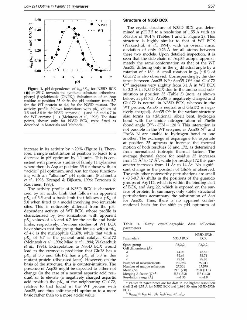
Table 1. X-ray crystallographic data collectionparameters
Parameters N35D BCXN35D-2FXb
BCX
Space group P212121 P212121
Cell dimensions (AÊ )a 44.05 43.83b 52.69 52.74c 78.61 78.80
Number of measurements 150,984 99,311Number of unique reflections 27,301 17,579Mean I/sI 21.1 (7.0) 25.8 (11.1)Merging R-factor (%)a,b 5.7 (15.2) 5.7 (14.2)Resolution range (AÊ ) 1-1.55 1-1.8
a Values in parentheses are for data in the highest resolutionshell (1.61-1.55 AÊ for N35D BCX and 1.86-1.80 AÊ for N35D-2FXbBCX).
b Rmerge � �hkl �ni � 0jIiÿIhklj/�hkl �n
i � 0Iihkl.
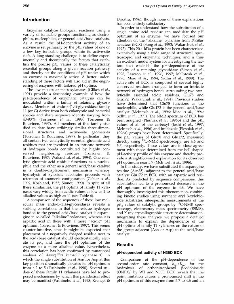
Figure 1. pH-dependence of kcat/Km for N35D BCX(*) at 25 �C towards the synthetic substrate orthonitro-phenyl b-xylobioside (ONPX2). Substitution of an Aspresidue at position 35 shifts the pH optimum from 5.7for the WT protein to 4.6 for the N35D mutant. Theactivity pro®le follows ionizations with pKa values of3.5 and 5.8 in the N35D enzyme (Ð) and 4.6 and 6.7 inthe WT enzyme (- - -) (McIntosh et al., 1996). The datapoints, shown only for N35D BCX, were ®tted asdescribed in Materials and Methods.
Low pH Optima in Family 11 Xylanases 257
increase in its activity by �20 % (Figure 1). There-fore, a single substitution at position 35 leads to adecrease in pH optimum by 1.1 units. This is con-sistent with previous studies of family 11 xylanaseswhere there is Asp at position 35 for those with an``acidic'' pH optimum, and Asn for those function-ing with an ``alkaline'' pH optimum (Fushinobuet al., 1998; Krengel & Dijkstra, 1996; Torronen &Rouvinen, 1995).
The activity pro®le of N35D BCX is character-ized by an acidic limb that follows an apparentpKa of 3.5 and a basic limb that follows a pKa of5.8 when ®tted to a model involving two ionizablesites. This is noticeably different from the pH-dependent activity of WT BCX, whose pro®le ischaracterized by two ionizations with apparentpKa values of 4.6 and 6.7 for the acidic and basiclimbs, respectively. Previous studies of WT BCXhave shown that the group that ionizes with a pKa
of 4.6 is the nucleophile Glu78, while that with apKa of 6.7 is the general acid catalyst Glu172(McIntosh et al., 1996; Miao et al., 1994; Wakarchuket al., 1994). Extrapolation to N35D BCX wouldlead to the erroneous prediction that Glu78 has apKa of 3.5 and Glu172 has a pKa of 5.8 in thismutant protein (discussed later). However, on thebasis of the structure, this is counter-intuitive. Thepresence of Asp35 might be expected to either notchange (in the case of a neutral aspartic acid resi-due), or to elevate (a negatively charged asparticacid residue) the pKa of the neighboring Glu172,relative to that found in the WT protein withAsn35, and thus shift the pH optimum to a morebasic rather than to a more acidic value.
Structure of N35D BCX
The crystal structure of N35D BCX was deter-mined at pH 7.5 to a resolution of 1.55 AÊ with anR-factor of 19.4 % (Tables 1 and 2; Figure 2). Thisstructure is highly similar to that of WT BCX(Wakarchuk et al., 1994), with an overall r.m.s.deviation of only 0.23 AÊ for all atoms betweenthese two models. Upon detailed inspection, it isseen that the side-chain of Asp35 adopts approxi-mately the same conformation as that of the WTAsn35, differing only in the w2 dihedral angle by arotation of �16 �. A small rotation in w3 (�8 �) ofGlu172 is also observed. Correspondingly, the dis-tance between Asn35 Nd2/Asp35 Od2 and Glu172Oe2 increases very slightly from 3.1 AÊ in WT BCXto 3.2 AÊ in N35D BCX due to the amino acid sub-stitution at position 35 (Table 3) (note, as shownbelow, at pH 7.5, Asp35 is negatively charged andGlu172 is neutral in N35D BCX, whereas in theWT protein, Asn35 is neutral and Glu172 is nega-tively charged). Asp35 Od2 in the variant structurealso forms an additional, albeit bent, hydrogenbond with the amide nitrogen atom of Phe36(bond angle Od2 � � �HN � 120 �). This interaction isnot possible in the WT enzyme, as Asn35 Nd2 andPhe36 N are unable to hydrogen bond to oneanother. The exchange of asparagine for aspartateat position 35 appears to increase the thermalmotion of both residues 35 and 172, as determinedfrom normalized isotropic thermal factors. Theaverage thermal factor for residue 35 increasesfrom 11 AÊ 2 to 17 AÊ 2, while for residue 172 this par-ameter increases from 11 AÊ 2 to 14 AÊ 2. No signi®-cant change in the position of Glu78 is observed.The only other noteworthy perturbations are small(�0.5-0.7 AÊ ) shifts in the positions of the guanidogroups of Arg112, which is within the binding cleftof BCX, and Arg122, which is exposed on the sur-face of protein. In summary, only subtle structuralperturbations accompany the substitution of Aspfor Asn35. Thus, there is no apparent confor-mational basis for the shift in pH optimum of
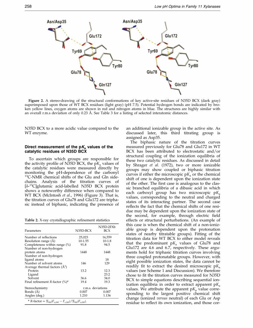
Figure 2. A stereo-drawing of the structural conformations of key active-site residues of N35D BCX (dark gray)superimposed upon those of WT BCX residues (light gray) (pH 7.5). Potential hydrogen bonds are indicated by bro-ken yellow lines, oxygen atoms are shown in red and nitrogen atoms in blue. The structures are highly similar withan overall r.m.s deviation of only 0.23 AÊ . See Table 3 for a listing of selected interatomic distances.
258 Low pH Optima in Family 11 Xylanases
N35D BCX to a more acidic value compared to theWT enzyme.
Direct measurement of the pKa values of thecatalytic residues of N35D BCX
To ascertain which groups are responsible forthe activity pro®le of N35D BCX, the pKa values ofthe catalytic residues were measured directly bymonitoring the pH-dependence of the carbonyl13C-NMR chemical shifts of the Glu and Gln side-chains. Analysis of the titration curves of[d-13C]glutamic acid-labelled N35D BCX proteinshows a noteworthy difference when compared toWT BCX (McIntosh et al., 1996) (Figure 3). That is,the titration curves of Glu78 and Glu172 are tripha-sic instead of biphasic, indicating the presence of
Table 2. X-ray crystallographic re®nement statistics
Parameters N35D-BCXN35D-2FXb
BCX
Number of reflections 25,023 16,559Resolution range (AÊ ) 10-1.55 10-1.8Completeness within range (%) 91.8 94.5Number of non-hydrogenprotein atoms 1448 1448Number of non-hydrogenligand atoms 18Number of solvent atoms 146 129Average thermal factors (AÊ 2)
Protein 13.2 12.3Ligand 23.2Solvent 36.6 34.0
Final refinement R-factor (%)a 19.4 19.3
Stereochemistry r.m.s. deviationsBonds (AÊ ) 0.007 0.007Angles (deg.) 1.210 1.136
a R-factor � �hkljFo,hkl ÿ Fc,hklj/�hkljFo,hklj.
an additional ionizable group in the active site. Asdiscussed later, this third titrating group isassigned as Asp35.
The biphasic nature of the titration curvesmeasured previously for Glu78 and Glu172 in WTBCX has been attributed to electrostatic and/orstructural coupling of the ionization equilibria ofthese two catalytic residues. As discussed in detailby Shrager et al. (1972), two or more ionizablegroups may show coupled or biphasic titrationcurves if either the microscopic pKa or the chemicalshift of one is dependent upon the ionization stateof the other. The ®rst case is analogous to the clas-sic branched equilibria of a dibasic acid in whicheach carboxyl group has two microscopic pKa
values, corresponding to the neutral and chargedstates of its interacting partner. The second casere¯ects the fact that the chemical shifts of one resi-due may be dependent upon the ionization state ofthe second, for example, through electric ®eldeffects or structural perturbations. (An example ofthis case is when the chemical shift of a non-ioniz-able group is dependent upon the protonationstates of nearby titratable groups). Fitting of thetitration data for WT BCX to either model revealsthat the predominant pKa values of Glu78 andGlu172 are 4.6 and 6.7, respectively. These argu-ments hold for triphasic titration curves involvingthree coupled protonatable groups. However, witheight possible ionization states, the data cannot bereadily ®t to extract the desired microscopic pKa
values (see Scheme 1 and Discussion). We thereforechose to ®t the titration curves measured for N35DBCX to simple equations describing sequential ion-ization equilibria in order to extract apparent pKa
values. We attribute the apparent pKa value corre-sponding to the largest positive chemical shiftchange (ionized versus neutral) of each Glu or Aspresidue to re¯ect its own ionization, and those cor-
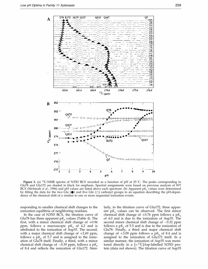
Figure 3. (a) 13C-NMR spectra of N35D BCX recorded as a function of pH at 25 �C. The peaks corresponding toGlu78 and Glu172 are shaded in black for emphasis. Spectral assignments were based on previous analysis of WTBCX (McIntosh et al., 1996) and pH values are listed above each spectrum. (b) Apparent pKa values were determinedby ®tting the data for the two Glu (*) and ®ve Gln (*) carbonyl groups to an equation describing the pH-depen-dence of the chemical shift of a residue to one or more sequential ionization events.
Low pH Optima in Family 11 Xylanases 259
responding to smaller chemical shift changes to theionization equilibria of neighboring residues.
In the case of N35D BCX, the titration curve ofGlu78 has three apparent pKa values (Table 4). The®rst, with a minor chemical shift change of �0.96ppm, follows a macroscopic pKa of 4.2 and isattributed to the ionization of Asp35. The second,with a major chemical shift change of �2.69 ppm,follows a pKa of 5.7 and is assigned to the ioniz-ation of Glu78 itself. Finally, a third, with a minorchemical shift change of ÿ0.39 ppm, follows a pKa
of 8.4 and re¯ects the ionization of Glu172. Simi-
larly, in the titration curve of Glu172, three appar-ent pKa values can be observed. The ®rst minorchemical shift change of �0.76 ppm follows a pKa
of 4.0 and is due to the ionization of Asp35. Thesecond minor chemical shift change of ÿ0.32 ppmfollows a pKa of 5.5 and is due to the ionization ofGlu78. Finally, a third and major chemical shiftchange of �3.09 ppm follows a pKa of 8.4 and isassigned to the ionization of Glu172 itself. In asimilar manner, the ionization of Asp35 was moni-tored directly in a [g-13C]Asp-labelled N35D pro-tein (data not shown). The titration curve of Asp35
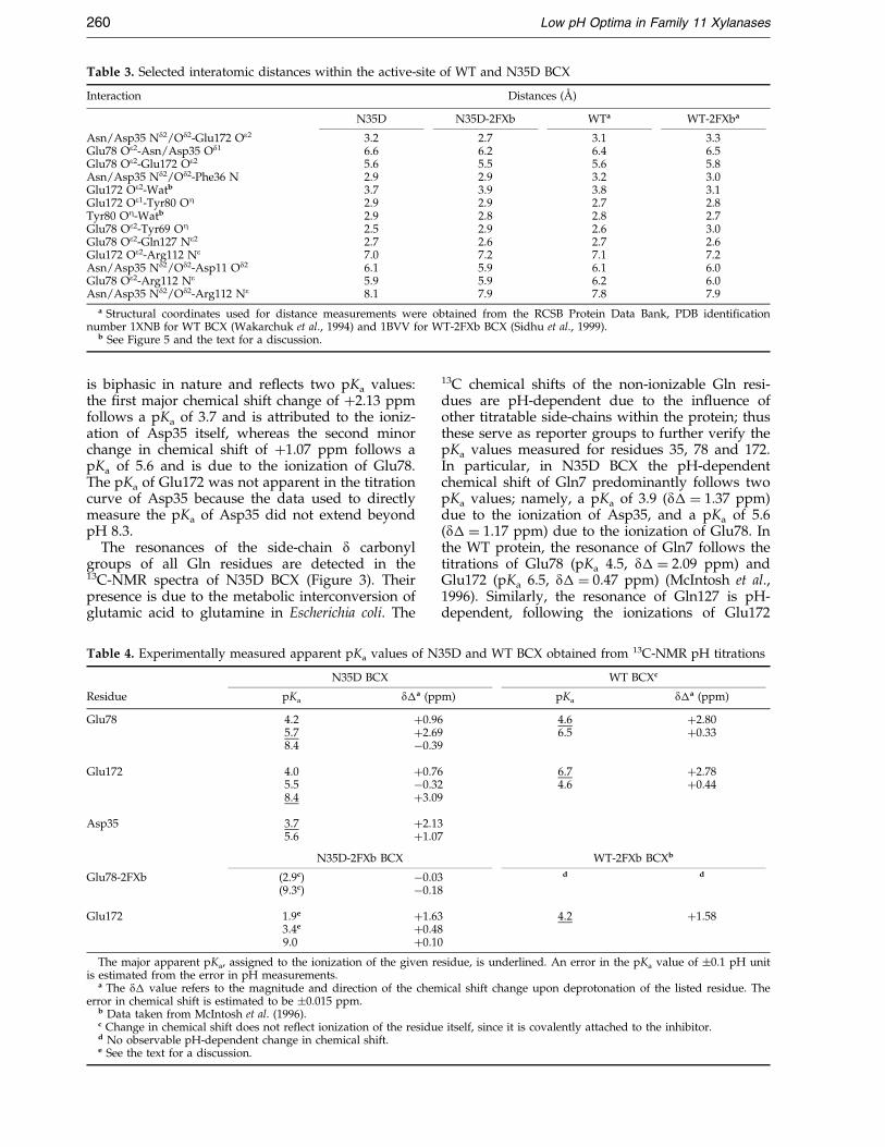
Table 3. Selected interatomic distances within the active-site of WT and N35D BCX
Interaction Distances (AÊ )
N35D N35D-2FXb WTa WT-2FXba
Asn/Asp35 Nd2/Od2-Glu172 Oe2 3.2 2.7 3.1 3.3Glu78 Oe2-Asn/Asp35 Od1 6.6 6.2 6.4 6.5Glu78 Oe2-Glu172 Oe2 5.6 5.5 5.6 5.8Asn/Asp35 Nd2/Od2-Phe36 N 2.9 2.9 3.2 3.0Glu172 Oe2-Watb 3.7 3.9 3.8 3.1Glu172 Oe1-Tyr80 OZ 2.9 2.9 2.7 2.8Tyr80 OZ-Watb 2.9 2.8 2.8 2.7Glu78 Oe2-Tyr69 OZ 2.5 2.9 2.6 3.0Glu78 Oe2-Gln127 Ne2 2.7 2.6 2.7 2.6Glu172 Oe2-Arg112 Ne 7.0 7.2 7.1 7.2Asn/Asp35 Nd2/Od2-Asp11 Od2 6.1 5.9 6.1 6.0Glu78 Oe2-Arg112 Ne 5.9 5.9 6.2 6.0Asn/Asp35 Nd2/Od2-Arg112 Ne 8.1 7.9 7.8 7.9
a Structural coordinates used for distance measurements were obtained from the RCSB Protein Data Bank, PDB identi®cationnumber 1XNB for WT BCX (Wakarchuk et al., 1994) and 1BVV for WT-2FXb BCX (Sidhu et al., 1999).
b See Figure 5 and the text for a discussion.
260 Low pH Optima in Family 11 Xylanases
is biphasic in nature and re¯ects two pKa values:the ®rst major chemical shift change of �2.13 ppmfollows a pKa of 3.7 and is attributed to the ioniz-ation of Asp35 itself, whereas the second minorchange in chemical shift of �1.07 ppm follows apKa of 5.6 and is due to the ionization of Glu78.The pKa of Glu172 was not apparent in the titrationcurve of Asp35 because the data used to directlymeasure the pKa of Asp35 did not extend beyondpH 8.3.
The resonances of the side-chain d carbonylgroups of all Gln residues are detected in the13C-NMR spectra of N35D BCX (Figure 3). Theirpresence is due to the metabolic interconversion ofglutamic acid to glutamine in Escherichia coli. The
Table 4. Experimentally measured apparent pKa values of N
N35D BCX
Residue pKa d�a (pp
Glu78 4.2 �0.965.7 �2.698.4 ÿ0.39
Glu172 4.0 �0.765.5 ÿ0.328.4 �3.09
Asp35 3.7 �2.135.6 �1.07
N35D-2FXb BCX
Glu78-2FXb (2.9c) ÿ0.03(9.3c) ÿ0.18
Glu172 1.9e �1.633.4e �0.489.0 �0.10
The major apparent pKa, assigned to the ionization of the given reis estimated from the error in pH measurements.
a The d� value refers to the magnitude and direction of the chemerror in chemical shift is estimated to be �0.015 ppm.
b Data taken from McIntosh et al. (1996).c Change in chemical shift does not re¯ect ionization of the residud No observable pH-dependent change in chemical shift.e See the text for a discussion.
13C chemical shifts of the non-ionizable Gln resi-dues are pH-dependent due to the in¯uence ofother titratable side-chains within the protein; thusthese serve as reporter groups to further verify thepKa values measured for residues 35, 78 and 172.In particular, in N35D BCX the pH-dependentchemical shift of Gln7 predominantly follows twopKa values; namely, a pKa of 3.9 (d� � 1.37 ppm)due to the ionization of Asp35, and a pKa of 5.6(d� � 1.17 ppm) due to the ionization of Glu78. Inthe WT protein, the resonance of Gln7 follows thetitrations of Glu78 (pKa 4.5, d� � 2.09 ppm) andGlu172 (pKa 6.5, d� � 0.47 ppm) (McIntosh et al.,1996). Similarly, the resonance of Gln127 is pH-dependent, following the ionizations of Glu172
35D and WT BCX obtained from 13C-NMR pH titrations
WT BCXc
m) pKa d�a (ppm)
4.6 �2.806.5 �0.33
6.7 �2.784.6 �0.44
WT-2FXb BCXb
d d
4.2 �1.58
sidue, is underlined. An error in the pKa value of �0.1 pH unit
ical shift change upon deprotonation of the listed residue. The
e itself, since it is covalently attached to the inhibitor.
Low pH Optima in Family 11 Xylanases 261
(pKa 8.4, d� � 0.03 ppm), Glu78 (pKa 5.6,d� � 0.65 ppm) and Asp35 (pKa 3.8, d� � 0.65ppm) in N35D BCX and of Glu172 (pKa 6.6,d� � 0.32 ppm) and Glu78 (pKa 4.5, d� � 0.76ppm) in the WT protein (McIntosh et al., 1996).
The analyses of the ionization behavior ofAsp35, Glu78 and Glu172 are further supported bydeuterium isotope shift measurements (Joshi et al.,1997; Ladner et al., 1975; Yamazaki et al., 1994). Anestimate of the protonation state of a carboxylgroup can be made by observing the difference inits 13C chemical shift in H2O versus 2H2O solutions.A neutral carboxylic acid group is expected toshow an isotope shift of � ÿ0.25 ppm between itsprotonated and deuterated forms, whereas a car-boxylate group would show no such effect.Measurement of the deuterium isotope shift of thed side-chain carbon atoms of Glu78 and Glu172 inN35D BCX protein reveals that at pH* 6.32, Glu78is deprotonated (no apparent isotope shift),whereas Glu172 is protonated (isotope shift ofÿ0.29 ppm). These data are consistent with themeasured pKa values of the three active-site car-boxyl groups in N35D BCX.
Based on both titration curves and isotope shifts,we conclude that the pKa values corresponding pri-marily to the ionizations of Asp35, Glu78 andGlu172 in N35D BCX are 3.7, 5.7 and 8.4, respect-ively. The small differences in the correspondingpKa values measured from the multiphasic titrationcurves of the Asp, Glu and Gln residues (Table 4)are attributed to dif®culties in accurately ®ttingthese data and the complex nature of the structuralor electrostatic interactions in this highly couplednetwork of ionizable side-chains. Regardless, thesedata clearly differ from the pKa values of 4.6 and6.8 measured for Glu78 and Glu172 in the WTenzyme, and con®rm the expectation that the sub-stitution of Asn35 by Asp elevates the pKa valuesof the nearby catalytic glutamic acid residues.
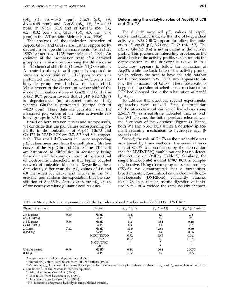
Table 5. Steady-state kinetic parameters for the hydrolysis of
Phenol substituent pKaa Protein
2,5-Dinitro 5.15 N35D(2,5-DNPX2) WTc
3,4-Dinitro 5.36 N35D(3,4-DNPX2) WTc
2-Nitro 7.22 N35D(ONPX2) WTd
N35D/E172QE172Qe
N35D/E78QE78Q
Unsubstituted 9.99 N35D(PhX2) WTe
Assays were carried out at pH 6.0 and 40 �C.a Phenol pKa values were taken from Tull & Withers (1994).b Values of kcat/Km were taken from the slope of the Lineweaver-
a non-linear ®t of the Michaelis-Menten equation.c Data taken from Ziser et al. (1995).d Data taken from Lawson et al. (1996).e Data taken from Lawson et al. (1997).f No detectable enzymatic hydrolysis (unpublished results).
Determining the catalytic roles of Asp35, Glu78and Glu172
The directly measured pKa values of Asp35,Glu78, and Glu172 indicate that the pH-dependentactivity of N35D BCX appears to follow the ioniz-ation of Asp35 (pKa 3.7) and Glu78 (pKa 5.7). ThepKa of Glu172 (8.4) is not apparent in the activitypro®le. This presents an interesting problem, as theacidic limb of the activity pro®le, which re¯ects thedeprotonation of the nucleophile Glu78 in WTBCX, now appears to follow the ionization ofAsp35, while the basic limb of the activity pro®le,which re¯ects the need to have the acid catalystGlu172 protonated in WT BCX, now appears to fol-low the ionization of Glu78. These observationsbegged the question of whether the mechanism ofBCX had changed due to the substitution of Asn35by Asp.
To address this question, several experimentalapproaches were utilized. First, determinationof the stereochemical course of hydrolysis using2,5-DNPX2 as a substrate indicated that, as withthe WT enzyme, the initial product released wasthe b anomer of the xylobiose (Figure 4). Hence,both WT and N35D BCX utilize a double-displace-ment retaining mechanism to hydrolyze aryl b-xylobiosides.
Second, the role of Glu78 as the nucleophile wasascertained by three methods. The essential func-tion of Glu78 was con®rmed by the observationthat the N35D/E78Q double mutant has no detect-able activity on ONPX2 (Table 5). Similarly, thesingle (nucleophile) mutant E78Q BCX is comple-tely inactive. Using electrospray mass spectrometry(ESMS), we demonstrated that a mechanism-based inhibitor, 2,4-dinitrophenyl 2-deoxy-2-¯uoro-b-xylobioside (DNP2FXb), covalently attachesto Glu78. In particular, tryptic digestion of inhib-ited N35D BCX yielded the same doubly charged,
aryl b-xylobiosides for N35D and WT BCX
kcatb (sÿ1) Km
b (mM) kcat/Kmb (sÿ1 mMÿ1)
14.8 6.7 2.676 2.2 358.2 48.1 0.188.3 3.4 2.714.5 25.6 0.569.6 14 0.660.72 33.3 0.0210.62 8.3 0.075
f f f
f f f
0.14 20.1 0.00700.051 8.7 0.0050
Burk plot, whereas values of kcat and Km were determined from

Figure 4. Stereochemical course of hydrolysis of ONPX2 by N35D BCX at 25 �C, 99.9 % 2H2O and pH* 4.8. 1H-NMRspectra were recorded as a function of time after addition of ONPX2 substrate (R � xylose, R0 � 20 dinitrophenyl). Thecontrol spectrum of ONPX2 with no enzyme (bottom) shows a doublet at 5.26 ppm (J � 7.0 Hz) corresponding to theaxial anomeric proton of the xylose residue nearest to the aryl leaving group (R0), Ha1, that is present in the b-anomerof the unhydrolyzed substrate. Initially (t < ten minutes) after the addition of N35D enzyme, the doublet at 5.26 ppmdisappears while a new doublet at 4.59 ppm (J � 7.9 Hz) appears. This is due to the axial proton, Ha2, of the initialb-anomeric product released and demonstrates that the mechanism of N35D BCX involves stereochemical retentionof anomeric con®guration of the product. As time proceeds (t � 20-210 minutes) mutarotation of the initial productoccurs, as is evident by the appearance of a second peak at 5.19 ppm (J � 3.5 Hz) due to the presence of the a-anomerat the reducing end of the xylobioside. The distorted peak near 4.8 ppm is due to the residual signal of water.
262 Low pH Optima in Family 11 Xylanases
covalently modi®ed peptide containing Glu78 (m/z of 826 in neutral loss mode) as observed in a pre-vious analysis of WT BCX (Miao et al., 1994; anddata not shown). Finally, the crystal structure ofthe N35D BCX glycosyl-enzyme intermediate(N35D-2FXb) clearly shows covalent modi®cationat Glu78 by the DNP2FXb inhibitor (details dis-cussed subsequently).
Third, the role of Glu172 was also probed bysite-directed mutagenesis. Analysis of the activityof the double mutant protein N35D/E172Q indi-cated that Glu172 still plays a primary role in func-tioning as the general acid/base catalyst in N35DBCX. This is not surprising, given the structuralsimilarity of the mutant and WT enzymes(Figure 2), and thus the conserved positioning ofthe side-chain of Glu172 with respect to the bind-ing site for xylosic substrates. Speci®cally, similarvalues of kcat/Km for hydrolysis of ONPX2 were
found for both the N35D/E172Q double mutantprotein (0.021 sÿ1 mMÿ1) and the E172Q singlemutant protein (0.075 sÿ1 mMÿ1) (Table 5). Notethat this synthetic substrate has an activated leav-ing group that needs less general acid catalyticassistance for departure. Therefore, activity isobserved, albeit at a substantially reduced rate,even with BCX variants lacking their general acidcatalyst.
Brùnsted analysis of the activity of N35D BCXtowards aryl bbb-xylobiosides
The enzymatic activity of N35D BCX wasmeasured using a number of different syntheticaryl b-xylobiosides of varying leaving group ability(Table 5). ONPX2 was chosen as a reference sub-strate to compare the activities of all the BCX var-iants considered in this study. As summarized in
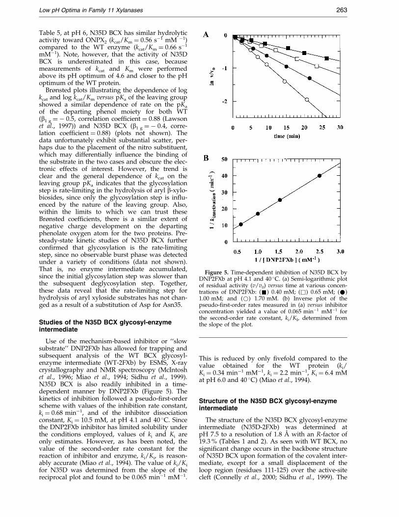
Figure 5. Time-dependent inhibition of N35D BCX byDNP2FXb at pH 4.1 and 40 �C. (a) Semi-logarithmic plotof residual activity (v/v0) versus time at various concen-trations of DNP2FXb: (&) 0.40 mM; (&) 0.65 mM; (*)1.00 mM; and (*) 1.70 mM. (b) Inverse plot of thepseudo-®rst-order rates measured in (a) versus inhibitorconcentration yielded a value of 0.065 minÿ1 mMÿ1 forthe second-order rate constant, ki/KI, determined fromthe slope of the plot.
Low pH Optima in Family 11 Xylanases 263
Table 5, at pH 6, N35D BCX has similar hydrolyticactivity toward ONPX2 (kcat/Km � 0.56 sÿ1 mM ÿ1)compared to the WT enzyme (kcat/Km � 0.66 sÿ1
mMÿ1). Note, however, that the activity of N35DBCX is underestimated in this case, becausemeasurements of kcat and Km were performedabove its pH optimum of 4.6 and closer to the pHoptimum of the WT protein.
Brùnsted plots illustrating the dependence of logkcat and log kcat/Km versus pKa of the leaving groupshowed a similar dependence of rate on the pKa
of the departing phenol moiety for both WT(b1 g � ÿ 0.5, correlation coef®cient � 0.88 (Lawsonet al., 1997)) and N35D BCX (b1 g � ÿ 0.4, corre-lation coef®cient � 0.88) (plots not shown). Thedata unfortunately exhibit substantial scatter, per-haps due to the placement of the nitro substituent,which may differentially in¯uence the binding ofthe substrate in the two cases and obscure the elec-tronic effects of interest. However, the trend isclear and the general dependence of kcat on theleaving group pKa indicates that the glycosylationstep is rate-limiting in the hydrolysis of aryl b-xylo-biosides, since only the glycosylation step is in¯u-enced by the nature of the leaving group. Also,within the limits to which we can trust theseBrùnsted coef®cients, there is a similar extent ofnegative charge development on the departingphenolate oxygen atom for the two proteins. Pre-steady-state kinetic studies of N35D BCX furthercon®rmed that glycosylation is the rate-limitingstep, since no observable burst phase was detectedunder a variety of conditions (data not shown).That is, no enzyme intermediate accumulated,since the initial glycosylation step was slower thanthe subsequent deglycosylation step. Together,these data reveal that the rate-limiting step forhydrolysis of aryl xyloside substrates has not chan-ged as a result of a substitution of Asp for Asn35.
Studies of the N35D BCX glycosyl-enzymeintermediate
Use of the mechanism-based inhibitor or ``slowsubstrate'' DNP2FXb has allowed for trapping andsubsequent analysis of the WT BCX glycosyl-enzyme intermediate (WT-2FXb) by ESMS, X-raycrystallography and NMR spectroscopy (McIntoshet al., 1996; Miao et al., 1994; Sidhu et al., 1999).N35D BCX is also readily inhibited in a time-dependent manner by DNP2FXb (Figure 5). Thekinetics of inhibition followed a pseudo-®rst-orderscheme with values of the inhibition rate constant,ki � 0.68 minÿ1, and of the inhibitor dissociationconstant, Ki � 10.5 mM, at pH 4.1 and 40 �C. Sincethe DNP2FXb inhibitor has limited solubility underthe conditions employed, values of ki and Ki areonly estimates. However, as has been noted, thevalue of the second-order rate constant for thereaction of inhibitor and enzyme, ki/Ki, is reason-ably accurate (Miao et al., 1994). The value of ki/Ki
for N35D was determined from the slope of thereciprocal plot and found to be 0.065 minÿ1 mMÿ1.
This is reduced by only ®vefold compared to thevalue obtained for the WT protein (ki/Ki � 0.34 minÿ1 mMÿ1, ki � 2.2 minÿ1, Ki � 6.4 mMat pH 6.0 and 40 �C) (Miao et al., 1994).
Structure of the N35D BCX glycosyl-enzymeintermediate
The structure of the N35D BCX glycosyl-enzymeintermediate (N35D-2FXb) was determined atpH 7.5 to a resolution of 1.8 AÊ with an R-factor of19.3 % (Tables 1 and 2). As seen with WT BCX, nosigni®cant change occurs in the backbone structureof N35D BCX upon formation of the covalent inter-mediate, except for a small displacement of theloop region (residues 111-125) over the active-sitecleft (Connelly et al., 2000; Sidhu et al., 1999). The
264 Low pH Optima in Family 11 Xylanases
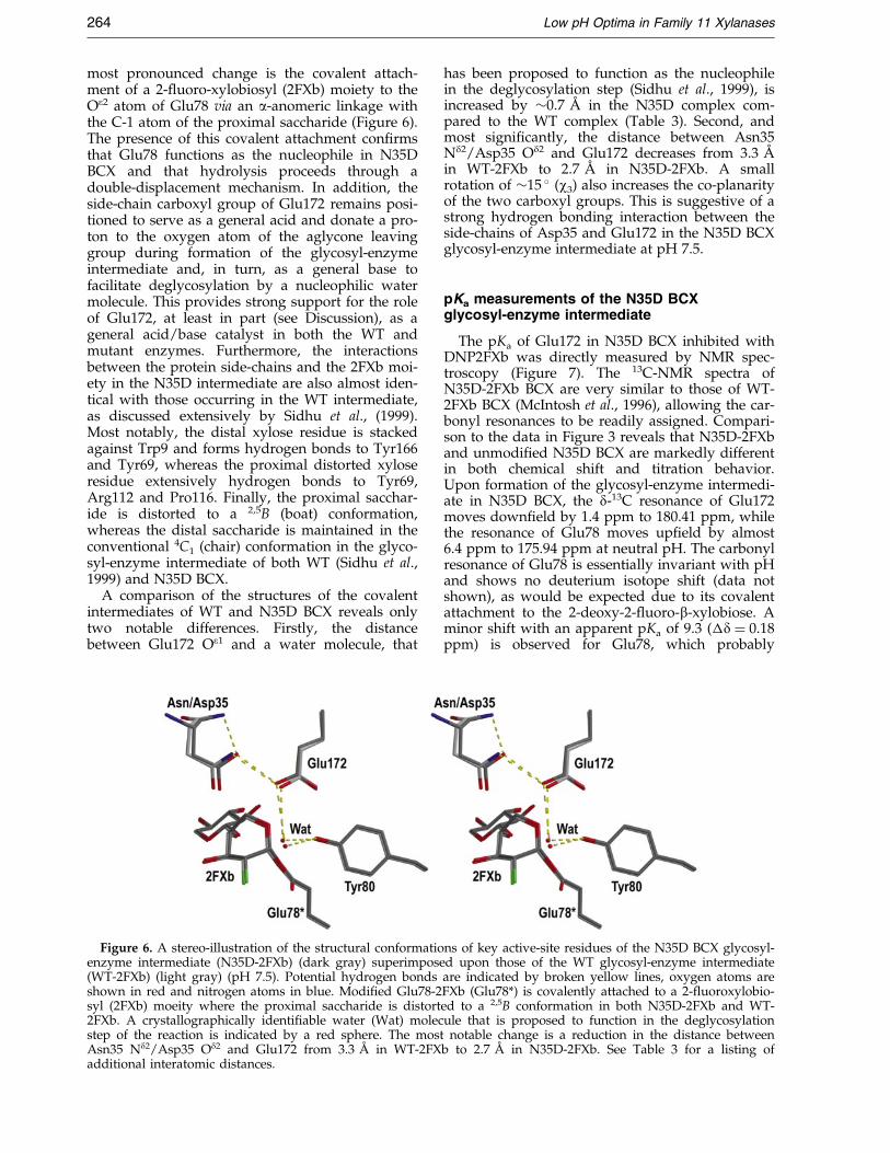
most pronounced change is the covalent attach-ment of a 2-¯uoro-xylobiosyl (2FXb) moiety to theOe2 atom of Glu78 via an a-anomeric linkage withthe C-1 atom of the proximal saccharide (Figure 6).The presence of this covalent attachment con®rmsthat Glu78 functions as the nucleophile in N35DBCX and that hydrolysis proceeds through adouble-displacement mechanism. In addition, theside-chain carboxyl group of Glu172 remains posi-tioned to serve as a general acid and donate a pro-ton to the oxygen atom of the aglycone leavinggroup during formation of the glycosyl-enzymeintermediate and, in turn, as a general base tofacilitate deglycosylation by a nucleophilic watermolecule. This provides strong support for the roleof Glu172, at least in part (see Discussion), as ageneral acid/base catalyst in both the WT andmutant enzymes. Furthermore, the interactionsbetween the protein side-chains and the 2FXb moi-ety in the N35D intermediate are also almost iden-tical with those occurring in the WT intermediate,as discussed extensively by Sidhu et al., (1999).Most notably, the distal xylose residue is stackedagainst Trp9 and forms hydrogen bonds to Tyr166and Tyr69, whereas the proximal distorted xyloseresidue extensively hydrogen bonds to Tyr69,Arg112 and Pro116. Finally, the proximal sacchar-ide is distorted to a 2,5B (boat) conformation,whereas the distal saccharide is maintained in theconventional 4C1 (chair) conformation in the glyco-syl-enzyme intermediate of both WT (Sidhu et al.,1999) and N35D BCX.
A comparison of the structures of the covalentintermediates of WT and N35D BCX reveals onlytwo notable differences. Firstly, the distancebetween Glu172 Oe1 and a water molecule, that
Figure 6. A stereo-illustration of the structural conformatioenzyme intermediate (N35D-2FXb) (dark gray) superimpose(WT-2FXb) (light gray) (pH 7.5). Potential hydrogen bondsshown in red and nitrogen atoms in blue. Modi®ed Glu78-2syl (2FXb) moeity where the proximal saccharide is distort2FXb. A crystallographically identi®able water (Wat) molecstep of the reaction is indicated by a red sphere. The mostAsn35 Nd2/Asp35 Od2 and Glu172 from 3.3 AÊ in WT-2FXadditional interatomic distances.
has been proposed to function as the nucleophilein the deglycosylation step (Sidhu et al., 1999), isincreased by �0.7 AÊ in the N35D complex com-pared to the WT complex (Table 3). Second, andmost signi®cantly, the distance between Asn35Nd2/Asp35 Od2 and Glu172 decreases from 3.3 AÊ
in WT-2FXb to 2.7 AÊ in N35D-2FXb. A smallrotation of �15 � (w3) also increases the co-planarityof the two carboxyl groups. This is suggestive of astrong hydrogen bonding interaction between theside-chains of Asp35 and Glu172 in the N35D BCXglycosyl-enzyme intermediate at pH 7.5.
pKa measurements of the N35D BCXglycosyl-enzyme intermediate
The pKa of Glu172 in N35D BCX inhibited withDNP2FXb was directly measured by NMR spec-troscopy (Figure 7). The 13C-NMR spectra ofN35D-2FXb BCX are very similar to those of WT-2FXb BCX (McIntosh et al., 1996), allowing the car-bonyl resonances to be readily assigned. Compari-son to the data in Figure 3 reveals that N35D-2FXband unmodi®ed N35D BCX are markedly differentin both chemical shift and titration behavior.Upon formation of the glycosyl-enzyme intermedi-ate in N35D BCX, the d-13C resonance of Glu172moves down®eld by 1.4 ppm to 180.41 ppm, whilethe resonance of Glu78 moves up®eld by almost6.4 ppm to 175.94 ppm at neutral pH. The carbonylresonance of Glu78 is essentially invariant with pHand shows no deuterium isotope shift (data notshown), as would be expected due to its covalentattachment to the 2-deoxy-2-¯uoro-b-xylobiose. Aminor shift with an apparent pKa of 9.3 (�d � 0.18ppm) is observed for Glu78, which probably
ns of key active-site residues of the N35D BCX glycosyl-d upon those of the WT glycosyl-enzyme intermediateare indicated by broken yellow lines, oxygen atoms areFXb (Glu78*) is covalently attached to a 2-¯uoroxylobio-ed to a 2,5B conformation in both N35D-2FXb and WT-ule that is proposed to function in the deglycosylationnotable change is a reduction in the distance between
b to 2.7 AÊ in N35D-2FXb. See Table 3 for a listing of
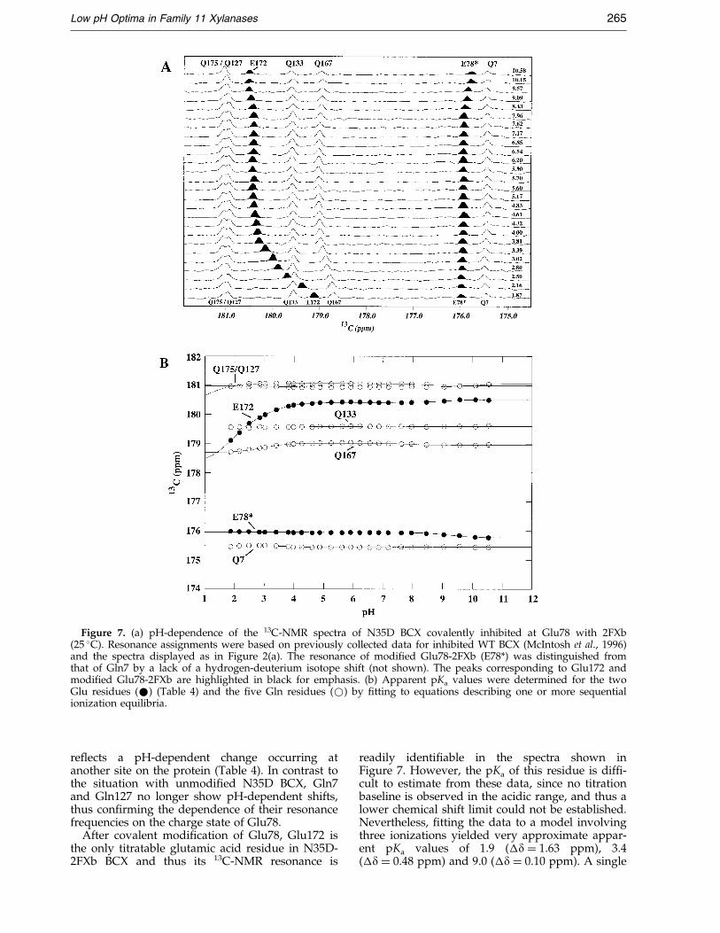
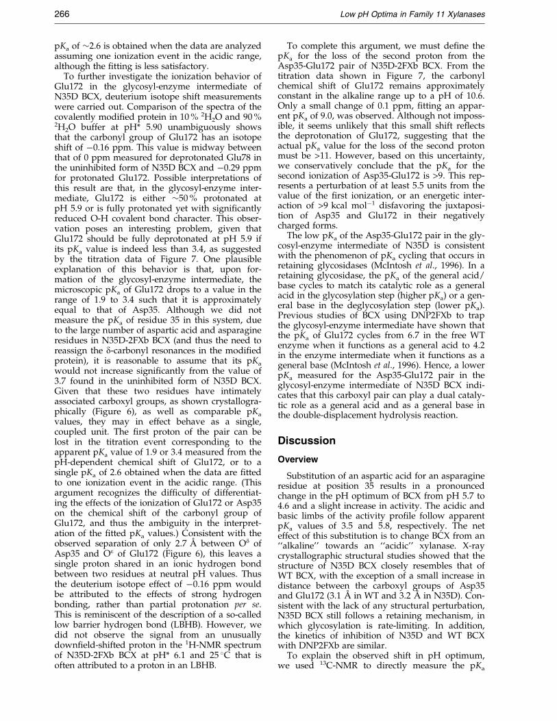
Figure 7. (a) pH-dependence of the 13C-NMR spectra of N35D BCX covalently inhibited at Glu78 with 2FXb(25 �C). Resonance assignments were based on previously collected data for inhibited WT BCX (McIntosh et al., 1996)and the spectra displayed as in Figure 2(a). The resonance of modi®ed Glu78-2FXb (E78*) was distinguished fromthat of Gln7 by a lack of a hydrogen-deuterium isotope shift (not shown). The peaks corresponding to Glu172 andmodi®ed Glu78-2FXb are highlighted in black for emphasis. (b) Apparent pKa values were determined for the twoGlu residues (*) (Table 4) and the ®ve Gln residues (*) by ®tting to equations describing one or more sequentialionization equilibria.
Low pH Optima in Family 11 Xylanases 265
re¯ects a pH-dependent change occurring atanother site on the protein (Table 4). In contrast tothe situation with unmodi®ed N35D BCX, Gln7and Gln127 no longer show pH-dependent shifts,thus con®rming the dependence of their resonancefrequencies on the charge state of Glu78.
After covalent modi®cation of Glu78, Glu172 isthe only titratable glutamic acid residue in N35D-2FXb BCX and thus its 13C-NMR resonance is
readily identi®able in the spectra shown inFigure 7. However, the pKa of this residue is dif®-cult to estimate from these data, since no titrationbaseline is observed in the acidic range, and thus alower chemical shift limit could not be established.Nevertheless, ®tting the data to a model involvingthree ionizations yielded very approximate appar-ent pKa values of 1.9 (�d � 1.63 ppm), 3.4(�d � 0.48 ppm) and 9.0 (�d � 0.10 ppm). A single
266 Low pH Optima in Family 11 Xylanases
pKa of �2.6 is obtained when the data are analyzedassuming one ionization event in the acidic range,although the ®tting is less satisfactory.
To further investigate the ionization behavior ofGlu172 in the glycosyl-enzyme intermediate ofN35D BCX, deuterium isotope shift measurementswere carried out. Comparison of the spectra of thecovalently modi®ed protein in 10 % 2H2O and 90 %2H2O buffer at pH* 5.90 unambiguously showsthat the carbonyl group of Glu172 has an isotopeshift of ÿ0.16 ppm. This value is midway betweenthat of 0 ppm measured for deprotonated Glu78 inthe uninhibited form of N35D BCX and ÿ0.29 ppmfor protonated Glu172. Possible interpretations ofthis result are that, in the glycosyl-enzyme inter-mediate, Glu172 is either �50 % protonated atpH 5.9 or is fully protonated yet with signi®cantlyreduced O-H covalent bond character. This obser-vation poses an interesting problem, given thatGlu172 should be fully deprotonated at pH 5.9 ifits pKa value is indeed less than 3.4, as suggestedby the titration data of Figure 7. One plausibleexplanation of this behavior is that, upon for-mation of the glycosyl-enzyme intermediate, themicroscopic pKa of Glu172 drops to a value in therange of 1.9 to 3.4 such that it is approximatelyequal to that of Asp35. Although we did notmeasure the pKa of residue 35 in this system, dueto the large number of aspartic acid and asparagineresidues in N35D-2FXb BCX (and thus the need toreassign the d-carbonyl resonances in the modi®edprotein), it is reasonable to assume that its pKa
would not increase signi®cantly from the value of3.7 found in the uninhibited form of N35D BCX.Given that these two residues have intimatelyassociated carboxyl groups, as shown crystallogra-phically (Figure 6), as well as comparable pKa
values, they may in effect behave as a single,coupled unit. The ®rst proton of the pair can belost in the titration event corresponding to theapparent pKa value of 1.9 or 3.4 measured from thepH-dependent chemical shift of Glu172, or to asingle pKa of 2.6 obtained when the data are ®ttedto one ionization event in the acidic range. (Thisargument recognizes the dif®culty of differentiat-ing the effects of the ionization of Glu172 or Asp35on the chemical shift of the carbonyl group ofGlu172, and thus the ambiguity in the interpret-ation of the ®tted pKa values.) Consistent with theobserved separation of only 2.7 AÊ between Od ofAsp35 and Oe of Glu172 (Figure 6), this leaves asingle proton shared in an ionic hydrogen bondbetween two residues at neutral pH values. Thusthe deuterium isotope effect of ÿ0.16 ppm wouldbe attributed to the effects of strong hydrogenbonding, rather than partial protonation per se.This is reminiscent of the description of a so-calledlow barrier hydrogen bond (LBHB). However, wedid not observe the signal from an unusuallydown®eld-shifted proton in the 1H-NMR spectrumof N35D-2FXb BCX at pH* 6.1 and 25 �C that isoften attributed to a proton in an LBHB.
To complete this argument, we must de®ne thepKa for the loss of the second proton from theAsp35-Glu172 pair of N35D-2FXb BCX. From thetitration data shown in Figure 7, the carbonylchemical shift of Glu172 remains approximatelyconstant in the alkaline range up to a pH of 10.6.Only a small change of 0.1 ppm, ®tting an appar-ent pKa of 9.0, was observed. Although not imposs-ible, it seems unlikely that this small shift re¯ectsthe deprotonation of Glu172, suggesting that theactual pKa value for the loss of the second protonmust be >11. However, based on this uncertainty,we conservatively conclude that the pKa for thesecond ionization of Asp35-Glu172 is >9. This rep-resents a perturbation of at least 5.5 units from thevalue of the ®rst ionization, or an energetic inter-action of >9 kcal molÿ1 disfavoring the juxtaposi-tion of Asp35 and Glu172 in their negativelycharged forms.
The low pKa of the Asp35-Glu172 pair in the gly-cosyl-enzyme intermediate of N35D is consistentwith the phenomenon of pKa cycling that occurs inretaining glycosidases (McIntosh et al., 1996). In aretaining glycosidase, the pKa of the general acid/base cycles to match its catalytic role as a generalacid in the glycosylation step (higher pKa) or a gen-eral base in the deglycosylation step (lower pKa).Previous studies of BCX using DNP2FXb to trapthe glycosyl-enzyme intermediate have shown thatthe pKa of Glu172 cycles from 6.7 in the free WTenzyme when it functions as a general acid to 4.2in the enzyme intermediate when it functions as ageneral base (McIntosh et al., 1996). Hence, a lowerpKa measured for the Asp35-Glu172 pair in theglycosyl-enzyme intermediate of N35D BCX indi-cates that this carboxyl pair can play a dual cataly-tic role as a general acid and as a general base inthe double-displacement hydrolysis reaction.
Discussion
Overview
Substitution of an aspartic acid for an asparagineresidue at position 35 results in a pronouncedchange in the pH optimum of BCX from pH 5.7 to4.6 and a slight increase in activity. The acidic andbasic limbs of the activity pro®le follow apparentpKa values of 3.5 and 5.8, respectively. The neteffect of this substitution is to change BCX from an``alkaline'' towards an ``acidic'' xylanase. X-raycrystallographic structural studies showed that thestructure of N35D BCX closely resembles that ofWT BCX, with the exception of a small increase indistance between the carboxyl groups of Asp35and Glu172 (3.1 AÊ in WT and 3.2 AÊ in N35D). Con-sistent with the lack of any structural perturbation,N35D BCX still follows a retaining mechanism, inwhich glycosylation is rate-limiting. In addition,the kinetics of inhibition of N35D and WT BCXwith DNP2FXb are similar.
To explain the observed shift in pH optimum,we used 13C-NMR to directly measure the pKa
Scheme 1.
Low pH Optima in Family 11 Xylanases 267
values of the catalytically essential residues in boththe free enzyme and in the glycosyl-enzyme inter-mediate of N35D BCX. In the free enzyme, theapparent pKa of the newly introduced Asp residueat position 35 is 3.7, while those of Glu78 andGlu172 are elevated from their WT values to 5.7and 8.4, respectively. The ionization states at pH 6.3expected from the pKa values of Asp35, Glu78 andGlu172 were con®rmed by deuterium isotope shiftmeasurements. Thus, the activity pro®le of N35DBCX appears to follow the pKa values of Asp35and Glu78, instead of Glu78 and Glu172 as in WTBCX. However, several experiments con®rmed thatGlu78 is still the nucleophile and that Glu172 stillcontributes to the role of the general acid catalyst.
The crystal structure of the N35D BCX glycosyl-enzyme intermediate is very similar to that of theWT glycosyl-enzyme intermediate, with a signi®-cantly notable exception. The side-chain carboxylgroups of Asp35 and Glu172 are now shifted closertogether (2.7 AÊ in N35D-2FX versus 3.3 AÊ in WT-2FXb) with a concomitant increase in co-planarity.The energetic driving force for the favorable juxta-position of these two carboxylate groups appar-ently arises from the formation of a rather shortand strong hydrogen bond resulting from the lossof the ®rst proton and the subsequent sharing ofthe second between these two groups. This con-clusion is supported by 13C-NMR titrations, whichindicate that Glu172 and Asp35 behave as a singlecoupled unit with a pKa in the range of 1.9 and 3.4for the loss of the ®rst proton and a pKa of >9 forthe loss of the second, combined with the obser-vation of an intermediate deuterium isotope shiftat pH 5.9, indicative of a shared proton bridgingthese two carboxylate groups.
pKa values of the catalytic residues
Previous NMR studies of WT BCX revealed thatGlu78 and Glu172 have coupled ionization equili-bria. Consistent with their catalytic roles, the majorpKa values for these two residues are 4.6 and 6.7,respectively. However, when their biphasictitration curves were ®tted to a model describingthe interaction of two ionizable groups, the micro-scopic pKa of Glu172 in the presence of a neutralGlu78 was found to be 5.5, and that of Glu78 inthe presence of a charged Glu172 was 5.8 (seeScheme 1 of McIntosh et al., 1996). Therefore, theelectrostatic interaction between the two nearbycarboxyl groups (Cd separation � 6.5 AÊ ) perturbstheir pKa values by �1.2 units. This is supportedby the observation that upon substitution of Glu78with Gln, the pKa of Glu172 drops to 4.2, whereasthe reciprocal mutation of Glu172 leads to a slightelevation of the pKa of Glu78 to 5.0. In both cases,the titration curves become monophasic. Thedifferences between the pKa values measured forthe mutant proteins and those determined from ananalysis of the biphasic titration curves of WT BCXsuggest that the coupling between the two gluta-mic acids is not purely electrostatic. Conformation-
al changes in the active-site upon ionization ormutation may also in¯uence the titration behaviorof Glu78 and Glu172.
Substitution of Asn35 to an aspartic acid residueleads to a dramatic change in the ionization prop-erties of BCX. Most notably, the apparent pKa
values of Glu78 and Glu172 are elevated to 5.7 and8.4, respectively, and both residues show triphasictitration curves. This undoubtedly arises from anadditional electrostatic interaction with Asp35,which has an apparent pKa of 3.7 and thus is pre-dominantly in a negatively charged state at pHvalues when Glu78 and Glu172 undergo deproto-nation. The greater perturbation of the pKa valueof Glu172 is consistent with the crystal structure ofN35D BCX, determined at pH 7.5. That is, thecarboxyl carbon atoms of Asp35 and Glu172 areseparated by 4.8 AÊ , whereas those of Asp35 andGlu78 are 8.5 AÊ apart (Figure 2 and Table 3).
The multiphasic titration curves recorded forAsp35, Glu78, and Glu172 clearly indicate a com-plex network of electrostatic, and possibly structur-al, interactions between these three residues. Asoutlined in Results, the apparent pKa values listedin Table 4 were determined by ®tting the NMR-detected titration curves of the active-site carboxylgroups to a simple model of sequential ionizationevents. However, to gain further insight into theelectrostatic interactions between Asp35, Glu78,and Glu172, it is useful to analyze their titrationdata to extract microscopic pKa values. Unfortu-nately, with eight possible ionization states, it isdif®cult to carry out this analysis in full. Usingonly data recorded in acidic solutions (e.g. withGlu172 protonated), a simultaneous ®tting of thetitration curves for Asp35 and Glu78 to a model oftwo coupled ionization equilibria yields the micro-scopic pKa values shown in Scheme 1:
The upper limb of this scheme shows the predo-minant ionization pathway of N35D BCX in whichAsp35 deprotonates with increasing pH beforeGlu78, whereas the lower corresponds to the minorpopulation of protein in which Glu78 is negativelycharged and Asp35 neutral. Note that the micro-scopic pKa of 5.54 for Glu78 along the upper limbof this coupled equilibrium curve is similar to theapparent pKa value of 5.7 listed in Table 4, whereasthat of 4.03 for Asp35 is somewhat higher than the
268 Low pH Optima in Family 11 Xylanases
corresponding value of 3.7 determined from asequential ionization model. This re¯ects the dif®-culty of extracting pKa values for complex bindingequilibria in proteins, as the chemical shifts ofinvolved residues are exquisitely sensitive to bothstructural and electrostatic factors, thus theequations describing their NMR behavior areunderdetermined experimentally. However, sup-port for Scheme 1 follows from the observationthat the microscopic pKa of 4.49 ®t for Glu78 in thepresence of a neutral Asp35 is very similar to thatof 4.6 measured for this catalytic residue in WTBCX, which contains a neutral asparagine residueat position 35. Recognizing that the accuracyof these ®ttings is uncertain, Scheme 1 suggeststhat the ionization of Asp35 elevates the pKa ofGlu78 by �1 unit, and vice versa, due to chargerepulsion, with an energetic coupling of2.303RT(�pKa) � 1.4 kcal molÿ1. The implicationsof this analysis for understanding the catalyticmechanism of N35D BCX will be discussed below.
Unfortunately, we were unable to extract micro-scopic pKa values for the coupling of Glu172 withGlu78 or Asp35 for the following two reasons.First, in contrast to the case of the WT protein,upon addition of acid, the 13Cd nuclei of Glu78 andGlu172 in N35D BCX show small down®eld shifts(i.e. opposite to that expected for protonation of acarboxylate) at pH values corresponding to eachother's pKa values. Therefore, it is not possible to®t these data to the equation describing the pH-dependence of the chemical shifts of two residueswith coupled ionization equilibria (Shrager et al.,1972). Second, NMR data were not recorded abovepH 8.3 for [g-13C]Asp-labelled N35D BCX, thusprecluding a mutual ®t of the titration curves ofAsp35 and Glu172 for microscopic pKa values.However, due to the large difference in theirapparent pKa values, the population of proteinwith Asp35 protonated and Glu172 deprotonatedmust be negligible.
Upon formation of a long-lived glycosyl-enzymeintermediate, the pKa value of Glu172 in WT-2FXbBCX drops from 6.7 to 4.2 (McIntosh et al., 1996).This pKa cycling allows the glutamate residue toperform its dual role as a general acid for the gly-cosylation step and general base for deglycosyla-tion. The reduction in the pKa value of Glu172 by2.5 units is attributed to the elimination of chargerepulsion from Glu78, due to formation of acovalent linkage to the 2-¯uoro-xylobiose, as wellas to possible changes in the structure andhydration of BCX (McIntosh et al., 1996). In a simi-lar fashion, formation of the N35D BCX glycosyl-enzyme intermediate leads to a dramatic change inthe titration behavior of Glu172. As seen inFigure 7, the pH-dependence of the d-13C reson-ance of Glu172 follows apparent pKa values of 1.9or 3.4 when ®tted to two sequential titrations or2.6 when ®tted to one ionization event in the acidicrange. The observation of an isotope shift of ÿ0.16ppm at pH* 5.90 for Glu172 indicates that this resi-
due is at least partially protonated at pH valuesabove these pKa values.
Based on this measured titration and deuter-ium isotope shift data, we conclude that Asp35and Glu172 are behaving as a highly coupleddicarboxylic acid system in N35D-2FXb BCX,with a ®rst pKa in the range of 1.9-3.4 and asecond estimated to be >9. This is remarkablefor two reasons. First, in the case of the WTenzyme, the pKa of Glu172 drops by only 2.5units upon covalent modi®cation of Glu78,whereas in N35D BCX, the pKa value associatedwith Glu172 falls by �5.8 units, from 8.4 to�2.6. Second, assuming that the pKa values forthe ®rst and second deprotonation steps of theAsp35-Glu172 pair are �2.6 and >9, this rep-resents an energetic interaction of >9 kcal molÿ1
disfavoring the juxtaposition of the two carboxy-late groups in their negatively charged forms. Apossible explanation for this behavior is seenfrom a comparison of the crystal structures ofN35D BCX in its free and modi®ed forms, deter-mined at pH 7.5. In the free enzyme, the dis-tance between Asp35 Od2 and Glu172 Oe2 is3.2 AÊ , whereas in the glycosyl-enzyme intermedi-ate, the separation is reduced to 2.7 AÊ (Table 3).The corresponding values for the WT protein are3.1 AÊ and 3.3 AÊ , respectively. This suggests thata relatively strong hydrogen bond exists betweenAsp35 and Glu172 in N35D-2FXb BCX, such thatone proton is shared between the two carboxylgroups with a net charge of ÿ1. As mentionedpreviously, this interaction shares manysimilarities to an LBHB, in that a proton ispartitioned somewhat equally between the twocarboxylate groups with ®rst ionization pKa
values that are approximately matched.However, the signature proton with a down-®eld-shifted resonance commonly attributed tothe presence of an LBHB is not detected. Also,although slightly less than the �2.8 AÊ oxygen-oxygen hydrogen bonding distance observed inwater, the Asp35-Glu172 separation is somewhatlonger than the value of <2.55 AÊ often cited forsuch an LBHB in model compounds (Clelandet al., 1998). Regardless, this close interactionbetween these two active-site residues, which isnot observed in WT-2FXb BCX, could accountfor the distinct pKa values measured for Glu172in the mutant enzyme. By way of comparison,in a study of the ionization of symmetric dicar-boxylic acids, McDaniel & Brown (1953) reportedthat the pKa values for cis-caronic acid were 2.3and 8.2. This strong energetic coupling wasattributed to a highly favorable hydrogen bondbetween a carboxylic acid and a carboxylategroup in this constrained ring system.
Reverse protonation mechanism
Based upon the pKa values of Glu78 (5.7) andGlu172 (8.4) determined by 13C-NMR, and by ana-logy to the WT enzyme, the pH optimum of N35D
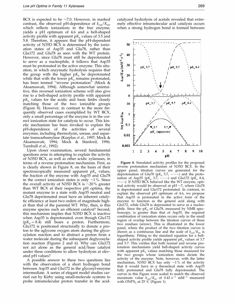
Figure 8. Simulated activity pro®les for the proposedreverse protonation mechanism of N35D BCX. In theupper panel, titration curves are generated for thedeprotonation of Glu78 (pKa 5.7, ± ± ±) and the proto-nation of Asp35 (pKa 3.7, - - - - -) and Glu172 (pKa 8.4,- - - - -). If N35D BCX behaved like the WT enzyme, opti-mal activity would be observed at pH �7, where Glu78is deprotonated and Glu172 protonated. In contrast, toexplain the observed pH optimum of 4.6, we proposethat Asp35 is protonated in the active form of theenzyme to function as the general acid along withGlu172, while Glu78 is depronated to serve as a nucleo-phile. Since the pKa of Glu78, measured by NMR spec-troscopy, is greater than that of Asp35, the requiredcombination of ionization states occurs only in the smallregion of overlap between the titration curves of thesetwo residues (arrow). This is illustrated in the lowerpanel, where the product of the two titration curves isshown as a continuous line and the scale of kcat/Km islogarithmic. Fitting to the standard equation for a bell-shaped activity pro®le yields apparent pKa values of 3.7and 5.7. This veri®es that both normal and reverse pro-tonation mechanisms yield bell-shaped activity curveswith apparent pKa values matching those measured forthe two groups whose ionization states dictate theactivity of the enzyme. Note, however, with the lattermechanism, N35D BCX has only �1 % of the activitythat it would have in a theoretical state with Asp35fully protonated and Glu78 fully deprotonated. Thecurves in this Figure were scaled to match the observedmaximum value kcat/Km of 0.43 sÿ1 mMÿ1 measuredwith ONPX at 25 �C (Figure 1).
Low pH Optima in Family 11 Xylanases 269
BCX is expected to be �7.0. However, in markedcontrast, the observed pH-dependence of kcat/Km,which re¯ects ionizations in the free enzyme,yields a pH optimum of 4.6 and a bell-shapedactivity pro®le with apparent pKa values of 3.5 and5.8. Therefore, it appears that the pH-dependentactivity of N35D BCX is determined by the ioniz-ation states of Asp35 and Glu78, rather thanGlu172 and Glu78 as seen with the WT protein.However, since Glu78 must still be deprotonatedto serve as a nucleophile, it follows that Asp35must be protonated in the active enzyme. This situ-ation, in which enzymatic hydrolysis requires thatthe group with the higher pKa be deprotonatedwhile that with the lower pKa remains protonated,has been termed ``reverse protonation'' (Mock &Aksamawati, 1994). Although somewhat unintui-tive, this reversed ionization scheme will also giverise to a bell-shaped activity pro®le with apparentpKa values for the acidic and basic limbs closelymatching those of the two ionizable groups(Figure 8). However, in contrast to the more fre-quently observed cases exempli®ed by WT BCX,only a small percentage of the enzyme is in the cor-rect ionization state for catalysis to occur. This kin-etic mechanism has been invoked to explain thepH-dependence of the activities of severalenzymes, including thermolysin, urease, and aspar-tate transcarbamylase (Karplus et al., 1997; Mock &Aksamawati, 1994; Mock & Stanford, 1996;Turnbull et al., 1992).
Upon closer examination, several fundamentalquestions arise in attempting to explain the activityof N35D BCX, as well as other acidic xylanases, interms of a reverse protonation mechanism. First, asis clearly shown in Figure 8, on the basis of theirspectroscopically measured apparent pKa values,the fraction of the enzyme with Asp35 and Glu78in the correct ionization states is only �1 %. Sincethe overall activity of N35D BCX is �20 % greaterthan WT BCX at their respective pH optima, themutant enzyme in a state with Asp35 neutral andGlu78 deprotonated must have an inherent cataly-tic ef®ciency at least two orders of magnitude high-er than that of the parental WT. Why, then, is thisenzyme species such an ef®cient catalyst? Second,this mechanism implies that N35D BCX is inactivewhen Asp35 is deprotonated, even though Glu172(pKa � 8.4) still bears a proton. Furthermore,Glu172 is positioned structurally to donate a pro-ton to the aglycone oxygen atom during the glyco-sylation reaction and to abstract a proton from awater molecule during the subsequent deglycosyla-tion reaction (Figures 2 and 6). Why can Glu172not act alone as the general acid/base catalystunder these conditions to allow hydrolysis at elev-ated pH values?
A possible answer to these two questions lieswith the observation of a short hydrogen bondbetween Asp35 and Glu172 in the glycosyl-enzymeintermediate. A series of elegant model studies car-ried out by Kirby using small-molecule systems toprobe intramolecular proton transfer in the acid-
catalyzed hydrolysis of acetals revealed that extre-mely effective intramolecular acid catalysis occurswhen a strong hydrogen bond is formed between
2
270 Low pH Optima in Family 11 Xylanases
the departing alcohol and the conjugate base formof the general acid catalyst in systems such as thatshown in Scheme 2 (Kirby, 1997). This stronghydrogen bond must exist, at least in part, in thetransition state leading to the product, thereby sta-bilizing it and facilitating hydrolysis.
The situation observed with N35D BCX is
Scheme 2.
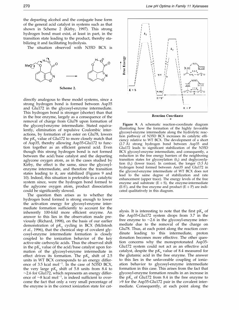
Figure 9. A schematic reaction-coordinate diagramillustrating how the formation of the highly favorableglycosyl-enzyme intermediate along the hydrolytic reac-tion pathway of N35D BCX increases its catalytic ef®-ciency relative to WT BCX. The development of a short(2.7 AÊ ) strong hydrogen bond between Asp35 andGlu172 leads to signi®cant stabilization of the N35DBCX glycosyl-enzyme intermediate, and consequently, areduction in the free energy barriers of the neighboringtransition states for glycosylation (k2) and deglycosyla-tion (k3) (lower trace). In contrast, the longer (3.3 AÊ )hydrogen bond formed between Asn35 and Glu172 inthe glycosyl-enzyme intermediate of WT BCX does notlead to the same degree of stabilization and rateenhancement (upper trace). The energy levels of the freeenzyme and substrate (E � S), the enzyme-intermediate(E-S0), and the free enzyme and product (E � P) are indi-cated qualitatively in this diagram.
directly analogous to these model systems, since astrong hydrogen bond is formed between Asp35and Glu172 in the glycosyl-enzyme intermediate.This hydrogen bond is stronger (shorter) than thatin the free enzyme, largely as a consequence of theremoval of charge from Glu78 upon formation ofthe glycosyl-enzyme intermediate. Stated equiva-lently, elimination of repulsive Coulombic inter-actions, by formation of an ester on Glu78, lowersthe pKa value of Glu172 to more closely match thatof Asp35, thereby allowing Asp35-Glu172 to func-tion together as an ef®cient general acid. Eventhough this strong hydrogen bond is not formedbetween the acid/base catalyst and the departingaglycone oxygen atom, as in the cases studied byKirby, the effect is the same, since the glycosyl-enzyme intermediate, and therefore the transitionstates leading to it, are stabilized (Figures 9 and10). Indeed, this situation is preferable in a catalyticsystem since, were the hydrogen bond formed tothe aglycone oxygen atom, product dissociationcould be signi®cantly slowed.
The question then arises as to whether thehydrogen bond formed is strong enough to lowerthe activation energy for glycosyl-enzyme inter-mediate formation suf®ciently to account for theinherently 100-fold more ef®cient enzyme. Ananswer to this lies in the observation made pre-viously (Richard, 1998), on the basis of our earlierdemonstration of pKa cycling in BCX (McIntoshet al., 1996), that the chemical step of covalent gly-cosyl-enzyme intermediate formation is closelycoupled to the ionization behavior of the keyactive-site carboxylic acids. Thus the observed shiftin the pKa value of the acid/base catalyst upon for-mation of the glycosyl-enzyme intermediate ineffect drives its formation. The pKa shift of 2.5units in WT BCX corresponds to an energy differ-ence of 3.5 kcal molÿ1. In the case of N35D BCX,the very large pKa shift of 5.8 units from 8.4 to�2.6 for Glu172, which represents an energy differ-ence of �8 kcal molÿ1, is indeed suf®cient to over-come the fact that only a very small percentage ofthe enzyme is in the correct ionization state for cat-
alysis. It is interesting to note that the ®rst pKa ofthe Asp35-Glu172 system drops from 3.7 in thefree enzyme to �2.6 in the glycosyl-enzyme inter-mediate due to the removal of the charge onGlu78. Thus, at each point along the reaction coor-dinate leading to this intermediate, protondonation becomes more effective. The other ques-tion concerns why the monoprotonated Asp35-Glu172 system could not act as an effective acidcatalyst, despite the pKa value of 8.4 measured forthe glutamic acid in the free enzyme. The answerto this lies in the unfavorable coupling of ioniz-ation behavior to glycosyl-enzyme intermediateformation in this case. This arises from the fact thatglycosyl-enzyme formation results in an increase inthe pKa of Glu172 from 8.4 in the free enzyme to>9 for the Asp35-Glu172 pair in the covalent inter-mediate. Consequently, at each point along the
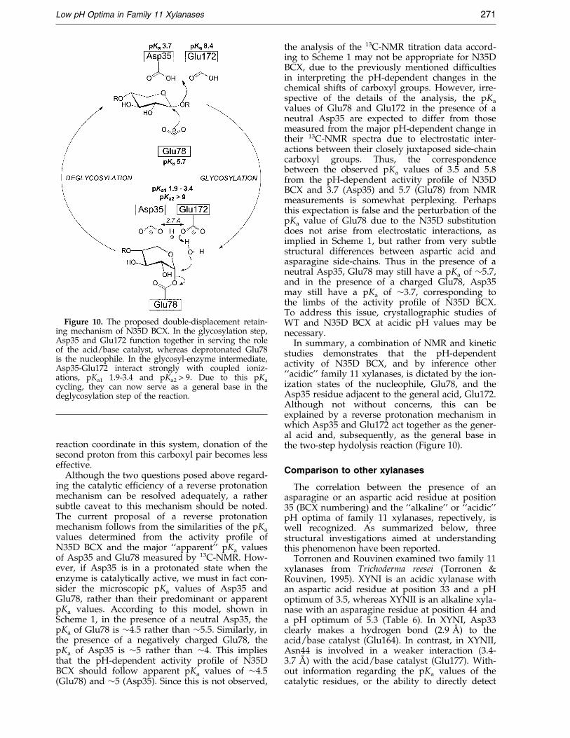
Figure 10. The proposed double-displacement retain-ing mechanism of N35D BCX. In the glycosylation step,Asp35 and Glu172 function together in serving the roleof the acid/base catalyst, whereas deprotonated Glu78is the nucleophile. In the glycosyl-enzyme intermediate,Asp35-Glu172 interact strongly with coupled ioniz-ations, pKa1 1.9-3.4 and pKa2 > 9. Due to this pKa
cycling, they can now serve as a general base in thedeglycosylation step of the reaction.
Low pH Optima in Family 11 Xylanases 271
reaction coordinate in this system, donation of thesecond proton from this carboxyl pair becomes lesseffective.
Although the two questions posed above regard-ing the catalytic ef®ciency of a reverse protonationmechanism can be resolved adequately, a rathersubtle caveat to this mechanism should be noted.The current proposal of a reverse protonationmechanism follows from the similarities of the pKa
values determined from the activity pro®le ofN35D BCX and the major ``apparent'' pKa valuesof Asp35 and Glu78 measured by 13C-NMR. How-ever, if Asp35 is in a protonated state when theenzyme is catalytically active, we must in fact con-sider the microscopic pKa values of Asp35 andGlu78, rather than their predominant or apparentpKa values. According to this model, shown inScheme 1, in the presence of a neutral Asp35, thepKa of Glu78 is �4.5 rather than �5.5. Similarly, inthe presence of a negatively charged Glu78, thepKa of Asp35 is �5 rather than �4. This impliesthat the pH-dependent activity pro®le of N35DBCX should follow apparent pKa values of �4.5(Glu78) and �5 (Asp35). Since this is not observed,
the analysis of the 13C-NMR titration data accord-ing to Scheme 1 may not be appropriate for N35DBCX, due to the previously mentioned dif®cultiesin interpreting the pH-dependent changes in thechemical shifts of carboxyl groups. However, irre-spective of the details of the analysis, the pKa
values of Glu78 and Glu172 in the presence of aneutral Asp35 are expected to differ from thosemeasured from the major pH-dependent change intheir 13C-NMR spectra due to electrostatic inter-actions between their closely juxtaposed side-chaincarboxyl groups. Thus, the correspondencebetween the observed pKa values of 3.5 and 5.8from the pH-dependent activity pro®le of N35DBCX and 3.7 (Asp35) and 5.7 (Glu78) from NMRmeasurements is somewhat perplexing. Perhapsthis expectation is false and the perturbation of thepKa value of Glu78 due to the N35D substitutiondoes not arise from electrostatic interactions, asimplied in Scheme 1, but rather from very subtlestructural differences between aspartic acid andasparagine side-chains. Thus in the presence of aneutral Asp35, Glu78 may still have a pKa of �5.7,and in the presence of a charged Glu78, Asp35may still have a pKa of �3.7, corresponding tothe limbs of the activity pro®le of N35D BCX.To address this issue, crystallographic studies ofWT and N35D BCX at acidic pH values may benecessary.
In summary, a combination of NMR and kineticstudies demonstrates that the pH-dependentactivity of N35D BCX, and by inference other``acidic'' family 11 xylanases, is dictated by the ion-ization states of the nucleophile, Glu78, and theAsp35 residue adjacent to the general acid, Glu172.Although not without concerns, this can beexplained by a reverse protonation mechanism inwhich Asp35 and Glu172 act together as the gener-al acid and, subsequently, as the general base inthe two-step hydolysis reaction (Figure 10).
Comparison to other xylanases
The correlation between the presence of anasparagine or an aspartic acid residue at position35 (BCX numbering) and the ``alkaline'' or ``acidic''pH optima of family 11 xylanases, repectively, iswell recognized. As summarized below, threestructural investigations aimed at understandingthis phenomenon have been reported.
Torronen and Rouvinen examined two family 11xylanases from Trichoderma reesei (Torronen &Rouvinen, 1995). XYNI is an acidic xylanase withan aspartic acid residue at position 33 and a pHoptimum of 3.5, whereas XYNII is an alkaline xyla-nase with an asparagine residue at position 44 anda pH optimum of 5.3 (Table 6). In XYNI, Asp33clearly makes a hydrogen bond (2.9 AÊ ) to theacid/base catalyst (Glu164). In contrast, in XYNII,Asn44 is involved in a weaker interaction (3.4-3.7 AÊ ) with the acid/base catalyst (Glu177). With-out information regarding the pKa values of thecatalytic residues, or the ability to directly detect
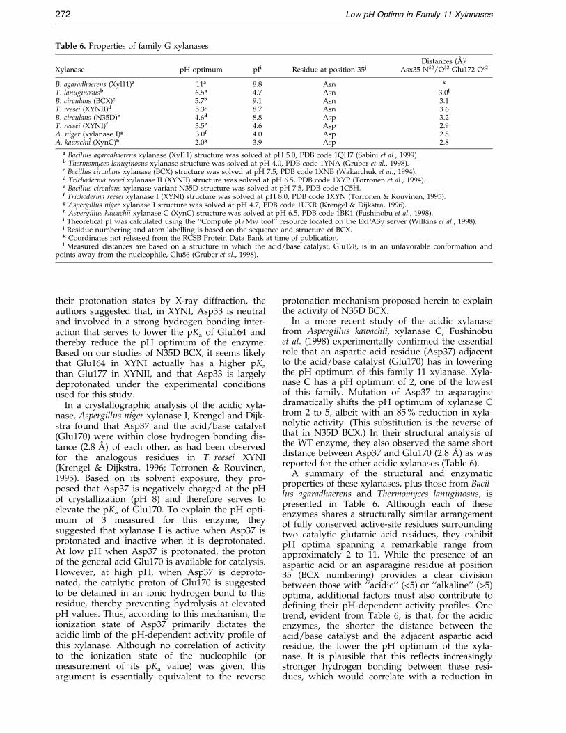
Table 6. Properties of family G xylanases
Distances (AÊ )j
Xylanase pH optimum pIi Residue at position 35j Asx35 Nd2/Od2-Glu172 Oe2
B. agaradhaerens (Xyl11)a 11a 8.8 Asn k
T. lanuginosusb 6.5a 4.7 Asn 3.0l
B. circulans (BCX)c 5.7b 9.1 Asn 3.1T. reesei (XYNII)d 5.3c 8.7 Asn 3.6B. circulans (N35D)e 4.6d 8.8 Asp 3.2T. reesei (XYNI)f 3.5e 4.6 Asp 2.9A. niger (xylanase I)g 3.0f 4.0 Asp 2.8A. kawachii (XynC)h 2.0g 3.9 Asp 2.8
a Bacillus agaradhaerens xylanase (Xyl11) structure was solved at pH 5.0, PDB code 1QH7 (Sabini et al., 1999).b Thermomyces lanuginosus xylanase structure was solved at pH 4.0, PDB code 1YNA (Gruber et al., 1998).c Bacillus circulans xylanase (BCX) structure was solved at pH 7.5, PDB code 1XNB (Wakarchuk et al., 1994).d Trichoderma reesei xylanase II (XYNII) structure was solved at pH 6.5, PDB code 1XYP (Torronen et al., 1994).e Bacillus circulans xylanase variant N35D structure was solved at pH 7.5, PDB code 1C5H.f Trichoderma reesei xylanase I (XYNI) structure was solved at pH 8.0, PDB code 1XYN (Torronen & Rouvinen, 1995).g Aspergillus niger xylanase I structure was solved at pH 4.7, PDB code 1UKR (Krengel & Dijkstra, 1996).h Aspergillus kawachii xylanase C (XynC) structure was solved at pH 6.5, PDB code 1BK1 (Fushinobu et al., 1998).i Theoretical pI was calculated using the ``Compute pI/Mw tool'' resource located on the ExPASy server (Wilkins et al., 1998).j Residue numbering and atom labelling is based on the sequence and structure of BCX.k Coordinates not released from the RCSB Protein Data Bank at time of publication.l Measured distances are based on a structure in which the acid/base catalyst, Glu178, is in an unfavorable conformation and
points away from the nucleophile, Glu86 (Gruber et al., 1998).
272 Low pH Optima in Family 11 Xylanases
their protonation states by X-ray diffraction, theauthors suggested that, in XYNI, Asp33 is neutraland involved in a strong hydrogen bonding inter-action that serves to lower the pKa of Glu164 andthereby reduce the pH optimum of the enzyme.Based on our studies of N35D BCX, it seems likelythat Glu164 in XYNI actually has a higher pKa
than Glu177 in XYNII, and that Asp33 is largelydeprotonated under the experimental conditionsused for this study.
In a crystallographic analysis of the acidic xyla-nase, Aspergillus niger xylanase I, Krengel and Dijk-stra found that Asp37 and the acid/base catalyst(Glu170) were within close hydrogen bonding dis-tance (2.8 AÊ ) of each other, as had been observedfor the analogous residues in T. reesei XYNI(Krengel & Dijkstra, 1996; Torronen & Rouvinen,1995). Based on its solvent exposure, they pro-posed that Asp37 is negatively charged at the pHof crystallization (pH 8) and therefore serves toelevate the pKa of Glu170. To explain the pH opti-mum of 3 measured for this enzyme, theysuggested that xylanase I is active when Asp37 isprotonated and inactive when it is deprotonated.At low pH when Asp37 is protonated, the protonof the general acid Glu170 is available for catalysis.However, at high pH, when Asp37 is deproto-nated, the catalytic proton of Glu170 is suggestedto be detained in an ionic hydrogen bond to thisresidue, thereby preventing hydrolysis at elevatedpH values. Thus, according to this mechanism, theionization state of Asp37 primarily dictates theacidic limb of the pH-dependent activity pro®le ofthis xylanase. Although no correlation of activityto the ionization state of the nucleophile (ormeasurement of its pKa value) was given, thisargument is essentially equivalent to the reverse
protonation mechanism proposed herein to explainthe activity of N35D BCX.
In a more recent study of the acidic xylanasefrom Aspergillus kawachii, xylanase C, Fushinobuet al. (1998) experimentally con®rmed the essentialrole that an aspartic acid residue (Asp37) adjacentto the acid/base catalyst (Glu170) has in loweringthe pH optimum of this family 11 xylanase. Xyla-nase C has a pH optimum of 2, one of the lowestof this family. Mutation of Asp37 to asparaginedramatically shifts the pH optimum of xylanase Cfrom 2 to 5, albeit with an 85 % reduction in xyla-nolytic activity. (This substitution is the reverse ofthat in N35D BCX.) In their structural analysis ofthe WT enzyme, they also observed the same shortdistance between Asp37 and Glu170 (2.8 AÊ ) as wasreported for the other acidic xylanases (Table 6).
A summary of the structural and enzymaticproperties of these xylanases, plus those from Bacil-lus agaradhaerens and Thermomyces lanuginosus, ispresented in Table 6. Although each of theseenzymes shares a structurally similar arrangementof fully conserved active-site residues surroundingtwo catalytic glutamic acid residues, they exhibitpH optima spanning a remarkable range fromapproximately 2 to 11. While the presence of anaspartic acid or an asparagine residue at position35 (BCX numbering) provides a clear divisionbetween those with ``acidic'' (<5) or ``alkaline'' (>5)optima, additional factors must also contribute tode®ning their pH-dependent activity pro®les. Onetrend, evident from Table 6, is that, for the acidicenzymes, the shorter the distance between theacid/base catalyst and the adjacent aspartic acidresidue, the lower the pH optimum of the xyla-nase. It is plausible that this re¯ects increasinglystronger hydrogen bonding between these resi-dues, which would correlate with a reduction in
Low pH Optima in Family 11 Xylanases 273
the pKa of the Asp and hence a shift in the acidiclimb of the activity pro®le of these enzymes tolower pH values. It would be particularly interest-ing to extend this analysis to the measurement ofthe same distances in the glycosyl-enzyme inter-mediates of these xylanases to see if the stabiliz-ation of the ``Asp-Glu'' general base is alsocorrespondingly greater.
A second, albeit imperfect, trend seen in Table 6is that naturally occuring acidic xylanases havelow pI values, whereas alkaline xylanases tend tohave high pI values. This is also somewhat coun-ter-intuitive as, for example, a low pI indicates anexcess of glutamic acid and aspartic acid residues,which by charge repulsion should disfavor lowpKa values for the active-site catalytic groups.However, with further inspection, we see that xyla-nases with pH optima below �6 have theoreticalpI values such that they should actually carry a netpositive charge at their respective pH optima (e.g.pI >pH optimum). This may help lower the pKa
values of the catalytic carboxylic acid side-chains,and thereby allow activity under acidic conditions.In contrast, the two xylanases with pH optimaabove �6 have pI values less than their pH optima,indicating that they will carry a net negativecharge when maximally active. This may serve toelevate the pKa values of the catalytic glutamicacid residues, as required for activity under alka-line conditions. The qualitative nature of thesearguments clearly highlights the need to exper-imentally and theoretically dissect the role ofglobal and local electrostatic interactions in estab-lishing the pKa values and hence pH optima ofenzymes. In closing, we note that the observedtrends in the pI values of the family 11 xylanasescould simply re¯ect requirements for stability orsolubility in the environmental conditions underwhich these enzymes have evolved to be active.
Comparison to other glycosidases
A similar triad of carboxylate groups, as presentin N35D BCX or acidic xylanases, is seen in otherglycosidases. However, in contrast to the family 11xylanases, a negatively charged aspartic acid resi-due near the acid/base catalyst is proposed toraise the pH optimum (pH 6.0) (Knegtel et al.,1995) of cyclodextrin glycosyl transferases(CGTase) (Klein et al., 1992; Strokopytov et al.,1995). In Bacillus circulans strain 251 CGTase, thenucleophile, Asp229, the acid/base catalyst,Glu257, and the aspartic acid group adjacent to it,Asp328, all occupy a geometrical arrangement verysimilar to that found in BCX. The interactionbetween these residues is strong in the unligandedstructure (2.7 AÊ separation between Glu257 Oe2
and Asp328 Od2) and weaker when the substrate isbound (3.6 AÊ ) (Strokopytov et al., 1995). In theiranalysis of A. niger xylanase I, Krengel & Dijkstra(1996) suggest a reason for the opposite effect of anegatively charged aspartate near the acid/basecatalyst in lowering the pH optimum of xylanases
while raising it in the CGTases. Upon substratebinding, Asp328 moves to form a new hydrogenbond with the substrate instead of with Glu257. Inthis manner, the proton on Glu257 is available forcatalysis at higher pH values because it is no long-er bound by Asp328 (Krengel & Dijkstra, 1996).
A similar arrangement of catalytic residues isobserved in the a-amylases. Functioning at a neu-tral pH optimum, pig pancreatic a-amylase (PPA)(Qian et al., 1994) contains the three residues analo-gous to those found in both the CGTases(Strokopytov et al., 1995) and the acidic family 11xylanases. In PPA, Glu233 is the proposed acid/base catalyst, with Asp300 adjacent to it, whileAsp197 functions as the nucleophile. Asp300 isfully conserved amongst a-amylases (Brayer et al.,1995) and, along with a chloride ion (Qian et al.,1994) that is situated 4.8 AÊ away from Glu233 inthe unliganded structure (Machius et al., 1996), isproposed to play a role in elevating the pKa of theacid/base catalyst (Glu233). In contrast to thefamily 11 xylanases, however, the distancesbetween the acid/base catalyst and the adjacentaspartic acid residue are greater in the a-amylases,being 6.2 AÊ between Glu233 Oe2 and Asp300 Od2 inPPA, and 3.9 AÊ in human pancreatic a-amylase(HPA) (Brayer et al., 1995).
Distinct from PPA and HPA, the acidic a-amy-lase from A. niger exhibits maximal activity at pH�3-4, yet also contains an adjacent aspartic acidresidue, Asp297, 3.8 AÊ away from the acid/basecatalyst, Glu230 (Boel et al., 1990). Interestingly,however, fungal a-amylases do not bind chlorideions in the active site, whereas animal a-amylasesdo (Brayer et al., 1995). This may further implicatethe evolutionary role of the negatively chargedchloride ion in shifting the pH optimum of animala-amylases to neutral pH by elevating the pKa ofthe acid/base catalyst. It remains to be seen, how-ever, if the fungal a-amylases from A. niger use thesame mechanism as the xylanases in lowering thepH optimum of the enzyme through an asparticacid residue adjacent to the acid/base catalyst.
Conclusion
The residue at position 35 plays a signi®cant rolein determining the pH optima of the family 11xylanases. Using site-directed mutagenesis, wewere able to con®rm what was known from natureand modify BCX, an ``alkaline'' xylanase, to func-tion under more acidic conditions without anyreduction in its net activity. Counter-intuitively,the presence of an aspartic acid residue adjacent tothe acid/base catalyst serves to lower the pHoptimum of N35D BCX, instead of raising it aswould be predicted from simple electrostatic con-siderations. Furthermore, we demonstrated with13C-NMR that Asp35 indeed elevates the pKa ofthe acid/base catalyst, Glu172, signi®cantly from6.7 to 8.4.
274 Low pH Optima in Family 11 Xylanases
To explain this paradox, we propose that N35DBCX, and by inference all ``acidic'' family 11 xyla-nases, functions using a reverse protonation mech-anism. In this catalytic scheme, Asp35 (pKa � 3.7)and Glu172 act together to ful®l the role of theacid/base catalyst, donating a proton to the glyco-sidic oxygen atom, while Glu78 (pKa � 5.7) stillserves as the nucleophile. Only a small percentageof the enzyme (�1 %) is in the requisite ionizationstate at this optimal pH. However, this form func-tions as a highly effective catalyst, since a stronghydrogen bond, formed between Asp35 andGlu172 in the glycosyl-enzyme intermediate, sub-stantially stabilizes the transition state for glycosyltransfer. The enzyme becomes inactive at higherpH, since loss of the second proton from theAsp35-Glu172 pair would remove this stronghydrogen bonding interaction and result in sub-stantial charge repulsion between these active-siteresidues. This study highlights the complexity ofpossible electrostatic interactions within a proteinand the diverse mechanisms by which the pH opti-ma of enzymes can be set.
Materials and Methods
Cloning, mutagenesis and protein expression
The synthetic gene encoding BCX was cloned into thepCW plasmid system under control of an inducible tacpromoter, as described (McIntosh et al., 1996; Sung et al.,1993; Wakarchuk et al., 1994). To create the genes encod-ing N35D, N35D/E78Q and N35D/E172Q, site-directedmutagenesis was performed using a QuickChange2 Site-Directed Mutagenesis Kit (Stratagene Cloning Systems,La Jolla, CA). The following oligonucleotide primerswere used in the mutagenesis of WT BCX to N35D BCX(based on complementarity to the synthetic BCX gene(Sung et al., 1993)): 50 GG TCT AAT ACT GGG GACTTC GTA GTC GG 30 and 50 CC GAC TAC GAA GTCCCC AGT ATT AGA CC 30. The double mutants,N35D/E78Q and N35D/E172Q BCX, were made by per-forming further site-directed mutagenesis on the N35DBCX gene using the following oligonucleotide primerpairs: for N35D/E78Q, 50 G CGT TCG CCA CTG ATTCAA TAT TAC GTT GTC G 30 and 50 C GAC AAC GTAATA TTG AAT CAG TGG CGA ACG C 30; for N35D/E172Q, 50 GTA ATG GCG ACC CAA GGC TAC CAGAGC 30 and 50 GCT CTG GTA GCC TTG GGT CGCCAT TAC 30. All other recombinant DNA procedures,such as plasmid isolation and puri®cation, were per-formed as recommended by the manufacturers. After thesequences were con®rmed by automated DNA sequen-cing, the mutagenized plasmids were transformed intoEscherichia coli strain BL21 (lDE3) using electroporationor calcium chloride-heat shock.
Proteins used for kinetic studies were expressed inE. coli strain BL21 grown in TYP medium at 37 �C untilthe time of induction (A600 � 0.5-0.6) and thereafter at30 �C until the cells were harvested 16 hours later. Pro-tein expression was induced by addition of IPTG to a®nal concentration of 0.75 mM. Puri®cation was per-formed as described, using SP-Sepharose2 ion-exchangechromatography followed by Sephacryl S-1002 HR size-exclusion chromatography (Pharmacia Biotech, Inc.)(Sung et al., 1993). Fresh column material was used for
different proteins to prevent any possible cross-contami-nation. Proteins were puri®ed to >95 % homogeneity asjudged by SDS-PAGE and Coomassie blue staining.Further characterization was performed using a PerkinElmer Sciex API III electrospray mass spectrometer withthe following results: N35D, observed 20,402(�3.5) Da,expected 20,402 Da; N35D/E78Q, observed 20,400(�3.5)Da, expected 20,401 Da; N35D/E172Q, observed20,401(�3.5) Da, expected 20,401 Da.
N35D BCX, 13C-enriched in the side-chain d-carbonylgroup of the glutamate and glutamine residues, wasexpressed as described (McIntosh et al., 1996). Bacteriawere grown in a synthetic medium (Anderson et al.,1993; McIntosh et al., 1996; Muchmore et al., 1989) con-taining 275-325 mg/l 99 % L-[d-13C]glutamate (TracerTechnology, Cambridge, MA). The isotopically labelledproteins were expressed and puri®ed as above, exceptthat the size-exclusion chromatography step was not per-formed. The [d-13C]Glu and -Gln-enriched xylanaseswere puri®ed to >90 % homogeneity as judged by SDS-PAGE and Coomassie blue staining. Further characteriz-ation was performed using electrospray mass spec-trometry, yielding an observed mass of 20,406(�3.5) Da(expected 20,409 Da) for isotopically labelled N35D BCX.The expected mass value was calculated assuming�100 % 13C enrichment of seven residues. Deviationsfrom observed and expected molecular masses re¯ectsisotopic dilution of the [d-13C]glutamate.
Enzyme kinetics
Steady-state kinetics
Several aryl b-xylobiosides were used as substratesin the assays described below: 2,5-dinitrophenyl b-xylo-bioside (2,5-DNPX2), �e440 nm,pH6.0 � 3.57 mMÿ1
cmÿ1 (where �e is the difference in molar absorptivitybetween the phenol and its corresponding xylobioside);3,4-dinitrophenyl b-xylobioside (3,4-DNPX2),�e400nm,pH6.0 � 11.71 mMÿ1 cmÿ1; orthonitrophenylb-xylobioside (ONPX2), �e400 nm,pH6.0 � 1.07 mMÿ1
cmÿ1; and phenyl b-xylobioside (PhX2), �e288nm,pH12.2 �2.17 mMÿ1 cmÿ1. Inhibition studies were performedusing 2,4-dinitrophenyl 2-deoxy-2-¯uoro-b-xylobioside(DNP2FXb) as a mechanism-based inhibitor. All sub-strates and inhibitors were synthesized and characterizedaccording to previously published procedures (Lawsonet al., 1997; Ziser et al., 1995). All other materials,unless otherwise stated, were obtained from the SigmaChemical Company. Spectrophotometric assays wereperformed on either a Pye Unicam 8700 or UV4 UV/Visspectrophotometer, both equipped with a circulatingwaterbath for temperature control. Assays were carriedout in 200 ml micro black-walled quartz cuvets with a1 cm path-length, as described (Lawson et al., 1997). ThepH values of assay solutions were measured using aCorning G-P Micro Combo2 electrode. Steady-statekinetic data were ®tted using the programs PlotData(TRIUMF, University of British Columbia) and GraFit(Leatherbarrow, 1998).
Assays used to determine the Michaelis-Mentensteady-state parameters, kcat and Km, utilized the appro-priate aryl b-xylobioside substrate in 20 mM Mes(pH 6.0), 50 mM NaCl, 0.1 % (w/v) bovine serum albu-min. Typically, substrate concentrations were variedfrom 0.2Km to 5Km. After 15 minutes preincubation at40 �C, 10 ml of enzyme at an appropriate concentration(2.0 � 10ÿ4 mM to 7.0 � 10ÿ2 mM ®nal) was added to190 ml of the assay solution. The rates of enzymatic

Low pH Optima in Family 11 Xylanases 275
hydrolysis of the aryl b-xylobiosides were determined bymonitoring the rate of phenol release at the appropriatewavelength in a continuous assay at 40 �C. Enzymeconcentrations and reaction times were chosen such thatless than 10 % of the total substrate was hydrolyzed overthe course of the measurement. In the case of PhX2, astopped assay was used to measure enzymatic hydroly-sis rates. After an appropriate time, 600 ml of 200 mMNa3PO4 (pH 12.3) buffer was added to terminate thereaction. The absorbance of the released phenol wasmeasured at 288 nm and corrected for both the spon-taneous hydrolysis of PhX2 and the background absor-bance of the enzyme. All other conditions were identicalwith those listed above for assays containing the otheraryl b-xylobiosides. Experimental rates measured at eachgiven substrate concentration were ®tted to the standardMichaelis-Menten expression to determine the par-ameters kcat and Km. Values of kcat/Km were determinedfrom the slope of a Lineweaver-Burke plot. Brùnstedplots (not shown) were constructed by plotting the log ofthe rate constant against the pKa of the leaving group,according to the Brùnsted equation. The Brùnsted coef®-cient, b1 g, was obtained from the slope of the plot deter-mined by a linear least-squares ®tting of the data usingthe program Gra®t (Leatherbarrow, 1998).
Assays used to determine the pH-dependence of kcat/Km employed low concentrations of ONPX2 substrate(0.35 mM), 50 mM NaCl, 0.1 % bovine serum albumin,and the appropriate buffer for a given pH range (pH 2-5:20 mM succinate; pH 5-7: 20 mM Mes; pH 7-8: 20 mMHepes; pH 8-11: 20 mM Ches). After 15 minutes preincu-bation at 25 �C, the enzymatic reaction was initiated byaddition of 10 ml of enzyme (3.0 � 10ÿ3 mM ®nal) to190 ml of the assay solution. Progress curves were fol-lowed by measuring the release of o-nitrophenolate at400 nm versus time until at least 80 % substrate depletionwas observed. The pH of each assay solution wasmeasured after completion of the reaction. An aliquot ofthe assay mix was then re-assayed at pH 6.0 to con®rmthe stability of the enzyme compared to an aliquot ofunassayed enzyme that had not been exposed to assayconditions. Experimental data were ®tted to a pseudo-®rst-order expression, which upon division by theenzyme concentration yielded kcat/Km values. This meth-od obviated the need to correct for the variation ofextinction coef®cient of ONPX2 with pH and eliminatedany errors associated with the determination of substrateconcentrations. The kcat/Km data were then plotted as afunction of pH and ®tted to a bell-shaped activity pro-®le, described in equation (1), from which apparent pKa
values were determined:
kcat
Km
� �obs
� kcat
Km
� �max
1
1� 10ÿpH
10ÿpKa1� 10ÿpKa2
10ÿpH
0BB@1CCA �1�
Pre-steady-state kinetics
Pre-steady-state kinetic measurements were taken formutant BCX proteins using a stopped-¯ow spectropho-tometer (Olis RSM-1000) with a 2 cm path-length celland a circulating waterbath, as described (Zechel et al.,1998). The dead-time of the instrument is 2.5 ms. Datawere collected at a rate of 1000 spectra/second over tenseconds. Assays consisted of various amounts of 2,5-DNPX2 substrate (0.30 mM to 2.20 mM) and enzyme
(1.0 � 10ÿ3 mM to 2.0 � 10ÿ3 mM) in 10 mM Mes(pH 6.0), 25 mM NaCl at 25 �C. The limited solubility of2,5-DNPX2 under the conditions described precludedassays containing higher concentrations of substrate.Since no pre-steady-state burst was observed withN35D, N35D/E78Q or N35D/E172Q BCX, further dataanalysis was not necessary.
Kinetics of inhibition
Inhibition studies of N35D BCX were performed usingpreviously described methods (Miao et al., 1994).Enzyme (1.5 � 10ÿ3 mM) was incubated at 40 �C in abuffer (pH 4.1) containing 20 mM succinate, 50 mMNaCl and 0.1 % bovine serum albumin in the presence ofDNP2FXb inhibitor at concentrations of 1.70 mM,1.00 mM, 0.65 mM or 0.40 mM. A control in which noinhibitor was added was also performed. Residual enzy-matic activity was determined by removing 5 ml aliquotsof the reaction mixture at various times and assaying ina solution of 2.89 mM ONPX2 in 20 mM Mes (pH 6.0),20 mM NaCl, 0.1 % bovine serum albumin by monitor-ing the release of o-nitrophenolate at 400 nm. Pseudo-®rst-order rate constants (kobs) at each inhibitor concen-tration were determined by ®tting the residual activity(v/vo) versus time data directly to a ®rst-order equationas described (Miao et al., 1994). Values of ki (inhibitionrate constant) and Ki (dissociation constant for the inhibi-tor) were determined by ®tting values of kobs to equation(2). The second-order rate constant, ki/Ki, was deter-mined from the slope of a reciprocal plot:
kobs � ki�DNP2FXb�Ki � �DNP2FXb� �2�
NMR
All NMR spectra were recorded using a VarianUnity2 spectrometer operating at 500 MHz for protons.
Titration curves
The [d-13C]Glu and -Gln-enriched N35D BCX proteinwas dialyzed or exchanged, using a microconcentrationdevice, into 10 mM sodium phosphate, 10 % 2H2O/90 %H2O, at pH* �6.0. The sample contained 1.20 mM ofselectively labelled N35D BCX in a ®nal volume of2.0 ml. One-dimensional 13C-NMR spectra were recordedas a function of pH* at 25 �C and processed as described(McIntosh et al., 1996). Chemical shifts were referencedto an external sample of DSS at 0.00 ppm.
The 13C-NMR spectra of [d-13C]Glu and -Gln-enrichedN35D BCX, covalently modi®ed with DNP2FXb, wererecorded and processed as described above for the unin-hibited protein. Solid DNP2FXb (2.00 mM ®nal) wasadded directly to the 13C-labelled protein, initially at1.20 mM in 2.0 ml of 10 mM sodium phosphate, 10 %2H2O/90 % H2O at pH* 6.21. The protein was completelymodi®ed within 30 minutes, as judged by 13C-NMRmeasurements, and was stable for the period over whichthe measurements were taken. The inhibition was con-®rmed by mass spectrometry (observed 20,677(�2) Da,expected 20,675 Da).
Titration curves were generated by recording 13C-NMR spectra of [d-13C]Glu and -Gln-labelled xylanasesas a function of pH* at 25 �C. Proteins were titrated

276 Low pH Optima in Family 11 Xylanases
using microliter aliquots of either 0.25-0.50 M HCl orNaOH. The pH* of the sample was determined using aCorning G-P Micro Combo2 electrode. After measuringthe acidic limb of the titration curve, the protein wasexchanged into neutral buffer, using a micro concentrat-ing device, to remove any excess salt and to avoid aggre-gation resulting from the addition of a large quantity ofbase. Titration of the basic limb was then carried out in amanner similar to that used for the acidic limb. Thesample was centrifuged periodically to remove anyprecipitate that formed during the titration. Individuald-carbon resonances of glutamate and glutamine side-chains of mutant xylanase proteins were assigned on thebasis of previous analysis of the WT spectra (McIntoshet al., 1996; Plesniak et al., 1996b). pKa values were deter-mined by non-linear least-squares ®tting of the observeddata to models involving one, two, or three sequentialionizations (equations (3)-(5)) (Shrager et al., 1972) usingthe program PlotData (TRIUMF, University of BritishColumbia). The inherent error of the pH measurementsis estimated to be �0.1 pH unit.
NMR-derived titration curves characterized by one,two, or three apparent pKa values are described byequation (3), (4), and (5), respectively. dobs is the chemicalshift of the residue being monitored, and di representsthe chemical shift of the residue in each ionization stateof the enzyme:
dobs � da10ÿpH � db10ÿpKa
10ÿpH � 10ÿpKa�3�
dobs � da10ÿ2pH � db10ÿ�pH�pKa1� � dc10ÿ�pKa1�pKa2�
10ÿ2pH � 10ÿ�pH�pKa1 � 10ÿ�pKa1�pKa2� �4�
dobs �da10ÿ�3pH� � db10ÿ�pKa3�2pH�
�dc10ÿ�pKa2�pKa3�pH� � dd10ÿ�pKa1�pKa2�pHa3�
10ÿ�3pH� � 10ÿ�pKa3�2pH� � 10ÿ�pKa2�pKa3�pH
�10ÿ�pKa1�pKa2�pKa3�
�5�
Selection of the appropriate model was based on the cri-terion of using the minimal number of ionization eventsto adequately ®t the observed titration data.
Deuterium isotope shifts
Deuterium isotope shifts of the [d-13C]Glu and -Glnside-chains of isotopically labelled N35D BCX weremeasured for both free and inhibited proteins. Spectrawere recorded at 25 �C and processed as describedabove. The initial sample conditions for the free proteinwere 10 mM sodium phosphate, 10 % 2H2O at pH* 6.32,0.72 mM N35D, while for the inhibited protein the initialconditions were 10 mM sodium phosphate, 10 % 2H2O atpH* 5.90, 1.20 mM N35D. After recording the initialspectra of these proteins in 10 % 2H2O, samples wereexchanged into 90 % 2H2O using a microconcentrationdevice, and the pH* was readjusted to the same value asin the respective 10 % 2H2O sample by addition of 0.1 MHCl.
Stereochemistry
The stereochemical course of hydrolysis of N35D BCXwas determined by adding 60 ml of enzyme (0.015 mMN35D ®nal) directly to a 5 mm NMR tube containing5.4 mM ONPX2, 10 mM sodium phosphate, 50 mMNaCl, 99.9 % 2H2O at pH* 4.8 (Lawson et al., 1996). One-
dimensional 1H spectra were recorded at 25 �C as a func-tion of time (t � 5, 10, 20, 25, 210 minutes) after additionof enzyme in order to monitor the hydrolysis and sub-sequent mutarotation of the anomeric proton of the xylo-biose unit proximal to the departing phenolate group.
Electrospray mass spectrometry
Labelling and identi®cation of the active-site nucleo-phile was performed as described (Lawson et al., 1996;Miao et al., 1994). N35D BCX (0.50 mM ®nal) was inhib-ited at 40 �C in a buffer (pH 4.1) containing 20 mM succi-nate, 50 mM NaCl, and DNPX2FXb (1.50 mM ®nal).After eight hours of inhibition, an aliquot of the inhibitedsample was analyzed using a Perkin Elmer Sciex API IIILC-ESMS system. A major peak corresponding to themass of the covalently labelled protein was observed(20,666(�2) Da) and a relatively minor peak correspond-ing to the mass of the unlabelled protein were observed(20,401(�2) Da). After heat-denaturing by boiling in awaterbath for two minutes and subsequent cooling toroom temperature, the covalently modi®ed N35D pro-tein (0.1 mM ®nal) was subject to proteolytic digestionfor seven hours using a solution containing 0.5 mg/mlpepsin and 200 mM sodium phosphate (pH 2.0). A con-trol experiment in which no DNP2FXb inhibitor wasadded was performed in parallel. Neutral loss MS/MSstudies on the labelled peptide were performed asdescribed for WT BCX (Miao et al., 1994). The triplequadrupole neutral loss scan mode was set to detect amass loss of m/z 133.5, corresponding to the loss of2FXb from a peptide ion in the doubly charged state.
X-ray crystallography
Crystals of N35D BCX were grown and soakedwith DNP2FXb at pH 7.5 as described for the WTenzyme (Sidhu et al., 1999). Diffraction data were col-lected from a single N35D BCX crystal and a singlecrystal of N35D-2FXb BCX on a Rigaku R-AXIS IICimaging plate area detector system using CuKa radi-ation supplied by a Rigaku RU300 rotating anodegenerator operating at 50 kV and 100 mA. Each dif-fraction data frame was exposed for 20 minutes,during which time the crystal was oscillated through1.2 �. Intensity data were integrated, scaled, andreduced to structure factor amplitudes with the HKLsuite of programs (Otwinowski & Minor, 1997).Because both types of crystals retained unit cells iso-morphous to WT BCX, the WT model, with residue35 truncated to alanine was used as the startingmodel in each case. These models were subjected torigid body, simulated annealing, positional, and indi-vidual isotropic thermal factor re®nement using X-PLOR (BruÈ nger, 1992) and the CCP4 Suite(Collaborative Computational Project Number 4, 1994).At this point Fo ÿ Fc difference electron density mapswere calculated and both Asp35 and the 2FXb sac-charide were built into observed density with the pro-gram O (Jones et al., 1991). The models were thenre®ned further with X-PLOR. A standard carbohydratetopology and parameter library was used in there®nement of the glycosyl-enzyme intermediate (Sidhuet al., 1999). The structural models were examinedperiodically during re®nement with Fo ÿ Fc, 2Fo ÿ Fc,and fragment-deleted difference electron density maps.Manual adjustments were made as necessary and sol-vent molecules were identi®ed. The validity of solvent

Low pH Optima in Family 11 Xylanases 277
molecules was assessed based on both hydrogenbonding potential to appropriate protein atoms andre®nement of a thermal factor of less than 75 AÊ 2. Thecoordinate error estimated from a Luzzati plot(Luzzati, 1952) is 0.18 AÊ for N35D BCX and 0.19 AÊ
for N35D-2FXb BCX. Structural illustrations usingatomic coordinates were generated using the programsBobscript (Kraulis, 1991) and Raster3D (Merrit &Murphy, 1994).
Protein Data Bank accession codes
Atomic coordinates and related structure factors havebeen deposited in the RCSB Protein Data Bank(Bernstein et al., 1977) with identi®cation codes 1C5H forN35D BCX and 1C5I for N35D-2FXb BCX.
Acknowledgments
We thank Dr Shouming He and Lloyd Mackenzie forassistance with the ESMS studies, Timothy Hiebert forsynthesizing various substrates, Nham Nguyen for crys-tallographic assistance and Drs Gideon Davies, WarrenWakarchuk, and Tony Warren for helpful discussionsthroughout this project. This work was funded by theGovernment of Canada's Network of Centres of Excel-lence Program supported by the Medical Research Coun-cil of Canada and the Natural Sciences and EngineeringResearch Council through PENCE Inc. L.P.M. acknowl-edges the Alexander von Humbolt-Stiftung for aResearch Fellowship.
References
Anderson, D. E., Lu, J., McIntosh, L. P. & Dahlquist,F. W. (1993). The Folding, Stability and Dynamics ofT4 Lysozyme: A Perspective Using Nuclear MagneticResonance (Clore, G. M. & Gronenborn, A. M., eds),McMillian Press, London.
Bernstein, F. C., Koetzle, T. F., Williams, G. J., Meyer,E. E., Jr, Brice, M. D., Rodgers, J. R., Kennard, O.,Shimanouchi, T. & Tasumi, M. (1977). The ProteinData Bank: a computer-based archival ®le formacromolecular structures. J. Mol. Biol. 112, 535-542.
Birsan, C., Johnson, P., Joshi, M., MacLeod, A.,McIntosh, L., Monem, V., Nitz, M., Rose, D. R.,Tull, D., Wakarchuck, W. W., Wang, Q., Warren,R. A., White, A. & Withers, S. G. (1998). Mechan-isms of cellulases and xylanases. Biochem. Soc.Trans. 26, 156-160.
Boel, E., Brady, L., Brzozowski, A. M., Derewenda, Z.,Dodson, G. G., Jensen, V. J., Peterson, S. B., Swift,H., Thim, L. & Woldike, H. F. (1990). Calcium bind-ing in a-amylases: an X-ray diffraction study at2.1 AÊ resolution of two enzymes from Aspergillus.Biochemistry, 29, 6244-6249.
Brayer, G. D., Luo, Y. & Withers, S. G. (1995). The struc-ture of human pancreatic alpha-amylase at 1.8 AÊ
resolution and comparisons with related enzymes.Protein Sci. 4, 1730-1742.
BruÈ nger, A. T. (1992). X-PLOR Version 3.1: A System forX-ray Crystallography and NMR, Yale UniversityPress, New Haven, CT.
Cleland, W. W., Frey, P. A. & Gerlt, J. A. (1998). Thelow barrier hydrogen bond in enzymatic catalysis.J. Biol. Chem. 273, 25529-25532.
Collaborative Computational Project Number 4 (1994).The CCP4 suite: programs for protein crystallogra-phy. Acta Crystallog. sect. D, 50, 760-763.
Connelly, G. P., Withers, S. G. & McIntosh, L. P. (2000).Analysis of the dynamic properties of Bacillus circu-lans xylanase upon formation of a covalent glyco-syl-enzyme intermediate. Protein Sci. 9, 512-524.
Fushinobu, S., Ito, K., Konno, M., Wakagi, T. &Matsuzawa, H. (1998). Crystallographic and muta-tional analyses of an extremely acidophilic andacid-stable xylanase: biased distribution of acidicresidues and importance of Asp37 for catalysis atlow pH. Protein Eng. 11, 1121-1128.
Gebler, J., Gilkes, N. R., Claeyssens, M., Wilson, D. B.,Beguin, P., Wakarchuk, W. W., Kilburn, D. G.,Miller, R. C., Jr, Warren, R. A. & Withers, S. G.(1992). Stereoselective hydrolysis catalyzed byrelated beta-1,4-glucanases and beta-1,4-xylanases.J. Biol. Chem. 267, 12559-12561.
Gilkes, N. R., Henrissat, B., Kilburn, D. G., Miller, R. C.,Jr & Warren, R. A. (1991). Domains in microbialbeta-1,4-glycanases: sequence conservation, func-tion, and enzyme families. Microbiol. Rev. 55, 303-315.
Gruber, K., Klintschar, G., Hayn, M., Schlacher, A.,Steiner, W. & Kratky, C. (1998). Thermophilic xyla-nase from Thermomyces lanuginosus: high-resolutionX-ray structure and modeling studies. Biochemistry,37, 13475-13485.
Jones, T. A., Zhou, J. Y., Cowan, S. W. & Kjeldgaard, M.(1991). Improved methods for building protein mol-ecules in electron density maps and the location oferrors in these maps. Acta Crystallog. sect. A, 47,110-119.
Joshi, M. D., Hedberg, A. & McIntosh, L. P. (1997).Complete measurement of the pKa values of thecarboxyl and imidazole groups in Bacillus circulansxylanase. Protein Sci. 6, 2667-2670.
Karplus, P. A., Pearson, M. A. & Hausinger, R. P.(1997). 70 Years of crystalline urease: what have welearned? Acc. Chem. Res. 30, 330-337.
Kirby, A. J. (1997). Ef®ciency of proton transfer catalysisin models and enzymes. Acc. Chem. Res. 30, 290-296.
Klein, C., Hollender, J., Bender, H. & Schulz, G. E.(1992). Catalytic center of cyclodextrin glycosyl-transferase derived from X-ray structure analysiscombined with site-directed mutagenesis. Biochemis-try, 31, 8740-8746.
Knegtel, R. M. A., Strokopytov, B., Penninga, D., Faber,O. G., Rozeboom, H. J., Kalk, K. H., Dijkhuizen, L.& Dijkstra, B. W. (1995). Crystallographic studies ofthe interaction of cyclodextrin glycosyltransferasefrom Bacillus circulans strain 251 with natural sub-strates and products. J. Biol. Chem. 270, 29256-29264.
Koshland, D. E. (1953). Stereochemistry and mechanismof enzymatic reactions. Biol. Rev. 28, 416-436.
Kraulis, P. J. (1991). MOLSCRIPT: a program to produceboth detailed and schematic plots of proteinstruc-tures. J. Appl. Crystallog. 24, 946-950.
Krengel, U. & Dijkstra, B. W. (1996). Three-dimensionalstructure of endo-1,4-beta-xylanase I from Aspergil-lus niger: molecular basis for its low pH optimum.J. Mol. Biol. 263, 70-78.
Ladner, H. K., Led, J. J. & Grant, D. M. (1975). Deuter-ium isotope effects on 13C chemical shifts in aminoacids and dipeptides. J. Magn. Reson. 20, 530-534.
Lawson, S. L., Wakarchuk, W. W. & Withers, S. G.(1996). Effects of both shortening and lengthening
278 Low pH Optima in Family 11 Xylanases
the active site nucleophile of Bacillus circulans xyla-nase on catalytic activity. Biochemistry, 35, 10110-10118.
Lawson, S. L., Wakarchuk, W. W. & Withers, S. G.(1997). Positioning the acid/base catalyst in a glyco-sidase: studies with Bacillus circulans xylanase. Bio-chemistry, 36, 2257-2265.
Leatherbarrow, R. J. (1998). Gra-Fit, Version 4.0, Eritha-cus Software Ltd., UK, Staines.
Luzzati, P. V. (1952). Traitment statistique des erreursdans la determination des structures cristallines.Acta Crystallog. sect. A, 50, 803-810.
Machius, M., Vertesy, L., Huber, R. & Wiegand, G.(1996). Carbohydrate and protein-based inhibitorsof porcine pancreatic a-amylase: structure analysisand comparison of their binding characteristics.J. Mol. Biol. 260, 409-421.
McDaniel, D. H. & Brown, H. C. (1953). Hydrogenbonding as a factor in the ionization of dicarboxylicacids. Science, 118, 370-372.
McIntosh, L. P., Hand, G., Johnson, P. E., Joshi, M. D.,Korner, M., Plesniak, L. A., Ziser, L., Wakarchuk,W. W. & Withers, S. G. (1996). The pKa of the gen-eral acid/base carboxyl group of a glycosidasecycles during catalysis: a 13C-NMR study of Bacilluscirculans xylanase. Biochemistry, 35, 9958-9966.
Merrit, E. A. & Murphy, M. E. (1994). Raster3D version2.0: a program for photorealistic molecular graphics.Acta Crystallog. sect. D, 50, 869-873.
Miao, S., Ziser, L., Aebersold, R. & Withers, S. G. (1994).Identi®cation of glutamic acid 78 as the active sitenucleophile in Bacillus subtilis xylanase using elec-trospray tandem mass spectrometry. Biochemistry,33, 7027-7032.
Mock, W. L. & Aksamawati, M. (1994). Binding to ther-molysin of phenolate-containing inhibitors necessi-tates a revised mechanism of catalysis. Biochem. J.302, 57-68.
Mock, W. L. & Stanford, D. J. (1996). Arazoformyldipeptide substrates for thermolysin. con®rmationof a reverse protonation catalytic mechanism. Bio-chemistry, 35, 7369-7377.
Muchmore, D. C., McIntosh, L. P., Russell, C. B.,Anderson, D. E. & Dahlquist, F. W. (1989).Expression and nitrogen-15 labeling of proteins forproton and nitrogen-15 nuclear magnetic resonance.Methods Enzymol. 177, 44-73.
Otwinowski, Z. & Minor, W. (1997). Processing of X-raydiffraction data collected in oscillation mode.Methods Enzymol. 276, 307-326.
Plesniak, L. A., Connelly, G. P., Wakarchuk, W. W. &McIntosh, L. P. (1996a). Characterization of a buriedneutral histidine residue in Bacillus circulans xyla-nase: NMR assignments, pH titration, and hydro-gen exchange. Protein Sci. 5, 2319-2328.
Plesniak, L. A., Wakarchuk, W. W. & McIntosh, L. P.(1996b). Secondary structure and NMR assignmentsof Bacillus circulans xylanase. Protein Sci. 5, 1118-1135.
Qian, M., Haser, R., Buisson, G., Duee, E. & Payan, F.(1994). The active center of a mammalian a-amy-lase. Structure of the complex of a pancreatica-amylase with a carbohydrate inhibitor re®ned to2.2 AÊ resolution. Biochemistry, 33, 6284-6294.
Richard, J. P. (1998). The enhancement of enzymatic rateaccelerations by Brùnsted acid-base catalysis. Bio-chemistry, 37, 4305-4309.
Sabini, E., Sulzenbacher, G., Dauter, M., Dauter, Z.,Jorgensen, P. L., Schulein, M., Dupont, C., Davies,
G. J. & Wilson, K. S. (1999). Catalysis and speci®cityin enzymatic glycoside hydrolysis: a 2,5B confor-mation for the glycosyl-enzyme intermediaterevealed by the structure of the Bacillus agaradhae-rens family 11 xylanase. Chem. Biol. 6, 483-492.
Shrager, R. I., Cohen, J. S., Heller, S. R., Sachs, D. H. &Schechter, A. N. (1972). Mathematical models forinteracting groups in nuclear magnetic resonancetitration curves. Biochemistry, 11, 541-547.
Sidhu, G., Withers, S. G., Nguyen, N. T., McIntosh, L. P.,Ziser, L. & Brayer, G. D. (1999). Sugar ring distor-tion in the glycosyl-enzyme intermediate of a familyG/11 xylanase. Biochemistry, 38, 5346-5354.
Sinnot, M. L. (1990). Catalytic mechanisms of enzymicglycosyl transfer. Chem. Rev. 90, 1171-1202.
Strokopytov, B., Penninga, D., Rozeboom, H. J., Kalk,K. H., Dijkhuizen, L. & Dijkstra, B. W. (1995). X-raystructure of cyclodextrin glycosyltransferase com-plexed with acarbose. Implications for the catalyticmechanism of glycosidases. Biochemistry, 34, 2234-2240.
Sung, W. L., Luk, C. K., Zahab, D. M. & Wakarchuk, W.(1993). Overexpression of the Bacillus subtilis andcirculans xylanases in Escherichia coli. Protein Expr.Purif. 4, 200-206.
Torronen, A. & Rouvinen, J. (1995). Structural compari-son of two major endo-1,4-xylanases from Tricho-derma reesei. Biochemistry, 34, 847-856.
Torronen, A. & Rouvinen, J. (1997). Structural and func-tional properties of low molecular weight endo-1,4-beta-xylanases. J. Biotechnol. 57, 137-149.
Torronen, A., Kubicek, C. P. & Henrissat, B. (1993).Amino acid sequence similarities between low mol-ecular weight endo-1,4-beta-xylanases and family Hcellulases revealed by clustering analysis. FEBSLetters, 321, 135-139.
Torronen, A., Harkki, A. & Rouvinen, J. (1994). Three-dimensional structure of endo-1,4-beta-xylanase IIfrom Trichoderma reesei: two conformational states inthe active site. EMBO J. 13, 2493-2501.
Tull, D. & Withers, S. G. (1994). Mechanisms of cellu-lases and xylanases: a detailed kinetic study ofthe exo-beta-1,4-glycanase from Cellulomonas ®mi.Biochemistry, 33, 6363-6370.
Turnbull, J. L., Waldrop, G. L. & Schachman, H. K.(1992). Ionization of amino acid residues involvedin the catalytic mechanism of aspartate transcarba-moylase. Biochemistry, 31, 1285-1294.
Wakarchuk, W., Methot, N., Lanthier, P., Sung, W.,Seligy, V., Yaguchi, M., To, R., Campbell, R. &Rose, D. (1992). The 20 kDa xylanase of Bacillus sub-tilis: a structure/function analysis. In Xylan andXylanases (Visor, J., et al., eds), pp. 439-442, ElsevierScience B. V., Amsterdam.
Wakarchuk, W. W., Campbell, R. L., Sung, W. L.,Davoodi, J. & Yaguchi, M. (1994). Mutational andcrystallographic analyses of the active site residuesof the Bacillus circulans xylanase. Protein Sci. 3, 467-475.
Wilkins, M. R., Gasteiger, E., Bairoch, A., Sanchez, J. C.,Williams, K. L., Appel, R. D. & Hochstrasser, D. F.(1998). Protein identi®cation and analysis tools inthe ExPASy server. In 2-D Proteome AnalysisProtocols (Link, A. J., ed.), Humana Press, NewJersey.
Yamazaki, T., Nicholson, L. K., Torchia, D. A.,Wing®eld, P., Stahl, S. J., Kaufman, J. D.,Eyermann, C. J., Hodge, C. N., Lam, P. Y. S., Ru,Y., Jadhav, P. K., Chang, C. & Weber, P. C. (1994).
Low pH Optima in Family 11 Xylanases 279
NMR and X-ray evidence that the HIV protease cat-alytic aspartyl groups are protonated in the com-plex formed by the protease and a non-peptidecyclic urea-based inhibitor. J. Am. Chem. Soc. 116,10791-10792.
Zechel, D. L., Konermann, L., Withers, S. G. & Douglas,D. J. (1998). Pre-steady state kinetic analysis of an
enzymatic reaction monitored by time-resolved elec-trospray ionization mass spectrometry. Biochemistry,37, 7664-7669.
Ziser, L., Setyawati, I. & Withers, S. G. (1995). Synthesesand testing of substrates and mechanism-basedinactivators for xylanases. Carbohydr. Res. 274, 137-153.
Edited by M. F. Summers
(Received 9 December 1999; received in revised form 23 March 2000; accepted 24 March 2000)